Search results
1000 results found for “Dysbindin”
Name
Description
Product #
Price
Quantity
Shipping Method
- View Data Sheet
Name :
ID2 HumanDescription:
Inhibitor of DNA Binding 2 Human Recombinant
DNA-binding protein inhibitor ID-2, bHLHb26, GIG8, ID2A, ID2H, MGC26389, Class B basic helix-loop-helix protein 26, Inhibitor of DNA binding 2.
Product # :
PRO-1383Price :
Quantity :
Shipping Method :
Shipped with Ice Packs
More Info
- description
- source
- formulation
- purity
- More Info
Description
ID2 Human Recombinant produced in E.Coli is a single, non-glycosylated polypeptide chain containing 154 amino acids (1-134 a.a.) and having a molecular mass of 17kDa. ID2 is fused to a 20 amino acid His-tag at N-terminus & purified by proprietary chromatographic techniques.
Source
E.coli.
Formulation
ID2 protein solution (1mg/ml) contains 20mM Tris-HCl buffer (pH8.0), 10% glycerol and 0.4M Urea.
Purity
Greater than 85% as determined by SDS-PAGE.
More Info
-
Introduction
Inhibitor of DNA Binding 2 (ID2) is a part of the inhibitor of DNA binding family, whose members are transcriptional regulators that contain a helix-loop-helix (HLH) domain but not a basic domain. Members of the ID family inhibit the functions of basic helix-loop-helix transcription factors in a dominant-negative way by suppressing their heterodimerization partners through the HLH domains. ID2 play a role in negatively regulating cell differentiation and may be an inhibitor of tissue-specific gene expression.
-
Synonyms
DNA-binding protein inhibitor ID-2, bHLHb26, GIG8, ID2A, ID2H, MGC26389, Class B basic helix-loop-helix protein 26, Inhibitor of DNA binding 2.
-
Physical Appearance
Sterile Filtered colorless solution.
-
Stability
Store at 4°C if entire vial will be used within 2-4 weeks. Store, frozen at -20°C for longer periods of time. For long term storage it is recommended to add a carrier protein (0.1% HSA or BSA).Avoid multiple freeze-thaw cycles.
-
Amino Acid Sequence
MGSSHHHHHH SSGLVPRGSH MKAFSPVRSV RKNSLSDHSL GISRSKTPVD DPMSLLYNMN DCYSKLKELV PSIPQNKKVS KMEILQHVID YILDLQIALD SHPTIVSLHH QRPGQNQASR TPLTTLNTDI SILSLQASEF PSELMSNDSK ALCG.
ProSpec's products are furnished for LABORATORY RESEARCH USE ONLY. The product may not be used as drugs, agricultural or pesticidal products, food additives or household chemicals.
- View Data Sheet
Name :
COL4A3BP HumanDescription:
Collagen Type IV Alpha 3 Binding Protein Human Recombinant
COL4A3BP, FLJ20597, Collagen type IV alpha-3-binding protein, GPBP, STARD11, CERT, HCERT, CERTL, Ceramide Transfer Protein, Goodpasture antigen-binding protein, StAR-related lipid transfer protein 11, START domain-containing protein 11.
Product # :
PRO-837Price :
Quantity :
Shipping Method :
Shipped with Ice Packs
More Info
- description
- source
- formulation
- purity
- More Info
Description
COL4A3BP Recombinant Human produced in E.Coli is a single, non-glycosylated polypeptide chain containing 290 amino acids (347-598 a.a.) and having a molecular mass of 33.1 kDa. The COL4A3BP is fused to 38 amino acid His-Tag at N-terminus and purified by proprietary chromatographic techniques.
Source
Escherichia Coli.
Formulation
COL4A3BP Human solution containing 20mM Tris HCL pH-8, 0.1M NaCl, & 10% glycerol.
Purity
Greater than 95.0% as determined by SDS-PAGE.
More Info
-
Introduction
COL4A3BP is a kinase that particularly phosphorylates the N-terminal region of the non-collagenous domain of the alpha 3 chain of type IV collagen, recognized as the Goodpasture antigen that is the outcome of an autoimmune reaction directed at COL4A3BP. One isoform of COL4A3BP participates in ceramide intracellular transport.
-
Synonyms
COL4A3BP, FLJ20597, Collagen type IV alpha-3-binding protein, GPBP, STARD11, CERT, HCERT, CERTL, Ceramide Transfer Protein, Goodpasture antigen-binding protein, StAR-related lipid transfer protein 11, START domain-containing protein 11.
-
Physical Appearance
Sterile filtered colorless solution.
-
Stability
Store at 4°C if entire vial will be used within 2-4 weeks. Store, frozen at -20°C for longer periods of time. For long term storage it is recommended to add a carrier protein (0.1% HSA or BSA).Avoid multiple freeze-thaw cycles.
-
Amino Acid Sequence
MRGSHHHHHH GMASMTGGQQ MGRDLYDDDD KDRWAGSMLH WPTSLPSGDA FSSVGTHRFV QKVEEMVQNH MTYSLQDVGG DANWQLVVEE GEMKVYRREV EENGIVLDPL KATHAVKGVT GHEVCNYFWN VDVRNDWETT IENFHVVETL ADNAIIIYQT HKRVWPASQR DVLYLSVIRK IPALTENDPE TWIVCNFSVD HDSAPLNNRC VRAKINVAMI CQTLVSPPEG NQEISRDNIL CKITYVANVN PGGWAPASVL RAVAKREYPK FLKRFTSYVQ EKTAGKPILF.
ProSpec's products are furnished for LABORATORY RESEARCH USE ONLY. The product may not be used as drugs, agricultural or pesticidal products, food additives or household chemicals.
- View Data Sheet
Name :
STX2 HumanDescription:
Syntaxin-2 Human Recombinant
Syntaxin 2, epimorphin, EPM, EPIM, STX2A, STX2B, STX2C.
Product # :
PRO-1146Price :
Quantity :
Shipping Method :
Shipped with Ice Packs
More Info
- description
- source
- formulation
- purity
- More Info
Description
STX2 Human Recombinant produced in E. coli is a single polypeptide chain containing 289 amino acids (1-264) and having a molecular mass of 33.6 kDa.STX2 is fused to a 25 amino acid His-tag at N-terminus & purified by proprietary chromatographic techniques.
Source
E.coli.
Formulation
The STX2 solution (0.25mg/1ml) contains 20mM Tris-HCl buffer (pH 8.0), 100mM NaCl, 1mM DTT and 50% glycerol.
Purity
Greater than 95% as determined by SDS-PAGE.
More Info
-
Introduction
Syntaxin-2 (STX2) is a member of the syntaxin/epimorphin family of proteins. Syntaxin family members are cellular receptors for transport vesicles which participate in exocytosis in neutrophils. STX2 regulates epithelial-mesenchymal interactions and epithelial cell morphogenesis and activation. STX2 is a t-SNARE which localizes to the apical plasma membrane and intracellular vesicular structures. In addition, STX2, along with SNAP-23, is essential for regulated surfactant secretion.
-
Synonyms
Syntaxin 2, epimorphin, EPM, EPIM, STX2A, STX2B, STX2C.
-
Physical Appearance
Sterile Filtered colorless solution.
-
Stability
Store at 4°C if entire vial will be used within 2-4 weeks. Store, frozen at -20°C for longer periods of time. For long term storage it is recommended to add a carrier protein (0.1% HSA or BSA).Avoid multiple freeze-thaw cycles.
-
Amino Acid Sequence
MGSSHHHHHH SSGLVPRGSH MGSHMMRDRL PDLTACRKND DGDTVVVVEK DHFMDDFFHQ VEEIRNSIDK ITQYVEEVKK NHSIILSAPN PEGKIKEELE DLNKEIKKTA NKIRAKLKAI EQSFDQDESG NRTSVDLRIR RTQHSVLSRK FVEAMAEYNE AQTLFRERSK GRIQRQLEIT GRTTTDDELE EMLESGKPSI FTSDIISDSQ ITRQALNEIE SRHKDIMKLE TSIRELHEMF MDMAMFVETQ GEMINNIERN VMNATDYVEH AKEETKKAIK YQSKARRKK
ProSpec's products are furnished for LABORATORY RESEARCH USE ONLY. The product may not be used as drugs, agricultural or pesticidal products, food additives or household chemicals.
- View Data Sheet
Name :
Activin-A Human ActiveDescription:
Activin-A Human Recombinant, Active
Inhba, Inhibin beta A, FSH releasing protein.
Product # :
CYT-145Price :
Quantity :
Shipping Method :
Shipped at Room temp
More Info
- description
- source
- formulation
- purity
- biological activity
- More Info
Description
Active form Activin-A Human Recombinant produced in e.coli is a homodimeric, non-glycosylated, polypeptide chain containing 2 x 117 amino acids and having a molecular weight of 26.2kDa.The Active form Activin-A is purified by standard chromatographic techniques.
Source
E.Coli.
Formulation
Human Activin-A was lyophilized from a concentrated 1mg/ml protein solution containing 0.1% TFA.
Purity
Greater than 95% as obsereved by SDS-PAGE.
Biological Activity
Biological activity is assessed by the ability to induce cytotoxicity of MPC-11 cells and was found to be 8.95ng/ml corresponding to a specific activity of 1.1 x 10⁵ units/mg.
More Info
-
Introduction
Activins are homodimers or heterodimers of the different β subunit isoforms, part of the TGFβ family. Mature Activin A has two 116 amino acids residues βA subunits (βA-βA). Activin displays an extensive variety of biological activities, including mesoderm induction, neural cell differentiation, bone remodelling, haematopoiesis, and reproductive physiology. Activins takes part in the production and regulation of hormones such as FSH, LH, GnRH and ACTH. Cells that are identified to express Activin A include fibroblasts, endothelial cells, hepatocytes, vascular smooth muscle cells, macrophages, keratinocytes, osteoclasts, bone marrow monocytes, prostatic epithelium, neurons, chondrocytes, osteoblasts, Leydig cells, Sertoli cells, and ovarian granulosa cells.
-
Synonyms
Inhba, Inhibin beta A, FSH releasing protein.
-
Physical Appearance
Lyophilized freeze dried powder.
-
Stability
Lyophilized Activin-A although stable at room temperature for 3 weeks, should be stored desiccated below -18°C. Upon reconstitution Activin-A should be stored at 4°C between 2-7 days and for future use below -18°C.For long term storage it is recommended to add a carrier protein (0.1% HSA or BSA).Please prevent freeze-thaw cycles.
-
Solubility
Human INHBA protein should be reconstituted in distilled pyrogen free water to a concentration of 100ug /ml which can then be further diluted to other aqueous solutions.
-
Amino Acid Sequence
MGLECDGKVN ICCKKQFFVS FKDIGWNDWI IAPSGYHANY CEGECPSHIA GTSGSSLSFH STVINHYRMR GHSPFANLKS CCVPTKLRPM SMLYYDDGQN IIKKDIQNMI VEECGCS.
-
Background
Title: Research on Activin A Human Recombinant: Molecular Characteristics, Signaling Pathways, Physiological Functions, and Therapeutic Potential
Introduction:
Activin A, a member of the transforming growth factor-beta (TGF-β) superfamily, is a multifunctional cytokine that plays a significant role in various biological processes in the human body. Its involvement in diverse physiological and pathological functions has garnered considerable attention in scientific research. This paper aims to provide an overview of Activin A, encompassing its molecular characteristics, signaling pathways, physiological functions, and therapeutic potential.
Activin A is encoded by the INHBA gene and is produced as a precursor protein that undergoes post-translational modifications to generate the mature form. The mature Activin A protein consists of two β-subunits held together by disulfide bonds. These structural features contribute to its functional properties and interactions with specific receptors.
Upon binding to its cell surface receptors, Activin A triggers intracellular signaling cascades, leading to various cellular responses. Canonical SMAD-dependent pathway as well as non-SMAD pathways, such as MAPK/ERK, PI3K/Akt, and JNK signaling, are activated by Activin A. The intricate network of signaling pathways enables Activin A to regulate diverse biological processes, including cell proliferation, differentiation, apoptosis, and tissue homeostasis.
Activin A exerts its physiological functions in a tissue-specific manner. It plays a critical role in embryonic development, particularly in organogenesis and patterning. Additionally, Activin A is involved in reproductive biology, where it participates in folliculogenesis, spermatogenesis, and hormonal regulation. It also contributes to neural development, immune system modulation, and skeletal homeostasis.
The multifunctional properties of Activin A have positioned it as a potential therapeutic target for various diseases. Its involvement in cancer, neurodegenerative disorders, fibrosis, and reproductive disorders has prompted extensive research to explore its therapeutic potential. Understanding the molecular mechanisms underlying Activin A's actions provides valuable insights for developing innovative therapeutic strategies.
In conclusion, Activin A is a versatile cytokine with diverse roles in human biology. This research aims to deepen our understanding of its molecular characteristics, signaling pathways, physiological functions, and therapeutic potential. By elucidating the complexities of Activin A, we strive to pave the way for novel therapeutic interventions in various human diseases.
ProSpec's products are furnished for LABORATORY RESEARCH USE ONLY. The product may not be used as drugs, agricultural or pesticidal products, food additives or household chemicals.
- View Data Sheet
Name :
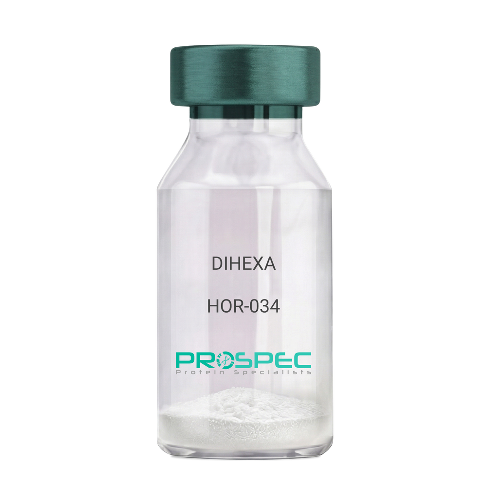
DIHEXADescription:
DIHEXA
Product # :
HOR-034Price :
Quantity :
Shipping Method :
Shipped at Room temp
More Info
- description
- formulation
- purity
- More Info
Description
DIHEXA Synthetic is a single, non-glycosylated polypeptide chain containing 3 amino acids, having a molecular mass of 504.28 Dalton and a Molecular formula of C27H44N4O5.
Formulation
The protein was lyophilized with no additives.
Purity
Greater than 97.0% as determined by analysis by RP-HPLC.
More Info
-
Physical Appearance
Sterile Filtered White lyophilized (freeze-dried) powder.
-
Stability
Lyophilized DIHEXA although stable at room temperature for 3 weeks, should be stored desiccated below -18°C. Upon reconstitution DIHEXA should be stored at 4°C between 2-7 days and for future use below -18°C. For long term storage it is recommended to add a carrier protein (0.1% HSA or BSA). Please prevent freeze-thaw cycles.
-
Solubility
It is recommended to reconstitute the lyophilized DIHEXA in sterile 18MΩ-cm H2O not less than 100 µg/ml, which can then be further diluted to other aqueous solutions.
-
Amino Acid Sequence
Hexanoyl-Tyr-Ile-Ahx-NH2.
-
Background
Dihexa, also known as N-hexanoic-Tyr-Ile-(6) aminohexanoic amide, is a potent and orally active small peptide that has been the focus of significant research due to its potential neurogenic and neuroprotective effects. This compound, derived from angiotensin IV, has been shown to possess a wide range of biological activities, including enhancing cognitive function, promoting neurogenesis, and potentially mitigating the effects of neurodegenerative diseases.
Dihexa's primary mechanism of action involves its interaction with hepatocyte growth factor (HGF) and its receptor, c-Met. By mimicking the effects of HGF, Dihexa can stimulate the c-Met receptor, leading to a cascade of events that promote neurogenesis and synaptic plasticity. Studies by Benoist et al. (2014) have demonstrated that Dihexa can enhance cognitive function in rats, suggesting potential applications in cognitive enhancement and the treatment of cognitive disorders.
In addition to its neurogenic effects, Dihexa has been shown to possess neuroprotective properties. Research by Kawas et al. (2017) found that Dihexa could protect neurons from apoptosis, suggesting potential applications in the treatment of neurodegenerative diseases such as Alzheimer's and Parkinson's disease.
Given its neurogenic and neuroprotective effects, Dihexa has been proposed as a potential therapeutic agent for a variety of conditions, including cognitive disorders, neurodegenerative diseases, and stroke. For instance, a study by Harding et al. (2018) found that Dihexa could improve outcomes in animal models of stroke, indicating its potential as a therapeutic agent in stroke recovery.
While research on Dihexa is promising, it is important to note that most studies have been conducted in animals or in vitro. More research is needed to fully understand the potential effects and applications of Dihexa in humans. However, the existing body of research suggests that Dihexa could be a promising tool in the treatment of cognitive disorders and neurodegenerative diseases.
ProSpec's products are furnished for LABORATORY RESEARCH USE ONLY. The product may not be used as drugs, agricultural or pesticidal products, food additives or household chemicals.
- View Data Sheet
Name :
Fra e 1.0101Description:
Allergen Fra e 1.0101 Recombinant
Allergen Fra e 1.0101, Fra e 1.0101, Fra e 1.
Product # :
PRO-2286Price :
Quantity :
Shipping Method :
Shipped with Ice Packs
More Info
- description
- source
- formulation
- purity
- More Info
Description
Recombinant Allergen Fra e 1.0101 produced in SF9 is a glycosylated, polypeptide chain having a calculated molecular mass of 17,794 Dalton. Fra e 1.0101 is expressed with a 10xHis tag at N-terminus and purified by proprietary chromatographic techniques.
Source
Sf9 insect cells.
Formulation
Fra e 1.0101 is supplied in 20mM HEPES buffer pH-7.9 and 6M Urea.
Purity
Greater than 80.0% as determined by SDS-PAGE.
More Info
-
Introduction
Allergen Fra e 1.0101 (Fra e 1.0101) causes an allergic reaction in humans.
-
Synonyms
Allergen Fra e 1.0101, Fra e 1.0101, Fra e 1.
-
Physical Appearance
Sterile Filtered clear solution.
-
Stability
Store at 4°C if entire vial will be used within 2-4 weeks. Store, frozen at -20°C for longer periods of time. Avoid multiple freeze-thaw cycles.
-
Immunological Functions
1. Binds IgE type human antibodies. 2. Immunodot test with positive/negative sera panels.
-
Molar extinction coefficient
14815; A280(1mg/ml)=0.833
ProSpec's products are furnished for LABORATORY RESEARCH USE ONLY. The product may not be used as drugs, agricultural or pesticidal products, food additives or household chemicals.
- View Data Sheet
Name :
BD14 MouseDescription:
Beta Defensin-14 Mouse Recombinant
Beta-defensin 14, BD-14, mBD-14, Defensin, beta 14, Defb14.
Product # :
CYT-945Price :
Quantity :
Shipping Method :
Shipped at Room temp
More Info
- description
- source
- formulation
- purity
- More Info
Description
Beta Defensin-14 Mouse Recombinant produced in E.coli is a single, non-glycosylated, polypeptide chain containing 45 amino acids and having a molecular mass of 5.2kDa.The BD14 is purified by proprietary chromatographic techniques.
Source
Escherichia Coli.
Formulation
BD-14 protein was lyophilized from a 0.2µm filtered concentrated solution in PBS, pH 7.4.
Purity
Greater than 96.0% as determined by:
(a) Analysis by RP-HPLC.
(b) Analysis by SDS-PAGE.More Info
-
Introduction
Alpha and Beta Defensins are cationic peptides with antimicrobial activity against Gram-negative and Gram-positive bacteria, fungi and enveloped viruses. These 2-6kDa proteins have vital roles in innate immune system. Mammalian Defensins are classified into alpha, beta and theta categories, based on their size and pattern of disulfide bonding. Beta-Defensins contain a six-cysteine motif which forms 3 intra-molecular disulfide bonds. Since beta-defensins are cationic peptides, they can therefore interact with the membrane of invading microbes, which are negative due to lipopolysaccharides (LPS) and lipoteichoic acid (LTA) found in the cell membrane. In addition, they can affect the stability of the membrane.
-
Synonyms
Beta-defensin 14, BD-14, mBD-14, Defensin, beta 14, Defb14.
-
Physical Appearance
Sterile Filtered White lyophilized (freeze-dried) powder.
-
Stability
Lyophilized Mouse BD14 although stable at room temperature for 3 weeks, should be stored desiccated below -18°C. Upon reconstitution BD-14 should be stored at 4°C between 2-7 days and for future use below -18°C.For long term storage it is recommended to add a carrier protein (0.1% HSA or BSA).Please prevent freeze-thaw cycles.
-
Solubility
It is recommended to reconstitute the lyophilized BD14 in sterile 18M-cm H2O not less than 100µg/ml, which can then be further diluted to other aqueous solutions.
-
Amino Acid Sequence
FLPKTLRKFF CRIRGGRCAV LNCLGKEEQI GRCSNSGRKC CRKKK.
-
Background
What is the molecular weight/Mw of BD14 Protein?
BD14 Protein has a total Mw of 5.2kDa.
What is the source or expression system of BD14 Protein?
Escherichia Coli.
What is the Purity of BD14 Protein?
BD14 Protein is >96% pure as determined by SDS-PAGE.
What is the Biological Activity of BD14 Protein?
The biological functionality of BD14 Protein will be determined in the future.
What is the amino acid sequence of BD14 Protein?
FLPKTLRKFF CRIRGGRCAV LNCLGKEEQI GRCSNSGRKC CRKKK.
What applications can BD14 Protein be used in?
BD14 Protein can probably be used in western blot, ELISA and Lateral Flow.
What is the endotoxin level for BD14 Protein?
The endotoxin level is minimal, BD14 Protein was purified using conventional chromatography techniques.
ProSpec's products are furnished for LABORATORY RESEARCH USE ONLY. The product may not be used as drugs, agricultural or pesticidal products, food additives or household chemicals.
- View Data Sheet
Name :
NT 3 HumanDescription:
Neurotrophin-3 Human Recombinant
Neurotrophic factor, Nerve growth factor-2, NGF-2, HDNF, NT-3.
Product # :
CYT-257Price :
Quantity :
Shipping Method :
Shipped at Room temp
More Info
- description
- source
- formulation
- purity
- biological activity
- More Info
Description
Neurotrophin-3 Human Recombinant produced in E.Coli is a non-glycosylated and non-covalently linked homodimer, containing 2x120 amino acid chains, having a total Mw of 27.5 kDa. The NT-3 is purified by proprietary chromatographic techniques.
Source
Escherichia Coli.
Formulation
Lyophilized from 0.1% TFA.
Purity
Greater than 95.0% as determined by analysis by SDS-PAGE.
Biological Activity
The ED50 as determined by the dose-dependent proliferation of neuroblastoma cell line expressing BR6 is 3.49 ng/ml.
More Info
-
Introduction
NT3 a member of the neurotrophin family, that controls survival and differentiation of mammalian neurons. This protein is closely related to both nerve growth factor and brain-derived neurotrophic factor. It may be involved in the maintenance of the adult nervous system, and may affect development of neurons in the embryo when it is expressed in human placenta. NTF3-deficient mice generated by gene targeting display severe movement defects of the limbs. The mature peptide of this protein is identical in all mammals examined including human, pig, rat and mouse.
-
Synonyms
Neurotrophic factor, Nerve growth factor-2, NGF-2, HDNF, NT-3.
-
Physical Appearance
Sterile Filtered White lyophilized (freeze-dried) powder.
-
Stability
Lyophilized NGF2 although stable at room temperature for 3 weeks, should be stored desiccated below -18°C. Upon reconstitution NGF-2 should be stored at 4°C between 2-7 days and for future use below -18°C.For long term storage it is recommended to add a carrier protein (0.1% HSA or BSA).Please prevent freeze-thaw cycles.
-
Solubility
It is recommended to reconstitute the lyophilized Neurotrophin-3 in sterile 18MΩ-cm H2O not less than 100µg/ml, which can then be further diluted to other aqueous solutions.
-
Amino Acid Sequence
MYAEHKSHRGE YSVCDSESLW VTDKSSAIDI RGHQVTVLGE IKTGNSPVKQ YFYETRCKEA RPVKNGCRGI DDKHWNSQCK TSQTYVRALT SENNKLVGWR WIRIDTSCVC ALSRKIGRT.
-
Protein content
Protein quantitation was carried out by two independent methods:1. UV spectroscopy at 280 nm using the absorbency value of 2.165 as the extinction coefficient for a 0.1% (1mg/ml) solution. This value is calculated by the PC GENE computer analysis program of protein sequences (IntelliGenetics). 2. Analysis by RP-HPLC, using a standard solution of NT-3 as a Reference Standard.
ProSpec's products are furnished for LABORATORY RESEARCH USE ONLY. The product may not be used as drugs, agricultural or pesticidal products, food additives or household chemicals.
- View Data Sheet
Name :
S100A10 HumanDescription:
S100 Calcium Binding Protein A10 Human Recombinant
Protein S100-A10, S100 calcium-binding protein A10, Calpactin-1 light chain, Calpactin I light chain, p10 protein, p11, Cellular ligand of annexin II, S100A10, ANX2LG, CAL1L, CLP11, 42C, p10, GP11, ANX2L, Ca[1], MGC111133.
Product # :
PRO-384Price :
Quantity :
Shipping Method :
Shipped with Ice Packs
More Info
- description
- source
- formulation
- purity
- More Info
Description
S100A10 Human Recombinant also called Calpactin light chain is expressed in E. coli having a molecular weight of 15.3kDa fused to an amino terminal hexahistidine tag.
Source
Escherichia Coli.
Formulation
S100-A10 is supplied in 1xPBS and 50% Glycerol.
Purity
Greater than 95% as determined by SDS-PAGE.
2 bands on Western blot at 15.3 and 30.6 kDa, respectively representing monomeric and dimeric form.More Info
-
Introduction
S100A10 is a member of the S100 family of proteins contains two EF-hand calcium-binding motifs and is thought to be involved in the regulation of a number of cellular processes including cell cycle progression and differentiation. S100 proteins are localized either in the cytoplasm or the nucleus of a wide range of cells. There are at least 13 members in the S100 gene family, which are located as a cluster on chromosome 1q21. S100A10 may function in exocytosis and endocytosis.
-
Synonyms
Protein S100-A10, S100 calcium-binding protein A10, Calpactin-1 light chain, Calpactin I light chain, p10 protein, p11, Cellular ligand of annexin II, S100A10, ANX2LG, CAL1L, CLP11, 42C, p10, GP11, ANX2L, Ca[1], MGC111133.
-
Stability
Store at 4°C if entire vial will be used within 1-2 weeks. Store, frozen at -20°C for longer periods of time. Avoid multiple freeze-thaw cycles.
-
Applications
S100A10 can be used directly as a positive control in Western blotting, ELISA, immunoprecipitation and other immunological experiments.
The biological activity of this product has not yet been tested.
ProSpec's products are furnished for LABORATORY RESEARCH USE ONLY. The product may not be used as drugs, agricultural or pesticidal products, food additives or household chemicals.
- View Data Sheet
Name :
S100A11 HumanDescription:
S100 Calcium Binding Protein A11 Human Recombinant
Protein S100-A11, S100 calcium-binding protein A11, Calgizzarin, MLN 70, S100A11, MLN70, S100C.
Product # :
PRO-385Price :
Quantity :
Shipping Method :
Shipped with Ice Packs
More Info
- description
- source
- formulation
- purity
- More Info
Description
S100A11 Human Recombinant is expressed in E. coli having a molecular weight of 17kDa fused to an amino terminal hexahistidine tag.
Source
Escherichia Coli.
Formulation
S100A11 is supplied in PBS and 50% glycerol.
Purity
Greater than 95% as determined by SDS-PAGE.
2 bands on Western blot at 17 and 34 kDa, respectively representing monomeric and dimeric form.More Info
-
Introduction
S100A11 is a member of the S100 family of proteins which contains two EF-hand calcium-binding motifs and is thought to be involved in the regulation of a number of cellular processes including cell cycle progression and differentiation. S100A11 may function in motility, invasion and tubulin polymerisation. S100 proteins are localized either in the cytoplasm or the nucleus of a wide range of cells. There are at least 13 members in the S100 gene family, which are located as a cluster on chromosome 1q21. Chromosomal rearrangements and altered expression of S100A11 have been implicated in tumor metastasis.
-
Synonyms
Protein S100-A11, S100 calcium-binding protein A11, Calgizzarin, MLN 70, S100A11, MLN70, S100C.
-
Stability
Store at 4°C if entire vial will be used within 1-2 weeks. Store, frozen at -20°C for longer periods of time. Avoid multiple freeze-thaw cycles.
-
Applications
S100A11 can be used directly as a positive control in Western blotting, ELISA, immunoprecipitation and other immunological experiments.
The biological activity of this product has not yet been tested.
ProSpec's products are furnished for LABORATORY RESEARCH USE ONLY. The product may not be used as drugs, agricultural or pesticidal products, food additives or household chemicals.
- View Data Sheet
Name :
DPP4 Human, HEKDescription:
Dipeptidyl-Peptidase 4 Human Recombinant, HEK
CD26, ADABP, ADCP2, DPPIV, TP103, DPP4, Dipeptidyl peptidase 4, Dipeptidyl peptidase IV, DPP IV, T-cell activation antigen CD26, Adenosine deaminase complexing protein 2, CD26 antigen.
Product # :
ENZ-1187Price :
Quantity :
Shipping Method :
Shipped with Ice Packs
More Info
- description
- source
- formulation
- purity
- biological activity
- More Info
Description
DPP4 Human Recombinant is a single, glycosylated polypeptide chain containing 977 amino acids (29-766a.a) and having a molecular mass of 112.1kDa (calculated). DPP4 is fused to a 239 amino acid hIgG-Tag at C-terminus and is purified by proprietary chromatographic techniques.
Source
HEK293 Cells.
Formulation
DPP4 protein solution (0.25mg/ml) contains 10% Glycerol and Phosphate-Buffered Saline (pH 7.4).
Purity
Greater than 95.0% as determined by Analysis by SDS-PAGE.
Biological Activity
Specific activity is > 15,000 pmol/min/ug in which one unit defined as the amount of enzyme that hydrolyze 1pmole of H-Gly-Pro-AMC.HBr to H-Gly-Pro and AMC per minute at pH 8.0 at 37℃.
The ED50 range ≤250 ng/ml is measured by its binding ability in a functional ELISA with MERS-CoV Spike S1 Subunit (CAT# sars-052)..
The ED50 range ≤200 ng/ml is measured by its binding ability in a functional ELISA with MERS-CoV Spike RBD (CAT# sars-054).
The ED50 range ≤120 ng/ml is measured by its binding ability in a functional ELISA with MERS-CoV Spike (CAT# sars-051).
More Info
-
Synonyms
CD26, ADABP, ADCP2, DPPIV, TP103, DPP4, Dipeptidyl peptidase 4, Dipeptidyl peptidase IV, DPP IV, T-cell activation antigen CD26, Adenosine deaminase complexing protein 2, CD26 antigen.
-
Physical Appearance
Sterile Filtered colorless solution.
-
Stability
Store at 4°C if entire vial will be used within 2-4 weeks. Store, frozen at -20°C for longer periods of time. For long term storage it is recommended to add a carrier protein (0.1% HSA or BSA).Avoid multiple freeze-thaw cycles.
-
Amino Acid Sequence
DGSMNKGTDD ATADSRKTYT LTDYLKNTYR LKLYSLRWIS DHEYLYKQEN NILVFNAEYG NSSVFLENST FDEFGHSIND YSISPDGQFI LLEYNYVKQW RHSYTASYDI YDLNKRQLIT EERIPNNTQW VTWSPVGHKL AYVWNNDIYV KIEPNLPSYR ITWTGKEDII YNGITDWVYE EEVFSAYSAL WWSPNGTFLA YAQFNDTEVP LIEYSFYSDE SLQYPKTVRV PYPKAGAVNP TVKFFVVNTD SLSSVTNATS IQITAPASML IGDHYLCDVT WATQERISLQ WLRRIQNYSV MDICDYDESS GRWNCLVARQ HIEMSTTGWV GRFRPSEPHF TLDGNSFYKI ISNEEGYRHI CYFQIDKKDC TFITKGTWEV IGIEALTSDY LYYISNEYKG MPGGRNLYKI QLSDYTKVTC LSCELNPERC QYYSVSFSKE AKYYQLRCSG PGLPLYTLHS SVNDKGLRVL EDNSALDKML QNVQMPSKKL DFIILNETKF WYQMILPPHF DKSKKYPLLL DVYAGPCSQK ADTVFRLNWA TYLASTENII VASFDGRGSG YQGDKIMHAI NRRLGTFEVE DQIEAARQFS KMGFVDNKRI AIWGWSYGGY VTSMVLGSGS GVFKCGIAVA PVSRWEYYDS VYTERYMGLP TPEDNLDHYR NSTVMSRAEN FKQVEYLLIH GTADDNVHFQ QSAQISKALV DVGVDFQAMW YTDEDHGIAS STAHQHIYTH MSHFIKQCFS LPKLLEPKSC DKTHTCPPCP APELLGGPSV FLFPPKPKDT LMISRTPEVT CVVVDVSHED PEVKFNWYVD GVEVHNAKTK PREEQYNSTY RVVSVLTVLH QDWLNGKEYK CKVSNKALPA PIEKTISKAK GQPREPQVYT LPPSRDELTK NQVSLTCLVK GFYPSDIAVE WESNGQPENN YKTTPPVLDS DGSFFLYSKL TVDKSRWQQG NVFSCSVMHE ALHNHYTQKS LSLSPGK.
-
Background
DPP4 protein, also known as Dipeptidyl peptidase-4 or CD26, is a cell surface protease with diverse functions in cell signaling and metabolism. This research aims to investigate the role of DPP4 protein in various physiological processes and its implications in disease pathogenesis. Understanding the biological significance of DPP4 can provide insights into its potential as a therapeutic target for several disorders.
Function of DPP4 Protein:
DPP4 protein is involved in the cleavage and regulation of several peptide hormones and chemokines, influencing their bioactivity and half-life. It is widely expressed in various tissues, including immune cells, endothelial cells, and epithelial cells. DPP4 can modulate immune responses, glucose metabolism, and neuropeptide signaling through enzymatic and non-enzymatic activities.
Role of DPP4 Protein in Immune Regulation:
DPP4 protein plays a role in immune cell activation and regulation. It is expressed on the surface of T cells, where it functions as a co-stimulatory molecule. DPP4 engagement on T cells promotes T cell activation, cytokine production, and adhesion to endothelial cells. Additionally, DPP4 can cleave and inactivate certain chemokines, thereby influencing chemotaxis and immune cell recruitment.
Implications of DPP4 Protein in Metabolic Disorders:
DPP4 protein is involved in glucose metabolism and insulin regulation. It cleaves incretin hormones, such as glucagon-like peptide-1 (GLP-1) and gastric inhibitory polypeptide (GIP), which play crucial roles in glucose homeostasis. Inhibition of DPP4 activity can enhance the action of these incretin hormones, leading to improved glycemic control. Therefore, DPP4 inhibitors have been developed as antidiabetic drugs.
Association of DPP4 Protein with Cardiovascular Diseases:
DPP4 protein has been implicated in the pathogenesis of cardiovascular diseases. Elevated DPP4 levels have been observed in patients with heart failure, atherosclerosis, and hypertension. DPP4 can contribute to endothelial dysfunction, inflammation, and vascular remodeling, which are key factors in the development and progression of cardiovascular disorders. Inhibition of DPP4 activity has shown potential as a therapeutic strategy in preclinical studies.
Given its involvement in various biological processes and disease pathogenesis, DPP4 protein has emerged as a potential therapeutic target. DPP4 inhibitors, which prevent the enzymatic activity of DPP4, have been developed for the treatment of type 2 diabetes. These inhibitors enhance the action of incretin hormones, leading to improved glycemic control. Additionally, ongoing research aims to explore the therapeutic potential of DPP4 inhibitors in other conditions, such as immune-mediated disorders and cardiovascular diseases.
Conclusion:
The investigation of DPP4 protein provides insights into its diverse functions in cell signaling, immune regulation, and metabolism. Understanding the role of DPP4 in disease pathogenesis opens avenues for the development of targeted therapies for conditions such as diabetes, cardiovascular diseases, and immune-mediated disorders. Further research on DPP4 protein and its associated pathways may uncover new therapeutic opportunities and improve patient outcomes.
ProSpec's products are furnished for LABORATORY RESEARCH USE ONLY. The product may not be used as drugs, agricultural or pesticidal products, food additives or household chemicals.
- View Data Sheet
Name :
PI16 HumanDescription:
Peptidase Inhibitor 16 Human Recombinant
Peptidase inhibitor 16, PI-16, Cysteine-rich secretory protein 9, CRISP-9, PSP94-binding protein, PI16, CRISP9, PSPBP, MSMBBP, MGC45378, DKFZp586B1817.
Product # :
ENZ-113Price :
Quantity :
Shipping Method :
Shipped at Room temp
More Info
- description
- source
- formulation
- purity
- More Info
Description
The Peptidase Inhibitor 16 Human Recombinant is produced in HEK293 cells and fused with a C-terminal Flag Tag (11 amino acids). The PI16 Flag Tagged Fusion Protein is 45.7kDa protein containing a total of 426 amino acid residues and purified by proprietary chromatographic techniques.
Source
HEK293 (Human Embryonic Kidney cell line).
Formulation
PI16 was filtered (0.4µm) and lyophilized from 0.5mg/ml in 20mM Tris and 50mM NaCl, pH 7.5.
Purity
Greater than 95.0% as determined by SDS-PAGE.
More Info
-
Introduction
Peptidase Inhibitor 16 (PI16) which a member of the CRISP family, is a putative serine protease inhibitor. PI16 interacts with PSP94/MSMB. PI16 is expressed in the prostate, testis, ovary and intestine. It also concentrates in prostate cancer patient's sera. PI16 may serve as a marker following prostatectomy for prostate cancer.
-
Synonyms
Peptidase inhibitor 16, PI-16, Cysteine-rich secretory protein 9, CRISP-9, PSP94-binding protein, PI16, CRISP9, PSPBP, MSMBBP, MGC45378, DKFZp586B1817.
-
Physical Appearance
Filtered White lyophilized (freeze-dried) powder.
-
Stability
Store lyophilized PI16 Human recombinant at -20°C. Aliquot the product after reconstitution to avoid repeated freezing/thawing cycles. Reconstituted protein can be stored at 4°C for a limited period of time; it does not show any change after two weeks at 4°C.
-
Solubility
It is recommended to add deionized water to prepare a working stock solution of 0.5mg/ml and let the lyophilized pellet dissolve completely. Product is not sterile! Please filter the product by an appropriate sterile filter before using it in the cell culture.
-
Amino Acid Sequence
LTDEEKRLMV ELHNLYRAQV SPPASDMLHM RWDEELAAFA KAYARQCVWG HNKERGRRGE NLFAITDEGM DVPLAMEEWH HEREHYNLSA ATCSPGQMCG HYTQVVWAKT ERIGCGSHFC EKLQGVEETN IELLVCNYEP PGNVKGKRPY QEGTPCSQCP SGYHCKNSLC EPIGSPEDAQ DLPYLVTEAP SFRATEASDS RKMGTPSSLA TGIPAFLVTE VSGSLATKAL PAVETQAPTS LATKDPPSMA TEAPPCVTTE VPSILAAHSL PSLDEEPVTF PKSTHVPIPK SADKVTDKTK VPSRSPENSL DPKMSLTGAR ELLPHAQEEA EAEAELPPSS EVLASVFPAQ DKPGELQATL DHTGHTSSKS LPNFPNTSAT ANATGGRALA LQSSLPGAEG PDKPSVVSGL NSGPGAAADYKDDDDK.
ProSpec's products are furnished for LABORATORY RESEARCH USE ONLY. The product may not be used as drugs, agricultural or pesticidal products, food additives or household chemicals.
- View Data Sheet
Name :
Midkine MouseDescription:
Midkine Mouse Recombinant
NEGF-2, Neurite Growth-Promoting Factor 2, MK, Neurite outgrowth-promoting protein, Midgestation and kidney protein, Amphiregulin-associated protein, ARAP, Neurite outgrowth-promoting factor 2, FLJ27379, Midkine, MK1, NEGF2.
Product # :
CYT-178Price :
Quantity :
Shipping Method :
Shipped at Room temp
More Info
- description
- source
- formulation
- purity
- biological activity
- More Info
Description
Midkine Mouse Recombinant produced in E.Coli is a single, non-glycosylated, polypeptide chain containing 120 amino acids and having a molecular mass of 13.3kDa.The Midkine Mouse is purified by proprietary chromatographic techniques.
Source
Escherichia Coli.
Formulation
Lyophilized from a 0.2um filtered concentrated solution in PBS, pH 7.4.
Purity
Greater than 95.0% as determined by HPLC and SDS-PAGE.
Biological Activity
Fully biologically active when compared to standard. Determined by its ability to chemoattract human neutrophils using a concentration range of 10-100 ng/ml corresponding to a specific activity of 10,000-100,000IU/mg.More Info
-
Introduction
Midkine (MK) is the product of a retinoic acid responsive gene, MK, and is a member of a family of heparin binding factors. It contains 121 amino acid residues including 10 conserved cysteine residues, all of which appear to be disulphide linked.
Midkine is expressed during embryogenesis, showing an expression pattern that suggests functions in neurogenesis, cell migration, secondary organogenetic induction, and mesoderm-epithelial interaction.
The widespread downregulation of MK in the adult human is reverted in a number of cancers, in which polypeptides are able to act as both transforming growth factors and promoters of angiogenesis.
Midkine (MK), induces chemotaxis of human neutrophils and was found to trigger mobilization of intracellular calcium of these cells.
Midkine induces histamine release from rat peritoneal mast cells with a rapid response in a dose dependent manner.
Midkine is also a potent stimulator of collagen and glycosaminoglycan synthesis. -
Synonyms
NEGF-2, Neurite Growth-Promoting Factor 2, MK, Neurite outgrowth-promoting protein, Midgestation and kidney protein, Amphiregulin-associated protein, ARAP, Neurite outgrowth-promoting factor 2, FLJ27379, Midkine, MK1, NEGF2.
-
Physical Appearance
Sterile Filtered White lyophilized (freeze-dried) powder.
-
Stability
Lyophilized Midkine Mouse although stable at room temperature for 3 weeks, should be stored desiccated below -18°C. Upon reconstitution Midkine Mouse should be stored at 4°C between 2-7 days and for future use below -18°C.Please prevent freeze-thaw cycles.
-
Solubility
It is recommended to reconstitute the lyophilized Midkine Mouse in sterile 18M-cm H2O not less than 100µg/ml, which can then be further diluted to other aqueous solutions.
-
Amino Acid Sequence
VAKKKEKVKK GSECSEWTWG PCTPSSKDCG MGFREGTCGA QTQRVHCKVP CNWKKEFGAD CKYKFESWGA CDGSTGTKAR QGTLKKARYN AQCQETIRVT KPCTSKTKSK TKAKKGKGKD
ProSpec's products are furnished for LABORATORY RESEARCH USE ONLY. The product may not be used as drugs, agricultural or pesticidal products, food additives or household chemicals.
- View Data Sheet
Name :
S100A3 MouseDescription:
S100 Calcium Binding Protein A3 Mouse Recombinant
Protein S100-A3, Protein S-100E, S100 calcium-binding protein A3, S100a3, S100E.
Product # :
PRO-1166Price :
Quantity :
Shipping Method :
Shipped with Ice Packs
More Info
- description
- source
- formulation
- purity
- More Info
Description
S100A3 Mouse Recombinant produced in E.Coli is a single, non-glycosylated polypeptide chain containing 125 amino acids (1-101 a.a) and having a molecular mass of 14.3kDa.S100A3 is fused to a 24 amino acid His-tag at N-terminus & purified by proprietary chromatographic techniques.
Source
Escherichia Coli.
Formulation
S100A3 protein solution (1mg/ml) containing 20mM Tris-HCl buffer, pH8.0, 10% glycerol, 2mM DTT and 150mM NaCl.
Purity
Greater than 95.0% as determined by SDS-PAGE.
More Info
-
Introduction
S100A3 is part of S100 family of proteins containing 2 EF-hand calcium-binding motifs. S100 proteins are localized in the cytoplasm/nucleus of a broad range of cells, and takes part in the regulation of several cellular processes such as cell cycle progression and differentiation. S100A3 has the largest number of cysteines of all S100 proteins. S100A3 has high affinity for Zinc, and is widely expressed in human hair cuticle.
-
Synonyms
Protein S100-A3, Protein S-100E, S100 calcium-binding protein A3, S100a3, S100E.
-
Physical Appearance
S100A3 is supplied as a sterile filtered clear solution.
-
Stability
Store at 4°C if entire vial will be used within 2-4 weeks. Store, frozen at -20°C for longer periods of time. For long term storage it is recommended to add a carrier protein (0.1% HSA or BSA).Avoid multiple freeze-thaw cycles.
-
Amino Acid Sequence
MGSSHHHHHH SSGLVPRGSH MGSHMTRPLE QAVAAIVCTF QEYAGRCGDK YKICQSELKE LLQKELPTWT PSEFRECDYN KFMSVLDTNK DCEVDFGEYV RSLASLCLYC HEYFKECPPE PPCPQ.
ProSpec's products are furnished for LABORATORY RESEARCH USE ONLY. The product may not be used as drugs, agricultural or pesticidal products, food additives or household chemicals.
- View Data Sheet
Name :
S100b MouseDescription:
S100 Calcium Binding Protein B Mouse Recombinant
Protein S100-B, S-100 protein beta chain, S-100 protein subunit beta, S100 calcium-binding protein B.
Product # :
PRO-2370Price :
Quantity :
Shipping Method :
Shipped with Ice Packs
More Info
- description
- source
- formulation
- purity
- More Info
Description
s100b Mouse Recombinant produced in E.Coli is a single, non-glycosylated, polypeptide chain containing 92 amino acids (1-92 a.a.) and having a molecular mass of 10.7kDa. The s100b is purified by proprietary chromatographic techniques.
Source
Escherichia Coli.
Formulation
The s100b protein solution (1mg/ml) contains Phosphate Buffered Saline (pH 7.4) and 10% glycerol.
Purity
Greater than 95.0% as determined by SDS-PAGE.
More Info
-
Introduction
S100b is a member of the S100 family of proteins which are a family of EF-hand calcium binding proteins that exist mostly as dimers of the 20 currently identified individual S100 monomers. The S100B homodimer is expressed in cells of the central nervous system, glial cells and in certain peripheral cells e.g. Schwann cells, melanocytes, adipocytes and chondrocytes. S100 proteins are localized either in the cytoplasm or the nucleus of a wide range of cells. S100 proteins are involved in the regulation of a number of cellular processes such as cell cycle progression and differentiation. There are at least 13 members in the S100 gene family, which are located as a cluster on chromosome 1q21; however, S100b is located at 21q22.3. The determination of S100B in serum levels may be used to monitor the extent of brain injury and malignant melanoma. S100b proteins may have a role in Neurite extension, proliferation of melanoma cells, stimulation of Ca2+ fluxes, inhibition of PKC-mediated phosphorylation, astrocytosis and axonal proliferation, and inhibition of microtubule assembly. Chromosomal rearrangements and altered expression of the S100b gene are implicated in several neurological, neoplastic, and other types of diseases, including Alzheimer's disease, Down's syndrome, epilepsy, amyotrophic lateral sclerosis, melanoma, and type I diabetes.
-
Synonyms
Protein S100-B, S-100 protein beta chain, S-100 protein subunit beta, S100 calcium-binding protein B.
-
Physical Appearance
Sterile filtered colorless solution.
-
Stability
Store at 4°C if entire vial will be used within 2-4 weeks. Store, frozen at -20°C for longer periods of time. For long term storage it is recommended to add a carrier protein (0.1% HSA or BSA).Avoid multiple freeze-thaw cycles.
-
Amino Acid Sequence
MSELEKAMVA LIDVFHQYSG REGDKHKLKK SELKELINNE LSHFLEEIKE QEVVDKVMET LDEDGDGECD FQEFMAFVAM VTTACHEFFE HE.
ProSpec's products are furnished for LABORATORY RESEARCH USE ONLY. The product may not be used as drugs, agricultural or pesticidal products, food additives or household chemicals.
- View Data Sheet
Name :
DEFB118 HumanDescription:
Beta Defensin 118 Human Recombinant
Beta Defensin 118, Beta-defensin 18, DEFB-18, Defensin, beta 118, Epididymal secretory protein 13.6, ESP13.6, DEFB118, C20orf63, DEFB18, ESC42, Beta-defensin 118 precursor.
Product # :
CYT-714Price :
Quantity :
Shipping Method :
Shipped with Ice Packs
More Info
- description
- source
- formulation
- purity
- More Info
- sds-page
Description
DEFB118 Human Recombinant produced in E.Coli is a single, non-glycosylated polypeptide chain containing 126 amino acids (21-123 a.a) and having a molecular mass of 13.8kDa.DEFB118 is fused to a 23 amino acid His-tag at N-terminus & purified by proprietary chromatographic techniques.
Source
Escherichia Coli.
Formulation
DEFB118 protein solution (0.5mg/ml) containing 20mM Tris-HCl buffer (pH8.0), 0.15M NaCl, 20% glycerol and 1mM DTT.
Purity
Greater than 90.0% as determined by SDS-PAGE.
sds-page
More Info
-
Introduction
Beta Defensin 118 (DEFB118) which is a member of the beta subfamily of defensins, is found in a cluster with other beta-defensin genes on the long arm of chromosome 20. Beta-defensins are antimicrobial peptides which provide protection for tissues and organs from infection by a diversity of microorganisms. DEFB118 protein’s expression is regulated by androgen, and the encoded protein binds to sperm and exhibits antibacterial activity.
-
Synonyms
Beta Defensin 118, Beta-defensin 18, DEFB-18, Defensin, beta 118, Epididymal secretory protein 13.6, ESP13.6, DEFB118, C20orf63, DEFB18, ESC42, Beta-defensin 118 precursor.
-
Physical Appearance
Sterile Filtered clear solution.
-
Stability
Store at 4°C if entire vial will be used within 2-4 weeks. Store, frozen at -20°C for longer periods of time. For long term storage it is recommended to add a carrier protein (0.1% HSA or BSA).Avoid multiple freeze-thaw cycles.
-
Amino Acid Sequence
MGSSHHHHHH SSGLVPRGSH MGSYSGEKKC WNRSGHCRKQ CKDGEAVKDT CKNLRACCIP SNEDHRRVPA TSPTPLSDST PGIIDDILTV RFTTDYFEVS SKKDMVEESE AGRGTETSLP NVHHSS.
-
Background
Title: Beta Defensin 118 Human Recombinant: Exploring its Role in Innate Immunity and Potential Therapeutic Applications
Abstract:
Beta defensin 118 (BD118) is a member of the beta defensin family, known for its antimicrobial properties and immune-modulatory functions. This research paper provides a comprehensive analysis of human recombinant BD118, focusing on its production, characterization, and potential applications in immune modulation and therapeutic interventions. The paper highlights the significance of BD118 in innate immunity and its role in host defense against microbial pathogens. Furthermore, it explores ongoing research and clinical trials investigating the therapeutic potential of recombinant BD118 in various inflammatory and infectious diseases. The information presented in this paper aims to enhance our understanding of human recombinant BD118 and its utility as a research tool and a potential immunotherapeutic agent.Introduction:
Beta defensin 118 (BD118) is a small cationic peptide that plays a critical role in the innate immune response against microbial pathogens. Human recombinant BD118, generated through genetic engineering techniques, offers a valuable tool for studying its immune-modulatory properties and exploring its therapeutic potential.Production and Characterization:
Recombinant BD118 is typically produced using expression systems such as bacteria or yeast. The protein is then purified and characterized to ensure its structural integrity and functional activity. Rigorous quality control measures are implemented to confirm the specificity and potency of the recombinant BD118.Role in Innate Immunity:
BD118 exhibits antimicrobial activity against a wide range of pathogens, including bacteria, fungi, and viruses. Additionally, it possesses immune-modulatory functions, such as the regulation of pro-inflammatory responses and the promotion of wound healing. Recombinant BD118 serves as a valuable tool for investigating the mechanisms underlying its immune-modulatory actions and exploring its potential as an immunotherapeutic agent.Therapeutic Implications:
Dysregulation of the immune system is associated with various inflammatory and infectious diseases. Recombinant BD118 holds promise as a potential therapeutic agent due to its antimicrobial properties and immune-modulatory functions. Ongoing research and clinical trials are investigating the therapeutic applications of recombinant BD118 in conditions such as skin infections, respiratory diseases, and inflammatory bowel disease.Conclusion:
Human recombinant BD118 represents a valuable research tool and a potential immunotherapeutic agent. Its production, characterization, and applications in immune modulation contribute to our understanding of innate immunity and the development of targeted therapeutic interventions. Continued research and clinical trials exploring the therapeutic potential of recombinant BD118 offer promising avenues for improving outcomes in inflammatory and infectious diseases.What is the molecular weight/Mw of DEFB118 Protein?
DEFB118 Protein has a total Mw of 13.8kDa.
What is the source or expression system of DEFB118 Protein?
Escherichia Coli.
What is the Purity of DEFB118 Protein?
DEFB118 Protein is >90% pure as determined by SDS-PAGE.
What is the Biological Activity of DEFB118 Protein?
The biological functionality of DEFB118 Protein will be determined in the future.
What is the amino acid sequence of DEFB118 Protein?
MGSSHHHHHH SSGLVPRGSH MGSYSGEKKC WNRSGHCRKQ CKDGEAVKDT CKNLRACCIP SNEDHRRVPA TSPTPLSDST PGIIDDILTV RFTTDYFEVS SKKDMVEESE AGRGTETSLP NVHHSS.
What applications can DEFB118 Protein be used in?
DEFB118 Protein can probably be used in western blot, ELISA and Lateral Flow.
What is the endotoxin level for DEFB118 Protein?
The endotoxin level is minimal, DEFB118 Protein was purified using conventional chromatography techniques.
ProSpec's products are furnished for LABORATORY RESEARCH USE ONLY. The product may not be used as drugs, agricultural or pesticidal products, food additives or household chemicals.
- View Data Sheet
Name :
DTYMK HumanDescription:
Deoxythymidylate Kinase Human Recombinant
Deoxythymidylate Kinase (Thymidylate Kinase), DTMP Kinase, CDC8, TMPK, TYMK, Thymidylate (DTMP) Kinase, EC 2.7.4.9, PP3731, Thymidylate kinase.
Product # :
PKA-005Price :
Quantity :
Shipping Method :
Shipped with Ice Packs
More Info
- description
- source
- formulation
- purity
- More Info
Description
DTYMK Human Recombinant produced in E.Coli is a single, non-glycosylated polypeptide chain containing 232 amino acids (1-212 a.a) and having a molecular mass of 26kDa. DTYMK is fused to a 20 amino acid His-tag at N-terminus & purified by proprietary chromatographic techniques.
Source
E.coli.
Formulation
DTYMK protein solution (1mg/ml) containing 20mM Tris-HCl buffer (pH 8.0), 20% glycerol, 2mM DTT and 0.1M NaCl.
Purity
Greater than 95% as determined by SDS-PAGE.
More Info
-
Introduction
Thymidylate kinase (DTYMK) is involved in pyrimidine metabolism. Specifically, DTYMK catalyzes the ATP-dependent conversion of dTMP (deoxythymidine monophosphate) to dTDP (deoxythymidine diphosphate), which then acts as one of the 4 nucleotides in DNA. Through its role in the catalytic creation of dTDP, DTYMK has an imperative role in the pathway of DNA synthesis and is assumed to be involved in cell cycle progression and cell growth.
-
Synonyms
Deoxythymidylate Kinase (Thymidylate Kinase), DTMP Kinase, CDC8, TMPK, TYMK, Thymidylate (DTMP) Kinase, EC 2.7.4.9, PP3731, Thymidylate kinase.
-
Physical Appearance
Sterile Filtered colorless solution.
-
Stability
Store at 4°C if entire vial will be used within 2-4 weeks. Store, frozen at -20°C for longer periods of time. For long term storage it is recommended to add a carrier protein (0.1% HSA or BSA).Avoid multiple freeze-thaw cycles.
-
Amino Acid Sequence
MGSSHHHHHH SSGLVPRGSH MAARRGALIV LEGVDRAGKS TQSRKLVEAL CAAGHRAELL RFPERSTEIG KLLSSYLQKK SDVEDHSVHL LFSANRWEQV PLIKEKLSQG VTLVVDRYAF SGVAFTGAKE NFSLDWCKQP DVGLPKPDLV LFLQLQLADA AKRGAFGHER YENGAFQERA LRCFHQLMKD TTLNWKMVDA SKSIEAVHED IRVLSEDAIR TATEKPLGEL WK.
ProSpec's products are furnished for LABORATORY RESEARCH USE ONLY. The product may not be used as drugs, agricultural or pesticidal products, food additives or household chemicals.
- View Data Sheet
Name :
CALB1 RatDescription:
Calbindin-1 Rat Recombinant
Calbindin, Vitamin D-dependent calcium-binding protein, avian-type, Calbindin D28, D-28K, Spot 35 protein, Calb1, CaBP28K, MGC93326.
Product # :
PRO-400Price :
Quantity :
Shipping Method :
Shipped at Room temp
More Info
- description
- source
- formulation
- purity
- More Info
Description
Recombinant Rat Calbindin-1 produced in E.Coli.The Rat CALB1 is purified by proprietary chromatographic techniques.
Source
Escherichia Coli.
Formulation
The protein was lyophilized from a concentrated solution (1mg/ml) containing 50mM NaHCO3.
Purity
Greater than 90% as determined by SDS-PAGE.
More Info
-
Introduction
Calbindins are Ca-binding proteins belonging to the troponin C superfamily. CALB28K/Calbindin1/CALB1 (D28K/Spot35 protein or cholecalcin, rat 261 aa; mouse 261 aa; human 261-aa, chromosome 8q21.3-q22.1) was originally described as 27-kDA induced by vitamin D in the duodenum of chicken. In mammals, it is expressed in the kidney, pancreatic islets, and brain. In brain, its synthesis is independent of vitamin D. CABP28K contains 4 active and 2 inactive EF-hand Ca-binding domains. The gene for CABP28K is clustered in the same region as carbonic anhydrase. The neurons in the brains of patients with Huntington disease are CAB28K depleted. There are two types of CaBPs: the "trigger"- and the "buffer"-CaBPs. The conformation of "trigger" type CaBPs changes upon Ca2+ binding and exposes regions on protein that interact with target molecules, thus altering their activity. The buffer-type CABP are thought to control the intracellular calcium concentration. Calbindin D-28K is found predominantly in subpopulations of central and peripheral nervous system neurons, and in certain epithelial cells involved in Ca2+ transport such as distal tubular cells and cortical collecting tubules of the kidney, and in enteric neuroendocrine cells.
-
Synonyms
Calbindin, Vitamin D-dependent calcium-binding protein, avian-type, Calbindin D28, D-28K, Spot 35 protein, Calb1, CaBP28K, MGC93326.
-
Physical Appearance
Sterile Filtered White lyophilized (freeze-dried) powder.
-
Stability
Lyophilized CABP28K although stable at room temperature for 3 weeks, should be stored desiccated below -18°C. Upon reconstitution CABP28K should be stored at 4°C between 2-7 days and for future use below -18°C.For long term storage it is recommended to add a carrier protein (0.1% HSA or BSA).Please prevent freeze-thaw cycles.
-
Solubility
It is recommended to reconstitute the lyophilized CABP28K in sterile 18MΩ-cm H2O not less than 100µg/ml, which can then be further diluted to other aqueous solutions.
-
Applications
CABP28K can be used for immunoblots, absorption experiments in immunohistochemistry, radioimmunoassay and intracellular injection.
For adsorption we suggest the following procedureA- Dilute 1 µl of the monoclonal antibody against calbindin D-28k in 5 ml of the usual buffer for immunohistochemistry (final dilution 1:5'000).
B- Add 1 µg of the recombinant protein to 1 ml of the diluted antibody solution and mix well.
C- Incubate for at least 6 hours in the cold.
D- Apply to tissue-sections and incubate for 3 days.
E- Complete the immunohistochemical reaction as usual (biotinylated second antibody, ABC-complex, DAB).
As a result, the immunostaining should be strongly reduced or even completely prevented.
ProSpec's products are furnished for LABORATORY RESEARCH USE ONLY. The product may not be used as drugs, agricultural or pesticidal products, food additives or household chemicals.
- View Data Sheet
Name :
Resistin MouseDescription:
Resistin Mouse Recombinant
Cysteine-rich secreted protein FIZZ3, Adipose tissue-specific secretory factor, ADSF, C/EBP-epsilon-regulated myeloid-specific secreted cysteine-rich protein, Cysteine-rich secreted protein A12-alpha-like 2, RSTN, XCP1, RETN1, MGC126603, MGC126609.
Product # :
CYT-1034Price :
Quantity :
Shipping Method :
Shipped at Room temp
More Info
- description
- source
- formulation
- purity
- More Info
Description
Resistin Mouse Recombinant produced in E.Coli is a non glycosylated, homodimeric polypeptide chain containing 2 x 95 amino acids and having a total molecular mass of 20.6kDa. The Resistin is purified by proprietary chromatographic techniques.
Source
Escherichia Coli.
Formulation
Lyophilized from a sterile filtered solution containing 0.1 % trifluoroacetic acid (TFA).
Purity
Greater than 95.0% as determined by SDS-PAGE.
More Info
-
Introduction
Resistin, a product of the RSTN gene, is a peptide hormone belonging to the class of cysteine-rich secreted proteins which is termed the RELM family, and is also described as ADSF (Adipose Tissue- Specific Secretory Factor) and FIZZ3 (Found in Inflammatory Zone). Human resistin contains 108 amino acids as a prepeptide, and its hydrofobic signal peptide is cleaved before its secretion. Resistin circulates in human blood as a dimeric protein consisting of two 92 amino acid polypeptides, which are disulfide-linked via Cys26.
Resistin may be an important link between obesity and insulin resistance. Mouse resistin, specifically produced and secreted by adipocyte, acts on skeletal muscle myocytes, hepatocytes and adipocytes themselves so that it reduces their sensitivity to insulin. Steppan et al. have suggested that resistin suppresses the ability of insulin to stimulate glucose uptake. They have also suggested that resistin is present at elevated levels in blood of obese mice, and is down regulated by fasting and antidiabetic drugs. Way et al., on the other hand, have found that resistin expression is severly suppressed in obesity and is stimulated by several antidiabetic drugs.
Other studies have shown that mouse resistin increases during the differentiation of adipocytes, but it also seems to inhibit adipogenesis. In contrast, the human adipogenic differentiation is likely to be associated with a down regulation of resistin gene expression. -
Synonyms
Cysteine-rich secreted protein FIZZ3, Adipose tissue-specific secretory factor, ADSF, C/EBP-epsilon-regulated myeloid-specific secreted cysteine-rich protein, Cysteine-rich secreted protein A12-alpha-like 2, RSTN, XCP1, RETN1, MGC126603, MGC126609.
-
Physical Appearance
Sterile Filtered White lyophilized (freeze-dried) powder.
-
Stability
Lyophilized Resistin although stable at room temperature for 3 weeks, should be stored desiccated below -18°C. Upon reconstitution Resistin Mouse should be stored at 4°C between 2-7 days and for future use below -18°C. For long term storage it is recommended to add a carrier protein (0.1% HSA or BSA). Please prevent freeze-thaw cycles.
-
Solubility
It is recommended to reconstitute the lyophilized Resistin in sterile 18MΩ-cm H2O at a concentration of 100µg/ml, which can then be further diluted to other aqueous solutions.
-
Amino Acid Sequence
MSSMPLCPID EAIDKKIKQD FNSLFPNAIK NIGLNCWTVS SRGKLASCPE GTAVLSCSCG SACGSWDIRE EKVCHCQCAR IDWTAARCCK LQVAS.
ProSpec's products are furnished for LABORATORY RESEARCH USE ONLY. The product may not be used as drugs, agricultural or pesticidal products, food additives or household chemicals.
- View Data Sheet
Name :
Activin B HumanDescription:
Activin-B Human Recombinant
Inhibin beta B (activin AB beta polypeptide), Inhibin, beta-2, Activin beta-B chain, MGC157939.
Product # :
CYT-058Price :
Quantity :
Shipping Method :
Shipped at Room temp
More Info
- description
- source
- formulation
- purity
- More Info
Description
Activin B human Recombinant produced in Nicotiana benthamiana plant is a beta-B single chain (aa 293-406) containing 123 amino acids (molecular formula C615H910N178O177S12). Activin B is fused to a 10-His-tag at the N-terminal having the total molecular mass of 14kDa and purified by standard chromatographic techniques.
Source
Nicotiana benthamiana plant
Formulation
Lyophilized from 1mg/ml solution in 0.05M Tris-HCl buffer pH 7.4.
Purity
Greater than 97.0% as determined by Analysis by SDS-PAGE.
More Info
-
Introduction
Inhibins are dimeric peptide hormones produced by female ovarian granulose cells and male Sertoli cells as well as a variety of other tissues. Inhibins have two isoforms, A and B, with the same alpha subunit but different beta subunits. Inhibin A is a dimer of alpha and beta A subunits, inhibin B is a dimer of alpha and beta B subunits.
Inhibins are thought to inhibit the production of follicle-stimulating hormone (FSH) by the pituitary gland. In addition, Inhibins are also thought to play a role in the control of gametogenesis, and embryonic and fetal development. -
Synonyms
Inhibin beta B (activin AB beta polypeptide), Inhibin, beta-2, Activin beta-B chain, MGC157939.
-
Physical Appearance
Sterile Filtered White lyophilized (freeze-dried) powder.
-
Stability
Lyophilized Activin B although stable at room temperature for 3 weeks, should be stored desiccated below -18°C. Upon reconstitution Activin B should be stored at 4°C between 2-7 days and for future use below -18°C. For long term storage it is recommended to add a carrier protein (0.1% HSA or BSA). Please prevent freeze-thaw cycles.
-
Solubility
It is recommended to reconstitute the lyophilized Activin B in sterile 18M-cm H2O not less than 100µg/ml, which can then be further diluted to other aqueous solutions.
-
Amino Acid Sequence
HHHHHHHHHH GLECDGRTNL CCRQQFFIDF RLIGWNDWII APTGYYGNYC EGSCPAYLAG VPGSASSFHT AVVNQYRMRG LNPGTVNSCC IPTKLSTMSM LYFDDEYNIV KRDVPNMIVE ECG
-
Background
What is the molecular weight / Mw of Activin B Protein?
Activin A Protein has a total Mw of 14 kDa.
What is the source or expression system of Activin B Protein?
Nicotinia
What is the Purity of Activin B Protein?
Activin B Protein is >97% pure as determined by SDS-PAGE.
What is the Biological Activity of Activin B Protein?
The biological functionality of Activin-B Protein will be determined in the future.
What is the endotoxin level for Activin B Protein?
The endotoxin level is minimal, ACTIVIN B Protein was purified using conventional chromatography techniques.
What is the amino acid sequence of ACTIVIN B Protein?
HHHHHHHHHH GLECDGRTNL CCRQQFFIDF RLIGWNDWII APTGYYGNYC EGSCPAYLAG VPGSASSFHT AVVNQYRMRG LNPGTVNSCC IPTKLSTMSM LYFDDEYNIV KRDVPNMIVE ECG
What applications can ACTIVIN B Protein be used in?
ACTIVIN A Protein can probably be used in western blot, ELISA and Lateral Flow.
ProSpec's products are furnished for LABORATORY RESEARCH USE ONLY. The product may not be used as drugs, agricultural or pesticidal products, food additives or household chemicals.
- View Data Sheet
Name :
IL18BP HumanDescription:
Interleukin-18 Binding Protein Human Recombinant
Interleukin 18 Binding Protein, MC51L-53L-54L Homolog Gene Product, Tadekinig-Alfa, IL-18BP, IL18BPa, Interleukin-18-binding protein, Tadekinig-alfa.
Product # :
CYT-728Price :
Quantity :
Shipping Method :
Shipped with Ice Packs
More Info
- description
- source
- formulation
- purity
- More Info
Description
IL18BP Human Recombinant produced in E.Coli is a single, non-glycosylated polypeptide chain containing 187 amino acids (31-194 a.a) and having a molecular mass of 20kDa. IL18BP is fused to a 23 amino acid His-tag at N-terminus & purified by proprietary chromatographic techniques.
Source
Escherichia Coli.
Formulation
IL18BP protein solution (1mg/ml) containing 20mM Tris-HCl buffer (pH 8.0), 0.4M UREA and 10% glycerol.
Purity
Greater than 90% as determined by SDS-PAGE.
More Info
-
Introduction
Interleukin-18 Binding Protein (IL18BP) serves as an inhibitor of the proinflammatory cytokine, IL18. IL18BP binds IL18, inhibits the binding of IL18 to its receptor, and consequently inhibits IL18-induced IFN-gamma production, resulting in reduced T-helper type 1 immune responses. The IL18BP protein is constitutively expressed and secreted in mononuclear cells. Elevated levels of IL18BP protein are detected in the intestinal tissues of patients with Crohn's disease.
-
Synonyms
Interleukin 18 Binding Protein, MC51L-53L-54L Homolog Gene Product, Tadekinig-Alfa, IL-18BP, IL18BPa, Interleukin-18-binding protein, Tadekinig-alfa.
-
Physical Appearance
Sterile Filtered clear solution.
-
Stability
Store at 4°C if entire vial will be used within 2-4 weeks. Store, frozen at -20°C for longer periods of time. For long term storage it is recommended to add a carrier protein (0.1% HSA or BSA).Avoid multiple freeze-thaw cycles.
-
Amino Acid Sequence
MGSSHHHHHH SSGLVPRGSH MGSTPVSQTT TAATASVRST KDPCPSQPPV FPAAKQCPAL EVTWPEVEVP LNGTLSLSCV ACSRFPNFSI LYWLGNGSFI EHLPGRLWEG STSRERGSTG TQLCKALVLE QLTPALHSTN FSCVLVDPEQ VVQRHVVLAQ LWAGLRATLP PTQEALPSSH SSPQQQG.
ProSpec's products are furnished for LABORATORY RESEARCH USE ONLY. The product may not be used as drugs, agricultural or pesticidal products, food additives or household chemicals.
- View Data Sheet
Name :
TNF a MouseDescription:
Tumor Necrosis Factor-Alpha Mouse Recombinant
TNF-alpha, Tumor necrosis factor ligand superfamily member 2, TNF-a, Cachectin, DIF, TNFA, TNFSF2.
Product # :
CYT-252Price :
Quantity :
Shipping Method :
Shipped at Room temp
More Info
- description
- source
- formulation
- purity
- biological activity
- More Info
Description
Tumor Necrosis Factor-a Mouse Recombinant produced in E. coli is a single, non glycosylated, polypeptide chain containing 157 amino acids and having a molecular mass of 17301.32 Dalton. The TNF-alpha is purified by standard chromatographic techniques.
Source
Escherichia Coli.
Formulation
Lyophilized from a 0.2µm filtered concentrated solution in PBS, pH 7.2.
Purity
Greater than 97.0% as determined by:
(a) Analysis by RP-HPLC.
(c) Analysis by SDS-PAGE.Biological Activity
The ED50 as determined by the cytolysis of murine L929 cells in the presence of Actinomycin D is < 0.1ng/ml, corresponding to a Specific Activity of 10,000,000 Units/mg.More Info
-
Introduction
Tumor necrosis factor is a cytokine involved in systemic inflammation and is a member of a group of cytokines that all stimulate the acute phase reaction. TNF is mainly secreted by macrophages.
TNF causes apoptotic cell death, cellular proliferation, differentiation, inflammation, tumorigenesis and viral replication, TNF is also involved in lipid metabolism, and coagulation. TNF's primary role is in the regulation of immune cells.
Dysregulation and, in particular, overproduction of TNF have been implicated in a variety of human diseases- autoimmune diseases, insulin resistance, and cancer. -
Synonyms
TNF-alpha, Tumor necrosis factor ligand superfamily member 2, TNF-a, Cachectin, DIF, TNFA, TNFSF2.
-
Physical Appearance
Sterile Filtered White lyophilized (freeze-dried) powder.
-
Stability
Lyophilized Tumor Necrosis Factor-a although stable at room temperature for 3 weeks, should be stored desiccated below -18°C. Upon reconstitution TNF-a should be stored at 4°C between 2-7 days and for future use below -18°C.For long term storage it is recommended to add a carrier protein (0.1% HSA or BSA).Please prevent freeze-thaw cycles.
-
Solubility
It is recommended to reconstitute the lyophilized Tumor Necrosis Factor-alpha in sterile 18M-cm H2O not less than 100µg/ml, which can then be further diluted to other aqueous solutions.
-
Amino Acid Sequence
MLRSSSQNSS DKPVAHVVAN HQVEEQLEWL SQRANALLAN GMDLKDNQLV VPADGLYLVY SQVLFKGQGC PDYVLLTHTV SRFAISYQEK VNLLSAVKSP CPKDTPEGAE LKPWYEPIYL GGVFQLEKGD QLSAEVNLPK YLDFAESGQV YFGVIAL
-
Background
Tumor Necrosis Factor-alpha (TNF-α) is a pro-inflammatory cytokine that plays a critical role in the regulation of immune responses, inflammation, and cell survival. It is primarily produced by activated macrophages, but can also be secreted by other immune cells, such as T cells, natural killer cells, and mast cells. TNF-α is involved in a wide range of physiological and pathological processes, including the defense against infections, the development of autoimmune diseases, and the progression of cancer.
TNF-α exerts its effects by binding to two distinct cell surface receptors, TNF receptor 1 (TNFR1) and TNF receptor 2 (TNFR2). Upon binding to its receptors, TNF-α activates multiple signaling pathways, including the nuclear factor-kappa B (NF-κB) pathway, the mitogen-activated protein kinase (MAPK) pathway, and the apoptotic pathway. These signaling pathways regulate various cellular processes, such as inflammation, cell proliferation, differentiation, and apoptosis.
In the context of infections, TNF-α plays a crucial role in the body's defense against pathogens. It promotes the recruitment and activation of immune cells, enhances the production of other pro-inflammatory cytokines, and stimulates the expression of adhesion molecules on endothelial cells, facilitating the migration of immune cells to the site of infection. TNF-α also helps to induce fever, which is an important component of the body's immune response to infections.
However, excessive or prolonged production of TNF-α can contribute to the development of chronic inflammatory diseases, such as rheumatoid arthritis, inflammatory bowel disease, and psoriasis. In these conditions, elevated levels of TNF-α promote the infiltration of immune cells into the affected tissues, leading to tissue damage and the perpetuation of inflammation. The central role of TNF-α in the pathogenesis of these diseases has led to the development of anti-TNF-α therapies, which have revolutionized the treatment of chronic inflammatory diseases. These therapies include monoclonal antibodies, such as infliximab and adalimumab, and soluble TNF receptor fusion proteins, such as etanercept. Anti-TNF-α therapies have been shown to be effective in reducing inflammation, improving symptoms, and slowing disease progression in patients with chronic inflammatory diseases.
In the context of cancer, TNF-α has complex and context-dependent effects on tumor development and progression. On one hand, TNF-α can promote anti-tumor immunity by activating immune cells and stimulating the production of other pro-inflammatory cytokines. On the other hand, chronic inflammation driven by TNF-α can promote tumor growth, angiogenesis, and metastasis. Therefore, the role of TNF-α in cancer is still an area of active research, and the development of TNF-α-targeted therapies for cancer remains a challenge.
In conclusion, TNF-α is a pro-inflammatory cytokine that plays a critical role in the regulation of immune responses, inflammation, and cell survival. Its involvement in various physiological and pathological processes has made it an important target for the development of therapies for chronic inflammatory diseases and cancer. Anti-TNF-α therapies have revolutionized the treatment of chronic inflammatory diseases, but the complex role of TNF-α in cancer remains an area of ongoing research. Understanding the precise mechanisms by which TNF-α contributes to disease pathogenesis will be crucial for the development of more effective and targeted therapies.
-
Protein content
Protein quantitation was carried out by two independent methods1. UV spectroscopy at 280 nm using the absorbency value of 1.24 as the extinction coefficient for a 0.1% (1mg/ml) solution. This value is calculated by the PC GENE computer analysis program of protein sequences (IntelliGenetics). 2. Analysis by RP-HPLC, using a calibrated solution of TNF-a as a Reference Standard.
ProSpec's products are furnished for LABORATORY RESEARCH USE ONLY. The product may not be used as drugs, agricultural or pesticidal products, food additives or household chemicals.
- View Data Sheet
Name :
MSI2 HumanDescription:
Musashi RNA-Binding Protein 2 Human Recombinant
MSI2H, RNA-binding protein Musashi homolog 2, Musashi-2, MSI2.
Product # :
PRO-1531Price :
Quantity :
Shipping Method :
Shipped with Ice Packs
More Info
- description
- source
- formulation
- purity
- More Info
Description
MSI2 Human Recombinant produced in E. coli is a single polypeptide chain containing 351 amino acids (1-328) and having a molecular mass of 37.6kDa. MSI2 is fused to a 23 amino acid His-tag at N-terminus & purified by proprietary chromatographic techniques.
Source
Escherichia Coli.
Formulation
The MSI2 solution (0.25mg/ml) contains 20mM Tris-HCl buffer (pH 8.0), 0.15M NaCl, 10% glycerol and 1mM DTT.
Purity
Greater than 90% as determined by SDS-PAGE.
More Info
-
Introduction
Musashi RNA-Binding Protein 2 (MSI2), is RNA binding protein which owns 2 conserved tandem RNA recognition motifs. MSI2 takes part in proliferation and maintenance of stem cells in the central nervous system. Comparable proteins in other species function as RNA-binding proteins and take part in posttranscriptional gene regulation.
-
Synonyms
MSI2H, RNA-binding protein Musashi homolog 2, Musashi-2, MSI2.
-
Physical Appearance
Sterile Filtered clear solution.
-
Stability
Store at 4°C if entire vial will be used within 2-4 weeks. Store, frozen at -20°C for longer periods of time. For long term storage it is recommended to add a carrier protein (0.1% HSA or BSA).Avoid multiple freeze-thaw cycles.
-
Amino Acid Sequence
MGSSHHHHHH SSGLVPRGSH MGSMEANGSQ GTSGSANDSQ HDPGKMFIGG LSWQTSPDSL RDYFSKFGEI RECMVMRDPT TKRSRGFGFV TFADPASVDK VLGQPHHELD SKTIDPKVAF PRRAQPKMVT RTKKIFVGGL SANTVVEDVK QYFEQFGKVE DAMLMFDKTT NRHRGFGFVT FENEDVVEKV CEIHFHEINN KMVECKKAQP KEVMFPPGTR GRARGLPYTM DAFMLGMGML GYPNFVATYG RGYPGFAPSY GYQFPGFPAA AYGPVAAAAV AAARGSGSNP ARPGGFPGAN SPGPVADLYG PASQDSGVGN YISAASPQPG SGFGHGIAGP LIATAFTNGY H.
ProSpec's products are furnished for LABORATORY RESEARCH USE ONLY. The product may not be used as drugs, agricultural or pesticidal products, food additives or household chemicals.
- View Data Sheet
Name :
MECP2 HumanDescription:
Methyl CpG Binding Protein 2 Human Recombinant
Methyl CpG binding protein 2 (Rett syndrome), MeCp-2 protein, AUTSX3, MRX16, MRX79, MRXS13, MRXSL, PPMX, RTT, Mental retardation, X-linked 16, DKFZp686A24160.
Product # :
PRO-212Price :
Quantity :
Shipping Method :
Shipped with Ice Packs
More Info
- description
- source
- formulation
- purity
- More Info
Description
MECP2 Human Recombinant is expressed in 293 cells. The protein contains 486 amino acids (1-486a.a.) and fused to an N-terminal Flag tag, having an Mw of 53.56kDa.
Source
Mammalian system, 293 cells.
Formulation
The MECP2 solution (0.45mg/ml) contains 50mM Tris, 135mM NaCl, 20% Glycerol, pH 7.5 and 200µg/ml FLAG peptide.
Purity
Greater than 80% as determined by SDS-PAGE.
More Info
-
Introduction
MECP2 is the key modificator of eukaryotic genomes and has a crucial part in mammalian development.
Human proteins MECP2, MBD1, MBD2, MBD3, and MBD4 form a family of nuclear proteins linked by the existence in each of a methyl-CpG binding domain (MBD). Each one of these proteins, with the exception of MBD3, can bind specifically to methylated DNA. In addition, MECP2, MBD1 and MBD2 can inhibit transcription from methylated gene promoters. Unlike other MBD family members, MECP2 is X-linked and subject to X inactivation. MECP2 is expendable in stem cells, but is vital for embryonic development. MECP2 gene mutations are the cause of most cases of Rett syndrome, a progressive neurologic developmental disorder and one of the most common reasons of mental retardation in females. -
Synonyms
Methyl CpG binding protein 2 (Rett syndrome), MeCp-2 protein, AUTSX3, MRX16, MRX79, MRXS13, MRXSL, PPMX, RTT, Mental retardation, X-linked 16, DKFZp686A24160.
-
Physical Appearance
Sterile Filtered colorless solution.
-
Stability
MECP2 although stable 4°C for 4 weeks, should be stored below -18°C. For long term storage it is recommended to add a carrier protein (0.1% HSA or BSA).Please prevent freeze-thaw cycles.
-
Amino Acid Sequence
MDYKDDDDKMVAGMLGLREEKSEDQDLQGLKDKPLKFKKVKKDKKEEKEGKHEPVQPSAHHSA
EPAEAGKAETSEGSGSAPAVPEASASPKQRRSIIRDRGPMYDDPTLPEGWTRKLKQRKSGRSAG
KYDVYLINPQGKAFRSKVELIAYFEKVGDTSLDPNDFDFTVTGRGSPSRREQKPPKKPKSPKAPGT
GRGRGRPKGSGTTRPKAATSEGVQVKRVLEKSPGKLLVKMPFQTSPGGKAEGGGATTSTQVMV
IKRPGRKRKAEADPQAIPKKRGRKPGSVVAAAAAEAKKKAVKESSIRSVQETVLPIKKRKTRETVSIE
VKEVVKPLLVSTLGEKSGKGLKTCKSPGRKSKESSPKGRSSSASSPPKKEHHHHHHHSESPKAPVP
LLPPLPPPPPEPESSEDPTSPPEPQDLSSSVCKEEKMPRGGSLESDGCPKEPAKTQPAVATAATAA
EKYKHRGEGERKDIVSSSMPRPNREEPVDSRTPVTERVS.
ProSpec's products are furnished for LABORATORY RESEARCH USE ONLY. The product may not be used as drugs, agricultural or pesticidal products, food additives or household chemicals.