Search results
638 results found for “mip (ccl3,4,9,15)”
Name
Description
Product #
Price
Quantity
Shipping Method
- View Data Sheet
Name :
EPHA2 AntibodyDescription:
EPH receptor A2, Mouse Anti Human
EPH receptor A2, Ephrin type-A receptor 2, Tyrosine-protein kinase receptor ECK, Epithelial cell kinase, EPHA2, ECK, ARCC2.
Product # :
ANT-425Price :
Quantity :
Shipping Method :
Shipped with Ice Packs
More Info
- formulation
- More Info
- Citations
Formulation
1mg/ml containing PBS, pH-7.4, & 0.1% Sodium Azide.
Citations
More Info
-
Introduction
EPHA2 is a member of the ephrin receptor subfamily of the protein-tyrosine kinase family. EPH and EPH-related receptors are implicated in mediating developmental events, particularly in the nervous system. Receptors in the EPH subfamily typically have a single kinase domain and an extracellular region containing a Cys-rich domain and 2 fibronectin type III repeats. The ephrin receptors are divided into two groups based on the similarity of their extracellular domain sequences and their affinities for binding ephrin-A and ephrin-B ligands. The EPHA2 gene encodes a protein that binds ephrin-A ligands. Mutations in the EPHA2 gene are the origin of certain genetically-related cataract disorders.
-
Synonyms
EPH receptor A2, Ephrin type-A receptor 2, Tyrosine-protein kinase receptor ECK, Epithelial cell kinase, EPHA2, ECK, ARCC2.
-
Immunogen
Anti-human EPHA2 mAb, is derived from hybridization of mouse F0 myeloma cells with spleen cells from BALB/c mice immunized with recombinant human EPHA2 amino acids 1-154 purified from E. coli.
-
Ig Subclass
Mouse IgG2b heavy chain and κ light chain.
-
Clone
P3D7AT.
-
Applications
EPHA2 antibody has been tested by ELISA, Western blot and immunohistochemistry analysis to assure specificity and reactivity. Since application varies, however, each investigation should be titrated by the reagent to obtain optimal results. Recommended dilution range for Western blot is 1:500 ~ 1:2,000 and immunohistochemistry analysis is 1:100~200. Recommended starting dilution for Western blot is 1:500 and Immunohistochemistry is 1:100.
-
Type
Mouse Anti Human Monoclonal.
-
Storage Procedures
For periods up to 1 month store at 4°C, for longer periods of time, store at -20°C. Prevent freeze thaw cycles.
-
Purification Method
EPHA2 antibody was purified from mouse ascitic fluids by protein-G affinity chromatography.
ProSpec's products are furnished for LABORATORY RESEARCH USE ONLY. The product may not be used as drugs, agricultural or pesticidal products, food additives or household chemicals.
- View Data Sheet
Name :
VAMP1 HumanDescription:
Synaptobrevin-1 Human Recombinant
Vesicle-associated membrane protein 1, SYB1, VAMP-1, Synaptobrevin-1, VAMP1, DKFZp686H12131.
Product # :
PRO-577Price :
Quantity :
Shipping Method :
Shipped with Ice Packs
More Info
- description
- source
- formulation
- purity
- More Info
- Citations
Description
VAMP1 Human Recombinant produced in E.Coli is a single, non-glycosylated polypeptide chain containing 111 amino acids (1-91) and having a molecular mass of 11.9 kDa. The VAMP-1 contains 20 amino acids His-Tag fused at N-terminus and purified by standard chromatography techniques.
Source
Escherichia Coli.
Formulation
The protein solution (1mg/ml) contains 1X PBS and 1mM EDTA.
Purity
Greater than 95.0% as determined by SDS-PAGE.
Citations
More Info
-
Introduction
Synaptobrevin 1(Vehicle-associated membrane, VAMP1) is one of the key proteins in the SNARE complex which is involved in regulated exocytosis. Synaptobrevin1 binds to t-SNAREs, syntaxin (STX) and SNAP25, after the fusion of synaptic vesicles to plasma membrane.
-
Synonyms
Vesicle-associated membrane protein 1, SYB1, VAMP-1, Synaptobrevin-1, VAMP1, DKFZp686H12131.
-
Physical Appearance
Sterile Filtered colorless solution.
-
Stability
Store at 4°C if entire vial will be used within 2-4 weeks. Store, frozen at -20°C for longer periods of time. For long term storage it is recommended to add a carrier protein (0.1% HSA or BSA).Avoid multiple freeze-thaw cycles.
-
Amino Acid Sequence
MGSSHHHHHH SSGLVPRGSH MSAPAQPPAE GTEGTAPGGG PPGPPPNMTS NRRLQQTQAQVEEVVDIIRV NVDKVLERDQ KLSELDDRAD ALQAGASQFE SSAAKLKRKY W.
ProSpec's products are furnished for LABORATORY RESEARCH USE ONLY. The product may not be used as drugs, agricultural or pesticidal products, food additives or household chemicals.
- View Data Sheet
Name :
ACP1 HumanDescription:
Acid Phosphatase-1 Human Recombinant
HAAP, MGC3499, MGC111030, ACP1, Low molecular weight phosphotyrosine protein phosphatase, LMW-PTPase, LMW-PTP, Low molecular weight cytosolic acid phosphatase, Red cell acid phosphatase 1, Adipocyte acid phosphatase.
Product # :
ENZ-408Price :
Quantity :
Shipping Method :
Shipped with Ice Packs
More Info
- description
- source
- formulation
- purity
- More Info
- Citations
Description
Recombinant Human ACP1 produced in E.Coli is a single, non-glycosylated polypeptide chain containing 178 amino acids (1-158 a.a.) and having a molecular mass of 20.1 kDa. ACP1 is fused to a 20 amino acid His-Tag at N-Terminus and purified by conventional chromatography techniques.
Source
Escherichia Coli.
Formulation
The ACP1 protein solution contains 20mM MES, pH-6, 0.1mM PMSF, 2mM EDTA and 10% Glycerol.
Purity
Greater than 95.0% as determined by SDS-PAGE.
Citations
More Info
-
Introduction
ACP1 is part of the phosphotyrosine protein. ACP1 functions as an acid phosphatase and a protein tyrosine phosphatase (PTPase) existing in all human tissues, including adipocytes. ACP1 enzyme hydrolyzes protein tyrosine phosphate to protein tyrosine and orthophosphate, and also orthophosphoric monoesters to alcohol and orthophosphate. ACP1 is present in adipocytes, thus playing a specific role in the regulation of adipose tissue. High levels of the ACP1 negatively regulate cell proliferation and growth of leiomyomas during dephosphorylation of the PDGF receptor. High significant differences in birth weight-placental weight relationships were observed among acid phosphatase locus 1 phenotypes.
-
Synonyms
HAAP, MGC3499, MGC111030, ACP1, Low molecular weight phosphotyrosine protein phosphatase, LMW-PTPase, LMW-PTP, Low molecular weight cytosolic acid phosphatase, Red cell acid phosphatase 1, Adipocyte acid phosphatase.
-
Physical Appearance
Sterile filtered colorless solution.
-
Stability
Store at 4°C if entire vial will be used within 2-4 weeks. Store, frozen at -20°C for longer periods of time. For long term storage it is recommended to add a carrier protein (0.1% HSA or BSA).Avoid multiple freeze-thaw cycles.
-
Amino Acid Sequence
MGSSHHHHHH SSGLVPRGSH MAEQATKSVL FVCLGNICRS PIAEAVFRKL VTDQNISENW VIDSGAVSDW NVGRSPDPRA VSCLRNHGIHTAHKARQITK EDFATFDYIL CMDESNLRDL NRKSNQVKTC KAKIELLGSY DPQKQLIIED PYYGNDSDFE TVYQQCVRCC RAFLEKAH.
-
Unit Definition
One unit is defined as the amount of enzyme that will hydrolyze 1nmole of p-nitrophenyl phosphate per minute at 37°C in MES pH5.0 using 10mM of substrate.
-
Specific Activity
> 15,000 Units per 1mg protein.
ProSpec's products are furnished for LABORATORY RESEARCH USE ONLY. The product may not be used as drugs, agricultural or pesticidal products, food additives or household chemicals.
- View Data Sheet
Name :
GCK HumanDescription:
Glucokinase/Hexokinase-4 Human Recombinant
Glucokinase, EC 2.7.1.2, Hexokinase-4, Hexokinase type IV, HK IV, HK4, Hexokinase-D, GCK, GK, GLK, HHF3, HKIV, HXKP, MODY2.
Product # :
PKA-236Price :
Quantity :
Shipping Method :
Shipped with Ice Packs
More Info
- description
- source
- formulation
- purity
- More Info
- Citations
Description
Glucokinase Human Recombinant produced in E.Coli is a single, non-glycosylated, polypeptide chain (aa 1-465) fused to a 20aa His tag at the N-terminal encoding the sequence of 485 amino acids and having a molecular mass of 54.3 kDa.HK4 is purified by proprietary chromatographic techniques.
Source
Escherichia Coli.
Formulation
The protein (1mg/ml) contains 20mM Tris-HCl pH-8.0 and 10% glycerol.
Purity
Greater than 95.0% as determined by SDS-PAGE.
Citations
More Info
-
Introduction
Hexokinases phosphorylate glucose to produce glucose-6-phosphate, thus committing glucose to the glycolytic pathway. Alternative splicing of Glucokinase results in three tissue-specific forms of glucokinase, one found in pancreatic islet beta cells and two found in liver. The protein localizes to the outer membrane of mitochondria. In contrast to other forms of hexokinase, HK4 is not inhibited by its product glucose-6-phosphate but remains active while glucose is abundant. Mutations in this gene have been associated with non-insulin dependent diabetes mellitus (NIDDM), maturity onset diabetes of the young, type 2 (MODY2) and persistent hyperinsulinemic hypoglycemia of infancy (PHHI).
-
Synonyms
Glucokinase, EC 2.7.1.2, Hexokinase-4, Hexokinase type IV, HK IV, HK4, Hexokinase-D, GCK, GK, GLK, HHF3, HKIV, HXKP, MODY2.
-
Physical Appearance
Sterile filtered colorless solution.
-
Stability
Store at 4°C if entire vial will be used within 2-4 weeks.Store, frozen at -20°C for longer periods of time. For long term storage it is recommended to add a carrier protein (0.1% HSA or BSA). Please avoid freeze-thaw cycles.
-
Amino Acid Sequence
MGSSHHHHHH SSGLVPRGSH MLDDRARMEA AKKEKVEQIL AEFQLQEEDL KKVMRRMQKE MDRGLRLETH EEASVKMLPT YVRSTPEGSE VGDFLSLDLG GTNFRVMLVK VGEGEEGQWS VKTKHQMYSI PEDAMTGTAE MLFDYISECI SDFLDKHQMK HKKLPLGFTF SFPVRHEDID KGILLNWTKG FKASGAEGNN VVGLLRDAIK RRGDFEMDVV AMVNDTVATM ISCYYEDHQC EVGMIVGTGC NACYMEEMQN VELVEGDEGR MCVNTEWGAF GDSGELDEFL LEYDRLVDES SANPGQQLYE KLIGGKYMGE LVRLVLLRLV DENLLFHGEA SEQLRTRGAF ETRFVSQVES DTGDRKQIYN ILSTLGLRPS TTDCDIVRRA CESVSTRAAH MCSAGLAGVI NRMRESRSED VMRITVGVDG SVYKLHPSFK ERFHASVRRL TPSCEITFIE SEEGSGRGAA LVSAVACKKA CMLGQ.
ProSpec's products are furnished for LABORATORY RESEARCH USE ONLY. The product may not be used as drugs, agricultural or pesticidal products, food additives or household chemicals.
- View Data Sheet
Name :
Leptin PufferfishDescription:
Leptin Pufferfish Recombinant
OB Protein, Obesity Protein, OBS, Obesity factor.
Product # :
CYT-530Price :
Quantity :
Shipping Method :
Shipped at Room temp
More Info
- description
- source
- formulation
- purity
- biological activity
- More Info
- Citations
Description
Leptin Pufferfish (Takifugu rubripes) Recombinant produced in E.Coli is a single, non-glycosylated, polypeptide chain having a molecular mass of 16 kDa. Bioactive Leptin Pufferfish (Takifugu rubripes) Recombinant was prepared according to the sequence published by Kurokawa et al. (2005)Peptides 26, 745-750 in two forms: monomer and covalent dimer. MS analysis revealed molecular masses of 15,291 and 30,585 Da, close to the theoretical values of 15,270 and 30,540 Da. CD spectra revealed high similarity to mammalian leptins. Other details of its preparation will be soon published by Yacobovitz et al (in press), General and Comparative Endocrinology.The Pufferfish Leptin is purified by proprietary chromatographic techniques.
Source
Escherichia Coli.
Formulation
The Pufferfish Leptin was lyophilized from a concentrated (0.85mg/ml) solution with 0.003mM NaHCO3.
Purity
Greater than 99.0% as determined by:
(a) Analysis by SEC-HPLC.
(b) Analysis by SDS-PAGE.Biological Activity
Biological active as evidenced by inducing proliferation of BAF/3 cells stably transfected with the long form of human leptin receptor. The affinity of human leptin receptors is considerably lower campared to mammalian leptins.Citations
More Info
-
Introduction
A 16-kDa peptide hormone secreted from white adipocytes and implicated in the regulation of food intake and energy balance. Leptin provides the key afferent signal from fat cells in the feedback system that controls body fat stores.
-
Synonyms
OB Protein, Obesity Protein, OBS, Obesity factor.
-
Physical Appearance
Sterile Filtered White lyophilized (freeze-dried) powder.
-
Stability
Lyophilized Pufferfish Leptin although stable at room temperature for 3 weeks, should be stored desiccated below -18°C. Upon reconstitution Leptin should be stored at 4°C between 2-7 days and for future use below -18°C.For long term storage it is recommended to add a carrier protein (0.1% HSA or BSA).Please prevent freeze-thaw cycles.
-
Solubility
It is recommended to reconstitute the lyophilized Pufferfish Leptin in sterile 0.4% NaHCO3 pH-9 not less than 100µg/ml, which can then be further diluted to other aqueous solutions.
-
Amino Acid Sequence
ALPGALDAMDVEKMKSKVTWKAQGLVARIDKHFPDRGLRFDTDKVE
GSTSVVASLESYNNLISDRFGGVSQIKTEISSLAGYLNHWREGNCQE
QQPKVWPRRNIFNHTVSLEALMRVREFLKLLQKNVDLLERC
-
Protein content
Protein quantitation was carried out by UV spectroscopy at 280 nm using the absorbency value of 1.28 as the extinction coefficient for a 0.1% (1mg/ml) solution. This value is calculated by the PC GENE computer analysis program of protein sequences (IntelliGenetics).
ProSpec's products are furnished for LABORATORY RESEARCH USE ONLY. The product may not be used as drugs, agricultural or pesticidal products, food additives or household chemicals.
- View Data Sheet
Name :
Dengue NS1 ST3, InsectDescription:
Dengue Virus NS1 Subtype 3 Recombinant, Insect Cells
Product # :
DEN-031Price :
Quantity :
Shipping Method :
Shipped with Ice Packs
More Info
- description
- source
- formulation
- purity
- More Info
- Citations
Description
Recombinant Dengue Virus NS1 Subtype 3 produced in Insect Cells is a polypeptide chain containing amino acids 775-1129 and having a molecular weight of approximately 50kDa. Dengue NS1 ST3 is purified by proprietary chromatographic technique.
Source
Insect cells.
Formulation
Dengue NS1 ST3 protein solution (1mg/ml) in 1xPBS, pH7.4, 0.1% Thimerosal, 5mM EDTA, 1µg/ml of Leupeptin, Aprotinin and Pepstatin A.
Purity
Protein is >95% pure as determined by 12.5% SDS-PAGE.
Citations
More Info
-
Introduction
Caused by one of four closely related virus serotypes of the genus Flavivirus, family Flaviviridae, each serotype is sufficiently different that there is no cross-protection and epidemics caused by multiple serotypes (hyperendemicity) can occur. In cell culture experiments and mice Morpholino antisense oligos have shown specific activity against Dengue virus.
-
Physical Appearance
Sterile filtered colorless solution.
-
Stability
Store at 4°C if entire vial will be used within 2-4 weeks. Store, frozen at -20°C for longer periods of time. For long term storage it is recommended to add a carrier protein (0.1% HSA or BSA).Avoid multiple freeze-thaw cycles.
-
Applications
Ag test control.
ProSpec's products are furnished for LABORATORY RESEARCH USE ONLY. The product may not be used as drugs, agricultural or pesticidal products, food additives or household chemicals.
- View Data Sheet
Name :
TNFRSF8 igG-His HumanDescription:
CD30 Ligand Receptor, IgG-His Tag Human Recombinant
Tumor Necrosis Factor Receptor Superfamily Member 8, Tumor Necrosis Factor Receptor Superfamily, Member 8, Lymphocyte Activation Antigen CD30, CD30L Receptor, Ki-1 Antigen, D1S166E, CD30, Cytokine Receptor CD30, CD30 Antigen, Ki-1.
Product # :
CYT-983Price :
Quantity :
Shipping Method :
Shipped with Ice Packs
More Info
- description
- source
- formulation
- purity
- More Info
- Citations
Description
TNFRSF8 produced in Sf9 Baculovirus cells is a single, glycosylated polypeptide chain containing 613 amino acids (19-379 a.a.) and having a molecular mass of 66.7kDa (Molecular size on SDS-PAGE will appear at approximately 25-100kDa). TNFRSF8 is expressed with a 252 amino acid hIgG-His-tag at C-Terminus and purified by proprietary chromatographic techniques.
Source
Sf9, Baculovirus cells.
Formulation
TNFRSF8 protein solution (0.5mg/ml) contains Phosphate Buffered Saline (pH 7.4) and 10% glycerol.
Purity
Greater than 85.0% as determined by SDS-PAGE.
Citations
More Info
-
Introduction
Tumor necrosis factor receptor superfamily member 8 (TNFRSF8) is a receptor for TNFSF8/CD30L. TNFRSF8 has a role in the regulation of cellular growth and transformation of activated lymphoblasts. In addition, the TNFRSF8 protein regulates gene expression via activation of NF-kappa-B. TNFRSF8 being a regulator of apoptosis, induces cell death or proliferation, depending on the cell type.
-
Synonyms
Tumor Necrosis Factor Receptor Superfamily Member 8, Tumor Necrosis Factor Receptor Superfamily, Member 8, Lymphocyte Activation Antigen CD30, CD30L Receptor, Ki-1 Antigen, D1S166E, CD30, Cytokine Receptor CD30, CD30 Antigen, Ki-1.
-
Physical Appearance
Sterile Filtered colorless solution.
-
Stability
Store at 4°C if entire vial will be used within 2-4 weeks. Store, frozen at -20°C for longer periods of time. For long term storage it is recommended to add a carrier protein (0.1% HSA or BSA). Avoid multiple freeze-thaw cycles.
-
Amino Acid Sequence
ADPFPQDRPF EDTCHGNPSH YYDKAVRRCC YRCPMGLFPT QQCPQRPTDC RKQCEPDYYL DEADRCTACV TCSRDDLVEK TPCAWNSSRV CECRPGMFCS TSAVNSCARC FFHSVCPAGM IVKFPGTAQK NTVCEPASPG VSPACASPEN CKEPSSGTIP QAKPTPVSPA TSSASTMPVR GGTRLAQEAA SKLTRAPDSP SSVGRPSSDP GLSPTQPCPE GSGDCRKQCE PDYYLDEAGR CTACVSCSRD DLVEKTPCAW NSSRTCECRP GMICATSATN SCARCVPYPI CAAETVTKPQ DMAEKDTTFE APPLGTQPDC NPTPENGEAP ASTSPTQSLL VDSQASKTLP IPTSAPVALS STGKAAAFES RACSLEPKSC DKTHTCPPCP APELLGGPSV FLFPPKPKDT LMISRTPEVT CVVVDVSHED PEVKFNWYVD GVEVHNAKTK PREEQYNSTY RVVSVLTVLH QDWLNGKEYK CKVSNKALPA PIEKTISKAK GQPREPQVYT LPPSRDELTK NQVSLTCLVK GFYPSDIAVE WESNGQPENN YKTTPPVLDS DGSFFLYSKL TVDKSRWQQG NVFSCSVMHE ALHNHYTQKS LSLSPGKHHH HHH.
ProSpec's products are furnished for LABORATORY RESEARCH USE ONLY. The product may not be used as drugs, agricultural or pesticidal products, food additives or household chemicals.
- View Data Sheet
Name :
IL 17A/F RatDescription:
Interleukin-17A/F Heterodimer Rat Recombinant
IL17A/F, IL17 A/F, IL-17A/F, IL-17 A/F, IL17AF, IL-17 AF, Interleukin-17 A/F, Interleukin-17 AF.
Product # :
CYT-677Price :
Quantity :
Shipping Method :
Shipped at Room temp
More Info
- description
- source
- formulation
- purity
- biological activity
- More Info
- Citations
Description
IL-17A/F Rat Recombinant produced in E.Coli is a heterodimeric, non-glycosylated polypeptide chain containing 1 monomeric subunit of each IL-17A & IL-17F. The dimer contains 269 amino acids and having a total molecular mass of 30.7 kDa. The IL-17A/F Rat is purified by proprietary chromatographic techniques.
Source
Escherichia Coli.
Formulation
Lyophilized from a concentrated (1mg/ml) solution containing no additives.
Purity
Greater than 98.0% as determined by HPLC and SDS-PAGE.
Biological Activity
The ED50 as determined by its ability to induce of IL-6 production from NIH-3T3 cells, is 15.3-23 ng/ml.Citations
More Info
-
Introduction
Rat IL-17A/F is a glycoprotein which is secreted as a disulfide-linked heterodimer. IL-17A/F consists of two proteins of the IL-17 family, IL-17A and IL17F. Proteins of the 6 homodimeric IL17 family show a cysteine knot motif that contains two disulfide-bonds. Rat IL17A is produced as a 155 a.a precursor that includes a 23 amino acids signal sequence and a 132 amino acid chain that includes an N-linked glycosylation site. Rat IL17F is produced as a 153 amino acid precursor with a 20 amino acid signal sequence and a 133 amino acid region. Similar to IL17A, IL17F also has an N-linked glycosylation site. Both proteins (IL17A & IL17F) share 50% amino acid sequence identity. Human IL17A & IL17F show approximately 60% homology in their amino acid sequence to mouse IL-17A and IL-17F. Interleukin-17A/F and IL17A, IL17F homodimers are manufactured by activted CD4+ T cells, called Th17. IL-23 causes Th17 lymphocytes to manufacture IL-17A/F. IL17RA and IL17RC form a heterodimer for the binding of IL17A and IL17F. IL-17A/F binds IL-17RA. Interleukin-17A/F induces chemokine production and airway neutrophilia with intermediate potency between IL17A (most potent) and IL17F (least potent).
-
Synonyms
IL17A/F, IL17 A/F, IL-17A/F, IL-17 A/F, IL17AF, IL-17 AF, Interleukin-17 A/F, Interleukin-17 AF.
-
Physical Appearance
Sterile Filtered White lyophilized (freeze-dried) powder.
-
Stability
Lyophilized Rat IL-17A/F although stable at room temperature for 3 weeks, should be stored desiccated below -18°C. Upon reconstitution Rat IL-17A/F should be stored at 4°C between 2-7 days and for future use below -18°C.For long term storage it is recommended to add a carrier protein (0.1% HSA or BSA).Please prevent freeze-thaw cycles.
-
Solubility
It is recommended to reconstitute the lyophilized Rat IL-17A/F in sterile water not more than 1mg/ml, which can then be further diluted to other aqueous solutions.
-
Amino Acid Sequence
IL-17A: MAVLIPQSSV CPNAEANNFL QNVKVNLKVL NSLSSKASSR RPSDYLNRST SPWTLSRNED PDRYPSVIWE AQCRHQRCVN AEGKLDHHMN SVLIQQEILV LKREPEKCPF TFRVEKMLVG VGCTCVSSIV RHAS.
IL-17F: MARRNPKVGL SALQKAGNCP PLEDNSVRVD IRIFNQNQGI SVPRDFQNRS SSPWDYNITR DPDRFPSEIA EAQCRHSGCI NAQGQEDGSM NSVPIQQEIL VLRREPQGCS NSFRLEKMLI KVGCTCVTPI VHHAA.
ProSpec's products are furnished for LABORATORY RESEARCH USE ONLY. The product may not be used as drugs, agricultural or pesticidal products, food additives or household chemicals.
- View Data Sheet
Name :
S100A7A HumanDescription:
S100 Calcium Binding Protein A7A Human Recombinant
NICE-2, NICE2, Protein S100-A7A, S100A15, S100A7f, S100A7L1, S100 calcium-binding protein A15, S100 calcium-binding protein A7A, S100 calcium-binding protein A7-like 1, S100A7A.
Product # :
PRO-756Price :
Quantity :
Shipping Method :
Shipped at Room temp
More Info
- description
- source
- formulation
- purity
- More Info
- Citations
Description
The S100A7A Human Recombinant is manufactured with N-terminal fusion of 9 amino acids His Tag. The S100A7A His -Tagged Fusion Protein is a 12.3kDa protein containing 109 amino acid residues and 9 additional amino acid residues - His Tag (underlined).
Source
Escherichia Coli.
Formulation
Filtered and lyophilized from 0.5mg/ml in 20mM Tris buffer and 50mM NaCl pH-7.5.
Purity
Greater than 90% as determined by SDS-PAGE.
Citations
More Info
-
Introduction
S100A7A takes part in epidermal differentiation and inflammation thus is significant for the pathogenesis of psoriasis and other diseases.
-
Synonyms
NICE-2, NICE2, Protein S100-A7A, S100A15, S100A7f, S100A7L1, S100 calcium-binding protein A15, S100 calcium-binding protein A7A, S100 calcium-binding protein A7-like 1, S100A7A.
-
Stability
Store lyophilized S100A7A Human recombinant at -20°C. Aliquot the product after reconstitution to avoid repeated freezing/thawing cycles. Reconstituted S100A7A can be stored at 4°C for a limited period of time; it does not show any change after two weeks at 4°C.
-
Solubility
Add pyrogen free water to a working concentration of 0.5mg/ml and let the lyophilized pellet dissolve completely. Product is not sterile! Please filter the product by an appropriate sterile filter before using it in the cell culture.
-
Amino Acid Sequence
MMKHHHHHHASNTQAERSIIG MIDMFHKYTG RDGKIEKPSL LTMMKENFPN FLSACDKKGI HYLATVFEKK DKNEDKKIDF SEFLSLLGDI AADYHKQSHG AAPCSGGSQ.
ProSpec's products are furnished for LABORATORY RESEARCH USE ONLY. The product may not be used as drugs, agricultural or pesticidal products, food additives or household chemicals.
- View Data Sheet
Name :
ENO1 AntibodyDescription:
Enolase-1, Mouse Anti Human
NNE, PPH, MPB1, MBP-1, ENO1L1, ENO1, Alpha-Enolase, Enolase-Alpha, 2-phospho-D-glycerate hydro-lyase, Non-neural enolase, Enolase 1, MPB-1, Phosphopyruvate hydratase, C-myc promoter-binding protein, Plasminogen-binding protein, MBPB1.
Product # :
ANT-647Price :
Quantity :
Shipping Method :
Shipped with Ice Packs
More Info
- formulation
- More Info
- Citations
Formulation
1mg/ml containing PBS, pH-7.4, 10% Glycerol and 0.02% Sodium Azide.
Citations
More Info
-
Introduction
ENO1 is a homodimeric soluble protein that encodes a smaller monomeric structural lens protein, tau-crystallin. ENO1 is a glycolytic enzyme expressed in mainly all tissues. ENO1 isoenzyme full length protein is found in the cytoplasm. The shorter protein is formed from another translation start that is restricted to the nucleus, and binds to a component in the c-myc promoter. ENO1 is involved in anaerobic metabolism under hypoxic conditions and plays a role as a cell surface plasminogen receptor during tissue invasion. Irregular expression of Enolase-1 is linked with tumor progression in several cases of breast and lung cancer. Enolase-1 is as an auto antigen associated with Hashimoto's encephalopathy and severe asthma. ENO1 is the target protein of serum anti-endothelial antibody in Behcet's disease.
-
Synonyms
NNE, PPH, MPB1, MBP-1, ENO1L1, ENO1, Alpha-Enolase, Enolase-Alpha, 2-phospho-D-glycerate hydro-lyase, Non-neural enolase, Enolase 1, MPB-1, Phosphopyruvate hydratase, C-myc promoter-binding protein, Plasminogen-binding protein, MBPB1.
-
Physical Appearance
Sterile filtered colorless solution.
-
Immunogen
Anti-human ENO1 mAb, is derived from hybridization of mouse F0 myeloma cells with spleen cells from BALB/c mice immunized with recombinant human ENO1 amino acids 1-434 purified from E. coli.
-
Ig Subclass
Mouse IgG2a heavy chain and k light chain.
-
Clone
PAT1G7AT.
-
Applications
ENO1 antibody has been tested by ELISA, Western blot analysis, Flow cytometry and ICC/IF to assure specificity and reactivity. Since application varies, however, each investigation should be titrated by the reagent to obtain optimal results.
-
Type
Mouse Anti Human Monoclonal.
-
Storage Procedures
For periods up to 1 month store at 4°C, for longer periods of time, store at -20°C. Prevent freeze thaw cycles.
-
Purification Method
ENO1 antibody was purified from mouse ascitic fluids by protein-A affinity chromatography.
ProSpec's products are furnished for LABORATORY RESEARCH USE ONLY. The product may not be used as drugs, agricultural or pesticidal products, food additives or household chemicals.
- View Data Sheet
Name :
Bax Mouse, GSTDescription:
Bax Mouse Recombinant, GST
Apoptosis regulator BAX membrane isoform alpha, Bax, Bcl2-associated X protein.
Product # :
PRO-647Price :
Quantity :
Shipping Method :
Shipped with Ice Packs
More Info
- description
- source
- formulation
- purity
- More Info
- Citations
Description
Bax Mouse Recombinant amino acid 1-171 produced in E.Coli is a single, non-glycosylated polypeptide chain. The Mouse Bax is purified by proprietary chromatographic techniques.
Source
Escherichia Coli.
Formulation
The 1mg/ml protein contains 10mM Tris-HCl pH-8, 1mM EDTA and 150mM NaCl, 10% glycerol, 0.1% tween & 10mM glutathione.
Purity
Greater than 95.0% as determined by SDS-PAGE.
Citations
More Info
-
Introduction
Bcl-2–associated X protein (Bax) belongs to the Bcl-2 protein family.
Bax is a pro-apoptotic Bcl-2 protein containing BH1, BH2 and BH3 domains.
Bax accelerates programmed cell death by binding to, and antagonizing the apoptosis repressor bcl2 or its adenovirus homolog e1b 19k protein. Bax induces the release of cytochrome c, activation of casp3, and thereby apoptosis. Bax is expressed in a wide variety of tissues, with highest levels in the testis and ovary.
Tumor suppressor protein p53 upregulates the expression of BAX. Bax has been shown to be involved in p53-mediated apoptosis. -
Synonyms
Apoptosis regulator BAX membrane isoform alpha, Bax, Bcl2-associated X protein.
-
Physical Appearance
Sterile Filtered colorless solution.
-
Stability
Store at 4°C if entire vial will be used within 2-4 weeks. Store, frozen at -20°C for longer periods of time. For long term storage it is recommended to add a carrier protein (0.1% HSA or BSA).Avoid multiple freeze-thaw cycles.
ProSpec's products are furnished for LABORATORY RESEARCH USE ONLY. The product may not be used as drugs, agricultural or pesticidal products, food additives or household chemicals.
- View Data Sheet
Name :
TIAF1 HumanDescription:
TGFB1-Induced Anti-Apoptotic Factor 1 Human Recombinant
TGFB1-Induced Anti-Apoptotic Factor 1, 12 KDa TGF-Beta-1-Induced Antiapoptotic Factor, Molecule Associated With Jak-3 N-Terminal, KIAA0216, MYO18A, MYSPDZ, MAJN, SPR210.
Product # :
PRO-1211Price :
Quantity :
Shipping Method :
Shipped with Ice Packs
More Info
- description
- source
- formulation
- purity
- More Info
- Citations
Description
TIAF1 Human Recombinant produced in E. coli is a single polypeptide chain containing 152 amino acids (1-115) and having a molecular mass of 16.6 kDa.TIAF1 is fused to a 37 amino acid His-tag at N-terminus & purified by proprietary chromatographic techniques.
Source
E.coli.
Formulation
The TIAF1 solution (0.5mg/1ml) contains 20mM Tris-HCl buffer (pH 8.0), 0.4M Urea and 10% glycerol.
Purity
Greater than 85% as determined by SDS-PAGE.
Citations
More Info
-
Introduction
TGFB1-induced anti-apoptotic factor 1 (TIAF1) hinders the cytotoxic effects of TNF-alpha and overexpressed TNF receptor adapters TRADD, FADD, and RIPK1. The TIAF1 protein is involved in TGF-beta1 inhibition of IkappaB-alpha expression and suppression of TNF-mediated IkappaB-alpha degradation. TIAF1 is not detected in normal kidney and liver. TIAF1 is up-regulated in chronic and acute allograft rejection: expressed in the inflammatory infiltrate and in tubular epithelial cells.
-
Synonyms
TGFB1-Induced Anti-Apoptotic Factor 1, 12 KDa TGF-Beta-1-Induced Antiapoptotic Factor, Molecule Associated With Jak-3 N-Terminal, KIAA0216, MYO18A, MYSPDZ, MAJN, SPR210.
-
Physical Appearance
Sterile Filtered colorless solution.
-
Stability
Store at 4°C if entire vial will be used within 2-4 weeks. Store, frozen at -20°C for longer periods of time. For long term storage it is recommended to add a carrier protein (0.1% HSA or BSA).Avoid multiple freeze-thaw cycles.
-
Amino Acid Sequence
MRGSHHHHHH GMASMTGGQQ MGRDLYDDDD KDRWGSHMSS PSSPFREQSF LCAAGDAGEE SRVQVLKNEV RRGSPVLLGW VEQAYADKCV CGPSAPPAPT PPSLSQRVMC NDLFKVNPFQ LQQFRADPST ASLLLCPGGL DHKLNLRGKA WG
ProSpec's products are furnished for LABORATORY RESEARCH USE ONLY. The product may not be used as drugs, agricultural or pesticidal products, food additives or household chemicals.
- View Data Sheet
Name :
NTRK3 MouseDescription:
Neurotrophic Receptor Tyrosine Kinase 3 Mouse Recombinant
NT-3 growth factor receptor, GP145-TrkC, Trk-C, Neurotrophic tyrosine kinase receptor type 3, TrkC tyrosine kinase, Ntrk3, TrkC, AW125844, Ntrk3_tv3
Product # :
CYT-1163Price :
Quantity :
Shipping Method :
Shipped with Ice Packs
More Info
- description
- source
- formulation
- purity
- More Info
- Citations
Description
NTRK3 Mouse Recombinant produced in Baculovirus is a single glycosylated polypeptide chain containing 404 amino acids (32-429 aa) and having a molecular mass of 45.4kDa.NTRK3 is fused to a 6 amino acid His tag at C-terminus and purified by proprietary chromatographic techniques.
Source
Sf9, Baculovirus cells.
Formulation
NTRK3 protein (0.25mg/ml) contains 10% glycerol and Phosphate-Buffered Saline (pH 7.4).
Purity
Greater than 90.0% as determined by SDS-PAGE.
Citations
More Info
-
Introduction
Neurotrophic Receptor Tyrosine Kinase 3 (NTRK3)belongs to the receptor tyrosine kinases family and is a high affinity catalytic receptor of neurotrophin NT-3. NTRK3 mediates multiple effects of this neurotrophic factor, which includes neuronal differentiation and survival. NTRK3 inducesvarious pleiotorpic responses in malignant cells, including enhanced tumor cell invasiveness and chemotoxis.Increased NTRK3 expression was observed in neuroblastoma, medulloblastoma, and in neuroectodermal brain tumors.
-
Synonyms
NT-3 growth factor receptor, GP145-TrkC, Trk-C, Neurotrophic tyrosine kinase receptor type 3, TrkC tyrosine kinase, Ntrk3, TrkC, AW125844, Ntrk3_tv3
-
Physical Appearance
Sterile Filtered colorless solution.
-
Stability
Store at 4°C if entire vial will be used within 2-4 weeks. Store, frozen at -20°C for longer periods of time. For long term storage it is recommended to add a carrier protein (0.1% HSA or BSA).Avoid multiple freeze-thaw cycles.
-
Amino Acid Sequence
CPANCVCSKT EINCRRPDDG NLFPLLEGQD SGNSNGNASI NITDISRNIT SIHIENWRGL HTLNAVDMEL YTGLQKLTIK NSGLRNIQPR AFAKNPHLRY INLSSNRLTT LSWQLFQTLS LRELRLEQNF FNCSCDIRWM QLWQEQGEAR LDSQSLYCIS ADGSQLPLFR MNISQCDLPE ISVSHVNLTV REGDNAVITC NGSGSPLPDV DWIVTGLQSI NTHQTNLNWT NVHAINLTLV NVTSEDNGFT LTCIAENVVG MSNASVALTV YYPPRVVSLV EPEVRLEHCI EFVVRGNPTP TLHWLYNGQP LRESKIIHMD YYQEGEVSEG CLLFNKPTHY NNGNYTLIAK NALGTANQTI NGHFLKEPFP ESTDFFDFES DASPTPPITV THKPEEDTHH HHHH
ProSpec's products are furnished for LABORATORY RESEARCH USE ONLY. The product may not be used as drugs, agricultural or pesticidal products, food additives or household chemicals.
- View Data Sheet
Name :
IL 1RA AntibodyDescription:
Interleukin-1 Receptor Antagonist, Mouse Anti-Human
IRAP, IL1F3, IL1RA, IL-1ra3, ICIL-1RA, IL1RN, IL1 inhibitor, IL-1ra, MGC10430.
Product # :
ANT-238Price :
Quantity :
Shipping Method :
Shipped at Room temp
More Info
- formulation
- More Info
- Citations
Formulation
1mg/ml in PBS (after reconstitution)
Citations
More Info
-
Introduction
Interleukin-1 ra is a member of the interleukin 1 cytokine family. This protein inhibits the activities of interleukin 1, alpha (IL1A) and interleukin 1, beta (IL1B), and modulates a variety of interleukin 1 related immune and inflammatory responses. This gene and five other closely related cytokine genes form a gene cluster spanning approximately 400 kb on chromosome 2. A polymorphism of this gene is reported to be associated with increased risk of osteoporotic fractures and gastric cancer. Four alternatively spliced transcript variants encoding distinct isoforms have been reported.
-
Synonyms
IRAP, IL1F3, IL1RA, IL-1ra3, ICIL-1RA, IL1RN, IL1 inhibitor, IL-1ra, MGC10430.
-
Solubility
Reconstitute with sterile H20. Mix gently, wash the sides of the vial and wait 30-60 seconds before use.
-
Immunogen
r.Human IL-1RA.
-
Ig Subclass
Mouse IgG2b.
-
Clone
NYR-hIL1RA.
-
Applications
Direct ELISA, Western Blot, Immuneprecipitation.
-
Titer
By direct ELISA, 1:10,000 dilution will yield 0.8 O.D using alkaline phosphatase conjugated rabbit anti-mouse Ig (Jackson Laboratories).
-
Shipping Conditions
Antibody is shipped lyophilized at ambient temperature.
-
Type
Mouse Anti Human Monoclonal.
-
Storage Procedures
In lyophilized form, for long periods, store at 4oC in a dry environment. After reconstitution, if not intended for use within a month, aliquot and store at -20oC.
-
Purification Method
Ion exchange.
ProSpec's products are furnished for LABORATORY RESEARCH USE ONLY. The product may not be used as drugs, agricultural or pesticidal products, food additives or household chemicals.
- View Data Sheet
Name :
IL4 Rhesus MacaqueDescription:
Interleukin-4 Rhesus Macaque Recombinant
Interleukin-4, IL-4, B-cell stimulatory factor 1, BSF-1, Lymphocyte stimulatory factor 1, IL4.
Product # :
CYT-172Price :
Quantity :
Shipping Method :
Shipped at Room temp
More Info
- description
- source
- formulation
- purity
- biological activity
- More Info
- Citations
Description
IL-4 Rhesus Macaque Recombinant produced in E.Coli is a single, non-glycosylated, polypeptide chain containing 129 amino acids and having a molecular mass of 14.9kDa.The IL4 Rhesus Macaque is purified by proprietary chromatographic techniques.
Source
Escherichia Coli.
Formulation
Lyophilized from a 0.2µm filtered concentrated solution in 1xPBS, pH 7.4.
Purity
Greater than 97.0% as determined by:
(a) Analysis by RP-HPLC.
(b) Analysis by SDS-PAGE.Biological Activity
The ED50 as determined by the dose-dependent stimulation of TF1 cells is less than 0.2ng/ml, corresponding to a specific activity of >5.0×10⁶ IU/mg.Citations
More Info
-
Introduction
IL4 is a pleiotropic cytokine produced by activated T cells. IL4 is a ligand for interleukin 4 receptor. The interleukin 4 receptor also binds to IL13, which may contribute to many overlapping functions of this cytokine and IL13. STAT6, a signal transducer and activator of transcription, has been shown to play a central role in mediating the immune regulatory signal of this cytokine. This gene, IL3, IL5, IL13, and CSF2 form a cytokine gene cluster on chromosome 5q, with this gene particularly close to IL13. IL4, IL13 and IL5 are found to be regulated coordinately by several long-range regulatory elements in an over 120 kilobase range on the chromosome. Two alternatively spliced transcript variants of this gene encoding distinct isoforms have been reported.
-
Synonyms
Interleukin-4, IL-4, B-cell stimulatory factor 1, BSF-1, Lymphocyte stimulatory factor 1, IL4.
-
Physical Appearance
Sterile Filtered White lyophilized (freeze-dried) powder.
-
Stability
Lyophilized Interleukin-4 although stable at room temperature for 3 weeks, should be stored desiccated below -18°C. Upon reconstitution IL4 should be stored at 4°C between 2-7 days and for future use below -18°C.Please prevent freeze-thaw cycles.
-
Solubility
It is recommended to reconstitute the lyophilized Interleikin-4 in sterile 18MΩ-cm H2O not less than 100µg/ml, which can then be further diluted to other aqueous solutions.
-
Amino Acid Sequence
HNCHIALREI IETLNSLTEQ KTLCTKLTIT DILAASKNTT EKETFCRAAT VLRQFYSHHE KDTRCLGATA QQFHRHKQLI RFLKRLDRNL WGLAGLNSCP VKEANQSTLE DFLERLKTIM REKYSKCSS.
-
Background
What is the molecular weight/Mw of IL 4 RHESUS MACAQUE Protein?
IL 4 RHESUS MACAQUE Protein has a total Mw of 14.9kDa.
What is the source or expression system of IL 4 RHESUS MACAQUE Protein?
Escherichia Coli.
What is the Purity of IL 4 RHESUS MACAQUE Protein?
IL 4 RHESUS MACAQUE Protein is >97% pure as determined by SDS-PAGE.
What is the Biological Activity of IL 4 RHESUS MACAQUE Protein?
The ED50 as determined by the dose-dependent stimulation of TF1 cells is less than 0.2ng/ml, corresponding to a specific activity of >5.0×10⁶ IU/mg.
What is the amino acid sequence of IL 4 RHESUS MACAQUE Protein?
HNCHIALREI IETLNSLTEQ KTLCTKLTIT DILAASKNTT EKETFCRAAT VLRQFYSHHE KDTRCLGATA QQFHRHKQLI RFLKRLDRNL WGLAGLNSCP VKEANQSTLE DFLERLKTIM REKYSKCSS.
What applications can IL 4 RHESUS MACAQUE Protein be used in?
IL 4 RHESUS MACAQUE Protein can probably be used in western blot, ELISA and Lateral Flow.
What is the endotoxin level for IL 4 RHESUS MACAQUE Protein?
The endotoxin level is minimal, IL 4 RHESUS MACAQUE Protein was purified using conventional chromatography techniques.
Who supplies recombinant IL 4 RHESUS MACAQUE that is in stock and available to order?
ProSpec has the protein in stock and available for immediate delivery.
Where can I order bulk mg quantities of IL 4 RHESUS MACAQUE?
ProSpec supplies bulk mg quantities at affordable prices that meets research and academic budgets.
ProSpec's products are furnished for LABORATORY RESEARCH USE ONLY. The product may not be used as drugs, agricultural or pesticidal products, food additives or household chemicals.
- View Data Sheet
Name :
PRL R HumanDescription:
Prolactin Soluble Receptor Human Recombinant
PRL-R, hPRLrI.
Product # :
CYT-595Price :
Quantity :
Shipping Method :
Shipped at Room temp
More Info
- description
- source
- formulation
- purity
- biological activity
- More Info
- Citations
Description
Extra Cellular Domain Prolactin Receptor Human Recombinant produced in E.Coli is a non-glycosylated, Polypeptide chain containsing 210 amino acids and having a molecular mass of 23.97 kDa. The Prolactin Receptor is purified by proprietary chromatographic techniques according to Bignon et al. (1994) JBC 269; 3318-24 and tested according to Gertler et al. (1996) JBC 271; 24482-91.
Source
Escherichia Coli.
Formulation
The Prolactin Receptor was lyophilized from a concentrated (0.4mg/ml) solution with 0.0045mM NaHCO3.
Purity
Greater than 97.0% as determined by:
(a) Analysis by SEC-HPLC.
(b) Analysis by SDS-PAGE.
(c) Gel filtration at pH 8 under non denaturative conditions.Biological Activity
Activity is determined by the dose-dependant inhibition of Prolactin stimuled proliferation of Nb2 cells and by high affinity binding of ovine Prolactin and other lactogenic hormones in 1:1 molar ratio.Citations
More Info
-
Introduction
Prolactin is a pituitary hormone that plays a role in the stimulation of milk production, salt and water regulation, growth, development and reproduction. The primary step in its action is the binding to a specific membrane receptor (prolactin receptor) which belongs to the superfamily of class 1 cytokine receptors. Prolactin is a hormone involved in a range of significant functions including ion transport and osmoregulation, stimulation of milk, protein synthesis as well as the regulation of numerous reproductive functions. Prolactin exerts its influence on different cell types through a signal transduction pathway which begins with the binding of the hormone to a transmembrane Prolactin receptor. PRLR varies in size (short and long forms) with tissue source and species, from ~40 kDa to 100 kDa. The PRL-R consists of at least 3 separate domains: an extracellular region with 5 cysteines which contains the prolactin binding site, a single transmembrane domain and a cytoplasmic region, the length of which appears to influence ligand binding and regulate cellular function.
-
Synonyms
PRL-R, hPRLrI.
-
Physical Appearance
Sterile filtered white lyophilized powder.
-
Stability
Lyophilized PRL-R although stable at room temperature for 1-2 weeks, should be stored desiccated below -18°C or preferably even at -80°C to prevent dimer formation. Upon reconstitution PRL-R should be stored sterile at 4°C between 2-7 days and for future use below -18°C. For long term storage at 4°C it is recommended to add a carrier protein (0.1% HSA or BSA). Please prevent freeze-thaw cycles as they cause oligomerization of the protein.
-
Solubility
It is recommended to reconstitute the lyophilized PRLR in sterile 18M-cm H2O not less than 100µg/ml and not more than 1 mg/ml, which can then be further diluted to other aqueous solutions.
-
Amino Acid Sequence
AGKPEIFKCRSPNKETFTCWWRPGTDGGLPTNYSLTYHREGETLMHECPDYITGGPNSCH
FGKQYTSMWRTYIMMVNATNQMGSSFSDELYVDVTYIVQPDPPLELAVEVKQPEDRKPYL
WIKWSPPTLIDLKTGWFTLLYEIRLKPEKAAEWEIHFAGQQTEFKILSLHPGQKYLVQVR
CKPDHGYWSAWSPATFIQIPSDFTMNDTTVW. -
Protein content
UV spectroscopy at 280 nm using the absorbency value of 2.63 as the extinction coefficient for a 0.1% (1mg/ml) solution. This value is calculated by the PC GENE computer analysis program of protein sequences (IntelliGenetics).
ProSpec's products are furnished for LABORATORY RESEARCH USE ONLY. The product may not be used as drugs, agricultural or pesticidal products, food additives or household chemicals.
- View Data Sheet
Name :
C1QBP AntibodyDescription:
Complement Component 1, Mouse Anti Human
p32, HABP1, gC1Qr, GC1QBP, SF2p32, gC1Q-R, Complement component 1 Q subcomponent-binding protein mitochondrial, Glycoprotein gC1qBP, C1qBP, GC1q-R protein, Hyaluronan-binding protein 1, Mitochondrial matrix protein p32, p33, C1QBP.
Product # :
ANT-556Price :
Quantity :
Shipping Method :
Shipped with Ice Packs
More Info
- formulation
- More Info
- Citations
Formulation
1mg/ml containing PBS, pH-7.4, 10% Glycerol and 0.02% Sodium Azide.
Citations
More Info
-
Introduction
C1QBP binds to the globular "heads" of c1q thus inhibiting c1 activation. C1QBP interacts with a wide range of ligands and is implicated in cell signaling. C1QBP associates with C1r and C1s in order to yield the first component of the serum complement system. C1QBP protein has been identified as the p32 subunit of pre-mRNA splicing factor SF2, as well as a hyaluronic acid-binding protein.
C1QBP is a new marker of tumor cells and tumor-associated macrophages/myeloid cells in hypoxic/metabolically deprived areas of tumors.
Mitochondrial C1QBP is a critical mediator of p14ARF-induced apoptosis.
C1QBP functions as a chemotactic factor for immature dendritic cells, and migration is mediated through ligation of both C1QBP and cC1qR/CR.
C1QBP overexpression successfully blocks mRNA accumulation from the adenovirus major late transcription unit (MLTU) and stimulates RNA polymerase II carboxy-terminal domain phosphorylation in virus-infected cells.
C1QBP binds with Hepacivirus core protein on CD8+ and CD4+ positive t-cells and inactivates lck and akt. -
Synonyms
p32, HABP1, gC1Qr, GC1QBP, SF2p32, gC1Q-R, Complement component 1 Q subcomponent-binding protein mitochondrial, Glycoprotein gC1qBP, C1qBP, GC1q-R protein, Hyaluronan-binding protein 1, Mitochondrial matrix protein p32, p33, C1QBP.
-
Physical Appearance
Sterile filtered colorless solution.
-
Immunogen
Anti-human C1QBP mAb, is derived from hybridization of mouse F0 myeloma cells with spleen cells from BALB/c mice immunized with a recombinant human C1QBP protein 74-282 amino acids purified from E. coli.
-
Ig Subclass
Mouse IgG1 heavy chain and k light chain.
-
Clone
PAT1G7AT.
-
Applications
C1QBP antibody has been tested by ELISA, Western blot analysis to assure specificity and reactivity. Since application varies, however, each investigation should be titrated by the reagent to obtain optimal results.
-
Type
Mouse Anti Human Monoclonal.
-
Storage Procedures
For periods up to 1 month store at 4°C, for longer periods of time, store at -20°C. Prevent freeze thaw cycles.
-
Purification Method
C1QBP antibody was purified from mouse ascitic fluids by protein-A affinity chromatography.
ProSpec's products are furnished for LABORATORY RESEARCH USE ONLY. The product may not be used as drugs, agricultural or pesticidal products, food additives or household chemicals.
- View Data Sheet
Name :
LAMTOR2 HumanDescription:
LAMTOR2 Human Recombinant
Ragulator complex protein LAMTOR2, Endosomal adaptor protein p14, Late endosomal/lysosomal Mp1-interacting protein, Late endosomal/lysosomal adaptor, MAPK, MTOR activator 2, Mitogen-activated protein-binding protein-interacting protein, Roadblock domain-containing protein 3, LAMTOR2, MAPBPIP, ROBLD3, p14, ENDAP, HSPC003, MAPKSP1AP, Ragulator2, RP11-336K24.9.
Product # :
PRO-1440Price :
Quantity :
Shipping Method :
Shipped with Ice Packs
More Info
- description
- source
- formulation
- purity
- More Info
- Citations
Description
LAMTOR2 Human Recombinant produced in E.Coli is a single, non-glycosylated polypeptide chain containing 148 amino acids (1-125 a.a) and having a molecular mass of 15.9kDa.LAMTOR2 is fused to a 23 amino acid His-tag at N-terminus & purified by proprietary chromatographic techniques.
Source
Escherichia Coli.
Formulation
LAMTOR2 protein solution (0.25mg/ml) containing 20mM Tris-HCl buffer (pH 8.0), 0.15M NaCl, 10% glycerol and 1mM DTT.
Purity
Greater than 90.0% as determined by SDS-PAGE.
Citations
More Info
-
Introduction
LAMTOR2 is a member of the GAMAD family. LAMTOR2 is highly conserved with a mouse protein linked with the cytoplasmic face of late endosomes and lysosomes. The mouse protein interacts with MAPK scaffold protein 1, which is a component of the mitogen-activated protein kinase pathway. LAMTOR2 gene mutation has been associated with a primary immunodeficiency syndrome in humans, proposing a role for this protein in endosomal biogenesis.
-
Synonyms
Ragulator complex protein LAMTOR2, Endosomal adaptor protein p14, Late endosomal/lysosomal Mp1-interacting protein, Late endosomal/lysosomal adaptor, MAPK, MTOR activator 2, Mitogen-activated protein-binding protein-interacting protein, Roadblock domain-containing protein 3, LAMTOR2, MAPBPIP, ROBLD3, p14, ENDAP, HSPC003, MAPKSP1AP, Ragulator2, RP11-336K24.9.
-
Physical Appearance
Sterile Filtered colorless solution.
-
Stability
Store at 4°C if entire vial will be used within 2-4 weeks. Store, frozen at -20°C for longer periods of time. For long term storage it is recommended to add a carrier protein (0.1% HSA or BSA).Avoid multiple freeze-thaw cycles.
-
Amino Acid Sequence
MGSSHHHHHH SSGLVPRGSH MGSMLRPKAL TQVLSQANTG GVQSTLLLNN EGSLLAYSGY GDTDARVTAA IASNIWAAYD RNGNQAFNED NLKFILMDCM EGRVAITRVA NLLLCMYAKE TVGFGMLKAK AQALVQYLEE PLTQVAAS.
ProSpec's products are furnished for LABORATORY RESEARCH USE ONLY. The product may not be used as drugs, agricultural or pesticidal products, food additives or household chemicals.
- View Data Sheet
Name :
TNFRSF10C AntibodyDescription:
TRAIL Receptor-3, Mouse Anti Human
Tumor Necrosis Factor Receptor Superfamily, Member 10c, Decoy Without An , Intracellular Domain, TRAILR3, DCR1, TRID, TNF-Related Apoptosis-Inducing Ligand Receptor, Antagonist Decoy Receptor For TRAIL/Apo-2L, Decoy TRAIL Receptor Without Death Domain, Lymphocyte Inhibitor Of TRAIL, Decoy Receptor,TRAIL-R, LIT,Tumor Necrosis Factor Receptor Superfamily Member 10C, TRAIL Receptor Without an Intracellular Domain, Cytotoxic TRAIL Receptor, TRAIL Receptor, CD263 Antigen, DCR1-TNFR, CD263.
Product # :
ANT-741Price :
Quantity :
Shipping Method :
Shipped with Ice Packs
More Info
- formulation
- More Info
- Citations
Formulation
1mg/ml containing PBS, pH-7.4, 10% Glycerol and 0.02% Sodium Azide.
Citations
More Info
-
Introduction
TRAIL Receptor-3, also known as TNFRSF10C belongs to theTNF-receptor super family. TNFRSF10C contains an extracellular TRAIL-binding domain as well as a transmembranedomain, however no cytoplasmic death domain. Furthermore, TNFRSF10C is not capable of inducing apoptosis, and is considered toact as an antagonistic receptor which protects cells from TRAIL-induced apoptosis.
-
Synonyms
Tumor Necrosis Factor Receptor Superfamily, Member 10c, Decoy Without An , Intracellular Domain, TRAILR3, DCR1, TRID, TNF-Related Apoptosis-Inducing Ligand Receptor, Antagonist Decoy Receptor For TRAIL/Apo-2L, Decoy TRAIL Receptor Without Death Domain, Lymphocyte Inhibitor Of TRAIL, Decoy Receptor,TRAIL-R, LIT,Tumor Necrosis Factor Receptor Superfamily Member 10C, TRAIL Receptor Without an Intracellular Domain, Cytotoxic TRAIL Receptor, TRAIL Receptor, CD263 Antigen, DCR1-TNFR, CD263.
-
Physical Appearance
Sterile filtered colorless solution.
-
Immunogen
Anti-human TNFRSF10C mAb, is derived from hybridization of mouse SP2/0 myeloma cells with spleen cells from BALB/c mice immunized with Recombinant human TNFRSF10C amino acids 26-236 purified from E. coli.
-
Ig Subclass
Mouse IgG1 heavy chain and κ light chain.
-
Clone
PAT1E12AT.
-
Applications
The antibody has been tested by ELISA, Western blot analysis, Flow cytometry and ICC/IF to assure specificity and reactivity. Since application varies, however, each investigation should be titrated by the reagent to obtain optimal results.
-
Type
Mouse Anti Human Monoclonal.
-
Storage Procedures
For periods up to 1 month store at 4°C, for longer periods of time, store at -20°C. Prevent freeze thaw cycles.
-
Purification Method
TNFRSF10C antibody was purified from mouse ascitic fluids by protein-A affinity chromatography.
ProSpec's products are furnished for LABORATORY RESEARCH USE ONLY. The product may not be used as drugs, agricultural or pesticidal products, food additives or household chemicals.
- View Data Sheet
Name :
HMGB2 HumanDescription:
High-Mobility Group Box 2 Human Recombinant
High mobility group (nonhistone chromosomal) protein B2, h mobility group box 2, HMG2.
Product # :
PRO-888Price :
Quantity :
Shipping Method :
Shipped with Ice Packs
More Info
- description
- source
- formulation
- purity
- More Info
- Citations
Description
HMGB2 Human Recombinant produced in Baculovirus is a single polypeptide chain containing 232 amino acids (1-209) and having a molecular mass of 26.4 kDa.The HMGB2 is fused to a 23 amino acid His-Tag at N-terminus and purified by proprietary chromatographic techniques.
Source
Baculovirus.
Formulation
The HMGB2 solution (0.5mg/ml) contains 20mM Tris-HCl buffer (pH 8.0), 1mM DTT, 0.1M NaCl and 30% glycerol.
Purity
Greater than 90% as determined by SDS-PAGE.
Citations
More Info
-
Introduction
HMGB2 belongs to the non-histone chromosomal high-mobility group protein family which are chromatin-associated and highly spread in the nucleus of higher eukaryotic cells. HMGB2 can successfully bend DNA and form DNA circles which indicates that HMGB2 facilitates cooperative interactions between cis-acting proteins by promoting DNA flexibility. Additionally, HMGB2 takes part in the final ligation step in DNA end-joining processes of DNA double-strand breaks repair and V(D)J recombination.
-
Synonyms
High mobility group (nonhistone chromosomal) protein B2, h mobility group box 2, HMG2.
-
Physical Appearance
Sterile Filtered colorless solution.
-
Stability
Store at 4°C if entire vial will be used within 2-4 weeks. Store, frozen at -20°C for longer periods of time. For long term storage it is recommended to add a carrier protein (0.1% HSA or BSA).Avoid multiple freeze-thaw cycles.
-
Amino Acid Sequence
MGSSHHHHHH SSGLVPRGSH TGSMGKGDPN KPRGKMSSYA FFVQTCREEH KKKHPDSSVN FAEFSKKCSE RWKTMSAKEK SKFEDMAKSD KARYDREMKN YVPPKGDKKG KKKDPNAPKR PPSAFFLFCS EHRPKIKSEH PGLSIGDTAK KLGEMWSEQS AKDKQPYEQK AAKLKEKYEK DIAAYRAKGK SEAGKKGPGR PTGSKKKNEP EDEEEEEEEE DEDEEEEDED EE.
ProSpec's products are furnished for LABORATORY RESEARCH USE ONLY. The product may not be used as drugs, agricultural or pesticidal products, food additives or household chemicals.
- View Data Sheet
Name :
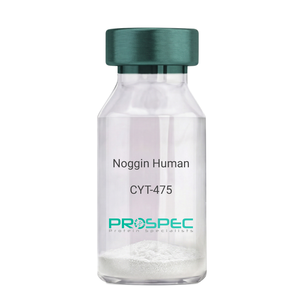
Noggin HumanDescription:
Noggin Human Recombinant
SYM1, SYNS1, NOG.
Product # :
CYT-475Price :
Quantity :
Shipping Method :
Shipped at Room temp
More Info
- description
- source
- formulation
- purity
- biological activity
- More Info
- Citations
Description
Noggin Human Recombinant produced in E.Coli is a non-glycosylated, non-disulfide-linked homodimer consisting of two 206 amino acid polypeptide chains, having a total molecular mass of approximately 46.3kDa. Noggin is purified by proprietary chromatographic techniques.
Source
Escherichia Coli.
Formulation
Lyophilized from a 0.2μm filtered solution in 30% CH3CN, 0.1% TFA.
Purity
Greater than 95.0% as determined by SDS-PAGE.
Biological Activity
The ED50 was determined by its ability to inhibit 5.0ng/ml of BMP-4 induced alkaline phosphatase production by murine ATDC-5 cells. The expected ED50 for this effect is < 3ng/ml of Noggin, corresponding to a Specific Activity of 3.3x10⁵units/mg.
Citations
More Info
-
Introduction
The secreted polypeptide noggin, encoded by the NOG gene, binds and inactivates members of the transforming growth factor-beta (TGF-beta) superfamily signaling proteins, such as bone morphogenetic protein-4 (BMP4). By diffusing through extracellular matrices more efficiently than members of the TGF-beta superfamily, noggin may have a principal role in creating morphogenic gradients. Noggin appears to have pleiotropic effect, both early in development as well as in later stages. It was originally isolated from Xenopus based on its ability to restore normal dorsal-ventral body axis in embryos that had been artificially ventralized by UV treatment. The results of the mouse knockout of noggin suggest that it is involved in numerous developmental processes, such as neural tube fusion and joint formation. Recently, several dominant human NOG mutations in unrelated families with proximal symphalangism (SYM1) and multiple synostoses syndrome (SYNS1) were identified; both SYM1 and SYNS1 have multiple joint fusion as their principal feature, and map to the same region (17q22) as NOG. All NOG mutations altered evolutionarily conserved amino acid residues. The amino acid sequence of human noggin is highly homologous to that of Xenopus, rat and mouse.
-
Synonyms
SYM1, SYNS1, NOG.
-
Physical Appearance
Sterile Filtered White lyophilized (freeze-dried) powder.
-
Stability
Lyophilized Noggin although stable at room temperature for 3 weeks, should be stored desiccated below -18°C. Upon reconstitution Noggin should be stored at 4°C between 2-7 days and for future use below -18°C.For long term storage it is recommended to add a carrier protein (0.1% HSA or BSA).Please prevent freeze-thaw cycles.
-
Solubility
It is recommended to be briefly centrifuged prior to opening to bring the contents to the bottom. Reconstitute in 10mM HCl to a concentration of 0.1-1.0 mg/ml. Further dilutions should be made in appropriate buffered solutions.
-
Amino Acid Sequence
MQHYLHIRPAPSDNLPLVDLIEHPDPIFDPKEKDLNETLLRSLLGGHYDPGFMATSPP
EDRPGGGGGAAGGAEDLAELDQLLRQRPSGAMPSEIKGLEFSEGLAQGKKQRLSKKLR
RKLQMWLWSQTFCPVLYAWNDLGSRFWPRYVKVGSCFSKRSCSVPEGMVCKPSKSVHL
TVLRWRCQRRGGQRCGWIPIQYPIISECKCSC. -
Background
Recombinant Human Noggin Growth Beta Factor: A Potent Inhibitor of Bone Morphogenetic Protein Signaling.
Abstract:
Recombinant human Noggin Growth Beta Factor (Noggin) is a highly conserved protein that acts as a potent antagonist of the Bone Morphogenetic Protein (BMP) signaling pathway.
Noggin plays a critical role in embryonic development, tissue homeostasis, and disease processes.
This research paper provides a comprehensive analysis of the molecular characteristics, signaling mechanisms, and diverse physiological functions of recombinant human Noggin.
Additionally, it explores the therapeutic implications of Noggin in various disorders. Synonyms such as SYM1, SYNS1, and NOG associated with Noggin are discussed throughout the paper to highlight their relevance in scientific literature.
Introduction:
- Recombinant human Noggin Growth Beta Factor (Noggin) is a protein with multifaceted roles in development, tissue homeostasis, and disease. This section introduces Noggin and its synonyms, including SYM1, SYNS1, and NOG, emphasizing their significance and relevance in scientific research.
Molecular Characteristics of Noggin :
- This section explores the molecular characteristics of Noggin, including its primary amino acid sequence, protein structure, and post-translational modifications. The interactions of Noggin with BMPs and other regulatory molecules are also discussed, highlighting the importance of these interactions in modulating BMP signaling.
Inhibition of BMP Signaling by Noggin:
- Noggin acts as a potent inhibitor of BMP signaling by binding to BMP ligands and preventing their interaction with BMP receptors. This section delves into the mechanisms through which Noggin interferes with BMP signaling, including competition for receptor binding and sequestration of BMPs in extracellular spaces. The implications of Noggin-mediated inhibition of BMP signaling in development and tissue homeostasis are also discussed.
Physiological Functions of Noggin:
- Noggin plays critical roles in various physiological processes, including embryonic development, neurogenesis, skeletal development, and joint formation. This section provides an in-depth analysis of Noggin's contributions to these processes, highlighting its role in maintaining proper tissue patterning, cell fate determination, and morphogenesis.
Therapeutic Implications of Noggin:
- The unique inhibitory properties of Noggin make it an attractive therapeutic candidate for various disorders. This section discusses the potential applications of Noggin in bone and joint diseases, neurological disorders, and cancer. Additionally, it explores the challenges and future prospects of utilizing Noggin as a therapeutic agent.
Clinical Studies and Translational Research:
- This section reviews clinical studies and translational research involving Noggin, emphasizing its potential in regenerative medicine and tissue engineering. It highlights ongoing efforts to develop Noggin-based therapeutics and discusses the promising results observed in preclinical and clinical studies.
ProSpec's products are furnished for LABORATORY RESEARCH USE ONLY. The product may not be used as drugs, agricultural or pesticidal products, food additives or household chemicals.
- View Data Sheet
Name :
EIF5A AntibodyDescription:
Eukaryotic Translation Initiation Factor 5A, Mouse Anti Human
EIF-5A, EIF5A1, eIF5AI, MGC99547, MGC104255, EIF5A, Eukaryotic translation initiation factor 5A-1, eIF-5A-1, eIF-5A1, Eukaryotic initiation factor 5A isoform 1, eIF-4D, Rev-binding factor.
Product # :
ANT-010Price :
Quantity :
Shipping Method :
Shipped with Ice Packs
More Info
- formulation
- More Info
- Citations
Formulation
1mg/ml containing PBS, pH-7.4, & 0.1% Sodium Azide.
Citations
More Info
-
Introduction
EIF5A is the single protein identified to contain remarkable amino acid formed by the action of deoxyhypusine synthase and deoxyhypusine hydroxylase using spermidine as the substrate. EIF5A takes part in the first step of peptide bond formation in translation, nevertheless further experiments implicates it as a universally conserved translation elongation factor. Modulation of EIF5A is connected to proliferation and cancer. Expression of EIF-5A is upregulated in the PBMCs of HIV-1 patients. EIF5A coordinates significant cellular processes like cell viability and senescence during its effects on the stability of certain mRNAs. Heat stress-induced loss of EIF-5A in a human pancreatic cancer cell line. EIF5A stability takes part in determining the fate of the particular cell type after severe heat stress.
-
Synonyms
EIF-5A, EIF5A1, eIF5AI, MGC99547, MGC104255, EIF5A, Eukaryotic translation initiation factor 5A-1, eIF-5A-1, eIF-5A1, Eukaryotic initiation factor 5A isoform 1, eIF-4D, Rev-binding factor.
-
Immunogen
Anti-human EIF5A mAb, is derived from hybridization of mouse F0 myeloma cells with spleen cells from BALB/c mice immunized with recombinant human EIF5A amino acids 1-154 purified from E. coli.
-
Ig Subclass
Mouse IgG1 heavy chain and κ light chain.
-
Clone
PAT2E9AT.
-
Applications
EIF5A antibody has been tested by ELISA, Western blot and Immunofluorescence analysis to assure specificity and reactivity. Since application varies, however, each investigation should be titrated by the reagent to obtain optimal results. Recommended dilution range for Western blot analysis and Immunofluorescence is 1:250 ~ 500.
Recommended starting dilution is 1:250. -
Type
Mouse Anti Human Monoclonal.
-
Storage Procedures
For periods up to 1 month store at 4°C, for longer periods of time, store at -20°C. Prevent freeze thaw cycles.
-
Purification Method
EIF5A antibody was purified from mouse ascitic fluids by protein-G affinity chromatography.
ProSpec's products are furnished for LABORATORY RESEARCH USE ONLY. The product may not be used as drugs, agricultural or pesticidal products, food additives or household chemicals.
- View Data Sheet
Name :
GLOD4 HumanDescription:
Glyoxalase Domain Containing 4 Human Recombinant
Glyoxalase domain containing protein 4, HC71, C17orf25, CGI-150, Chromosome 17 open reading frame 25.
Product # :
PRO-894Price :
Quantity :
Shipping Method :
Shipped with Ice Packs
More Info
- description
- source
- formulation
- purity
- More Info
- Citations
Description
GLOD4 Human Recombinant produced in E.coli is a single, non-glycosylated polypeptide chain containing 318 amino acids (1-298) and having a molecular mass of 35.3 kDa.The GLOD4 is fused to a 20 amino acid His-Tag at N-terminus and purified by proprietary chromatographic techniques.
Source
E.coli.
Formulation
The GLOD4 solution (1mg/ml) contains 20mM Tris-HCl buffer (pH 8.0), 1mM DTT, 0.1M NaCl and 20% glycerol.
Purity
Greater than 95% as determined by SDS-PAGE.
Citations
More Info
-
Introduction
GLOD4 is a part of the glyoxalase system which is a group of enzymes that is in charge of the detoxification of methylglyoxal and other reactive aldehydes that are usually produced in metabolism. The mitochondrial protein GLOD4 is known to interact with NUDT9.
-
Synonyms
Glyoxalase domain containing protein 4, HC71, C17orf25, CGI-150, Chromosome 17 open reading frame 25.
-
Physical Appearance
Sterile Filtered colorless solution.
-
Stability
Store at 4°C if entire vial will be used within 2-4 weeks. Store, frozen at -20°C for longer periods of time. For long term storage it is recommended to add a carrier protein (0.1% HSA or BSA).Avoid multiple freeze-thaw cycles.
-
Amino Acid Sequence
MGSSHHHHHH SSGLVPRGSH MAARRALHFV FKVGNRFQTA RFYRDVLGMK VLRHEEFEEG CKAACNGPYD GKWSKTMVGF GPEDDHFVAE LTYNYGVGDY KLGNDFMGIT LASSQAVSNA RKLEWPLTEV AEGVFETEAP GGYKFYLQNR SLPQSDPVLK VTLAVSDLQK SLNYWCNLLG MKIYEKDEEK QRALLGYADN QCKLELQGVK GGVDHAAAFG RIAFSCPQKE LPDLEDLMKR ENQKILTPLV SLDTPGKATV QVVILADPDG HEICFVGDEA FRELSKMDPE GSKLLDDAMA ADKSDEWFAK HNKPKASG
ProSpec's products are furnished for LABORATORY RESEARCH USE ONLY. The product may not be used as drugs, agricultural or pesticidal products, food additives or household chemicals.
- View Data Sheet
Name :
OTUB1 HumanDescription:
Ubiquitin Aldehyde Binding 1 Human Recombinant
Ubiquitin thioesterase OTUB1, Otubain-1, OTU domain-containing ubiquitin aldehyde-binding protein 1, Ubiquitin-specific-processing protease OTUB1, Deubiquitinating enzyme OTUB1, OTUB1, OTB1, OTU1, HSPC263, MGC4584, FLJ20113, FLJ40710, MGC111158.
Product # :
PRO-711Price :
Quantity :
Shipping Method :
Shipped with Ice Packs
More Info
- description
- source
- formulation
- purity
- More Info
- Citations
Description
OTUB1 Human Recombinant fused with a 20 amino acid His tag at N-terminus produced in E.Coli is a single, non-glycosylated, polypeptide chain containing 291 amino acids (1- 271 a.a.) and having a molecular mass of 33.4kDa.The OTUB1 is purified by proprietary chromatographic techniques.
Source
Escherichia Coli.
Formulation
The OTUB1 solution contains 20mM Tris buffer (pH 8.0) and 10% glycerol.
Purity
Greater than 95.0% as determined by SDS-PAGE.
Citations
More Info
-
Introduction
Otubain 1 (OTUB1) belongs to the ovarian tumor (OUT) superfamily of predicted cysteine proteases and inhibits cytokine gene transcription in the immune system through its interaction with a ubiquitin protease and E3 ubiquitin ligase. OTUB1 is a highly specific ubiquitin iso-peptidase, it cleaves ubiquitin from branched poly-ubiquitin chains but not from ubiquitinated substrates. OTUB1 is believed to work in specific ubiquitin-dependent pathways, possibly by providing an editing function of polyubiquitin chain growth. OTUB1 is a hydrolase that removes conjugated ubiquitin from proteins in vitro and may therefore have a significant regulatory role in the level of protein turnover by preventing degradation. Additionally, OTUB1 is a regulator of T-cell anergy, a phenomenon that occurs when T-cells are rendered impassive to antigen re-challenge and no longer respond to their cognate antigen. OTUB1 acts via its interaction with RNF128/GRAIL, which is an essential inductor of CD4 T-cell anergy.
-
Synonyms
Ubiquitin thioesterase OTUB1, Otubain-1, OTU domain-containing ubiquitin aldehyde-binding protein 1, Ubiquitin-specific-processing protease OTUB1, Deubiquitinating enzyme OTUB1, OTUB1, OTB1, OTU1, HSPC263, MGC4584, FLJ20113, FLJ40710, MGC111158.
-
Physical Appearance
Sterile Filtered colorless solution.
-
Stability
Store at 4°C if entire vial will be used within 2-4 weeks. Store, frozen at -20°C for longer periods of time. For long term storage it is recommended to add a carrier protein (0.1% HSA or BSA).Avoid multiple freeze-thaw cycles.
-
Amino Acid Sequence
MGSSHHHHHH SSGLVPRGSH MAAEEPQQQK QEPLGSDSEG VNCLAYDEAI MAQQDRIQQE IAVQNPLVSE RLELSVLYKE YAEDDNIYQQ KIKDLHKKYS YIRKTRPDGN CFYRAFGFSH LEALLDDSKE LQRFKAVSAK SKEDLVSQGF TEFTIEDFHN TFMDLIEQVE KQTSVADLLA SFNDQSTSDY LVVYLRLLTS GYLQRESKFF EHFIEGGRTV KEFCQQEVEP MCKESDHIHI IALAQALSVS IQVEYMDRGE GGTTNPHIFP EGSEPKVYLL YRPGHYDILY K.
ProSpec's products are furnished for LABORATORY RESEARCH USE ONLY. The product may not be used as drugs, agricultural or pesticidal products, food additives or household chemicals.