Skip to content

High-quality research proteins.

  • 1-800-805-6531

  • Mon - Sat 8.00-18.00, Sun - Closed

  • info@prospecbio.com

  • PO Box 6591 East Brunswick NJ 08816

  • Products
    • Cytokines
    • Growth factors
    • Chemokines
    • Cd antigens
    • Neurotrophins
    • Hormones
    • Enzymes
    • Viral antigens
    • Recombinant proteins
    • Natural proteins
    • Monoclonal antibodies
    • Polyclonal antibodies
    • Thermal Packaging
  • About
    • Company
    • Founder
    • Contribution
  • Customers
    • Eastern Asia
    • Eastern Europe
    • North America
    • South America
    • South East Asia
    • Southern Asia
    • Western Europe
  • Ordering
    • Ordering Method
    • Ordering - Credit Card
    • Ordering - PO
    • Shipping Fees
    • FAQs
  • Services
    • DNA Cloning
    • Protein Expression
    • Monoclonal Antibody
    • Fermentation
    • Technology License
  • Contact us
Log in

Country/region

  • USD $ | Albania
  • USD $ | Andorra
  • USD $ | Argentina
  • USD $ | Armenia
  • USD $ | Australia
  • USD $ | Austria
  • USD $ | Azerbaijan
  • USD $ | Belarus
  • USD $ | Belgium
  • USD $ | Bolivia
  • USD $ | Brazil
  • USD $ | Bulgaria
  • USD $ | Cambodia
  • USD $ | Canada
  • USD $ | Chile
  • USD $ | China
  • USD $ | Colombia
  • USD $ | Costa Rica
  • USD $ | Croatia
  • USD $ | Cyprus
  • USD $ | Czechia
  • USD $ | Denmark
  • USD $ | Estonia
  • USD $ | Finland
  • USD $ | France
  • USD $ | Georgia
  • USD $ | Germany
  • USD $ | Greece
  • USD $ | Hong Kong SAR
  • USD $ | Hungary
  • USD $ | Iceland
  • USD $ | India
  • USD $ | Ireland
  • USD $ | Israel
  • USD $ | Italy
  • USD $ | Japan
  • USD $ | Kazakhstan
  • USD $ | Latvia
  • USD $ | Liechtenstein
  • USD $ | Lithuania
  • USD $ | Madagascar
  • USD $ | Malta
  • USD $ | Mexico
  • USD $ | Moldova
  • USD $ | Monaco
  • USD $ | Netherlands
  • USD $ | New Zealand
  • USD $ | North Macedonia
  • USD $ | Norway
  • USD $ | Panama
  • USD $ | Paraguay
  • USD $ | Peru
  • USD $ | Philippines
  • USD $ | Poland
  • USD $ | Portugal
  • USD $ | Romania
  • USD $ | Singapore
  • USD $ | Slovakia
  • USD $ | Slovenia
  • USD $ | South Africa
  • USD $ | South Korea
  • USD $ | Spain
  • USD $ | Sri Lanka
  • USD $ | Sweden
  • USD $ | Switzerland
  • USD $ | Taiwan
  • USD $ | Thailand
  • USD $ | Türkiye
  • USD $ | Ukraine
  • USD $ | United Kingdom
  • USD $ | United States
  • USD $ | Uruguay
  • USD $ | Vatican City
  • USD $ | Venezuela
  • USD $ | Vietnam
  • Facebook
  • Instagram
logoProSpec - Recombinant Protein Manufacturer for academic and R&D industry
Become a distributor
Account Log in
Wishlist
Cart Cart(0)
  • Products
    +
    • Cytokines
    • Growth factors
    • Chemokines
    • Cd antigens
    • Neurotrophins
    • Hormones
    • Enzymes
    • Viral antigens
    • Recombinant proteins
    • Natural proteins
    • Monoclonal antibodies
    • Polyclonal antibodies
    • Thermal Packaging
  • About
    +
    • Company
    • Founder
    • Contribution
  • Customers
    +
    • Eastern Asia
    • Eastern Europe
    • North America
    • South America
    • South East Asia
    • Southern Asia
    • Western Europe
  • Ordering
    +
    • Ordering Method
    • Ordering - Credit Card
    • Ordering - PO
    • Shipping Fees
    • FAQs
  • Services
    +
    • DNA Cloning
    • Protein Expression
    • Monoclonal Antibody
    • Fermentation
    • Technology License
  • Contact us

Item added to your cart

View cart
View All
  • Cytokines
  • Growth factors
  • Chemokines
  • Cd antigens
  • Neurotrophins
  • Hormones
  • Enzymes
  • Viral antigens
  • Recombinant proteins
  • Natural proteins
  • Monoclonal antibodies
  • Polyclonal antibodies
  • Thermal Packaging
  • Tumor Necrosis Factor

    Tumor Necrosis Factor

    View All
  • 4-1BB

    4-1BB

    View All
  • Adiponectin

    Adiponectin

    View All
  • AIF1

    AIF1

    View All
  • Other Cytokines

    Other Cytokines

    View All
  • AITRL

    AITRL

    View All
  • Angiopoietin

    Angiopoietin

    View All
  • Apolipoprotein

    Apolipoprotein

    View All
  • B-Cell Activating Factor

    B-Cell Activating Factor

    View All
  • Beta Defensin

    Beta Defensin

    View All
  • Bone Morphogenetic Protein

    Bone Morphogenetic Protein

    View All
  • B type Natriuretic Peptide

    B type Natriuretic Peptide

    View All
  • BST

    BST

    View All
  • Betacellulin

    Betacellulin

    View All
  • Cardiotrophin

    Cardiotrophin

    View All
  • Activin

    Activin

    View All
  • Fibroblast Growth Factor

    Fibroblast Growth Factor

    View All
  • Other Growth Factors

    Other Growth Factors

    View All
  • CSF

    CSF

    View All
  • CTGF

    CTGF

    View All
  • IGFBP

    IGFBP

    View All
  • VEGF Protein

    VEGF Protein

    View All
  • EGF

    EGF

    View All
  • Epigen

    Epigen

    View All
  • Erythropoietin

    Erythropoietin

    View All
  • Insulin-Like Growth Factor

    Insulin-Like Growth Factor

    View All
  • Growth Hormone

    Growth Hormone

    View All
  • HDGF

    HDGF

    View All
  • Hepatocyte Growth Factor

    Hepatocyte Growth Factor

    View All
  • Insulin

    Insulin

    View All
  • BCA-1/ BLC (CXCL13)

    BCA-1/ BLC (CXCL13)

    View All
  • BRAK (CXCL14)

    BRAK (CXCL14)

    View All
  • C-10 (CCL6)

    C-10 (CCL6)

    View All
  • Other Chemokines

    Other Chemokines

    View All
  • MEC (CCL28)

    MEC (CCL28)

    View All
  • LD78-beta (CCL3L1)

    LD78-beta (CCL3L1)

    View All
  • CTACK (CCL27)

    CTACK (CCL27)

    View All
  • CXCL16

    CXCL16

    View All
  • CXCL17

    CXCL17

    View All
  • Platelet Factor-4 (CXCL4)

    Platelet Factor-4 (CXCL4)

    View All
  • ENA-78 (CXCL5)

    ENA-78 (CXCL5)

    View All
  • Eotaxin (CCL11,24,26)

    Eotaxin (CCL11,24,26)

    View All
  • Exodus-2 (CCL21)

    Exodus-2 (CCL21)

    View All
  • Fractalkine (CX3CL1)

    Fractalkine (CX3CL1)

    View All
  • CXCL6

    CXCL6

    View All
  • Other CD Antigens

    Other CD Antigens

    View All
  • CD14

    CD14

    View All
  • CD164

    CD164

    View All
  • CD1

    CD1

    View All
  • CD2

    CD2

    View All
  • CD200

    CD200

    View All
  • CD207

    CD207

    View All
  • CD226

    CD226

    View All
  • CD23

    CD23

    View All
  • CD244

    CD244

    View All
  • CD247

    CD247

    View All
  • CD27

    CD27

    View All
  • CD274

    CD274

    View All
  • CD300

    CD300

    View All
  • CD33

    CD33

    View All
  • Other Neurotrophins

    Other Neurotrophins

    View All
  • Beta-NGF

    Beta-NGF

    View All
  • BDNF

    BDNF

    View All
  • CDNF

    CDNF

    View All
  • CNTF

    CNTF

    View All
  • GDNF

    GDNF

    View All
  • Glia Maturation Factor

    Glia Maturation Factor

    View All
  • MANF

    MANF

    View All
  • Midkine

    Midkine

    View All
  • Neuroglobin

    Neuroglobin

    View All
  • Neurotrophic factors

    Neurotrophic factors

    View All
  • Neuregulin

    Neuregulin

    View All
  • Neuropilin

    Neuropilin

    View All
  • Pigment Epithelium-Derived Factor

    Pigment Epithelium-Derived Factor

    View All
  • Pleiotrophin

    Pleiotrophin

    View All
  • Peptide Hormones

    Peptide Hormones

    View All
  • Other Hormones

    Other Hormones

    View All
  • HCG

    HCG

    View All
  • Endothelin

    Endothelin

    View All
  • Exendin

    Exendin

    View All
  • FSH

    FSH

    View All
  • Glucagon

    Glucagon

    View All
  • GHRP

    GHRP

    View All
  • GLP

    GLP

    View All
  • Thyrostimulin

    Thyrostimulin

    View All
  • Inhibin A

    Inhibin A

    View All
  • LHRH

    LHRH

    View All
  • Melanotan

    Melanotan

    View All
  • Procalcitonin

    Procalcitonin

    View All
  • PTH

    PTH

    View All
  • Peroxiredoxin

    Peroxiredoxin

    View All
  • Peptidase

    Peptidase

    View All
  • Synthetase

    Synthetase

    View All
  • Transferase

    Transferase

    View All
  • Hydrolase

    Hydrolase

    View All
  • Dehydrogenase

    Dehydrogenase

    View All
  • ACE2

    ACE2

    View All
  • Esterase

    Esterase

    View All
  • Protein Kinases

    Protein Kinases

    View All
  • Other Enzymes

    Other Enzymes

    View All
  • Phosphatase

    Phosphatase

    View All
  • Deaminase

    Deaminase

    View All
  • Oxygenase

    Oxygenase

    View All
  • Adenylate Kinase

    Adenylate Kinase

    View All
  • Lyase

    Lyase

    View All
  • Chagas

    Chagas

    View All
  • SARS

    SARS

    View All
  • Influenza

    Influenza

    View All
  • ASFV

    ASFV

    View All
  • Astrovirus

    Astrovirus

    View All
  • Borrelia

    Borrelia

    View All
  • Helicobacter Pylori

    Helicobacter Pylori

    View All
  • Chikungunya

    Chikungunya

    View All
  • Chlamydia

    Chlamydia

    View All
  • Cytomegalo

    Cytomegalo

    View All
  • Dengue

    Dengue

    View All
  • EBV

    EBV

    View All
  • Ebola

    Ebola

    View All
  • Feline Leukemia Virus

    Feline Leukemia Virus

    View All
  • Hepatitis A

    Hepatitis A

    View All
  • Other

    Other

    View All
  • Actin

    Actin

    View All
  • ADAM

    ADAM

    View All
  • Complement Component

    Complement Component

    View All
  • Ag85

    Ag85

    View All
  • Eukaryotic Translation Initiation Factor

    Eukaryotic Translation Initiation Factor

    View All
  • Anterior Gradient Protein

    Anterior Gradient Protein

    View All
  • Heat Shock Protein

    Heat Shock Protein

    View All
  • Alpha-2-HS-Glycoprotein

    Alpha-2-HS-Glycoprotein

    View All
  • Hemoglobin

    Hemoglobin

    View All
  • Allergy

    Allergy

    View All
  • Anaplasma

    Anaplasma

    View All
  • Angiogenin

    Angiogenin

    View All
  • Ankyrin Repeat Domain

    Ankyrin Repeat Domain

    View All
  • Annexin

    Annexin

    View All
  • Other Natural Proteins

    Other Natural Proteins

    View All
  • Aprotinin

    Aprotinin

    View All
  • Transferrin

    Transferrin

    View All
  • Avidin

    Avidin

    View All
  • Anti Coagulation Factors

    Anti Coagulation Factors

    View All
  • Natural Albumin

    Natural Albumin

    View All
  • Natural Coagulation Factors

    Natural Coagulation Factors

    View All
  • Fibronectin

    Fibronectin

    View All
  • Thrombin

    Thrombin

    View All
  • Anti Human Enzyme

    Anti Human Enzyme

    View All
  • Other Monoclonal Antibodies

    Other Monoclonal Antibodies

    View All
  • Anti Human Cytokine

    Anti Human Cytokine

    View All
  • Anti Human Heat Shock Protein

    Anti Human Heat Shock Protein

    View All
  • Anti Mouse Lymphocyte

    Anti Mouse Lymphocyte

    View All
  • Anti Human Chemokine

    Anti Human Chemokine

    View All
  • Anti Human Lymphocyte

    Anti Human Lymphocyte

    View All
  • Anti Viral Monoclonal

    Anti Viral Monoclonal

    View All
  • Anti Mouse Cytokine

    Anti Mouse Cytokine

    View All
  • Anti-GST Monoclonal

    Anti-GST Monoclonal

    View All
  • Other Polyclonal Antibodies

    Other Polyclonal Antibodies

    View All
  • Anti Viral

    Anti Viral

    View All
  • Anti-GST Monoclonal

    Anti-GST Monoclonal

    View All
Back

Search results

638 results found for “mip (ccl3,4,9,15)”

Name

Description

Product #

Price

Quantity

Shipping Method

  • View Data Sheet

    Name :

    BMP6 Human

    Description:

    Bone Morphogenetic protein-6 Human Recombinant

    Bone morphogenetic protein 6, BMP-6, VG-1-related protein, VG-1-R, VGR-1, BMP6, VGR, VGR1.

    Product # :

    CYT-754

    Price :

    Quantity :

    Shipping Method :

    Ice Icon

    Shipped with Ice Packs

    Add To Cart

    More Info

    • description
    • source
    • formulation
    • purity
    • More Info
    • sds-page
    • Citations

    Description

    BMP6 Human Recombinant produced in E.coli is a single, non-glycosylated polypeptide chain containing 164 amino acids (375-513) and having a molecular mass of 18kDa.BMP6 is fused to a 25 amino acid His-tag at N-terminus & purified by proprietary chromatographic techniques.

    Source

    Escherichia Coli.

    Formulation

    The BMP6 solution (0.25mg/ml) contains 10mM Sodium citrate buffer (pH 3.5) and 10% glycerol.

    Purity

    Greater than 90.0% as determined by SDS-PAGE.

    sds-page

    BMP6-sds-page - Product image 1

    Citations

    More Info

    • Introduction

      The bone morphogenetic proteins (BMPs) are a family of secreted signaling molecules, which can induce ectopic bone growth. Various BMPs are part of the transforming growth factor-beta (TGFB) superfamily. BMPs were initially identified by an ability of demineralized bone extract to induce endochondral osteogenesis in vivo in an extraskeletal site. Based upon its expression early in embryogenesis, BMP6 has a suggested role in early development. Moreover, the fact that the BMP6 is closely related to BMP5 and BMP7 leads to an assumption of possible bone inductive activity.

    • Synonyms

      Bone morphogenetic protein 6, BMP-6, VG-1-related protein, VG-1-R, VGR-1, BMP6, VGR, VGR1.

    • Physical Appearance

      Sterile filtered colorless solution.

    • Stability

      Store at 4°C if entire vial will be used within 2-4 weeks. Store, frozen at -20°C for longer periods of time. For long term storage it is recommended to add a carrier protein (0.1% HSA or BSA). Avoid multiple freeze-thaw cycles.

    • Amino Acid Sequence

      MGSSHHHHHH SSGLVPRGSH MGSHMSASSR RRQQSRNRST QSQDVARVSS ASDYNSSELK TACRKHELYV SFQDLGWQDW IIAPKGYAAN YCDGECSFPL NAHMNATNHA IVQTLVHLMN PEYVPKPCCA PTKLNAISVL YFDDNSNVIL KKYRNMVVRA CGCH.

    • Background

      Bone Morphogenetic Protein-6 Human Recombinant: Unraveling its Therapeutic Potential in Tissue Engineering and Regenerative Medicine

      Abstract:

      Bone Morphogenetic Protein-6 (BMP-6) human recombinant is a critical member of the bone morphogenetic protein family, known for its pivotal role in tissue development, repair, and regeneration. This research paper aims to provide a comprehensive analysis of BMP-6, including its characteristics, signaling pathways, and potential therapeutic applications. Moreover, innovative methodologies for the production and optimization of BMP-6 human recombinant are proposed, shedding light on its future implications in the field of tissue engineering and regenerative medicine.

      Introduction:

      Tissue engineering and regenerative medicine have emerged as promising approaches to address the challenges associated with tissue repair and regeneration. BMP-6, a prominent member of the BMP family, plays a key role in regulating cellular responses during tissue development and healing. This paper explores the distinctive features of BMP-6 and presents novel approaches for the production and optimization of BMP-6 human recombinant, aiming to unlock its therapeutic potential in diverse regenerative contexts.

      Characteristics and Signaling Pathways:

      BMP-6 is a secreted growth factor belonging to the transforming growth factor-beta (TGF-β) superfamily. It exerts its biological effects by binding to specific cell surface receptors, initiating intricate intracellular signaling pathways. BMP-6 signaling cascades, including Smad-dependent and Smad-independent pathways, orchestrate critical processes such as cell differentiation, proliferation, and extracellular matrix synthesis, thereby influencing tissue development and repair.

      Production of BMP-6 Human Recombinant:

      Efficient production methodologies are vital for harnessing the therapeutic potential of BMP-6 human recombinant. Recombinant protein expression systems, including mammalian cells or baculovirus-insect cell systems, have been employed for the production of functional BMP-6. Optimization strategies, such as codon optimization, signal peptide engineering, and protein folding enhancement, have been implemented to enhance the yield and bioactivity of BMP-6 recombinant protein.

      Potential Therapeutic Applications:

      BMP-6 human recombinant holds immense promise in the field of tissue engineering and regenerative medicine. Its regulatory role in bone formation, cartilage regeneration, and wound healing positions it as a potential therapeutic candidate for the treatment of skeletal disorders, osteoarthritis, and tissue injuries. Furthermore, the ability of BMP-6 to modulate cell behavior and tissue remodeling indicates its wider therapeutic applications in various regenerative processes.

      Conclusion:

      BMP-6 human recombinant emerges as a crucial regulator in tissue engineering and regenerative medicine, offering significant potential for tissue repair and regeneration. Optimizing production methodologies and further unraveling its signaling mechanisms will undoubtedly enhance its therapeutic applications. Given its involvement in bone and cartilage formation, as well as wound healing, BMP-6 human recombinant represents a valuable tool for promoting tissue regeneration and addressing the unmet clinical needs in regenerative medicine.

      What is the molecular weight/Mw of BMP6 Protein?
      BMP6 Protein has a total Mw of 18kDa.

      What is the source or expression system of BMP6 Protein?
      Escherichia Coli.

      What is the Purity of BMP6 Protein?
      BMP6 Protein is >90% pure as determined by SDS-PAGE.

      What is the Biological Activity of BMP6 Protein?
      The biological functionality of BMP6 Protein will be determined in the future.

      What is the amino acid sequence of BMP6 Protein?
      MGSSHHHHHH SSGLVPRGSH MGSHMSASSR RRQQSRNRST QSQDVARVSS ASDYNSSELK TACRKHELYV SFQDLGWQDW IIAPKGYAAN YCDGECSFPL NAHMNATNHA IVQTLVHLMN PEYVPKPCCA PTKLNAISVL YFDDNSNVIL KKYRNMVVRA CGCH.

      What applications can BMP6 Protein be used in?
      BMP6 Protein can probably be used in western blot, ELISA and Lateral Flow.

      What is the endotoxin level for BMP6 Protein?
      The endotoxin level is minimal, BMP6 Protein was purified using conventional chromatography techniques.

    ProSpec's products are furnished for LABORATORY RESEARCH USE ONLY. The product may not be used as drugs, agricultural or pesticidal products, food additives or household chemicals.

    Bmp6 Human
  • View Data Sheet

    Name :

    Bet v 1.0101, Sf9

    Description:

    Major pollen allergen Bet v 1-A Recombinant, Sf9

    Major pollen allergen Bet v 1-A, Allergen Bet v I-A, Bet v 1-A, BETVIA, BETVI.

    Product # :

    PRO-2282

    Price :

    Quantity :

    Shipping Method :

    Ice Icon

    Shipped with Ice Packs

    Add To Cart

    More Info

    • description
    • source
    • formulation
    • purity
    • More Info
    • Citations

    Description

    Recombinant Major pollen allergen Bet v 1-A produced in SF9 is a glycosylated, polypeptide chain having a calculated molecular mass of 18,942 Dalton. Bet v 1.0101 is expressed with a 10xHis tag at N-terminus and purified by proprietary chromatographic techniques.

    Source

    Sf9 insect cells.

    Formulation

    Bet v 1.0101 protein solution is supplied in 20mM HEPES buffer pH-8.0, 200mM NaCl and 20% glycerol.

    Purity

    Greater than 95.0% as determined by SDS-PAGE.

    Citations

    More Info

    • Introduction

      Major pollen allergen Bet v 1-A (Bet v 1.0101) causes an allergic reaction in humans. Bet v 1.0101 is a cause of type I allergic reactions in Europe, North America and the former Soviet Union states, observed in early spring.

    • Synonyms

      Major pollen allergen Bet v 1-A, Allergen Bet v I-A, Bet v 1-A, BETVIA, BETVI.

    • Physical Appearance

      Sterile Filtered clear solution.

    • Stability

      Store at 4°C if entire vial will be used within 2-4 weeks. Store, frozen at -20°C for longer periods of time. Avoid multiple freeze-thaw cycles.

    • Immunological Functions

      1. Binds IgE type human antibodies. 2. Immunodot test with positive/negative sera panels.

    ProSpec's products are furnished for LABORATORY RESEARCH USE ONLY. The product may not be used as drugs, agricultural or pesticidal products, food additives or household chemicals.

    Bet V 10101
  • View Data Sheet

    Name :

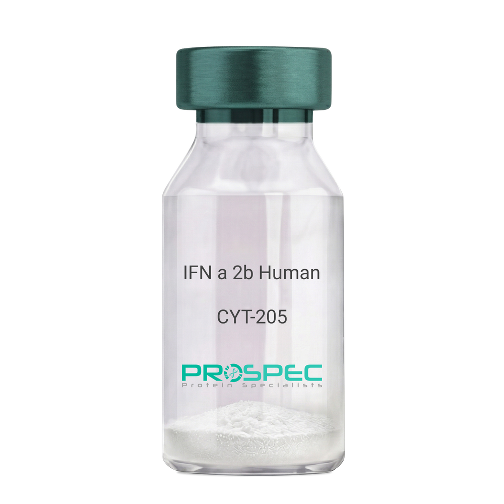
    IFN a 2b Human

    Description:

    IFN-Alpha 2b Human Recombinant

    IFN alpha 2b, IFNA, INFA2, MGC125764, MGC125765.

    Product # :

    CYT-205

    Price :

    Quantity :

    Shipping Method :

    Room Temp Icon

    Shipped at Room temp

    Add To Cart

    More Info

    • description
    • source
    • formulation
    • purity
    • biological activity
    • More Info
    • Citations

    Description

    IFN-a 2b Human Recombinant produced in E.Coli is a single, non-glycosylated, polypeptide chain containing 166 amino acids and having a molecular mass of 19400 Dalton. The difference between IFNA2A and IFNA2B is in the amino acid present at position 23. IFN-alpha 2a has a lysine at that position 23 while IFN-alpha 2b has arginine. The IFN-alpha 2b gene was obtained from human leukocytes. The IFN-a 2b is purified by proprietary chromatographic techniques.

    Source

    Escherichia Coli.

    Formulation

    Lyophilized from a (1 mg/ml) solution in containing 2.3 mg Sodium phosphate dibasic and 0.55 mg sodium phosphate monobasic buffer.

    Purity

    Greater than 98.0% as determined by(a) Analysis by RP-HPLC.
    (b) Analysis by SDS-PAGE.

    Biological Activity

    The specific activity as determined in a viral resistance assay using bovine kidney MDBK cells was found to be 260,000,000 IU/ mg.

    Citations

    More Info

    • Introduction

      IFN-alpha is produced by macrophages and has antiviral activities. IFN stimulates the production of two enzymes: protein kinase and an oligoadenylate synthetase.

    • Synonyms

      IFN alpha 2b, IFNA, INFA2, MGC125764, MGC125765.

    • Physical Appearance

      Sterile Filtered White lyophilized (freeze-dried) powder.

    • Stability

      Lyophilized IFN although stable at room temperature for 3 weeks, should be stored desiccated below -18°C. Upon reconstitution IFN alpha 2b should be stored at 4°C between 2-7 days and for future use below -18°C.For long term storage it is recommended to add a carrier protein (0.1% HSA or BSA).Please prevent freeze-thaw cycles.

    • Solubility

      It is recommended to reconstitute the lyophilized IFN-alpha 2b in sterile 18MΩ-cm H2O not less than 100µg/ml, which can then be further diluted to other aqueous solutions.

    • Amino Acid Sequence

      MCDLPQTHSL GSRRTLMLLA QMRRISLFSC LKDRHDFGFP QEEFGNQFQK AETIPVLHEM IQQIFNLFST KDSSAAWDET LLDKFYTELY QQLNDLEACV IQGVGVTETP LMKEDSILAV RKYFQRITLY LKEKKYSPCA WEVVRAEIMR SFSLSTNLQE SLRSKE

    • Background

      IFN-alpha forms are produced by monocytes/macrophages, lymphoblastoid cells, fibroblasts, and a number of different cell types following induction by viruses, nucleic acids, glucocorticoid hormones, and low-molecular weight substances (n-butyrate, 5-bromodeoxy uridine). At least 23 different variants of IFN-alpha are known. The individual proteins have molecular masses between 19-26 kDa and consist of proteins with lengths of 156-166 and 172 amino acids. All IFN-alpha subtypes possess a common conserved sequence region between amino acid positions 115-151 while the amino-terminal ends are variable. Many IFN-alpha subtypes differ in their sequences at only one or two positions. Naturally occurring variants also include proteins truncated by 10 amino acids at the carboxy-terminal end. Disulfide bonds are formed between cysteines at positions 1/98 and 29/138. The disulfide bond 29/138 is essential for biological activity while the 1/98 bond can be reduces without affecting biological activity. All IFN-alpha forms contain a potential glycosylation site but most subtypes are not glycosylated. In contrast to IFN-gamma IFN-alpha proteins are stable at pH2.

      There are at least 23 different IFN-alpha genes. They have a length of 1-2 kb and are clustered on human chromosome 9p22. It is not known whether all these genes are expressed following stimulation of the cells. In some cell systems expression of some subtypes (IFN-alpha-1, IFN-alpha-2, IFN-alpha-4) is stronger than those of others. IFN-alpha genes do not contain intron sequences found in many other eukaryotic genes (see also: gene expression). Based upon the structures two types of IFN-alpha genes, designated class 1 and II, are distinguished. They encode proteins of 156-166 and172 amino acids, respectively. Deletions covering 9p22 are observed frequently in cells of lymphoblastoid leukemias. It is not known to date whether this is of significance with respect to IFN expression.

      The gene encoding the IFN-alpha receptor maps to human chromosome 21q22.1. IFN-alpha and IFN-beta are thought to bind to the same IFN binding subunit which is expressed in 100-5000 copies in IFN-alpha sensitive and -resistant cells and is associated with other as yet unidentified proteins. The IFN-omega (Omega IFN) also binds to the IFN-alpha/IFN-beta receptor. Another receptor expressed on B-lymphocytes is identical with CD21. This receptor also binds Epstein-Barr virus through its gp350/220 coat protein.Signal transduction mechanisms elicited after binding of IFN-alpha to its receptors involves tyrosine phosphorylation (see also: PTK; protein tyrosine kinase) of various non-receptor tyrosine kinases belonging to the Janus kinases. Soluble forms of the IFN-alpha receptor, corresponding to truncated forms of the extracellular domain of the cell surface IFN-alpha receptor, have been found in human serum and in normal human urine.

      All known subtypes of IFN-alpha show the same antiviral antiparasitic, antiproliferative activities in suitable bioassays although they may differ in relative activities. Human IFN-alpha is also a potent antiviral substance in murine, porcine, and bovine cell systems. Human IFN-alpha is less active in rodent cells. Site-directed mutagenesis techniques have been used to create some variants of certain subtypes (IFN-alpha-2) that display approximately 100-fold enhanced antiviral activities in mouse cells.

      What is the molecular weight/Mw of IFN A 2B HUMAN Protein?
      IFN A 2B HUMAN Protein has a total Mw of 19.4kDa.

      What is the source or expression system of IFN A 2B HUMAN Protein?
      Escherichia Coli.

      What is the Purity of IFN A 2B HUMAN Protein?
      IFN A 2B HUMAN Protein is >98% pure as determined by SDS-PAGE.

      What is the Biological Activity of IFN A 2B HUMAN Protein?
      The specific activity as determined in a viral resistance assay using bovine kidney MDBK cells was found to be 260,000,000 IU/ mg.

      What is the amino acid sequence of IFN A 2B HUMAN Protein?
      MCDLPQTHSL GSRRTLMLLA QMRRISLFSC LKDRHDFGFP QEEFGNQFQK AETIPVLHEM IQQIFNLFST KDSSAAWDET LLDKFYTELY QQLNDLEACV IQGVGVTETP LMKEDSILAV RKYFQRITLY LKEKKYSPCA WEVVRAEIMR SFSLSTNLQE SLRSKE
      What applications can IFN A 2B HUMAN Protein be used in?
      IFN A 2B HUMAN Protein can probably be used in western blot, ELISA and Lateral Flow.

      What is the endotoxin level for IFN A 2B HUMAN Protein?
      The endotoxin level is minimal, IFN A 2B HUMAN Protein was purified using conventional chromatography techniques.

    • Protein content

      Protein quantitation was carried out by two independent methods1. UV spectroscopy at 280 nm using the absorbency value of 0.924 as the extinction coefficient for a 0.1% (1mg/ml) solution. This value is calculated by the PC GENE computer analysis program of protein sequences (IntelliGenetics). 2. Analysis by RP-HPLC, using a calibrated solution of IFN alpha 2b as a Reference Standard.

    ProSpec's products are furnished for LABORATORY RESEARCH USE ONLY. The product may not be used as drugs, agricultural or pesticidal products, food additives or household chemicals.

    Interferon Alpha 2B Human
  • View Data Sheet

    Name :

    ART4 Human

    Description:

    ADP-Ribosyltransferase 4 Human Recombinant

    ARTC4, DOK1, CD297 Antigen, Dombrock Blood Group, ADP-Ribosyltransferase 4, Ecto-ADP-Ribosyltransferase 4.

    Product # :

    ENZ-1072

    Price :

    Quantity :

    Shipping Method :

    Ice Icon

    Shipped with Ice Packs

    Add To Cart

    More Info

    • description
    • source
    • formulation
    • purity
    • More Info
    • Citations

    Description

    ART4 produced in Sf9 Insect cells is a single, glycosylated polypeptide chain containing 248 amino acids (47-285 a.a.) and having a molecular mass of 28.8kDa (Molecular size on SDS-PAGE will appear at approximately 28-40kDa). ART4 is expressed with a 9 amino acid His tag at C-Terminus and purified by proprietary chromatographic techniques.

    Source

    Sf9, Insect cells.

    Formulation

    ART4 protein solution (0.25mg/ml) contains Buffered Saline (pH 7.4) and 10% glycerol.

    Purity

    Greater than 85% as determined by SDS-PAGE.

    Citations

    More Info

    • Introduction

      Ecto-ADP-Ribosyltransferase 4 or ART4 is typed as a glycosylphosphatidylinositol (GPI)-linked membrane protein which carries Dombrock blood group antigens. The development of this protein takes place at the highest levels in the fetal liver. the process of the ART4 development is regulated while the erythroid differentiation process occurs. ART4 is a is part of the Ribosyltransferase family.

    • Synonyms

      ARTC4, DOK1, CD297 Antigen, Dombrock Blood Group, ADP-Ribosyltransferase 4, Ecto-ADP-Ribosyltransferase 4.

    • Physical Appearance

      Sterile Filtered colorless solution.

    • Stability

      Store at 4°C if entire vial will be used within 2-4 weeks. Store, frozen at -20°C for longer periods of time. For long term storage it is recommended to add a carrier protein (0.1% HSA or BSA). Avoid multiple freeze-thaw cycles.

    • Amino Acid Sequence

      ADPSEVAIKI DFDFAPGSFD DQYQGCSKQV MEKLTQGDYF TKDIEAQKNY FRMWQKAHLA WLNQGKVLPQ NMTTTHAVAI LFYTLNSNVH SDFTRAMASV ARTPQQYERS FHFKYLHYYL TSAIQLLRKD SIMENGTLCY EVHYRTKDVH FNAYTGATIR FGQFLSTSLL KEEAQEFGNQ TLFTIFTCLG APVQYFSLKK EVLIPPYELF KVINMSYHPR GDWLQLRSTG NLSTYNCQLL KAHHHHHH.

    ProSpec's products are furnished for LABORATORY RESEARCH USE ONLY. The product may not be used as drugs, agricultural or pesticidal products, food additives or household chemicals.

    Art4 Human
  • View Data Sheet

    Name :

    PGAM1 Antibody

    Description:

    Phosphoglycerate Mutase 1, Mouse Anti Human

    Phosphoglycerate mutase isozyme B, PGAM-B, PGAMA.

    Product # :

    ANT-577

    Price :

    Quantity :

    Shipping Method :

    Ice Icon

    Shipped with Ice Packs

    Add To Cart

    More Info

    • formulation
    • More Info
    • Citations

    Formulation

    1mg/ml containing PBS, pH-7.4, 10% Glycerol and 0.02% Sodium Azide.

    Citations

    More Info

    • Introduction

      PGAM1 is part of the phosphoglycerate mutase family. PGAM1 is an essential component of glucose and 2,3-BPGA (2,3-bisphosphoglycerate) metabolism and catalyzes the reversible reaction of 3-phosphoglycerate (3-PGA) to 2-phosphoglycerate (2-PGA) in the glycolytic pathway. PGAM1 is a dimeric enzyme containing, in different tissues, different proportions of a slow-migrating muscle (MM) isozyme, a fast-migrating brain (BB) isozyme, and a hybrid form (MB). PGAM1 mutations lead to muscle phosphoglycerate mutase deficiency, a.k.a. glycogen storage disease X.

    • Synonyms

      Phosphoglycerate mutase isozyme B, PGAM-B, PGAMA.

    • Physical Appearance

      Sterile filtered colorless solution.

    • Immunogen

      Anti-human PGAM1 mAb, is derived from hybridization of mouse F0 myeloma cells with spleen cells from BALB/c mice immunized with recombinant human PGAM1 amino acids 1-254 purified from E. coli.

    • Ig Subclass

      Mouse IgG2a heavy chain and l light chain.

    • Clone

      PAT1G4AT.

    • Applications

      PGAM1 antibody has been tested by ELISA, Western blot analysis, ICC/IF and Flow cytometry to assure specificity and reactivity. Since application varies, however, each investigation should be titrated by the reagent to obtain optimal results.

    • Type

      Mouse Anti Human Monoclonal.

    • Storage Procedures

      For periods up to 1 month store at 4°C, for longer periods of time, store at -20°C. Prevent freeze thaw cycles.

    • Purification Method

      PGAM1 antibody was purified from mouse ascitic fluids by protein-A affinity chromatography.

    ProSpec's products are furnished for LABORATORY RESEARCH USE ONLY. The product may not be used as drugs, agricultural or pesticidal products, food additives or household chemicals.

    Pgam1 Antibody
  • View Data Sheet

    Name :

    LSM5 Antibody

    Description:

    LSM5 Homolog, U6 Small Nuclear RNA Associated, Mouse Anti Human

    LSM5 homolog, U6 small nuclear RNA associated (S.cerevisiae), YER146W, U6 snRNA-associated Sm-like protein LSm5.

    Product # :

    ANT-608

    Price :

    Quantity :

    Shipping Method :

    Ice Icon

    Shipped with Ice Packs

    Add To Cart

    More Info

    • formulation
    • More Info
    • Citations

    Formulation

    1mg/ml containing PBS, pH-7.4, 10% Glycerol and 0.02% Sodium Azide.

    Citations

    More Info

    • Introduction

      LSM5 also known as LSM5 Homolog, U6 Small Nuclear RNA Associated belongs to the Sm protein family. This family is identified in a variety of organisms based on sequence homology. Sm-like proteins contain the Sm sequence motif, which consists of 2 regions separated by a linker of variable length which folds as a loop. In addition, the Sm-like proteins are thought to create a stable heteromer present in tri-snRNP particles, which are important for pre-mRNA splicing. Among the diseases associated with LSM5 are spinal muscular atrophy, and muscular atrophy.

    • Synonyms

      LSM5 homolog, U6 small nuclear RNA associated (S.cerevisiae), YER146W, U6 snRNA-associated Sm-like protein LSm5.

    • Physical Appearance

      Sterile filtered colorless solution.

    • Immunogen

      Anti-human LSM5 mAb, is derived from hybridization of mouse F0 myeloma cells with spleen cells from BALB/c mice immunized with a recombinant human LSM5 protein 1-91 amino acids purified from E. coli.

    • Ig Subclass

      Mouse IgG2a heavy chain and k light chain.

    • Clone

      PAT25F8AT.

    • Applications

      LSM5 antibody has been tested by ELISA, Western blot analysis, Flow cytometry and ICC/IF to assure specificity and reactivity. Since application varies, however, each investigation should be titrated by the reagent to obtain optimal results.

    • Type

      Mouse Anti Human Monoclonal.

    • Storage Procedures

      For periods up to 1 month store at 4°C, for longer periods of time, store at -20°C. Prevent freeze thaw cycles.

    • Purification Method

      LSM5 antibody was purified from mouse ascitic fluids by protein-A affinity chromatography.

    ProSpec's products are furnished for LABORATORY RESEARCH USE ONLY. The product may not be used as drugs, agricultural or pesticidal products, food additives or household chemicals.

    Lsm5 Antibody
  • View Data Sheet

    Name :

    IL 28A Human

    Description:

    Interleukin-28A Human Recombinant

    Interleukin-28A, IL-28A, IFN-Lambda 2, IFN-Lambda 2, Cytokine ZCYTO20, IL28A, IFNL2, ZCYTO20.

    Product # :

    CYT-602

    Price :

    Quantity :

    Shipping Method :

    Room Temp Icon

    Shipped at Room temp

    Add To Cart

    More Info

    • description
    • source
    • formulation
    • purity
    • More Info
    • Citations

    Description

    IL-28A human recombinant produced in E.Coli is a single, non-glycosylated, polypeptide chain containing 175 amino acids and having a molecular mass of 19.6 kDa.

    Source

    Escherichia Coli.

    Formulation

    Lyophilized from a 0.2 μm filtered solution containing no additives.

    Purity

    Greater than 85% as determined by(a) Analysis by RP-HPLC.
    (b) Analysis by SDS-PAGE.

    Citations

    More Info

    • Introduction

      IL-28A is distantly related to type I IFNs and the IL-10 family. Expression of IL-28A is induced by viral infection which interacts with a heterodimeric class II cytokine receptor that consists of interleukin 10 receptor, beta (IL10RB) and interleukin 28 receptor, alpha. IL-28A exhibits common features with type I IFNs such as antiviral activity, antiproliferative activity and in vivo antitumour activity.
      IL-28A acts similarly to IFNs, but is less effective generally and has activity in a more limited range of cell lines. IFN-ambda 1, IFN-lambda 2 and IFN-lambda 3 are closely positioned genes on human chromosome 19.
      IL-28A induces ELR(-) CXC chemokine mRNA in human peripheral blood mononuclear cells, in an IFN-gamma-independent manner.
      IL-28A is able to generate tolerogenic DCs, an activity that could thwart IFN-beta functions. IL-28A produced in response to viral infection, activates both monocytes and macrophages producing a restricted panel of cytokines and therefore is an important factor in activating innate immune responses at the site of viral infection.

    • Synonyms

      Interleukin-28A, IL-28A, IFN-Lambda 2, IFN-Lambda 2, Cytokine ZCYTO20, IL28A, IFNL2, ZCYTO20.

    • Physical Appearance

      Sterile Filtered White lyophilized (freeze-dried) powder.

    • Stability

      Lyophilized IFN-Lambda 2 although stable at room temperature for 3 weeks, should be stored desiccated below -18°C. Upon reconstitution IFN-Lambda 2 Recombinant should be stored at 4°C between 2-7 days and for future use below -18°C.For long term storage it is recommended to add a carrier protein (0.1% HSA or BSA).Please prevent freeze-thaw cycles.

    • Solubility

      It is recommended to reconstitute the lyophilized IL-28A in sterile 18MΩ-cm H2O not less than 100µg/ml, which can then be further diluted to other aqueous solutions.

    • Amino Acid Sequence

      VPVAR LHGALPDARG CHIAQFKSLS PQELQAFKRAKDALEESLLL KDCRCHSRLF PRTWDLRQLQ VRERPMALEA ELALTLKVLE ATADTDPALV DVLDQPLHTL HHILSQFRAC IQPQPTAGPR TRGRLHHWLY RLQEAPKKES PGCLEASVTFNLFRLLTRDL NCVASGDLCV.

    ProSpec's products are furnished for LABORATORY RESEARCH USE ONLY. The product may not be used as drugs, agricultural or pesticidal products, food additives or household chemicals.

    Il28A Human
  • View Data Sheet

    Name :

    ULBP1 Human

    Description:

    UL16 Binding Protein 1 Human Recombinant

    UL16 Binding Protein 1, Retinoic Acid Early Transcript 1I, alcan-beta, NKG2D Ligand 1, NKG2DL1, RAET1I, N2DL-1.

    Product # :

    CYT-166

    Price :

    Quantity :

    Shipping Method :

    Ice Icon

    Shipped with Ice Packs

    Add To Cart

    More Info

    • description
    • source
    • formulation
    • purity
    • More Info
    • Citations

    Description

    ULBP1 Human Recombinant produced in E.coli is a single, non-glycosylated polypeptide chain containing 216 amino acids (26-216) and having a molecular mass of 25.0 kDa. ULBP1 is fused to a 25 amino acid His-tag at N-terminus AND purified by using conventional chromatography techniques.

    Source

    Escherichia Coli.

    Formulation

    The ULBP1 solution (0.5mg/ml) contains 20mM Tris-HCl buffer (pH 8.0), 1mM DTT, 150mM NaCl and 30% glycerol.

    Purity

    Greater than 90.0% as determined by SDS-PAGE.

    Citations

    More Info

    • Introduction

      ULBP1 together with at least ULBP2 and ULBP3, is ligand for the NKG2D receptor. ULBPs stimulate various signaling pathways in primary NK cells, which result in cytokines and chemokines production. In CMV infected cells, ULBP1 cooperates with soluble CMV glycoprotein UL16 to inhibit the interaction with the NKG2D receptor providing a mechanism by which CMV infected cells escape the immune system. Additionally, UL16 retains ULBP1 to the ER and cis-Golgi apparatus to prevent it from reaching cell surface.

    • Synonyms

      UL16 Binding Protein 1, Retinoic Acid Early Transcript 1I, alcan-beta, NKG2D Ligand 1, NKG2DL1, RAET1I, N2DL-1.

    • Physical Appearance

      Sterile Filtered colorless solution.

    • Stability

      Store at 4°C if entire vial will be used within 2-4 weeks. Store, frozen at -20°C for longer periods of time. For long term storage it is recommended to add a carrier protein (0.1% HSA or BSA).Avoid multiple freeze-thaw cycles.

    • Amino Acid Sequence

      MGSSHHHHHH SSGLVPRGSH MGSHMGWVDT HCLCYDFIIT PKSRPEPQWC EVQGLVDERP FLHYDCVNHK AKAFASLGKK VNVTKTWEEQ TETLRDVVDF LKGQLLDIQV ENLIPIEPLT LQARMSCEHE AHGHGRGSWQ FLFNGQKFLL FDSNNRKWTA LHPGAKKMTE KWEKNRDVTM FFQKISLGDC KMWLEEFLMY WEQMLDPTKP PSLAPG

    ProSpec's products are furnished for LABORATORY RESEARCH USE ONLY. The product may not be used as drugs, agricultural or pesticidal products, food additives or household chemicals.

    Ulbp1 Human
  • View Data Sheet

    Name :

    ACTA1 Antibody

    Description:

    Actin, Mouse Anti Human

    ACTA, Actin, actin, alpha 1, skeletal muscle, alpha skeletal muscle actin, alpha skeletal muscle, alpha-actin-1, ASMA, CFTD, CFTDM, MPFD, NEM1, NEM2, NEM3.

    Product # :

    ANT-749

    Price :

    Quantity :

    Shipping Method :

    Ice Icon

    Shipped with Ice Packs

    Add To Cart

    More Info

    • formulation
    • More Info
    • Citations

    Formulation

    1mg/ml containing PBS, pH-7.4, 10% Glycerol and 0.02% Sodium Azide.

    Citations

    More Info

    • Introduction

      Actin is one of the most highly-conserved proteins, differing by no more than 20% in species as diverse as algae and humans .Actin or ACTA is aprotein localized in the I band of the myofibrils in the muscle, Acting together with myosin.Actin takes part in the contraction and relaxation of muscle. Actin exists as a monomer in low salt concentrations, but filaments form rapidly as salt concentration rises, with the consequent hydrolysis of ATP.Each actin protomer binds one molecule of ATP and has one high affinity site for either calcium or magnesium ions, as well as several low affinity sites. It occurs in globular (G-actin) and fibrous (F-actin) forms. Actin is found in all eukaryotic cells (except for nematode sperm). Its other activitiessuch as cell motility, cell division and cytokinesis, vesicle and organelle movement, cell signaling, and the establishment and maintenance of cell junctions and cell shape.

    • Synonyms

      ACTA, Actin, actin, alpha 1, skeletal muscle, alpha skeletal muscle actin, alpha skeletal muscle, alpha-actin-1, ASMA, CFTD, CFTDM, MPFD, NEM1, NEM2, NEM3.

    • Physical Appearance

      Sterile filtered colorless solution.

    • Immunogen

      Anti-human ACTA1 mAb, is derived from hybridization of mouse F0 myeloma cells with spleen cells from BALB/c mice immunized with recombinant human ACTA1 amino acids 13-377 purified from E. coli.

    • Ig Subclass

      Mouse IgG2a heavy chain and κ light chain.

    • Clone

      PAT2F5AT.

    • Applications

      ACTA1 antibody has been tested by ELISA, Western blot analysis and ICC/IF to assure specificity and reactivity. Since application varies, however, each investigation should be titrated by the reagent to obtain optimal results.

    • Type

      Mouse Anti Human Monoclonal.

    • Storage Procedures

      For periods up to 1 month store at 4°C, for longer periods of time, store at -20°C. Prevent freeze thaw cycles.

    • Purification Method

      ACTA1 antibody was purified from mouse ascitic fluids by protein-A affinity chromatography.

    ProSpec's products are furnished for LABORATORY RESEARCH USE ONLY. The product may not be used as drugs, agricultural or pesticidal products, food additives or household chemicals.

    Actin Antibody
  • View Data Sheet

    Name :

    CBR1 PAT2D6AT Antibody

    Description:

    Carbonyl Reductase-1 PAT2D6AT, Mouse Anti Human

    CBR, hCBR1, SDR21C1, CBR1, Carbonyl reductase [NADPH] 1, NADPH-dependent carbonyl reductase 1, Prostaglandin-E(2) 9-reductase, Prostaglandin 9-ketoreductase, 15-hydroxyprostaglandin dehydrogenase [NADP+], CRN.

    Product # :

    ANT-584

    Price :

    Quantity :

    Shipping Method :

    Ice Icon

    Shipped with Ice Packs

    Add To Cart

    More Info

    • formulation
    • More Info
    • Citations

    Formulation

    1mg/ml containing PBS, pH-7.4, 10% Glycerol and 0.02% Sodium Azide.

    Citations

    More Info

    • Introduction

      CBR1 is one of numerous monomeric, NADPH-dependent oxidoreductases having ubiquistly specificity for carbonyl compounds. CBR1 is broadly distributed in human tissues. CBR1 metabolizes toxic environmental quinones and pharmacological relevant substrates such as the anticancer doxorubicin. CBR1 converts prostaglandin E2 to prostaglandin F2-alpha.

    • Synonyms

      CBR, hCBR1, SDR21C1, CBR1, Carbonyl reductase [NADPH] 1, NADPH-dependent carbonyl reductase 1, Prostaglandin-E(2) 9-reductase, Prostaglandin 9-ketoreductase, 15-hydroxyprostaglandin dehydrogenase [NADP+], CRN.

    • Physical Appearance

      Sterile filtered colorless solution.

    • Immunogen

      Anti-human CBR1 mAb, is derived from hybridization of mouse F0 myeloma cells with spleen cells from BALB/c mice immunized with recombinant human CBR1 amino acids 1-277 purified from E. coli.

    • Ig Subclass

      Mouse IgG2a heavy chain and k light chain.

    • Clone

      PAT2D6AT.

    • Applications

      CBR1 antibody has been tested by ELISA, Western blot analysis, ICC/IF and Flow cytometry to assure specificity and reactivity. Since application varies, however, each investigation should be titrated by the reagent to obtain optimal results.

    • Type

      Mouse Anti Human Monoclonal.

    • Storage Procedures

      For periods up to 1 month store at 4°C, for longer periods of time, store at -20°C. Prevent freeze thaw cycles.

    • Purification Method

      CBR1 antibody was purified from mouse ascitic fluids by protein-A affinity chromatography.

    ProSpec's products are furnished for LABORATORY RESEARCH USE ONLY. The product may not be used as drugs, agricultural or pesticidal products, food additives or household chemicals.

    Cbr1 Pat2D6At Antibody
  • View Data Sheet

    Name :

    CSNK1A1 Antibody

    Description:

    Casein kinase I isoform alpha, Mouse Anti Human

    Casein kinase I isoform alpha, CKI-alpha, CK1, CSNK1A1, HLCDGP1, PRO2975.

    Product # :

    ANT-442

    Price :

    Quantity :

    Shipping Method :

    Ice Icon

    Shipped with Ice Packs

    Add To Cart

    More Info

    • formulation
    • More Info
    • Citations

    Formulation

    1mg/ml containing PBS, pH-7.4, & 0.1% Sodium Azide.

    Citations

    More Info

    • Introduction

      Caseine Kinase 1 alpha belongs is a member of the protein kinase superfamily, CK1 Ser/Thr protein kinase family, Casein kinase I subfamily. The CK1 isoforms - alpha, beta, gamma, delta, epsilon and their splice variants are involved in diverse cellular processes including membrane trafficking, circadian rhythm, cell cycle progression, chromosome segregation, apoptosis and cellular differentiation. Possibly CSNK1A1 can phosphorylate a large number of proteins and is involved in Wnt signaling where it phosphorylates CTNNB1 on Ser45. CSNK1A1 also interacts with the Axin complex.

    • Synonyms

      Casein kinase I isoform alpha, CKI-alpha, CK1, CSNK1A1, HLCDGP1, PRO2975.

    • Immunogen

      Anti-human CSNK1A1 mAb, is derived from hybridization of mouse F0 myeloma cells with spleen cells from BALB/c mice immunized with recombinant human CSNK1A1 amino acids 1-337 purified from E. coli.

    • Ig Subclass

      Mouse IgG2b heavy chain and κ light chain.

    • Clone

      PAT2E2AT.

    • Applications

      CSNK1A1 antibody has been tested by ELISA and Western blot analysis to assure specificity and reactivity. Since application varies, however, each investigation should be titrated by the reagent to obtain optimal results. Recommended dilution range for Western blot analysis is 1:1000 ~ 2000. Recommended starting dilution is 1:1000.

    • Type

      Mouse Anti Human Monoclonal.

    • Storage Procedures

      For periods up to 1 month store at 4°C, for longer periods of time, store at -20°C. Prevent freeze thaw cycles.

    • Purification Method

      CSNK1A1 antibody was purified from mouse ascitic fluids by protein-G affinity chromatography.

    ProSpec's products are furnished for LABORATORY RESEARCH USE ONLY. The product may not be used as drugs, agricultural or pesticidal products, food additives or household chemicals.

    Csnk1A1 Antibody
  • View Data Sheet

    Name :

    GPT Human, Active

    Description:

    Glutamic-Pyruvate Transaminase Human Recombinant, Active

    Alanine aminotransferase 1, ALT1, EC 2.6.1.2, Glutamate pyruvate transaminase 1, GPT 1, Glutamic--alanine transaminase 1, Glutamic--pyruvic transaminase 1, GPT, AAT1, GPT1.

    Product # :

    ENZ-280

    Price :

    Quantity :

    Shipping Method :

    Ice Icon

    Shipped with Ice Packs

    Add To Cart

    More Info

    • description
    • source
    • formulation
    • purity
    • biological activity
    • More Info
    • Citations

    Description

    Alanine Aminotransferase Human Recombinant produced in E.Coli is a homodimer, non-glycosylated, polypeptide chain containing 495a.a and having a molecular mass of 54,479 Dalton. The amino acid sequence is the same as that of native form of human liver ALT.The ALT is purified by proprietary chromatographic techniques.

    Source

    Escherichia Coli.

    Formulation

    The protein was dialyzed against 40mM sodium acetate buffer (pH 5.5), 1mM DTT,1mM EDTA, 5mM 2-oxoglutarate and 0.1mM pyridoxal-5'-phosphate.

    Purity

    Greater than 95.0% as determined by Analysis by SDS-PAGE.

    Biological Activity

    The specific activity was found to be 839 U/mg.

    Citations

    More Info

    • Introduction

      Alanine transaminase or ALT is a transaminaseenzyme.
      ALT is found in serumand in various bodily tissues, but is most commonly associated with the liver; It catalyzes the transfer of an aminogroup from alanineto a-ketoglutarate, the products of this reversible transaminationreaction being pyruvateand glutamatealanine+ a-ketoglutarate= pyruvate+ glutamate
      It is commonly measured clinically as a part of a diagnostic liver function test, to determine liver health. It is also called serum glutamate pyruvate transaminase (SGPT) or alanine aminotransferase (ALAT). Diagnostically, it is almost always measured in units/litre (U/L).

    • Synonyms

      Alanine aminotransferase 1, ALT1, EC 2.6.1.2, Glutamate pyruvate transaminase 1, GPT 1, Glutamic--alanine transaminase 1, Glutamic--pyruvic transaminase 1, GPT, AAT1, GPT1.

    • Physical Appearance

      Sterile liquid formulation.

    • Stability

      AAT1 although stable at 10°C for 5 days, should be stored below -18°C.Please prevent freeze-thaw cycles.

    ProSpec's products are furnished for LABORATORY RESEARCH USE ONLY. The product may not be used as drugs, agricultural or pesticidal products, food additives or household chemicals.

    Alanine Aminotransferase Human
  • View Data Sheet

    Name :

    SIX6 Human

    Description:

    SIX Homeobox 6 Human Recombinant

    Homeobox protein SIX6, Homeodomain protein OPTX2, Optic homeobox 2, Sine oculis homeobox homolog 6, SIX6, OPTX2, SIX9, OPTX2, MCOPCT2.

    Product # :

    PRO-1253

    Price :

    Quantity :

    Shipping Method :

    Ice Icon

    Shipped with Ice Packs

    Add To Cart

    More Info

    • description
    • source
    • formulation
    • purity
    • More Info
    • Citations

    Description

    SIX6 Human Recombinant produced in E.Coli is a single, non-glycosylated polypeptide chain containing 269 amino acids (1-246 a.a) and having a molecular mass of 30.1kDa.SIX6 is fused to a 23 amino acid His-tag at N-terminus & purified by proprietary chromatographic techniques.

    Source

    Escherichia Coli.

    Formulation

    SIX6 protein solution (0.5mg/ml) containing 20mM Tris-HCl buffer (pH 8.0), 0.2M NaCl, 40% glycerol, 5mM DTT and 2mM EDTA.

    Purity

    Greater than 90.0% as determined by SDS-PAGE.

    Citations

    More Info

    • Introduction

      SIX Homeobox 6 (SIX6) is a member of the SIX/Sine oculis homeobox family contains 1 homeobox DNA-binding domain. The SIX6 protein is essential in embryonic development by providing necessary instructions for the formation of the forebrain and eye development. SIX6 is expressed in the developing and adult retina, hypothalamic and the pituitary regions. SIX6 gene defects are the cause of microphthalmia isolated with cataract type 2.

    • Synonyms

      Homeobox protein SIX6, Homeodomain protein OPTX2, Optic homeobox 2, Sine oculis homeobox homolog 6, SIX6, OPTX2, SIX9, OPTX2, MCOPCT2.

    • Physical Appearance

      Sterile Filtered colorless solution.

    • Stability

      Store at 4°C if entire vial will be used within 2-4 weeks. Store, frozen at -20°C for longer periods of time. For long term storage it is recommended to add a carrier protein (0.1% HSA or BSA).Avoid multiple freeze-thaw cycles.

    • Amino Acid Sequence

      MGSSHHHHHH SSGLVPRGSH MGSMFQLPIL NFSPQQVAGV CETLEESGDV ERLGRFLWSL PVAPAACEAL NKNESVLRAR AIVAFHGGNY RELYHILENH KFTKESHAKL QALWLEAHYQ EAEKLRGRPL GPVDKYRVRK KFPLPRTIWD GEQKTHCFKE RTRNLLREWY LQDPYPNPSK KRELAQATGL TPTQVGNWFK NRRQRDRAAA AKNRLQQQVL SQGSGRALRA EGDGTPEVLG VATSPAASLS SKAATSAISI TSSDSECDI.

    ProSpec's products are furnished for LABORATORY RESEARCH USE ONLY. The product may not be used as drugs, agricultural or pesticidal products, food additives or household chemicals.

    Six6 Human
  • View Data Sheet

    Name :

    ANXA1 Antibody

    Description:

    Annexin A1, Mouse Anti Human

    ANX1, LPC1, ANXA1, Lipocortin I, Calpactin II, Chrombindin-9, p35, Annexin-1, Phospholipase A2 inhibitory protein, Annexin I, Annexin A1.

    Product # :

    ANT-562

    Price :

    Quantity :

    Shipping Method :

    Ice Icon

    Shipped with Ice Packs

    Add To Cart

    More Info

    • formulation
    • More Info
    • Citations

    Formulation

    1mg/ml containing PBS, pH-7.4, 10% Glycerol and 0.02% Sodium Azide.

    Citations

    More Info

    • Introduction

      ANXA1 is part of the family of Ca(2+)-dependent phospholipid binding proteins which have a Mw between 35kDa-40kDa and are situated on the cytosolic face of the plasma membrane. ANXA1 protein has a Mw of 40kDa, with phospholipase A2 inhibitory activity to bind from two to four calcium ions with high affinity. Since phospholipase A2 is necessary for the biosynthesis of the potent mediators of inflammation, prostaglandins and leukotrienes, ANXA1 might have potential anti-inflammatory activity. ANXA1 promotes membrane fusion and iplays a role in exocytosis. The recognition of ANXA1 protein by immunocytochemical leads a simple, highly sensitive and specific assay for diagnosis of hairy cell leukemia.

    • Synonyms

      ANX1, LPC1, ANXA1, Lipocortin I, Calpactin II, Chrombindin-9, p35, Annexin-1, Phospholipase A2 inhibitory protein, Annexin I, Annexin A1.

    • Physical Appearance

      Sterile filtered colorless solution.

    • Immunogen

      Anti-human ANXA1 mAb, clone PAT2G5AT, is derived from hybridization of mouse F0 myeloma cells with spleen cells from BALB/c mice immunized with a recombinant human ANXA1 protein 1-346 amino acids purified from E. coli.

    • Ig Subclass

      Mouse IgG1 heavy chain and k light chain.

    • Clone

      PAT2G5AT

    • Applications

      The antibody has been tested by ELISA, Western blot analysis to assure specificity and reactivity. Since application varies, however, each investigation should be titrated by the reagent to obtain optimal results.

    • Type

      Mouse Anti Human Monoclonal.

    • Storage Procedures

      For periods up to 1 month store at 4°C, for longer periods of time, store at -20°C. Prevent freeze thaw cycles.

    • Purification Method

      ANXA1 antibody was purified from mouse ascitic fluids by protein-A affinity chromatography.

    ProSpec's products are furnished for LABORATORY RESEARCH USE ONLY. The product may not be used as drugs, agricultural or pesticidal products, food additives or household chemicals.

    Anxa1 Antibody
  • View Data Sheet

    Name :

    iL22RA (Y51A) Mouse, PEG

    Description:

    Interleukin-22 Receptor Antagonist (Y51A), PEG Mouse Recombinant

    Product # :

    CYT-1248

    Price :

    Quantity :

    Shipping Method :

    Room Temp Icon

    Shipped at Room temp

    Add To Cart

    More Info

    • description
    • source
    • formulation
    • purity
    • biological activity
    • More Info
    • Citations

    Description

    Interleukin 22 Y51A mutan Mouse Recombinant is a single non-glycosilated polypeptide chain containing 147 amino acids and additional Ala at N-terminus. The iL22RA is bound to 20 kDa PEG molecule at N-terminus, resulting in 36.0 kDa. The Mouse iL22RA (Y51A) Pegylated runs as a 50 kDa due to enlarged hydrodymanic volume. iL22RA Mutant Mouse Recombinant was purified by proprietary chromatographic techniques.

    Source

    Escherichia coli.

    Formulation

    The Mouse iL22RA (Y51A) was lyophilized from a concentrated (1mg/ml) solution with 0.003mM NaHCO3.

    Purity

    Greater than 98.0% as determined by:

    (a) Gel filtration analysis.

    (b) Analysis by SDS-PAGE.

    Biological Activity

    Pegylated Mouse iL22RA inhibits mouse IL-22 induced STAT3 phosphorylation in HepG cells. Its inhibitory acrtivity in vitro is ~ 10-20% compared to the non-pegylated mIL22 (Y51A) mutant.

    Citations

    More Info

    • Physical Appearance

      White lyophilized (freeze-dried) powder.

    • Stability

      Lyophilized iL22RA (Y51A) Mutant Mouse Recombinant although stable at room temperature for several weeks, should be stored desiccated below -18°C. Upon reconstitution at > 0.1 mg/ml and up to 2mM and filter sterilization iL22RA mutant can be stored at 4°C or even room temperature for several weeks making it suitable for long term infusion studies using osmotic pumps. At lower concentration addition of a carrier protein (0.1% HSA or BSA) is suggested. Please prevent freeze-thaw cycles.

    • Solubility

      It is recommended to reconstitute the lyophilized iL22RA (Y51A) Mutant Mouse Recombinant in sterile water or sterile 0.4% NaHCO3 adjusted to pH 8-9, not less than 100µg/ml, which can then be further diluted with other aqueous solutions.

    • Background

      IL-22 is a part of the IL-10 family of regulatory cytokines and produced by several populations of immune cells at a site of inflammation. Members of this family share partial homology in their amino acid sequences, but varies in their biological functions. IL-22 takes effect on non-hematopoietic cells. IL-22 takes part in wound healing and in protection against microbs. Produced by T lymphocytes, IL-22 inhibits IL-4 production by Th2 cells, and induces acute phase reactants in the pancreas and liver.

    • Protein content

      Protein quantization was carried out by UV spectroscopy at 280 nm using the absorbency value of 0.18 as the extinction coefficient for a 0.1% (1mg/ml) solution at pH 8.0. This value is calculated by the DNAman computer analysis program of protein sequences.

    ProSpec's products are furnished for LABORATORY RESEARCH USE ONLY. The product may not be used as drugs, agricultural or pesticidal products, food additives or household chemicals.

    Il22Ra Mouse Peg
  • View Data Sheet

    Name :

    FABP4 Antibody

    Description:

    Fatty Acid Binding Protein 4, Mouse Anti Human

    Fatty acid-binding protein adipocyte, AFABP, Fatty acid-binding protein 4, Adipocyte lipid-binding protein, ALBP, A-FABP, FABP4.

    Product # :

    ANT-350

    Price :

    Quantity :

    Shipping Method :

    Ice Icon

    Shipped with Ice Packs

    Add To Cart

    More Info

    • formulation
    • More Info
    • Citations

    Formulation

    1mg/ml containing PBS, pH-7.4, 0.02% Sodium Azide and 10% Glycerol.

    Citations

    More Info

    • Introduction

      Adipocyte fatty acid binding protein FABP4 is a 15 kDa member of the intracellular fatty acid binding protein (FABP) family, which is known for the ability to bind fatty acids and related compounds (bile acids or retinoids) in an internal cavity. FABP4 is expressed in a differentiation-dependent fashion in adipocytes and is a critical gene in the regulation of the biological function of these cells. In mice, targeted mutations in FABP4 provide significant protection from hyperinsulinemia and insulin resistance in the context of both dietary and genetic obesity. Adipocytes obtained from FABP4-deficient mice also have reduced efficiency of ipolysis in vitro and in vivo, and these mice exhibited moderately improved systemic dyslipidemia. Recent studies also demonstrated FABP4 expression in macrophages upon differentiation and activation. In these cells, FABP4 modulates inflammatory responses and cholesterol ester accumulation, and total or macrophage-specific FABP4 deficiency confers dram

    • Synonyms

      Fatty acid-binding protein adipocyte, AFABP, Fatty acid-binding protein 4, Adipocyte lipid-binding protein, ALBP, A-FABP, FABP4.

    • Immunogen

      Anti-human FABP4 mAb is derived from hybridization of mouse F0 myeloma cells with spleen cells from BALB/c mice immunized with recombinant human FABP4 amino acids 1-132 purified from E. coli.

    • Ig Subclass

      Mouse IgG1 heavy chain and κ light chain.

    • Clone

      P3F4AT.

    • Applications

      FABP4 antibody has been tested by ELISA and Western blot analysis to assure specificity and reactivity. Since application varies, however, each investigation should be titrated by the reagent to obtain optimal results. Recommended dilution range for Western blot analysis is 1:500 ~ 1,000. Recommended starting dilution is 1:500.

    • Type

      Mouse Anti Human Monoclonal.

    • Storage Procedures

      For periods up to 1 month store at 4°C, for longer periods of time, store at -20°C. Prevent freeze thaw cycles.

    • Purification Method

      FABP4 antibody was purified from mouse ascitic fluids by protein-G affinity chromatography.

    ProSpec's products are furnished for LABORATORY RESEARCH USE ONLY. The product may not be used as drugs, agricultural or pesticidal products, food additives or household chemicals.

    Fabp4 Antibody
  • View Data Sheet

    Name :

    HNRNPA1 Human

    Description:

    Heterogeneous Nuclear Ribonucleoprotein A1 Human Recombinant

    Heterogeneous nuclear ribonucleoprotein A1, hnRNP A1, Helix-destabilizing protein, Single-strand RNA-binding protein, hnRNP core protein A1, HNRNPA1, HNRPA1, HNRPA1L3, hnRNP-A1.

    Product # :

    PRO-1038

    Price :

    Quantity :

    Shipping Method :

    Ice Icon

    Shipped with Ice Packs

    Add To Cart

    More Info

    • description
    • source
    • formulation
    • purity
    • More Info
    • Citations

    Description

    HNRNPA1 Human Recombinant produced in E.Coli is a single, non-glycosylated polypeptide chain containing 343 amino acids (1-320 a.a.) and having a molecular mass of 36.6kDa.HNRNPA1 is fused to a 23 amino acid His-tag at N-terminus & purified by proprietary chromatographic techniques.

    Source

    Escherichia Coli.

    Formulation

    HNRNPA1 protein solution (0.5mg/ml) containing 20mM Tris-HCl buffer (pH8.0), 40% glycerol, 0.15M NaCl and 1mM DTT.

    Purity

    Greater than 85.0% as determined by SDS-PAGE.

    Citations

    More Info

    • Introduction

      Heterogeneous nuclear ribonucleoprotein A1 (HNRNPA1) is a member of the A/B subfamily of ubiquitously expressed heterogeneous nuclear ribonucleoproteins (hnRNPs). HNRNPA1 is involved in the packing of pre-mRNA into hnRNP particles, transport of poly(A) mRNA from the nucleus to the cytoplasm and may control splice site selection. The HNRNPA1 protein may also have a role in HCV RNA replication. In addition, HNRNPA1 is believed to have a key role in the formation of specific myometrial protein species in parturition.
      The hnRNP proteins have distinctive nucleic acid binding properties. HNRNPA1 has 2 repeats of quasi-RRM domains that bind to RNAs. HNRNPA1 is one of the most copious core proteins of hnRNP complexes and it is restricted to the nucleoplasm. The HNRNPA1 protein, along with other hnRNP proteins, is exported from the nucleus, most likely bound to mRNA, and is immediately re-imported. The M9 domain of HNRNPA1 functions as both a nuclear localization and nuclear export signal.

    • Synonyms

      Heterogeneous nuclear ribonucleoprotein A1, hnRNP A1, Helix-destabilizing protein, Single-strand RNA-binding protein, hnRNP core protein A1, HNRNPA1, HNRPA1, HNRPA1L3, hnRNP-A1.

    • Physical Appearance

      Sterile Filtered clear solution.

    • Stability

      Store at 4°C if entire vial will be used within 2-4 weeks. Store, frozen at -20°C for longer periods of time. For long term storage it is recommended to add a carrier protein (0.1% HSA or BSA).Avoid multiple freeze-thaw cycles.

    • Amino Acid Sequence

      MGSSHHHHHH SSGLVPRGSH MGSMSKSESP KEPEQLRKLF IGGLSFETTD ESLRSHFEQW GTLTDCVVMR DPNTKRSRGF GFVTYATVEE VDAAMNARPH KVDGRVVEPK RAVSREDSQR PGAHLTVKKI FVGGIKEDTE EHHLRDYFEQ YGKIEVIEIM TDRGSGKKRG FAFVTFDDHD SVDKIVIQKY HTVNGHNCEV RKALSKQEMA SASSSQRGRS GSGNFGGGRG GGFGGNDNFG RGGNFSGRGG FGGSRGGGGY GGSGDGYNGF GNDGSNFGGG GSYNDFGNYN NQSSNFGPMK GGNFGGRSSG PYGGGGQYFA KPRNQGGYGG SSSSSSYGSG RRF.

    ProSpec's products are furnished for LABORATORY RESEARCH USE ONLY. The product may not be used as drugs, agricultural or pesticidal products, food additives or household chemicals.

    Hnrnpa1 Human
  • View Data Sheet

    Name :

    Leptin Protein

    Description:

    Leptin Human Recombinant

    OB Protein, Obesity Protein, OBS, Obesity factor.

    Product # :

    CYT-228

    Price :

    Quantity :

    Shipping Method :

    Room Temp Icon

    Shipped at Room temp

    Add To Cart

    More Info

    • description
    • source
    • formulation
    • purity
    • biological activity
    • More Info
    • Citations

    Description

    Leptin Human Recombinant produced in E.Coli is a single, non-glycosylated, polypeptide chain containing 146 amino acids and having a molecular mass of 16 kDa.The Leptin is purified by proprietary chromatographic techniques.

    Source

    Escherichia Coli.

    Formulation

    The protein was lyophilized from a concentrated (1mg/ml) solution with 0.0045mM NaHCO3.

    Purity

    Greater than 95.0% as determined by:
    (a) Analysis by Gel filtration.
    (b) Analysis by SDS-PAGE.

    Biological Activity

    Biological Activity is evidenced by inducing proliferation of BAF/3 cells stably transfected with the long form of human leptin receptor.

    Citations

    More Info

    • Introduction

      A 16-kDa peptide hormone secreted from white adipocytes and implicated in the regulation of food intake and energy balance. Leptin provides the key afferent signal from fat cells in the feedback system that controls body fat stores.

    • Synonyms

      OB Protein, Obesity Protein, OBS, Obesity factor.

    • Physical Appearance

      Sterile Filtered White lyophilized (freeze-dried) powder.

    • Stability

      Lyophilized Leptin although stable at room temperature for 3 weeks, should be stored desiccated below -18°C. Upon reconstitution Leptin should be stored at 4°C between 2-7 days and for future use below -18°C. For long term storage it is recommended to add a carrier protein (0.1% HSA or BSA). Please prevent freeze-thaw cycles.

    • Solubility

      It is recommended to reconstitute the lyophilized Leptin in sterile water or 0.4% NaHCO3 pH-8-9, not less than 100µg/ml, which can then be further diluted to other aqueous solutions.

    • Amino Acid Sequence

      The sequence of the first five N-terminal amino acids was determined and was found to be Ala-Val-Pro-Ile-Gln.

    • Protein content

      Protein quantitation was carried out by UV spectroscopy at 280 nm using the absorbency value of 0.87 as the extinction coefficient for a 0.1% (1mg/ml) solution. This value is calculated by the PC GENE computer analysis program of protein sequences (IntelliGenetics).

    ProSpec's products are furnished for LABORATORY RESEARCH USE ONLY. The product may not be used as drugs, agricultural or pesticidal products, food additives or household chemicals.

    Leptin Human
  • View Data Sheet

    Name :

    PSMB4 Human

    Description:

    Proteasome Subunit Beta Type 4 Human Recombinant

    Proteasome (prosome, macropain) subunit beta type 4, PROS26, HN3, Macropain beta chain, Multicatalytic endopeptidase complex beta chain, Proteasome chain 3, 26 kDa prosomal protein, HsN3, PROS-26, hsBPROS26, EC 3.4.25.1.

    Product # :

    ENZ-197

    Price :

    Quantity :

    Shipping Method :

    Ice Icon

    Shipped with Ice Packs

    Add To Cart

    More Info

    • description
    • source
    • formulation
    • purity
    • More Info
    • Citations

    Description

    PSMB4 Human Recombinant produced in E.Coli is a single, non-glycosylated polypeptide chain containing 240 amino acids (46-264 a.a.) and having a molecular mass of 26.6kDa.PSMB4 is fused to a 21 amino acid His-tag at N-terminus & purified by proprietary chromatographic techniques.

    Source

    Escherichia Coli.

    Formulation

    PSMB4 protein solution (0.5mg/ml) containing 20mM Tris-HCl buffer (pH8.0), 100mM NaCl, 1mM DTT and 30% glycerol.

    Purity

    Greater than 90.0% as determined by SDS-PAGE.

    Citations

    More Info

    • Introduction

      PSMB4 is a member of the peptidase T1B family. The 20S Proteasome chamber holds structural alpha subunits and predominantly catalytic beta subunits. The two external rings in the proteasome contain seven alpha subunits each and the two internal rings contain of seven beta subunits each. PSMB4 is a beta subunit of the 20S Proteasome that is spread all over the eukaryotic cells in large quantities and cleaves peptides in an ATP/ubiquitin-dependent process in a non-lysosomal pathway.

    • Synonyms

      Proteasome (prosome, macropain) subunit beta type 4, PROS26, HN3, Macropain beta chain, Multicatalytic endopeptidase complex beta chain, Proteasome chain 3, 26 kDa prosomal protein, HsN3, PROS-26, hsBPROS26, EC 3.4.25.1.

    • Physical Appearance

      Sterile Filtered colorless solution.

    • Stability

      Store at 4°C if entire vial will be used within 2-4 weeks. Store, frozen at -20°C for longer periods of time. For long term storage it is recommended to add a carrier protein (0.1% HSA or BSA).Avoid multiple freeze-thaw cycles.

    • Amino Acid Sequence

      MGSSHHHHHH SSGLVPRGSH MTQNPMVTGT SVLGVKFEGG VVIAADMLGS YGSLARFRNI SRIMRVNNST MLGASGDYAD FQYLKQVLGQ MVIDEELLGD GHSYSPRAIH SWLTRAMYSR RSKMNPLWNT MVIGGYADGE SFLGYVDMLG VAYEAPSLAT GYGAYLAQPL LREVLEKQPV LSQTEARDLV ERCMRVLYYR DARSYNRFQI ATVTEKGVEI EGPLSTETNW DIAHMISGFE

    ProSpec's products are furnished for LABORATORY RESEARCH USE ONLY. The product may not be used as drugs, agricultural or pesticidal products, food additives or household chemicals.

    Psmb4 Human
  • View Data Sheet

    Name :

    PPM1G Human

    Description:

    Protein Phosphatase 1G Human Recombinant

    Protein Phosphatase 1G, PP2CG, PPP2CG, MGC1675, MGC2870, PP2C GAMMA, EC 3.1.3.16, Protein phosphatase 2C isoform gamma, PP2C-gamma, Protein phosphatase magnesium-dependent 1 gamma, Protein phosphatase 1C, PPM1G, PPM1C.

    Product # :

    ENZ-368

    Price :

    Quantity :

    Shipping Method :

    Ice Icon

    Shipped with Ice Packs

    Add To Cart

    More Info

    • description
    • source
    • formulation
    • purity
    • More Info
    • Citations

    Description

    PPM1G Human Recombinant fused with His-tag at N-terminus produced in E.Coli is a single, non-glycosylated, polypeptide chain containing 250 amino acids and having a molecular mass of 27 kDa.The PPM1G is purified by proprietary chromatographic techniques.

    Source

    Escherichia Coli.

    Formulation

    The PPM1G solution (1mg/ml) contains 25mM Tris pH-7.5, 1mM DTT, 1mM EDTA, 2mM b-ME and 20% glycerol.

    Purity

    Greater than 95.0% as determined by SDS-PAGE.

    Citations

    More Info

    • Introduction

      PPM1G is part of the PP2C family of Ser/Thr protein phosphatases which are known to be negative regulators of cell stress response pathways. PPM1G is accountable for the dephosphorylation of Pre-mRNA splicing factors, an important factor for the formation of functional spliceosome. PPM1G regulates cell cycle progression.
      PPM1G mediates histone dephosphorylation/exchange in response to DNA damage or checkpoint recovery in higher eukaryotes.
      The degradation of p21/WAF1 induced by PPM1G is mediated in a proteasome-dependent manner.
      Protein phosphatase 1G regulates assembly and function of the beta-catenin degradation complex.

    • Synonyms

      Protein Phosphatase 1G, PP2CG, PPP2CG, MGC1675, MGC2870, PP2C GAMMA, EC 3.1.3.16, Protein phosphatase 2C isoform gamma, PP2C-gamma, Protein phosphatase magnesium-dependent 1 gamma, Protein phosphatase 1C, PPM1G, PPM1C.

    • Physical Appearance

      Sterile Filtered colorless solution.

    • Stability

      Store at 4°C if entire vial will be used within 2-4 weeks. Store, frozen at -20°C for longer periods of time. For long term storage it is recommended to add a carrier protein (0.1% HSA or BSA).Avoid multiple freeze-thaw cycles.

    • Amino Acid Sequence

      MGSSHHHHHH SSGLVPRGSH MEGKEEPGSD SGTTAVVALI RGKQLIVANA GDSRCVVSEA GKALDMSYDH KPEDEVELAR IKNAGGKVTM DGRVNGGLNL SRAIGDHFYK RNKNLPPEEQ MISALPDIKV LTLTDDHEFM VIACDGIWNV MSSQEVVDFI QSKISQRDEN GELRLLSSIV EELLDQCLAP DTSGDGTGCD NMTCIIICFK PRNTAELQPE SGKRKLEEVL STEGAEENGN SDKKKKAKRD.

    ProSpec's products are furnished for LABORATORY RESEARCH USE ONLY. The product may not be used as drugs, agricultural or pesticidal products, food additives or household chemicals.

    Ppm1G Human
  • View Data Sheet

    Name :

    GOR E.Coli

    Description:

    Glutathione Oxidoreductase E.Coli Recombinant

    Glutathione reductase, GR, GRase, gor, b3500, JW3467.

    Product # :

    ENZ-574

    Price :

    Quantity :

    Shipping Method :

    Ice Icon

    Shipped with Ice Packs

    Add To Cart

    More Info

    • description
    • source
    • formulation
    • purity
    • biological activity
    • More Info
    • Citations

    Description

    GOR E.Coli Recombinant produced in E.coli is a single, non-glycosylated polypeptide chain containing 473 amino acids (1-450) and having a molecular mass of 51.2kDa.GOR is fused to a 23 amino acid His-tag at N-terminus & purified by proprietary chromatographic techniques.

    Source

    Escherichia Coli.

    Formulation

    The GOR solution (1mg/ml) contains 20mM Tris-HCl buffer (pH8.0), 10% glycerol, 0.1M NaCl and 1mM DTT.

    Purity

    Greater than 90.0% as determined by SDS-PAGE.

    Biological Activity

    The specific activity is > 52 units/ml.
    One unit will reduce 1.0 umol of oxidized glutathione per minute at pH 7.5 at 25°C.

    Citations

    More Info

    • Introduction

      Glutathione reductase (Gor) is a member of the class-I pyridine nucleotide disulfide oxidoreductase family. The main role of the Gor protein is to uphold high levels of reduced glutathione in the cytosol. With the associated oxidation of NADPH, Gor transforms oxidized glutathione to the reduced form. The active site of the Gor protein is a redox-active disulfide bond.

    • Synonyms

      Glutathione reductase, GR, GRase, gor, b3500, JW3467.

    • Physical Appearance

      Sterile filtered colorless solution.

    • Stability

      Store at 4°C if entire vial will be used within 2-4 weeks.Store, frozen at -20°C for longer periods of time. For long term storage it is recommended to add a carrier protein (0.1% HSA or BSA).Avoid multiple freeze-thaw cycles.

    • Amino Acid Sequence

      MGSSHHHHHH SSGLVPRGSH MGSMTKHYDY IAIGGGSGGI ASINRAAMYG QKCALIEAKE LGGTCVNVGC VPKKVMWHAA QIREAIHMYG PDYGFDTTIN KFNWETLIAS RTAYIDRIHT SYENVLGKNN VDVIKGFARF VDAKTLEVNG ETITADHILI ATGGRPSHPD IPGVEYGIDS
      DGFFALPALP ERVAVVGAGY IAVELAGVIN GLGAKTHLFV RKHAPLRSFD PMISETLVEV MNAEGPQLHT NAIPKAVVKN TDGSLTLELE DGRSETVDCL IWAIGREPAN DNINLEAAGV KTNEKGYIVV DKYQNTNIEG IYAVGDNTGA VELTPVAVAA GRRLSERLFN NKPDEHLDYS
      NIPTVVFSHP PIGTVGLTEP QAREQYGDDQ VKVYKSSFTA MYTAVTTHRQ PCRMKLVCVG SEEKIVGIHG IGFGMDEMLQ GFAVALKMGA TKKDFDNTVA IHPTAAEEFV TMR.

    ProSpec's products are furnished for LABORATORY RESEARCH USE ONLY. The product may not be used as drugs, agricultural or pesticidal products, food additives or household chemicals.

    Gor Ecoli
  • View Data Sheet

    Name :

    CTSS Antibody

    Description:

    Cathepsin-S, Mouse Anti Human

    Cathepsin S, MGC3886, CTSS, Cathepsin-S.

    Product # :

    ANT-485

    Price :

    Quantity :

    Shipping Method :

    Ice Icon

    Shipped with Ice Packs

    Add To Cart

    More Info

    • formulation
    • More Info
    • Citations

    Formulation

    1mg/ml containing PBS, pH-7.4, 10% Glycerol and 0.02% Sodium Azide.

    Citations

    More Info

    • Introduction

      Cathepsin S (CTSS) belongs to the peptidase C1 family. CTSS is a lysosomal cysteine proteinase that participates in the degradation of antigenic proteins to peptides for presentation on MHC class II molecules. CTSS functions as an elastase over a broad pH range in alveolar macrophages.

    • Synonyms

      Cathepsin S, MGC3886, CTSS, Cathepsin-S.

    • Physical Appearance

      Sterile filtered colorless solution.

    • Immunogen

      Anti-human CTSS mAb, clone PAT1F9A, is derived from hybridization of mouse F0 myeloma cells with spleen cells from BALB/c mice immunized with a recombinant human CTSS protein 17-331 amino acids purified from E. coli.

    • Ig Subclass

      Mouse IgG2b heavy chain and Kappa light chain.

    • Clone

      PAT1F9AT.

    • Applications

      The antibody has been tested by ELISA, Western blot analysis to assure specificity and reactivity. Since application varies, however, each investigation should be titrated by the reagent to obtain optimal results. Recommended starting dilution is 1:500.

    • Type

      Mouse Anti Human Monoclonal.

    • Storage Procedures

      For periods up to 1 month store at 4°C, for longer periods of time, store at -20°C. Prevent freeze thaw cycles.

    • Purification Method

      CTSS antibody was purified from mouse ascitic fluids by protein-A affinity chromatography.

    ProSpec's products are furnished for LABORATORY RESEARCH USE ONLY. The product may not be used as drugs, agricultural or pesticidal products, food additives or household chemicals.

    Ctss Antibody
  • View Data Sheet

    Name :

    IL 20 Human

    Description:

    Interleukin-20 Human Recombinant

    IL10D, ZCYTO10, IL-20, MGC96907, Four alpha helix cytokine Zcyto10.

    Product # :

    CYT-347

    Price :

    Quantity :

    Shipping Method :

    Room Temp Icon

    Shipped at Room temp

    Add To Cart

    More Info

    • description
    • source
    • formulation
    • purity
    • biological activity
    • More Info
    • Citations

    Description

    Interleukin-20 Human Recombinant produced in E.Coli is a single, non-glycosylated homodimeric polypeptide chain containing 2 x 153 amino acids and having a total molecular mass of 35,212 Dalton. The IL-20 is purified by proprietary chromatographic techniques.

    Source

    Escherichia Coli.

    Formulation

    Lyophilized carrier free.

    Purity

    Greater than 95.0% as determined by:
    (a) Analysis by RP-HPLC.
    (b) Analysis by SDS-PAGE.

    Biological Activity

    The Biological Activity was determined by measuring the dose dependent activation of STAT3 in human epidermal keratinocytes (HEKa cells).Significant STAT3 activation is observed with 100 ng/mL of recombinant human IL-20 corresponding to a Specific Activity of 10,000IU/mg.

    Citations

    More Info

    • Introduction

      IL20 is a cytokine structurally related to interleukin 10 (IL10). IL20 cytokine has been shown to transduce its signal through signal transducer and activator of transcription 3 (STAT3) in keratinocytes. A specific receptor for this cytokine is found to be expressed in skin and upregulated dramatically in psoriatic skin, suggesting a role for this protein in epidermal function and psoriasis.

    • Synonyms

      IL10D, ZCYTO10, IL-20, MGC96907, Four alpha helix cytokine Zcyto10.

    • Physical Appearance

      Sterile Filtered White lyophilized (freeze-dried) powder.

    • Stability

      Lyophilized Interleukin-20 although stable at room temperature for 3 weeks, should be stored desiccated below -18°C. Upon reconstitution IL20 should be stored at 4°C between 2-7 days and for future use below -18°C.For long term storage it is recommended to add a carrier protein (0.1% HSA or BSA).Please prevent freeze-thaw cycles.

    • Solubility

      It is recommended to reconstitute the lyophilized Interleukin -20 in sterile 18MΩ-cm H2O at a concentration of 0.5mg/ml or less.

    • Amino Acid Sequence

      The sequence of the first five N-terminal amino acids was determined and was found to be Met-Leu-Lys-Thr-Leu.

    ProSpec's products are furnished for LABORATORY RESEARCH USE ONLY. The product may not be used as drugs, agricultural or pesticidal products, food additives or household chemicals.

    Il 20 Human
  • View Data Sheet

    Name :

    OSM Rat

    Description:

    Oncostatin-M Rat Recombinant

    Oncostatin-M, OSM, OncoM.

    Product # :

    CYT-169

    Price :

    Quantity :

    Shipping Method :

    Room Temp Icon

    Shipped at Room temp

    Add To Cart

    More Info

    • description
    • source
    • formulation
    • purity
    • biological activity
    • More Info
    • Citations

    Description

    OSM Rat Recombinant produced in E.coli is a single, non-glycosylated polypeptide chain containing 214 amino acids and having a molecular mass of 24.3kDa.The OSM is purified by proprietary chromatographic techniques.

    Source

    Escherichia Coli.

    Formulation

    OSM protein was lyophilized from a 0.2µm filtered concentrated solution in 1xPBS, pH 7.4.

    Purity

    Greater than 95.0% as determined by:
    (a) Analysis by RP-HPLC.
    (b) Analysis by SDS-PAGE.

    Biological Activity

    The ED50 as determined by the dose-dependent stimulation of the proliferation of rat C6 cells is < 5.0 µg/ml, corresponding to a specific activity of > 200 units/mg.

    Citations

    More Info

    • Introduction

      Oncostatin M is a member of a cytokine family that includes leukemia-inhibitory factor, granulocyte colony-stimulating factor, and interleukin 6. This gene encodes a growth regulator which inhibits the proliferation of a number of tumor cell lines. It regulates cytokine production, including IL-6, G-CSF and GM-CSF from endothelial cells.

    • Synonyms

      Oncostatin-M, OSM, OncoM.

    • Physical Appearance

      Sterile Filtered White lyophilized (freeze-dried) powder.

    • Stability

      Lyophilized Oncostatin M although stable at room temperature for 3 weeks, should be stored desiccated below -18°C. Upon reconstitution Oncostatin should be stored at 4°C between 2-7 days and for future use below -18°C.For long term storage it is recommended to add a carrier protein (0.1% HSA or BSA).Please prevent freeze-thaw cycles.

    • Solubility

      It is recommended to reconstitute the lyophilized Oncostatin M in sterile 18MΩ-cm H2O not less than 100µg/ml, which can then be further diluted to other aqueous solutions.

    • Amino Acid Sequence

      KRGCSSSSPK LLSQLKSQAN ITGNTASLLE PYILHQNLNT LTLRAACTEH PVAFPSEDML RQLSKPDFLS TVHATLGRVW HQLGAFRQQF PKIQDFPELE RARQNIQGIR NNVYCMARLL HPPLEIPEPT QADSGTSRPT TTAPGIFQIK IDSCRFLWGY HRFMGSVGRV FEEWGDGSRR SR RHSPLWAW LKGDHRIRPS RSSQSAMLRS LVPR.

    ProSpec's products are furnished for LABORATORY RESEARCH USE ONLY. The product may not be used as drugs, agricultural or pesticidal products, food additives or household chemicals.

    Osm Rat
  • 1
  • …
  • 16
  • 17
  • 18
  • 19
  • 20
  • …
  • 27
Your browser does not support the video tag.

Products

  • Cytokines
  • Growth Factors
  • Chemokines
  • CD Antigens
  • Neurotrophins
  • Hormones
  • Enzymes
  • Viral Antigens
  • Recombinant Proteins
  • Natural Proteins
  • Monoclonal Antibodies
  • Polyclonal Antibodies
  • Thermal Packaging

About

  • Company
  • Founder
  • Contribution
  • Contact Us
  • My Account

Ordering

  • Ordering Method
  • Ordering - Credit Card
  • Ordering PO
  • Shipping Fees
  • FAQ's

Legal

  • Terms & Conditions
  • Privacy
  • Site Map
  • info@prospecbio.com
  • Facebook
  • Instagram
  • Linkedin Profile

© 2026 ProSpec-Tany TechnoGene Ltd. All Rights Reserved.

  • Choosing a selection results in a full page refresh.
  • Opens in a new window.