Search results
1000 results found for “eph receptor”
Name
Description
Product #
Price
Quantity
Shipping Method
- View Data Sheet
Name :
BMPR1A HumanDescription:
Bone Morphogenetic Protein Receptor Type IA Human Recombinant
BMPR-1A, BMP-R1A, BMPR1A, BMR1A, CD292, CD-292, Serine/threonine-protein kinase receptor R5, SKR5, ALK-3, ACVRLK3, EC 2.7.11.30, CD292 antigen.
Product # :
CYT-380Price :
Quantity :
Shipping Method :
Shipped at Room temp
More Info
- description
- source
- formulation
- purity
- biological activity
- More Info
- Citations
Description
BMPR1A Human Recombinant extracellular domain produced in baculovirus is a monomeric, glycosylated, Polypeptide chain fused with 6xHis tag at C-terminus and having a molecular mass of 23 kDa. The BMR1A is purified by proprietary chromatographic techniques.
Source
Insect Cells.
Formulation
CD292 was lyophilized from a concentrated (1mg/ml) sterile solution containing 1X PBS.
Purity
Greater than 90.0% as determined by
(a) Analysis by RP-HPLC.
(b) Analysis by SDS-PAGE.Biological Activity
Measured by its ability to inhibit recombinant human BMP-2 induced alkaline phosphatase production by C2C12 myogenic cells. The ED50 for this effect is typically 1-3 µg/ml in the presence of 500 ng/ml of recombinant human BMP-2 corresponding to a Specific Activity of 2,000 units/mg.Citations
More Info
-
Introduction
The bone morphogenetic protein (BMP) receptors are a family of transmembrane serine/threonine kinases that include the type I receptors BMPR1A and BMPR1B and the type II receptor BMPR2. These receptors are also closely related to the receptors, ACVR1 and ACVR2. The ligands of these receptors are members of the TGF-beta superfamily. TGF-betas transduce their signals through the formation of heteromeric complexes with 2 different types of serine (threonine) kinase receptors: type I receptors of about 50-55 kD and type II receptors of about 70-80 kD. Type II receptors bind ligands in the absence of type I receptors, but they require their respective type I receptors for signaling, whereas type I receptors require their respective type II receptors for ligand binding.
-
Synonyms
BMPR-1A, BMP-R1A, BMPR1A, BMR1A, CD292, CD-292, Serine/threonine-protein kinase receptor R5, SKR5, ALK-3, ACVRLK3, EC 2.7.11.30, CD292 antigen.
-
Physical Appearance
Sterile Filtered White lyophilized (freeze-dried) powder.
-
Stability
Lyophilized Bone Morphogenetic Protein Receptor 1A although stable at room temperature for 3 weeks, should be stored desiccated below -18°C. Upon reconstitution BMPR1A should be stored at 4°C between 2-7 days and for future use below -18°C.For long term storage it is recommended to add a carrier protein (0.1% HSA or BSA).Please prevent freeze-thaw cycles.
-
Solubility
It is recommended to reconstitute the lyophilized ALK-3 in sterile PBS not less than 100 µg/ml, which can then be further diluted to other aqueous solutions.
-
Background
Bone Morphogenetic Protein Receptor Type IA Human Recombinant: Exploring the Potential of a Key Regulator in Bone Development
Abstract:
Bone Morphogenetic Protein Receptor Type IA (BMPR1A) human recombinant is a crucial regulator in bone development and homeostasis. This research paper provides a comprehensive analysis of BMPR1A, including its characteristics, signaling pathways, and potential therapeutic applications. Additionally, innovative methodologies for the production and optimization of BMPR1A human recombinant are proposed, shedding light on its future implications in the field of regenerative medicine.
Introduction:
Bone development and maintenance rely on intricate signaling pathways, with BMPR1A playing a pivotal role in bone morphogenesis. This paper explores the unique features of BMPR1A and presents novel approaches for its production and optimization, aiming to uncover its therapeutic potential in bone-related disorders.
Characteristics and Signaling Pathways:
BMPR1A belongs to the serine/threonine kinase receptor family and is expressed predominantly in skeletal tissues. It binds bone morphogenetic proteins (BMPs), initiating intracellular signaling cascades that regulate osteoblast differentiation and bone formation. BMPR1A activates the Smad-dependent and Smad-independent pathways, leading to the activation of transcription factors involved in bone-specific gene expression.
Production of BMPR1A Human Recombinant:
Efficient production methodologies are critical for harnessing the therapeutic potential of BMPR1A human recombinant. Mammalian cell-based expression systems, such as Chinese hamster ovary (CHO) cells, have been utilized to ensure proper folding and post-translational modifications. Optimization strategies, including codon optimization and vector engineering, have been employed to enhance production efficiency. Purification techniques, such as affinity chromatography and size exclusion chromatography, have been optimized to obtain high-quality BMPR1A recombinant protein.
Potential Therapeutic Applications:
BMPR1A human recombinant holds significant promise in regenerative medicine. Disruption of BMP signaling has been implicated in skeletal disorders, including bone fractures, osteoporosis, and skeletal dysplasias. Modulating BMPR1A activity using BMPR1A human recombinant may provide a targeted therapeutic approach for promoting bone regeneration, fracture healing, and bone tissue engineering. Furthermore, BMPR1A signaling plays a role in other tissues, such as the cardiovascular system and nervous system, suggesting broader therapeutic applications.
Conclusion:
BMPR1A human recombinant represents a crucial regulator in bone development and holds immense potential in regenerative medicine. Optimizing production methodologies and further understanding its signaling pathways will enhance its clinical utility. With its implications in skeletal disorders and potential applications in other tissues, BMPR1A human recombinant stands as a promising tool for promoting bone regeneration and tissue engineering.
What is the molecular weight/Mw of BMPR1A Protein?
BMPR1A Protein has a total Mw of 23kDa.
What is the source or expression system of BMPR1A Protein?
Insect Cells.
What is the Purity of BMPR1A Protein?
BMPR1A Protein is >90% pure as determined by SDS-PAGE.
What is the Biological Activity of BMPR1A Protein?
Measured by its ability to inhibit recombinant human BMP-2 induced alkaline phosphatase production by C2C12 myogenic cells. The ED50 for this effect is typically 1-3 µg/ml in the presence of 500 ng/ml of recombinant human BMP-2 corresponding to a Specific Activity of 2,000 units/mg.
What applications can BMPR1A Protein be used in?
BMPR1A Protein can probably be used in western blot, ELISA and Lateral Flow.
What is the endotoxin level for BMPR1A Protein?
The endotoxin level is minimal, BMPR1A Protein was purified using conventional chromatography techniques.
ProSpec's products are furnished for LABORATORY RESEARCH USE ONLY. The product may not be used as drugs, agricultural or pesticidal products, food additives or household chemicals.
- View Data Sheet
Name :
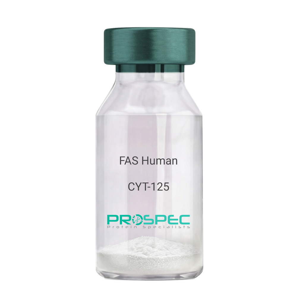
FAS HumanDescription:
sFas Receptor Human Recombinant
Tumor necrosis factor receptor superfamily member 6, Apo-1 antigen, Apoptosis-mediating surface antigen FAS, FASLG receptor, CD95, FAS, APT1, FAS1, APO-1, FASTM, ALPS1A, TNFRSF6.
Product # :
CYT-125Price :
Quantity :
Shipping Method :
Shipped at Room temp
More Info
- description
- source
- formulation
- purity
- biological activity
- More Info
- Citations
Description
sFas Receptor Human Recombinant produced in E.coli is a single, non-glycosylated polypeptide chain containing 157 amino acids and having a molecular mass of 17.6kDa.The FAS is purified by proprietary chromatographic techniques.
Source
Escherichia Coli.
Formulation
FAS protein was lyophilized from a 0.2µm filtered concentrated solution in 1×PBS, pH 7.4.
Purity
Greater than 95.0% as determined by:
(a) Analysis by RP-HPLC.
(b) Analysis by SDS-PAGE.Biological Activity
The ED50 was determined by its ability to inhibit the cytotoxicity of Jurkat cells is between 10-15 µg/ml in the presence of 2ng/ml of hFasL.Citations
More Info
-
Introduction
Fas and Fas Ligand (FasL) are members of the TNF superfamily and are type I and type II transmembrane proteins, respectively. Binding of FasL to Fas initiates apoptosis in Fas-bearing cells. The apoptosis mechanism involves the recruitment of pro-caspase 8 through an adaptor molecule named FADD followed by processing of the pro-enzyme to active forms. These active caspases subsequently cleave a variety of cellular substrates leading to the eventual cell death. sFasR is able to inhibit FasL-induced apoptosis by acting as a decoy receptor whicht serves as a sink for FasL. The full length Fas Receptor is a 319 a.a type I transmembrane protein, which contains a 157 a.a extracellular domain, a 17 a.a transmembrane domain, and 145 a.a cytoplasmic domain. The mature human Fas ECD shares 55%, 58%, a.a sequence identity with the mouse, rat, Fas, respectively.
-
Synonyms
Tumor necrosis factor receptor superfamily member 6, Apo-1 antigen, Apoptosis-mediating surface antigen FAS, FASLG receptor, CD95, FAS, APT1, FAS1, APO-1, FASTM, ALPS1A, TNFRSF6.
-
Physical Appearance
Sterile Filtered White lyophilized (freeze-dried) powder.
-
Stability
Lyophilized FAS although stable at room temperature for 3 weeks, should be stored desiccated below -18°C. Upon reconstitution FAS should be stored at 4°C between 2-7 days and for future use below -18°C. For long term storage it is recommended to add a carrier protein (0.1% HSA or BSA). Please prevent freeze-thaw cycles.
-
Solubility
It is recommended to reconstitute the lyophilized FAS in sterile 18M-cm H2O not less than 100µg/ml, which can then be further diluted to other aqueous solutions.
-
Amino Acid Sequence
MRLSSKSVNA QVTDINSKGL ELRKTVTTVE TQNLEGLHHD GQFCHKPCPP GERKARDCTV NGDEPDCVPC QEGKEYTDKA HFSSKCRRCR LCDEGHGLEV EINCTRTQNT KCRCKPNFFC NSTVCEHCDP CTKCEHGIIK ECTLTSNTKC KEEGSRS.
-
Background
What is the molecular weight/Mw of FAS Protein?
FAS Protein has a total Mw of 17.6kDa.
What is the source or expression system of FAS Protein?
Escherichia Coli.
What is the Purity of FAS Protein?
FAS Protein is >95% pure as determined by SDS-PAGE.
What is the Biological Activity of FAS Protein?
The ED50 was determined by its ability to inhibit the cytotoxicity of Jurkat cells is between 10-15 µg/ml in the presence of 2ng/ml of hFasL.
What is the amino acid sequence of FAS Protein?
MRLSSKSVNA QVTDINSKGL ELRKTVTTVE TQNLEGLHHD GQFCHKPCPP GERKARDCTV NGDEPDCVPC QEGKEYTDKA HFSSKCRRCR LCDEGHGLEV EINCTRTQNT KCRCKPNFFC NSTVCEHCDP CTKCEHGIIK ECTLTSNTKC KEEGSRS.
What applications can FAS Protein be used in?
FAS Protein can probably be used in western blot, ELISA and Lateral Flow.
What is the endotoxin level for FAS Protein?
The endotoxin level is minimal, FAS Protein was purified using conventional chromatography techniques..
ProSpec's products are furnished for LABORATORY RESEARCH USE ONLY. The product may not be used as drugs, agricultural or pesticidal products, food additives or household chemicals.
- View Data Sheet
Name :
M6PR (242-277) HumanDescription:
Mannose-6-Phosphate Receptor (242-277) Human Recombinant
CD-MPR, MPR46, MPR 46, 46-KDa Mannose 6-Phosphate Receptor, MPR-46.
Product # :
PRO-2828Price :
Quantity :
Shipping Method :
Shipped at Room temp
More Info
- description
- source
- formulation
- purity
- More Info
- Citations
Description
The M6PRHuman is created as a recombinant protein with a 4kda N-terminal fusion of His Tag. The M6PRHis-Tagged Fusion Protein, produced in E. coli, is a 9kDa protein containing 36 amino acid residues of the M6PRHuman, 242-277 amino acids.
Source
Escherichia Coli.
Formulation
Each mg was lyophilized with 1xPBS, 0.4% SDS and 4mM DTT.
Purity
Greater than 90% as determined by SDS-PAGE.
Citations
More Info
-
Synonyms
CD-MPR, MPR46, MPR 46, 46-KDa Mannose 6-Phosphate Receptor, MPR-46.
-
Physical Appearance
Filtered White lyophilized (freeze-dried) powder.
-
Stability
Store lyophilized M6PRat -20°C. Aliquot the product after reconstitution to avoid repeated freezing/thawing cycles. Reconstituted protein can be stored at 4°C for a limited period of time; it does not show any change after two weeks at 4°C.
-
Solubility
It is recommended to add deionized water to prepare a working stock solution of approximately 0.5mg/ml and let the lyophilized pellet dissolve completely. Product is not sterile! Please filter the product by an appropriate sterile filter before using it on cell culture.
-
Background
Mannose 6-phosphate receptors are membrane proteins which take part in the intracellular transport of lysosomal enzymes.
These receptors recognize and sort lysosomal hydrolases, facilitate their delivery to lysosomes via endocytic pathways.
M6PR binds to mannose 6-phosphate (M6P) residues on newly synthesized lysosomal enzymes in the Golgi apparatus, directing them to lysosomes.
After delivering enzymes to lysosomes, M6PRs are recycled back to the cell surface. This mechanism is critical for maintaining cellular homeostasis and efficient enzyme delivery.
M6PRs may also play a role in signaling pathways, influencing cellular responses to stress and metabolic changes.
Mutations or dysfunction in M6PRs can cause lysosomal storage disorders, emphasizing their importance in human health.
Recombinant M6PR has significant implications in therapeutic development, mainly in understanding lysosomal storage diseases and enhancing enzyme replacement therapies.
ProSpec's products are furnished for LABORATORY RESEARCH USE ONLY. The product may not be used as drugs, agricultural or pesticidal products, food additives or household chemicals.
- View Data Sheet
Name :
PTPRC HumanDescription:
Protein Tyrosine Phosphatase Receptor Type C Human Recombinant
LCA, LY5, B220, CD45, T200, GP180, Receptor-type tyrosine-protein phosphatase C, Leukocyte common antigen, L-CA, PTPRC.
Product # :
ENZ-775Price :
Quantity :
Shipping Method :
Shipped with Ice Packs
More Info
- description
- source
- formulation
- purity
- More Info
- Citations
Description
PTPRC Human Recombinant produced in E.coli is a single, non-glycosylated polypeptide chain containing 257 amino acids (1031-1251a.a) and having a molecular mass of 29.6kDa. PTPRC is fused to a 36 amino acid His-tag at N-terminus & purified by proprietary chromatographic techniques.
Source
Escherichia Coli.
Formulation
The PTPRC solution (0.25mg/ml) contains 20mM Tris-HCl buffer (pH 8.0), 0.15M NaCl, 10% glycerol and 1mM DTT.
Purity
Greater than 95% as determined by SDS-PAGE.
Citations
More Info
-
Introduction
Protein Tyrosine Phosphatase Receptor Type C, also known as PTPRC, which is a vital regulator of T- and B-cell antigen receptor signaling, is also a part of the protein tyrosine phosphatase (PTP) family. PTPs are signaling molecules which regulate a variety of cellular processes including differentiation, cell growth, mitosis, and oncogenic transformation. PTPRC contains an extracellular domain, a single transmembrane segment and 2 tandem intracytoplasmic catalytic domains, and therefore is classified as a receptor type PTP.
-
Synonyms
LCA, LY5, B220, CD45, T200, GP180, Receptor-type tyrosine-protein phosphatase C, Leukocyte common antigen, L-CA, PTPRC.
-
Physical Appearance
Sterile Filtered colorless solution.
-
Stability
Store at 4°C if entire vial will be used within 2-4 weeks. Store, frozen at -20°C for longer periods of time. For long term storage it is recommended to add a carrier protein (0.1% HSA or BSA). Avoid multiple freeze-thaw cycles.
-
Amino Acid Sequence
MRGSHHHHHH GMASMTGGQQ MGRDLYDDDD KDRWGSVMIA AQGPLKETIG DFWQMIFQRK VKVIVMLTEL KHGDQEICAQ YWGEGKQTYG DIEVDLKDTD KSSTYTLRVF ELRHSKRKDS RTVYQYQYTN WSVEQLPAEP KELISMIQVV KQKLPQKNSS EGNKHHKSTP LLIHCRDGSQ QTGIFCALLN LLESAETEEV VDIFQVVKAL RKARPGMVST FEQYQFLYDV IASTYPAQNG QVKKNNHQED KIEFDNE.
ProSpec's products are furnished for LABORATORY RESEARCH USE ONLY. The product may not be used as drugs, agricultural or pesticidal products, food additives or household chemicals.
- View Data Sheet
Name :
NFNB E.ColiDescription:
Dihydropteridine Reductase E.Coli Recombinant
DPRA, NFSB, NFSI, NTR.
Product # :
ENZ-423Price :
Quantity :
Shipping Method :
Shipped with Ice Packs
More Info
- description
- source
- formulation
- purity
- More Info
- Citations
Description
NFNB Recombinant produced in E.Coli is a single, non-glycosylated polypeptide chain containing 237 amino acids (1-217 a.a.) and having a molecular mass of 26 kDa. The NFNB is fused to a 20 amino acid His-Tag at N-terminus and purified by proprietary chromatographic techniques.
Source
Escherichia Coli.
Formulation
1mg/ml solution containing 20mM Tris pH-8, 1mM DTT, 0.05M NaCl & 10% glycerol.
Purity
Greater than 95.0% as determined by SDS-PAGE.
Citations
More Info
-
Introduction
NFNB demonstrates the capability to reduce quinines. NFNB enzyme activates prodrugs in antibody directed enzyme prodrug therapy. NFNB reduces nitrofurazone, quinones and anti-tumor agent CB1954 (5-(aziridin-1-yl)-2,4-dinitrobenzamide). The reduction of CB1954 results in the generation of cytotoxic species.
-
Synonyms
DPRA, NFSB, NFSI, NTR.
-
Physical Appearance
Sterile filtered colorless solution.
-
Stability
Store at 4°C if entire vial will be used within 2-4 weeks. Store, frozen at -20°C for longer periods of time. For long term storage it is recommended to add a carrier protein (0.1% HSA or BSA). Avoid multiple freeze-thaw cycles.
-
Amino Acid Sequence
MGSSHHHHHH SSGLVPRGSH MDIISVALKR HSTKAFDASK KLTPEQAEQI KTLLQYSPSS TNSQPWHFIV ASTEEGKARV AKSAAGNYVF NERKMLDASH VVVFCAKTAM DDVWLKLVVD QEDADGRFAT PEAKAANDKG RKFFADMHRK DLHDDAEWMA KQVYLNVGNF LLGVAALGLD AVPIEGFDAA ILDAEFGLKE KGYTSLVVVP VGHHSVEDFN ATLPKSRLPQ NITLTEV.
ProSpec's products are furnished for LABORATORY RESEARCH USE ONLY. The product may not be used as drugs, agricultural or pesticidal products, food additives or household chemicals.
- View Data Sheet
Name :
EG VEGF MouseDescription:
Endocrine Gland Vascular Endothelial Growth Factor Mouse Recombinant
PK1, Prokineticin 1, EG-VEGF, Prok1, Endocrine-gland-derived vascular endothelial growth factor.
Product # :
CYT-825Price :
Quantity :
Shipping Method :
Shipped at Room temp
More Info
- description
- source
- formulation
- purity
- More Info
- Citations
Description
EG-VEGF Mouse Recombinant produced in E.Coli is a single, non-glycosylated, polypeptide chain containing 86 amino acids and having a molecular mass of 9.6kDa.
Source
Escherichia Coli.
Formulation
Lyophilized from a 0.2µm filtered concentrated solution in PBS pH7.4 and 3% Trehalose.
Purity
Greater than 95.0% as determined by:
(a) Analysis by RP-HPLC.
(b) Analysis by SDS-PAGE.Citations
More Info
-
Introduction
Endocrine gland-derived vascular endothelial growth factor (EG-VEGF) induces proliferation, migration, and fenestration in capillary endothelial cells derived from endocrine glands. Its expression is induced by hypoxia and is restricted to the steroidogenic glands (ovary, testis, adrenal, and placenta). Its expression is often complementary to the expression of VEGF (MIM 192240), suggesting that these molecules function in a coordinated manner. EG-VEGF potently contracts gastrointestinal (gi) smooth muscle. Induces proliferation, migration and fenestration (the formation of membrane discontinuities) in capillary endothelial cells derived from endocrine glands. Has little or no effect on a variety of other endothelial and non-endothelial cell types.
-
Synonyms
PK1, Prokineticin 1, EG-VEGF, Prok1, Endocrine-gland-derived vascular endothelial growth factor.
-
Physical Appearance
Sterile Filtered White lyophilized (freeze-dried) powder.
-
Stability
Lyophilized EG-VEGF Human Recombinant although stable at room temperature for 3 weeks, should be stored desiccated below -18°C. Upon reconstitution EG-VEGF should be stored at 4°C between 2-7 days and for future use below -18°C.Please prevent freeze-thaw cycles.
-
Solubility
It is recommended to reconstitute the lyophilized Endocrine Gland Vascular Endothelial Growth Factor in sterile 18MΩ-cm H2O not less than 100µg/ml, which can then be further diluted to other aqueous solutions.
-
Amino Acid Sequence
AVITGACERD IQCGAGTCCA ISLWLRGLRL CTPLGREGEE CHPGSHKIPF LRKRQHHTCP CSPSLLCSRF PDGRYRCFRD LKNANF.
-
Background
What is the molecular weight/Mw of EG-VEGF Protein?
EG-VEGF Protein has a total Mw of 9.6kDa.
What is the source or expression system of EG-VEGF Protein?
Escherichia Coli.
What is the Purity of EG-VEGF Protein?
EG-VEGF Protein is >95% pure as determined by SDS-PAGE.
What is the Biological Activity of EG-VEGF Protein?
The biological functionality of EG-VEGF Protein will be determined in the future.
What is the amino acid sequence of EG-VEGF Protein?
AVITGACERD IQCGAGTCCA ISLWLRGLRL CTPLGREGEE CHPGSHKIPF LRKRQHHTCP CSPSLLCSRF PDGRYRCFRD LKNANF.
What applications can EG-VEGF Protein be used in?
EG-VEGF Protein can probably be used in western blot, ELISA and Lateral Flow.
What is the endotoxin level for EG-VEGF Protein?
The endotoxin level is minimal, EG-VEGF Protein was purified using conventional chromatography techniques.
ProSpec's products are furnished for LABORATORY RESEARCH USE ONLY. The product may not be used as drugs, agricultural or pesticidal products, food additives or household chemicals.
- View Data Sheet
Name :
ELOB MouseDescription:
Elongin B Mouse Recombinant
Transcription elongation factor B polypeptide 2, TCEB2, RNA polymerase IItranscription factor SIII subunit B, SIII p18, EloB, Elongin 18 kDa subunit.
Product # :
PRO-2550Price :
Quantity :
Shipping Method :
Shipped with Ice Packs
More Info
- description
- source
- formulation
- purity
- More Info
- Citations
Description
ELOB Mouse Recombinant produced in E.Coli is a single, non-glycosylated polypeptide chain of 141 amino acids ( 1-118 a.a.) having a molecular mass of 15.6 kDa. The Recombinant Mouse ELOB is fused to a 23 amino acid His-tag at N-terminus & purified by proprietary chromatographic techniques.
Source
Escherichia Coli.
Formulation
The protein (1mg/ml) contains PBS pH-7.4 containing 1mM DTT and 10% Glycerol.
Purity
Greater than 95.0% as determined by SDS-PAGE.
Citations
More Info
-
Introduction
Elongin B (Elob) is a subunit of the transcription factor B (SIII) complex. SIII complex is a general transcription elongation factor that increases the RNA polymerase II transcription elongation past template-encoded arresting sites. The SIII complex comprised of a transcriptionally active subunit (A) and 2 regulatory subunits (B and C). Subunit A is transcriptionally active and its transcription activity is enhanced by binding to the dimeric complex of the SIII regulatory subunits B and C. The von Hippel-Lindau tumor suppressor protein binds to elongin B and C and inhibits transcription elongation.
-
Synonyms
Transcription elongation factor B polypeptide 2, TCEB2, RNA polymerase IItranscription factor SIII subunit B, SIII p18, EloB, Elongin 18 kDa subunit.
-
Physical Appearance
Sterile filtered colorless solution.
-
Stability
Store at 4°C if entire vial will be used within 2-4 weeks. Store, frozen at -20°C for longer periods of time. For long term storage it is recommended to add a carrier protein (0.1% HSA or BSA). Avoid multiple freeze-thaw cycles.
-
Amino Acid Sequence
MGSSHHHHHH SSGLVPRGSH MGSMDVFLMI RRHKTTIFTD AKESSTVFEL KRIVEGILKR PPEEQRLYKD DQLLDDGKTL GECGFTSQTA RPQAPATVGL AFRADDTFEA LRIEPFSSPP ELPDVMKPQD SGGSANEQAV Q
ProSpec's products are furnished for LABORATORY RESEARCH USE ONLY. The product may not be used as drugs, agricultural or pesticidal products, food additives or household chemicals.
- View Data Sheet
Name :
ACVR1 HumanDescription:
Activin A Receptor Type 1 Human Recombinant
ACVR1A, ALK2, ACVR1, ACTRI, ACTR-I, ACVRLK2, FOP, SKR1, TSRI, Activin receptor type I, Activin receptor-like kinase 2, ALK-2, TSR-I, Serine/threonine-protein kinase receptor R1, TGF-B superfamily receptor type I.
Product # :
CYT-1140Price :
Quantity :
Shipping Method :
Shipped with Ice Packs
More Info
- description
- source
- formulation
- purity
- More Info
- sds-page
- Citations
Description
ACVR1 produced in Sf9 Baculovirus cells is a single, glycosylated polypeptide chain containing 342 amino acids (21-123a.a.) and having a molecular mass of 38.4kDa. ACVR1 is expressed with a 239 amino acid hIgG-His-Tag at C-Terminus and purified by proprietary chromatographic techniques.
Source
Sf9, Baculovirus cells.
Formulation
ACVR1 protein solution (0.25mg/ml) contains Phosphate Buffered Saline (pH 7.4) and 10% glycerol.
Purity
Greater than 90.0% as determined by SDS-PAGE.
sds-page
Citations
More Info
-
Introduction
Activin A Receptor Type 1 (ACVR1) is a member of TGF-beta serine/threonine kinase receptor family. ACVR1 forms a receptor complex contains2 type II and 2 type I transmembrane serine/threonine kinases. Type II receptors phosphorylate and activate type I receptors which autophosphorylate,bind and activate SMAD transcriptional regulators. ACVR1 takes part in left-right pattern formation during embryogenesis and is also essential in the BMP pathway which is responsible for the development and repair of the skeletal system.ACVR1 is linked to Fibrodysplasia Ossificans Progressiva which isknown for the formation of heterotopic bone throughout the body.
-
Synonyms
ACVR1A, ALK2, ACVR1, ACTRI, ACTR-I, ACVRLK2, FOP, SKR1, TSRI, Activin receptor type I, Activin receptor-like kinase 2, ALK-2, TSR-I, Serine/threonine-protein kinase receptor R1, TGF-B superfamily receptor type I.
-
Physical Appearance
Sterile filtered colorless solution.
-
Stability
Store at 4°C if entire vial will be used within 2-4 weeks.Store, frozen at -20°C for longer periods of time. For long term storage it is recommended to add a carrier protein (0.1% HSA or BSA).Avoid multiple freeze-thaw cycles.
-
Amino Acid Sequence
MEDEKPKVNP KLYMCVCEGL SCGNEDHCEG QQCFSSLSIN DGFHVYQKGC FQVYEQGKMT
CKTPPSPGQA VECCQGDWCN RNITAQLPTK GKSFPGTQNF HLELEPKSCD KTHTCPPCPA
PELLGGPSVF LFPPKPKDTL MISRTPEVTC VVVDVSHEDP EVKFNWYVDG VEVHNAKTKP
REEQYNSTYR VVSVLTVLHQ DWLNGKEYKC KVSNKALPAP IEKTISKAKG QPREPQVYTL
PPSRDELTKN QVSLTCLVKG FYPSDIAVEW ESNGQPENNY KTTPPVLDSD GSFFLYSKLT
VDKSRWQQGN VFSCSVMHEA LHNHYTQKSL SLSPGKHHHH HH. -
Background
Functional Implications and Therapeutic Prospects of Activin A Receptor Type 1 Human Recombinant
1. Abstract
This study illuminates the functional roles and potential therapeutic applications of Activin A Receptor Type 1 Human Recombinant (ACVR1), a crucial protein in the TGF-beta superfamily signaling pathway. Through a comprehensive review of its structure, signaling mechanism, biological functions, and disease associations, this paper aims to elucidate the current understanding of ACVR1 and its potential therapeutic implications in various disease states.
2. Introduction
The Activin A Receptor Type 1 Human Recombinant, abbreviated as ACVR1, is a receptor protein vital for transmitting cellular signals in the Transforming Growth Factor-beta (TGF-beta) superfamily pathway. Known to play pivotal roles in organogenesis, bone growth, and cell differentiation, the ACVR1 and its functions present vast therapeutic potential.
3. Structure and Signaling of ACVR1
ACVR1 is a transmembrane serine/threonine kinase receptor, characterized by an extracellular ligand-binding domain and an intracellular kinase domain for signal transduction. Binding of ligands such as Activin A leads to the formation of heteromeric complexes with type II receptors, triggering phosphorylation events that activate downstream signaling pathways.
4. Biological Functions of ACVR1
Being a part of the TGF-beta superfamily signaling pathway, ACVR1 is implicated in a broad spectrum of biological processes. It is crucial for embryonic development, cellular proliferation, differentiation, apoptosis, and homeostasis. It also plays a significant role in bone morphogenesis, contributing to skeletal patterning and growth.
5. ACVR1 in Disease Pathology
The dysregulation of ACVR1 has been associated with various pathological conditions, including Fibrodysplasia Ossificans Progressiva (FOP), a rare genetic disorder characterized by progressive ossification of soft tissues. Mutations in ACVR1 lead to enhanced BMP signaling, causing aberrant bone formation. This highlights the critical role of ACVR1 in skeletal homeostasis and disease.
6. Therapeutic Potential of ACVR1
Given the central role of ACVR1 in cellular signaling and its association with disease, it presents a promising target for therapeutic intervention. Strategies to modulate ACVR1 signaling could potentially ameliorate symptoms of diseases like FOP, offering promising avenues for novel therapeutic approaches.
7. Conclusion and Future Perspectives
While our understanding of ACVR1's functional roles has expanded significantly over the years, much remains to be elucidated. Further research into the precise molecular mechanisms of ACVR1 and its pathway will pave the way for therapeutic advances, enhancing our capability to combat various diseases.
What is the molecular weight / Mw of ACVR1 Protein?
ACVR1 Protein has a total Mw of 38.4kDa.
What is the source or expression system of ACVR1 Protein?
Sf9, Baculovirus cells.
What is the Purity of ACVR1 Protein?
ACVR1 Protein is >90% pure as determined by SDS-PAGE.
What is the Biological Activity of ACVR1 Protein?
The biological functionality of ACVR1 Protein will be determined in the future.
What is the endotoxin level for ACVR1 Protein?
The endotoxin level is minimal, ACVR1 Protein was purified using conventional chromatography techniques.
What is the amino acid sequence of ACVR1 Protein?
MEDEKPKVNP KLYMCVCEGL SCGNEDHCEG QQCFSSLSIN DGFHVYQKGC FQVYEQGKMT
CKTPPSPGQA VECCQGDWCN RNITAQLPTK GKSFPGTQNF HLELEPKSCD KTHTCPPCPA
PELLGGPSVF LFPPKPKDTL MISRTPEVTC VVVDVSHEDP EVKFNWYVDG VEVHNAKTKP
REEQYNSTYR VVSVLTVLHQ DWLNGKEYKC KVSNKALPAP IEKTISKAKG QPREPQVYTL
PPSRDELTKN QVSLTCLVKG FYPSDIAVEW ESNGQPENNY KTTPPVLDSD GSFFLYSKLT
VDKSRWQQGN VFSCSVMHEA LHNHYTQKSL SLSPGKHHHH HH.
What applications can ACVR1 Protein be used in?
ACVR1 Protein can probably be used in western blot, ELISA and Lateral Flow
ProSpec's products are furnished for LABORATORY RESEARCH USE ONLY. The product may not be used as drugs, agricultural or pesticidal products, food additives or household chemicals.
- View Data Sheet
Name :
SSR2 HumanDescription:
Signal Sequence Receptor, Beta Human Recombinant
HSD25, TLAP, TRAP-BETA, TRAPB, Translocon-associated protein subunit beta, Signal sequence receptor subunit beta, SSR-beta.
Product # :
PRO-1380Price :
Quantity :
Shipping Method :
Shipped with Ice Packs
More Info
- description
- source
- formulation
- purity
- More Info
- Citations
Description
SSR2 Human Recombinant produced in E.Coli is a single, non-glycosylated polypeptide chain containing 155 amino acids (18-149a.a) and having a molecular mass of 16.8kDa. SSR2 is fused to a 23 amino acid His-tag at N-terminus & purified by proprietary chromatographic techniques.
Source
E.coli.
Formulation
SSR2 protein solution (0.5mg/ml) containing 20mM Tris-HCl buffer (pH8.0), 10% glycerol and 0.1M NaCl.
Purity
Greater than 90% as determined by SDS-PAGE.
Citations
More Info
-
Introduction
The signal sequence receptor (SSR) is a glycosylated endoplasmic reticulum membrane receptor related with protein translocation across the ER membrane. The SSR consists of 2 subunits, a 34-kD glycoprotein (alpha-SSR or SSR1) and a 22-kD glycoprotein (beta-SSR or SSR2). The human beta-signal sequence receptor gene (SSR2) maps to chromosome bands 1q21-q23. Diseases correlated with SSR2 include calcaneonavicular coalition, and osteosarcoma, and among its related super-pathways are Viral mRNA Translation and Generic Transcription Pathway.
-
Synonyms
HSD25, TLAP, TRAP-BETA, TRAPB, Translocon-associated protein subunit beta, Signal sequence receptor subunit beta, SSR-beta.
-
Physical Appearance
Sterile Filtered colorless solution.
-
Stability
Store at 4°C if entire vial will be used within 2-4 weeks. Store, frozen at -20°C for longer periods of time. For long term storage it is recommended to add a carrier protein (0.1% HSA or BSA).Avoid multiple freeze-thaw cycles.
-
Amino Acid Sequence
MGSSHHHHHH SSGLVPRGSH MGSEEGARLL ASKSLLNRYA VEGRDLTLQY NIYNVGSSAA LDVELSDDSF PPEDFGIVSG MLNVKWDRIA PASNVSHTVV LRPLKAGYFN FTSATITYLA QEDGPVVIGS TSAPGQGGIL AQREFDRRFS PHFLD.
ProSpec's products are furnished for LABORATORY RESEARCH USE ONLY. The product may not be used as drugs, agricultural or pesticidal products, food additives or household chemicals.
- View Data Sheet
Name :
EGF RatDescription:
Epidermal Growth Factor Rat Recombinant
Urogastrone, URG, EGF.
Product # :
CYT-669Price :
Quantity :
Shipping Method :
Shipped at Room temp
More Info
- description
- source
- formulation
- purity
- biological activity
- More Info
- Citations
Description
Epidermal Growth Factor Rat Recombinant produced in E.Coli is a single, non-glycosylated, polypeptide chain containing 53 amino acids and having a molecular mass of 6151 Dalton. The Rat EGF is purified by proprietary chromatographic techniques.
Source
Escherichia Coli.
Formulation
Rat EGF was lyophilized from a 0.2µm filtered concentrated (1.0mg/ml) solution in PBS, pH 7.4.
Purity
Greater than 98.0% as determined by(a) Analysis by RP-HPLC.
(b) Analysis by SDS-PAGE.Biological Activity
The ED50 as calculated by the dose-dependant proliferation of murine BALB/c 3T3 cells is less than 0.1ng/ml, corresponding to a specific activity of > 10,000,000 units/mg.Citations
More Info
-
Introduction
Epidermal growth factor has a profound effect on the differentiation of specific cells in vivo and is a potent mitogenic factor for a variety of cultured cells of both ectodermal and mesodermal origin. The EGF precursor is believed to exist as a membrane-bound molecule which is proteolytically cleaved to generate the 53-amino acid peptide hormone that stimulates cells to divide.
EGF stimulates the growth of various epidermal and epithelial tissues in vivo and in vitro and of some fibroblasts in cell culture. -
Synonyms
Urogastrone, URG, EGF.
-
Physical Appearance
Sterile Filtered White lyophilized (freeze-dried) powder.
-
Stability
Lyophilized Rat EGF although stable at room temperature for 3 weeks, should be stored desiccated below -18°C. Upon reconstitution Rat EGF should be stored at 4°C between 2-7 days and for future use below -18°C. For long term storage it is recommended to add a carrier protein (0.1% HSA or BSA).Please prevent freeze-thaw cycles.
-
Solubility
It is recommended to reconstitute the lyophilized Rat EGF in sterile water not less than 100µg/ml, which can then be further diluted to other aqueous solutions.
-
Amino Acid Sequence
NSNTGCPPSY DGYCLNGGVC MYVESVDRYV CNCVIGYIGE RCQHRDLRWW KLR.
-
Background
Pioneering Insights into Epidermal Growth Factor Rat Recombinant: Unraveling Signaling Dynamics and Therapeutic Implications
Abstract:
This research paper delves into the unexplored landscape of Epidermal Growth Factor Rat Recombinant (EGF-RR), delving into its intricate molecular attributes, signaling pathways, and potential therapeutic applications. By employing advanced methodologies encompassing protein expression, receptor binding assays, and bioinformatics analyses, this study sheds light on the complex interplay between EGF-RR and cellular responses, offering a new perspective for therapeutic interventions.
Introduction:
Epidermal Growth Factor (EGF) holds a key role in cellular regulation. This paper navigates the intricacies of Epidermal Growth Factor Rat Recombinant (EGF-RR), focusing on its unique molecular properties and its potential therapeutic implications.
Protein Expression and Purification:
The paper delves into the meticulous engineering of EGF-RR, involving gene optimization for enhanced expression. Protein purification strategies, such as affinity chromatography, are employed to obtain highly purified EGF-RR for subsequent analyses.
Receptor Binding Assays and Ligand Interaction:
Advanced receptor binding assays elucidate the interaction of EGF-RR with its cognate receptor. By quantifying binding affinities and kinetic rates, the study unveils the nuances of EGF-RR's engagement with its receptor, shedding light on potential structural determinants.
Cellular Signaling Pathways and Functional Responses:
Through in vitro cellular assays, the study unravels the intricate signaling pathways initiated by EGF-RR. Quantitative phosphoproteomic analyses expose the dynamic phosphorylation events triggered by EGF-RR, providing insights into its role in cellular proliferation, migration, and differentiation.
Bioinformatics Insights and Molecular Modeling:
Utilizing advanced bioinformatics tools, molecular dynamics simulations provide a deeper understanding of EGF-RR's interactions with its receptor and potential downstream effectors. Structural modeling unveils the conformational changes driving signaling cascades.
Therapeutic Prospects and Novel Avenues:
The molecular insights into EGF-RR's signaling dynamics open avenues for therapeutic exploration. Targeted interventions harnessing EGF-RR's potential in wound healing and tissue regeneration, as well as its role in modulating cancer microenvironments, emerge as promising prospects.
Challenges and Future Directions:
Despite progress, challenges such as deciphering context-dependent signaling responses remain. Future research should focus on unraveling the intricate cross-talk between different signaling pathways and exploring EGF-RR's role in specific disease contexts.
Conclusion:
In a convergence of advanced methodologies and visionary insights, Epidermal Growth Factor Rat Recombinant emerges as a captivating subject. Its distinctive molecular attributes and complex cellular interplay offer potential avenues for therapeutic interventions, ushering in a new era of precision medicine.
What is the molecular weight/Mw of EGF RAT Protein?
EGF RAT Protein has a total Mw of 6.1kDa.
What is the source or expression system of EGF RAT Protein?
Escherichia Coli.
What is the Purity of EGF RAT Protein?
EGF RAT Protein is >98% pure as determined by SDS-PAGE.
What is the Biological Activity of EGF RAT Protein?
The ED50 as calculated by the dose-dependant proliferation of murine BALB/c 3T3 cells is less than 0.1ng/ml, corresponding to a specific activity of > 10,000,000 units/mg.
What is the amino acid sequence of EGF RAT Protein?
NSNTGCPPSY DGYCLNGGVC MYVESVDRYV CNCVIGYIGE RCQHRDLRWW KLR.
What applications can EGF RAT Protein be used in?
EGF RAT Protein can probably be used in western blot, ELISA and Lateral Flow.
What is the endotoxin level for EGF RAT Protein?
The endotoxin level is minimal, EGF RAT Protein was purified using conventional chromatography techniques.
ProSpec's products are furnished for LABORATORY RESEARCH USE ONLY. The product may not be used as drugs, agricultural or pesticidal products, food additives or household chemicals.
- View Data Sheet
Name :
PEF1 HumanDescription:
Penta-EF-Hand Domain Containing 1 Human Recombinant
Peflin, PEF protein with a long N-terminal hydrophobic domain, Penta-EF hand domain-containing protein 1, PEF1, ABP32, PEF1A.
Product # :
PRO-099Price :
Quantity :
Shipping Method :
Shipped with Ice Packs
More Info
- description
- source
- formulation
- purity
- More Info
- Citations
Description
PEF1 Human Recombinant fused with a 36 amino acid His tag at N-terminus produced in E.Coli is a single, non-glycosylated, polypeptide chain containing 320 amino acids (1-284 a.a.) and having a molecular mass of 34.5kDa. The PEF1 is purified by proprietary chromatographic techniques.
Source
Escherichia Coli.
Formulation
The PEF1 solution (0.25 mg/ml) contains 20mM Tris-HCl buffer (pH 8.0), 1mM DTT, 50% glycerol and 0.2M NaCl.
Purity
Greater than 85.0% as determined by SDS-PAGE.
Citations
More Info
-
Introduction
Penta-EF-hand domain containing 1 (PEF1) is a member of the penta-EF-hand protein family, which shares similarities to the apoptosis linked gene ALG-2. PEF1 forms heterodimers with PDCD6/ALG2 and dissociates from PDCD6/ALG2 in the presence of Ca (2+). Furthermore, PEF1 proteins bind calcium and participate in a various calcium-dependent processes in vertebrates.
-
Synonyms
Peflin, PEF protein with a long N-terminal hydrophobic domain, Penta-EF hand domain-containing protein 1, PEF1, ABP32, PEF1A.
-
Physical Appearance
Sterile Filtered colorless solution.
-
Stability
Store at 4°C if entire vial will be used within 2-4 weeks. Store, frozen at -20°C for longer periods of time. For long term storage it is recommended to add a carrier protein (0.1% HSA or BSA).Avoid multiple freeze-thaw cycles.
-
Amino Acid Sequence
MRGSHHHHHH GMASMTGGQQ MGRDLYDDDD KDRWGSMASY PYRQGCPGAA GQAPGAPPGS
YYPGPPNSGG QYGSGLPPGG GYGGPAPGGP YGPPAGGGPY GHPNPGMFPS GTPGGPYGGA
APGGPYGQPP PSSYGAQQPG LYGQGGAPPN VDPEAYSWFQ SVDSDHSGYI SMKELKQALV
NCNWSSFNDE TCLMMINMFD KTKSGRIDVY GFSALWKFIQ QWKNLFQQYD RDRSGSISYT
ELQQALSQMG YNLSPQFTQL LVSRYCPRSA NPAMQLDRFI QVCTQLQVLT EAFREKDTAV
QGNIRLSFED FVTMTASRML.
ProSpec's products are furnished for LABORATORY RESEARCH USE ONLY. The product may not be used as drugs, agricultural or pesticidal products, food additives or household chemicals.
- View Data Sheet
Name :
EHF HumanDescription:
Ets Homologous Factor Human Recombinant
ESE3, ESE3B, ESEJ, ETS homologous factor, hEHF, ETS domain-containing transcription factor, Epithelium-specific Ets transcription factor 3, EHF.
Product # :
PRO-1436Price :
Quantity :
Shipping Method :
Shipped with Ice Packs
More Info
- description
- source
- formulation
- purity
- More Info
- Citations
Description
EHF Human Recombinant produced in E. coli is a single polypeptide chain containing 323 amino acids (1-300) and having a molecular mass of 37.3kDa. EHF is fused to a 23 amino acid His-tag at N-terminus & purified by proprietary chromatographic techniques.
Source
E.coli.
Formulation
The EHF solution (1mg/ml) contains 20mM Tris-HCl buffer (pH 8.0), 0.4M Urea and 10% glycerol.
Purity
Greater than 90% as determined by SDS-PAGE.
Citations
More Info
-
Introduction
ETS homologous factor (EHF) is a part of the ESE subfamily of Ets transcription factors. Ets factors are an important kind of transcriptional regulators which take part in hema-topoiesis, angiogenesis, organogenesis, oncogenesis and specification of neuronal connectivity. EHF is expressed solely in a subset of epithelial cells, with highest expression noticed in glandular epithelium of the prostate, pancreas, salivary gland and trachea. EHF transactivates the c-Met promoter via 3 high affinity binding sites, which support the notion that EHF contribute to branching morphogenesis.
-
Synonyms
ESE3, ESE3B, ESEJ, ETS homologous factor, hEHF, ETS domain-containing transcription factor, Epithelium-specific Ets transcription factor 3, EHF.
-
Physical Appearance
Sterile Filtered colorless solution.
-
Stability
Store at 4°C if entire vial will be used within 2-4 weeks. Store, frozen at -20°C for longer periods of time. For long term storage it is recommended to add a carrier protein (0.1% HSA or BSA).Avoid multiple freeze-thaw cycles.
-
Amino Acid Sequence
MGSSHHHHHH SSGLVPRGSH MGSMILEGGG VMNLNPGNNL LHQPPAWTDS YSTCNVSSGF FGGQWHEIHP QYWTKYQVWE WLQHLLDTNQ LDANCIPFQE FDINGEHLCS MSLQEFTRAA GTAGQLLYSN LQHLKWNGQC SSDLFQSTHN VIVKTEQTEP SIMNTWKDEN YLYDTNYGST VDLLDSKTFC RAQISMTTTS HLPVAESPDM KKEQDPPAKC HTKKHNPRGT HLWEFIRDIL LNPDKNPGLI KWEDRSEGVF RFLKSEAVAQ LWGKKKNNSS MTYEKLSRAM RYYYKREILE RVDGRRLVYK FGKNARGWRE NEN.
ProSpec's products are furnished for LABORATORY RESEARCH USE ONLY. The product may not be used as drugs, agricultural or pesticidal products, food additives or household chemicals.
- View Data Sheet
Name :
EIF3K HumanDescription:
Eukaryotic Translation Initiation Factor 3K Human Recombinant
Eukaryotic Translation Initiation Factor 3 Subunit K, PLAC-24, eIF3k, eIF-3 p25, eIF-3 p28, EIF3S12, HSPC029, M9, PRO1474, PTD001, Muscle-specific gene M9 protein, ARG134, MSTP001, muscle specific, Eukaryotic Translation Initiation Factor 3 subunit 12.
Product # :
PRO-179Price :
Quantity :
Shipping Method :
Shipped with Ice Packs
More Info
- description
- source
- formulation
- purity
- More Info
- Citations
Description
EIF3K produced in E.Coli is a single, non-glycosylated polypeptide chain containing 238 amino acids (1-218a.a.) and having a molecular mass of 27.2 kDa. EIF3K is fused to a 20 amino acid His-tag at N-terminus & purified by proprietary chromatographic techniques.
Source
Escherichia Coli.
Formulation
The EIF3K protein solution (1mg/1ml) is formulated in 20mM Tris-HCl buffer (pH8.0), 100mM NaCl, 1mM DTT and 20% glycerol.
Purity
Greater than 95% as determined by SDS-PAGE.
Citations
More Info
-
Introduction
EIF3K is a member of the eIF3 subunit K family. EIF3K is the smallest subunit of eIF3 and it interacts with a number of other subunits of eIF3 and the 40S ribosomal subunit. EIF3K is conserved among high eukaryotes, including mammals, insects, and plants, and it is universally expressed in human tissues. EIF3K is distributed both in nucleus and cytoplasm and colocalized with cyclin D3, a regulatory subunit of cyclin-dependent kinase 4 (Cdk4).
-
Synonyms
Eukaryotic Translation Initiation Factor 3 Subunit K, PLAC-24, eIF3k, eIF-3 p25, eIF-3 p28, EIF3S12, HSPC029, M9, PRO1474, PTD001, Muscle-specific gene M9 protein, ARG134, MSTP001, muscle specific, Eukaryotic Translation Initiation Factor 3 subunit 12.
-
Physical Appearance
Sterile Filtered clear solution.
-
Stability
Store at 4°C if entire vial will be used within 2-4 weeks. Store, frozen at -20°C for longer periods of time. For long term storage it is recommended to add a carrier protein (0.1% HSA or BSA).Avoid multiple freeze-thaw cycles.
-
Amino Acid Sequence
MGSSHHHHHH SSGLVPRGSH MAMFEQMRAN VGKLLKGIDR YNPENLATLE RYVETQAKEN AYDLEANLAV LKLYQFNPAF FQTTVTAQIL LKALTNLPHT DFTLCKCMID QAHQEERPIR QILYLGDLLE TCHFQAFWQA LDENMDLLEG ITGFEDSVRK FICHVVGITY QHIDRWLLAE MLGDLSDSQL KVWMSKYGWS ADESGQIFIC SQEESIKPKN IVEKIDFDSV SSIMASSQ
ProSpec's products are furnished for LABORATORY RESEARCH USE ONLY. The product may not be used as drugs, agricultural or pesticidal products, food additives or household chemicals.
- View Data Sheet
Name :
ENHO HumanDescription:
Energy Homeostasis Associated Human Recombinant
Adropin, Energy homeostasis-associated protein, ENHO, C9orf165, UNQ470.
Product # :
PRO-1569Price :
Quantity :
Shipping Method :
Shipped at Room temp
More Info
- description
- source
- formulation
- purity
- More Info
- Citations
Description
ENHO Human Recombinant produced in E.Coli is a single, non-glycosylated, polypeptide chain (a.a 34-76) containing 121 amino acids including extra 78 N-terminal amino acids. The total molecular mass is 13.05kDa (calculated).
Source
Escherichia Coli.
Formulation
ENHO filtered (0.4 µm) and lyophilized from 0.5mg/ml in phosphate buffered saline.
Purity
Greater than 90.0% as determined by SDS-PAGE.
Citations
More Info
-
Introduction
Energy Homeostasis Associated (ENHO) participates in glucose homeostasis maintenance and lipid metabolism. ENHO is expressed in the liver and the brain. The role of ENHO in obesity or diabetes is studied.
-
Synonyms
Adropin, Energy homeostasis-associated protein, ENHO, C9orf165, UNQ470.
-
Physical Appearance
Filtered White lyophilized (freeze-dried) powder.
-
Stability
Store lyophilized protein at -20°C. Aliquot the product after reconstitution to avoid repeated freezing/thawing cycles. Reconstituted protein can be stored at 4°C for a limited period of time; it does not show any change after two weeks at 4°C.
-
Solubility
It is recommended to add deionized water to prepare a working stock solution of approximately 0.5mg/ml and let the lyophilized pellet dissolve completely. ENHO is not sterile! Please filter the product by an appropriate sterile filter before using it in the cell culture.
-
Amino Acid Sequence
MGGKSNGEKK YIVGFKQGFK SCAKKEDVIS EKGGKLQKCF KYVDAASATL NEKAVEELKK DPSVAYVEED KLFKALTSCHSRSADVDSLS ESSPNSSPGP CPEKAPPPQK PSHEGSYLLQ P.
ProSpec's products are furnished for LABORATORY RESEARCH USE ONLY. The product may not be used as drugs, agricultural or pesticidal products, food additives or household chemicals.
- View Data Sheet
Name :
EEF1A1 HumanDescription:
Eukaryotic Translation Elongation Factor 1 Alpha 1 Human Recombinant
Eukaryotic Translation Elongation Factor 1 Alpha 1, EF1A, EEF1A, LENG7, Leukocyte Receptor Cluster (LRC) Member 7, Elongation Factor Tu, Eukaryotic Elongation Factor 1 A-1, Leukocyte Receptor Cluster Member 7, EF-1-alpha-1, EF-Tu, eEF1A-1, CCS-3, CCS3, EE1A1, EEF-1, GRAF-1EF, HNGC:16303, PTI1, Cervical Cancer Suppressor 3, CTCL Tumor Antigen, EF1a-Like Protein, Elongation Factor 1 Alpha Subunit, Elongation Factor 1-Alpha 1, Eukaryotic Translation Elongation Factor 1 Alpha 1-Like 14, Glucocorticoid Receptor AF-1 Specific Elongation Factor, Prostate Tumor-Inducing Protein 1, Translation Elongation Factor 1 Alpha 1-Like 14.
Product # :
PRO-1910Price :
Quantity :
Shipping Method :
Shipped with Ice Packs
More Info
- description
- source
- formulation
- purity
- More Info
- Citations
Description
EEF1A1 Human Recombinant produced in E.Coli is a single, non-glycosylated polypeptide chain containing 462 amino acids (1-462 a.a) and having a molecular mass of 50kDa.
Source
Escherichia Coli.
Formulation
EEF1A1 protein solution (1mg/ml) containing 20mM Tris-HCl buffer (pH 8.0), 0.4M Urea and 10% glycerol.
Purity
Greater than 90.0% as determined by SDS-PAGE.
Citations
More Info
-
Introduction
EEF1A1 is an isoform of the alpha subunit of the elongation factor-1 complex, which is responsible forthe enzymatic release of aminoacyl tRNAs to the ribosome. EEF1A1 is expressed in brain, placenta, lung, liver, kidney, and pancreas, and the other isoform (alpha 2) is expressed in brain, heart and skeletal muscle. EEF1A1 is recognized as an autoantigen in 66% of patients with Felty syndrome. EEF1A1 has multiple copies on numerous chromosomes, part of them, if not all, symbolize different pseudogenes. Among the diseases associated with EEF1A1 are tinea nigra and andcervical cancer.
-
Synonyms
Eukaryotic Translation Elongation Factor 1 Alpha 1, EF1A, EEF1A, LENG7, Leukocyte Receptor Cluster (LRC) Member 7, Elongation Factor Tu, Eukaryotic Elongation Factor 1 A-1, Leukocyte Receptor Cluster Member 7, EF-1-alpha-1, EF-Tu, eEF1A-1, CCS-3, CCS3, EE1A1, EEF-1, GRAF-1EF, HNGC:16303, PTI1, Cervical Cancer Suppressor 3, CTCL Tumor Antigen, EF1a-Like Protein, Elongation Factor 1 Alpha Subunit, Elongation Factor 1-Alpha 1, Eukaryotic Translation Elongation Factor 1 Alpha 1-Like 14, Glucocorticoid Receptor AF-1 Specific Elongation Factor, Prostate Tumor-Inducing Protein 1, Translation Elongation Factor 1 Alpha 1-Like 14.
-
Physical Appearance
Sterile Filtered clear solution.
-
Stability
Store at 4°C if entire vial will be used within 2-4 weeks. Store, frozen at -20°C for longer periods of time.For long term storage it is recommended to add a carrier protein (0.1% HSA or BSA).Please avoid freeze thaw cycles.
-
Amino Acid Sequence
MGKEKTHINI VVIGHVDSGK STTTGHLIYK CGGIDKRTIE KFEKEAAEMG KGSFKYAWVL DKLKAERERG ITIDISLWKF ETSKYYVTII DAPGHRDFIK NMITGTSQAD CAVLIVAAGV GEFEAGISKN GQTREHALLA YTLGVKQLIV GVNKMDSTEP PYSQKRYEEI VKEVSTYIKK IGYNPDTVAF VPISGWNGDN MLEPSANMPW FKGWKVTRKD GNASGTTLLE ALDCILPPTR PTDKPLRLPL QDVYKIGGIG TVPVGRVETG VLKPGMVVTF APVNVTTEVK SVEMHHEALS EALPGDNVGF NVKNVSVKDV RRGNVAGDSK NDPPMEAAGF TAQVIILNHP GQISAGYAPV LDCHTAHIAC KFAELKEKID RRSGKKLEDG PKFLKSGDAA IVDMVPGKPM CVESFSDYPP LGRFAVRDMR QTVAVGVIKA VDKKAAGAGK ITKSAQKAQK AK
ProSpec's products are furnished for LABORATORY RESEARCH USE ONLY. The product may not be used as drugs, agricultural or pesticidal products, food additives or household chemicals.
- View Data Sheet
Name :
PMSGDescription:
Pregnant Mare Serum Gonadotropin
Product # :
HOR-272Price :
Quantity :
Shipping Method :
Shipped at Room temp
More Info
- description
- source
- formulation
- More Info
- Citations
Description
PMSG is a complex glycoprotein obtained from the serum of pregnant mares. This 43-63 kda protein is capable of supplementing and being substituted for the follicle stimulating and interstitial cell-stimulating hormone of the anterior pituitary gland in both the male and female. Thus PMSG-Intervet stimulates development of the ovarian follicle in the female.
Source
Serum of pregnant mares.
Formulation
The PMSG was lyophilized with no additives.
Citations
More Info
-
Introduction
PMSG Hormone is a well know used hormone together with progestogen to increase ovulation just before to artificial insemination. PMSG hormone is a placental glycoprotein produced from the serum of pregnant mares. PMSG comprises of an alfa subunit and a beta subunit. PMSG hormone is secreted from endometrial cups within the pregnant mare uterus aging from 40 to 130 days into their maturation, and once extracted, it can been used to promote artificially estrus in female animals. These assemblies produce PMSG hormone to induce mare's ovarian and repsouctive structures. PMSG can induce the growth of follicles by ovaries and results in ovulatation. PMSG hormone has an about a 4 day half-life of bioactivity in species other than horses. The extended biological activity can cause ovarian stimulation and ovulation. However, PMSG use alone often causes cystic ovarian disease because of the unrestrained ovarian stimulation and due to the sugar molecules which decrease clearance of the hormone. PMSG is more likely to be used than other pituitary hormones due to the extended circulatory half-life. PMSG solely exhibits luteinizing hormone like activity, however in other animal classes it has FSH & LH like activity.
-
Physical Appearance
Sterile Filtered White lyophilized (freeze-dried) powder.
-
Stability
Lyophilized PMSG although stable at room temperature for 3 weeks, should be stored between 2-8°C.
-
Solubility
It is recommended to reconstitute the lyophilized PMSG in sterile 18M-cm H2O at a concentration of 1000 IU/ml, which can then be further diluted to other aqueous solutions.
-
Background
What is the molecular weight/Mw of PMSG Protein?
PMSG Protein has a total Mw of 43-63 kDa.
What is the source or expression system of PMSG Protein?
Serum of pregnant mares.
What is the Biological Activity of PMSG Protein?
The biological functionality of PMSG Protein will be determined in the future.
What applications can PMSG Protein be used in?
PMSG Protein can probably be used in western blot, ELISA and Lateral Flow.
What is the endotoxin level for PMSG Protein?
The endotoxin level is minimal, PMSG Protein was purified using conventional chromatography techniques.
Who supplies PMSG that is in stock and available to order?
ProSpec has the protein in stock and available for immediate delivery.
Where can I order bulk mg quantities of PMSG?
ProSpec supplies bulk mg quantities at affordable prices that meets research and academic budgets.
ProSpec's products are furnished for LABORATORY RESEARCH USE ONLY. The product may not be used as drugs, agricultural or pesticidal products, food additives or household chemicals.
- View Data Sheet
Name :
EIF3K (50-94) HumanDescription:
Eukaryotic Translation Initiation Factor 3K (50-94 a.a.) Human Recombinant
PLAC-24, eIF3k, eIF-3 p25, eIF-3 p28, EIF3S12.
Product # :
PRO-2833Price :
Quantity :
Shipping Method :
Shipped at Room temp
More Info
- description
- source
- formulation
- purity
- More Info
- Citations
Description
The EIF3KHuman is created as a recombinant protein with a 4kda N-terminal fusion of His Tag. The EIF3KHis-Tagged Fusion Protein, produced in E. coli, is a 10kDa protein containing 45 amino acid residues of the EIF3KHuman, 50-94 amino acids.
Source
Escherichia Coli.
Formulation
Each mg was lyophilized with 1xPBS, 0.4% SDS and 4mM DTT.
Purity
Greater than 90% as determined by SDS-PAGE.
Citations
More Info
-
Synonyms
PLAC-24, eIF3k, eIF-3 p25, eIF-3 p28, EIF3S12.
-
Physical Appearance
Filtered White lyophilized (freeze-dried) powder.
-
Stability
Store lyophilized EIF3Kat -20°C. Aliquot the product after reconstitution to avoid repeated freezing/thawing cycles. Reconstituted protein can be stored at 4°C for a limited period of time; it does not show any change after two weeks at 4°C.
-
Solubility
It is recommended to add deionized water to prepare a working stock solution of approximately 0.5mg/ml and let the lyophilized pellet dissolve completely. Product is not sterile! Please filter the product by an appropriate sterile filter before using it on cell culture.
-
Background
Eukaryotic Translation Initiation Factor 3K (EIF3K) is a part of the eIF3 subunit K family. EIF3K is the smallest subunit of eIF3 and it interacts with a few other subunits of the 40S ribosomal subunit and eIF3. EIF3K is found both in nucleus and cytoplasm and colocalized with cyclin D3, a regulatory subunit of cyclin-dependent kinase 4. EIF3K is conserved among high eukaryotes, including mammals, plants and insects and is expressed in human tissues.
ProSpec's products are furnished for LABORATORY RESEARCH USE ONLY. The product may not be used as drugs, agricultural or pesticidal products, food additives or household chemicals.
- View Data Sheet
Name :
EMC2 HumanDescription:
ER Membrane Protein Complex Subunit 2 Human Recombinant
ER Membrane Protein Complex Subunit 2, KIAA0103, Tetratricopeptide Repeat Domain 35, Tetratricopeptide Repeat Protein 35, TPR Repeat Protein 35, TTC35.
Product # :
PRO-1613Price :
Quantity :
Shipping Method :
Shipped with Ice Packs
More Info
- description
- source
- formulation
- purity
- More Info
- Citations
Description
EMC2 Human Recombinant produced in E.coli is a single, non-glycosylated polypeptide chain containing 320 amino acids (1-297) and having a molecular mass of 37.2kDa.EMC2 is fused to a 23 amino acid His-tag at N-terminus & purified by proprietary chromatographic techniques.
Source
Escherichia Coli.
Formulation
The EMC2 solution contains 20mM Tris-HCl buffer (pH 8.0), 0.15M NaCl, 2mM DTT and 40% glycerol.
Purity
Greater than 90% as determined by SDS-PAGE.
Citations
More Info
-
Introduction
EMC2 is an element of the ER membrane protein complex. EMC2 is a member of the EMC2 family and holds 3 TPR repeats.
-
Synonyms
ER Membrane Protein Complex Subunit 2, KIAA0103, Tetratricopeptide Repeat Domain 35, Tetratricopeptide Repeat Protein 35, TPR Repeat Protein 35, TTC35.
-
Physical Appearance
Sterile filtered colorless solution.
-
Stability
Store at 4°C if entire vial will be used within 2-4 weeks. Store, frozen at -20°C for longer periods of time. For long term storage it is recommended to add a carrier protein (0.1% HSA or BSA).Avoid multiple freeze-thaw cycles.
-
Amino Acid Sequence
MGSSHHHHHH SSGLVPRGSH MGSMAKVSEL YDVTWEEMRD KMRKWREENS RNSEQIVEVG EELINEYASK LGDDIWIIYE QVMIAALDYG RDDLALFCLQ ELRRQFPGSH RVKRLTGMRF EAMERYDDAI QLYDRILQED PTNTAARKRK IAIRKAQGKN VEAIRELNEY LEQFVGDQEA WHELAELYIN EHDYAKAAFC LEELMMTNPH NHLYCQQYAE VKYTQGGLEN LELSRKYFAQ ALKLNNRNMR ALFGLYMSAS HIASNPKASA KTKKDNMKYA SWAASQINRA YQFAGRSKKE TKYSLKAVED MLETLQITQS.
ProSpec's products are furnished for LABORATORY RESEARCH USE ONLY. The product may not be used as drugs, agricultural or pesticidal products, food additives or household chemicals.
- View Data Sheet
Name :
EMAP II HumanDescription:
Endothelial-Monocyte Activating Polypeptide II Human Recombinant
AIMP1, EMAP2, EMAP-2, EMAPII, SCYE1, Multisynthetase complex auxiliary component p43, Endothelial monocyte-activating polypeptide 2, EMAP-II, p43.
Product # :
CYT-607Price :
Quantity :
Shipping Method :
Shipped at Room temp
More Info
- description
- source
- formulation
- purity
- biological activity
- More Info
- Citations
Description
EMAP-II Recombinant Human produced in E.Coli is a single, non-glycosylated polypeptide chain containing 166 amino acids and having a molecular mass of 18.3 kDa. The EMAP-II is purified by proprietary chromatographic techniques.
Source
Escherichia Coli.
Formulation
Lyophilized from a concentrated (1mg/ml) solution in water containing 20mM sodium Phosphate buffer pH=7.5 and 130mM sodium chloride.
Purity
Greater than 98.0% as determined by(a) Analysis by RP-HPLC.
(b) Analysis by SDS-PAGE.Biological Activity
Determined by the apoptotic effect on MCF-7 cells using a concentration of 20-30 ng/ml.Citations
More Info
-
Introduction
EMAP-II also called SCYE1 is a tumor derived cytokine that plays a role in a wide variety of activities on endothelial cells, monocytes and neutrophils. EMAP-II inhibits endothelial cell proliferation, vasculogenesis, neovessel formation, and can induce apoptosis. It is also chemotactic towards neutrophils and monocytes and induces myeloperoxidase activity from neutrophils. EMAP-II clinical value is inhibiting angiogenesis of vascular beds and suppressing the growth of primary and secondary tumors with no affect to normal tissues. SCYE1is specifically induced by apoptosis, and it is involved in the control of angiogenesis, inflammation, and wound healing. The release of this SCYE1 renders the tumor-associated vasculature sensitive to tumor necrosis factor. The precursor protein is identical to the p43 subunit, which is associated with the multi-tRNA synthetase complex, and it modulates aminoacylation activity of tRNA synthetase in normal cells. EMAP-2 plays a role in in the stimulation of inflammatory responses after proteolytic cleavage in tumor cells.
-
Synonyms
AIMP1, EMAP2, EMAP-2, EMAPII, SCYE1, Multisynthetase complex auxiliary component p43, Endothelial monocyte-activating polypeptide 2, EMAP-II, p43.
-
Physical Appearance
Sterile Filtered White lyophilized (freeze-dried) powder.
-
Stability
Lyophilized EMAP-II although stable at room temperature for 3 weeks, should be stored desiccated below -18°C. Upon reconstitution EMAP-II should be stored at 4°C between 2-7 days and for future use below -18°C.Please prevent freeze-thaw cycles.
-
Solubility
It is recommended to reconstitute the lyophilized EMAP-II in sterile 18MΩ-cm H2O not less than 100µg/ml, which can then be further diluted to other aqueous solutions.
-
Amino Acid Sequence
SKPIDVSRLD LRIGCIITAR KHPDADSLYV EEVDVGEIAP RTVVSGLVNH VPLEQM QNRM VILLCNLKPA KMRGVLSQAM VMCASSPEKI EILAPPNGSV PGDRITFDAF PGEPDKELNP KKKIWEQIQP DLHTNDECVA TYKGVPFEVK GKGVCRAQTM SNSGIK.
-
Background
What is the molecular weight/Mw of EMAP II HUMAN Protein?
EMAP II HUMAN Protein has a total Mw of 18.3kDa.
What is the source or expression system of EMAP II HUMAN Protein?
Escherichia Coli.
What is the Purity of EMAP II HUMAN Protein?
EMAP II HUMAN Protein is >98% pure as determined by SDS-PAGE.
What is the Biological Activity of EMAP II HUMAN Protein?
Determined by the apoptotic effect on MCF-7 cells using a concentration of 20-30 ng/ml.
What is the amino acid sequence of EMAP II HUMAN Protein?
SKPIDVSRLD LRIGCIITAR KHPDADSLYV EEVDVGEIAP RTVVSGLVNH VPLEQM QNRM VILLCNLKPA KMRGVLSQAM VMCASSPEKI EILAPPNGSV PGDRITFDAF PGEPDKELNP KKKIWEQIQP DLHTNDECVA TYKGVPFEVK GKGVCRAQTM SNSGIK.
What applications can EMAP II HUMAN Protein be used in?
EMAP II HUMAN Protein can probably be used in western blot, ELISA and Lateral Flow.
What is the endotoxin level for EMAP II HUMAN Protein?
The endotoxin level is minimal, EMAP II HUMAN Protein was purified using conventional chromatography techniques
ProSpec's products are furnished for LABORATORY RESEARCH USE ONLY. The product may not be used as drugs, agricultural or pesticidal products, food additives or household chemicals.
- View Data Sheet
Name :
Epigen Human, HisDescription:
Epigen Human Recombinant, His Tag
EPG, Epigen, PRO9904, ALGV3072, FLJ75542, EPGN, Epithelial mitogen.
Product # :
CYT-794Price :
Quantity :
Shipping Method :
Shipped with Ice Packs
More Info
- description
- source
- formulation
- purity
- More Info
- Citations
Description
EPGN Human Recombinant produced in E.Coli is a single, non-glycosylated polypeptide chain containing 111 amino acids (23-110 a.a) and having a molecular mass of 12.1kDa.EPGN is fused to a 23 amino acid His-tag at N-terminus & purified by proprietary chromatographic techniques.
Source
Escherichia Coli.
Formulation
EPGN protein solution (1mg/ml) containing 20mM Tris-HCl buffer (pH8.0), 10% glycerol and 0.4M Urea.
Purity
Greater than 90.0% as determined by SDS-PAGE.
Citations
More Info
-
Introduction
EPGN is an EGF-related polypeptide growth factor that signals through the ErbB receptor-1. EPGN is produced in numerous tissues, including the testis, liver, heart and in certain tumor cells. EPGN is mitogenic for fibroblasts and epithelial cells. Human EPGN is originally synthesized as a glycosylated 14.7 kDa transmembrane precursor protein, which is processed by proteolytic cleavage to produce a mature soluble sequence.
-
Synonyms
EPG, Epigen, PRO9904, ALGV3072, FLJ75542, EPGN, Epithelial mitogen.
-
Physical Appearance
Sterile Filtered clear solution.
-
Stability
Store at 4°C if entire vial will be used within 2-4 weeks. Store, frozen at -20°C for longer periods of time. For long term storage it is recommended to add a carrier protein (0.1% HSA or BSA).Avoid multiple freeze-thaw cycles.
-
Amino Acid Sequence
MGSSHHHHHH SSGLVPRGSH MGSAAVTVTP PITAQQGNWT VNKTEADNIE GPIALKFSHL CLEDHNSYCI NGACAFHHEL EKAICRCFTG YTGERCEHLT LTSYAVDSYE K
-
Background
What is the molecular weight/Mw of EPIGEN Protein?
EPIGEN Protein has a total Mw of 12.1kDa.
What is the source or expression system of EPIGEN Protein?
Escherichia Coli.
What is the Purity of EPIGEN Protein?
EPIGEN Protein is >90% pure as determined by SDS-PAGE.
What is the Biological Activity of EPIGEN Protein?
The biological functionality of EPIGEN Protein will be determined in the future.
What is the amino acid sequence of EPIGEN Protein?
MGSSHHHHHH SSGLVPRGSH MGSAAVTVTP PITAQQGNWT VNKTEADNIE GPIALKFSHL CLEDHNSYCI NGACAFHHEL EKAICRCFTG YTGERCEHLT LTSYAVDSYE K
What applications can EPIGEN Protein be used in?
EPIGEN Protein can probably be used in western blot, ELISA and Lateral Flow.
What is the endotoxin level for EPIGEN Protein?
The endotoxin level is minimal, EPIGEN Protein was purified using conventional chromatography techniques.
ProSpec's products are furnished for LABORATORY RESEARCH USE ONLY. The product may not be used as drugs, agricultural or pesticidal products, food additives or household chemicals.
- View Data Sheet
Name :
APOD Human, HEKDescription:
Apolipoprotein-D Human Recombinant, HEK
Apolipoprotein D, Apo-D, ApoD.
Product # :
CYT-768Price :
Quantity :
Shipping Method :
Shipped at Room temp
More Info
- description
- source
- formulation
- purity
- More Info
- Citations
Description
Apolipoprotein-D Human Recombinant produced in HEK cells is a single, glycosylated, polypeptide chain (aa 21-189) containing a total of 175 amino acids, having a molecular mass of 20.1kDa (calculated) and fused to a 6 aa His tag at C-Terminus.The Human APOD is purified by proprietary chromatographic techniques.
Source
HEK 293.
Formulation
Filtered (0.4µm) and lyophilized from 0.5mg/ml in 0.05M phosphate buffer and 0.075M NaCl, pH 7.4.
Purity
Greater than 80.0% as determined by SDS-PAGE.
Citations
More Info
-
Introduction
Apolipoprotein-D is mainly associated with high density lipoproteins in human plasma. Apolipoprotein-D is an atypical apolipoprotein and, based on its primary structure, Apolipoprotein-D is a member of the lipocalin family. Lipocalins adopt a beta-barrel tertiary structure and transport small hydrophobic ligands. Apolipoprotein-D binds cholesterol, progesterone, pregnenolone, bilirubin and arachidonic acid.
Apolipoprotein-D is expressed in numerous tissues having high levels of expression in spleen, testes and brain. Apolipoprotein-D is present at high concentrations in the cyst fluid of women with gross cystic disease of the breast, a condition associated with increased risk of breast cancer. Apolipoprotein-D accumulates in regenerating peripheral nerves and in the cerebrospinal fluid of patients with neurodegenerative conditions, such as Alzheimer's disease. Apolipoprotein-D participates in maintenance and repair within the central and peripheral nervous systems. Apolipoprotein-D is a multi-ligand, multi-functional transporter and transports a ligand from 1 cell to another within an organ, scavenge a ligand within an organ for transport to the blood or could transport a ligand from the circulation to specific cells within a tissue. -
Synonyms
Apolipoprotein D, Apo-D, ApoD.
-
Physical Appearance
Filtered White lyophilized (freeze-dried) powder.
-
Stability
Store lyophilized protein at -20°C. Aliquot the product after reconstitution to avoid repeated freezing/thawing cycles. Reconstituted protein can be stored at 4°C for a limited period of time.
-
Solubility
It is recommended to add deionized water to a working concentration of 0.5mg/ml and let the lyophilized pellet dissolve completely. Product is not sterile! Please filter the product by an appropriate sterile filter before using it in the cell culture.
-
Amino Acid Sequence
QAFHLGKCPN PPVQENFDVN KYLGRWYEIE KIPTTFENGR CIQANYSLME NGKIKVLNQE LRADGTVNQI EGEATPVNLT EPAKLEVKFS WFMPSAPYWI LATDYENYAL VYSCTCIIQL FHVDFAWILA RNPNLPPETV DSLKNILTSN NIDVKKMTVT DQVNCPKLSH HHHHH.
-
Background
What is the molecular weight/Mw of APO D Protein?
APO D Protein has a total Mw of 20.1kDa.
What is the source or expression system of APO D Protein?
HEK 293.
What is the Purity of APO D Protein?
APO D Protein is >80% pure as determined by SDS-PAGE.
What is the Biological Activity of APO D Protein?
The biological functionality of APO D Protein will be determined in the future.
What is the amino acid sequence of APO D Protein?
QAFHLGKCPN PPVQENFDVN KYLGRWYEIE KIPTTFENGR CIQANYSLME NGKIKVLNQE LRADGTVNQI EGEATPVNLT EPAKLEVKFS WFMPSAPYWI LATDYENYAL VYSCTCIIQL FHVDFAWILA RNPNLPPETV DSLKNILTSN NIDVKKMTVT DQVNCPKLSH HHHHH.
What applications can APO D Protein be used in?
APO D Protein can probably be used in western blot, ELISA and Lateral Flow.
What is the endotoxin level for APO D Protein?
The endotoxin level is minimal, APO D Protein was purified using conventional chromatography techniques.
ProSpec's products are furnished for LABORATORY RESEARCH USE ONLY. The product may not be used as drugs, agricultural or pesticidal products, food additives or household chemicals.
- View Data Sheet
Name :
EIF2S1 HumanDescription:
Eukaryotic Translation Initiation Factor 2 Subunit 1 Alpha Human Recombinant
Eukaryotic translation initiation factor 2 subunit 1, Eukaryotic translation initiation factor 2 subunit alpha, eIF-2-alpha, EIF-2alpha, EIF-2A, EIF2, EIF-2, EIF2A, EIF-2A.
Product # :
PRO-845Price :
Quantity :
Shipping Method :
Shipped with Ice Packs
More Info
- description
- source
- formulation
- purity
- More Info
- Citations
Description
EIF2S1 Recombinant Human produced in E.Coli is a single, non-glycosylated polypeptide chain containing 335 amino acids (1-315 a.a.) and having a molecular mass of 38.2 kDa. The EIF2S1 is fused to a 20 amino acid His-Tag at N-terminus and purified by proprietary chromatographic techniques.
Source
Escherichia Coli.
Formulation
0.5mg/ml solution containing 20mM Tris-HCl pH-8, 0.1M NaCl & 10% glycerol.
Purity
Greater than 85% as determined by SDS-PAGE.
Citations
More Info
-
Introduction
EIF2S1 participates in the premature steps of protein synthesis by forming a ternary complex with GTP and initiator tRNA which binds to a 40S ribosomal subunit, followed by mRNA binding to create a 43S pre-initiation complex. Junction of the 60S ribosomal subunit to form the 80S initiation complex is preceded by hydrolysis of the GTP bound to eIF-2 and release of an eIF-2-GDP binary complex. In order for eIF-2 to recycle and catalyze another round of initiation, the GDP bound to eIF-2 should exchange with GTP by way of a reaction catalyzed by eIF-2B.
-
Synonyms
Eukaryotic translation initiation factor 2 subunit 1, Eukaryotic translation initiation factor 2 subunit alpha, eIF-2-alpha, EIF-2alpha, EIF-2A, EIF2, EIF-2, EIF2A, EIF-2A.
-
Physical Appearance
Sterile filtered colorless solution.
-
Stability
Store at 4°C if entire vial will be used within 2-4 weeks. Store, frozen at -20°C for longer periods of time. For long term storage it is recommended to add a carrier protein (0.1% HSA or BSA).Avoid multiple freeze-thaw cycles.
-
Amino Acid Sequence
MGSSHHHHHH SSGLVPRGSH MPGLSCRFYQ HKFPEVEDVV MVNVRSIAEM GAYVSLLEYN NIEGMILLSE LSRRRIRSIN KLIRIGRNEC VVVIRVDKEK GYIDLSKRRV SPEEAIKCED KFTKSKTVYS ILRHVAEVLE YTKDEQLESL FQRTAWVFDD KYKRPGYGAY DAFKHAVSDP SILDSLDLNE DEREVLINNI NRRLTPQAVK IRADIEVACY GYEGIDAVKE ALRAGLNCST ENMPIKINLI APPRYVMTTT TLERTEGLSV LSQAMAVIKE KIEEKRGVFN VQMEPKVVTD TDETELARQM ERLERENAEV DGDDDAEEME AKAED
ProSpec's products are furnished for LABORATORY RESEARCH USE ONLY. The product may not be used as drugs, agricultural or pesticidal products, food additives or household chemicals.
- View Data Sheet
Name :
ENTPD6 MouseDescription:
Ectonucleoside Triphosphate Diphosphohydrolase 6 Mouse Recombinant
ectonucleoside triphosphate diphosphohydrolase 6 isoform1, 2700026H11Rik, Cd39l, Cd39l2, dJ738P15.3, NTPDa, NTPDase-6, Entpd6.
Product # :
PRO-2672Price :
Quantity :
Shipping Method :
Shipped with Ice Packs
More Info
- description
- source
- formulation
- purity
- biological activity
- More Info
- Citations
Description
ENTPD6 Mouse Recombinant produced in HEK293 cells is a single, glycosylated polypeptide chain (33-455 a.a) containing 433 amino acids and having a molecular mass of 47.3 kDa.ENTPD6 is fused to a 6 amino acid His-tag at C-terminus & purified by proprietary chromatographic techniques.
Source
HEK293 cells.
Formulation
ENTPD6 protein (0.25mg/ml) contains 20% glycerol and Phosphate-Buffered Saline (pH 7.4).
Purity
Greater than 95.0% as determined by SDS-PAGE.
Biological Activity
Specific activity is > 80,000 pmol/min/ug, and is defined as the amount of enzyme that hydrolyze GDP per minute at pH 7.5 at 25C.
Citations
More Info
-
Introduction
Ectonucleoside Triphosphate Diphosphohydrolase 6 or ENTPD6 is an enzyme, akin to E-type nucleotidases. E-type nucleotidases, for instance CD39, are responsible for extracellular nucleotides catabolism. ENTPD6 has four apyrase-conserved areas like all to E-type nucleotidases. Rather spliced transcript agents that codes various isoforms were discovered for the DNA segment.
-
Synonyms
ectonucleoside triphosphate diphosphohydrolase 6 isoform1, 2700026H11Rik, Cd39l, Cd39l2, dJ738P15.3, NTPDa, NTPDase-6, Entpd6.
-
Physical Appearance
Sterile Filtered colorless solution.
-
Stability
Store at 4°C if entire vial will be used within 2-4 weeks. Store, frozen at -20°C for longer periods of time. For long term storage it is recommended to add a carrier protein (0.1% HSA or BSA).Avoid multiple freeze-thaw cycles.
-
Amino Acid Sequence
DGSMKWHRAS AAQAFFTIAG AASGARWTQQ AFSSPGSAAR GHEVFYGIMF DAGSTGTRIH VFQFARPPGE TPTLTHETFK ALKPGLSAYA DDVEKSAQGI QELLNVAKQH IPYDFWKATP LVLKATAGLR LLPGEKAQKL LQKVKEVFKA SPFLVGDDCV SIMNGTDEGV SAWITVNFLT GSLKTPGSSS VGMLDLGGGS TQITFLPRVE GTLQASPPGH LTALQMFNRT
YKLYSYSYLG LGLMSARLAI LGGVEGKPAE NDKELVSPCL SPRFRGEWEH AEVTYRISGQ KAVGLYELCA SRVSEVLRNK VHRTEEAQHV DFYAFSYYYD LAASFGLIDA EKGGSLVVGD FEIAAKYVCR TLETQPPSSP FACMDLTYIS LLLHEFGFPG DKVLKLARKI DNVETSWALG AIFHYIDSLK RQKVPALHHH HHH
ProSpec's products are furnished for LABORATORY RESEARCH USE ONLY. The product may not be used as drugs, agricultural or pesticidal products, food additives or household chemicals.
- View Data Sheet
Name :
TREM1 HumanDescription:
Triggering Receptor Expressed on Myeloid Cells 1 Human Recombinant
Triggering receptor expressed on myeloid cells 1, Triggering receptor expressed on monocytes 1, TREM-1, TREM1.
Product # :
PRO-457Price :
Quantity :
Shipping Method :
Shipped with Ice Packs
More Info
- description
- source
- formulation
- purity
- More Info
- Citations
Description
TREM1 Human Recombinant produced in E.Coli is a single, non-glycosylated polypeptide chain containing 209 amino acids (21-205 a.a.) and having a molecular mass of 23.3kDa.TREM1 is fused to a 24 amino acid His-tag at N-terminus & purified by proprietary chromatographic techniques.
Source
Escherichia Coli.
Formulation
TREM1 protein solution (0.5mg/ml) containing 20mM Tris-HCl buffer (pH 8.0), 0.15M NaCl, 30% glycerol and 1mM DTT.
Purity
Greater than 85.0% as determined by SDS-PAGE.
Citations
More Info
-
Introduction
TREM1 is a transmembrane receptor protein that is involved in monocytic activation and in the inflammatory response. TREM1 is expressed on the surface of neutrophils, mature monocytes and macrophages. TREM1 stimulates neutrophil and monocyte-mediated inflammatory responses. TREM1 also triggers the release of pro-inflammatory chemokines and cytokines, as well as increased surface expression of cell activation markers. Furthermore, TREM1 is an amplifier of inflammatory responses that are triggered by bacterial and fungal infections and is a fundamental mediator of septic shock. The expression of the soluble form of TREM1 increases during sepsis and can consequently be used as a biological marker for septic shock. TREM1 is strongly expressed in acute inflammatory lesions caused by bacteria and fungi. Increased TREM1 expression on monocytes is related to both infectious and noninfectious inflammatory processes.
TREM1 is vastly expressed in adult liver, lung and spleen than in corresponding fetal tissue. TREM1 is also expressed in the lymph node, placenta, spinal cord and heart tissues. TREM1 expression is more elevated in peripheral blood leukocytes than in the bone marrow and in normal cells than malignant cells. TREM1 is expressed at low levels in the early development of the hematopoietic system and in the promonocytic stage and at high levels in mature monocytes. -
Synonyms
Triggering receptor expressed on myeloid cells 1, Triggering receptor expressed on monocytes 1, TREM-1, TREM1.
-
Physical Appearance
Sterile Filtered clear solution.
-
Stability
Store at 4°C if entire vial will be used within 2-4 weeks. Store, frozen at -20°C for longer periods of time. For long term storage it is recommended to add a carrier protein (0.1% HSA or BSA).Please avoid freeze thaw cycles.
-
Amino Acid Sequence
MGSSHHHHHH SSGLVPRGSH MGSMATKLTE EKYELKEGQT LDVKCDYTLE KFASSQKAWQ IIRDGEMPKT LACTERPSKN SHPVQVGRII LEDYHDHGLL RVRMVNLQVE DSGLYQCVIY QPPKEPHMLF DRIRLVVTKG FSGTPGSNEN STQNVYKIPP TTTKALCPLY TSPRTVTQAP PKSTADVSTP DSEINLTNVT DIIRVPVFN.
ProSpec's products are furnished for LABORATORY RESEARCH USE ONLY. The product may not be used as drugs, agricultural or pesticidal products, food additives or household chemicals.