Search results
1000 results found for “glypican”
Name
Description
Product #
Price
Quantity
Shipping Method
- View Data Sheet
Name :
PON1 Human, HEKDescription:
Paraoxonase-1 Human Recombinant, HEK
Serum paraoxonase/arylesterase 1, Serum aryldialkylphosphatase 1, Aromatic esterase 1, A-esterase 1 , Serum aryldialkylphosphatase 1, paraoxonase 1, K-45, ESA, PON, MVCD5
Product # :
ENZ-1154Price :
Quantity :
Shipping Method :
Shipped with Ice Packs
More Info
- description
- source
- formulation
- purity
- biological activity
- More Info
- Citations
Description
PON1 Human Recombinant produced in HEK cells is a single, glycosylated, polypeptide chain (16-355 a.a) containing a total of 346 amino acids, having a molecular mass of 39.0kDa. PON1 is fused to a 6 amino acid His-tag at C-terminus,and is purified by proprietary chromatographic techniques.
Source
HEK293 Cells.
Formulation
The PON1 solution (0.25mg/ml) contains 20% Glycerol and Phosphate-Buffered Saline (pH 7.4).
Purity
Greater than 90.0% as determined by SDS-PAGE.
Biological Activity
Specific activity is > 2,500 pmol/min/ug. Defined by the amount of enzyme that hydrolyzes 1pmole of pnitrophenyl acetate to p-nitrophenol per minute at pH 7.5 at 37˚C.
Citations
More Info
-
Introduction
Paraoxonase-1 or PON1 is part of the paraoxonase group of proteins. PON1 is an enzyme, responsible to the toxic metabolites of a different of organophosphorus insecticides hydrolyzation. Furthermore, PON1 is a dominant anti-atherosclerotic part of HDL. The enzyme needs PPAR-gamma for activation, leading to synthesis and release of paraoxonase 1 from the liver tissue, resulting in atherosclerosis reduction. PON1 has many qualities for atheroprotective through inflammatory lipid peroxides metabolism. This enzyme can hydrolyze a large number of substrates, for example cyclic carbonates, lactones, nerve gases etc.
-
Synonyms
Serum paraoxonase/arylesterase 1, Serum aryldialkylphosphatase 1, Aromatic esterase 1, A-esterase 1 , Serum aryldialkylphosphatase 1, paraoxonase 1, K-45, ESA, PON, MVCD5
-
Physical Appearance
Sterile Filtered colorless solution.
-
Stability
Store at 4°C if entire vial will be used within 2-4 weeks. Store, frozen at -20°C for longer periods of time. For long term storage it is recommended to add a carrier protein (0.1% HSA or BSA).Avoid multiple freeze-thaw cycles.
-
Amino Acid Sequence
LFRNHQSSYQ TRLNALREVQ PVELPNCNLV KGIETGSEDL EILPNGLAFI SSGLKYPGIK SFNPNSPGKI LLMDLNEEDP TVLELGITGS KFDVSSFNPH GISTFTDEDN AMYLLVVNHP DAKSTVELFK FQEEEKSLLH LKTIRHKLLP NLNDIVAVGP EHFYGTNDHY FLDPYLQSWE MYLGLAWSYV VYYSPSEVRV VAEGFDFANG INISPDGKYV YIAELLAHKI HVYEKHANWT LTPLKSLDFN TLVDNISVDP ETGDLWVGCH PNGMKIFFYD SENPPASEVL RIQNILTEEP KVTQVYAENG TVLQGSTVAS VYKGKLLIGT VFHKALYCEL HHHHHH
ProSpec's products are furnished for LABORATORY RESEARCH USE ONLY. The product may not be used as drugs, agricultural or pesticidal products, food additives or household chemicals.
- View Data Sheet
Name :
HPSE ActiveDescription:
Recombinant Human Heparanase-1 Active
Product # :
ENZ-1032Price :
Quantity :
Shipping Method :
Shipped with Ice Packs
More Info
- description
- formulation
- purity
- biological activity
- More Info
- Citations
Description
Heparanase Active Enzyme is produced in CHO cells.The protein is purified by several orthogonal chromatography steps.
Formulation
Heparanase Active Enzyme is supplied in20mM Acetate buffer and 750mM NaCl pH 5.4.
Purity
Greater than 95.0% as determined by:
(a) Analysis by RP-HPLC.
(b) Analysis by SDS-PAGE.Biological Activity
The specific activity of Heparanase Active Enzyme in-house standard is about 0.7 Units (1 unit = 1 μmole of reducing ends of heparan sulfate substrate formed per minute per mg Heparanase Active Enzyme at 37°C). The enzymatic activity of each Heparanase Active Enzyme batch is comparable to the standard as determined by activity assay in which immobilized heparan, released due to heparanase activity, is quantified colorimetrically. Recommended reaction buffer: 20 mM Citrate Phosphate buffer, pH 5.4; 50mM NaCl; 1mM CaCl2.
Citations
More Info
-
Introduction
Heparanase is an endo β-D-glucuronidase, which degrades heparan sulfate side chains of heparan sulfate proteoglycans (HSPGs) in the extracellular matrix. Heparanase plays an important role in ECM degradation, facilitating the migration and extravasation of tumor cells and inflammatory leukocytes (1,2,3). Upon degradation, heparanase releases growth factors and cytokines that stimulate cell proliferation and chemotaxis (4,5). Heparanase is a heterodimer comprised of a 50 kDa subunit harboring the active site and a 8 kDa subunit. It is produced as a latent 65 kDa precursor and proteolytically processed to its active form (1,6). Heparanase is highly expressed in myeloid leukocytes (i.e. neutrophils) in platelets and in human placenta. Human heparanase was found to be upregulated in various types of primary tumors, correlating in some cases with increased tumor invasiveness and vascularity and with poor prospective survival (7,8).
-
Data Sheet
To view the FULL VERSION data sheet click Heparanase-1 Active Enzyme
-
Specificity
Heparanase Active Enzyme is identified by Western blot analysis with polyclonalrabbit anti-HPA1 antibodies as 2 subunits of 8-kDa and 50-kDa.
-
Shipping Conditions
Heparanase Active Enzyme is shipped frozen on dry ice unless stated otherwise by the customer. Shipping fees to N. America and W. Europe is $400 Shipping fees to Asia, Australia and E. Europe is $500
-
Storage Procedures
Store at –80ºC, avoid repeated freeze-thaw cycles.
ProSpec's products are furnished for LABORATORY RESEARCH USE ONLY. The product may not be used as drugs, agricultural or pesticidal products, food additives or household chemicals.
- View Data Sheet
Name :
LIF HumanDescription:
Leukemia Inhibitory Factor Human Recombinant
CDF, HILDA, D-FACTOR, Differentiation- stimulating factor, Melanoma-derived LPL inhibitor, MLPLI, Emfilermin, Leukemia inhibitory factor, LIF, DIA.
Product # :
CYT-644Price :
Quantity :
Shipping Method :
Shipped at Room temp
More Info
- description
- source
- formulation
- purity
- biological activity
- More Info
- Citations
Description
Leukemia Inhibitory Factor (LIF) Human Recombinant produced in E.Coli is a single, non-glycosylated, polypeptide chain containing 180 amino acids and having a molecular mass of 19.7kDa. The Leukemia Inhibitory Factor (LIF) is purified by proprietary chromatographic techniques.
Source
Escherichia Coli.
Formulation
Leukemia Inhibitory Factor (LIF) was lyophilized from a concentrated (1mg/ml) sterile solution containing 1xPBS pH 7.4.
Purity
Greater than 98.0% as determined by:
(a) Analysis by RP-HPLC.
(b) Analysis by SDS-PAGE.Biological Activity
The ED50 was determined by the M1 cell differentiation assay is < 0.01 ng/ml, corresponding to a specific activity of 100,000,000IU/mg.Citations
More Info
-
Introduction
Leukemia Inhibitory Factor also called LIF is a lymphoid factor that promotes long-term maintenance of embryonic stem cells by suppressing spontaneous differentiation. Leukemia Inhibitory Factor has several functions such as cholinergic neuron differentiation, control of stem cell pluripotency, bone & fat metabolism, mitogenesis of factor dependent cell lines & promotion of megakaryocyte production in vivo. Human and mouse LIF exhibit a 78% identity in its amino acid sequence.
-
Synonyms
CDF, HILDA, D-FACTOR, Differentiation- stimulating factor, Melanoma-derived LPL inhibitor, MLPLI, Emfilermin, Leukemia inhibitory factor, LIF, DIA.
-
Physical Appearance
Sterile Filtered White lyophilized (freeze-dried) powder.
-
Stability
Lyophilized Leukemia Inhibitory Factor (LIF) although stable at room temperature for 3 weeks, should be stored desiccated below -18°C. Upon reconstitution Leukemia Inhibitory Factor (LIF) should be stored at 4°C between 2-7 days and for future use below -18°C.For long term storage it is recommended to add a carrier protein (0.1% HSA or BSA).Please prevent freeze-thaw cycles.
-
Solubility
It is recommended to reconstitute the lyophilized Leukemia Inhibitory Factor (LIF) in sterile water not less than 100µg/ml, which can then be further diluted to other aqueous solutions.
-
Amino Acid Sequence
SPLPITPVNA TCAIRHPCHN NLMNQIRSQL AQLNGSANAL FILYYTAQGE PFPNNLDKLC GPNVTDFPPF HANGTEKAKL VELYRIVVYL GTSLGNITRD QKILNPSALS LHSKLNATAD ILRGLLSNVL CRLCSKYHVG HVDVTYGPDT SGKDVFQKKK LGCQLLGKYK QIIAVLAQAF.
-
Background
Leukemia Inhibitory Factor (LIF) Background
Leukemia Inhibitory Factor (LIF) is a cytokine within the interleukin-6 family. It is influential in the hypothalamus and involved in energy balance.
LIF stimulates ACTH secretion and affects the stress and immune response in the body. Administration of LIF has been shown to correct low plasma ACTH and repair hypothalamic-pituitary-adrenal (HPA) axis responses.
Function and Applications of LIF
LIF prevents the proliferation of myeloid leukemia cells by inducing their terminal differentiation. Beyond cancer, LIF influences bone metabolism, embryogenesis, and inflammation.
Moreover, it supports the self-renewal of stem cells in culture by activating Stat3, thus preventing spontaneous differentiation, and is studied for potential benefits in fertility treatments.
Structure and Interactions
LIF's structure consists of a four alpha-helix bundle similar to other hematopoietic cytokines. It interacts with components such as the ciliary neurotrophic factor (CNTF) through multimeric receptors, influencing both cytokine's effects on cells. This interaction is crucial for forming high-affinity binding sites that mediate their biological activities.
ProSpec's products are furnished for LABORATORY RESEARCH USE ONLY. The product may not be used as drugs, agricultural or pesticidal products, food additives or household chemicals.
- View Data Sheet
Name :
F7 HumanDescription:
Coagulation Factor VIIa Human Recombinant
Coagulation factor VII, EC 3.4.21.21, Serum prothrombin conversion accelerator, SPCA, Proconvertin, Eptacog alfa, F7.
Product # :
PRO-331Price :
Quantity :
Shipping Method :
Shipped at Room temp
More Info
- description
- source
- formulation
- purity
- biological activity
- More Info
- Citations
Description
Factor VIIa Human Recombinant produced in BHK is a glycosylated polypeptide two-chain dimer consisting of 406 amino acids with a molecular weight of 50kD.The Factor-VIIa is purified by proprietary chromatographic techniques.
Source
BHK cells (Baby Hamster Kidney Cells).
Formulation
The protein 1 mg/ml was lyophilized after from a sterile solution containing 10mg sucrose pH-6.
Purity
Greater than 98.0% as determined by analysis by SDS-PAGE.
Biological Activity
The potency per mg was tested and found to be 50,000Units/mg.Citations
More Info
-
Introduction
Coagulation factor VII is a vitamin K-dependent factor which is essential for hemostasis. It circulates in the blood as a zymogen which is later converted to an active form by factor IXa, factor Xa, factor XIIa, or thrombin by minor proteolysis. Upon activation of factor VII, a heavy chain with a catalytic domain and a light chain with 2 EGF-like domains are generated, and the two chains are held together by a disulfide bond. The presence of factor III and calcium ions further activates the coagulation cascade by converting factor IX to factor IXa and/or factor X to factor Xa. Alternative splicing of factor VII results in 2 transcripts. Defects in coagulation factor VII can cause coagulopathy. Coagulation factor VII initiates the extrinsic pathway of blood coagulation. Minor proteolysis converts factor VII to factor VIIa by factors Xa, XIIa, IXa, or thrombin. Factor VIIa also converts factor IX to factor IXa in the presence of tissue factor and calcium.
-
Synonyms
Coagulation factor VII, EC 3.4.21.21, Serum prothrombin conversion accelerator, SPCA, Proconvertin, Eptacog alfa, F7.
-
Physical Appearance
Sterile Filtered White lyophilized (freeze-dried) powder.
-
Stability
Lyophilized Factor-VIIa although stable at room temperature for 3 weeks, should be stored desiccated below -18°C. Upon reconstitution Factor-VIIa should be stored at 4°C between 2-7 days and for future use below -18°C.Please prevent freeze-thaw cycles.
-
Solubility
It is recommended to reconstitute the lyophilized Factor-VIIa in sterile 18MΩ-cm H2O not less than 100µg/ml, which can then be further diluted to other aqueous solutions.
ProSpec's products are furnished for LABORATORY RESEARCH USE ONLY. The product may not be used as drugs, agricultural or pesticidal products, food additives or household chemicals.
- View Data Sheet
Name :
VBP1 HumanDescription:
Von Hippel-Lindau Binding Protein 1 Human Recombinant
Prefoldin subunit 3, HIBBJ46, Von Hippel-Lindau-binding protein 1, VBP-1, VHL-binding protein 1, VBP1, PFDN3, PFD3.
Product # :
PRO-1325Price :
Quantity :
Shipping Method :
Shipped with Ice Packs
More Info
- description
- source
- formulation
- purity
- More Info
- Citations
Description
VBP1 Human Recombinant produced in E.Coli is a single, non-glycosylated polypeptide chain containing 220 amino acids (1-197 a.a) and having a molecular mass of 25kDa.VBP1 is fused to a 23 amino acid His-tag at N-terminus & purified by proprietary chromatographic techniques.
Source
Escherichia Coli.
Formulation
VBP1 protein solution (0.5mg/ml) containing 20mM Tris-HCl buffer (pH 8.0), 0.2M NaCl, 50% glycerol and 2mM DTT.
Purity
Greater than 90.0% as determined by SDS-PAGE.
Citations
More Info
-
Introduction
Prefoldin subunit 3 (VBP1) is a member of the prefoldin subunit alpha family. VBP1 interacts with the Von Hippel-Lindau protein in order to create an intracellular complex. Since VBP1 serves as a chaperone protein, it is assumed to have a role in the transport of the Von Hippel-Lindau protein from the perinuclear granules to the nucleus or cytoplasm. VBP1 binds specifically to cytosolic chaperonin (c-CPN) and transfers target proteins to it. VBP1 also binds to nascent polypeptide chain and stimulates folding in an environment in which there are numerous competing pathways for nonnative proteins.
-
Synonyms
Prefoldin subunit 3, HIBBJ46, Von Hippel-Lindau-binding protein 1, VBP-1, VHL-binding protein 1, VBP1, PFDN3, PFD3.
-
Physical Appearance
Sterile Filtered colorless solution.
-
Stability
Store at 4°C if entire vial will be used within 2-4 weeks. Store, frozen at -20°C for longer periods of time. For long term storage it is recommended to add a carrier protein (0.1% HSA or BSA).Avoid multiple freeze-thaw cycles.
-
Amino Acid Sequence
MGSSHHHHHH SSGLVPRGSH MGSMAAVKDS CGKGEMATGN GRRLHLGIPE AVFVEDVDSF MKQPGNETAD TVLKKLDEQY QKYKFMELNL AQKKRRLKGQ IPEIKQTLEI LKYMQKKKES TNSMETRFLL ADNLYCKASV PPTDKVCLWL GANVMLEYDI DEAQALLEKN LSTATKNLDS LEEDLDFLRD QFTTTEVNMA RVYNWDVKRR NKDDSTKNKA.
ProSpec's products are furnished for LABORATORY RESEARCH USE ONLY. The product may not be used as drugs, agricultural or pesticidal products, food additives or household chemicals.
- View Data Sheet
Name :
LUM MouseDescription:
Lumican Mouse Recombinant
Lum, Ldc, SLRR2D, Lumican, Keratan sulfate proteoglycan lumican, KSPG lumican.
Product # :
PRO-2609Price :
Quantity :
Shipping Method :
Shipped with Ice Packs
More Info
- description
- source
- formulation
- purity
- More Info
- Citations
Description
LUM Mouse Recombinant produced in Sf9 Baculovirus cells is a single, glycosylated polypeptide chain containing 328 amino acids (19-338 a.a) and having a molecular mass of 37.5kDa (Migrates at 40-57kDa on SDS-PAGE under reducing conditions).LUM is fused to an 8 amino acid His-tag at C-terminus & purified by proprietary chromatographic techniques.
Source
Sf9, Baculovirus cells.
Formulation
LUM protein solution (0.5mg/ml) containing Phosphate Buffered Saline (pH 7.4) and 20% glycerol.
Purity
Greater than 95.0% as determined by SDS-PAGE.
Citations
More Info
-
Introduction
Lumican also known as LUM is a part of the small leucine-rich proteoglycan (SLRP) family which comprises decorin, biglycan, fibromodulin, keratocan, epiphycan, and osteoglycin. Furthermore, we can see that in these bifunctional molecules, the protein moiety binds collagen fibrils and the highly charged hydrophilic glycosaminoglycans regulate interfibrillar spacings. Lumican is the main keratan sulfate proteoglycan of the cornea however LUM is also distributed in interstitial collagenous matrices throughout the body. Lumican regulates collagen fibril organization and circumferential growth, corneal transparency, epithelial cell migration and tissue repair.
-
Synonyms
Lum, Ldc, SLRR2D, Lumican, Keratan sulfate proteoglycan lumican, KSPG lumican.
-
Physical Appearance
Sterile Filtered colorless solution.
-
Stability
Store at 4°C if entire vial will be used within 2-4 weeks. Store, frozen at -20°C for longer periods of time. For long term storage it is recommended to add a carrier protein (0.1% HSA or BSA).Avoid multiple freeze-thaw cycles.
-
Amino Acid Sequence
QYYDYDIPLF MYGQISPNCA PECNCPHSYP TAMYCDDLKL KSVPMVPPGI KYLYLRNNQI
DHIDEKAFEN VTDLQWLILD LLENSKIK GKVFSKLKQL KKLHINYNNL TESVGPLPKS
LQDLQLTNNK ISKLGSFDGL VNLTFIYLQH NQLKEDAVSA SLKGLKSLEY LDLSFNQMSK
LPAGLPTSLL TLYLDNNKIS NIPDEYFKRF TGLQYLRLSH NELADSGVPG NSFNISSLLE
LDLSYNKLKS IPTVNENLEN YYLEVNELEK FDVKSFCKIL GPLSYSKIKH LRLDGNPLTQ SSLPPDMYEC LRVANEITVN VEHHHHHH.
ProSpec's products are furnished for LABORATORY RESEARCH USE ONLY. The product may not be used as drugs, agricultural or pesticidal products, food additives or household chemicals.
- View Data Sheet
Name :
GCA HumanDescription:
Grancalcin Human Recombinant
Grancalcin EF-hand calcium binding protein, GCL, Grancalcin penta-EF-hand protein.
Product # :
PRO-080Price :
Quantity :
Shipping Method :
Shipped with Ice Packs
More Info
- description
- source
- formulation
- purity
- More Info
- Citations
Description
GCA produced in E.Coli is a single, non-glycosylated polypeptide chain containing 237 amino acids (1-217.a.) and having a molecular mass of 26.1kDa. GCA is fused to a 20 amino acid His-tag at N-terminus & purified by proprietary chromatographic techniques.
Source
Escherichia Coli.
Formulation
The GCA protein solution (1mg/1ml) is formulated in 20mM Tris-HCl buffer (pH8.0) 0.1M NaCl and 20% glycerol.
Purity
Greater than 95% as determined by SDS-PAGE.
Citations
More Info
-
Introduction
Grancalcin is calcium-binding protein which is particularly abundant in human neutrophils. GCA is a member of the penta EF-hand (PEF) subfamily of EF-hand proteins, who also comprises calpain, sorcin, peflin, and ALG-2. GCA undergoes essential conformational changes upon binding of calcium, which subsequently exposes hydrophobic amino acid residues, that direct the protein to hydrophobic surfaces. GCA cooperates with L-plastin, a protein known to have actin bundling activity, which suggests that GCA has a part in the regulation of neutrophils adhesion.
-
Synonyms
Grancalcin EF-hand calcium binding protein, GCL, Grancalcin penta-EF-hand protein.
-
Physical Appearance
Sterile Filtered clear solution.
-
Stability
Store at 4°C if entire vial will be used within 2-4 weeks. Store, frozen at -20°C for longer periods of time.Please avoid freeze thaw cycles.
-
Amino Acid Sequence
MGSSHHHHHH SSGLVPRGSH MAYPGYGGGF GNFSIQVPGM QMGQPVPETG PAILLDGYSG PAYSDTYSSA GDSVYTYFSA VAGQDGEVDA EELQRCLTQS GINGTYSPFS LETCRIMIAM LDRDHTGKMG FNAFKELWAA LNAWKENFMT VDQDGSGTVE HHELRQAIGL MGYRLSPQTL TTIVKRYSKN GRIFFDDYVA CCVKLRALTD FFRKRDHLQQ GSANFIYDDF LQGTMAI
ProSpec's products are furnished for LABORATORY RESEARCH USE ONLY. The product may not be used as drugs, agricultural or pesticidal products, food additives or household chemicals.
- View Data Sheet
Name :
GH HumanDescription:
Growth Hormone Human Recombinant
GH1, GH, GHN, GH-N, hGH-N,Pituitary growth hormone, Growth hormone 1, Somatotropin.
Product # :
CYT-202Price :
Quantity :
Shipping Method :
Shipped at Room temp
More Info
- description
- source
- formulation
- purity
- biological activity
- More Info
- Citations
Description
GH Human Recombinant produced in E.Coli is a single, non-glycosylated, polypeptide chain containing 191 amino acids and having a molecular mass of 22kDa. Growth Hormone is purified by proprietary chromatographic techniques.
Source
Escherichia Coli.
Formulation
GH protein lyophilized from a 0.2µm filtered concentrated solution containing mannitol, and glycine.
Purity
Greater than 96.0% as determined by SDS-PAGE.
Biological Activity
The ED50 was determined by the proliferation assay of rat lymphoma NB2-11 cells and was found to be less than 0.1ng/ml.
Citations
More Info
-
Introduction
GH is a member of the somatotropin/prolactin family of hormones which play an important role in growth control. The gene, along with four other related genes, is located at the growth hormone locus on chromosome 17 where they are interspersed in the same transcriptional orientation; an arrangement which is thought to have evolved by a series of gene duplications. The five genes share a remarkably high degree of sequence identity. Alternative splicing generates additional isoforms of each of the five growth hormones, leading to further diversity and potential for specialization. This particular family member is expressed in the pituitary but not in placental tissue as is the case for the other four genes in the growth hormone locus. Mutations in or deletions of the gene lead to growth hormone deficiency and short stature.
-
Synonyms
GH1, GH, GHN, GH-N, hGH-N,Pituitary growth hormone, Growth hormone 1, Somatotropin.
-
Physical Appearance
Sterile Filtered White lyophilized (freeze-dried) powder.
-
Stability
Lyophilized GH although stable at room temperature for 3 weeks, should be stored desiccated below -18°C. Upon reconstitution HGH should be stored at 4°C between 2-7 days and for future use below -18°C.For long term storage it is recommended to add a carrier protein (0.1% HSA or BSA).Please prevent freeze-thaw cycles.
-
Solubility
It is recommended to reconstitute the lyophilized HGH in sterile 18MΩ-cm H2O not less than 100µg/ml, which can then be further diluted to other aqueous solutions.
-
Amino Acid Sequence
FPTIPLSRLF DNAMLRAHRL HQLAFDTYQE FEEAYIPKEQ KYSFLQNPQT SLCFSESIPT PSNREETQQK SNLELLRISL LLIQSWLEPV QFLRSVFANS LVYGASDSNV YDLLKDLEEG IQTLMGRLED GSPRTGQIFK QTYSKFDTNS HNDDALLKNY GLLYCFRKDM DKVETFLRIV QCRSVEGSCG F.
ProSpec's products are furnished for LABORATORY RESEARCH USE ONLY. The product may not be used as drugs, agricultural or pesticidal products, food additives or household chemicals.
- View Data Sheet
Name :
GDF15 HumanDescription:
Growth and Differentiation Factor 15 Human Recombinant
GDF-15, MIC1, MIC-1, NAG-1, PDF, PLAB, PTGFB, Growth/differentiation factor 15, Placental bone morphogenetic protein, Placental TGF-beta, Macrophage inhibitory cytokine 1, Prostate differentiation factor, NSAID-activated gene 1 protein, NSAID-regulated gene 1 protein, NRG-1, GDF15.
Product # :
CYT-335Price :
Quantity :
Shipping Method :
Shipped at Room temp
More Info
- description
- source
- formulation
- purity
- More Info
- Citations
Description
GDF15 Human Recombinant produced in E.Coli is a homodimeric, non-glycosylated, Polypeptide chain containing 2x113 amino acids and having a molecular mass of 24.8 kDa. The GDF15 is purified by proprietary chromatographic techniques.
Source
Escherichia Coli.
Formulation
GDF15 is lyophilized from a sterile filtered solution containing 0.1% Trifluoroacetic Acid (TFA).
Purity
Greater than 95.0% as determined by SDS-PAGE.
Citations
More Info
-
Introduction
GDF15 is part of the TGF-Beta superfamily that is involved in regulating inflammatory and apoptotic pathways in injured tissues and throughout disease processes. GDF15 is most abundant in the liver. Its expression in liver can be considerably up-regulated in during injury of organs such as liver, kidney, heart and lung. GDF-15 promotes proliferation or growth arrest and differentiation due to differences in cellular differentiation. GDF15 prevents apoptosis in cerebellar granule neurons by activating Akt and inhibiting endogenously active ERK. GDF15 is a novel autocrine/endocrine factor that antagonizes the hypertrophic response and loss of ventricular performance.
-
Synonyms
GDF-15, MIC1, MIC-1, NAG-1, PDF, PLAB, PTGFB, Growth/differentiation factor 15, Placental bone morphogenetic protein, Placental TGF-beta, Macrophage inhibitory cytokine 1, Prostate differentiation factor, NSAID-activated gene 1 protein, NSAID-regulated gene 1 protein, NRG-1, GDF15.
-
Physical Appearance
Sterile Filtered White lyophilized (freeze-dried) powder.
-
Stability
Lyophilized GDF15 although stable at room temperature for 3 weeks, should be stored desiccated below -18°C. Upon reconstitution GDF15 should be stored at 4°C between 2-7 days and for future use below -18°C.For long term storage it is recommended to add a carrier protein (0.1% HSA or BSA).Please prevent freeze-thaw cycles.
-
Solubility
It is recommended to reconstitute the lyophilized GDF15 in sterile 18M-cm H2O at a concentration of 100µg/ml, which can then be further diluted to other aqueous solutions.
-
Amino Acid Sequence
MARNGDHCPL GPGRCCRLHT VRASLEDLGW ADWVLSPREV QVTMCIGACP SQFRAANMHA QIKTSLHRLK PDTVPAPCCV PASYNPMVLI QKTDTGVSLQ TYDDLLAKDC HCI.
-
Background
What is the molecular weight/Mw of GDF15 HUMAN Protein?
GDF15 HUMAN Protein has a total Mw of 24.8kDa.
What is the source or expression system of GDF15 HUMAN Protein?
Escherichia Coli.
What is the Purity of GDF15 HUMAN Protein?
GDF15 HUMAN Protein is >95% pure as determined by SDS-PAGE.
What is the Biological Activity of GDF15 HUMAN Protein?
The biological functionality of GDF15 HUMAN Protein will be determined in the future.
What is the amino acid sequence of GDF15 HUMAN Protein?
MARNGDHCPL GPGRCCRLHT VRASLEDLGW ADWVLSPREV QVTMCIGACP SQFRAANMHA QIKTSLHRLK PDTVPAPCCV PASYNPMVLI QKTDTGVSLQ TYDDLLAKDC HCI.
What applications can GDF15 HUMAN Protein be used in?
GDF15 HUMAN Protein can probably be used in western blot, ELISA and Lateral Flow.
What is the endotoxin level for GDF15 HUMAN Protein?
The endotoxin level is minimal, GDF15 HUMAN Protein was purified using conventional chromatography techniques.
ProSpec's products are furnished for LABORATORY RESEARCH USE ONLY. The product may not be used as drugs, agricultural or pesticidal products, food additives or household chemicals.
- View Data Sheet
Name :
GHBP Human, Sf9Description:
Growth Hormone Binding Protein Human Recombinant, Sf9
GHR, GHBP, GHIP.
Product # :
CYT-1152Price :
Quantity :
Shipping Method :
Shipped with Ice Packs
More Info
- description
- source
- formulation
- purity
- biological activity
- More Info
- Citations
Description
GHBP Human produced in Sf9 Baculovirus cells is a single, glycosylated polypeptide chain containing 254 amino acids (19-264aa) and having a molecular mass of 29.4kDa.GHBP is fused to a 8 amino acid His tag at C-terminus and purified by proprietary chromatographic techniques.
Source
Sf9, Baculovirus cells.
Formulation
The GHBP solution (1mg/ml) contains 10% Glycerol and Phosphate-Buffered Saline (pH 7.4).
Purity
Greater than 95.0% as determined by SDS-PAGE.
Biological Activity
Measured by ability to inhibit GH-induced proliferation assay using Nb2-11 Rat lymphoma cells in the presence of 1.25ng/ml of human growth hormone. The ED50 for this effect is equal or less than 10ng/ml.
Citations
More Info
-
Introduction
Growth Hormone Binding Protein or GHR, is a protein, part of the cytokine receptor superfamily. GHR binds to 2 receptors, therefore it enhances signal transduction via dimerization of receptors. In elevated levels, growth hormone operates as an antagonist due to high variance in the binding sites affinities. The antagonist operation can be embellished even more when the binding site is reduced its affinity.
-
Synonyms
GHR, GHBP, GHIP.
-
Physical Appearance
Sterile Filtered colorless solution.
-
Stability
Store at 4°C if entire vial will be used within 2-4 weeks. Store, frozen at -20°C for longer periods of time. For long term storage it is recommended to add a carrier protein (0.1% HSA or BSA).Avoid multiple freeze-thaw cycles.
-
Amino Acid Sequence
FSGSEATAAI LSRAPWSLQS VNPGLKTNSS KEPKFTKCRS PERETFSCHW TDEVHHGTKN LGPIQLFYTR RNTQEWTQEW KECPDYVSAG ENSCYFNSSF TSIWIPYCIK LTSNGGTVDE KCFSVDEIVQ PDPPIALNWT LLNVSLTGIH ADIQVRWEAP RNADIQKGWM VLEYELQYKE VNETKWKMMD PILTTSVPVY SLKVDKEYEV RVRSKQRNSG NYGEFSEVLY VTLPQMSQFT
CEEDFYLEHH HHHH
ProSpec's products are furnished for LABORATORY RESEARCH USE ONLY. The product may not be used as drugs, agricultural or pesticidal products, food additives or household chemicals.
- View Data Sheet
Name :
Borrelia Spielmanii OspCDescription:
Borrelia Spielmanii Outer Surface Protein C Recombinant
Product # :
BOR-009Price :
Quantity :
Shipping Method :
Shipped with Ice Packs
More Info
- description
- source
- formulation
- purity
- More Info
- Citations
Description
Recombinant Borrelia Spielmanii Outer Surface Protein C produced in E.coli is a non-glycosylated, polypeptide chain having a calculated molecular mass of 24kDa. Borrelia Spielmanii OspC is expressed with a -6x His tag at N-terminus and purified by proprietary chromatographic techniques.
Source
Escherichia Coli.
Formulation
Borrelia Spielmanii OspC is supplied in 20mM HEPES buffer pH-8, 200mM NaCl and 20% glycerol.
Purity
Greater than 95.0% as determined by SDS-PAGE.
Citations
More Info
-
Introduction
Borrelia belongs to a genus of bacteria of the spirochete phylum. Borrelia causes borreliosis, which is a zoonotic, vector-borne disease transmitted mainly by ticks and some by lice, depending on the species. Of the 36 known species of Borrelia, 12 are distinguished to cause Lyme disease or borreliosis and are transmitted by ticks. The main Borrelia species causing Lyme disease are Borrelia burgdorferi, Borrelia afzelii, and Borrelia garinii. The Borrelia genus members have a linear chromosome which is about 900 kbp in length as well as an excess of both linear and circular plasmids in the 5-220 kbp size range. The plasmids are atypical, as compared to most bacterial plasmids, since they contain many paralogous sequences, a large number of pseudogenes and, in some cases, essential genes. Moreover, a number of the plasmids have features suggesting that they are prophages.
-
Physical Appearance
Sterile Filtered clear solution.
-
Stability
Store at 4°C if entire vial will be used within 2-4 weeks. Store, frozen at -20°C for longer periods of time. Avoid multiple freeze-thaw cycles.
-
Immunological Functions
1. Binds IgG- and IgM-type human antibodies.2. Immunodot test with Lyme disease positive/negative plasma; suitable for LTT (lymphocyte transformation test).
-
Applications
Western blot with patient sample.
ProSpec's products are furnished for LABORATORY RESEARCH USE ONLY. The product may not be used as drugs, agricultural or pesticidal products, food additives or household chemicals.
- View Data Sheet
Name :
SRGN Human, HEKDescription:
Serglycin Human Recombinant, HEK
Serglycin, PRG, PRG1, PPG, Proteoglycan 1 secretory granule, Hematopoetic proteoglycan core protein, Platelet proteoglycan core protein, proteoglycan protein core for mast cell secretory granule.
Product # :
PRO-2046Price :
Quantity :
Shipping Method :
Shipped with Ice Packs
More Info
- description
- source
- formulation
- purity
- More Info
- Citations
Description
Serglycin Human Recombinant produced in HEK cells is a single, glycosylated, polypeptide chain (Tyr28-Leu158) containing a total of 137 amino acids, having a calculated molecular mass of 15.5kDa and fused to a 6 aa His tag at C-Terminus.
Source
HEK 293.
Formulation
SRGN filtered (0.4µm) solution in phosphate buffered saline and 20% (w/v) glycerol.
Purity
Greater than 95.0% as determined by SDS-PAGE.
Citations
More Info
-
Introduction
SRGN is identified as a hematopoietic cell granule proteoglycan. Proteoglycans stored in the secretory granules of various hematopoietic cells also hold a protease-resistant peptide core, and is vital for neutralizing hydrolytic enzymes. SRGN is related to the macromolecular complex of granzymes and perforin that acts as a intermediary of granule-mediated apoptosis.
-
Synonyms
Serglycin, PRG, PRG1, PPG, Proteoglycan 1 secretory granule, Hematopoetic proteoglycan core protein, Platelet proteoglycan core protein, proteoglycan protein core for mast cell secretory granule.
-
Physical Appearance
Filtered colorless solution.
-
Stability
Store at 4°C if entire vial will be used within 2-4 weeks. Store, frozen at -20°C for longer periods of time. For long term storage it is recommended to add a carrier protein (0.1% HSA or BSA).Avoid multiple freeze-thaw cycles.
-
Amino Acid Sequence
YPTRRARYQW VRCNPDSNSA NCLEEKGPMF ELLPGESNKI PRLRTDLFPK TRIQDLNRIF PLSEDYSGSG FGSGSGSGSG SGSGFLTEME QDYQLVDESD AFHDNLRSLD RNLPSDSQDL GQHGLEEDFM L HHHHHH.
ProSpec's products are furnished for LABORATORY RESEARCH USE ONLY. The product may not be used as drugs, agricultural or pesticidal products, food additives or household chemicals.
- View Data Sheet
Name :
GDF5 HumanDescription:
Growth Differentiation Factor-5 Human Recombinant
Cartilage-derived morphogenetic protein-1, CDMP-1, LAP4, SYNS2, GDF-5, Radotermin, CDMP1, GDF5, Growth differentiation factor 5, BMP-14.
Product # :
CYT-442Price :
Quantity :
Shipping Method :
Shipped at Room temp
More Info
- description
- source
- formulation
- purity
- biological activity
- More Info
- Citations
Description
Growth Differentiation Factor 5 Human Recombinant produced in E.Coli is a homodimer, non-glycosylated polypeptide chain containing 2 x 120 amino acids and having a total molecular mass of 27.4kDa. To enable bacterial expression the N-terminal sequence of Ala-Pro-Leu-Thr was replaced with a Lys.GDF5 is purified by proprietary chromatographic techniques.
Source
Escherichia Coli.
Formulation
The protein was lyophilized without any additives.
Purity
Greater than 98.0% as determined by:
(a) Analysis by RP-HPLC.
(b) Analysis by SDS-PAGE.Biological Activity
GDF-5 activity as determined by the induction of alkaline phosphatase activity in ATDC5 cells is typically 10-20ng/ml.Citations
More Info
-
Introduction
GDF-5 is a member of the bone morphogenetic protein (BMP) family and the TGF-beta superfamily. This group of proteins is characterized by a polybasic proteolytic processing site which is cleaved to produce a mature protein containing seven conserved cysteine residues. The members of this family are regulators of cell growth and differentiation in both embryonic and adult tissues. Mutations in this gene are associated with acromesomelic dysplasia, Hunter-Thompson type; brachydactyly, type C; and chondrodysplasia, Grebe type. These associations confirm that the gene product plays a role in skeletal development.
-
Synonyms
Cartilage-derived morphogenetic protein-1, CDMP-1, LAP4, SYNS2, GDF-5, Radotermin, CDMP1, GDF5, Growth differentiation factor 5, BMP-14.
-
Physical Appearance
Sterile Filtered White lyophilized (freeze-dried) powder.
-
Stability
Lyophilized Growth Differentiation Factor 5 although stable at room temperature for 3 weeks, should be stored desiccated below -18°C. Upon reconstitution Growth Differentiation Factor-5 should be stored at 4°C between 2-7 days and for future use below -18°C.For long term storage it is recommended to add a carrier protein (0.1% HSA or BSA).Please prevent freeze-thaw cycles.
-
Solubility
It is recommended to reconstitute the lyophilized Growth Differentiation Factor-5 in sterile 18MΩ-cm H2O not less than 100µg/ml, which can then be further diluted to other aqueous solutions.
-
Amino Acid Sequence
APSATRQGKR PSKNLKARCS RKALHVNFKD MGWDDWIIAP LEYEAFHCEG LCEFPLRSHL EPTNHAVIQT LMNSMDPEST PPTCCVPTRL SPISILFIDS ANNVVYKQYE DMVVESCGCR.
-
Background
What is the molecular weight/Mw of GDF5 HUMAN Protein?
GDF5 HUMAN Protein has a total Mw of 27.4kDa.
What is the source or expression system of GDF5 HUMAN Protein?
Escherichia Coli.
What is the Purity of GDF5 HUMAN Protein?
GDF5 HUMAN Protein is >98% pure as determined by SDS-PAGE.
What is the Biological Activity of GDF5 HUMAN Protein?
GDF-5 activity as determined by the induction of alkaline phosphatase activity in ATDC5 cells is typically 10-20ng/ml.
What is the amino acid sequence of GDF5 HUMAN Protein?
APSATRQGKR PSKNLKARCS RKALHVNFKD MGWDDWIIAP LEYEAFHCEG LCEFPLRSHL EPTNHAVIQT LMNSMDPEST PPTCCVPTRL SPISILFIDS ANNVVYKQYE DMVVESCGCR.
What applications can GDF5 HUMAN Protein be used in?
GDF5 HUMAN Protein can probably be used in western blot, ELISA and Lateral Flow.
What is the endotoxin level for GDF5 HUMAN Protein?
The endotoxin level is minimal, GDF5 HUMAN Protein was purified using conventional chromatography techniques.
-
Protein content
Protein quantitation was carried out by two independent methods:1. UV spectroscopy at 280 nm using the absorbency value of 1.15 as the extinction coefficient for a 0.1% (1mg/ml) solution. This value is calculated by the PC GENE computer analysis program of protein sequences (IntelliGenetics). 2. Analysis by RP-HPLC, using a standard solution of GDF5 as a Reference Standard.
ProSpec's products are furnished for LABORATORY RESEARCH USE ONLY. The product may not be used as drugs, agricultural or pesticidal products, food additives or household chemicals.
- View Data Sheet
Name :
LGALS8 MouseDescription:
Galectin-8 Mouse Recombinant
Galectin-8, Gal-8, LGALS-8, AI326142, D13Ertd524e, 1200015E08Rik.
Product # :
CYT-185Price :
Quantity :
Shipping Method :
Shipped with Ice Packs
More Info
- description
- source
- formulation
- purity
- biological activity
- More Info
- SDS-PAGE
- Citations
Description
LGALS8 mouse Recombinant produced E. coli is a single polypeptide chain containing 339 amino acids (1-316) and having a molecular mass of 38kDa.LGALS8 is fused to a 23 amino acid His-tag at N-terminus & purified by proprietary chromatographic techniques.
Source
Escherichia Coli.
Formulation
The LGALS8 solution (0.5mg/ml) contains 20mM Tris-HCl buffer (pH 8.0), 0.15M NaCl, 20% glycerol and 1mM DTT.
Purity
Greater than 90.0% as determined by SDS-PAGE.
Biological Activity
The ED50 for this effect is ≤ 2ug/ml. Measured by its ability to agglutinate human red blood cells.
SDS-PAGE
Citations
More Info
-
Introduction
LGALS8 is a prostate-specific antigen that is solely overexpressed in malignant tumors and thus is a supplementary specific identifier of malignancies. LGALS8 is part of the galectin gene family which facilitates both cell-cell and cell matrix interactions in a method parallel to the selectin subgroup of C-type lectins.
-
Synonyms
Galectin-8, Gal-8, LGALS-8, AI326142, D13Ertd524e, 1200015E08Rik.
-
Physical Appearance
Sterile Filtered colorless solution.
-
Stability
Store at 4°C if entire vial will be used within 2-4 weeks. Store, frozen at -20°C for longer periods of time. For long term storage it is recommended to add a carrier protein (0.1% HSA or BSA).Avoid multiple freeze-thaw cycles.
-
Amino Acid Sequence
MGSSHHHHHH SSGLVPRGSH MGSMLSLNNL QNIIYNPIIP YVGTITEQLK PGSLIVIRGH VPKDSERFQV DFQLGNSLKP RADVAFHFNP RFKRSSCIVC NTLTQEKWGW EEITYDMPFR KEKSFEIVFM VLKNKFQVAV NGRHVLLYAH RISPEQIDTV GIYGKVNIHS IGFRFSSDLQ
SMETSALGLT QINRENIQKP GKLQLSLPFE ARLNASMGPG RTVVIKGEVN TNARSFNVDL VAGKTRDIAL HLNPRLNVKA FVRNSFLQDA WGEEERNITC FPFSSGMYFE MIIYCDVREF KVAINGVHSL EYKHRFKDLS SIDTLSVDGD IRLLDVRSW. -
Background
What is the molecular weight/Mw of LGALS8 MOUSE Protein?
LGALS8 MOUSE Protein has a total Mw of 38kDa.
What is the source or expression system of LGALS8 MOUSE Protein?
Escherichia Coli.
What is the Purity of LGALS8 MOUSE Protein?
LGALS8 MOUSE Protein is >90% pure as determined by SDS-PAGE.
What is the Biological Activity of LGALS8 MOUSE Protein?
The biological functionality of LGALS8 MOUSE Protein will be determined in the future.
What is the amino acid sequence of LGALS8 MOUSE Protein?
MGSSHHHHHH SSGLVPRGSH MGSMLSLNNL QNIIYNPIIP YVGTITEQLK PGSLIVIRGH VPKDSERFQV DFQLGNSLKP RADVAFHFNP RFKRSSCIVC NTLTQEKWGW EEITYDMPFR KEKSFEIVFM VLKNKFQVAV NGRHVLLYAH RISPEQIDTV GIYGKVNIHS IGFRFSSDLQ
SMETSALGLT QINRENIQKP GKLQLSLPFE ARLNASMGPG RTVVIKGEVN TNARSFNVDL VAGKTRDIAL HLNPRLNVKA FVRNSFLQDA WGEEERNITC FPFSSGMYFE MIIYCDVREF KVAINGVHSL EYKHRFKDLS SIDTLSVDGD IRLLDVRSW.
What applications can LGALS8 MOUSE Protein be used in?
LGALS8 MOUSE Protein can probably be used in western blot, ELISA and Lateral Flow.
What is the endotoxin level for LGALS8 MOUSE Protein?
The endotoxin level is minimal, LGALS8 MOUSE Protein was purified using conventional chromatography techniques.
-
Assay Conditions
1. Mix equal volumes of human blood and Alsever’s solution (pH 7.0). (Alsever’s solution: NaCl 0.42g, Sodium citric acid 0.8g, Citric acid 0.055g, D-glucose 2.05g in DW100 ml).2. Centrifuge at 15000rpm for 10 minutes and wash 4 times with PBS.3. Dilute packed cells in a 0.5mg/ml trypsin-EDTA solution to give 4% red cell suspension.4. Incubate for 1 hour at 37°C and wash 4 times with PBS.5. Dilute packed cells in PBS to give a 4% red cell suspension.6. Load 50µl of 0.5%BSA-in-0.15M-NaCl solution and 25µl of 4%-Red-Cell-in-PBS in U shaped wells.7. Add 25µl of serial diluted galectin protein in PBS to each well plate (Round bottom 96 well plate).8. Incubate for 30 minutes at room temperature to observe visible agglutination.
ProSpec's products are furnished for LABORATORY RESEARCH USE ONLY. The product may not be used as drugs, agricultural or pesticidal products, food additives or household chemicals.
- View Data Sheet
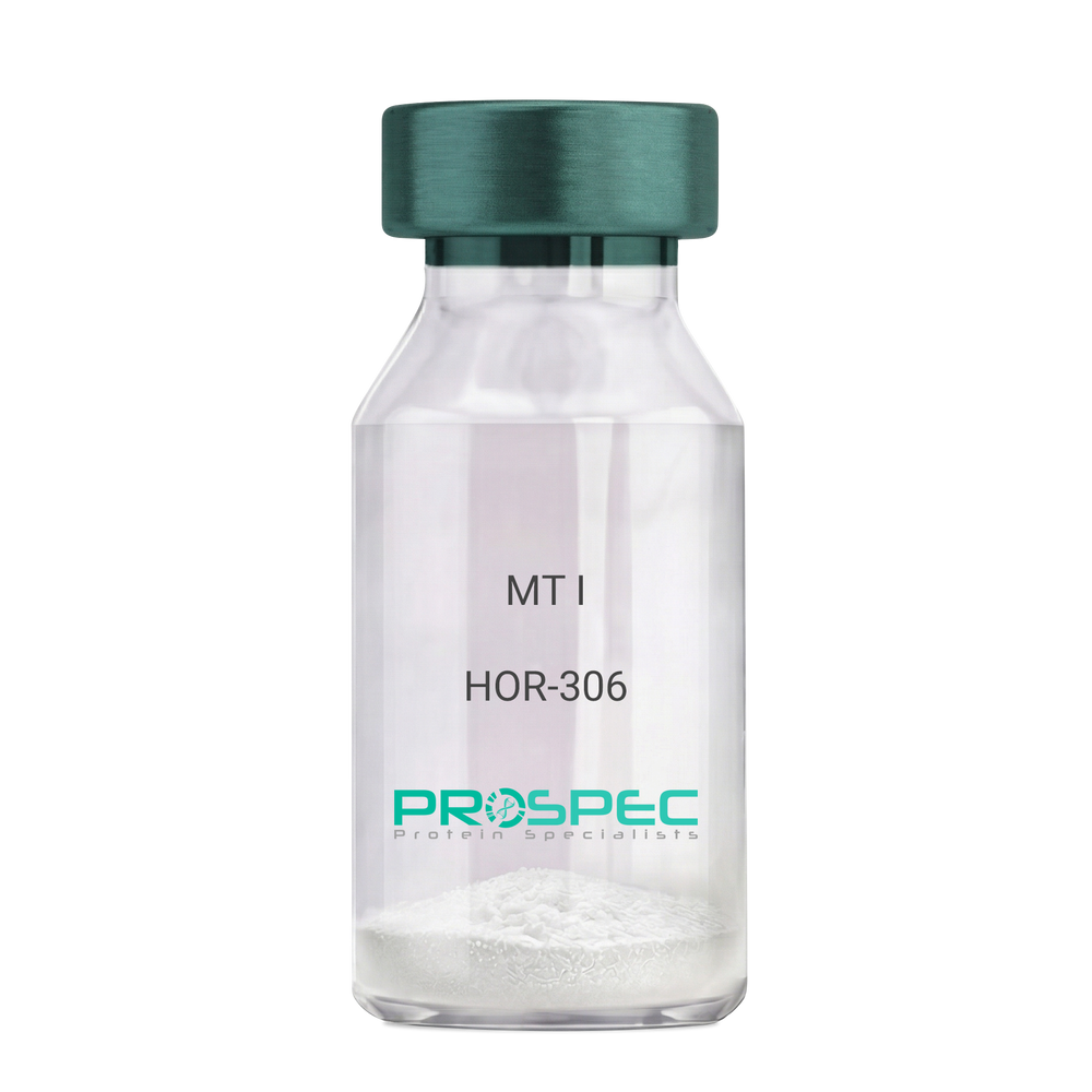
Name :
Melanotan-1Description:
Melanotan-I
Melanotan-I, MT-I, Melanotan-1.
Product # :
HOR-306Price :
Quantity :
Shipping Method :
Shipped at Room temp
More Info
- description
- formulation
- purity
- More Info
- Citations
Description
Melanotan-I has the amino acid sequence of Ser-Tyr-Ser-Nle-Glu-His-D-Phe-Arg-Trp-Gly-Lys-Pro-Val and a molecular weight of 1647.4 Dalton.
Formulation
The protein (1mg/ml) was lyophilized with no additives.
Purity
Greater than 98.0% as determined by RP-HPLC.
Citations
More Info
-
Synonyms
Melanotan-I, MT-I, Melanotan-1.
-
Physical Appearance
Sterile Filtered off-White lyophilized (freeze-dried) powder.
-
Stability
Lyophilized Melanotan-I although stable at room temperature for 3 weeks, should be stored desiccated below -18°C. Upon reconstitution MT-I should be stored at 4°C between 2-7 days and for future use below -18°C.For long term storage it is recommended to add a carrier protein (0.1% HSA or BSA).Please prevent freeze-thaw cycles.
-
Solubility
It is recommended to reconstitute the lyophilized Melanotan-I in sterile 1% acetic acid not less than 100 µg/ml, which can then be further diluted to other aqueous solutions.
-
Amino Acid Sequence
Ser-Tyr-Ser-Nle-Glu-His-D-Phe-Arg-Trp-Gly-Lys-Pro-Val
-
Background
What is the molecular weight/Mw of MELANOTAN-1 Protein?
MELANOTAN-1 Protein has a total Mw of 1.647kDa.
What is the Purity of MELANOTAN-1 Protein?
MELANOTAN-1 Protein is >98% pure as determined by SDS-PAGE.
What is the Biological Activity of MELANOTAN-1 Protein?
The biological functionality of MELANOTAN-1 Protein will be determined in the future.
What is the amino acid sequence of MELANOTAN-1 Protein?
Ser-Tyr-Ser-Nle-Glu-His-D-Phe-Arg-Trp-Gly-Lys-Pro-Val
What applications can MELANOTAN-1 Protein be used in?
MELANOTAN-1 Protein can probably be used in western blot, ELISA and Lateral Flow.
What is the endotoxin level for MELANOTAN-1 Protein?
The endotoxin level is minimal, MELANOTAN-1 Protein was purified using conventional chromatography techniques.
Who supplies MELANOTAN-1 that is in stock and available to order?
ProSpec has the protein in stock and available for immediate delivery.
Where can I order bulk mg quantities of MELANOTAN-1?
ProSpec supplies bulk mg quantities at affordable prices that meets research and academic budgets.
ProSpec's products are furnished for LABORATORY RESEARCH USE ONLY. The product may not be used as drugs, agricultural or pesticidal products, food additives or household chemicals.
- View Data Sheet
Name :
GDF15 MouseDescription:
Growth and Differentiation factor 15 Mouse Recombinant
Growth/differentiation factor 15, GDF-15.
Product # :
CYT-857Price :
Quantity :
Shipping Method :
Shipped with Ice Packs
More Info
- description
- source
- formulation
- purity
- More Info
- sds-page
- Citations
Description
GDF15 Mouse Recombinant produced in E.Coli is a single, non-glycosylated polypeptide chain containing 138 amino acids (189-303 a.a) and having a molecular mass of 14.9kDa. GDF15 is fused to a 23 amino acid His-tag at N-terminus & purified by proprietary chromatographic techniques.
Source
Escherichia Coli.
Formulation
GDF15 protein solution (1.0mg/ml) containing 20mM Phosphate buffer (pH 8.0) and 10% glycerol.
Purity
Greater than 90.0% as determined by SDS-PAGE.
sds-page
Citations
More Info
-
Introduction
GDF15 is part of the TGF-Beta superfamily which is involved in regulating inflammatory and apoptotic pathways in injured tissues and throughout disease processes. GDF15 is most abundant in the liver. Its expression in liver can be considerably up-regulated in during injury of organs such as liver, kidney, heart and lung. GDF-15 promotes proliferation or growth arrest and differentiation due to differences in cellular differentiation. GDF15 prevents apoptosis in cerebellar granule neurons by activating Akt and inhibiting endogenously active ERK. GDF15 is a novel autocrine/endocrine factor that antagonizes the hypertrophic response and loss of ventricular performance.
-
Synonyms
Growth/differentiation factor 15, GDF-15.
-
Physical Appearance
Sterile Filtered colorless solution.
-
Stability
Store at 4°C if entire vial will be used within 2-4 weeks. Store, frozen at -20°C for longer periods of time. For long term storage it is recommended to add a carrier protein (0.1% HSA or BSA).Avoid multiple freeze-thaw cycles.
-
Amino Acid Sequence
MGSSHHHHHH SSGLVPRGSH MGSSAHAHPR DSCPLGPGRC CHLETVQATL EDLGWSDWVL SPRQLQLSMC VGECPHLYRS ANTHAQIKAR LHGLQPDKVP APCCVPSSYT PVVLMHRTDS GVSLQTYDDL VARGCHCA.
-
Background
What is the molecular weight/Mw of GDF15 MOUSE Protein?
GDF15 MOUSE Protein has a total Mw of 14.9kDa.
What is the source or expression system of GDF15 MOUSE Protein?
Escherichia Coli.
What is the Purity of GDF15 MOUSE Protein?
GDF15 MOUSE Protein is >90% pure as determined by SDS-PAGE.
What is the Biological Activity of GDF15 MOUSE Protein?
The biological functionality of GDF15 MOUSE Protein will be determined in the future.
What is the amino acid sequence of GDF15 MOUSE Protein?
MGSSHHHHHH SSGLVPRGSH MGSSAHAHPR DSCPLGPGRC CHLETVQATL EDLGWSDWVL SPRQLQLSMC VGECPHLYRS ANTHAQIKAR LHGLQPDKVP APCCVPSSYT PVVLMHRTDS GVSLQTYDDL VARGCHCA.
What applications can GDF15 MOUSE Protein be used in?
GDF15 MOUSE Protein can probably be used in western blot, ELISA and Lateral Flow.
What is the endotoxin level for GDF15 MOUSE Protein?
The endotoxin level is minimal, GDF15 MOUSE Protein was purified using conventional chromatography techniques.
ProSpec's products are furnished for LABORATORY RESEARCH USE ONLY. The product may not be used as drugs, agricultural or pesticidal products, food additives or household chemicals.
- View Data Sheet
Name :
Vaspin Human, HisDescription:
Vaspin Human Recombinant, His Tag
Serpin A12 precursor, Visceral adipose-specific serpin, Visceral adipose tissue- derived serine protease inhibitor, Vaspin, OL-64, SERPINA12, Serine (or cysteine) proteinase inhibitor, clade A, antitrypsin, alpha-1 antiproteinase.
Product # :
CYT-459Price :
Quantity :
Shipping Method :
Shipped with Ice Packs
More Info
- description
- source
- formulation
- purity
- More Info
- Citations
Description
Vaspin Human Recombinant produced in E.Coli is a single, non-glycosylated, His Tag, polypeptide chain containing 415 amino acids and having a molecular mass of 47 kDa.The Vaspin is purified by proprietary chromatographic techniques.
Source
Escherichia Coli.
Formulation
Vaspin in 20mM Tris pH-8, 0.2mM PMSF and 10% glycerol.
Purity
Greater than 90.0% as determined by SDS-PAGE.
Citations
More Info
-
Introduction
Vaspin (visceral adipose-specific SERPIN), a newly identified adipokine, which is a member of serine protease inhibitor family. Vaspin is also a unique insulin sensitizing adipocytokine in obesity. A recent publication indicates that induction of human vaspin mRNA expression in adipose tissue is regulated in a fat depot-specific manner and could be associated with parameters of obesity, insulin resistance, and glucose metabolism.
-
Synonyms
Serpin A12 precursor, Visceral adipose-specific serpin, Visceral adipose tissue- derived serine protease inhibitor, Vaspin, OL-64, SERPINA12, Serine (or cysteine) proteinase inhibitor, clade A, antitrypsin, alpha-1 antiproteinase.
-
Physical Appearance
Sterile Filtered colorless solution.
-
Stability
Liquid Vaspin although stable at 10°C for 1 week, should be stored below -18°C. For long term storage it is recommended to add a carrier protein (0.1% HSA or BSA).Please prevent freeze-thaw cycles.
-
Amino Acid Sequence
MGSSHHHHHH SSGLVPRGSH MLKPSFSPRN YKALSEVQGW KQRMAAKELA RQNMDLGFKL LKKLAFYNPG RNIFLSPLSI STAFSMLCLG AQDSTLDEIK QGFNFRKMPE KDLHEGFHYI IHELTQKTQD LKLSIGNTLF IDQRLQPQRK FLEDAKNFYS AETILTNFQN LEMAQKQIND FISQKTHGKI NNLIENIDPG TVMLLANYIF FRARWKHEFD PNVTKEEDFF LEKNSSVKVP MMFRSGIYQV GYDDKLSCTI LEIPYQKNIT AIFILPDEGK LKHLEKGLQV DTFSRWKTLL SRRVVDVSVP RLHMTGTFDL KKTLSYIGVS KIFEEHGDLT KIAPHRSLKV GEAVHKAELK MDERGTEGAA GTGAQTLPME TPLVVKIDKP YLLLIYSEKI PSVLFLGKIV NPIGK.
ProSpec's products are furnished for LABORATORY RESEARCH USE ONLY. The product may not be used as drugs, agricultural or pesticidal products, food additives or household chemicals.
- View Data Sheet
Name :
GAPDH Human, ActiveDescription:
Glyceraldehyde-3-Phosphate Dehydrogenase Human Recombinant, Active
G3PD, GAPD, MGC88685, GAPDH, Glyceraldehyde-3-Phosphate Dehydrogenase.
Product # :
ENZ-985Price :
Quantity :
Shipping Method :
Shipped with Ice Packs
More Info
- description
- source
- formulation
- purity
- biological activity
- More Info
- Citations
Description
GAPDH Human Recombinant produced in E. coli is a single polypeptide chain containing 335 amino acids (1-335) and having a molecular mass of 36kDa. The GAPDH is purified by proprietary chromatographic techniques.
Source
Escherichia Coli.
Formulation
The GAPDH protein (1 mg/ml) contains 20mM Tris-HCl buffer pH-8, 1mM EDTA, 1mM DTT and 20% glycerol.
Purity
Greater than 95.0% as determined by SDS-PAGE.
Biological Activity
Specific activity is > 50 units/mg, and is defined as the amount of enzyme that convert 1.0 umole of glyceraldehyde-3-phosphate to 1,3-Bisphosphoglycerate per minute at pH 8.5 at 37C.
Citations
More Info
-
Introduction
GAPDH is a catalytic enzyme normally known to play a role in glycolysis. GAPDH exists as a tetramer composed of 36-kDa subunits and has a range of intracellular functions. GAPDH catalyzes the reversible reduction of 1,3-bisphosphoglycerate to glyceraldehyde 3-phosphophate in the presence of NADPH. Besides functioning as a glycolytic enzyme in cytoplasm, GAPDH has function in intracellular processes such as membrane fusion, microtubule bundling, phosphotransferase activity, nuclear RNA export, DNA replication and DNA repair. GAPDH catalyzes a vital energy-yielding step in carbohydrate metabolism, the reversible oxidative phosphorylation of glyceraldehyde-3-phosphate in the presence of inorganic phosphate and nicotinamide adenine dinucleotide (NAD). The enzyme exists as a tetramer of identical chains.
-
Synonyms
G3PD, GAPD, MGC88685, GAPDH, Glyceraldehyde-3-Phosphate Dehydrogenase.
-
Physical Appearance
Sterile filtered colorless solution.
-
Stability
Store at 4°C if entire vial will be used within 2-4 weeks. Store, frozen at -20°C for longer periods of time. For long term storage it is recommended to add a carrier protein (0.1% HSA or BSA).Avoid multiple freeze-thaw cycles.
-
Amino Acid Sequence
MGKVKVGVNG FGRIGRLVTR AAFNSGKVDI VAINDPFIDL NYMVYMFQYD STHGKFHGTV KAENGKLVIN GNPITIFQER DPSKIKWGDA GAEYVVESTG VFTTMEKAGA HLQGGAKRVI ISAPSADAPM FVMGVNHEKY DNSLKIISNA SCTTNCLAPL AKVIHDNFGI VEGLMTTVHA ITATQKTVDG PSGKLWRDGR GALQNIIPAS TGAAKAVGKV IPELNGKLTG MAFRVPTANV SVVDLTCRLE KPAKYDDIKK VVKQASEGPL KGILGYTEHQ VVSSDFNSDT HSSTFDAGAG IALNDHFVKL ISWYDNEFGY SNRVVDLMAH MASKE.
ProSpec's products are furnished for LABORATORY RESEARCH USE ONLY. The product may not be used as drugs, agricultural or pesticidal products, food additives or household chemicals.
- View Data Sheet
Name :
METRN MouseDescription:
Meteorin Mouse Recombinant
Meteorin, Hypoxia/reoxygenation regulatory factor, Metrn, 1810034B16Rik, Hyrac.
Product # :
PRO-2241Price :
Quantity :
Shipping Method :
Shipped with Ice Packs
More Info
- description
- source
- formulation
- purity
- More Info
- Citations
Description
METRN produced in Sf9 Baculovirus cells is a single, glycosylated polypeptide chain containing 276 amino acids (22-291 a.a.) and having a molecular mass of 30.2kDa (Migrates at 28-40kDa on SDS-PAGE under reducing conditions). METRN is expressed with a 6 amino acid His tag at C-Terminus and purified by proprietary chromatographic techniques.
Source
Sf9, Baculovirus cells.
Formulation
METRN protein solution (0.25mg/ml) contains Phosphate Buffered Saline (pH 7.4) and 10% glycerol.
Purity
Greater than 90.0% as determined by SDS-PAGE.
Citations
More Info
-
Introduction
Meteorin (METRN) is involved in both glial cell differentiation and axonal network formation during neurogenesis. METRN promotes astrocyte differentiation and transforms cerebellar astrocytes into radial glia. Moreover, the METRN protein stimulates axonal extension in small and intermediate neurons of sensory ganglia by activating nearby satellite glia.
-
Synonyms
Meteorin, Hypoxia/reoxygenation regulatory factor, Metrn, 1810034B16Rik, Hyrac.
-
Physical Appearance
Sterile Filtered colorless solution.
-
Stability
Store at 4°C if entire vial will be used within 2-4 weeks. Store, frozen at -20°C for longer periods of time. For long term storage it is recommended to add a carrier protein (0.1% HSA or BSA). Avoid multiple freeze-thaw cycles.
-
Amino Acid Sequence
GYSEDRCSWR GSGLTQEPGS VGQLTLDCTE GAIEWLYPAG ALRLTLGGPD PGTRPSIVCL RPERPFAGAQ VFAERMTGNL ELLLAEGPDL AGGRCMRWGP RERRALFLQA TPHRDISRRV AAFRFELHED QRAEMSPQAQ GLGVDGACRP CSDAELLLAA CTSDFVIHGT IHGVAHDTEL QESVITVVVA RVIRQTLPLF KEGSSEGQGR ASIRTLLRCG VRPGPGSFLF MGWSRFGEAW LGCAPRFQEF SRVYSAALTT HLNPCEMALD HHHHHH.
ProSpec's products are furnished for LABORATORY RESEARCH USE ONLY. The product may not be used as drugs, agricultural or pesticidal products, food additives or household chemicals.
- View Data Sheet
Name :
LGALS3 Mouse, ActiveDescription:
Galectin-3 Mouse Recombinant, BioActive
Lectin galactose binding soluble 3, Lectin, galactose binding, soluble 3, CBP35, GAL3, GALBP, GALIG, LGALS2, MAC2.
Product # :
CYT-1151Price :
Quantity :
Shipping Method :
Shipped with Ice Packs
More Info
- description
- source
- formulation
- purity
- biological activity
- More Info
- sds-page
- Citations
Description
LGALS3 Mouse Recombinant produced in E.Coli is a single, non-glycosylated polypeptide chain containing 287 amino acids ( 1-264 a.a) and having a molecular mass of 29.8kDa.LGALS3 is fused to a 23 amino acid His-tag at N-terminus & purified by proprietary chromatographic techniques.
Source
E.coli.
Formulation
LGALS3 protein (0.5mg/ml) contains 20mM Tris-HCl buffer (pH 8.0), 0.15M NaCl, 50% glycerol,1mM DTT and 2mM EDTA.
Purity
Greater than 95% as determined by SDS-PAGE.
Biological Activity
Measured by its ability to agglutinate human red blood cells. The ED50 for this effect is ≥ 25ug/ml.
sds-page
Citations
More Info
-
Introduction
Galectin 3, or LGALS3, is a protein which belongs to the animal lectins family, that binds betagalactoside residues selectively. LGALS3 is originated and leaves cells through ectocytosis. The protein is capable of inhibition apoptosis and the development of cancer. Galectin 3 is found in epithelial tissues in organisms, it can be located in dendritic cells, Kupffer cells, macrophages etc. LGALS3 levels elevated when inflammation is generating, cell proliferation, trans-activation by viral proteins and cell differentiation.
-
Synonyms
Lectin galactose binding soluble 3, Lectin, galactose binding, soluble 3, CBP35, GAL3, GALBP, GALIG, LGALS2, MAC2.
-
Physical Appearance
Sterile Filtered colorless solution.
-
Stability
Store at 4°C if entire vial will be used within 2-4 weeks. Store, frozen at -20°C for longer periods of time. For long term storage it is recommended to add a carrier protein (0.1% HSA or BSA).Avoid multiple freeze-thaw cycles.
-
Amino Acid Sequence
MGSSHHHHHH SSGLVPRGSH MGSMADSFSL NDALAGSGNP NPQGYPGAWG NQPGAGGYPG AAYPGAYPGQ APPGAYPGQA PPGAYPGQAP PSAYPGPTAP GAYPGPTAPG AYPGSTAPGA FPGQPGAPGA YPSAPGGYPA AGPYGVPAGP LTVPYDLPLP GGVMPRMLIT IMGTVKPNAN RIVLDFRRGN DVAFHFNPRF NENNRRVIVC NTKQDNNWGK EERQSAFPFE SGKPFKIQVL VEADHFKVAV NDAHLLQYNH RMKNLREISQ LGISGDITLT SANHAMI.
-
Background
What is the molecular weight/Mw of LGALS3 MOUSE Protein?
LGALS3 MOUSE Protein has a total Mw of 29.8kDa.
What is the source or expression system of LGALS3 MOUSE Protein?
Escherichia Coli.
What is the Purity of LGALS3 MOUSE Protein?
LGALS3 MOUSE Protein is >95% pure as determined by SDS-PAGE.
What is the Biological Activity of LGALS3 MOUSE Protein?
Measured by its ability to agglutinate human red blood cells. The ED50 for this effect is ≥ 25ug/ml.
What is the amino acid sequence of LGALS3 MOUSE Protein?
MGSSHHHHHH SSGLVPRGSH MGSMADSFSL NDALAGSGNP NPQGYPGAWG NQPGAGGYPG AAYPGAYPGQ APPGAYPGQA PPGAYPGQAP PSAYPGPTAP GAYPGPTAPG AYPGSTAPGA FPGQPGAPGA YPSAPGGYPA AGPYGVPAGP LTVPYDLPLP GGVMPRMLIT IMGTVKPNAN RIVLDFRRGN DVAFHFNPRF NENNRRVIVC NTKQDNNWGK EERQSAFPFE SGKPFKIQVL VEADHFKVAV NDAHLLQYNH RMKNLREISQ LGISGDITLT SANHAMI.
What applications can LGALS3 MOUSE Protein be used in?
LGALS3 MOUSE Protein can probably be used in western blot, ELISA and Lateral Flow.
ProSpec's products are furnished for LABORATORY RESEARCH USE ONLY. The product may not be used as drugs, agricultural or pesticidal products, food additives or household chemicals.
- View Data Sheet
Name :
PGLYRP1 Human, HEKDescription:
Peptidoglycan Recognition Protein 1 Human Recombinant, HEK
PGLYRP, PGRP, PGRP-S, PGRPS, TAG7, TNFSF3L, Peptidoglycan recognition protein 1, peptidoglycan recognition protein short.
Product # :
PRO-2726Price :
Quantity :
Shipping Method :
Shipped with Ice Packs
More Info
- description
- source
- formulation
- purity
- biological activity
- More Info
- Citations
Description
PGLYRP1 Human Recombinant produced in HEK293 Cells is a single, glycosylated polypeptide chain containing 184 amino acids (22-196 a.a) and having a molecular mass of 20.5 kDa.PGLYRP1 is expressed with a 6 amino acid His tag at C-Terminus and purified by proprietary chromatographic techniques.
Source
HEK293 Cells.
Formulation
The PGLYRP1 solution (0.25mg/1ml) contains phosphate buffered saline (pH7.4) and 10% glycerol.
Purity
Greater than 95.0% as determined by SDS-PAGE.
Biological Activity
Measured by its binding ability in a functional ELISA with Peptidoglycan. The ED50 range ≤ 30 ng/ml.
Citations
More Info
-
Introduction
Peptidoglycan Recognition Protein 1 (PGLYRP1) is a member of the N-acetylmuramoyl-L-alanine amidase 2 family. PGLYRP1 binds to peptidoglycan of bacteria and affects the peptidoglycan biosynthesis. PGLYRP1 displays bactericidal activity towards Gram-positive bacteria and is bacteriostatic towards Gram-negative bacteria. PGLYRP1 has a role in innate immunity. PGLYRP1 is highly expressed in the bone marrow, and weakly expressed in the kidney, liver, small intestine, spleen, thymus, peripheral leukocyte, lung, fetal spleen and neutrophils.
-
Synonyms
PGLYRP, PGRP, PGRP-S, PGRPS, TAG7, TNFSF3L, Peptidoglycan recognition protein 1, peptidoglycan recognition protein short.
-
Physical Appearance
Sterile filtered colorless solution.
-
Stability
Store at 4°C if entire vial will be used within 2-4 weeks.Store, frozen at -20°C for longer periods of time.For long term storage it is recommended to add a carrier protein (0.1% HSA or BSA).Avoid multiple freeze-thaw cycles.
-
Amino Acid Sequence
DGSQETEDPA CCSPIVPRNE WKALASECAQ HLSLPLRYVV VSHTAGSSCN TPASCQQQAR NVQHYHMKTL GWCDVGYNFL IGEDGLVYEG RGWNFTGAHS GHLWNPMSIG ISFMGNYMDR VPTPQAIRAA QGLLACGVAQ GALRSNYVLK GHRDVQRTLS PGNQLYHLIQ NWPHYRSPHH HHHH
ProSpec's products are furnished for LABORATORY RESEARCH USE ONLY. The product may not be used as drugs, agricultural or pesticidal products, food additives or household chemicals.
- View Data Sheet
Name :
GABARAPL2 HumanDescription:
GABA(A) Receptor-Associated Protein Like 2 Human Recombinant
GABA(A) receptor-associated protein-like 2, GATE-16, GEF2, ATG8, ATG8C, Ganglioside expression factor 2, General protein transport factor p16, Golgi-associated ATPase enhancer of 16 kDa, MAP1 light chain 3-related protein, gamma-aminobutyric acid receptor-associated protein-like 2, MAP1 light chain 3 related protein, FLC3A.
Product # :
PRO-079Price :
Quantity :
Shipping Method :
Shipped with Ice Packs
More Info
- description
- source
- formulation
- purity
- More Info
- Citations
Description
GABARAPL2 produced in E.Coli is a single, non-glycosylated polypeptide chain containing 137 amino acids (1-117a.a.) and having a molecular mass of 15.8kDa. GABARAPL2 is fused to a 20 amino acid His-tag at N-terminus & purified by proprietary chromatographic techniques.
Source
Escherichia Coli.
Formulation
The GABARAPL2 protein solution (0.5mg/1ml) is formulated in 20mM Tris-HCl buffer (pH8.0), 0.2M NaCl, 5mM DTT and 20% glycerol.
Purity
Greater than 90% as determined by SDS-PAGE.
Citations
More Info
-
Introduction
GABARAPL2 takes part in intra Golgi traffic and modulates intra Golgi transport by coupling between NSF activity and SNAREs activation. GABARAPL2 first stimulates the ATPase activity of NSF which in turn stimulates the association with GOSR1.
-
Synonyms
GABA(A) receptor-associated protein-like 2, GATE-16, GEF2, ATG8, ATG8C, Ganglioside expression factor 2, General protein transport factor p16, Golgi-associated ATPase enhancer of 16 kDa, MAP1 light chain 3-related protein, gamma-aminobutyric acid receptor-associated protein-like 2, MAP1 light chain 3 related protein, FLC3A.
-
Physical Appearance
Sterile Filtered clear solution.
-
Stability
Store at 4°C if entire vial will be used within 2-4 weeks. Store, frozen at -20°C for longer periods of time.Please avoid freeze thaw cycles.
-
Amino Acid Sequence
MGSSHHHHHH SSGLVPRGSH MKWMFKEDHS LEHRCVESAK IRAKYPDRVP VIVEKVSGSQ IVDIDKRKYL VPSDITVAQF MWIIRKRIQL PSEKAIFLFV DKTVPQSSLT MGQLYEKEKD EDGFLYVAYS GENTFGF
ProSpec's products are furnished for LABORATORY RESEARCH USE ONLY. The product may not be used as drugs, agricultural or pesticidal products, food additives or household chemicals.
- View Data Sheet
Name :
Periostin Human, HEKDescription:
Periostin Human Recombinant, HEK
OSF-2, Periostin, Osteoblast Specific Factor 2, PN OSF-2, PDLPOSTN, POSTN, MGC119510, MGC119511, PN, RP11-412K4.1.
Product # :
CYT-835Price :
Quantity :
Shipping Method :
Shipped at Room temp
More Info
- description
- source
- formulation
- purity
- More Info
- Citations
Description
Periostin Human Recombinant produced in HEK cells is a single, glycosylated, polypeptide chain (Asn22-Gln836) containing a total of 821 amino acids, having a calculated molecular mass of 91.8kDa and fused to a 6 aa His tag at C-Terminus.
Source
HEK 293.
Formulation
Periostin was filtered (0.4 µm) and lyophilized from 0.5mg/ml solution in phosphate buffered saline and 5% trehalose.
Purity
Greater than 38.0% as determined by SDS-PAGE.
Citations
More Info
-
Introduction
Periostin is a disulfide linked 90 kDa, 811 amino acid protein originally isolated as a osteoblast-specific factor that functions as a cell adhesion molecule for preosteoblasts and is thought to be involved in osteoblast recruitment, attachment and spreading. Additionally, periostin expression has previously been shown to be significantly increased by both transforming growth factor beta-1(TGFbeta1) and bone morphogenetic protein (BMP-2). OSF-2 has a typical signal sequence, followed by a cysteine-rich domain, a fourfold repeated domain and a C-terminal domain. The fourfold repeated domain of OSF-2 shows homology with the insect protein fasciclin
Periostin mRNA is expressed in the developing mouse embryonic and fetal heart, and that it is localized to the endocardial cushions that ultimately divide the primitive heart tube into a four-chambered heart. -
Synonyms
OSF-2, Periostin, Osteoblast Specific Factor 2, PN OSF-2, PDLPOSTN, POSTN, MGC119510, MGC119511, PN, RP11-412K4.1.
-
Physical Appearance
Filtered White lyophilized (freeze-dried) powder.
-
Stability
Store lyophilized protein at -20°C. Aliquot the product after reconstitution to avoid repeated freezing/thawing cycles. Reconstituted protein can be stored at 4°C for a limited period of time.
-
Solubility
It is recommended to add deionized water to prepare a working stock solution of approximately 0.5 mg/ml and let the lyophilized pellet dissolve completely. Periostin is not sterile! Please filter the product by an appropriate sterile filter before using it in the cell culture.
-
Amino Acid Sequence
NNHYDKILAH SRIRGRDQGP NVCALQQILG TKKKYFSTCK NWYKKSICGQ KTTVLYECCP GYMRMEGMKG CPAVLPIDHV YGTLGIVGAT TTQRYSDASK LREEIEGKGS FTYFAPSNEA WDNLDSDIRR GLESNVNVEL LNALHSHMIN KRMLTKDLKN GMIIPSMYNN LGLFINHYPN GVVTVNCARI IHGNQIATNG VVHVIDRVLT QIGTSIQDFI EAEDDLSSFR AAAITSDILE ALGRDGHFTL FAPTNEAFEK LPRGVLERIM GDKVASEALM KYHILNTLQC SESIMGGAVF ETLEGNTIEI GCDGDSITVN GIKMVNKKDI VTNNGVIHLI DQVLIPDSAK QVIELAGKQQ TTFTDLVAQL GLASALRPDG EYTLLAPVNN AFSDDTLSMD QRLLKLILQN HILKVKVGLN ELYNGQILET IGGKQLRVFV YRTAVCIENS CMEKGSKQGR NGAIHIFREI IKPAEKSLHE KLKQDKRFST FLSLLEAADL KELLTQPGDW TLFVPTNDAF KGMTSEEKEI LIRDKNALQN IILYHLTPGV FIGKGFEPGV TNILKTTQGS KIFLKEVNDT LLVNELKSKE SDIMTTNGVI HVVDKLLYPA DTPVGNDQLL EILNKLIKYI QIKFVRGSTF KEIPVTVYTT KIITKVVEPK IKVIEGSLQP IIKTEGPTLT KVKIEGEPEF RLIKEGETIT EVIHGEPIIK KYTKIIDGVP VEITEKETRE ERIITGPEIK YTRISTGGGE TEETLKKLLQ EEVTKVTKFI EGGDGHLFED EEIKRLLQGD TPVRKLQANK KVQGSRRRLR EGRSQHHHHH H.
ProSpec's products are furnished for LABORATORY RESEARCH USE ONLY. The product may not be used as drugs, agricultural or pesticidal products, food additives or household chemicals.
- View Data Sheet
Name :
SPOCK3 MouseDescription:
Sparc/Osteonectin 3 Mouse Recombinant
Testican-3, SPARC/osteonectin, CWCV, Kazal-like domains proteoglycan 3, Spock3.
Product # :
PRO-2328Price :
Quantity :
Shipping Method :
Shipped with Ice Packs
More Info
- description
- source
- formulation
- purity
- More Info
- Citations
Description
SPOCK3 Mouse Recombinant produced in Sf9 Baculovirus cells is a single, glycosylated polypeptide chain containing 423 amino acids (22-436 a.a.) and having a molecular mass of 47.9kDa (Migrates at 40-57kDa on SDS-PAGE under reducing conditions).SPOCK3 is expressed with an 8 amino acid His tag at C-Terminus and purified by proprietary chromatographic techniques.
Source
Sf9, Baculovirus cells.
Formulation
SPOCK3 protein solution (0.25mg/ml) containing Phosphate Buffered Saline (pH 7.4) and 10% glycerol.
Purity
Greater than 90% as determined by SDS-PAGE.
Citations
More Info
-
Introduction
Testican-3 (Spock3) is a nervous system-expressed heparan sulfate proteoglycan belonging to a subgroup of the BM-40/SPARC/osteonectin family, whose role of in brain development is unclear. Spock3 inhibits the processing of pro-matrix metalloproteinase 2(MMP-2) by MT1-MMP and MT3-MMP. Spock3 is mostly confined to the developmental stage of the brain.
-
Synonyms
Testican-3, SPARC/osteonectin, CWCV, Kazal-like domains proteoglycan 3, Spock3.
-
Physical Appearance
Sterile Filtered colorless solution.
-
Stability
Store at 4°C if entire vial will be used within 2-4 weeks. Store, frozen at -20°C for longer periods of time. For long term storage it is recommended to add a carrier protein (0.1% HSA or BSA).Avoid multiple freeze-thaw cycles.
-
Amino Acid Sequence
AAAAVAVAGG RSDGGNFLDE KQWLTTISQY DKEVGQWNKF RDEVEDDYFR TWNPGKPFDQ ALDPAKDPCL KTKCSRHKVC ITQDAQTALC ISHRRLTHSM KEVGGSHKQW RGLPSSTCKP CPIAYASPVC GSDGHSYSSQ CKLEYQACVL GKQISIKCEG RCPCPSDKSM NIGRNVKRAC
SDLEFREVAN RLRDWFKALH ESGSQNKKTK ALLRPERSRF DTSILPICKD SLGWMFNRLD TNYDLLLDQS ELGSIYLDKN EQCTKAFFNS CDTYKDSLIS NNEWCYCFQR QQDPPCHTEL SNIQKRQGIK KLLGQYIPLC DEDGYYKPTQ CHGSVGQCWC VDRYGNEVVG SRINGVADCA
IDFEISGDFA SGDFREWTDD EGEEDDIMND KDDIEDDDED EGDDDDDGDV HDGYILEHHH HHH
ProSpec's products are furnished for LABORATORY RESEARCH USE ONLY. The product may not be used as drugs, agricultural or pesticidal products, food additives or household chemicals.