Search results
1000 results found for “cyclin”
Name
Description
Product #
Price
Quantity
Shipping Method
- View Data Sheet
Name :
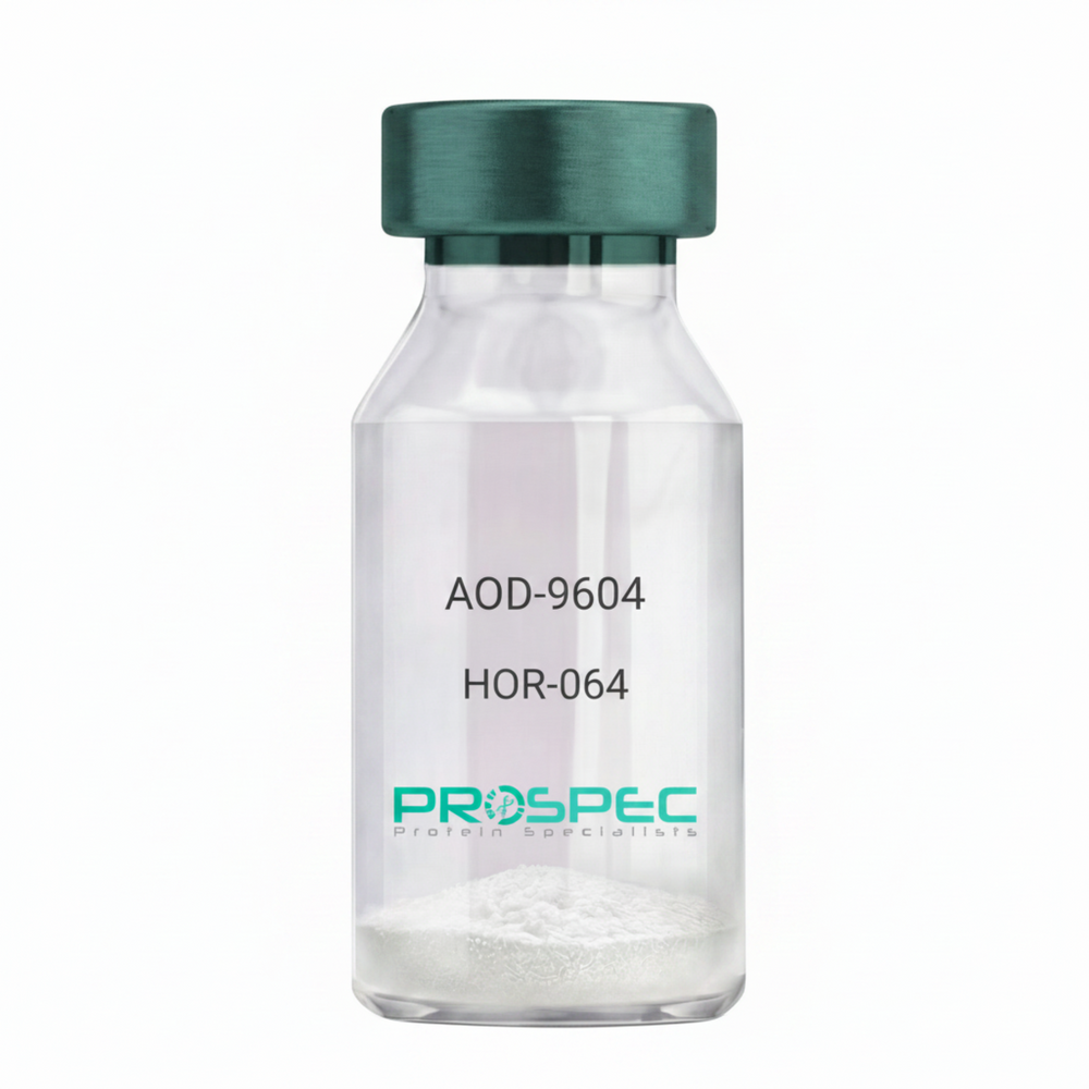
AOD-9604Description:
AOD-9604
Product # :
HOR-064Price :
Quantity :
Shipping Method :
Shipped at Room temp
More Info
- description
- formulation
- purity
- More Info
Description
AOD-9604 is a synthetic single, non-glycosylated polypeptide chain containing 16 amino acids, having a molecular mass of 1815 Dalton and a Molecular formula of C78H123N23O23 S2.
Formulation
The protein was lyophilized with no additives.
Purity
Greater than 97.0% as determined by analysis by RP-HPLC.
More Info
-
Physical Appearance
Sterile Filtered White lyophilized (freeze-dried) powder.
-
Stability
Lyophilized AOD-9604 although stable at room temperature for 3 weeks, should be stored desiccated below -18°C. Upon reconstitution AOD-9604 should be stored at 4°C between 2-7 days and for future use below -18°C.
For long term storage it is recommended to add a carrier protein (0.1% HSA or BSA).
Please prevent freeze-thaw cycles.
-
Solubility
It is recommended to reconstitute the lyophilized AOD-9604 in sterile 18MΩ-cm H2O not less than 100 µg/ml, which can then be further diluted to other aqueous solutions.
-
Amino Acid Sequence
H-Tyr-Leu-Arg-Ile-Val-Gln-Cys-Arg-SerVal-Glu-Gly-Ser-Cys-Gly-Phe-OH (Disulfide bond).
-
Background
AOD-9604 is a synthetic analog of the C-terminal fragment (177–191) AOD-9604 stimulates lipolysis (fat breakdown) without the unwanted growth-promoting or insulin-antagonizing effects of full-length hGH. AOD-9604 is a potent lipolytic tool in animal models and researchers focus on its regenerative properties in joint health and its ability to modulate beta3 adrenergic receptors without the systemic risks of growth hormone.
ProSpec's products are furnished for LABORATORY RESEARCH USE ONLY. The product may not be used as drugs, agricultural or pesticidal products, food additives or household chemicals.
- View Data Sheet
Name :
SSU72 PAT45E2AT AntibodyDescription:
SSU72 RNA Polymerase II CTD Phosphatase, Clone PAT45E2AT, Mouse Anti Human
SSU72 RNA polymerase II CTD phosphatase homolog (S. cerevisiae), HSPC182, CTD phosphatase SSU72, Ssu72 RNA polymerase II CTD phosphatase homolog (yeast), PNAS-120, RNA polymerase II subunit A C-terminal domain phosphatase SSU72, EC 3.1.3.16.
Product # :
ANT-701Price :
Quantity :
Shipping Method :
Shipped with Ice Packs
More Info
- formulation
- More Info
Formulation
1mg/ml containing PBS, pH-7.4, 10% Glycerol and 0.02% Sodium Azide.
More Info
-
Introduction
SSU72 is an extremely conserved homologue of yeast Ssu72, a CTD phosphatase and a component of the polyadenylation/termination machinery. SSU72 interacts with TFIIB, Rb and DNAM-1 and operates to catalyze the dephosphorylation of target proteins, and taking part in RNA processing and termination via dephosphorylation of Pol II. SSU72 is found in multiple alternatively spliced isoforms.
-
Synonyms
SSU72 RNA polymerase II CTD phosphatase homolog (S. cerevisiae), HSPC182, CTD phosphatase SSU72, Ssu72 RNA polymerase II CTD phosphatase homolog (yeast), PNAS-120, RNA polymerase II subunit A C-terminal domain phosphatase SSU72, EC 3.1.3.16.
-
Physical Appearance
Sterile filtered colorless solution.
-
Immunogen
Anti-human SSU72 mAb, is derived from hybridization of mouse F0 myeloma cells with spleen cells from BALB/c mice immunized with recombinant human SSU72 amino acids 1-194 purified from E. coli.
-
Ig Subclass
Mouse IgG1 heavy chain and κ light chain.
-
Clone
PAT45E2AT.
-
Applications
SSU72 antibody has been tested by ELISA and Western blot analysis to assure specificity and reactivity. Since application varies, however, each investigation should be titrated by the reagent to obtain optimal results.
-
Type
Mouse Anti Human Monoclonal.
-
Storage Procedures
For periods up to 1 month store at 4°C, for longer periods of time, store at -20°C. Prevent freeze thaw cycles.
-
Purification Method
SSU72 antibody was purified from mouse ascitic fluids by protein-A affinity chromatography.
ProSpec's products are furnished for LABORATORY RESEARCH USE ONLY. The product may not be used as drugs, agricultural or pesticidal products, food additives or household chemicals.
- View Data Sheet
Name :
TBCEL AntibodyDescription:
Tubulin Folding Cofactor E-Like, Mouse Anti Human
Tubulin Folding Cofactor E-Like, E-Like, LRRC351, Leucine Rich Repeat Containing Catastrophin, Tubulin-Specific Chaperone E-Like.
Product # :
ANT-576Price :
Quantity :
Shipping Method :
Shipped with Ice Packs
More Info
- formulation
- More Info
Formulation
1mg/ml containing PBS, pH-7.4, 10% Glycerol and 0.02% Sodium Azide.
More Info
-
Introduction
TBCEL, is a factor that is in charge of the microtubule cytoskeleton in determining cell behavior. TBCEL plays a role as a regulator of tubulin stability. While widely expressed in testis, TBCEL is also present in several tissues at a much lower level. TBCEL comprises of seven LRR (leucine-rich) repeats, one LRRCT domain and one ubiquitin-like domain. The gene that translates TBCEL consists of 66,704 bases and maps to human chromosome 11q23.3. Chromosome 11 houses over 1,400 genes and consist of nearly 4% of the human genome. Jervell and Lange-Nielsen syndrome, Jacobsen syndrome, Niemann-Pick disease, hereditary angioedema and Smith-Lemli-Opitz syndrome are associated with defects in genes that map to chromosome 11.
-
Synonyms
Tubulin Folding Cofactor E-Like, E-Like, LRRC351, Leucine Rich Repeat Containing Catastrophin, Tubulin-Specific Chaperone E-Like.
-
Physical Appearance
Sterile filtered colorless solution.
-
Immunogen
Anti-human TBCEL mAb, is derived from hybridization of mouse F0 myeloma cells with spleen cells from BALB/c mice immunized with a recombinant human TBCEL protein 1-424 amino acids purified from E. coli.
-
Ig Subclass
Mouse IgG2b heavy chain and k light chain.
-
Clone
PAT1B10AT.
-
Applications
TBCEL antibody has been tested by ELISA, Western blot analysis, ICC/IF and Flow cytometry to assure specificity and reactivity. Since application varies, however, each investigation should be titrated by the reagent to obtain optimal results.
-
Type
Mouse Anti Human Monoclonal.
-
Storage Procedures
For periods up to 1 month store at 4°C, for longer periods of time, store at -20°C. Prevent freeze thaw cycles.
-
Purification Method
KRAS antibody was purified from mouse ascitic fluids by protein-A affinity chromatography.
ProSpec's products are furnished for LABORATORY RESEARCH USE ONLY. The product may not be used as drugs, agricultural or pesticidal products, food additives or household chemicals.
- View Data Sheet
Description:
Vasoactive Intestinal Peptide
Product # :
HOR-043Price :
Quantity :
Shipping Method :
Shipped at Room temp
More Info
- description
- formulation
- purity
- More Info
Description
Vasoactive Intestinal Peptide Synthetic is a single, non-glycosylated polypeptide chain containing 28 amino acids, having a molecular mass of 3325 Dalton and a Molecular formula of C147H238N44O42S .
Formulation
The protein was lyophilized with no additives.
Purity
Greater than 97.0% as determined by analysis by RP-HPLC.
More Info
-
Physical Appearance
Sterile Filtered White lyophilized (freeze-dried) powder.
-
Stability
Lyophilized Vasoactive Intestinal Peptide although stable at room temperature for 3 weeks, should be stored desiccated below -18°C. Upon reconstitution Vasoactive Intestinal Peptide should be stored at 4°C between 2-7 days and for future use below -18°C. For long term storage it is recommended to add a carrier protein (0.1% HSA or BSA). Please prevent freeze-thaw cycles.
-
Solubility
It is recommended to reconstitute the lyophilized Vasoactive Intestinal Peptide in sterile 18MΩ-cm H2O not less than 100 µg/ml, which can then be further diluted to other aqueous solutions.
-
Amino Acid Sequence
H-His-Ser-Asp-Ala-Val-Phe-Thr-Asp-Asn-Tyr-Thr-Arg-Leu-Arg-Lys-Gln-Met-Ala-Val-Lys-Lys-Tyr-Leu-Asn-Ser-Ile-Leu-Asn-NH2.
-
Background
Vasoactive Intestinal Peptide (VIP), a prominent member of the glucagon-secretin peptide superfamily, plays a multifaceted role as a neuropeptide and neurotransmitter. This research paper aims to provide an extensive analysis of VIP, exploring its biochemical properties, diverse physiological functions, and potential therapeutic applications in various disease conditions.
Vasoactive Intestinal Peptide (VIP), originally identified for its potent vasodilatory properties in the gastrointestinal system, has since been recognized for its wide-ranging physiological effects throughout the body. As a neuropeptide and neurotransmitter.
VIP, a 28-amino acid peptide, exhibits a diverse range of biological activities due to its interactions with multiple G protein-coupled receptors (GPCRs) from the VIP/PACAP receptor family (Harmar et al., 2012). These receptors are expressed in various tissues and cells, enabling VIP to exert its pleiotropic effects.
VIP is involved in the regulation of numerous physiological processes. It functions as a potent vasodilator, modulates smooth muscle activity in the gastrointestinal tract, and participates in neurotransmission in the central and peripheral nervous systems (Lelievre et al., 2008). Additionally, VIP has immunomodulatory properties, influencing immune cell functions and inflammatory responses (Delgado et al., 2004).
VIP serves as a neurotransmitter in the central nervous system, where it contributes to various neuronal functions, including learning, memory, and sensory perception (Deng et al., 2015). Moreover, VIP has demonstrated neuroprotective effects in neurodegenerative disorders, making it a potential therapeutic target for neuroprotection and neuroregeneration (Delgado et al., 2002).
VIP's diverse physiological functions offer potential therapeutic applications in various disease contexts. Research has explored its potential in the treatment of inflammatory diseases, neurodegenerative disorders, and gastrointestinal disorders (Delgado et al., 2013). Furthermore, VIP-based therapies are being investigated for their neuroprotective potential in brain injuries and stroke.
As the research on VIP continues to progress, further investigation is warranted to uncover the precise mechanisms of action and potential clinical applications in medicine. VIP stands as a remarkable example of a neuropeptide with extensive physiological roles and therapeutic potential in various disease conditions.
What is the molecular weight/Mw of VASOACTIVE INTESTINAL PEPTIDE Protein?
VASOACTIVE INTESTINAL PEPTIDE Protein has a total Mw of 3.32kDa.
What is the Purity of VASOACTIVE INTESTINAL PEPTIDE Protein?
VASOACTIVE INTESTINAL PEPTIDE Protein is >97% pure as determined by SDS-PAGE.
What is the Biological Activity of VASOACTIVE INTESTINAL PEPTIDE Protein?
The biological functionality of VASOACTIVE INTESTINAL PEPTIDE Protein will be determined in the future.
What is the amino acid sequence of VASOACTIVE INTESTINAL PEPTIDE Protein?
H-His-Ser-Asp-Ala-Val-Phe-Thr-Asp-Asn-Tyr-Thr-Arg-Leu-Arg-Lys-Gln-Met-Ala-Val-Lys-Lys-Tyr-Leu-Asn-Ser-Ile-Leu-Asn-NH2.
What applications can VASOACTIVE INTESTINAL PEPTIDE Protein be used in?
VASOACTIVE INTESTINAL PEPTIDE Protein can probably be used in western blot, ELISA and Lateral Flow.
What is the endotoxin level for VASOACTIVE INTESTINAL PEPTIDE Protein?
The endotoxin level is minimal, VASOACTIVE INTESTINAL PEPTIDE Protein was purified using conventional chromatography techniques.
ProSpec's products are furnished for LABORATORY RESEARCH USE ONLY. The product may not be used as drugs, agricultural or pesticidal products, food additives or household chemicals.
- View Data Sheet
Name :
CLEC7A HumanDescription:
C-Type Lectin Domain Family 7, Member A Human Recombinant
BGR, Dendritic Cell-Associated C-Type Lectin-1, Dendritic Cell-Associated C-Type Lectin 1, C-Type Lectin Domain Family 7, Member A, Lectin-Like Receptor 1, CD369 Antigen, CANDF4, SCARE2, CD369, C-Type Lectin Domain Containing 7A, C-Type Lectin Domain Family 7 Member A, C-Type (Calcium Dependent, Carbohydrate-Recognition Domain) Lectin, Superfamily Member 12, C-Type Lectin Superfamily Member 12, DC-Associated C-Type Lectin 1, Beta-Glucan Receptor, Dectin-1, CLECSF12, DECTIN1.
Product # :
PRO-2634Price :
Quantity :
Shipping Method :
Shipped with Ice Packs
More Info
- description
- source
- formulation
- purity
- More Info
Description
CLEC7A Human Recombinant produced in Sf9 Baculovirus cells is a single, glycosylated polypeptide chain containing 183 amino acids (71-244 a.a) and having a molecular mass of 21kDa. CLEC7A is fused to a 9 amino acid His-tag at C-terminus & purified by proprietary chromatographic techniques.
Source
Sf9, Baculovirus cells.
Formulation
The CLEC7A solution (0.5mg/1ml) contains phosphate buffered saline (pH7.4) and 10% glycerol.
Purity
Greater than 95.0% as determined by SDS-PAGE.
More Info
-
Introduction
C-type lectin domain family 7 member A 1 or CLEC7A is a protein, that in the innate immune system, acts against fungal pathogens. CLEC7A can be found in the immune system response cells such as monocytes, macrophages & neutrophils, or in dendritic and T cells. The protein is enhanced by macrophages by using GM-CSF, IL-4, or IL-13, or diminishes by dexamethasone, IL-10 and LPS.
-
Synonyms
BGR, Dendritic Cell-Associated C-Type Lectin-1, Dendritic Cell-Associated C-Type Lectin 1, C-Type Lectin Domain Family 7, Member A, Lectin-Like Receptor 1, CD369 Antigen, CANDF4, SCARE2, CD369, C-Type Lectin Domain Containing 7A, C-Type Lectin Domain Family 7 Member A, C-Type (Calcium Dependent, Carbohydrate-Recognition Domain) Lectin, Superfamily Member 12, C-Type Lectin Superfamily Member 12, DC-Associated C-Type Lectin 1, Beta-Glucan Receptor, Dectin-1, CLECSF12, DECTIN1.
-
Physical Appearance
Sterile Filtered clear solution.
-
Stability
Store at 4°C if entire vial will be used within 2-4 weeks. Store, frozen at -20°C for longer periods of time. For long term storage it is recommended to add a carrier protein (0.1% HSA or BSA).Avoid multiple freeze-thaw cycles.
-
Amino Acid Sequence
ADPRHNSGRN PEEKDNFLSR NKENHKPTES SLDEKVAPSK ASQTTGGFSQ SCLPNWIMHG KSCYLFSFSG NSWYGSKRHC SQLGAHLLKI DNSKEFEFIE SQTSSHRINA FWIGLSRNQS EGPWFWEDGS AFFPNSFQVR NTVPQESLLH NCVWIHGSEV YNQICNTSSY SICEKELHHH HHH.
ProSpec's products are furnished for LABORATORY RESEARCH USE ONLY. The product may not be used as drugs, agricultural or pesticidal products, food additives or household chemicals.
- View Data Sheet
Name :
CMV Pp28Description:
Cytomegalo Virus Pp28 (UL99) Recombinant
Product # :
CMV-212Price :
Quantity :
Shipping Method :
Shipped with Ice Packs
More Info
- description
- source
- formulation
- purity
- More Info
Description
The E.Coli derived recombinant protein contains the CMV Pp28 (UL99) immunodominant regions, 130-160 amino acids.
Source
Escherichia Coli.
Formulation
50mM Tris-Hcl pH 7.2, 1mM EDTA and 50% glycerol.
Purity
CMV Pp28 protein is >95% pure as determined by 10% PAGE (coomassie staining).
More Info
-
Introduction
The human cytomegalovirus UL99-encoded pp28 is a myristylated phosphoprotein that is a constituent of the virion. The pp28 protein is positioned within the tegument of the virus particle, a protein structure that resides between the capsid and envelope. In the infected cell, pp28 is found in a cytoplasmic compartment derived from the Golgi apparatus, where the virus buds into vesicles to acquire its final membrane.
-
Stability
CMV Pp28 protein although stable at 4°C for 1 week, should be stored below -18°C. Please prevent freeze thaw cycles.
-
Applications
CMV Pp28 antigen is suitable for ELISA and Western blots, excellent antigen for detection of CMV with minimal specificity problems.
-
Specificity
Immunoreactive with sera of CMV-infected individuals.
-
Purification Method
Purified by GS-4B Sepharose-Affinity Purification.
ProSpec's products are furnished for LABORATORY RESEARCH USE ONLY. The product may not be used as drugs, agricultural or pesticidal products, food additives or household chemicals.
- View Data Sheet
Name :
ACE HumanDescription:
Angiotensin Converting Enzyme Human Recombinant
Angiotensin-converting enzyme, ACE, Dipeptidyl carboxypeptidase I, Kininase II, CD_antigen: CD143, DCP, DCP1, ACE1, CD143
Product # :
ENZ-1156Price :
Quantity :
Shipping Method :
Shipped with Ice Packs
More Info
- description
- source
- formulation
- purity
- biological activity
- More Info
Description
ACE Human produced in Sf9 Baculovirus cells is a single, glycosylated polypeptide chain containing 1235 amino acids (30-1256 a.a.) and having a molecular mass of 142kDa. ACE is expressed with an 8 amino acid His tag at C-Terminus and purified by proprietary chromatographic techniques.
Source
Sf9, Baculovirus cells.
Formulation
ACE protein solution (0.25mg/ml) contains Phosphate Buffered Saline (pH 7.4) and 10% glycerol.
Purity
Greater than 90.0% as determined by SDS-PAGE.
Biological Activity
Specific activity is > 1,000 pmol/min/mg. Defined by the amount of enzyme that cleaves 1pmol of McaRPPGFSAFK(Dnp)-OH per minute at 25C˚.
More Info
-
Introduction
Angiotensin Converting Enzyme (ACE) is a zinc metallopeptidase vital for blood pressure control and salt and water metabolism.ACE converts angiotensin I to angiotensin II by release of the terminal His-Leu which causes the vasoconstrictor activity of angiotensin to increase.ACE inactivates bradykinin, a potent vasodilator and has also a glycosidase activity which releases GPI-anchored proteins from the membrane by cleaving the mannose linkage in the GPI moiety.
-
Synonyms
Angiotensin-converting enzyme, ACE, Dipeptidyl carboxypeptidase I, Kininase II, CD_antigen: CD143, DCP, DCP1, ACE1, CD143
-
Physical Appearance
Sterile Filtered colorless solution.
-
Stability
Store at 4°C if entire vial will be used within 2-4 weeks. Store, frozen at -20°C for longer periods of time. For long term storage it is recommended to add a carrier protein (0.1% HSA or BSA).Avoid multiple freeze-thaw cycles.
-
Amino Acid Sequence
LDPGLQPGNF SADEAGAQLF AQSYNSSAEQ VLFQSVAASW AHDTNITAEN ARRQEEAALL SQEFAEAWGQ KAKELYEPIW QNFTDPQLRR IIGAVRTLGS ANLPLAKRQQ YNALLSNMSR IYSTAKVCLP NKTATCWSLD PDLTNILASS RSYAMLLFAW EGWHNAAGIP LKPLYEDFTA LSNEAYKQDG FTDTGAYWRS WYNSPTFEDD LEHLYQQLEP LYLNLHAFVR RALHRRYGDR YINLRGPIPA HLLGDMWAQS WENIYDMVVP FPDKPNLDVT STMLQQGWNA THMFRVAEEF FTSLELSPMP PEFWEGSMLE KPADGREVVC HASAWDFYNR KDFRIKQCTR VTMDQLSTVH HEMGHIQYYL QYKDLPVSLR RGANPGFHEA IGDVLALSVS TPEHLHKIGL LDRVTNDTES DINYLLKMAL EKIAFLPFGY LVDQWRWGVF SGRTPPSRYN FDWWYLRTKY QGICPPVTRN ETHFDAGAKF HVPNVTPYIR YFVSFVLQFQ FHEALCKEAG YEGPLHQCDI YRSTKAGAKL RKVLQAGSSR PWQEVLKDMV GLDALDAQPL LKYFQPVTQW LQEQNQQNGE VLGWPEYQWH PPLPDNYPEG IDLVTDEAEA SKFVEEYDRT SQVVWNEYAE ANWNYNTNIT TETSKILLQK NMQIANHTLK YGTQARKFDV NQLQNTTIKR IIKKVQDLER AALPAQELEE YNKILLDMET TYSVATVCHP NGSCLQLEPD LTNVMATSRK YEDLLWAWEG WRDKAGRAIL QFYPKYVELI NQAARLNGYV DAGDSWRSMY ETPSLEQDLE RLFQELQPLY LNLHAYVRRA LHRHYGAQHI NLEGPIPAHL LGNMWAQTWS NIYDLVVPFP SAPSMDTTEA MLKQGWTPRR MFKEADDFFT SLGLLPVPPE FWNKSMLEKP TDGREVVCHA SAWDFYNGKD FRIKQCTTVN LEDLVVAHHE MGHIQYFMQY KDLPVALREG ANPGFHEAIG DVLALSVSTP KHLHSLNLLS SEGGSDEHDI NFLMKMALDK IAFIPFSYLV DQWRWRVFDG SITKENYNQE WWSLRLKYQG LCPPVPRTQG DFDPGAKFHI PSSVPYIRYF VSFIIQFQFH EALCQAAGHT GPLHKCDIYQ SKEAGQRLAT AMKLGFSRPW PEAMQLITGQ PNMSASAMLS YFKPLLDWLR TENELHGEKL GWPQYNWTPN SARSEGPLPD SGRVSFLGLD LDAQQARVEH HHHHH
ProSpec's products are furnished for LABORATORY RESEARCH USE ONLY. The product may not be used as drugs, agricultural or pesticidal products, food additives or household chemicals.
- View Data Sheet
Name :
IL 1 alpha RatDescription:
Interleukin-1 alpha Rat Recombinant
Hematopoietin-1, Lymphocyte-activating factor (LAF), Endogenous Pyrogen (EP), Leukocyte Endogenous Mediator (LEM), Mononuclear Cell Factor (MCF), IL-1 alpha,IL1, IL-1A, IL1F1.
Product # :
CYT-381Price :
Quantity :
Shipping Method :
Shipped at Room temp
More Info
- description
- source
- formulation
- purity
- biological activity
- More Info
Description
Interleukin-1A Rat Recombinant produced in E.Coli is single, a non-glycosylated, Polypeptide chain containing 155 amino acids and having a molecular mass of 17703 Dalton. The IL-1A is purified by proprietary chromatographic techniques.
Source
Escherichia Coli.
Formulation
The protein was lyophilized from a concentrated (1mg/ml) sterile solution containing 50mM Tris-HCl, pH=8.
Purity
Greater than 98.0% as determined by:
(a) Analysis by RP-HPLC.
(b) Analysis by SDS-PAGE.Biological Activity
The ED50 as determined by the dose-dependant stimulation of murine D10S cells is < 0.005 ng/ml, corresponding to a Specific Activity of 200,000,000IU/mg.More Info
-
Introduction
Interleukin-1 alpha is a proinflammatory cytokine produced by a wide variety of cell types, including macrophages, osteoblasts, monocytes and hepatocytes. Circulating levels of are normally low and only rise after stimulation by agents such as those produced byinflammation, infection or microbial endotoxins. IL-1 alpha possesses a wide variety of biological activities and exerts its effects by binding to specific cell surface receptors.
-
Synonyms
Hematopoietin-1, Lymphocyte-activating factor (LAF), Endogenous Pyrogen (EP), Leukocyte Endogenous Mediator (LEM), Mononuclear Cell Factor (MCF), IL-1 alpha,IL1, IL-1A, IL1F1.
-
Physical Appearance
Sterile Filtered White lyophilized (freeze-dried) powder.
-
Stability
Lyophilized Interleukin-1a although stable at room temperature for 3 weeks, should be stored desiccated below -18°C. Upon reconstitution IL1A should be stored at 4°C between 2-7 days and for future use below -18°C. For long term storage it is recommended to add a carrier protein (0.1% HSA or BSA).Please prevent freeze-thaw cycles.
-
Solubility
It is recommended to reconstitute the lyophilized Interleukin 1a in sterile 18MΩ-cm H2O not less than 100µg/ml, which can then be further diluted to other aqueous solutions.
-
Amino Acid Sequence
The sequence of the first five N-terminal amino acids was determined and was found to be Ala-Pro-His-Ser-Phe.
ProSpec's products are furnished for LABORATORY RESEARCH USE ONLY. The product may not be used as drugs, agricultural or pesticidal products, food additives or household chemicals.
- View Data Sheet
Name :
IL 13 Rat 109 a.a.Description:
Interleukin-13 109 a.a. Rat Recombinant
NC300, ALRH, BHR1, P600, IL-13.
Product # :
CYT-182Price :
Quantity :
Shipping Method :
Shipped at Room temp
More Info
- description
- source
- formulation
- purity
- biological activity
- More Info
Description
Interleukin-13 Rat Recombinant produced in E.Coli is a single, non-glycosylated polypeptide chain containing 109 amino acids and having a molecular mass of 11.9 kDa. The IL-13 is purified by proprietary chromatographic techniques.
Source
Escherichia Coli.
Formulation
The protein (1mg/ml) was lyophilized in PBS, pH7.4.
Purity
Greater than 95% as determined by SDS-PAGE.
Biological Activity
ED50 range = 40ng/ml, corresponding to a specific activity of > 25,000IU/mg as determined by the dose dependent proliferation of human TF-1 cells. Optimal concentration for individual application should be determined by a dose response assay.More Info
-
Introduction
IL13 is an immunoregulatory cytokine produced primarily by activated Th2 cells. IL-13 is involved in several stages of B-cell maturation and differentiation. It up-regulates CD23 and MHC class II expression, and promotes IgE isotype switching of B cells. This cytokine down-regulates macrophage activity, thereby inhibits the production of pro-inflammatory cytokines and chemokines. This cytokine is found to be critical to the pathogenesis of allergen-induced asthma but operates through mechanisms independent of IgE and eosinophils. This gene, IL3, IL5, IL4, and CSF2 form a cytokine gene cluster on chromosome 5q, with this gene particularly close to IL4.
-
Synonyms
NC300, ALRH, BHR1, P600, IL-13.
-
Physical Appearance
Sterile Filtered White lyophilized (freeze-dried) powder.
-
Stability
Lyophilized Interleukin-13 although stable at room temperature for 3 weeks, should be stored desiccated below -18°C. Upon reconstitution IL13 should be stored at 4°C between 2-7 days and for future use below -18°C.For long term storage it is recommended to add a carrier protein (0.1% HSA or BSA).Please prevent freeze-thaw cycles.
-
Solubility
It is recommended to reconstitute the lyophilized Interleukin 13 in sterile 18MΩ-cm H2O not less than 100µg/ml, which can then be further diluted to other aqueous solutions.
-
Amino Acid Sequence
VRRSTSPPVA LRELIEELSN ITQDQKTSLC NSSIVWSVDI TAGGFCAALE SLTNISSCNA IHRTQRILNG LCNQKASDVA SSPPDTKIEV AQFISKLLNY SKQLFRYGH.
-
Protein content
Protein quantitation was carried out by two independent methods:1. UV spectroscopy at 280 nm using the absorbency value of 0.69 as the extinction coefficient for a 0.1% (1mg/ml) solution. This value is calculated by the PC GENE computer analysis program of protein sequences (IntelliGenetics). 2. Analysis by RP-HPLC, using a calibrated solution of IL-13 as a Reference Standard.
ProSpec's products are furnished for LABORATORY RESEARCH USE ONLY. The product may not be used as drugs, agricultural or pesticidal products, food additives or household chemicals.
- View Data Sheet
Name :
Omentin HumanDescription:
Omentin Human Recombinant
Intelectin-1, HL1, LFR, HL-1, INTL, ITLN, hIntL.
Product # :
CYT-301Price :
Quantity :
Shipping Method :
Shipped at Room temp
More Info
- description
- source
- formulation
- purity
- More Info
Description
Omentin Human Recombinant is produced in E.Coli by recombinant DNA technology is a single, polypeptide chain containing 313 amino acids and having a molecular mass of 35 kDa. Intelectin is purified by proprietary chromatographic techniques.
Source
E.Coli.
Formulation
Each mg of lyophilized powder contains 10mM NaP, pH-7.5 and 5:1 mannitol to protein.
Purity
Greater than 95.0% as determined by SDS-PAGE.
More Info
-
Introduction
Omentin is a recently recognized gene highly localized to the mental tissue (visceral adipose tissue). Omentin is present in the stromal vascular cells in the adipose tissue rather than in the adipocytes. Omentin is predominantly expressed in the visceral adipose tissue than the subcutaneous tissue, with the omentin mRNA being 150 times higher in the visceral adipose tissue. Omentin has also been detected in human blood using western blot analysis, and seems to increase INSstimulated glucose uptake in 3T3-L1 adipocytes in mice. Omentin seems to increase Akt phosphorylation irrespective of INS presence. Its role in glucose metabolism and obesity remains to be described; an INS-sensitizing action is possible.Differences in Omentin expression has been noted in adipose tissue from normals and patients with inflammatory bowel disease although its significance is unknown.
-
Synonyms
Intelectin-1, HL1, LFR, HL-1, INTL, ITLN, hIntL.
-
Physical Appearance
Sterile Filtered White lyophilized (freeze-dried) powder.
-
Stability
Lyophilized Intelectin although stable at room temperature for 3 weeks, should be stored desiccated below -18°C. Upon reconstitution Intelectin should be stored at 4°C between 2-7 days and for future use below -18°C.For long term storage it is recommended to add a carrier protein (0.1% HSA or BSA).Please prevent freeze-thaw cycles.
-
Solubility
It is recommended to reconstitute the lyophilized Omentin in sterile 18MΩ-cm H2O not less than 100µg/ml, which can then be further diluted to other aqueous solutions.
-
Amino Acid Sequence
MNQLSFLLFL IATTRGWSTD EANTYFKEWTCSSSPSLPRS CKEIKDECPS AFDGLYFLRT ENGVIYQTFC DMTSGGGGWT LVASVHENDM RGKCTVGDRW SSQQGSKADY PEGDGNWANY NTFGSAEAAT SDDYKNPGYY DIQAKDLGIW HVPNKSPMQH WRNSSLLRYR TDTGFLQTLG HNLFGIYQKY PVKYGEGKCW TDNGPVIPVV YDFGDAQKTA SYYSPYGQRE FNNERAANAL CAGMRVTGCN TEHHCIGGGG YFPEASPQQC GDFSGFDWSG YGTHVGYSSS REITEAAVLLFYR.
ProSpec's products are furnished for LABORATORY RESEARCH USE ONLY. The product may not be used as drugs, agricultural or pesticidal products, food additives or household chemicals.
- View Data Sheet
Name :
IL 7 MouseDescription:
Interleukin-7 Mouse Recombinant
Lymphopoietin 1 (LP-1), pre-B cell factor, IL-7.
Product # :
CYT-372Price :
Quantity :
Shipping Method :
Shipped at Room temp
More Info
- description
- source
- formulation
- purity
- biological activity
- More Info
Description
Interleukin-7 Mouse Recombinant produced in E.Coli is a single, non-glycosylated polypeptide chain containing 129 amino acids and having a molecular mass of 14.9kDa.
Source
Escherichia Coli.
Formulation
Filtered (0.2µm) and lyophilized from a concentrated (1mg/ml) solution in 1×PBS, pH7.4 and 2% trehalose.
Purity
Greater than 96.0% as determined by:
(a) Analysis by RP-HPLC.
(b) Analysis by SDS-PAGE.Biological Activity
The ED50was determined by the dose-dependent stimulation of the proliferation of murine 2E8 cells is less than 0.2 ng/ml, corresponding to a specific activity of >5.0×10⁶ IU/mg.More Info
-
Introduction
Interleukin-7 is a potent lymphoid cell growth factor produced primarily by stromal cells. IL-7 has been shown to support the proliferation and differentiation of pre B- and early T cells as well as displaying a biological effect on cells of NK and myeloid lineages.
-
Synonyms
Lymphopoietin 1 (LP-1), pre-B cell factor, IL-7.
-
Physical Appearance
Sterile Filtered White lyophilized (freeze-dried) powder.
-
Stability
Lyophilized Interleukin-7 although stable at room temperature for 3 weeks, should be stored desiccated below -18°C. Upon reconstitution IL7 should be stored at 4°C between 2-7 days and for future use below -18°C.For long term storage it is recommended to add a carrier protein (0.1% HSA or BSA).Please prevent freeze-thaw cycles.
-
Solubility
It is recommended to reconstitute the lyophilized Interleukin 7 in sterile 18MΩ-cm H2O not less than 100µg/ml, which can then be further diluted to other aqueous solutions.
-
Amino Acid Sequence
ECHIKDKEGK AYESVLMISI DELDKMTGTD SNCPNNEPNF FRKHVCDDTK EAAFLNRAAR KLKQFLKMNI SEEFNVHLLT VSQGTQTLVN CTSKEEKNVK EQKKNDACFL KRLLREIKTC WNKILKGSI.
ProSpec's products are furnished for LABORATORY RESEARCH USE ONLY. The product may not be used as drugs, agricultural or pesticidal products, food additives or household chemicals.
- View Data Sheet
Name :
IL 9 MouseDescription:
Interleukin-9 Mouse Recombinant
P40, HP40, T-cell growth factor p40, IL-9, P40 cytokine.
Product # :
CYT-373Price :
Quantity :
Shipping Method :
Shipped at Room temp
More Info
- description
- source
- formulation
- purity
- biological activity
- More Info
Description
Interleukin-9 Mouse Recombinant produced in E.Coli is a single, non-glycosylated single polypeptide chain containing 127 amino acids and having a molecular mass of 14.3kDa. The IL-9 is purified by proprietary chromatographic techniques.
Source
Escherichia Coli.
Formulation
Lyophilized from a concentrated (1mg/ml) solution 10mM Na2PO4, pH 7.5.
Purity
Greater than 96.0% as determined by SDS-PAGE.
Biological Activity
The ED50 as determined by the dose-dependant stimulation of human MO7e cells is < 0.5 ng/ml, corresponding to a Specific Activity of 2,000,000IU/mg.More Info
-
Introduction
Factor that is thought to be a regulator of hematopoiesis. It has been shown to enhance the growth of human mast cells and megakaryoblastic leukemic cells as well as murine helper t-cell clones. IL-9 is a glycoprotein with a molecular weight of 32-39 that is derived from T-cells, and maps to human chromosome 5.
-
Synonyms
P40, HP40, T-cell growth factor p40, IL-9, P40 cytokine.
-
Physical Appearance
Sterile Filtered White lyophilized (freeze-dried) powder.
-
Stability
Lyophilized Interleukin-9 although stable at room temperature for 3 weeks, should be stored desiccated below -18°C. Upon reconstitution IL9 should be stored at 4°C between 2-7 days and for future use below -18°C.For long term storage it is recommended to add a carrier protein (0.1% HSA or BSA).Please prevent freeze-thaw cycles.
-
Solubility
It is recommended to reconstitute the lyophilized Interleukin 9 in sterile 18MΩ-cm H2O not less than 100µg/ml, which can then be further diluted to other aqueous solutions.
-
Amino Acid Sequence
MQRCSTTWGI RDTNYLIENL KDDPPSKCSC SGNVTSCLCL SVPTDDCTTP CYREGLLQLT NATQKSRLLP VFHRVKRIVE VLKNITCPSF SCEKPCNQTM AGNTMSFLKS LLGTFQKTEM QRQKSRP.
-
Protein content
Protein quantitation was carried out by two independent methods:1. UV spectroscopy at 280 nm using the absorbency value of 0.6 as the extinction coefficient for a 0.1% (1mg/ml) solution. This value is calculated by the PC GENE computer analysis program of protein sequences (IntelliGenetics). 2. Analysis by RP-HPLC, using a standard solution of IL-9 as a Reference Standard.
ProSpec's products are furnished for LABORATORY RESEARCH USE ONLY. The product may not be used as drugs, agricultural or pesticidal products, food additives or household chemicals.
- View Data Sheet
Name :
KLK8 MouseDescription:
Kallikrein-8 Mouse Recombinant
Ovasin, PRSS19, TADG14, NRPN, NP, Kallikrein 8 (Neuropsin/Ovasin) 2 EC 3.4.21.118, Kallikrein-8, Neuropsin, EC 3.4.21 61, HNP, HK8
Product # :
ENZ-1014Price :
Quantity :
Shipping Method :
Shipped with Ice Packs
More Info
- description
- source
- formulation
- purity
- More Info
Description
KLK8 Mouse Recombinant produced in Sf9 is a single, glycosylated polypeptide chain containing 240 amino acids (29-260) and having a molecular mass of 26.5kDa (Molecular size on SDS-PAGE will appear at approximately 28-40kDa). The KLK8 is fused to an 8 amino acid His-Tag at C-terminus and purified by proprietary chromatographic techniques.
Source
Sf9, Baculovirus cells.
Formulation
KLK8 protein 0.5mg/ml is supplied in PBS, pH-7.4, and 10% Glycerol.
Purity
Greater than 95% as determined by SDS-PAGE.
More Info
-
Introduction
Kallikrein-8 is a serine protease which degrades various proteins such as casein, fibrinogen, kininogen, fibronectin and collagen type IV. Kallikrein-8 takes part in the formation and maturation of orphan and small synaptic boutons in the Schaffer-collateral pathway, regulates Schaffer-collateral long-term potentiation in the hippocampus and is essential for memory acquisition and synaptic plasticity. Kallikrein-8 participates in the secondary phase of pathogenesis following spinal cord injury and also takes part in skin desquamation and keratinocyte proliferation.
-
Synonyms
Ovasin, PRSS19, TADG14, NRPN, NP, Kallikrein 8 (Neuropsin/Ovasin) 2 EC 3.4.21.118, Kallikrein-8, Neuropsin, EC 3.4.21 61, HNP, HK8
-
Physical Appearance
Sterile Filtered colorless solution.
-
Stability
Store at 4°C if entire vial will be used within 2-4 weeks. Store, frozen at -20°C for longer periods of time. For long term storage it is recommended to add a carrier protein (0.1% HSA or BSA).Avoid multiple freeze-thaw cycles.
-
Amino Acid Sequence
QGSKILEGRE CIPHSQPWQA ALFQGERLIC GGVLVGDRWV LTAAHCKKQK YSVRLGDHSL QSRDQPEQEI QVAQSIQHPC YNNSNPEDHS HDIMLIRLQN SANLGDKVKP VQLANLCPKV GQKCIISGWG TVTSPQENFP NTLNCAEVKI YSQNKCERAY PGKITEGMVC AGSSNGADTC QGDSGGPLVC DGMLQGITSW GSDPCGKPEK PGVYTKICRY TTWIKKTMDN RDLEHHHHHH.
ProSpec's products are furnished for LABORATORY RESEARCH USE ONLY. The product may not be used as drugs, agricultural or pesticidal products, food additives or household chemicals.
- View Data Sheet
Name :
C-JUN HumanDescription:
Jun Proto-Oncogene Human Recombinant
Transcription factor AP-1, Activator protein 1, AP1, Proto-oncogene c-jun, V-jun avian sarcoma virus 17 oncogene homolog, p39, c-Jun.
Product # :
PKA-323Price :
Quantity :
Shipping Method :
Shipped at Room temp
More Info
- description
- source
- formulation
- purity
- biological activity
- More Info
Description
C-JUN amino acids 1-81 produced in E.coli, is a non-glycosylated, polypeptide chain having a molecular mass of 52 kDa.C-JUN is a maltose binding protein (MBP) fusion protein with an amino-terminal polyhistidine tag and purified by proprietary chromatographic techniques.
Source
Escherichia Coli.
Formulation
C-JUN is supplied as lyophilized powder containing no additives.
Purity
Greater than 95% as determined by SDS-PAGE.
Biological Activity
C-Jun is phosphorylatable in vitro, using either recombinant active JNK1 or JNK2, or with JNK immunoprecipitated from stimulated cells. This phosphorylation can be monitored by Western blot analysis using an antibody directed to c-Jun [pS73], in conjunction with chemiluminescence detection methods. Optimization of the cell stimulation protocol, cell lysis procedure, and reaction conditions may be required for each specific application.More Info
-
Introduction
C-JUN is a gene which, in combination with c-Fos, forms the AP-1early response transcription factor. C-JUN is activated by the JNKpathway. C-JUN is the putative transforming gene of avian sarcoma virus 17. C-JUN is a protein which is highly similar to the viral protein, and which interacts directly with specific target DNA sequences to regulate gene expression. The C-JUN gene is intronless and is mapped to 1p32-p31, a chromosomal region involved in both translocations and deletions in human malignancies.
-
Synonyms
Transcription factor AP-1, Activator protein 1, AP1, Proto-oncogene c-jun, V-jun avian sarcoma virus 17 oncogene homolog, p39, c-Jun.
-
Physical Appearance
Sterile Filtered White lyophilized (freeze-dried) powder.
-
Stability
Store at 4°C if entire vial will be used within 1-2 weeks. Store, frozen at -20°C for longer periods of time. Avoid multiple freeze-thaw cycles.
-
Solubility
It is recommended to centrifuge the vial prior to opening in order to bring the contents to the bottom. The reconstitution of the lyophilized c-Jun is recommended in 40mM Tris, pH 7.5, to a concentration of 0.2-1.0 mg/ml.
-
Note
Kinase activity may vary depending on the substrate and reaction conditions.
ProSpec's products are furnished for LABORATORY RESEARCH USE ONLY. The product may not be used as drugs, agricultural or pesticidal products, food additives or household chemicals.
- View Data Sheet
Name :
ITAC (63-87) HumanDescription:
ITAC (63-87 a.a.) Human Recombinant
ITAC, I-TAC, CXCL-11, CXCL11.
Product # :
CHM-049Price :
Quantity :
Shipping Method :
Shipped at Room temp
More Info
- description
- source
- formulation
- purity
- More Info
Description
The I-TAC Human is created as a recombinant protein with a 4kda N-terminal fusion of His Tag. The I-TAC His-Tagged Fusion Protein, produced in E. coli, is a 9kDa protein containing 25 amino acid residues of the I-TACHuman, 63-87 amino acids.
Source
Escherichia Coli.
Formulation
Each mg was lyophilized with 1xPBS, 0.4% SDS and 4mM DTT.
Purity
Greater than 90% as determined by SDS-PAGE.
More Info
-
Synonyms
ITAC, I-TAC, CXCL-11, CXCL11.
-
Physical Appearance
Filtered White lyophilized (freeze-dried) powder.
-
Stability
Store lyophilized I-TAC at -20°C. Aliquot the product after reconstitution to avoid repeated freezing/thawing cycles. Reconstituted protein can be stored at 4°C for a limited period of time; it does not show any change after two weeks at 4°C.
-
Solubility
It is recommended to add deionized water to prepare a working stock solution of approximately 0.5mg/ml and let the lyophilized pellet dissolve completely. Product is not sterile! Please filter the product by an appropriate sterile filter before using it on cell culture.
-
Background
I-TAC is a small cytokine belongs to the CXC chemokinen family which also called inducible T-cell alpha chemoattractant and IP-9. I-TAC is expressed mainly in peripheral blood leukocytes, liver and pancreas with moderate levels in spleen, thymus and lung and low levels in small intestine, placenta and prostate. IFN-g and IFN-b induces strongly gene expression of I-TAC. The I-TAC chemokine elicits its effects on its target cells by interacting with the cell surface chemokine receptor CXCR3, with a higher affinity than do the other ligands for this receptor, CXCL9 and CXCL10.
ProSpec's products are furnished for LABORATORY RESEARCH USE ONLY. The product may not be used as drugs, agricultural or pesticidal products, food additives or household chemicals.
- View Data Sheet
Name :
CMV Pp65Description:
Cytomegalo Virus Pp65 (UL83) Recombinant
Product # :
CMV-215Price :
Quantity :
Shipping Method :
Shipped with Ice Packs
More Info
- description
- source
- formulation
- purity
- More Info
Description
The E.Coli derived 52.2 kDa recombinant protein contains the CMV Pp65 (UL83) immunodominant regions, 297-510 amino acids. Recombinant CMV-Pp65 is fused to a 26 kDa GST tag.
Source
Escherichia Coli.
Formulation
CMV PP65 protein solution is supplied in PBS.
Purity
CMV Pp65 protein is >90% pure as determined by SDS-PAGE.
More Info
-
Introduction
CMV belongs to the Betaherpesvirinae subfamily of Herpesviridae which includes herpes simplex virustypes 1 and 2, varicella-zoster virus, and Epstein-Barrvirus. The herpesviruses share a characteristic ability to remain latentover long periods. CMV is a double-stranded linear DNA virus with 162 hexagonal protein capsomeres surrounded by a lipid membrane. CMV has the largest genome of the herpes viruses, ranging from 230-240 kilobase pairs. Human CMV is composed of unique and inverted repeats that include the existence of 4 genome isomers caused by inversion of L-S genome components (class E). Replication may be divided into immediate early, delayed early, and late gene expression based on time of synthesis after infection. The DNA is replicated by rolling circles. In vitro, CMV replicates in human fibroblasts.
-
Stability
CMV Pp65 protein although stable at 4°C for 1 week, should be stored below -18°C. Please prevent freeze thaw cycles.
-
Applications
CMV Pp65 antigen is suitable for ELISA and Western blots, excellent antigen for detection of CMV with minimal specificity problems.
-
Specificity
Strongly reacts with human CMV positive serum.
-
Purification Method
CMV Pp65 was purified by proprietary chromatographic technique.
ProSpec's products are furnished for LABORATORY RESEARCH USE ONLY. The product may not be used as drugs, agricultural or pesticidal products, food additives or household chemicals.
- View Data Sheet
Name :
FGF23 HumanDescription:
Fibroblast Growth Factor-23 Human Recombinant
Tumor-derived hypophosphatemia-inducing factor, HYPF, ADHR, HPDR2, PHPTC, FGF23, FGF-23, Fibroblast Growth Factor-23.
Product # :
CYT-020Price :
Quantity :
Shipping Method :
Shipped at Room temp
More Info
- description
- source
- formulation
- purity
- biological activity
- More Info
Description
Fibroblast Growth Factor-23 Human Recombinant produced in E.Coli is a single, non-glycosylated polypeptide chain containing a total of 228 amino acids and having a molecular mass of 22.5kDa. The FGF-23 is and purified by chromatographic techniques.
Source
Escherichia Coli.
Formulation
The FGF-23 protein (0.5mg/ml) was lyophilized from a 0.2µm filtered concentrated solution in PBS, pH 7.4.
Purity
Greater than 95.0% as determined by SDS-PAGE.
Biological Activity
The biological activity of FGF-23 was measured in a cell proliferation assay using NIH/3T3 mouse embryonic fibroblasts. The ED50 for this effect is typically 0.05-0.5µg/ml in the presence of 5µg/ml of Recombinant Mouse Klotho and 10 µg/ml of HPR.
More Info
-
Introduction
FGF-23 is a member of the fibroblast growth factor (FGF) family. FGF family members possess broad mitogenic and cell survival activities and are involved in a variety of biological processes including embryonic development, cell growth, morphogenesis, tissue repair, tumor growth and invasion. FGF-23 inhibits renal tubular phosphate transport. The FGF-23 gene was identified by its mutations associated with autosomal dominant hypophosphatemic rickets (ADHR), an inherited phosphate wasting disorder. Abnormally high level expression of FGF-23 was found in oncogenic hypophosphatemic osteomalacia (OHO), a phenotypically similar disease caused by abnormal phosphate metabolism. FGF-23 mutations have also been shown to cause familial tumoral calcinosis with hyperphosphatemia.
-
Synonyms
Tumor-derived hypophosphatemia-inducing factor, HYPF, ADHR, HPDR2, PHPTC, FGF23, FGF-23, Fibroblast Growth Factor-23.
-
Physical Appearance
Sterile Filtered white lyophilized powder.
-
Stability
Lyophilized FGF-23 although stable at room temperature for 3 weeks, should be stored desiccated below -18°C. Upon reconstitution FGF-23 should be stored at 4°C between 2-7 days and for future use below -18°C.For long term storage it is recommended to add a carrier protein (0.1% HSA or BSA).Please prevent freeze-thaw cycles.
-
Solubility
It is recommended to reconstitute the lyophilized FGF-23 in sterile 18M-cm H2O not less than 100µg/ml, which can then be further diluted to other aqueous solutions.
-
Amino Acid Sequence
MYPNASPLLG SSWGGLIHLY TATARNSYHL QIHKNGHVDG APHQTIYSAL MIRSEDAGFV VITGVMSRRY LCMDFRGNIF GSHYFDPENC RFQHQTLENG YDVYHSPQYH FLVSLGRAKR AFLPGMNPPP YSQFLSRRNE IPLIHFNTPI PRRHTRSAED DSERDPLNVL KPRARMTPAP ASCSQELPSA EDNSPMASDP LGVVRGGRVN THAGGTGPEG CRPFAKFI.
-
Background
Before it was discovered in 2000, there was a hypothesis that a similar type of protein existed that performed many of the functions we see in FGF23. This was originally referred to as phosphatonin. Various effects were described and noted by researchers including inhibition of production and inhibition of secretion of parathyroid hormone. Derived from the bone Fibroblast Growth Factor 23 is a phosphaturic hormone. It increases phosphate excretion when acting on the kidney and also suppresses the biosynthesis of 1,25(OH)2D3.
Mechanism
Despite various research studies into the topic, many of the mechanisms for the regulations of FGF23 production remain a mystery to the scientific community. While we know that mutations in PHEX, ENPP1 and DMP1 result in the increased expression of FGF23, it is unclear why this occurs. This also means that currently, it is not possible to regulate the production of FGF23 either. We also do not know how signals from FGF23 regulate vitamin D metabolism. However, understanding these types of mechanisms could offer information needed to provide better treatment for deranged bone and mineral metabolism.Interactions
Molecular interactions involving FGF-23, vitamin D and klotho do provide the solution needed to regulate phosphate levels within the body. Furthermore, an interaction between Vitamin D and FGF3 can have an impact on renal phosphate balance. As well as this, when in the presence of klotho, FGF3 does actually increase bioactivity and begins to change systemic phosphate homeostasis.Function
Based on research it seems that the main function for FGF23 is the regulation of phosphate concentration in plasma. It seems to be secreted from the osteocytes due to elevated levels . When acting upon the kidneys, the hormone reduces the expression of NPT2. This is a sodium-phosphate cotransporter found in the proximal tube.
As such, it appears as though FGF23 is able to reduce the reabsorption, all the while maxing the excretion of phosphate. It has also been suggested that the hormone is able to suppress 1-alpha-hydroxylase. If this is the case, it can limit its potential to activate vitamin D and thus impair the ability for calcium absorption.Structure
FGF243 is located on the chromosome 12. It is composed of three exons. The crystal structure of FGF23 is completely different from the common conformation typically adopted by paracrine-acting FGFs. Instead, there is a conformation of the HPR region between beta strands 10 and 12. As well as this, there is a cleft between the other HPR-binding region, the beta1-beta12 loop and this one. This comes before a direct interaction between HPR sulfate and FGF23s backbone atoms. Due to this, endocrine function is benefitted and HPR-binding affinity is reduced for the lipangs.
Certain mutations that cause the protein to be completely resistant to proteolytic cleavage does trigger a surge in activity of the protein and the renal phosphate loss typically found in certain human diseases including hypophosphatemic rickets.
Studies have also revealed that FGF23 is overproduced in certain tumors including phosphaturic mesenchymal tumors. Furthermore a reduced level of activity for this protein is believed to lead to higher phosphate levels and familial tumor calcinosis clinical syndrome.What is the molecular weight/Mw of FGF23 HUMAN Protein?
FGF23 HUMAN Protein has a total Mw of 22.5kDa.
What is the source or expression system of FGF23 HUMAN Protein?
Escherichia Coli.
What is the Purity of FGF23 HUMAN Protein?
FGF23 HUMAN Protein is >95X% pure as determined by SDS-PAGE.
What is the Biological Activity of FGF23 HUMAN Protein?
The biological activity of FGF-23 was measured in a cell proliferation assay using NIH/3T3 mouse embryonic fibroblasts. The ED50 for this effect is typically 0.05-0.5µg/ml in the presence of 5µg/ml of Recombinant Mouse Klotho and 10 µg/ml of HPR.
What is the amino acid sequence of FGF23 HUMAN Protein?
MYPNASPLLG SSWGGLIHLY TATARNSYHL QIHKNGHVDG APHQTIYSAL MIRSEDAGFV VITGVMSRRY LCMDFRGNIF GSHYFDPENC RFQHQTLENG YDVYHSPQYH FLVSLGRAKR AFLPGMNPPP YSQFLSRRNE IPLIHFNTPI PRRHTRSAED DSERDPLNVL KPRARMTPAP ASCSQELPSA EDNSPMASDP LGVVRGGRVN THAGGTGPEG CRPFAKFI.
What applications can FGF23 HUMAN Protein be used in?
FGF23 HUMAN Protein can probably be used in western blot, ELISA and Lateral Flow.
What is the endotoxin level for FGF23 HUMAN Protein?
The endotoxin level is minimal, FGF23 HUMAN Protein was purified using conventional chromatography techniques.
ProSpec's products are furnished for LABORATORY RESEARCH USE ONLY. The product may not be used as drugs, agricultural or pesticidal products, food additives or household chemicals.
- View Data Sheet
Name :
GDF15 Human, HisDescription:
Growth and Differentiation Factor 15 Human Recombinant, His Tag
GDF-15, MIC1, MIC-1, NAG-1, PDF, PLAB, PTGFB, Growth/differentiation factor 15, Placental bone morphogenetic protein, Placental TGF-beta, Macrophage inhibitory cytokine 1, Prostate differentiation factor, NSAID-activated gene 1 protein, NSAID-regulated gene 1 protein, NRG-1, GDF15.
Product # :
CYT-691Price :
Quantity :
Shipping Method :
Shipped with Ice Packs
More Info
- description
- source
- formulation
- purity
- More Info
Description
Recombinant Human GDF15 produced in E.Coli is a single, non-glycosylated polypeptide chain containing 151 amino acids (195-308) and having a molecular mass of 16.7 kDa(molecular weight on SDS-PAGE will appear higher).GDF15 is expressed with a 36 amino acid His tag fused at N-Terminus and purified by proprietary chromatographic techniques.
Source
Escherichia Coli.
Formulation
The GDF15 protein solution contains 10mM sodium citrate, pH-3.5 & 10% glycerol.
Purity
Greater than 95.0% as determined by SDS-PAGE.
More Info
-
Introduction
GDF15 is part of the TGF-Beta superfamily that is involved in regulating inflammatory and apoptotic pathways in injured tissues and throughout disease processes. GDF15 is most abundant in the liver. Its expression in liver can be considerably up-regulated in during injury of organs such as liver, kidney, heart and lung. GDF-15 promotes proliferation or growth arrest and differentiation due to differences in cellular differentiation. GDF15 prevents apoptosis in cerebellar granule neurons by activating Akt and inhibiting endogenously active ERK. GDF15 is a novel autocrine/endocrine factor that antagonizes the hypertrophic response and loss of ventricular performance.
-
Synonyms
GDF-15, MIC1, MIC-1, NAG-1, PDF, PLAB, PTGFB, Growth/differentiation factor 15, Placental bone morphogenetic protein, Placental TGF-beta, Macrophage inhibitory cytokine 1, Prostate differentiation factor, NSAID-activated gene 1 protein, NSAID-regulated gene 1 protein, NRG-1, GDF15.
-
Physical Appearance
Sterile filtered colorless solution.
-
Stability
Store at 4°C if entire vial will be used within 2-4 weeks. Store, frozen at -20°C for longer periods of time. For long term storage it is recommended to add a carrier protein (0.1% HSA or BSA). Avoid multiple freeze-thaw cycles.
-
Amino Acid Sequence
MRGSHHHHHH GMASMTGGQQ MGRDLYDDDD KDRWGSMARA RNGDHCPLGP GRCCRLHTVR ASLEDLGWAD WVLSPREVQV TMCIGACPSQ FRAANMHAQI KTSLHRLKPD TVPAPCCVPA SYNPMVLIQK TDTGVSLQTY DDLLAKDCHC I.
-
Background
What is the molecular weight/Mw of GDF15 HUMAN, HIS Protein?
GDF15 HUMAN, HIS Protein has a total Mw of 16.7kDa.
What is the source or expression system of GDF15 HUMAN, HIS Protein?
Escherichia Coli.
What is the Purity of GDF15 HUMAN, HIS Protein?
GDF15 HUMAN, HIS Protein is >95% pure as determined by SDS-PAGE.
What is the Biological Activity of GDF15 HUMAN, HIS Protein?
The biological functionality of GDF15 HUMAN, HIS Protein will be determined in the future.
What is the amino acid sequence of GDF15 HUMAN, HIS Protein?
MRGSHHHHHH GMASMTGGQQ MGRDLYDDDD KDRWGSMARA RNGDHCPLGP GRCCRLHTVR ASLEDLGWAD WVLSPREVQV TMCIGACPSQ FRAANMHAQI KTSLHRLKPD TVPAPCCVPA SYNPMVLIQK TDTGVSLQTY DDLLAKDCHC I.
What applications can GDF15 HUMAN, HIS Protein be used in?
GDF15 HUMAN, HIS Protein can probably be used in western blot, ELISA and
Lateral Flow.
What is the endotoxin level for GDF15 HUMAN, HIS Protein?
The endotoxin level is minimal, GDF15 HUMAN, HIS Protein was purified using conventional chromatography techniques.
ProSpec's products are furnished for LABORATORY RESEARCH USE ONLY. The product may not be used as drugs, agricultural or pesticidal products, food additives or household chemicals.
- View Data Sheet
Name :
LGALS4 MouseDescription:
Galectin-4 Mouse Recombinant
gal-4 , Galectin-4, Lactose-binding lectin 4, lectin galactoside-binding soluble 4.
Product # :
CYT-187Price :
Quantity :
Shipping Method :
Shipped with Ice Packs
More Info
- description
- source
- formulation
- purity
- biological activity
- More Info
- SDS-PAGE
Description
LGALS4 Mouse Recombinant produced in E.Coli is a single, non-glycosylated polypeptide chain containing 349 amino acids (1-326a.a) and having a molecular mass of 38.8kDa.LGALS4 is fused to a 23 amino acid His-tag at N-terminus & purified by proprietary chromatographic techniques.
Source
Escherichia Coli.
Formulation
LGALS4 protein solution (0.5mg/ml) containing 20mM Tris-HCl buffer (pH 8.0), 0.1M NaCl, 10% glycerol and 1mM DTT.
Purity
Greater than 90.0% as determined by SDS-PAGE.
Biological Activity
The ED50 was measured by its ability to agglutinate human red blood cells and was found to be <5 ug/ml.
SDS-PAGE
More Info
-
Introduction
Galectin-4 is a member of the subfamily of galectins composed of two carbohydrate recognition domains having similar peptide chains. The galectins are a family of beta-galactoside-binding proteins having a role in modulating cell-cell and cell-matrix interactions, which inhibits chronic inflammations, GVHD, and allergic responses. LGALS4 expression is limited to small intestine, colon, and rectum, and it is underexpressed in colorectal cancer. LGALS4 binds as an endogenous ligand to glycosphingolipids having 3-O-sulfated Gal residues and bind as well to cholesterol-3-sulfate. LGALS4 takes part in cell adhesion. LGALS4 plays a role in crosslinking the lateral cell membranes of the surface-lining epithelial cells, thus supporting epithelial integrity against mechanical stress exerted by the bowel lume. LGALS4 is in charge of intestinal inflammation via selective regulation of peripheral and mucosal T-cell cell cycle, in addition to cell death by apoptosis of T-cells by a pathway independent of the activation of caspases. LGALS4 blockade decreases TNF-alpha inhibitor induced T-cell death. LGALS4 decreases pro-inflammatory cytokine secretion including IL-6 & IL-17.
-
Synonyms
gal-4 , Galectin-4, Lactose-binding lectin 4, lectin galactoside-binding soluble 4.
-
Physical Appearance
Sterile filtered colorless solution.
-
Stability
Store at 4°C if entire vial will be used within 2-4 weeks. Store, frozen at -20°C for longer periods of time. For long term storage it is recommended to add a carrier protein (0.1% HSA or BSA).Avoid multiple freeze-thaw cycles.
-
Amino Acid Sequence
MGSSHHHHHH SSGLVPRGSH MGSMAYVPAP GYQPTYNPTL PYKRPIPGGL SVGMSVYIQG MAKENMRRFH VNFAVGQDDG ADVAFHFNPR FDGWDKVVFN TMQSGQWGKE EKKKSMPFQK GKHFELVFMV MPEHYKVVVN GNSFYEYGHR LPVQMVTHLQ VDGDLELQSI NFLGGQPAAA PYPGAMTIPA YPAGSPGYNP PQMNTLPVMT GPPVFNPRVP YVGALQGGLT VRRTIIIKGY VLPTARNFVI NFKVGSSGDI ALHLNPRIGD SVVRNSFMNG SWGAEERKVA YNPFGPGQFF DLSIRCGMDR FKVFANGQHL FDFSHRFQAF QMVDTLEING DITLSYVQI.
-
Background
What is the molecular weight/Mw of LGALS4 MOUSE Protein?
LGALS4 MOUSE Protein has a total Mw of 38.8kDa.
What is the source or expression system of LGALS4 MOUSE Protein?
Escherichia Coli.
What is the Purity of LGALS4 MOUSE Protein?
LGALS4 MOUSE Protein is >90% pure as determined by SDS-PAGE.
What is the Biological Activity of LGALS4 MOUSE Protein?
The ED50 was measured by its ability to agglutinate human red blood cells and was found to be <5 ug/ml.
What is the amino acid sequence of LGALS4 MOUSE Protein?
MGSSHHHHHH SSGLVPRGSH MGSMAYVPAP GYQPTYNPTL PYKRPIPGGL SVGMSVYIQG MAKENMRRFH VNFAVGQDDG ADVAFHFNPR FDGWDKVVFN TMQSGQWGKE EKKKSMPFQK GKHFELVFMV MPEHYKVVVN GNSFYEYGHR LPVQMVTHLQ VDGDLELQSI NFLGGQPAAA PYPGAMTIPA YPAGSPGYNP PQMNTLPVMT GPPVFNPRVP YVGALQGGLT VRRTIIIKGY VLPTARNFVI NFKVGSSGDI ALHLNPRIGD SVVRNSFMNG SWGAEERKVA YNPFGPGQFF DLSIRCGMDR FKVFANGQHL FDFSHRFQAF QMVDTLEING DITLSYVQI.
What applications can LGALS4 MOUSE Protein be used in?
LGALS4 MOUSE Protein can probably be used in western blot, ELISA and Lateral Flow.
What is the endotoxin level for LGALS4 MOUSE Protein?
The endotoxin level is minimal, LGALS4 MOUSE Protein was purified using conventional chromatography techniques.
ProSpec's products are furnished for LABORATORY RESEARCH USE ONLY. The product may not be used as drugs, agricultural or pesticidal products, food additives or household chemicals.
- View Data Sheet
Description:
Hepatocyte Growth Factor Human Recombinant, HEK
Scatter Factor (SF), Hepatopoietin (HPTA), HGF, HGFB, F-TCF.
Product # :
CYT-090Price :
Quantity :
Shipping Method :
Shipped at Room temp
More Info
- description
- source
- formulation
- purity
- biological activity
- More Info
Description
HGF Human Recombinant produced in HEK cells is a glycosylated disulfide-linked heterodimer, containing 697 a.a. (Gln-32 to Ser-728) having a total molecular weight of 80kDa. The HGF is purified by proprietary chromatographic techniques.
Source
HEK.
Formulation
The HGF was lyophilized from a solution (1mg/ml) containing 10mM Sodium phosphate, 150mM NaCl, 0.01% Tween 80 and 100mM L-Arginine, pH 6.5.
Purity
Greater than 90.0% as determined by SDS-PAGE.
Biological Activity
The activity was measured in a cell proliferation assay using 4MBr-5 rhesus monkey epithelial cells (ATCC CCL-208). The EC50 for this effect is typically 20-40ng/ml.
More Info
-
Introduction
Hepatocyte Growth Factor (HGF) is a multifunctional growth factor which regulates both cell growth and cell motility. It exerts a strong mitogenic effect on hepatocytes and primary epithelial cells. HGF synergizes with Interleukin-3 and GM-CSF to stimulate colony formation of hematopoietic progenitor cells in vitro and may, therefore, also modulate hematopoiesis.
-
Synonyms
Scatter Factor (SF), Hepatopoietin (HPTA), HGF, HGFB, F-TCF.
-
Physical Appearance
Sterile Filtered White lyophilized (freeze-dried) powder.
-
Stability
Lyophilized HGF although stable at room temperature for 3 weeks, should be stored desiccated below -18°C. Upon reconstitution HGF should be stored at 4°C between 2-7 days and for future use below -18°C.For long term storage it is recommended to add a carrier protein (0.1% HSA or BSA).Please prevent freeze-thaw cycles.
-
Solubility
It is recommended to reconstitute the lyophilized Hepatocyte Growth Factor in sterile 18MΩ-cm H2O not less than 100µg/ml, which can then be further diluted to other aqueous solutions.
-
Amino Acid Sequence
32-QRKRRNTIHE FKKSAKTTLI KIDPALKIKT KKVNTADQCA NRCTRNKGLP FTCKAFVFDK ARKQCLWFPF NSMSSGVKKE FGHEFDLYEN KDYIRNCIIG KGRSYKGTVS ITKSGIKCQP WSSMIPHEHS FLPSSYRGKD LQENYCRNPR GEEGGPWCFT SNPEVRYEVC DIPQCSEVEC MTCNGESYRG LMDHTESGKI CQRWDHQTPH RHKFLPERYP DKGFDDNYCR NPDGQPRPWC YTLDPHTRWE YCAIKTCADN TMNDTDVPLE TTECIQGQGE GYRGTVNTIW NGIPCQRWDS QYPHEHDMTP ENFKCKDLRE NYCRNPDGSE SPWCFTTDPN IRVGYCSQIP NCDMSHGQDC YRGNGKNYMG NLSQTRSGLT CSMWDKNMED LHRHIFWEPD ASKLNENYCR NPDDDAHGPW CYTGNPLIPW DYCPISRCEG DTTPTIVNLD HPVISCAKTK QLRVVNGIPT RTNIGWMVSL RYRNKHICGG SLIKESWVLT ARQCFPSRDL KDYEAWLGIH DVHGRGDEKC KQVLNVSQLV YGPEGSDLVL MKLARPAVLD DFVSTIDLPN YGCTIPEKTS CSVYGWGYTG LINYDGLLRV AHLYIMGNEK CSQHHRGKVT LNESEICAGA EKIGSGPCEG DYGGPLVCEQ HKMRMVLGVI VPGRGCAIPN RPGIFVRVAY YAKWIHKIIL TYKVPQS-728
-
Background
What is the molecular weight/Mw of HEPATOCYTE GROWTH FACTOR HUMAN,HEK Protein?
HEPATOCYTE GROWTH FACTOR HUMAN,HEK Protein has a total Mw of 80kDa.
What is the source or expression system of HEPATOCYTE GROWTH FACTOR HUMAN,HEK Protein?
HEK.
What is the Purity of HEPATOCYTE GROWTH FACTOR HUMAN,HEK Protein?
HEPATOCYTE GROWTH FACTOR HUMAN,HEK Protein is >90% pure as determined by SDS-PAGE.
What is the Biological Activity of HEPATOCYTE GROWTH FACTOR HUMAN,HEK Protein?
The activity was measured in a cell proliferation assay using 4MBr-5 rhesus monkey epithelial cells (ATCC CCL-208). The EC50 for this effect is typically 20-40ng/ml.
What is the amino acid sequence of HEPATOCYTE GROWTH FACTOR HUMAN,HEK Protein?
32-QRKRRNTIHE FKKSAKTTLI KIDPALKIKT KKVNTADQCA NRCTRNKGLP FTCKAFVFDK ARKQCLWFPF NSMSSGVKKE FGHEFDLYEN KDYIRNCIIG KGRSYKGTVS ITKSGIKCQP WSSMIPHEHS FLPSSYRGKD LQENYCRNPR GEEGGPWCFT SNPEVRYEVC DIPQCSEVEC MTCNGESYRG LMDHTESGKI CQRWDHQTPH RHKFLPERYP DKGFDDNYCR NPDGQPRPWC YTLDPHTRWE YCAIKTCADN TMNDTDVPLE TTECIQGQGE GYRGTVNTIW NGIPCQRWDS QYPHEHDMTP ENFKCKDLRE NYCRNPDGSE SPWCFTTDPN IRVGYCSQIP NCDMSHGQDC YRGNGKNYMG NLSQTRSGLT CSMWDKNMED LHRHIFWEPD ASKLNENYCR NPDDDAHGPW CYTGNPLIPW DYCPISRCEG DTTPTIVNLD HPVISCAKTK QLRVVNGIPT RTNIGWMVSL RYRNKHICGG SLIKESWVLT ARQCFPSRDL KDYEAWLGIH DVHGRGDEKC KQVLNVSQLV YGPEGSDLVL MKLARPAVLD DFVSTIDLPN YGCTIPEKTS CSVYGWGYTG LINYDGLLRV AHLYIMGNEK CSQHHRGKVT LNESEICAGA EKIGSGPCEG DYGGPLVCEQ HKMRMVLGVI VPGRGCAIPN RPGIFVRVAY YAKWIHKIIL TYKVPQS-728
What applications can HEPATOCYTE GROWTH FACTOR HUMAN,HEK Protein be used in?
HEPATOCYTE GROWTH FACTOR HUMAN,HEK Protein can probably be used in western blot, ELISA and Lateral Flow.
What is the endotoxin level for HEPATOCYTE GROWTH FACTOR HUMAN,HEK Protein?
The endotoxin level is minimal, HEPATOCYTE GROWTH FACTOR HUMAN,HEK Protein was purified using conventional chromatography techniques.
ProSpec's products are furnished for LABORATORY RESEARCH USE ONLY. The product may not be used as drugs, agricultural or pesticidal products, food additives or household chemicals.
- View Data Sheet
Name :
DHH (C23II) His HumanDescription:
Desert HedgeHog (C23II) Human Recombinant, His Tag
HHG-3, Desert Hedgehog homolog, MGC35145, Desert hedgehog protein, DHH.
Product # :
CYT-763Price :
Quantity :
Shipping Method :
Shipped with Ice Packs
More Info
- description
- source
- formulation
- purity
- More Info
Description
DHH (C23II) His Human Recombinant produced in E.coli is a single, non-glycosylated polypeptide chain containing 201 amino acids (23-198) and having a molecular mass of 22.4kDa.DHH (C23II) His is fused to a 24 amino acid His-tag at N-terminus & purified by proprietary chromatographic techniques.
Source
Escherichia Coli.
Formulation
The DHH (C23II) His solution (0.25mg/ml) contains 20mM Tris-HCl buffer (pH 7.5), 0.15M NaCl, 1mM DTT and 10% glycerol.
Purity
Greater than 90% as determined by SDS-PAGE.
More Info
-
Introduction
DHH is part of the Hedgehog family which encodes signaling molecules that are involved in regulating morphogenesis. DHH protein is a precursor that is autocatalytically cleaved, the N-terminal portion is soluble and contains the signalling activity while the C-terminal portion is involved in precursor processing. Additionally, the C-terminal product covalently attaches a cholesterol moiety to the N-terminal product, restricting the N-terminal product to the cell surface and preventing it from freely diffusing throughout the organism. Defects in DHH protein have been associated with partial gonadal dysgenesis (PGD) accompanied by minifascicular polyneuropathy. DHH plays a role both male gonadal differentiation and perineurial development.
DHH plays a role in intercellular signaling which is essential for a variety of patterning events during development. DHH functions as a spermatocyte survival factor in the testes & is essential for testes development. -
Synonyms
HHG-3, Desert Hedgehog homolog, MGC35145, Desert hedgehog protein, DHH.
-
Physical Appearance
Sterile Filtered clear solution.
-
Stability
Store at 4°C if entire vial will be used within 2-4 weeks. Store, frozen at -20°C for longer periods of time. For long term storage it is recommended to add a carrier protein (0.1% HSA or BSA).Avoid multiple freeze-thaw cycles.
-
Amino Acid Sequence
MGSSHHHHHH SSGLVPRGSH MGSMIIGPGR GPVGRRRYAR KQLVPLLYKQ FVPGVPERTL GASGPAEGRV ARGSERFRDL VPNYNPDIIF KDEENSGADR LMTERCKERV NALAIAVMNM WPGVRLRVTE GWDEDGHHAQ DSLHYEGRAL DITTSDRDRN KYGLLARLAV EAGFDWVYYE SRNHVHVSVK ADNSLAVRAG G
ProSpec's products are furnished for LABORATORY RESEARCH USE ONLY. The product may not be used as drugs, agricultural or pesticidal products, food additives or household chemicals.
- View Data Sheet
Name :
I TAC HumanDescription:
I-TAC Human Recombinant (CXCL11)
Small inducible cytokine B11, CXCL11, I-TAC, IP-9, H174, Beta-R1, chemokine (C-X-C motif) ligand 11, IP9, b-R1, SCYB11, SCYB9B, MGC102770.
Product # :
CHM-334Price :
Quantity :
Shipping Method :
Shipped at Room temp
More Info
- description
- source
- formulation
- purity
- biological activity
- More Info
Description
I-TAC Human Recombinant produced in E.Coli is a single, non-glycosylated, polypeptide chain containing 73 amino acids and having a molecular mass of 8300 Dalton. The I-TAC is purified by proprietary chromatographic techniques.
Source
Escherichia Coli.
Formulation
Lyophilized from a 0.2?m filtered concentrated (0.5mg/ml) solution in 20mM PB, pH 7.4, 100mM NaCl.
Purity
Greater than 97.0% as determined by:
(a) Analysis by RP-HPLC.
(b) Analysis by SDS-PAGE.Biological Activity
Determined by its ability to chemoattract human IL-2 activated T-Lymphocytes using a concentration range of 0.1-10.0 ng/ml.More Info
-
Introduction
Chemokine (C-X-C motif) ligand 11 (CXCL11) is a small cytokine belonging to the CXC chemokinen family that is also called inducible T-cell alpha chemoattractant (I-TAC) and (IP-9). I-TAC is highly expressed in peripheral blood leukocytes, pancreas and liver, with moderate levels in thymus, spleen and lung and low expression levels were in small intestine, placenta and prostate. Gene expression of CXCL11 is strongly induced by IFN-g and IFN-b, and weakly induced by IFN-a. The I-TAC chemokine elicits its effects on its target cells by interacting with the cell surface chemokine receptor CXCR3, with a higher affinity than do the other ligands for this receptor, CXCL9 and CXCL10. I-TAC is chemotactic for activated T cells. The CXCL11 gene is located on human chromosome 4 along with many other members of the CXC chemokine family.
-
Synonyms
Small inducible cytokine B11, CXCL11, I-TAC, IP-9, H174, Beta-R1, chemokine (C-X-C motif) ligand 11, IP9, b-R1, SCYB11, SCYB9B, MGC102770.
-
Physical Appearance
Sterile Filtered White lyophilized (freeze-dried) powder.
-
Stability
Lyophilized I-TAC although stable at room temperature for 3 weeks, should be stored desiccated below -18°C. Upon reconstitution I-TAC should be stored at 4°C between 2-7 days and for future use below -18°C. For long term storage it is recommended to add a carrier protein (0.1% HSA or BSA).Please prevent freeze-thaw cycles.
-
Solubility
It is recommended to reconstitute the lyophilized I-TAC in sterile 18MΩ-cm H2O not less than 100µg/ml, which can then be further diluted to other aqueous solutions.
-
Amino Acid Sequence
FPMFKRGRCLCIGPGVKAVKVADIEKASIMYPSNNCDKIEVIITLKENKGQRCLNPKSKQARLIIKKVERKNF.
ProSpec's products are furnished for LABORATORY RESEARCH USE ONLY. The product may not be used as drugs, agricultural or pesticidal products, food additives or household chemicals.
- View Data Sheet
Name :
BD 1 HumanDescription:
Beta Defensin-1 Human Recombinant
Beta-defensin 1, BD-1, Defensin beta 1, hBD-1, HBD1, HBP1, DEFB1, HBD-1, HBP-1, DEFB101, DEFB-1, MGC51822.
Product # :
CYT-564Price :
Quantity :
Shipping Method :
Shipped at Room temp
More Info
- description
- source
- formulation
- purity
- biological activity
- More Info
Description
Beta Defensin-1 Human Recombinant produced in E.Coli is a single, non-glycosylated polypeptide chain containing 47 amino acids and having a molecular mass of 5 kDa.The BD-1 is purified by proprietary chromatographic techniques.
Source
Escherichia Coli.
Formulation
The Human BD-1 was lyophilized from a concentrated (1mg/ml) solution containing 20mM PBS pH-7.4 and 130mM sodium chloride.
Purity
Greater than 98.0% as determined by(a) Analysis by RP-HPLC.
(b) Analysis by SDS-PAGE.Biological Activity
Determined by its ability to chemoattract CD34+ dendritic cells using a concentration range of 100-1000ng/ml corresponding to a specific activity of 1,000-10,000IU/mg.More Info
-
Synonyms
Beta-defensin 1, BD-1, Defensin beta 1, hBD-1, HBD1, HBP1, DEFB1, HBD-1, HBP-1, DEFB101, DEFB-1, MGC51822.
-
Physical Appearance
Sterile Filtered White lyophilized (freeze-dried) powder.
-
Stability
Lyophilized Beta Defensin-1 Recombinant although stable at room temperature for 3 weeks, should be stored desiccated below -18°C. Upon reconstitution BD-1 should be stored at 4°C between 2-7 days and for future use below -18°C.Please prevent freeze-thaw cycles.
-
Solubility
It is recommended to reconstitute the lyophilized Beta Defensin-1 in sterile 18MΩ-cm H2O not less than 100µg/ml, which can then be further diluted to other aqueous solutions.
-
Amino Acid Sequence
GNFLTGLGHR SDHYNCVSSG GQCLYSACPI FTKIQGTCYR GKAKCCK.
-
Background
Beta Defensin-1 Human Recombinant: Unveiling its Potential in Innate Immunity and Therapeutic Applications
Abstract:
Beta Defensin-1 (BD-1), a member of the defensin family, plays a crucial role in innate immunity and host defense. This research paper provides an overview of BD-1 human recombinant, exploring its molecular characteristics, antimicrobial properties, and therapeutic applications. Understanding the multifaceted role of BD-1 offers new avenues for developing innovative immunotherapies. This article offers a concise analysis of BD-1, highlighting its impact on innate immunity and its therapeutic potential.Introduction:
Innate immunity serves as the first line of defense against invading pathogens. BD-1, a key peptide within the defensin family, exhibits broad-spectrum antimicrobial activity and plays a pivotal role in host defense mechanisms. This paper provides an overview of BD-1, shedding light on its structure, function, and therapeutic potential.BD-1 Structure and Function:
BD-1 is a cationic peptide with a conserved cysteine motif that confers its antimicrobial properties. It acts by disrupting the integrity of microbial cell membranes, leading to microbial death. Additionally, BD-1 exhibits immunomodulatory effects by stimulating immune cell recruitment and cytokine production.Antimicrobial Properties and Therapeutic Applications:
BD-1 demonstrates antimicrobial activity against a wide range of pathogens, including bacteria, fungi, and viruses. Its ability to combat multidrug-resistant strains makes it an attractive candidate for the development of novel antimicrobial therapies. Furthermore, BD-1's immunomodulatory effects contribute to its potential in treating inflammatory and infectious diseases.Therapeutic Potential of BD-1 Human Recombinant:
BD-1 human recombinant holds significant promise in the field of immunotherapy. Strategies aimed at enhancing BD-1 expression or delivering exogenous BD-1 may help boost innate immune responses in patients with compromised immune systems or chronic infections. Furthermore, BD-1-based therapeutics could be developed to combat antibiotic-resistant infections and prevent biofilm formation.Challenges and Future Directions:
While BD-1 shows immense therapeutic potential, challenges must be addressed. Further research is necessary to optimize the delivery methods of BD-1 and evaluate its long-term safety and efficacy. Additionally, understanding the interplay between BD-1 and other immune factors will aid in developing combinatorial approaches for enhanced therapeutic outcomes.Conclusion:
BD-1 human recombinant represents a promising avenue for developing novel immunotherapies and combating antimicrobial resistance. Understanding the molecular mechanisms and functional implications of BD-1 in innate immunity opens new horizons for innovative treatments. Continued research in this field has the potential to revolutionize the field of immunotherapy and improve patient outcomes.What is the molecular weight/Mw of BD1 Protein?
BD1 Protein has a total Mw of 5kDa.
What is the source or expression system of BD1 Protein?
Escherichia Coli.
What is the Purity of BD1 Protein?
BD1 Protein is >98% pure as determined by SDS-PAGE.
What is the Biological Activity of BD1 Protein?
Determined by its ability to chemoattract CD34+ dendritic cells using a concentration range of 100-1000ng/ml corresponding to a specific activity of 1,000-10,000IU/mg.
What is the amino acid sequence of BD1 Protein?
GNFLTGLGHR SDHYNCVSSG GQCLYSACPI FTKIQGTCYR GKAKCCK.
What applications can BD1 Protein be used in?
BD1 Protein can probably be used in western blot, ELISA and Lateral Flow.
What is the endotoxin level for BD1 Protein?
The endotoxin level is minimal, BD1 Protein was purified using conventional chromatography techniques.
ProSpec's products are furnished for LABORATORY RESEARCH USE ONLY. The product may not be used as drugs, agricultural or pesticidal products, food additives or household chemicals.
- View Data Sheet
Name :
KLK2 HumanDescription:
Kallikrein-2 Human Recombinant
hK2, KLK2A2, Kallikrein-2, Glandular kallikrein-1, hGK-1, issue kallikrein-2, KLK2.
Product # :
ENZ-719Price :
Quantity :
Shipping Method :
Shipped with Ice Packs
More Info
- description
- source
- formulation
- purity
- More Info
Description
KLK2 Human Recombinant produced in E. coli is a single polypeptide chain containing 260 amino acids (25-261) and having a molecular mass of 28.5kDa. KLK2 is fused to a 23 amino acid His-tag at N-terminus & purified by proprietary chromatographic techniques.
Source
Escherichia Coli.
Formulation
The KLK2 solution (1mg/ml) contains 20mM Tris-HCl buffer (pH 8.0), 0.4M urea and 10% glycerol.
Purity
Greater than 90% as determined by SDS-PAGE.
More Info
-
Introduction
KLK2 is a part of the grandular kallikrein protein family whose Members are engaged in a diverse array of biological functions. Kallikreins are a subgroup of serine proteases which are clustered on chromosome 19. KLK2 is a highly active trypsin-like serine protease which selectively cleaves at arginine remains. KLK2 is mostly expressed in prostatic tissue and is accountable for cleaving pro-prostate-specific antigen into its enzymatically active form. KLK2 is greatly expressed in prostate tumor cells and may possibly be a prognostic maker for prostate cancer risk.
-
Synonyms
hK2, KLK2A2, Kallikrein-2, Glandular kallikrein-1, hGK-1, issue kallikrein-2, KLK2.
-
Physical Appearance
Sterile Filtered colorless solution.
-
Stability
Store at 4°C if entire vial will be used within 2-4 weeks. Store, frozen at -20°C for longer periods of time. For long term storage it is recommended to add a carrier protein (0.1% HSA or BSA).Avoid multiple freeze-thaw cycles.
-
Amino Acid Sequence
MGSSHHHHHH SSGLVPRGSH MGSIVGGWEC EKHSQPWQVA VYSHGWAHCG GVLVHPQWVL TAAHCLKKNS QVWLGRHNLF EPEDTGQRVP VSHSFPHPLY NMSLLKHQSL RPDEDSSHDL MLLRLSEPAK ITDVVKVLGL PTQEPALGTT CYASGWGSIE PEEFLRPRSL QCVSLHLLSN DMCARAYSEK VTEFMLCAGL WTGGKDTCGG DSGGPLVCNG VLQGITSWGP EPCALPEKPA VYTKVVHYRK WIKDTIAANP.
ProSpec's products are furnished for LABORATORY RESEARCH USE ONLY. The product may not be used as drugs, agricultural or pesticidal products, food additives or household chemicals.