Search results
1000 results found for “Peroxisomal Biogenesis Factor”
Name
Description
Product #
Price
Quantity
Shipping Method
- View Data Sheet
Name :
MFGE8 MouseDescription:
Milk Fat Globule-EGF Factor 8 Protein Mouse Recombinant
Milk fat globule-EGF factor 8 protein, isoform CRA_a, Putative uncharacterized protein, Mfge8, mCG_6301.
Product # :
CYT-1000Price :
Quantity :
Shipping Method :
Shipped with Ice Packs
More Info
- description
- source
- formulation
- purity
- More Info
Description
MFGE8 Mouse Recombinant produced in Sf9 Baculovirus cells is a single, glycosylated polypeptide chain containing 413 amino acids (23-426a.a.) and having a molecular mass of 46kDa (Molecular size on SDS-PAGE will appear at approximately 40-57kDa).MFGE8 is expressed with a 6 amino acid His-tag at C-Terminus and purified by proprietary chromatographic techniques.
Source
Sf9, Baculovirus cells.
Formulation
MFGE8 protein solution (0.25mg/ml) contains Phosphate Buffered Saline (pH 7.4) and 10% glycerol.
Purity
Greater than 90.0% as determined by SDS-PAGE.
More Info
-
Introduction
Milk fat globule-EGF factor 8 protein (Mfge8) is pleiotropic secreted glycoprotein which promotes mammary gland morphogenesis, angiogenesis, and tumor progression. Mfge8 has also an imperative role in tissue homeostasis and the prevention of inflammation. Mfge8 functions as a bridge between phosphatidylserine on apoptotic cells and Integrin alpha V beta 3 on phagocytes, leading to the clearance of apoptotic debris.
-
Synonyms
Milk fat globule-EGF factor 8 protein, isoform CRA_a, Putative uncharacterized protein, Mfge8, mCG_6301.
-
Physical Appearance
Sterile filtered colorless solution.
-
Stability
Store at 4°C if entire vial will be used within 2-4 weeks. Store, frozen at -20°C for longer periods of time. For long term storage it is recommended to add a carrier protein (0.1% HSA or BSA).Avoid multiple freeze-thaw cycles.
-
Amino Acid Sequence
ADLASGDFCD SSLCLNGGTC LTGQDNDIYC LCPEGFTGLV CNETERGPCS PNPCYNDAKC LVTLDTQRGD IFTEYICQCP VGYSGIHCET GCSTQLGMEG GAIADSQISA SSVYMGFMGL QRWGPELARL YRTGIVNAWT ASNYDSKPWI QVNLLRKMRV SGVMTQGASR AGRAEYLKTF KVAYSLDGRK FEFIQDESGG DKEFLGNLDN NSLKVNMFNP TLEAQYIKLY PVSCHRGCTL RFELLGCELH GCSEPLGLKN NTIPDSQMSA SSSYKTWNLR AFGWYPHLGR LDNQGKINAW TAQSNSAKEW LQVDLGTQRQ VTGIITQGAR DFGHIQYVAS YKVAHSDDGV QWTVYEEQGS SKVFQGNLDN NSHKKNIFEK PFMARYVRVL PVSWHNRITL RLELLGCHHH HHH.
-
Background
An Insight into Milk Fat Globule-EGF Factor 8 Protein (Mouse Recombinant): Characteristics, Applications, and Future Directions
Abstract
The Milk Fat Globule-EGF Factor 8 (MFG-E8) protein, particularly in its mouse recombinant form, has emerged as a pivotal player in multiple physiological processes. This paper aims to elucidate its unique characteristics, delve into current methodologies employed in its study, and chart the trajectory for future research directions.
Introduction
Milk Fat Globule-EGF Factor 8 (MFG-E8) is a glycoprotein known for its vital role in several cellular processes, including cell signaling, apoptosis, and phagocytosis. The recombinant form of MFG-E8 derived from mouse models offers a valuable tool for researchers in deciphering its biological relevance.
Characteristics of MFG-E8 (Mouse Recombinant)
1. Structural Profile: The MFG-E8 protein harbors EGF-like domains, which enable its participation in numerous cellular signaling events.
2. Expression Spectrum: While originally identified in mammary epithelial cells, its expression spectrum extends to macrophages, dendritic cells, and other tissues.
3. Biochemical Activity: It plays a key role in facilitating the phagocytic clearance of apoptotic cells by bridging these cells to phagocytes.
Methodologies Employed in MFG-E8 Research
1. Production of Recombinant MFG-E8: Using bacterial expression systems, such as E. coli, mouse MFG-E8 DNA is introduced, followed by protein purification techniques like gel filtration chromatography.
2. Assays: The phagocytosis assays employing fluorescently tagged apoptotic cells and phagocytes enable researchers to quantify MFG-E8's effectiveness in apoptotic cell clearance.
3. Immunoblotting: Through SDS-PAGE and Western blotting, the expression and purification of MFG-E8 can be monitored and validated.
4. Knockout Models: MFG-E8 knockout mice models help decipher its in vivo significance, especially concerning its immune-regulatory roles.
Original Ideas & Implications
The potential for MFG-E8, particularly in its mouse recombinant form, to serve as a therapeutic agent in autoimmune disorders remains a tantalizing prospect. Considering its role in apoptotic cell clearance, dysregulation in MFG-E8 might be implicated in the etiology of autoimmune disorders. Thus, targeting this protein therapeutically may pave the way for innovative treatments.
Conclusions & Future Directions
While the recombinant MFG-E8 protein has elucidated much about the biological implications of this protein, the horizon is rife with potential. Future research might focus on its therapeutic potential, particularly in autoimmunity and inflammation.
ProSpec's products are furnished for LABORATORY RESEARCH USE ONLY. The product may not be used as drugs, agricultural or pesticidal products, food additives or household chemicals.
- View Data Sheet
Name :
M CSF Human, HisDescription:
Macrophage Colony Stimulating Factor Human Recombinant, His Tag
CSF-1, Lanimostim, MCSF, MGC31930, M-CSF.
Product # :
CYT-695Price :
Quantity :
Shipping Method :
Shipped with Ice Packs
More Info
- description
- source
- formulation
- purity
- More Info
Description
Macrophage Colony Stimulating Factor Human Recombinant produced in E.coli is a single, non-glycosylated, polypeptide chain containing 179 amino acids (33-190 a.a.) and having a total molecular mass of 20.7 kDa.MCSF is fused to a 20 amino acid His Tag at N-terminus and purified by proprietary chromatographic techniques.
Source
Escherichia Coli.
Formulation
The MCSF protein solution contains 20mM Tris-HCl, pH-8, 2mM DTT & 10% Glycerol.
Purity
Greater than 90.0% as determined by SDS-PAGE.
More Info
-
Introduction
Granulocyte/Macrophage Colony-Stimulating Factors are cytokines that act in hematopoiesis by controlling the production, differentiation, and function of 2 related white cell populations of the blood, the granulocytes and the monocytes-macrophages. CSF-1 induces cells of the monocyte/macrophage lineage. It plays a role in immunological defenses, bone metabolism, lipoproteins clearance, fertility and pregnancy.
-
Synonyms
CSF-1, Lanimostim, MCSF, MGC31930, M-CSF.
-
Physical Appearance
Sterile filtered colorless solution.
-
Stability
Store at 4°C if entire vial will be used within 2-4 weeks. Store, frozen at -20°C for longer periods of time. For long term storage it is recommended to add a carrier protein (0.1% HSA or BSA).Avoid multiple freeze-thaw cycles.
-
Amino Acid Sequence
MGSSHHHHHH SSGLVPRGSH MEEVSEYCSH MIGSGHLQSL QRLIDSQMET SCQITFEFVD QEQLKDPVCY LKKAFLLVQD IMEDTMRFRD NTPNAIAIVQ LQELSLRLKS CFTKDYEEHD KACVRTFYET PLQLLEKVKN VFNETKNLLD KDWNIFSKNC NNSFAECSSQ DVVTKPDCN.
ProSpec's products are furnished for LABORATORY RESEARCH USE ONLY. The product may not be used as drugs, agricultural or pesticidal products, food additives or household chemicals.
- View Data Sheet
Name :
PCCDescription:
Prothrombin Complex
PCC, Prothrombin Complex.
Product # :
PRO-439Price :
Quantity :
Shipping Method :
Shipped at Room temp
More Info
- description
- formulation
- More Info
Description
Prothrombin complex is a double virus–inactivated concentrate, containing equal concentrations of Factors II, VIII, IX, X and Heparin.
Formulation
Lyophilized Powder from 0.1M sodium chloride and 10mM sodium citrate buffer.
More Info
-
Introduction
Prothrombin Complex Concentrate (PCC) is a combination of several blood clotting factors II, VII, IX and X. PCC is used in the treatment of the congenital bleeding disorders associated with Factors II, VII, IX, and X deficiencies. PCC reverses the effect of warfarin (a coumarin anti-coagulant) and is used in cases of significant bleeding in patients with a coagulopathy, for example, gastrointestinal haemorrhage or intracranial haemorrhage. The PCC is also extensively used in treating acquired coagulation abnormalities secondary to vitamin K deficiency, warfarin ingestion, and various types of liver disease.
Hepatitis, anaphylaxis and thrombosis have been reported as complications of prothrombin complex concentrates administration. -
Synonyms
PCC, Prothrombin Complex.
-
Physical Appearance
Sterile Filtered White lyophilized (freeze-dried) powder.
-
Stability
Lyophilized Prothrombin-Complex although stable at room temperature for 3 weeks, should be stored desiccated below -18°C. Upon reconstitution Prothrombin-Complex should be stored at 4°C between 2-7 days and for future use below -18°C.For long term storage it is recommended to add a carrier protein (0.1% HSA or BSA).Please prevent freeze-thaw cycles.
-
Solubility
It is recommended to reconstitute the lyophilized Prothrombin Complex in sterile 18MΩ-cm H2O.
ProSpec's products are furnished for LABORATORY RESEARCH USE ONLY. The product may not be used as drugs, agricultural or pesticidal products, food additives or household chemicals.
- View Data Sheet
Name :
AGO2 (1-200) HumanDescription:
Argonaute 2 (1-200 a.a.) Human Recombinant
Protein argonaute-2, Argonaute2, hAgo2, Argonaute RISC catalytic component 2, Eukaryotic translation initiation factor 2C 2, eIF-2C 2, eIF2C 2, PAZ Piwi domain protein, PPD, AGO2, EIF2C2, Protein slicer.
Product # :
PRO-2578Price :
Quantity :
Shipping Method :
Shipped at Room temp
More Info
- description
- source
- formulation
- purity
- More Info
Description
AGO2 (1-200) Human Recombinant is a single, non-glycosylated, polypeptide chain containing 210 amino acids (1-200 a.a) and having a molecular mass of 23.7kDa (calculated). AGO2 (1-200) is fused to a 10 a.a His tag at N-terminal.
Source
Escherichia Coli.
Formulation
AGO2 (1-200) filtered (0.4 µm) and lyophilized from 0.5mg/ml in 50mM acetate buffer, pH 4.
Purity
Greater than 90.0% as determined by SDS-PAGE.
More Info
-
Introduction
The Argonaute protein is part of the RISC or RNA-induced silencing complex, as so, the protein has a key part in the slicing processes of RNA. The RNA interference (RNAi) is being held by RISC. Small non-coding RNA fragments bond to the Argonaute proteins, through base pairing, eventually leads to the cleavage of messenger RNA or translation suppression.
-
Synonyms
Protein argonaute-2, Argonaute2, hAgo2, Argonaute RISC catalytic component 2, Eukaryotic translation initiation factor 2C 2, eIF-2C 2, eIF2C 2, PAZ Piwi domain protein, PPD, AGO2, EIF2C2, Protein slicer.
-
Physical Appearance
Filtered White lyophilized (freeze-dried) powder.
-
Stability
Store lyophilized protein at -20°C. Aliquot the product after reconstitution to avoid repeated freezing/thawing cycles. Reconstituted protein can be stored at 4°C for a limited period of time; it does not show any change after two weeks at 4°C.
-
Solubility
It is recommended to add 0.1M acetate buffer, pH 4 to prepare a working stock solution of approximately 0.5mg/ml and let the lyophilized pellet dissolve completely. AGO2 (1-200) is not sterile! Please filter the product by an appropriate sterile filter before using it in the cell culture.
-
Amino Acid Sequence
MKHHHHHHAS MYSGAGPALA PPAPPPPIQG YAFKPPPRPD FGTSGRTIKL QANFFEMDIP KIDIYHYELD IKPEKCPRRV NREIVEHMVQ HFKTQIFGDR KPVFDGRKNL YTAMPLPIGR DKVELEVTLP GEGKDRIFKV SIKWVSCVSL QALHDALSGR LPSVPFETIQ ALDVVMRHLP SMRYTPVGRS FFTASEGCSN PLGGGREVW.
ProSpec's products are furnished for LABORATORY RESEARCH USE ONLY. The product may not be used as drugs, agricultural or pesticidal products, food additives or household chemicals.
- View Data Sheet
Name :
TFRC NativeDescription:
Transferrin Receptor Human
Transferrin receptor protein 1, TR, TfR, TfR1, Trfr, T9, p90, CD_antigen: CD71, Transferrin receptor, serum form, sTfR, TFRC, CD71
Product # :
PRO-2742Price :
Quantity :
Shipping Method :
Shipped with Ice Packs
More Info
- description
- source
- formulation
- More Info
Description
Human Transferrin Receptor Protein produced in human serum tissue having a molecular mass of 85kDa.
Source
Human serum.
Formulation
TFRC Native solution (0.2µm filtered) contains 250mM TRIS-HCl buffer, 0.15M NaCl, 0.09% NaN3, pH 8.0.
More Info
-
Introduction
Transferrin receptor protein 1 (TFRC) is required for iron delivery from transferring to cells. The TFRC protein is a transmembrane glycoprotein comprised of 2 disulfide-linked monomers joined by 2 disulfide bonds. Each monomer will bind one holo-transferrin molecule producing an iron-Tf-TfR complex which enters the cell by endocytosis.
-
Synonyms
Transferrin receptor protein 1, TR, TfR, TfR1, Trfr, T9, p90, CD_antigen: CD71, Transferrin receptor, serum form, sTfR, TFRC, CD71
-
Physical Appearance
Sterile Filtered brown solution.
-
Stability
Store at 4°C if entire vial will be used within 2-4 weeks. Store, frozen at -20°C for longer periods of time. For long term storage it is recommended to add a carrier protein (0.1% HSA or BSA). Avoid multiple freeze-thaw cycles.
-
Human Virus Test
Starting material donor has been tested and certified negative for antibodies to HIV-1, HIV-2, HCV, HBSAG, Parvovirus B19, Syphilis and HIV/HBV/HCV (PCR).
ProSpec's products are furnished for LABORATORY RESEARCH USE ONLY. The product may not be used as drugs, agricultural or pesticidal products, food additives or household chemicals.
- View Data Sheet
Name :
PI3Kb HumanDescription:
Phosphoinositide 3-kinase beta p110β/p85α Human Recombinant
Phosphoinositide 3-kinase beta p110b/p85a, PI3Kb, PI3Kb.
Product # :
PKA-333Price :
Quantity :
Shipping Method :
Shipped with Ice Packs
More Info
- description
- source
- formulation
- purity
- biological activity
- More Info
Description
Phosphoinositide 3-kinase beta Human Recombinant is a glycosilated protein having a molecular weight as follows: p85a chain 83.5 kDa, p110b chain 124.3 kDa.
Source
Sf9 insect cells.
Formulation
0.9 mg/ml solution in PBS and 2mM MgCl2.
Purity
Greater than 90.0% as determined by SDS Page.
Biological Activity
The specific activity was found to be 3,000 units/mg (1 unit is defined as 1 picomole phosphate transferred to PIP2 per minute).
More Info
-
Introduction
The PI3Kb isoform can be activated by insulin via the insulin receptor to initiate a cascade of events that control cell growth and metabolism. The activation of
PI3Kb is mediated by the p85 regulatory subunit binding to tyrosine phosphorylated insulin receptor substrate (IRS) proteins (e.g. IRS-1 and IRS-2).
It was also shown that PI3Kb is involved in apoptosis in human colon carcinoma cells. Injection of neutralizing antibodies specific to p110b in WiDr,
HCT116 and CO 115 adenocarcinoma cells inhibited de novo DNA synthesis.
PI3Kb is the major PI3K isoform required for apoptotic cell and Fc-g receptor mediated phagocytosis shown for primary mouse macrophages and the Jurkat human leukemia T cell line.
It was shown by several research groups that the catalytic subunit of PI3Kb can be activated by Gbg subunits of G-protein coupled receptors. -
Synonyms
Phosphoinositide 3-kinase beta p110b/p85a, PI3Kb, PI3Kb.
-
Physical Appearance
Sterile filtered liquid formualtion.
-
Stability
Store at 4°C if entire vial will be used within 2-4 weeks. Store, frozen at -20°C for longer periods of time. For long term storage it is recommended to add a carrier protein (0.1% HSA or BSA).Avoid multiple freeze-thaw cycles.
ProSpec's products are furnished for LABORATORY RESEARCH USE ONLY. The product may not be used as drugs, agricultural or pesticidal products, food additives or household chemicals.
- View Data Sheet
Name :
CYSH E.ColiDescription:
Phosphoadenosine phosphosulfate reductase E.Coli Recombinant
Phosphoadenosine phosphosulfate reductase, 3'-phosphoadenylylsulfate reductase, PAPS reductase, thioredoxin dependent, PAPS sulfotransferase, PAdoPS reductase, cysH, b2762, JW2732.
Product # :
ENZ-131Price :
Quantity :
Shipping Method :
Shipped with Ice Packs
More Info
- description
- source
- formulation
- purity
- More Info
Description
CYSH produced in E.Coli is a single, non-glycosylated polypeptide chain containing 264 amino acids (1-244 a.a.) and having a molecular mass of 30.1kDa.CYSH is fused to a 20 amino acid His-tag at N-terminus & purified by proprietary chromatographic techniques.
Source
Escherichia Coli.
Formulation
CYSH protein solution (0.5mg/ml) conteins 20% Glycerol, Phosphate-Buffered Saline (pH 7.4) and 1mM DTT.
Purity
Greater than 90.0% as determined by SDS-PAGE.
More Info
-
Introduction
CysH (Phosphoadenosine phosphosulfate reductase) is a member of the PAPS reductase family, specifically those acting on a sulfur group of donors with a disulfide as acceptor. The 3 substrates of the CysH enzyme are adenosine 3',5'-bisphosphate, sulfite, and thioredoxin disulfide, whereas its two products are 3'-phosphoadenylyl sulfate and thioredoxin.
-
Synonyms
Phosphoadenosine phosphosulfate reductase, 3'-phosphoadenylylsulfate reductase, PAPS reductase, thioredoxin dependent, PAPS sulfotransferase, PAdoPS reductase, cysH, b2762, JW2732.
-
Physical Appearance
Sterile filtered colorless solution.
-
Stability
CYSH E.Coli Recombinant although stable at 4°C for 1 week, should be stored below -18°C. Please prevent freeze thaw cycles.
-
Amino Acid Sequence
MGSSHHHHHH SSGLVPRGSH MSKLDLNALN ELPKVDRILA LAETNAELEK LDAEGRVAWA LDNLPGEYVL SSSFGIQAAV SLHLVNQIRP DIPVILTDTG YLFPETYRFI DELTDKLKLN LKVYRATESA AWQEARYGKL WEQGVEGIEK YNDINKVEPM NRALKELNAQ TWFAGLRREQ SGSRANLPVL AIQRGVFKVL PIIDWDNRTI YQYLQKHGLK YHPLWDEGYL SVGDTHTTRK WEPGMAEEET RFFGLKRECG LHEG.
ProSpec's products are furnished for LABORATORY RESEARCH USE ONLY. The product may not be used as drugs, agricultural or pesticidal products, food additives or household chemicals.
- View Data Sheet
Name :
IDNK E.ColiDescription:
Thermosensitive Gluconokinase E.Coli Recombinant
Thermosensitive gluconokinase, Gluconate kinase 1, idnK.
Product # :
PKA-060Price :
Quantity :
Shipping Method :
Shipped with Ice Packs
More Info
- description
- source
- formulation
- purity
- More Info
Description
IDNK E.Coli Recombinant produced in E.Coli is a single, non-glycosylated polypeptide chain containing 210 amino acids (1-187 a.a) and having a molecular mass of 23.4kDa. IDNK is fused to a 23 amino acid His-tag at N-terminus & purified by proprietary chromatographic techniques.
Source
E.coli.
Formulation
IDNK protein solution (1mg/ml) containing 20mM Tris-HCl buffer (pH 8.0), 0.15M NaCl, 10% glycerol and 1mM DTT.
Purity
Greater than 95% as determined by SDS-PAGE.
More Info
-
Introduction
Thermosensitive Gluconokinase, also known as IDNK is a 187 a.a protein which is a member of the gluconokinase gntK/gntV family. IDNK catalyzes theconversion of ATP and D-gluconate to ADP and 6-phospho-D-gluconate. In addition, IDNK is considered to take part in gender determination, deletion of the distal portion of 9p is able to lead to a development of male to female sex reversal, the phenotype of a female with a male X, Y genotype.
-
Synonyms
Thermosensitive gluconokinase, Gluconate kinase 1, idnK.
-
Physical Appearance
Sterile Filtered colorless solution.
-
Stability
Store at 4°C if entire vial will be used within 2-4 weeks. Store, frozen at -20°C for longer periods of time. For long term storage it is recommended to add a carrier protein (0.1% HSA or BSA).Avoid multiple freeze-thaw cycles.
-
Amino Acid Sequence
MGSSHHHHHH SSGLVPRGSH MGSMAGESFI LMGVSGSGKT LIGSKVAALL SAKFIDGDDL HPAKNIDKMS QGIPLSDEDR LPWLERLNDA SYSLYKKNET GFIVCSSLKK QYRDILRKGS PHVHFLWLDG DYETILARMQ RRAGHFMPVA LLKSQFEALE RPQADEQDIV RIDINHDIAN VTEQCRQAVL AIRQNRICAK EGSASDQRCE.
ProSpec's products are furnished for LABORATORY RESEARCH USE ONLY. The product may not be used as drugs, agricultural or pesticidal products, food additives or household chemicals.
- View Data Sheet
Name :
BMP 7 HumanDescription:
Bone Morphogenetic Protein-7 Human Recombinant
Osteogenic Protein 1, BMP-7.
Product # :
CYT-333Price :
Quantity :
Shipping Method :
Shipped at Room temp
More Info
- description
- source
- formulation
- purity
- More Info
Description
Bone Morphogenetic Protein-7 Human Recombinant produced in E.Coli is a monomeric, non-glycosylated, polypeptide chain containing 139 amino acids and having a molecular mass of 15679.97 Dalton. The BMP-7 is purified by proprietary chromatographic techniques.
Source
Escherichia Coli.
Formulation
BMP-7 was lyophilized from a concentrated (1mg/ml) sterile solution containing 10mM sodium citrate pH=3.5.
Purity
Greater than 95.0% as determined by:
(a) Analysis by RP-HPLC.
(b) Analysis by SDS-PAGE.More Info
-
Introduction
The bone morphogenetic proteins (BMPs) are a family of secreted signaling molecules that can induce ectopic bone growth. Many BMPs are part of the transforming growth factor-beta (TGFB) superfamily. BMPs were originally identified by an ability of demineralized bone extract to induce endochondral osteogenesis in vivo in an extraskeletal site. Based on its expression early in embryogenesis, the BMP encoded by this gene has a proposed role in early development. In addition, the fact that this BMP is closely related to BMP5 and BMP7 has lead to speculation of possible bone inductive activity.
-
Synonyms
Osteogenic Protein 1, BMP-7.
-
Physical Appearance
Sterile Filtered White lyophilized (freeze-dried) powder.
-
Stability
Lyophilized BMP-7 although stable at room temperature for 3 weeks, should be stored desiccated below -18°C. Upon reconstitution BMP 7 Human should be stored at 4°C between 2-7 days and for future use below -18°C.For long term storage it is recommended to add a carrier protein (0.1% HSA or BSA).Please prevent freeze-thaw cycles.
-
Solubility
It is recommended to briefly centrifuge the vial prior to opening to bring the contents to the bottom. Reconstitute in 20mM-100mM acetic acid at a concentration of 0.1-0.5mg per ml. Stock solutions should be apportioned into working aliquots and stored at <-20°C. Further dilutions should be made in appropriate buffered solutions.
-
Amino Acid Sequence
The sequence of the first five N-terminal amino acids was determined and was found to be Ser-Thr-Gly-Ser-Lys.
-
Background
Bone Morphogenetic Protein-7 Human Recombinant: A Comprehensive Review
Abstract:
Bone Morphogenetic Protein-7 (BMP-7) is a crucial member of the transforming growth factor-beta (TGF-β) superfamily with diverse roles in development, tissue repair, and regeneration.
This research paper provides a comprehensive review of BMP-7 Human Recombinant, focusing on its structure, signaling pathways, and diverse functions. Additionally, the paper explores the therapeutic potential of BMP-7 modulation.
Introduction:
BMP-7 is a multifunctional growth factor that plays a significant role in skeletal development and tissue homeostasis. This paper aims to provide an extensive review of BMP-7 Human Recombinant, highlighting its importance in various biological processes and its potential therapeutic applications.
Structure and Function of BMP-7:
BMP-7 is a disulfide-linked homodimeric protein composed of two subunits. It binds to specific cell surface receptors, activating downstream signaling pathways, including the Smad pathway and non-Smad signaling cascades. These pathways regulate cellular processes such as proliferation, differentiation, and apoptosis.
Skeletal Development and Regeneration:
BMP-7 is a key regulator of bone formation and remodeling. It promotes osteoblast differentiation and bone mineralization, contributing to skeletal development and repair. BMP-7 also plays a role in cartilage formation and chondrogenesis.
Tissue Repair and Regeneration:
Beyond its skeletal functions, BMP-7 is involved in tissue repair and regeneration in various organs, including the kidney, liver, and heart. It promotes the regeneration of damaged tissues by stimulating cell proliferation, angiogenesis, and extracellular matrix remodeling.
Therapeutic Potential:
Due to its regenerative and reparative properties, BMP-7 has attracted significant attention as a potential therapeutic agent. It has been investigated for its applications in bone regeneration, cartilage repair, and the treatment of kidney and liver diseases. Clinical trials exploring the therapeutic efficacy of BMP-7 are ongoing.
Challenges and Future Perspectives:
Despite the promising therapeutic potential of BMP-7, challenges remain, including optimizing its delivery systems, understanding its dosage and duration of treatment, and managing potential side effects. Future research should focus on unraveling the intricate mechanisms of BMP-7 signaling, developing targeted therapies, and enhancing its clinical applications.
What is the molecular weight/Mw of BMP7 Protein?
BMP7 Protein has a total Mw of 15kDa.
What is the source or expression system of BMP7 Protein?
Escherichia Coli.
What is the Purity of BMP7 Protein?
BMP7 Protein is >95% pure as determined by SDS-PAGE.
What is the Biological Activity of BMP7 Protein?
The biological functionality of BMP7 Protein will be determined in the future.
What is the amino acid sequence of BMP7 Protein?
BMP7 Protein is composed from 139 amino acids.
What applications can BMP7 Protein be used in?
BMP7 Protein can probably be used in western blot, ELISA and Lateral Flow.
What is the endotoxin level for BMP7 Protein?
The endotoxin level is minimal, BMP7 Protein was purified using conventional chromatography techniques.
ProSpec's products are furnished for LABORATORY RESEARCH USE ONLY. The product may not be used as drugs, agricultural or pesticidal products, food additives or household chemicals.
- View Data Sheet
Name :
TACO1 HumanDescription:
Translational Activator of Mitochondrially Encoded Cytochrome C Oxidase Human Recombinant
Translational Activator Of Mitochondrially Encoded Cytochrome C Oxidase, CCDC44, Coiled-Coil Domain-Containing Protein 44, Coiled-Coil Domain Containing 44, Clone HQ0477 PRO0477p, Translational Activator Of Cytochrome C Oxidase 1, Translational Activator Of Mitochondrially-Encoded Cytochrome C Oxidase I.
Product # :
PRO-1746Price :
Quantity :
Shipping Method :
Shipped with Ice Packs
More Info
- description
- source
- formulation
- purity
- More Info
Description
TACO1 Human Recombinant produced in E.Coli is a single, non-glycosylated polypeptide chain containing 260 amino acids (61-297 a.a) and having a molecular mass of 28.8kDa.TACO1 is fused to a 23 amino acid His-tag at N-terminus & purified by proprietary chromatographic techniques.
Source
Escherichia Coli.
Formulation
TACO1 protein solution (0.5mg/ml) containing 20mM Tris-HCl buffer (pH 8.0), 0.15M NaCl, 10% glycerol and 1mM DTT.
Purity
Greater than 95% as determined by SDS-PAGE.
More Info
-
Introduction
Translational activator of mitochondrially encoded cytochrome c oxidase I (TACO1) belongs to the TACO1 family. TACO1 is a mitochondrial protein which functions as a translational activator of mitochondrially-encoded cytochrome c oxidase 1. Mutations in this gene are associated with Leigh syndrome.
-
Synonyms
Translational Activator Of Mitochondrially Encoded Cytochrome C Oxidase, CCDC44, Coiled-Coil Domain-Containing Protein 44, Coiled-Coil Domain Containing 44, Clone HQ0477 PRO0477p, Translational Activator Of Cytochrome C Oxidase 1, Translational Activator Of Mitochondrially-Encoded Cytochrome C Oxidase I.
-
Physical Appearance
Sterile Filtered clear solution.
-
Stability
Store at 4°C if entire vial will be used within 2-4 weeks. Store, frozen at -20°C for longer periods of time.For long term storage it is recommended to add a carrier protein (0.1% HSA or BSA).Please avoid freeze thaw cycles.
-
Amino Acid Sequence
MGSSHHHHHH SSGLVPRGSH MGSNKWSKVR HIKGPKDVER SRIFSKLCLN IRLAVKEGGP NPEHNSNLAN ILEVCRSKHM PKSTIETALK MEKSKDTYLL YEGRGPGGSS LLIEALSNSS HKCQADIRHI LNKNGGVMAV GARHSFDKKG VIVVEVEDRE KKAVNLERAL EMAIEAGAEDVKETEDEEER NVFKFICDAS SLHQVRKKLD SLGLCSVSCA LEFIPNSKVQ LAEPDLEQAA HLIQALSNHE DVIHVYDNIE
ProSpec's products are furnished for LABORATORY RESEARCH USE ONLY. The product may not be used as drugs, agricultural or pesticidal products, food additives or household chemicals.
- View Data Sheet
Name :
PEBP1 HumanDescription:
Phosphatidylethanolamine Binding Protein 1 Human Recombinant
Phosphatidylethanolamine-binding protein 1, Prostatic-binding protein, HCNPpp, Neuropolypeptide h3, Raf kinase inhibitor protein, PEBP-1, RKIP, PEBP1, PBP, PEBP, HCNP.
Product # :
PRO-722Price :
Quantity :
Shipping Method :
Shipped with Ice Packs
More Info
- description
- source
- formulation
- purity
- More Info
Description
PEBP1 Human Recombinant produced in E.Coli is a single, non-glycosylated, polypeptide chain containing 187 amino acids (1-187 a.a.) and having a molecular mass of 21kDa.The PEBP1 is purified by proprietary chromatographic techniques.
Source
Escherichia Coli.
Formulation
The PEBP1 protein solution contains 20mM Tris-HCl buffer (pH8.0), 1mM DTT and 10% glycerol.
Purity
Greater than 95.0% as determined by SDS-PAGE.
More Info
-
Introduction
PEBP1 (Phosphatidylethanolamine binding protein 1) belongs to the phosphatidylethanolamine-binding protein family and a serine protease inhibitor that inhibits thrombin, neuropsin. PEBP1 plays a key modulatory part in several protein kinase signaling cascades. PKC phosphorylates PEBP1, resulting in the release of Raf-1 and activation of MEK and ERK. PEBP1 is expressed in many tissues and implicated in the regulation of such physiological processes as membrane biosynthesis, spermatogenesis, neural development, and metastasis suppression.
PEBP1 binds ATP, opioids and phosphatidylethanolamine, however it has lower affinity for phosphatidylinositol and phosphatidylcholine. PEBP1 may also be involved in the function of the presynaptic cholinergic neurons of the CNS. PEBP1 increases the production of choline acetyltransferase although not acetylcholinesterase. Furtheremore, PEBP1 functions in potentially sequestering toxic compounds, including locostatin which may have harmful effects on cells.
Loss of PEBP1 expression may have a significant role as prognostic marker in Gastrointestinal stromal tumors. In addition, PEBP1 is found differentially expressed in the Wernicke's Area from schizophrenia patients. PEBP1 is also, an invasion suppressor protein in nasopharyngeal carcinoma. -
Synonyms
Phosphatidylethanolamine-binding protein 1, Prostatic-binding protein, HCNPpp, Neuropolypeptide h3, Raf kinase inhibitor protein, PEBP-1, RKIP, PEBP1, PBP, PEBP, HCNP.
-
Physical Appearance
Sterile Filtered colorless solution.
-
Stability
Store at 4°C if entire vial will be used within 2-4 weeks. Store, frozen at -20°C for longer periods of time. For long term storage it is recommended to add a carrier protein (0.1% HSA or BSA).Avoid multiple freeze-thaw cycles.
-
Amino Acid Sequence
MPVDLSKWSG PLSLQEVDEQ PQHPLHVTYA GAAVDELGKV LTPTQVKNRP TSISWDGLDS GKLYTLVLTD PDAPSRKDPK YREWHHFLVV NMKGNDISSG TVLSDYVGSG PPKGTGLHRY VWLVYEQDRP LKCDEPILSN RSGDHRGKFK VASFRKKYEL RAPVAGTCYQ AEWDDYVPKL YEQLSGK.
ProSpec's products are furnished for LABORATORY RESEARCH USE ONLY. The product may not be used as drugs, agricultural or pesticidal products, food additives or household chemicals.
- View Data Sheet
Name :
PGP HumanDescription:
Phosphoglycolate Phosphatase Human Recombinant
Phosphoglycolate phosphatase, PGP, PGPase.
Product # :
ENZ-692Price :
Quantity :
Shipping Method :
Shipped with Ice Packs
More Info
- description
- source
- formulation
- purity
- More Info
Description
PGP produced in E.Coli is a single, non-glycosylated polypeptide chain containing 345 amino acids (1-321 a.a.) and having a molecular mass of 36.5kDa. PGP is fused to a 24 amino acid His-tag at N-terminus & purified by proprietary chromatographic techniques.
Source
Escherichia Coli.
Formulation
The PGP protein solution (0.5mg/1ml) is formulated in 20mM Tris-HCl buffer (pH 8.0), 0.15M NaCl, 10% glycerol and 1mM DTT.
Purity
Greater than 95% as determined by SDS-PAGE.
More Info
-
Introduction
Phosphoglycolate phosphatase (PGP) is discovered in all tissues including red cells, lymphocytes and cultured fibroblasts (at protein level). PGP is most active in skeletal muscle and cardiac muscle. The catalytic activity of PGP is 2-phosphoglycolate + H2O = glycolate + phosphate. Diseases associated with PGP include tardive dyskinesia and polycystic kidney disease.
-
Synonyms
Phosphoglycolate phosphatase, PGP, PGPase.
-
Physical Appearance
Sterile Filtered colorless solution.
-
Stability
Store at 4°C if entire vial will be used within 2-4 weeks.Store, frozen at -20°C for longer periods of time. For long term storage it is recommended to add a carrier protein (0.1% HSA or BSA).Avoid multiple freeze-thaw cycles.
-
Amino Acid Sequence
MGSSHHHHHH SSGLVPRGSH MGSHMAAAEA GGDDARCVRL SAERAQALLA DVDTLLFDCD GVLWRGETAV PGAPEALRAL RARGKRLGFI TNNSSKTRAA YAEKLRRLGF GGPAGPGASL EVFGTAYCTA LYLRQRLAGA PAPKAYVLGS PALAAELEAV GVASVGVGPE PLQGEGPGDW LHAPLEPDVR AVVVGFDPHF SYMKLTKALR YLQQPGCLLV GTNMDNRLPL ENGRFIAGTG CLVRAVEMAA QRQADIIGKP SRFIFDCVSQ EYGINPERTV MVGDRLDTDI LLGATCGLKT ILTLTGVSTL GDVKNNQESD CVSKKKMVPD FYVDSIADLL PALQG.
ProSpec's products are furnished for LABORATORY RESEARCH USE ONLY. The product may not be used as drugs, agricultural or pesticidal products, food additives or household chemicals.
- View Data Sheet
Name :
PLA2G2A HumanDescription:
Secreted Phospholipase A2-IIA Human Recombinant
MOM1, PLA2, PLA2B, PLA2L, PLA2S, PLAS1, sPLA2, Phospholipase A2 membrane associated, EC 3.1.1.4, Phosphatidylcholine 2-acylhydrolase, Group IIA phospholipase A2, GIIC sPLA2, Non-pancreatic secretory phospholipase A2, NPS-PLA2, sPLA2-IIA, PLA2G2A.
Product # :
ENZ-290Price :
Quantity :
Shipping Method :
Shipped at Room temp
More Info
- description
- source
- formulation
- purity
- More Info
Description
Secreted Phospholipase A2-IIA Human Recombinant is manufactured with N-terminal fusion of HisTag. PLA2G2A His-Tagged Fusion Protein is 15.8 kDa containing 124 amino acid residues of the human secreted phospholipase A2-IIA and 16 additional amino acid residues – HisTag.
Source
Escherichia Coli.
Formulation
Lyophilized from 0.5 mg/ml in 0.05M Acetate buffer pH-4.
Purity
Greater than 95% as determined by SDS-PAGE.
More Info
-
Introduction
Phospholipase A2 (PLA2) catalyzes the hydrolysis of the sn-2 position of membrane glycerophospholipids to liberate arachidonic acid (AA), a precursor of eicosanoids including prostaglandins and leukotrienes. The same reaction also produces lysophosholipids, which represent another class of lipid mediators.
The secretory PLA2 (sPLA2) family, in which 10 isozymes have been identified, consists of lowmolecular weight, Ca2+-requiring secretory enzymes that have been implicated in a number of biological processes, such as modification of eicosanoid generation, inflammation, and host defense.
This enzyme has been proposed to hydrolyze phosphatidylcholine (PC) in lipoproteins to liberate lyso- PC and free fatty acids in the arterial wall, thereby facilitating the accumulation of bioactive lipids and modified lipoproteins in atherosclerotic foci.
In mice, sPLA2 expression significantly influences HDL particle size and composition and demonstrate that an induction of sPLA2 is required for the decrease in plasma HDL cholesterol in response to inflammatory stimuli. Instillation of bacteria into the bronchi was associated with surfactant degradation and a decrease in large:small ratio of surfactant aggregates in rats.
sPLA2-IIA can exert beneficial action in the context of infectious diseases since recent studies have shown that this enzyme exhibits potent bactericidal effects. Induction of the synthesis of sPLA2-IIA is generally initiated by endotoxin and a limited number of cytokines via paracrine and/or autocrine processes. -
Synonyms
MOM1, PLA2, PLA2B, PLA2L, PLA2S, PLAS1, sPLA2, Phospholipase A2 membrane associated, EC 3.1.1.4, Phosphatidylcholine 2-acylhydrolase, Group IIA phospholipase A2, GIIC sPLA2, Non-pancreatic secretory phospholipase A2, NPS-PLA2, sPLA2-IIA, PLA2G2A.
-
Physical Appearance
Filtered lyophilized (freeze-dried) powder.
-
Stability
Store lyophilized protein at -20°C. Aliquot the product after reconstitution to avoid repeated freezing/thawing cycles. Reconstituted protein can be stored at 4°C for a limited period of time; it does not show any change after two weeks at 4°C.
-
Solubility
It is recommended to add 0.1M Acetate buffer pH-4 to prepare a working stock solution of approximately 0.5mg/ml and let the lyophilized pellet dissolve completely. For conversion into higher pH value, an intensive dilution by relevant buffer to a concentration of 10μg/ml is recomended. In higher concentrations the solubility of the PLA2G2A antigen is limited. Product is not sterile! Please filter the product by an appropriate sterile filter before using it in the cell culture.
-
Amino Acid Sequence
MRGSHHHHHH GMASHMNLVN FHRMIKLTTG KEAALSYGFY GCHCGVGGRG SPKDATDRCC VTHDCCYKRL EKRGCGTKFL SYKFSNSGSR ITCAKQDSCR SQLCECDKAA ATCFARNKTT YNKKYQYYSN KHCRGSTPRC.
ProSpec's products are furnished for LABORATORY RESEARCH USE ONLY. The product may not be used as drugs, agricultural or pesticidal products, food additives or household chemicals.
- View Data Sheet
Name :
UPP1 E.coliDescription:
Uridine Phosphorylase E.coli Recombinant
UPASE, UDRPASE, UPP, UDP.
Product # :
ENZ-258Price :
Quantity :
Shipping Method :
Shipped with Ice Packs
More Info
- description
- source
- formulation
- purity
- More Info
Description
UPP1 E.Coli Recombinant protein produced in E.Coli is a single, non-glycosylated, polypeptide chain containing 273 amino acids (1-253) and having a molecular mass of 29.3 kDa. UPP1 is fused to a 20 amino acid His Tag at N-terminus and is purified by proprietary chromatographic techniques.
Source
Escherichia Coli.
Formulation
UPP1 solution (1mg/ml) contains 20 mM Tris-HCl buffer (pH 8.0), 1mM DTT, 50mM NaCl and 10% glycerol.
Purity
Greater than 95.0% as determined by SDS-PAGE.
More Info
-
Introduction
UPP1 catalyzes the reversible phosphorylytic cleavage of uridine and deoxyuridine to uracil and ribose- or deoxyribose-1-phosphate which are used as carbon and energy sources or in the release of pyrimidine bases for nucleotide synthesis. UPP1 is part of the family of glycosyltransferases, specifically the pentosyltransferases. Pyrimidine nucleoside phosphorylases add ribose or deoxyribose to pyrimidine bases to form nucleosides that can be incorporated into RNA or DNA.
-
Synonyms
UPASE, UDRPASE, UPP, UDP.
-
Physical Appearance
Sterile filtered colorless solution.
-
Stability
Store at 4°C if entire vial will be used within 2-4 weeks. Store, frozen at -20°C for longer periods of time. For long term storage it is recommended to add a carrier protein (0.1% HSA or BSA).Avoid multiple freeze-thaw cycles.
-
Amino Acid Sequence
MGSSHHHHHH SSGLVPRGSH MSKSDVFHLG LTKNDLQGAT LAIVPGDPDR VEKIAALMDK PVKLASHREF TTWRAELDGK PVIVCSTGIG GPSTSIAVEE LAQLGIRTFL RIGTTGAIQP HINVGDVLVT TASVRLDGAS LHFAPLEFPA VADFECTTAL VEAAKSIGAT THVGVTASSD TFYPGQERYD TYSGRVVRHF KGSMEEWQAM GVMNYEMESA TLLTMCASQG LRAGMVAGVI VNRTQQEIPN AETMKQTESH AVKIVVEAAR RLL.
ProSpec's products are furnished for LABORATORY RESEARCH USE ONLY. The product may not be used as drugs, agricultural or pesticidal products, food additives or household chemicals.
- View Data Sheet
Name :
GYG1 HumanDescription:
Glycogenin-1 Human Recombinant
Glycogenin-1, GYG1, GYG.
Product # :
ENZ-431Price :
Quantity :
Shipping Method :
Shipped with Ice Packs
More Info
- description
- source
- formulation
- purity
- More Info
Description
GYG1 Human Recombinant fused with a 32 amino acid His-T7 tag at N-terminus produced in E.Coli is a single, non-glycosylated, polypeptide chain containing 365 amino acids (1-333 a.a.) and having a molecular mass of 41.2kDa.The GYG1 is purified by proprietary chromatographic techniques.
Source
Escherichia Coli.
Formulation
The GYG1 solution contains 20mM Tris-HCl buffer (pH 8.0), 1mM DTT and 10% glycerol.
Purity
Greater than 90.0% as determined by SDS-PAGE.
More Info
-
Introduction
Glycogenin-1 (GYG1) is an enzyme involved in glycogen biosynthesis. GYG1 is the chief enzyme involved in glycogen polymerisation. Glycogenin-1 is vital for the function of self-glucosylates, using an inter-subunit mechanism, to form an oligosaccharide primer which acts as substrate for glycogen synthase. In addition, GYG1 has a role in regulating glycogen metabolism and the achievement of maximal glycogen levels in skeletal muscle. GYG1 mRNA and protein content and activity increase in the muscle during recovery from prolonged and exhaustive exercise. GYG1 is inactivated with glycogen catabolism which concurs with an increase in glycogenin gene expression as exercise and glycogenolysis advance. Glycogenin will remain covalently attached to the reducing end of the glycogen molecule.
-
Synonyms
Glycogenin-1, GYG1, GYG.
-
Physical Appearance
Sterile Filtered colorless solution.
-
Stability
Store at 4°C if entire vial will be used within 2-4 weeks. Store, frozen at -20°C for longer periods of time. For long term storage it is recommended to add a carrier protein (0.1% HSA or BSA).Avoid multiple freeze-thaw cycles.
-
Amino Acid Sequence
MHHHHHHMAS MTGGQQMGRD LYDDDDKDRW GSMTDQAFVT LTTNDAYAKG ALVLGSSLKQ HRTTRRLVVL ATPQVSDSMR KVLETVFDEV IMVDVLDSGD SAHLTLMKRP ELGVTLTKLH CWSLTQYSKC VFMDADTLVL ANIDDLFDRE ELSAAPDPGW PDCFNSGVFV YQPSVETYNQ LLHLASEQGS FDGGDQGILN TFFSSWATTD IRKHLPFIYN LSSISIYSYL PAFKVFGASA KVVHFLGRVK PWNYTYDPKT KSVKSEAHDP NMTHPEFLIL WWNIFTTNVL PLLQQFGLVK DTCSYVNVED VSGAISHLSL GEIPAMAQPF VSSEERKERW EQGQADYMGA DSFDNIKRKL DTYLQ.
ProSpec's products are furnished for LABORATORY RESEARCH USE ONLY. The product may not be used as drugs, agricultural or pesticidal products, food additives or household chemicals.
- View Data Sheet
Name :
YWHAG HumanDescription:
Tyr-3/Trp-5 Monooxygenase Activation Protein Gamma Human Recombinant
14-3-3 protein gamma, Protein kinase C inhibitor protein 1, KCIP-1, YWHAG, Tyrosine 3-monooxygenase/tryptophan 5-monooxygenase activation protein, gamma polypeptide.
Product # :
PKA-238Price :
Quantity :
Shipping Method :
Shipped with Ice Packs
More Info
- description
- source
- formulation
- purity
- More Info
Description
YWHAG Human Recombinant produced in E.Coli is a single, non-glycosylated, polypeptide chain containing 247 amino acids (1-247) and having a molecular mass of 28 kDa. YWHAG is purified by proprietary chromatographic techniques.
Source
Escherichia Coli.
Formulation
YWHAG solution containing 20mM Tris 7.5.
Purity
Greater than 95.0% as determined by SDS-PAGE.
More Info
-
Introduction
The 14-3-3 family of proteins plays a key regulatory role in signal transduction, checkpoint control, apoptotic and nutrient-sensing pathways. 14-3-3 proteins are highly conserved and ubiquitously expressed. There are at least seven isoforms that have been identified in mammals. The 14-3-3gamma, a subtype of the 14-3-3 family of proteins, was thought to be brain and neuron-specific. It has been shown to interact with RAF1 and protein kinase C, proteins involved in various signal transduction pathways.
-
Synonyms
14-3-3 protein gamma, Protein kinase C inhibitor protein 1, KCIP-1, YWHAG, Tyrosine 3-monooxygenase/tryptophan 5-monooxygenase activation protein, gamma polypeptide.
-
Physical Appearance
Sterile filtered colorless solution.
-
Stability
Store at 4°C if entire vial will be used within 2-4 weeks. Store, frozen at -20°C for longer periods of time. For long term storage it is recommended to add a carrier protein (0.1% HSA or BSA).Avoid multiple freeze-thaw cycles.
-
Amino Acid Sequence
MVDREQLVQK ARLAEQAERY DDMAAAMKNV TELNEPLSNE ERNLLSVAYK NVVGARRSSW RVISSIEQKT SADGNEKKIE MVRAYREKIE KELEAVCQDV LSLLDNYLIK NCSETQYESK VFYLKMKGDY YRYLAEVATG EKRATVVESS EKAYSEAHEI SKEHMQPTHP IRLGLALNYS VFYYEIQNAP EQACHLAKTA FDDAIAELDT LNEDSYKDST LIMQLLRDNL TLWTSDQQDD DGGEGNN.
ProSpec's products are furnished for LABORATORY RESEARCH USE ONLY. The product may not be used as drugs, agricultural or pesticidal products, food additives or household chemicals.
- View Data Sheet
Name :
F9 AntibodyDescription:
Coagulation Factor-IX, Mouse Antibody
Product # :
ANT-229Price :
Quantity :
Shipping Method :
Shipped at Room temp
More Info
- formulation
- More Info
Formulation
1mg/ml in PBS (after reconstitution).
More Info
-
Introduction
Factor IX is a glycoprotein, which is synthesized in the liver. The domain structure of factor IX is similar to that of the other vitamin K dependent coagulation factors. The NH2-terminal region contains 12 g-carboxyglutamic acid (gla) residues, which facilitate the calcium dependent binding of factor IX to negatively charged phospholipid surfaces. Two domains which are homologous to epidermal growth factor (EGF) span the region between the NH2-terminal gla domain and the activation peptide (Ala-146 to Arg-180). Factor IX is activated by either factor XIa or the factor VIIa/tissue factor/phospholipid complex. Cleavage at site A yields the intermediate IXa, which is subsequently converted to the fully active form IXab by cleavage at site B. The NH2-terminal light chain (GLA and EGF domains) remains covalently attached to the COOH-terminal heavy chain by a disulfide bond. The serine protease catalytic triad (Ser-365, His 221, Asp-269) is located in the heavy chain. Factor IXab is the catalytic component of the “intrinsic factor Xase complex” (factor VIIIa/IXa/Ca2+/phospholipid) which proteolytically activates factor X to factor Xa.
-
Solubility
Reconstitute with H20 to get a 1 mg/ml concentration. Mix gently, wash the sides of the vial and wait 30-60 seconds before use.
-
Immunogen
Human Factor-IX.
-
Ig Subclass
Mouse IgG1.
-
Clone
NYRhFIX.
-
Applications
Direct ELISA, Western Blot, imunohistochemistry. For W.B. use 1µg/ml of antibody.
-
Titer
By direct ELISA, 1:10,000 dilution will yield 0.5 O.D using alkaline phosphatase conjugated rabbit anti-mouse Ig (Jackson Laboratories).
-
Shipping Conditions
Antibody is shipped lyophilized at ambient temperature.
-
Type
Mouse Antibody Monoclonal.
-
Storage Procedures
In lyophilized form, for long periods, store at 4 C in a dry environment. After reconstitution, if not intended for use within a month, aliquot and store at -20 C.
-
Purification Method
Protein A.
ProSpec's products are furnished for LABORATORY RESEARCH USE ONLY. The product may not be used as drugs, agricultural or pesticidal products, food additives or household chemicals.
- View Data Sheet
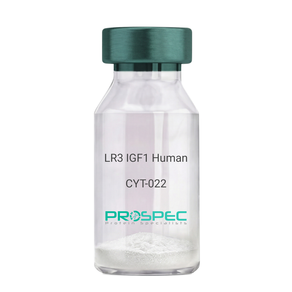
Name :
IGF1 LR3 HumanDescription:
LR3 Insulin Like Growth Factor-1 Human Recombinant
R3 IGF1, R3 IGF-1, R3IGF1, R3IGF-1, LONG IGF1, LONG IGF-1, LONG R3 IGF1, LONG R3IGF1, LONG R3 IGF-1, LONG R3IGF-1.
Product # :
CYT-022Price :
Quantity :
Shipping Method :
Shipped at Room temp
More Info
- description
- source
- formulation
- purity
- biological activity
- More Info
- sds-page
Description
The LR3 is a long-term analog of human IGF-1, specifically designed and manufactured for mammalian cell culture to support large-scale manufacturing of recombinant biopharmaceuticals. Recombinant Human LR3 Insulin Like Growth Factor-1 produced in E.Coli is a single, non-glycosylated, polypeptide chain containing 83 amino acids and having a molecular mass of 9.1kDa.
Source
Escherichia Coli.
Formulation
Lyophilized from a 0.2µm filtered concentrated solution in 20mM PB, pH 7.2.
Purity
Greater than 97.0% as determined by SDS-PAGE.
Biological Activity
The ED50 as determined by the stimulation of protein synthesis in L6 myoblasts is less than 10ng/ml, corresponding to a specific activity of 100,000units/mg.sds-page
More Info
-
Introduction
IGF-1 (Insulin-like growth factor-1) is a major hormonal mediator of statural growth. Under regular circumstances, GH (growth hormone) binds to its receptor in the liver, and other tissues, and stimulates the synthesis/secretion of IGF-1. In target tissues, the Type 1 IGF receptor, that is homologous to the insulin receptor, is activated by IGF-1, leading to intracellular signaling which stimulates multiple processes leading to statural growth. IGF-1 metabolic actions are partly directed at stimulating the uptake of glucose, fatty acids, and amino acids so that metabolism supports growing tissues.
-
Synonyms
R3 IGF1, R3 IGF-1, R3IGF1, R3IGF-1, LONG IGF1, LONG IGF-1, LONG R3 IGF1, LONG R3IGF1, LONG R3 IGF-1, LONG R3IGF-1.
-
Physical Appearance
Sterile Filtered White lyophilized (freeze-dried) powder.
-
Stability
Lyophilized LR3 IGF1 although stable at room temperature for 3 weeks, should be stored desiccated below -18°C. Upon reconstitution the LR3 IGF1 should be stored at 4°C between 2-7 days and for future use below -18°C.For long term storage it is recommended to add a carrier protein (0.1% HSA or BSA).Please prevent freeze-thaw cycles.
-
Solubility
It is recommended to reconstitute the lyophilized LR3 IGF1 in sterile 18M-cm H2O at a concentration of 100µg/ml, which can then be further diluted to other aqueous solutions.
-
Amino Acid Sequence
MFPAMPLSSLFVNGPRTLCGAELVDALQFVCGDRGFYFNKPTGYGSSSRRAPQTGIV DECCFRSCDLRRLEMYCAPLKPAKSA.
-
Background
What is the molecular weight/Mw of IGF1 LR3 HUMAN Protein?
IGF1 LR3 HUMAN Protein has a total Mw of 9.1kDa.
What is the source or expression system of IGF1 LR3 HUMAN Protein?
Escherichia Coli.
What is the Purity of IGF1 LR3 HUMAN Protein?
IGF1 LR3 HUMAN Protein is >98% pure as determined by SDS-PAGE.
What is the Biological Activity of IGF1 LR3 HUMAN Protein?
The ED50 as determined by the stimulation of protein synthesis in L6 myoblasts is less than 10ng/ml, corresponding to a specific activity of 100,000units/mg.
What is the amino acid sequence of IGF1 LR3 HUMAN Protein?
MFPAMPLSSLFVNGPRTLCGAELVDALQFVCGDRGFYFNKPTGYGSSSRRAPQTGIV DECCFRSCDLRRLEMYCAPLKPAKSA.
What applications can IGF1 LR3 HUMAN Protein be used in?
IGF1 LR3 HUMAN Protein can probably be used in western blot, ELISA and Lateral Flow.
What is the endotoxin level for IGF1 LR3 HUMAN Protein?
The endotoxin level is minimal, IGF1 LR3 HUMAN Protein was purified using conventional chromatography techniques.
ProSpec's products are furnished for LABORATORY RESEARCH USE ONLY. The product may not be used as drugs, agricultural or pesticidal products, food additives or household chemicals.
- View Data Sheet
Name :
HK2 AntibodyDescription:
Hexokinase-2, Mouse Anti Human
Hexokinase-2, EC 2.7.1.1, HK2, Hexokinase type II, HK II, Muscle form hexokinase, HXK2, DKFZp686M1669.
Product # :
ANT-378Price :
Quantity :
Shipping Method :
Shipped with Ice Packs
More Info
- formulation
- More Info
Formulation
1mg/ml containing PBS, pH-7.4, & 0.1% Sodium Azide.
More Info
-
Introduction
Hexokinases phosphorylate glucose to produce glucose-6-phosphate, thus committing glucose to the glycolytic pathway. Hexokinase 2 is the predominant form found in skeletal muscle. It localizes to the outer membrane of mitochondria. Expression of this gene is insulin-responsive, and studies in rat suggest that it is involved in the increased rate of glycolysis seen in rapidly growing cancer cells.
-
Synonyms
Hexokinase-2, EC 2.7.1.1, HK2, Hexokinase type II, HK II, Muscle form hexokinase, HXK2, DKFZp686M1669.
-
Immunogen
Anti-human Hexokinase-2 mAb is derived from hybridization of mouse F0 myeloma cells with spleen cells from BALB/c mice immunized with recombinant human Hexokinase-2 amino acids 1-917 purified from E. coli.
-
Ig Subclass
Mouse IgG1 heavy chain and κ light chain.
-
Clone
P1A7AT.
-
Applications
Hexokinase-2 antibody has been tested by ELISA and Western blot analysis to assure specificity and reactivity. Since application varies, however, each investigation should be titrated by the reagent to obtain optimal results. Recommended dilution range for Western blot analysis is 1:1,000 ~ 3,000. Recommended starting dilution is 1:2,000.
-
Type
Mouse Anti Human Monoclonal.
-
Storage Procedures
For periods up to 1 month store at 4°C, for longer periods of time, store at -20°C. Prevent freeze thaw cycles.
-
Purification Method
Hexokinase-2 antibody was purified from mouse ascitic fluids by protein-G affinity chromatography.
ProSpec's products are furnished for LABORATORY RESEARCH USE ONLY. The product may not be used as drugs, agricultural or pesticidal products, food additives or household chemicals.
- View Data Sheet
Name :
IGFBP2 MouseDescription:
Insulin Like Growth Factor Binding Protein-2 Mouse Recombinant
IBP-2, IGF-binding protein 2, IGFBP-2, mIGFBP-2, Igfbp-2, insulin-like growth factor binding protein 2 isoform 1, insulin-like growth factor binding protein 2.
Product # :
CYT-1229Price :
Quantity :
Shipping Method :
Shipped with Ice Packs
More Info
- description
- source
- formulation
- purity
- biological activity
- More Info
- SDS-PAGE
Description
IGFBP2 Mouse Recombinant produced in HEK293 cells is a single, glycosylated polypeptide chain (a.a 35-305) containing 277 amino acids and having a molecular mass of 30.3kDa. IGFBP2 is fused to a 6 amino acid His tag at C-terminus and purified by proprietary chromatographic techniques.
Source
HEK293 cells.
Formulation
IGFBP2 protein (1mg/ml) contains Phosphate-Buffered Saline (pH 7.4) and 10% glycerol.
Purity
Greater than 85.0% as determined by SDS-PAGE.
Biological Activity
The ED50 range is ≤ 0.7 ug/ml which measured by its ability to inhibit proliferation using MCF-7 human breast cancer cells in the presence of Mouse IGF-2.
SDS-PAGE
More Info
-
Synonyms
IBP-2, IGF-binding protein 2, IGFBP-2, mIGFBP-2, Igfbp-2, insulin-like growth factor binding protein 2 isoform 1, insulin-like growth factor binding protein 2.
-
Physical Appearance
Sterile Filtered colorless solution.
-
Stability
Store at 4°C if entire vial will be used within 2-4 weeks. Store, frozen at -20°C for longer periods of time. For long term storage it is recommended to add a carrier protein (0.1% HSA or BSA). Avoid multiple freeze-thaw cycles.
-
Amino Acid Sequence
EVLFRCPPCT PERLAACGPP PDAPCAELVR EPGCGCCSVC ARQEGEACGV YIPRCAQTLR CYPNPGSELP LKALVTGAGT CEKRRVGTTP QQVADSDDDH SEGGLVENHV DGTMNMLGGG SSAGRKPLKS GMKELAVFRE KVNEQHRQMG KGAKHLSLEE PKKLRPPPAR TPCQQELDQV LERISTMRLP DDRGPLEHLY SLHIPNCDKH GRYNLKQCKM SLNGQRGECW CVNPNTGKPI QGAPTIRGDP ECHLFYNEQQ ETGGAHAQSV QHHHHHH.
-
Background
Insulin-like growth factor-binding protein 2 (IGFBP2) is a key regulator of insulin-like growth factor (IGF) signaling pathway, playing crucial roles in cell proliferation, differentiation, and survival. IGFBP2, a member of the IGFBP family, modulates the bioavailability and activity of IGFs by binding to them and regulating their interaction with cell surface receptors. In recent years, IGFBP2 has emerged as a promising target for research due to its involvement in various physiological processes and its implications in several diseases, including cancer, metabolic disorders, and neurodegenerative conditions.
What is the molecular weight/Mw of IGFBP2 MOUSE Protein?
IGFBP2 MOUSE Protein has a total Mw of 30.3kDa.
What is the source or expression system of IGFBP2 MOUSE Protein?
HEK293 cells.
What is the Purity of IGFBP2 MOUSE Protein?
IGFBP2 MOUSE Protein is >85% pure as determined by SDS-PAGE.
What is the Biological Activity of IGFBP2 MOUSE Protein?
The ED50 range is ≤ 0.7 ug/ml which measured by its ability to inhibit proliferation using MCF-7 human breast cancer cells in the presence of Mouse IGF-2.
What is the amino acid sequence of IGFBP2 MOUSE Protein?
EVLFRCPPCT PERLAACGPP PDAPCAELVR EPGCGCCSVC ARQEGEACGV YIPRCAQTLR CYPNPGSELP LKALVTGAGT CEKRRVGTTP QQVADSDDDH SEGGLVENHV DGTMNMLGGG SSAGRKPLKS GMKELAVFRE KVNEQHRQMG KGAKHLSLEE PKKLRPPPAR TPCQQELDQV LERISTMRLP DDRGPLEHLY SLHIPNCDKH GRYNLKQCKM SLNGQRGECW CVNPNTGKPI QGAPTIRGDP ECHLFYNEQQ ETGGAHAQSV QHHHHHH.
What applications can IGFBP2 MOUSE Protein be used in?
IGFBP2 MOUSE Protein can probably be used in western blot, ELISA and Lateral Flow.
What is the endotoxin level for IGFBP2 MOUSE Protein?
The endotoxin level is minimal, IGFBP2 MOUSE Protein was purified using conventional chromatography techniques.
ProSpec's products are furnished for LABORATORY RESEARCH USE ONLY. The product may not be used as drugs, agricultural or pesticidal products, food additives or household chemicals.
- View Data Sheet
Name :
DCXR Human, BioactiveDescription:
Dicarbonyl/L-Xylulose Reductase Human Recombinant, Bioactive
DCR, HCR2, HCRII, KIDCR, P34H, SDR20C1, Dicarbonyl/L-Xylulose Reductase, EC=1.1.1.10, Carbonyl reductase II, Kidney dicarbonyl reductase, Sperm surface protein P34H.
Product # :
ENZ-1029Price :
Quantity :
Shipping Method :
Shipped with Ice Packs
More Info
- description
- source
- formulation
- purity
- biological activity
- More Info
Description
DCXR Recombinant Human produced in E.Coli is a single, non-glycosylated polypeptide chain containing 264 amino acids (1-244 a.a.) and having a molecular mass of 28 kDa. The DCXR is fused to a 20 amino acids His-Tag at N-terminus and purified by proprietary chromatographic techniques.
Source
Escherichia Coli.
Formulation
The DCXR (0.5mg/ml) solution containing 20mM Tris-HCl buffer (pH 8.0), 1mM DTT, 20% glycerol and 50mM NaCl.
Purity
Greater than 95.0% as determined by SDS-PAGE.
Biological Activity
Specific activity is > 1,800 pmol/min/ug and is defined as the amount of enzyme that oxidize 1pmole of xylitol to L-xylulose per minute at pH 10.0 at 37C.More Info
-
Introduction
DCXR catalyzes the NADPH-dependent reduction of numerous pentoses, tetroses, trioses, alpha-dicarbonyl molecules and L-xylulose. DCXR takes part in the uronate cycle of glucose metabolism. DCXR participates in the water absorption and cellular osmoregulation in the proximal renal tubules by producing xylitol, an osmolyte, thus preventing osmolytic stress from occurring in the renal tubules.
-
Synonyms
DCR, HCR2, HCRII, KIDCR, P34H, SDR20C1, Dicarbonyl/L-Xylulose Reductase, EC=1.1.1.10, Carbonyl reductase II, Kidney dicarbonyl reductase, Sperm surface protein P34H.
-
Physical Appearance
Sterile filtered colorless solution.
-
Stability
Store at 4°C if entire vial will be used within 2-4 weeks. Store, frozen at -20°C for longer periods of time. For long term storage it is recommended to add a carrier protein (0.1% HSA or BSA).Avoid multiple freeze-thaw cycles.
-
Amino Acid Sequence
MGSSHHHHHH SSGLVPRGSH MELFLAGRRV LVTGAGKGIG RGTVQALHAT GARVVAVSRT QADLDSLVRE CPGIEPVCVD LGDWEATERA LGSVGPVDLL VNNAAVALLQ PFLEVTKEAF DRSFEVNLRA VIQVSQIVAR GLIARGVPGA IVNVSSQCSQ RAVTNHSVYC STKGALDMLT KVMALELGPH KIRVNAVNPT VVMTSMGQAT WSDPHKAKTM LNRIPLGKFA EVEHVVNAIL FLLSDRSGMT TGSTLPVEGG FWAC.
ProSpec's products are furnished for LABORATORY RESEARCH USE ONLY. The product may not be used as drugs, agricultural or pesticidal products, food additives or household chemicals.
- View Data Sheet
Name :
FGF 9 MouseDescription:
Fibroblast Growth Factor-9 Mouse Recombinant
GAF (Glia-activating factor), HBGF-9, MGC119914, MGC119915, FGF-9.
Product # :
CYT-349Price :
Quantity :
Shipping Method :
Shipped at Room temp
More Info
- description
- source
- formulation
- purity
- biological activity
- More Info
Description
Fibroblast Growth Factor-9 Mouse Recombinant produced in E.Coli is a single, non-glycosylated, polypeptide chain containing 205 amino acids and having a molecular mass of 23308 Dalton.The FGF-9 Mouse Recombinant is purified by proprietary chromatographic techniques.
Source
Escherichia Coli.
Formulation
The protein was lyophilized from 10mM Tris, pH 8.0, 0.15M Amonium Sulfate.
Purity
Greater than 95.0% as determined by:
(a) Analysis by RP-HPLC.
(b) Analysis by SDS-PAGE.Biological Activity
The ED50, calculated by the dose-dependant proliferation of BAF3 cells expressing FGF receptors (measured by 3H-thymidine uptake) is <0.5 ng/ml, corresponding to a specific activity of 2MU/mg.More Info
-
Introduction
Rat and mouse FGF-9 show a very high homology to human FGF-9. The transcripts for FGF-9 have been found in brain and in kidney tissue. Fibroblast Growth Factor-9 is a member of the fibroblast growth factor (FGF) family. FGF family members possess broad mitogenic and cell survival activities, and are involved in a variety of biological processes, including embryonic development, cell growth, morphogenesis, tissue repair, tumor growth and invasion. FGF9 was isolated as a secreted factor that exhibits a growth-stimulating effect on cultured glial cells. In nervous system, this protein is produced mainly by neurons and may be important for glial cell development. Expression of the mouse homolog of this gene was found to be dependent on Sonic hedgehog (Shh) signaling. Mice lacking the homolog gene displayed a male-to-female sex reversal phenotype, which suggested a role in testicular embryogenesis Fibroblast Growth Factor 9 may have a role in glial cell growth and differentiation during development, gliosis during repair and regeneration of brain tissue after damage, differentiation and survival of neuronal cells, and growth stimulation of glial tumors.
-
Synonyms
GAF (Glia-activating factor), HBGF-9, MGC119914, MGC119915, FGF-9.
-
Physical Appearance
Sterile Filtered White lyophilized (freeze-dried) powder.
-
Stability
Lyophilized Fibroblast Growth Factor-9 although stable at room temperature for 3 weeks, should be stored desiccated below -18°C. Upon reconstitution FGF9 Mouse Recombinant should be stored at 4°C between 2-7 days and for future use below -18°C.For long term storage it is recommended to add a carrier protein (0.1% HSA or BSA).Please prevent freeze-thaw cycles.
-
Solubility
It is recommended to reconstitute the lyophilized Fibroblast Growth Factor 9 in sterile 18MΩ-cm H2O not less than 100µg/ml, which can then be further diluted to other aqueous solutions.
-
Amino Acid Sequence
The sequence of the first five N-terminal amino acids was determined and was found to be Pro-Leu-Gly-Glu-Val.
-
Background
What is the molecular weight/Mw of FGF9 Protein?
FGF9 Protein has a total Mw of 23.3kDa.
What is the source or expression system of FGF9 Protein?
Escherichia Coli.
What is the Purity of FGF9 Protein?
FGF9 Protein is >95% pure as determined by SDS-PAGE.
What is the Biological Activity of FGF9 Protein?
The ED50, calculated by the dose-dependant proliferation of BAF3 cells expressing FGF receptors (measured by 3H-thymidine uptake) is <0.5 ng/ml, corresponding to a specific activity of 2MU/mg.
What is the amino acid sequence of FGF9 Protein?
FGF9 Protein is composed from 205 amino acids.
What applications can FGF9 Protein be used in?
FGF9 Protein can probably be used in western blot, ELISA and Lateral Flow.
What is the endotoxin level for FGF9 Protein?
The endotoxin level is minimal, FGF9 Protein was purified using conventional chromatography techniques.
ProSpec's products are furnished for LABORATORY RESEARCH USE ONLY. The product may not be used as drugs, agricultural or pesticidal products, food additives or household chemicals.
- View Data Sheet
Name :
p38a/SAPK2 HumanDescription:
p38a/SAPK2 Human Recombinant
Mitogen-activated protein kinase 14, EC 2.7.11.24, Mitogen-activated protein kinase p38 alpha, MAP kinase p38 alpha, Cytokine suppressive anti-inflammatory drug-binding protein, CSAID-binding protein, CSBP, MAX-interacting protein 2, MAP kinase MXI2, SAPK2A, RK, p38, EXIP, Mxi2, CSBP1, CSBP2, CSPB1, PRKM14, PRKM15, p38ALPHA.
Product # :
PKA-217Price :
Quantity :
Shipping Method :
Shipped with Ice Packs
More Info
- description
- source
- formulation
- purity
- More Info
Description
p38/SAPK2 is a non-glycosilated polypeptide produced by phosphorylation of the purified p38 alpha with MKK6 having a molecular mass of 42.7 kDa.
Source
Escherichia Coli.
Formulation
p38/SAPK2 is supplied in 25mM Tris-HCl, 150mM NaCl, 1mM DTT, 50% glycerol, pH 8.5.
Purity
Greater than 95% as determined by SDS-PAGE.
More Info
-
Introduction
p38a/SAPK2 is a member of the MAP kinase family. MAP kinases act as an integration point for multiple biochemical signals, and are involved in a wide variety of cellular processes such as proliferation, differentiation, transcription regulation and development. This kinase is activated by various environmental stresses and proinflammatory cytokines. The activation requires its phosphorylation by MAP kinase kinases (MKKs), or its autophosphorylation triggered by the interaction of MAP3K7IP1/TAB1 protein with this kinase. The substrates of this kinase include transcription regulator ATF2, MEF2C, and MAX, cell cycle regulator CDC25B, and tumor suppressor p53, which suggest the roles of this kinase in stress related transcription and cell cycle regulation, as well as in genotoxic stress response. Four alternatively spliced transcript variants of this gene encoding distinct isoforms have been reported.
-
Synonyms
Mitogen-activated protein kinase 14, EC 2.7.11.24, Mitogen-activated protein kinase p38 alpha, MAP kinase p38 alpha, Cytokine suppressive anti-inflammatory drug-binding protein, CSAID-binding protein, CSBP, MAX-interacting protein 2, MAP kinase MXI2, SAPK2A, RK, p38, EXIP, Mxi2, CSBP1, CSBP2, CSPB1, PRKM14, PRKM15, p38ALPHA.
-
Physical Appearance
Sterile Filtered clear solution.
-
Stability
Store at 4°C if entire vial will be used within 2-4 weeks. Store, frozen at -20°C for longer periods of time. Avoid multiple freeze-thaw cycles.
-
Unit Definition
No protease activity detectable, specific activity is 2.357.900 U*/mg (*1 U = 1 pmol/min transferred to myelin basic protein at 30°C).
ProSpec's products are furnished for LABORATORY RESEARCH USE ONLY. The product may not be used as drugs, agricultural or pesticidal products, food additives or household chemicals.
- View Data Sheet
Name :
FLT1 D7 HumanDescription:
Vascular Endothelial Growth Factor Receptor-1 D1-7 Human Recombinant
FLT-1, FLT1, Tyrosine-protein kinase receptor FLT, Flt-1, Tyrosine-protein kinase FRT, Fms-like tyrosine kinase 1, VEGFR-1.
Product # :
PKA-241Price :
Quantity :
Shipping Method :
Shipped at Room temp
More Info
- description
- source
- formulation
- purity
- biological activity
- More Info
Description
Soluble FLT1 Human Recombinant fused with the Fc part of human IgG1 produced in baculovirus is disulfide-linked homodimeric, glycosylated, polypeptide containing 751 amino acids and having a molecular mass of 130 kDa. The soluble receptor protein contains only the first 7 extracellular domains (Met1-Thr751), which contain all the information necessary for high affinity ligand binding. The FLT1 fc/Chimera is purified by proprietary chromatographic techniques.
Source
Insect Cells.
Formulation
FLT1 D1-7 was lyophilized from a concentrated (1 mg/ml) sterile solution containing PBS Buffer, pH 7.4.
Purity
Greater than 95.0% as determined by SDS-PAGE.
Biological Activity
The activity of FLT1/Fc was determined by its ability to inhibit the VEGF-dependent proliferation of human umbilical vein endothelial cells. The ED50 for this effect is typically 10-30 ng/ml, corresponding to a specific activity of 33,333.33-100,000 units/mg.More Info
-
Introduction
Endothelial cells express three different vascular endothelial growth factor (VEGF) receptors, belonging to the family of receptor tyrosine kinases (RTKs). They are named VEGFR-1 (Flt-1), VEGFR-2 (KDR/Flk-1), and VEGFR-3 (Flt-4). Their expression is almost exclusively restricted to endothelial cells, but VEGFR-1 can also be found on monocytes. All VEGF-receptors have seven immunoglobulin-like extracellular domains, a single transmembrane region and an intracellular split tyrosine kinase domain. VEGFR-2 has a lower affinity for VEGF than the Flt-1 receptor, but a higher signalling activity. Mitogenic activity in endothelial cells is mainly mediated by VEGFR-2 leading to their proliferation. Differential splicing of the flt-1 gene leads to the formation of a secreted, soluble variant of VEGFR-1 (sVEGFR-1). No naturally occurring, secreted forms of VEGFR-2 have so far been reported. The binding of VEGF165 to VEGFR-2 is dependent on heparin.
-
Synonyms
FLT-1, FLT1, Tyrosine-protein kinase receptor FLT, Flt-1, Tyrosine-protein kinase FRT, Fms-like tyrosine kinase 1, VEGFR-1.
-
Physical Appearance
Sterile Filtered White lyophilized (freeze-dried) powder.
-
Stability
Lyophilized FLT-1 although stable at room temperature for 3 weeks, should be stored desiccated below -18C. Upon reconstitution FLT1 should be stored at 4C between 2-7 days and for future use below -18C. For long term storage it is recommended to add a carrier protein (0.1% HSA or BSA). Please prevent freeze-thaw cycles.
-
Solubility
It is recommended to reconstitute the lyophilized FLT1 Fc/Chimera in PBS not less than 50µg/ml, which can then be further diluted to other aqueous solutions.
-
Amino Acid Sequence
MVSYWDTGVL LCALLSCLLL TGSSSGSKLK DPELSLKGTQ HIMQAGQTLH LQCRGEAAHK WSLPEMVSKE SERLSITKSA CGRNGKQFCS TLTLNTAQAN HTGFYSCKYL AVPTSKKKET ESAIYIFISD TGRPFVEMYS EIPEIIHMTE GRELVIPCRV TSPNITVTLK KFPLDTLIPD GKRIIWDSRK GFIISNATYK EIGLLTCEAT VNGHLYKTNY LTHRQTNTII DVQISTPRPV KLLRGHTLVL NCTATTPLNT RVQMTWSYPD EKNKRASVRR RIDQSNSHAN IFYSVLTIDK MQNKDKGLYT CRVRSGPSFK SVNTSVHIYD KAFITVKHRK QQVLETVAGK RSYRLSMKVK AFPSPEVVWL KDGLPATEKS ARYLTRGYSL IIKDVTEEDA GNYTILLSIK QSNVFKNLTA TLIVNVKPQI YEKAVSSFPD PALYPLGSRQ ILTCTAYGIP QPTIKWFWHP CNHNHSEARC DFCSNNEESF ILDADSNMGN RIESITQRMA IIEGKNKMAS TLVVADSRIS GIYICIASNK VGTVGRNISF YITDVPNGFH VNLEKMPTEG EDLKLSCTVN KFLYRDVTWI LLRTVNNRTM HYSISKQKMA ITKEHSITLN LTIMNVSLQD SGTYACRARN VYTGEEILQK KEITIRDQEA PYLLRNLSDH TVAISSSTTL DCHANGVPEP QITWFKNNHK IQQEPGIILG PGSSTLFIER VTEEDEGVYH CKATNQKGSV ESSAYLTVQG TAASDKTHTC PPCPAPELLG GPSVFLFPPK PKDTLMISRT PEVTCVVVDV SHEDPEVKFN WYVDGVEVHN AKTKPREEQY NSTYRVVSVL TVLHQDWLNG KEYKCKVSNK ALPAPIEKTI S.
ProSpec's products are furnished for LABORATORY RESEARCH USE ONLY. The product may not be used as drugs, agricultural or pesticidal products, food additives or household chemicals.