Search results
1000 results found for “BATF”
Name
Description
Product #
Price
Quantity
Shipping Method
- View Data Sheet
Name :
TSTD3 HumanDescription:
Thiosulfate Sulfurtransferase Like Domain Containing 3 Human Recombinant
Thiosulfate Sulfurtransferase (Rhodanese)-Like Domain Containing 3, Rhodanese Domain-Containing Protein 3, Thiosulfate Sulfurtransferase/Rhodanese-Like Domain-Containing Protein 3.
Product # :
ENZ-723Price :
Quantity :
Shipping Method :
Shipped with Ice Packs
More Info
- description
- source
- formulation
- purity
- More Info
Description
TSTD3 Human Recombinant produced in E.Coli is a single, non-glycosylated polypeptide chain containing 120 amino acids (1-97 a.a) and having a molecular mass of 13.7kDa.TSTD3 is fused to a 23 amino acid His-tag at N-terminus & purified by proprietary chromatographic techniques.
Source
Escherichia Coli.
Formulation
TSTD3 protein solution (1mg/ml) containing 20mM Tris-HCl buffer (pH 8.0), 0.15M NaCl, 10% glycerol and 1mM DTT.
Purity
Greater than 90.0% as determined by SDS-PAGE.
More Info
-
Introduction
Thiosulfate Sulfurtransferase Like Domain Containing 3, also known as TSTD3 contains 1 rhodanese domain which is characterized by an active site cysteine residue that has the ability to bind sulfane sulfur and catalyse sulfur transfer.
-
Synonyms
Thiosulfate Sulfurtransferase (Rhodanese)-Like Domain Containing 3, Rhodanese Domain-Containing Protein 3, Thiosulfate Sulfurtransferase/Rhodanese-Like Domain-Containing Protein 3.
-
Physical Appearance
Sterile Filtered clear solution.
-
Stability
Store at 4°C if entire vial will be used within 2-4 weeks. Store, frozen at -20°C for longer periods of time. For long term storage it is recommended to add a carrier protein (0.1% HSA or BSA).Avoid multiple freeze-thaw cycles.
-
Amino Acid Sequence
MGSSHHHHHH SSGLVPRGSH MGSMKIEKCG WSEGLTSIKG NCHNFYTAIS KDVTYKELKN LLNSKNIMLI DVREIWEILE YQKIPESINV PLDEVGEALQ MNPRDFKEKY NEVKPSKSDS
ProSpec's products are furnished for LABORATORY RESEARCH USE ONLY. The product may not be used as drugs, agricultural or pesticidal products, food additives or household chemicals.
- View Data Sheet
Name :
LGALS7 MouseDescription:
Galectin-7 Mouse Recombinant
Galectin-7, Gal-7, HKL-14, PI7, p53-induced gene 1 protein, LGALS7, PIG1, LGALS7B, GAL7, LGALS7A.
Product # :
CYT-184Price :
Quantity :
Shipping Method :
Shipped with Ice Packs
More Info
- description
- source
- formulation
- purity
- More Info
- SDS-PAGE
Description
LGALS7 mouse Recombinant produced E. coli is a single polypeptide chain containing 159 amino acids (1-136) and having a molecular mass of 17.6kDa.LGALS7 is fused to a 23 amino acid His-tag at N-terminus & purified by proprietary chromatographic techniques.
Source
Escherichia Coli.
Formulation
The LGALS7 solution (1mg/ml) contains 20mM Tris-HCl buffer (pH 8.0), 0.1M NaCl, 20% glycerol and 2mM DTT.
Purity
Greater than 95.0% as determined by SDS-PAGE.
SDS-PAGE
More Info
-
Introduction
Galectins are a family of animal lectins with an affinity for beta-galactosides. This family has at least 14 identified members. Galectins share similarities in the CRD (the carbohydrate recognition domain). Galectins are synthesized as cytosolic proteins. Though localized principally in the cytoplasm and lacking a classical signal peptide, galectins can also be stimulated to secretion by non-classical pathways or alternatively targeted to the nucleus. Galectins are involved in modulating cell-cell and cell-matrix interactions. Human Galectin-7 belongs to the prototypical Galectins containing a single CRD, which is initially identified in human epidermis as a monomer. The Galectin-7 expression is induced by tumor suppressor protein p53 and associated with apoptosis. Galectin-7 is a pro-apoptotic protein which functions intracellularlly upstream of JNK activation and mitochondrial cytochrome c release. The correlation of Galectin-7 with the UV-induced apoptosis of keratinocytes presents a critical mechanism in the maintenance of epidermal homeostasis. Human Galectin-7 is localized in both nucleus and cytoplasm.
-
Synonyms
Galectin-7, Gal-7, HKL-14, PI7, p53-induced gene 1 protein, LGALS7, PIG1, LGALS7B, GAL7, LGALS7A.
-
Physical Appearance
Sterile Filtered colorless solution.
-
Stability
Store at 4°C if entire vial will be used within 2-4 weeks. Store, frozen at -20°C for longer periods of time. For long term storage it is recommended to add a carrier protein (0.1% HSA or BSA).Avoid multiple freeze-thaw cycles.
-
Amino Acid Sequence
MGSSHHHHHH SSGLVPRGSH MGSMSATQHK TSLPQGVRVG TVMRIRGMVP DQAGRFHVNL LCGEEQGADA ALHFNPRLDT SEVVFNTKEQ GKWGREERGT GIPFERGQPF EVLLIATEEG FKAVVGDDEY LHFHHRMPPA RVRLVEVGGD VQLHSVKIF.
-
Background
What is the molecular weight/Mw of LGALS7 MOUSE Protein?
LGALS7 MOUSE Protein has a total Mw of 17.6kDa.
What is the source or expression system of LGALS7 MOUSE Protein?
Escherichia Coli.
What is the Purity of LGALS7 MOUSE Protein?
LGALS7 MOUSE Protein is >95% pure as determined by SDS-PAGE.
What is the Biological Activity of LGALS7 MOUSE Protein?
The biological functionality of LGALS7 MOUSE Protein will be determined in the future.
What is the amino acid sequence of LGALS7 MOUSE Protein?
MGSSHHHHHH SSGLVPRGSH MGSMSATQHK TSLPQGVRVG TVMRIRGMVP DQAGRFHVNL LCGEEQGADA ALHFNPRLDT SEVVFNTKEQ GKWGREERGT GIPFERGQPF EVLLIATEEG FKAVVGDDEY LHFHHRMPPA RVRLVEVGGD VQLHSVKIF.
What applications can LGALS7 MOUSE Protein be used in?
LGALS7 MOUSE Protein can probably be used in western blot, ELISA and Lateral Flow.
What is the endotoxin level for LGALS7 MOUSE Protein?
The endotoxin level is minimal, LGALS7 MOUSE Protein was purified using conventional chromatography techniques.
ProSpec's products are furnished for LABORATORY RESEARCH USE ONLY. The product may not be used as drugs, agricultural or pesticidal products, food additives or household chemicals.
- View Data Sheet
Name :
PRTFDC1 HumanDescription:
Phosphoribosyl Transferase Domain Containing 1 Human Recombinant
Phosphoribosyltransferase domain-containing protein 1, PRTFDC1, HHGP.
Product # :
ENZ-142Price :
Quantity :
Shipping Method :
Shipped with Ice Packs
More Info
- description
- source
- formulation
- purity
- More Info
Description
PRTFDC1 produced in E.Coli is a single, non-glycosylated polypeptide chain containing 248 amino acids (1-225 a.a.) and having a molecular mass of 28.1kDa.PRTFDC1 is fused to a 23 amino acid His-tag at N-terminus & purified by proprietary chromatographic techniques.
Source
Escherichia Coli.
Formulation
PRTFDC1 protein solution (1mg/ml) containing 20mM Tris-HCl buffer (pH8.0), 40% glycerol, 0.1M NaCl and 1mM DTT.
Purity
Greater than 95.0% as determined by SDS-PAGE.
More Info
-
Introduction
Phosphoribosyltransferase domain-containing protein 1 (PRTFDC1) is a member of the purine/pyrimidine phosphoribosyltransferase family. PRTFDC1 has a low, barely measurable phosphoribosyltransferase activity (in vitro). PRTFDC1 can bind GMP, IMP and alpha-D-5-phosphoribosyl 1-pyrophosphate (PRPP). PRTFDC1 is not expected to impact purine metabolism or GMP salvage.
-
Synonyms
Phosphoribosyltransferase domain-containing protein 1, PRTFDC1, HHGP.
-
Physical Appearance
Sterile Filtered colorless solution.
-
Stability
PRTFDC1 Human Recombinant although stable at 4°C for 1 week, should be stored below -18°C. Please prevent freeze thaw cycles.
-
Amino Acid Sequence
MGSSHHHHHH SSGLVPRGSH MGSMAGSSEE APDYGRGVVI MDDWPGYDLN LFTYPQHYYG DLEYVLIPHG IIVDRIERLA KDIMKDIGYS DIMVLCVLKG GYKFCADLVE HLKNISRNSD RFVSMKVDFI RLKSYRNDQS MGEMQIIGGD DLSTLAGKNV LIVEDVVGTG RTMKALLSNI EKYKPNMIKV ASLLVKRTSR SDGFRPDYAG FEIPNLFVVG YALDYNEYFR DLNHICVINE HGKEKYRV.
ProSpec's products are furnished for LABORATORY RESEARCH USE ONLY. The product may not be used as drugs, agricultural or pesticidal products, food additives or household chemicals.
- View Data Sheet
Name :
CNPY3 HumanDescription:
Canopy 3 Homolog Human Recombinant
CAG4A, ERDA5, PRAT4A, TNRC5, Protein canopy homolog 3, CTG repeat protein 4a, Expanded repeat-domain protein CAG/CTG 5, Protein associated with TLR4, Trinucleotide repeat-containing gene 5 protein,CNPY3.
Product # :
PRO-1386Price :
Quantity :
Shipping Method :
Shipped with Ice Packs
More Info
- description
- source
- formulation
- purity
- More Info
Description
CNPY3 Human Recombinant produced in E.Coli is a single, non-glycosylated polypeptide chain containing 271 amino acids (31-278 a.a.) and having a molecular mass of 29.9kDa. CNPY3 is fused to a 23 amino acid His-tag at N-terminus & purified by proprietary chromatographic techniques.
Source
E.coli.
Formulation
CNPY3 protein solution (0.5mg/ml) contains 20mM Tris-HCl buffer (pH 8.0), 0.15M NaCl, 10% glycerol and 1mM DTT.
Purity
Greater than 85% as determined by SDS-PAGE.
More Info
-
Introduction
Canopy 3 homolog (CNPY3) is a 271 a.aToll-like receptor specific co-chaperone for HSP90B1. CNPY3 is necessitated for proper TLR folding, excluding that of TLR3, and so controls TLR exit from the endoplasmic reticulum. Therefore, CNPY3i s required for both innate and adaptive immune responses.
-
Synonyms
CAG4A, ERDA5, PRAT4A, TNRC5, Protein canopy homolog 3, CTG repeat protein 4a, Expanded repeat-domain protein CAG/CTG 5, Protein associated with TLR4, Trinucleotide repeat-containing gene 5 protein,CNPY3.
-
Physical Appearance
Sterile Filtered colorless solution.
-
Stability
Store at 4°C if entire vial will be used within 2-4 weeks. Store, frozen at -20°C for longer periods of time. For long term storage it is recommended to add a carrier protein (0.1% HSA or BSA).Avoid multiple freeze-thaw cycles.
-
Amino Acid Sequence
MGSSHHHHHH SSGLVPRGSH MGSGPSQAGA EENDWVRLPS KCEVCKYVAV ELKSAFEETG KTKEVIGTGY GILDQKASGV KYTKSDLRLI EVTETICKRL LDYSLHKERT GSNRFAKGMS ETFETLHNLV HKGVKVVMDI PYELWNETSA EVADLKKQCD VLVEEFEEVI EDWYRNHQEE DLTEFLCANH VLKGKDTSCL AEQWSGKKGD TAALGGKKSK KKSSRAKAAG GRSSSSKQRK ELGGLEGDPS PEEDEGIQKA SPLTHSPPDE L.
ProSpec's products are furnished for LABORATORY RESEARCH USE ONLY. The product may not be used as drugs, agricultural or pesticidal products, food additives or household chemicals.
- View Data Sheet
Name :
pykF E.ColiDescription:
Pyruvate Kinase I E.Coli Recombinant
Pyruvate kinase I, PK-1, pykF, b1676, JW1666.
Product # :
PKA-028Price :
Quantity :
Shipping Method :
Shipped with Ice Packs
More Info
- description
- source
- formulation
- purity
- More Info
Description
pykF E.Coli Recombinant produced in E.Coli is a single, non-glycosylated polypeptide chain containing 494 amino acids (1-470 a.a) and having a molecular mass of 53.3kDa.pykF is fused to a 24 amino acid His-tag at N-terminus & purified by proprietary chromatographic techniques.
Source
E.coli.
Formulation
pykF protein solution (1mg/ml) containing 20mM Tris-HCl buffer (pH8.0) and 10% glycerol.
Purity
Greater than 95% as determined by SDS-PAGE.
More Info
-
Introduction
pykF (pyruvate kinase I) is a member of the pyruvate kinase family. pykF is an enzyme involved in glycolysis. pykF catalyzes the transfer of a phosphate group from phosphoenolpyruvate (PEP) to ADP, yielding one molecule of pyruvate and one molecule of ATP, a process which also requires a Magnesium ion. This is the final step in the glycolytic pathway, which produces pyruvate molecules, the ultimate product of aerobic glycolysis.
-
Synonyms
Pyruvate kinase I, PK-1, pykF, b1676, JW1666.
-
Physical Appearance
Sterile Filtered colorless solution.
-
Stability
Store at 4°C if entire vial will be used within 2-4 weeks. Store, frozen at -20°C for longer periods of time. For long term storage it is recommended to add a carrier protein (0.1% HSA or BSA).Avoid multiple freeze-thaw cycles.
-
Amino Acid Sequence
MGSSHHHHHH SSGLVPRGSH MGSHMKKTKI VCTIGPKTES EEMLAKMLDA GMNVMRLNFS HGDYAEHGQR IQNLRNVMSK TGKTAAILLD TKGPEIRTMK LEGGNDVSLK AGQTFTFTTD KSVIGNSEMV AVTYEGFTTD LSVGNTVLVD DGLIGMEVTA IEGNKVICKV LNNGDLGENK GVNLPGVSIA LPALAEKDKQ DLIFGCEQGV DFVAASFIRK RSDVIEIREH LKAHGGENIH IISKIENQEG LNNFDEILEA SDGIMVARGD LGVEIPVEEV IFAQKMMIEK CIRARKVVIT ATQMLDSMIK NPRPTRAEAG DVANAILDGT DAVMLSGESA KGKYPLEAVS IMATICERTD RVMNSRLEFN NDNRKLRITE AVCRGAVETA EKLDAPLIVV ATQGGKSARA VRKYFPDATI LALTTNEKTA HQLVLSKGVV PQLVKEITST DDFYRLGKEL ALQSGLAHKG DVVVMVSGAL VPSGTTNTAS VHVL.
ProSpec's products are furnished for LABORATORY RESEARCH USE ONLY. The product may not be used as drugs, agricultural or pesticidal products, food additives or household chemicals.
- View Data Sheet
Name :
VEGI HumanDescription:
Human Vascular Endothelial Growth Inhibitor Recombinant
Tumor necrosis factor ligand superfamily member 15, TNFSF-15, TNFSF15, TNF ligand-related molecule 1, VEGI, TL-1, TL1, TL1A, VEGI192A, VEGI-192, MGC129934, MGC129935.
Product # :
CYT-517Price :
Quantity :
Shipping Method :
Shipped at Room temp
More Info
- description
- source
- formulation
- purity
- biological activity
- More Info
Description
TNFSF15 Human Recombinant produced in E.Coli is a single, non-glycosylated, polypeptide chain containing 180 amino acids and having a molecular mass of 20.5kDa. The TNFSF15 is purified by proprietary chromatographic techniques.
Source
Escherichia Coli.
Formulation
The TNFSF15 was lyophilized from a 0.2µm filtered concentrated solution in PBS, pH 7.4 with 0.02% Tween-20.
Purity
Greater than 95.0% as determined by:
(a) Analysis by RP-HPLC.
(b) Analysis by SDS-PAGE.Biological Activity
The ED50 as determined by its ability to induce apoptosis using human TF-1 cells is less than 20ng/ml, corresponding to a specific activity of > 5.0×10⁴ IU/mg.More Info
-
Introduction
TNFSF15 is a cytokine that belongs to the tumor necrosis factor (TNF) ligand family. This protein is abundantly expressed in endothelial cells, but is not expressed in either B or T cells. The expression of TNFSF15 is inducible by TNF and IL-1 alpha. This cytokine is a ligand for receptor TNFRSF25 and decoy receptor TNFRSF21/DR6. It can activate NF-kappaB and MAP kinases, and acts as an autocrine factor to induce apoptosis in endothelial cells. TNFSF15 is also found to inhibit endothelial cell proliferation, and thus may function as an angiogenesis inhibitor. An additional isoform encoded by an alternatively spliced transcript variant has been reported but the sequence of this transcript has not been determined.
-
Synonyms
Tumor necrosis factor ligand superfamily member 15, TNFSF-15, TNFSF15, TNF ligand-related molecule 1, VEGI, TL-1, TL1, TL1A, VEGI192A, VEGI-192, MGC129934, MGC129935.
-
Physical Appearance
Sterile Filtered White lyophilized (freeze-dried) powder.
-
Stability
TNFSF15 although stable at room temperature for 3 weeks, should be stored desiccated below -18°C. Upon reconstitution VEGI should be stored at 4°C between 2-7 days and for future use below -18°C.For long term storage it is recommended to add a carrier protein (0.1% HSA or BSA).Please prevent freeze-thaw cycles.
-
Solubility
It is recommended to reconstitute the lyophilized TNFSF15 in sterile 18MΩ-cm H2O not less than 100 µg/ml, which can then be further diluted to other aqueous solutions.
-
Amino Acid Sequence
MQLTKGRLHFSHPLSHTKHISPFVTDAPLRADGDKPRAHL
TVVRQTPTQHFKNQFPALHWEHELGLAFTKNRMNYTNKF
LLIPESGDYFIYSQVTFRGMTSECSEIRQAGRPNKPDSIT
VVITKVTDSYPEPTQLLMGTKSVCEVGSNWFQPIYLGAM
FSLQEGDKLMVNVSDISLVDYTKEDKTFFGAFLL.
ProSpec's products are furnished for LABORATORY RESEARCH USE ONLY. The product may not be used as drugs, agricultural or pesticidal products, food additives or household chemicals.
- View Data Sheet
Name :
rGFPDescription:
Green Fluorescent Protein Recombinant
Green fluorescent protein, GFP.
Product # :
PRO-687Price :
Quantity :
Shipping Method :
Shipped with Ice Packs
More Info
- description
- source
- formulation
- purity
- More Info
Description
rGFP Aequorea victoria produced in E.Coli is a single, non-glycosylated polypeptide chain containing 238 amino acids (1-238 a.a.) and having a molecular mass of 26.8 kDa.rGFP is purified by proprietary chromatographic techniques.
Source
Escherichia Coli.
Formulation
The Green Fluorescent Protein solution (1mg/ml) contains 20mM Tris-HCl, pH-8 and 10% Glycerol.
Purity
Greater than 95.0% as determined by SDS-PAGE.
More Info
-
Introduction
rGFP, also known as Green Fluorescent Protein, is a protein produced by the jellyfish (Aequorea Victoria) that produces bioluminescence in the green zone of the noticeable spectrum. Green Fluorescent Protein is a useful and ubiquitous instrument for producing chimeric proteins, where it functions as a fluorescent protein tag. rGFP is expressed in most known cell types and is used as a noninvasive fluorescent marker in living cells and organisms. Green Fluorescent Protein permits a broad range of applications where it has functioned as a cell lineage tracer, reporter of gene expression, or as a measure of protein-protein interactions.
-
Synonyms
Green fluorescent protein, GFP.
-
Physical Appearance
Sterile filtered yellowish solution.
-
Stability
Store at 4°C if entire vial will be used within 2-4 weeks. Store, frozen at -20°C for longer periods of time. For long term storage it is recommended to add a carrier protein (0.1% HSA or BSA).Avoid multiple freeze-thaw cycles.
-
Amino Acid Sequence
MSKGEELFTG VVPILVELDG DVNGHKFSVS GEGEGDATYG KLTLKFICTT GKLPVPWPTL VTTFSYGVQC FSRYPDHMKQ HDFFKSAMPEGYVQERTIFF KDDGNYKTRA EVKFEGDTLV NRIELKGIDF KEDGNILGHK LEYNYNSHNV YIMADKQKNG IKVNFKIRHN IEDGSVQLAD
HYQQNTPIGD GPVLLPDNHY LSTQSALSKD PNEKRDHMVL LEFVTAAGIT HGMDELYK.
ProSpec's products are furnished for LABORATORY RESEARCH USE ONLY. The product may not be used as drugs, agricultural or pesticidal products, food additives or household chemicals.
- View Data Sheet
Name :
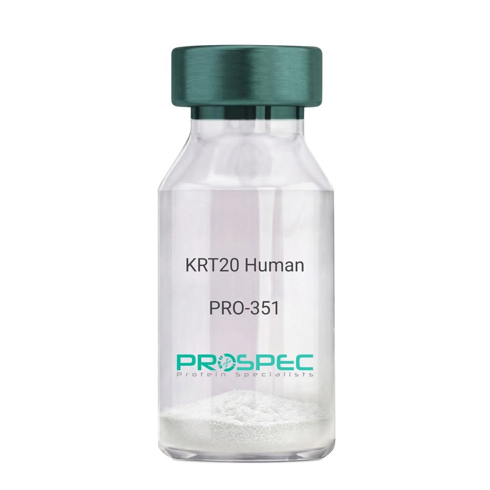
KRT20 HumanDescription:
Cytokeratin 20 Human Recombinant
Keratin type I cytoskeletal 20, Cytokeratin-20, CK-20, Keratin-20, K20, Protein IT, KRT20, CD20, CK20, KRT21, MGC35423.
Product # :
PRO-351Price :
Quantity :
Shipping Method :
Shipped at Room temp
More Info
- description
- source
- formulation
- purity
- More Info
Description
Cytokeratin 20 Human Recombinant produced in E.Coli is a single,non-glycosylated polypeptide chain having a molecular mass of 48,553 Dalton. The KRT20 is purified by proprietary chromatographic techniques.
Source
Escherichia Coli.
Formulation
The protein (1mg/1ml) was lyophilized after from a sterile solution containing 30mM Tris-HCL pH-8, 9.5M urea, 2mM DDT, 2mM EDTA and 10mM methylammonium chloride.
Purity
Greater than 95.0% as determined by SDS-PAGE.
More Info
-
Introduction
KRT20 is a member of the keratin family. The keratins are intermediate filament proteins responsible for the structural integrity of epithelial cells and are subdivided into cytokeratins and hair keratins. The type I cytokeratins consist of acidic proteins which are arranged in pairs of heterotypic keratin chains. This cytokeratin is a major cellular protein of mature enterocytes and goblet cells and is specifically expressed in the gastric and intestinal mucosa. The type I cytokeratin genes are clustered in a region of chromosome 17q12-q21.
-
Synonyms
Keratin type I cytoskeletal 20, Cytokeratin-20, CK-20, Keratin-20, K20, Protein IT, KRT20, CD20, CK20, KRT21, MGC35423.
-
Physical Appearance
Sterile Filtered White lyophilized (freeze-dried) powder.
-
Stability
Lyophilized KRT20 although stable at room temperature for 3 weeks, should be stored desiccated below -18°C. Upon reconstitution KRT20 should be stored at 4°C between 2-7 days and for future use below -18°C.For long term storage it is recommended to add a carrier protein (0.1% HSA or BSA).Please prevent freeze-thaw cycles.
-
Solubility
It is recommended to reconstitute the lyophilized KRT20 in sterile 18MΩ-cm H2O not less than 100µg/ml, which can then be further diluted to other aqueous solutions.
-
Reconstitution to filaments
Performed by mixing equimolar amounts of cytokeratins of type I and type II at concentrations of approx. 0.5 mg/ml, both dissolved in 9.5 M urea buffer (see above). Protofilaments and filament complexes are obtained by dialyzing the resulting polypeptide solution stepwise to a concentration of 4 M urea and then to low salt condition (50 mM NaCI, 2 mM dithiothreitol, 10 mM Tris-HCI, pH 7.4). For immunization purposes, the solution can be further dialyzed against PBS (phosphate buffered saline, e.g. Dulbecco's PBS).
ProSpec's products are furnished for LABORATORY RESEARCH USE ONLY. The product may not be used as drugs, agricultural or pesticidal products, food additives or household chemicals.
- View Data Sheet
Name :
LACTB E.coliDescription:
Beta Lactamase E.coli Recombinant
b-Lactamase, EC 3.5.2.6, TEM-1.
Product # :
ENZ-351Price :
Quantity :
Shipping Method :
Shipped at Room temp
More Info
- description
- source
- formulation
- purity
- biological activity
- More Info
Description
Recombinant E.coli Beta-Lactamase produced in E.Coli is a single, non-glycosylated polypeptide chain containing 264 amino acids and having a molecular mass of approximately 28.9 kDa. Beta Lactamase is purified by proprietary chromatographic techniques.
Source
Escherichia Coli.
Formulation
Lyophilized from a concentrated solution in 100mM Tris, pH7.0.
Purity
Greater than 95.0% as determined by SDS-PAGE.
Biological Activity
One unit will hydrolyze 1.0 μmole of benzyl penicillin at pH 7.0 at 25°C, in presence of EDTA.
More Info
-
Introduction
Beta-lactamase is a type of enzyme (EC 3.5.2.6) produced by some bacteria that is responsible for their resistance to beta-lactam antibiotics like penicillins, cephalosporins, cephamycins and carbapenems. These antibiotics have a common element in their molecular structure: a four-atom ring known as a beta-lactam. The lactamase enzyme breaks that ring open, deactivating the molecule's antibacterial properties.
-
Synonyms
b-Lactamase, EC 3.5.2.6, TEM-1.
-
Physical Appearance
Sterile Filtered White lyophilized (freeze-dried) powder.
-
Stability
Lyophilized Beta Lactamase although stable at room temperature for 3 weeks, should be stored desiccated below -18°C. Upon reconstitution Beta Lactamase Recombinant should be stored at 4°C between 2-7 days and for future use below -18°C. For long term storage it is recommended to add a carrier protein (0.1% HSA or BSA). Please prevent freeze-thaw cycles.
-
Solubility
It is recommended to reconstitute the lyophilized Beta Lactamase in sterile 18MΩ-cm H2O at a concentration of 100 µg/ml, which can then be further diluted to other aqueous solutions. The Beta Lactamase should be used in pH 7.0- 8.0 and in temperature not higher then 45°c.
-
Amino Acid Sequence
MHPETLVK VKDAEDQLGA RVGYIELDLN SGKILESFRP EERFPMMSTF KVLLCGAVLS RVDAGQEQLG RRIHYSQNDL VEYSPVTEKH LTDGMTVREL CSAAITMSDN TAANLLLTTI GGPKELTAFL HNMGDHVTRL DRWEPELNEA IPNDERDTTM PAAMATTLRK LLTGELLTLA SRQQLIDWME ADKVAGPLLR SALPAGWFIA DKSGAGERGS RGIIAALGPD GKPSRIVVIY TTGSQATMDE RNRQIAEIGA SLIKHW.
ProSpec's products are furnished for LABORATORY RESEARCH USE ONLY. The product may not be used as drugs, agricultural or pesticidal products, food additives or household chemicals.
- View Data Sheet
Name :
BETV6.0102Description:
Allergenic Isoflavone Reductase-Like Protein Bet v 6.0102 Recombinant
Allergenic isoflavone reductase-like protein Bet v 6.0102, BETV6.0102.
Product # :
ALR-018Price :
Quantity :
Shipping Method :
Shipped with Ice Packs
More Info
- description
- source
- formulation
- purity
- More Info
Description
Recombinant BETV6.0102 produced in SF9 is a glycosylated, polypeptide chain having a calculated molecular mass of 36,636 Dalton. BETV6.0102 is expressed with a 10xHis tag at N-terminus and purified by proprietary chromatographic techniques.
Source
Sf9 insect cells.
Formulation
BETV6.0102 is supplied in 20mM HEPES buffer pH-7.9 and 6M Urea.
Purity
Greater than 80.0% as determined by SDS-PAGE.
More Info
-
Introduction
BETV6.0102 allergen is one of Isoflavone reductases which were found in pear, orange, apple, mango, lychee and other plants. Isoflavone reductases are plant defense proteins which induced by plant stress, e.g. high salinity, freeze or drought.
-
Synonyms
Allergenic isoflavone reductase-like protein Bet v 6.0102, BETV6.0102.
-
Physical Appearance
Sterile Filtered clear solution.
-
Stability
Store at 4°C if entire vial will be used within 2-4 weeks. Store, frozen at -20°C for longer periods of time. Avoid multiple freeze-thaw cycles.
-
Immunological Functions
1. Binds IgE type human antibodies. 2. Immunodot test with positive/negative sera panels.
ProSpec's products are furnished for LABORATORY RESEARCH USE ONLY. The product may not be used as drugs, agricultural or pesticidal products, food additives or household chemicals.
- View Data Sheet
Name :
IL-12 HumanDescription:
Interleukin-12 Human Recombinant
NKSF, CTL maturation factor (TCMF), Cytotoxic lymphocyte maturation factor (CLMF), TSF, Edodekin-alpha, IL-12.
Product # :
CYT-101Price :
Quantity :
Shipping Method :
Shipped at Room temp
More Info
- description
- source
- formulation
- purity
- biological activity
- More Info
Description
Interleukin-12 Human Recombinant produced in HEK cells is a glycosylated heterodimer, having a total molecular weight of 57kDa.The IL12 is purified by proprietary chromatographic techniques.
Source
HEK.
Formulation
IL12 was lyophilized from a 0.2µm filtered solution containing 1xPBS.
Purity
Greater than 95% as obsereved by SDS-PAGE.
Biological Activity
The specific activity was determined by the dose-dependent release of IFN-gamma from the human NK92 cell line in presence of 20ng/mL rIL-2.
The EC50 is 0.5ng/ml.More Info
-
Introduction
IL-12 is a heterodimeric cytokine that stimulates the production of IFNgamma from T-cells and natural killer cells, and also induces differentiation of Th1 helper cells. IL-12 is an initiator of cell-mediated immunity.
-
Synonyms
NKSF, CTL maturation factor (TCMF), Cytotoxic lymphocyte maturation factor (CLMF), TSF, Edodekin-alpha, IL-12.
-
Physical Appearance
Sterile Filtered White lyophilized (freeze-dried) powder.
-
Stability
Lyophilized IL-12 although stable at room temperature for 3 weeks, should be stored desiccated below -18°C. Upon reconstitution IL12 should be stored at 4°C between 2-7 days and for future use below -18°C.For long term storage it is recommended to add a carrier protein (0.1% HSA or BSA).Please prevent freeze-thaw cycles.
-
Solubility
It is recommended to reconstitute the lyophilized IL12 in sterile PBS containing 0.1% endotoxin-free recombinant HSA.
-
Amino Acid Sequence
IL-12 is a heterodimer of IL-12A and IL-12B linked through a disulfide-bond between cysteines in red in sequences below.
>IL-12 A
RNLPVATPDPGMFPCLHHSQNLLRAVSNMLQKARQTLEFYPCTSEEIDHEDITKDKTSTVEACLP
LELTKNESCLNSRETSFITNGSCLASRKTSFMMALCLSSIYEDLKMYQVEFKTMNAKLLMDPKRQ
IFLDQNMLAVIDELMQALNFNSETVPQKSSLEEPDFYKTKIKLCILLHAFRIRAVTIDRVMSYLNAS.
>IL-12B
IWELKKDVYVVELDWYPDAPGEMVVLTCDTPEEDGITWTLDQSSEVLGSGKTLTIQVKEFGDAG
QYTCHKGGEVLSHSLLLLHKKEDGIWSTDILKDQKEPKNKTFLRCEAKNYSGRFTCWWLTTISTD
LTFSVKSSRGSSDPQGVTCGAATLSAERVRGDNKEYEYSVECQEDSACPAAEESLPIEVMVDAV
HKLKYENYTSSFFIRDIIKPDPPKNLQLKPLKNSRQVEVSWEYPDTWSTPHSYFSLTFCVQVQGK
SKREKKDRVFTDKTSATVICRKNASISVRAQDRYYSSSWSEWASVPCS.
ProSpec's products are furnished for LABORATORY RESEARCH USE ONLY. The product may not be used as drugs, agricultural or pesticidal products, food additives or household chemicals.
- View Data Sheet
Name :
Leptin-A TilapiaDescription:
Leptin-A Tilapia Recombinant
Product # :
CYT-1109Price :
Quantity :
Shipping Method :
Shipped at Room temp
More Info
- description
- source
- formulation
- purity
- biological activity
- More Info
Description
Leptin-A Tilapia Recombinant produced in E.Coli is a single, non-glycosylated polypeptide chain containing 161 amino acids and having a molecular mass of 16,491 Dalton. The Leptin-A Tilapia is purified by proprietary chromatographic techniques.
Source
Escherichia Coli.
Formulation
Lyophilized from a concentrated (1mg/ml) solution containing NaHCO3 at 1:2 salt: protein ratio.
Purity
Greater than 95.0% as determined by:
(a) Gel filtration analysis.
(b) Analysis by SDS-PAGE.Biological Activity
Tilapia leptins were found to be biologically active in promoting proliferation of BAF/3 cells stably transfected with the long form of human leptin receptor, but their activity was lower than that of mammalian leptin. Furthermore, the Tilapia leptins were biologically active in promoting STAT‐LUC activation in COS7 cells transfected with Tilapia leptin receptor but not in cells transfected with human leptin receptor. Tilapia Leptin A was more active than Tilapia Leptin B.
More Info
-
Introduction
Leptin is a protein hormone. It is mainly produced in adipose cells that regulate energy homeostasis by restraining hunger. Leptin ties to nuclear receptors in the hypothalamus (arcuate nucleus). Similar to insulin resistance in type II diabetes, in obesity there is a decrease in the sensitivity towards leptin, ending in a failure to identify satiety, even in high levels of energy stores or leptin itself. Full-length cDNA encoding 2 leptin sequences (tLepA and tLepB) and 1 leptin receptor sequence (tLepR) exists in tilapia (Oreochromis niloticus). The full-length cDNA of tLepR is 3423 bp, encoding a protein of 1140 amino acid which contained all functionally important domains conserved among vertebrate leptin receptors. The cDNAs of tLepA and tLepB are 486 bp and 459 bp in length, encoding proteins of 161 aa and 152 aa, respectively. The three-dimensional structures of tLepA and tLepB demonstrates strong conservation of tertiary structure with that of human leptin comprised of 4 helixes.
-
Physical Appearance
Sterile Filtered White lyophilized (freeze-dried) powder.
-
Stability
Lyophilized Leptin-A Tilapia although stable at room temperature for 3 weeks, should be stored desiccated below -18°C. Upon reconstitution Leptin-A Tilapia should be stored at 4°C between 2-7 days and for future use below -18°C. For long term storage it is recommended to add a carrier protein (0.1% HSA or BSA). Please prevent freeze-thaw cycles.
-
Solubility
It is recommended to reconstitute the lyophilized Leptin-A Tilapia in sterile water or 0.4% NaHCO3 adjusted to pH 8-9, not less than 100µg/ml, which can then be further diluted to other aqueous solutions.
-
Amino Acid Sequence
The first six N-terminal amino acids of recombinant Tilapia leptin A are Ala-Pro-Leu-Pro-Val-Glu.
-
Protein content
Protein quantitation was carried out by UV spectroscopy at 280 nm using the absorbency value of 1.33 for 1 mg/ml Leptin-A Tilapia as the extinction coefficient for a 0.1% (1mg/ml) solution at pH 8.0. This value is calculated by the DNAman computer analysis program of protein sequences.
ProSpec's products are furnished for LABORATORY RESEARCH USE ONLY. The product may not be used as drugs, agricultural or pesticidal products, food additives or household chemicals.
- View Data Sheet
Name :
Thyroglobulin Human, BiotinDescription:
Thyroglobulin Human, Biotinylated
Thyroglobulin, TGN, AITD3, TG.
Product # :
PRO-2563Price :
Quantity :
Shipping Method :
Shipped with Ice Packs
More Info
- description
- source
- formulation
- purity
- More Info
Description
Human Thyroglobulin is a biotinylated, glycosylated, polypeptide chain having a total molecular mass of 662 kDa (331 kDa per subunit).
Source
Native, Isolated from human thyroid glands.
Formulation
Human Thyroglobulin biotinylated is supplied at a 20mM HEPES buffer pH-7.6, 150mM NaCl and 40% Sucrose (w/v).
Purity
Greater than 80% as determined by SDS-PAGE.
More Info
-
Introduction
Thyroglobulin (TG) represents one of the main autoantigenic targets in autoimmune thyroid disease of humans. TG is a large globular dimeric glycoprotein with a total molecular weight of 660 kDa, which occupies a key precursor role in the biosynthesis of the thyroid hormones. Approximately 75% of the total protein content of the thyroid follicle consists of TG. The thyroid gland uses the Thyroglobulin in order to produce the thyroid hormones thyroxine (T4) and triiodothyronine (T3). Thyroglobulin is produced by the thyroid epithelial cells (thyrocytes) which form spherical follicles. Thyroglobulin is subsequently secreted and stored in the follicular lumen.
Patients with Hashimoto's thyroiditis or Graves' disease, frequently develop antibodies against Thyroglobulin. Tg-specific antibodies help in the diagnosis of the above diseases, however they also may be present in apparently healthy euthyroid individuals. Blood Thyroglobulin levels can be used as a tumor marker for certain kinds of thyroid cancer, and the may also be elevated in cases of Graves' disease. -
Synonyms
Thyroglobulin, TGN, AITD3, TG.
-
Physical Appearance
Sterile Filtered solution.
-
Stability
Store at 4°C if entire vial will be used within 2-4 weeks. Store, frozen at -20°C for longer periods of time. Avoid multiple freeze-thaw cycles.
-
Immunological Functions
1. Binds IgG type human auto antibodies.2. Auto antibodies to thyroglobulin recognize conformation dependent epitopes. 3.Functional Streptavidin based ELISA test (analysis of positive/negative samples.)
ProSpec's products are furnished for LABORATORY RESEARCH USE ONLY. The product may not be used as drugs, agricultural or pesticidal products, food additives or household chemicals.
- View Data Sheet
Name :
FUT5 HumanDescription:
Fucosyltransferase 5 Human Recombinant
FUT-5
Product # :
ENZ-1199Price :
Quantity :
Shipping Method :
Shipped at Room temp
More Info
- description
- source
- formulation
- purity
- More Info
Description
The FUT5 Human is created as a recombinant protein with a 4kda N-terminal fusion of His Tag. The FUT5 His-Tagged Fusion Protein produced in E. coli, is a 30kDa protein containing 172 amino acid residues of the FUT5 Human, 203-374 amino acids.
Source
Escherichia Coli.
Formulation
Each mg was lyophilized with 1xPBS, 0.4% SDS and 4mM DTT.
Purity
Greater than 90% as determined by SDS-PAGE.
More Info
-
Synonyms
FUT-5
-
Physical Appearance
Filtered White lyophilized (freeze-dried) powder.
-
Stability
Store lyophilized FUT5 at -20°C. Aliquot the product after reconstitution to avoid repeated freezing/thawing cycles. Reconstituted protein can be stored at 4°C for a limited period of time; it does not show any change after two weeks at 4°C.
-
Solubility
It is recommended to add deionized water to prepare a working stock solution of approximately 0.5mg/ml and let the lyophilized pellet dissolve completely. Product is not sterile! Please filter the product by an appropriate sterile filter before using it on cell culture.
-
Background
Fucosyltransferase 5 also known as FUT5 is a glycosyltransferase which takes part in the biosynthesis of glycolipids and glycoproteins. FUT5 mainly catalyzes the transfer of fucose (a monosaccharide) to the type 2 chain of oligosaccharides (Galβ1-4GlcNAc). FUT5 takes an important part in various biological processes which include regulation of inflammation, cell-cell interactions and immune response modulation. FUT5 is expressed mainly in tissues such as the pancreas, liver and various immune cells.
ProSpec's products are furnished for LABORATORY RESEARCH USE ONLY. The product may not be used as drugs, agricultural or pesticidal products, food additives or household chemicals.
- View Data Sheet
Name :
NDUFS2 HumanDescription:
Histidine NADH Dehydrogenase Fe-S Protein 2 Human Recombinant
CI-49 , NADH dehydrogenase [ubiquinone] iron-sulfur protein 2, mitochondrial, Complex I-49kD, CI-49kD, NADH-ubiquinone oxidoreductase 49 kDa subunit, NDUFS2.
Product # :
ENZ-737Price :
Quantity :
Shipping Method :
Shipped with Ice Packs
More Info
- description
- source
- formulation
- purity
- More Info
Description
NDUFS2 Human Recombinant produced in E.coli is a single, non-glycosylated polypeptide chain containing 410 amino acids (77-463a.a) and having a molecular mass of 46.5kDa. NDUFS2 is fused to a 23 amino acid His-tag at N-terminus & purified by proprietary chromatographic techniques.
Source
Escherichia Coli.
Formulation
The NDUFS2 solution (0.25mg/ml) contains 20mM Tris-HCl buffer (pH 8.0), 0.4M Urea and 10% glycerol.
Purity
Greater than 80% as determined by SDS-PAGE.
More Info
-
Introduction
NDUFS2 is a core subunit of the mitochondrial membrane respiratory chain NADH dehydrogenase (Complex I) which is a part of the minimal assembly required for catalysis. Complex I takes part in the transfer of electrons from NADH to the respiratory chain. Histidine NADH Dehydrogenase Fe-S Protein 2 (NDUFS2) is required for catalytic activity. Imperfections in NDUFS2 are the source of complex I mitochondrial respiratory chain deficiency, which is characterized by many symptoms including liver failure, cardiomyopathy and neurodegeneration.
-
Synonyms
CI-49 , NADH dehydrogenase [ubiquinone] iron-sulfur protein 2, mitochondrial, Complex I-49kD, CI-49kD, NADH-ubiquinone oxidoreductase 49 kDa subunit, NDUFS2.
-
Physical Appearance
Sterile Filtered clear solution.
-
Stability
Store at 4°C if entire vial will be used within 2-4 weeks. Store, frozen at -20°C for longer periods of time. For long term storage it is recommended to add a carrier protein (0.1% HSA or BSA).Avoid multiple freeze-thaw cycles.
-
Amino Acid Sequence
MGSSHHHHHH SSGLVPRGSH MGSVKNITLN FGPQHPAAHG VLRLVMELSG EMVRKCDPHI GLLHRGTEKL IEYKTYLQAL PYFDRLDYVS MMCNEQAYSL AVEKLLNIRP PPRAQWIRVL FGEITRLLNH IMAVTTHALD LGAMTPFFWL FEEREKMFEF YERVSGARMH AAYIRPGGVH QDLPLGLMDD IYQFSKNFSL RLDELEELLT NNRIWRNRTI DIGVVTAEEA LNYGFSGVML RGSGIQWDLR KTQPYDVYDQ VEFDVPVGSR GDCYDRYLCR VEEMRQSLRI IAQCLNKMPP GEIKVDDAKV SPPKRAEMKT SMESLIHHFK LYTEGYQVPP GATYTAIEAP KGEFGVYLVS DGSSRPYRCK IKAPGFAHLA GLDKMSKGHM LADVVAIIGT QDIVFGEVDR.
ProSpec's products are furnished for LABORATORY RESEARCH USE ONLY. The product may not be used as drugs, agricultural or pesticidal products, food additives or household chemicals.
- View Data Sheet
Name :
SIL1 HumanDescription:
SIL1 Human Recombinant
SIL1 homolog endoplasmic reticulum chaperone (S. cerevisiae), nucleotide exchange factor SIL1, Marinesco-Sjogren syndrome, BiP-associated protein, ULG5, MSS, BAP.
Product # :
PRO-1187Price :
Quantity :
Shipping Method :
Shipped with Ice Packs
More Info
- description
- source
- formulation
- purity
- More Info
Description
SIL1 Human Recombinant produced in E. coli is a single polypeptide chain containing 439 amino acids (32-461) and having a molecular mass of 50.0 kDa.SIL1 is fused to a 8 amino acid His-tag at C-terminus & purified by proprietary chromatographic techniques.
Source
E.coli.
Formulation
The SIL1 solution (0.5mg/ml) contains 20mM Tris-HCl buffer (pH 8.0), 100mM Nacl and 10% glycerol.
Purity
Greater than 90% as determined by SDS-PAGE.
More Info
-
Introduction
SIL1 is localized to endoplasmic reticulum (ER). SIL1 is an N-linked glycoprotein with an N-terminal ER targeting sequence, 2 putative N-glycosylation sites, and a C-terminal ER retention signal. SIL1 acts as a nucleotide exchange factor for another unfolded protein response protein. Mutations in SIL1 are linked to Marinesco-Sjogren syndrome.
-
Synonyms
SIL1 homolog endoplasmic reticulum chaperone (S. cerevisiae), nucleotide exchange factor SIL1, Marinesco-Sjogren syndrome, BiP-associated protein, ULG5, MSS, BAP.
-
Physical Appearance
Sterile Filtered colorless solution.
-
Stability
Store at 4°C if entire vial will be used within 2-4 weeks. Store, frozen at -20°C for longer periods of time. For long term storage it is recommended to add a carrier protein (0.1% HSA or BSA).Avoid multiple freeze-thaw cycles.
-
Amino Acid Sequence
MHQNLKEFAL TNPEKSSTKE TERKETKAEE ELDAEVLEVF HPTHEWQALQ PGQAVPAGSH VRLNLQTGER EAKLQYEDKF RNNLKGKRLD INTNTYTSQD LKSALAKFKE GAEMESSKED KARQAEVKRL FRPIEELKKD FDELNVVIET DMQIMVRLIN KFNSSSSSLE EKIAALFDLE YYVHQMDNAQ DLLSFGGLQV VINGLNSTEP LVKEYAAFVL GAAFSSNPKV QVEAIEGGAL QKLLVILATE QPLTAKKKVL FALCSLLRHF PYAQRQFLKL GGLQVLRTLV QEKGTEVLAV RVVTLLYDLV TEKMFAEEEA ELTQEMSPEK LQQYRQVHLL PGLWEQGWCE ITAHLLALPE HDAREKVLQT LGVLLTTCRD RYRQDPQLGR TLASLQAEYQ VLASLELQDG EDEGYFQELL GSVNSLLKEL RLEHHHHHH.
ProSpec's products are furnished for LABORATORY RESEARCH USE ONLY. The product may not be used as drugs, agricultural or pesticidal products, food additives or household chemicals.
- View Data Sheet
Name :
EGF Mouse, His ActiveDescription:
Epidermal Growth Factor, His Active Mouse Recombinant
AI790464, Pro-epidermal growth factor, URG.
Product # :
CYT-1054Price :
Quantity :
Shipping Method :
Shipped with Ice Packs
More Info
- description
- source
- formulation
- purity
- biological activity
- More Info
- SDS-PAGE
Description
EGF Mouse Recombinant produced in E.Coli is a single, non-glycosylated polypeptide chain containing 77 amino acids (977-1029 a.a) and having a molecular mass of 8.6kDa.EGF is fused to a 24 amino acid His-tag at N-terminus & purified by proprietary chromatographic techniques.
Source
Escherichia Coli.
Formulation
EGF protein solution (0.25mg/ml) contains 10% glycerol, 20mM Tris-HCl (pH 8.0), 0.1M NaCl & 2mM DTT.
Purity
Greater than 95.0% as determined by SDS-PAGE.
Biological Activity
Measured in a cell proliferation assay using mouse Balb/3T3 cell. The ED50 for this effect less or equal to 1ng/ml.
SDS-PAGE
More Info
-
Introduction
Pro-Epidermal Growth Factor Isoform 1 or EGF, is a globular peptide (77aa residues) which includes three intra molecular disulfide bonds. This protein acts as a growth factor that mediates the growth and proliferation of different epithelial & epidermal cells. Among other processes that EGF is part of are inhibition of gastric secretion and wound healing. EGF is a ligand for class I tyrosine kinase receptor (c-erbB).
-
Synonyms
AI790464, Pro-epidermal growth factor, URG.
-
Physical Appearance
Sterile Filtered clear solution.
-
Stability
Store at 4°C if entire vial will be used within 2-4 weeks. Store, frozen at -20°C for longer periods of time. For long term storage it is recommended to add a carrier protein (0.1% HSA or BSA).Avoid multiple freeze-thaw cycles.
-
Amino Acid Sequence
MGSSHHHHHH SSGLVPRGSH MGSMNSYPGC PSSYDGYCLN GGVCMHIESL DSYTCNCVIG YSGDRCQTRD LRWWELR.
-
Background
Deciphering Epidermal Growth Factor Signaling: Unveiling the Potential of His-Tagged Active Mouse Recombinant
Abstract:
This research paper delves into the intricate realm of Epidermal Growth Factor (EGF) signaling, focusing on the novel application of Histidine (His)-tagged Active Mouse Recombinant EGF. By employing sophisticated methodologies encompassing protein engineering, receptor binding assays, and cellular response analyses, this study sheds light on the multifaceted molecular attributes and therapeutic prospects of this innovative variant.
Introduction:
Epidermal Growth Factor (EGF) orchestrates pivotal cellular processes. This paper delves into the intricate signaling dynamics of EGF, with a spotlight on the innovative approach of utilizing His-Tagged Active Mouse Recombinant EGF to unravel its complexities and therapeutic potential.
Protein Engineering and His-Tag Integration:
The study embarks on strategic protein engineering, introducing a Histidine (His) Tag to the Active Mouse Recombinant EGF. This His-Tag facilitates purification and subsequent analyses, enabling a comprehensive exploration of EGF signaling.
Receptor Binding Assays and Ligand Interaction:
Advanced receptor binding assays unravel the nuances of EGF's interaction with its cognate receptor, including affinities and kinetics. By employing His-Tagged EGF, the study dissects the impact of the tag on receptor binding, shedding light on its potential implications on downstream signaling.
Cellular Responses and Pathway Activation:
In vitro cellular assays unveil the complex web of signaling pathways triggered by EGF. The study employs high-throughput techniques to investigate the intricate cascades initiated by His-Tagged Active Mouse Recombinant EGF, providing insights into its potential role in cell proliferation, migration, and survival.
Structural Dynamics and Conformational Insights:
In-depth biophysical analyses, including nuclear magnetic resonance (NMR) spectroscopy, delve into the structural dynamics of His-Tagged EGF. This sheds light on potential conformational changes induced by the tag and their impact on receptor binding affinity.
Therapeutic Implications and Future Prospects:
The integration of a His Tag not only facilitates purification but also offers avenues for targeted therapies. His-Tagged EGF could serve as a platform for tailored drug delivery, enhancing the precision of interventions in various pathologies.
Challenges and Future Research Directions:
While the His Tag offers immense potential, challenges such as potential interference with receptor binding warrant scrutiny. Future research should focus on optimizing the positioning of the tag and exploring its impact on downstream signaling cascades.
Conclusion:
In a synthesis of innovative methodologies and visionary insights, the integration of a His Tag into Active Mouse Recombinant EGF emerges as a paradigm-shifting approach. This technique not only enriches our understanding of EGF signaling dynamics but also presents exciting prospects for personalized therapeutic interventions.
What is the molecular weight/Mw of EGF MOUSE, HIS ACTIVE Protein?
EGF MOUSE, HIS ACTIVE Protein has a total Mw of 8.6kDa.
What is the source or expression system of EGF MOUSE, HIS ACTIVE Protein?
Escherichia Coli.
What is the Purity of EGF MOUSE, HIS ACTIVE Protein?
EGF MOUSE, HIS ACTIVE Protein is >95% pure as determined by SDS-PAGE.
What is the Biological Activity of EGF MOUSE, HIS ACTIVE Protein?
Measured in a cell proliferation assay using mouse Balb/3T3 cell. The ED50 for this effect less or equal to 1ng/ml.
What is the amino acid sequence of EGF MOUSE, HIS ACTIVE Protein?
MGSSHHHHHH SSGLVPRGSH MGSMNSYPGC PSSYDGYCLN GGVCMHIESL DSYTCNCVIG YSGDRCQTRD LRWWELR.
What applications can EGF MOUSE, HIS ACTIVE Protein be used in?
EGF MOUSE, HIS ACTIVE Protein can probably be used in western blot, ELISA and Lateral Flow.
What is the endotoxin level for EGF MOUSE, HIS ACTIVE Protein?
The endotoxin level is minimal, EGF MOUSE, HIS ACTIVE Protein was purified using conventional chromatography techniques..
ProSpec's products are furnished for LABORATORY RESEARCH USE ONLY. The product may not be used as drugs, agricultural or pesticidal products, food additives or household chemicals.
- View Data Sheet
Name :
FGF 2 RatDescription:
Fibroblast Growth Factor-basic Rat Recombinant
HBGH-2, HBGF-2, Prostatropin, FGF-2, FGB-b, Fibroblast Growth Factor-basic, Basic fibroblast growth factor, bFGF, Heparin-binding growth factor 2.
Product # :
CYT-608Price :
Quantity :
Shipping Method :
Shipped at Room temp
More Info
- description
- source
- formulation
- purity
- biological activity
- More Info
Description
bFGF Rat Recombinant (FGF-2) produced in E.Coli is a single, non-glycosylated, polypeptide chain containing 146 amino acids and having a molecular mass of 16.4 kDa.The bFGF2 is purified by proprietary chromatographic techniques.
Source
Escherichia Coli.
Formulation
FGF-b was lyophilized from 1mg/ml solution after extensive dialysis against 10mM Na2PO4, pH 7.5 and 50mM NaCl.
Purity
Greater than 95.0% as determined by SDS-PAGE.
Biological Activity
The ED50 range as determined by the dose-dependant proliferation of NR6R-3T3 cells was found to be 0.903ng/ml, corresponding to a specific activity of 1.1x10⁷ units/mg.
More Info
-
Introduction
FGF-basic is a member of the fibroblast growth factor (FGF) family. FGF family members bind heparin and possess broad mitogenic and angiogenic activities. This protein has been implicated in diverse biological processes, such as limb and nervous system development, wound healing, and tumor growth. The mRNA for this gene contains multiple polyadenylation sites, and is alternatively translated from AUG and non-AUG (CUG) initiation codons resulting in five different isoforms with distinct properties. The CUG-initiated isoforms are localized in the nucleus and are responsible for the intracrine effect, whereas, the AUG-initiated form is mostly cytosolic and is responsible for the paracrine and autocrine effects of this FGF.
The heparin-binding growth factors are angiogenic agents in vivo and are potent mitogens for a variety of cell types in vitro. there are differences in the tissue distribution and concentration of these 2 growth factors. -
Synonyms
HBGH-2, HBGF-2, Prostatropin, FGF-2, FGB-b, Fibroblast Growth Factor-basic, Basic fibroblast growth factor, bFGF, Heparin-binding growth factor 2.
-
Physical Appearance
Sterile Filtered White lyophilized (freeze-dried) powder.
-
Stability
Lyophilized Rat bFGF although stable at room temperature for 3 weeks, should be stored desiccated below -18°C. Upon reconstitution Rat FGF-2 should be stored at 4°C between 2-7 days and for future use below -18°C.For long term storage it is recommended to add a carrier protein (0.1% HSA or BSA).Please prevent freeze-thaw cycles.
-
Solubility
It is recommended to reconstitute the lyophilized Rat bFGF in sterile 18MΩ-cm H2O not less than 100µg/ml, which can then be further diluted to other aqueous solutions.
-
Amino Acid Sequence
MPALPEDGGGA FPPGHFKDPK RLYCKNGGFF LRIHPDGRVD GVREKSDPHV KLQLQAEERG VVSIKGVCAN RYLAMKEDGR LLASKCVTEE CFFFERLESN NYNTYRSRKY SSWYVALKRT GQYKLGSKTG PGQKAILFLP MSAKS.
-
Background
What is the molecular weight/Mw of FGF 2 RAT Protein?
FGF 2 RAT Protein has a total Mw of 16.4kDa.
What is the source or expression system of FGF 2 RAT Protein?
Escherichia Coli.
What is the Purity of FGF 2 RAT Protein?
FGF 2 RAT Protein is >95% pure as determined by SDS-PAGE.
What is the Biological Activity of FGF 2 RAT Protein?
The ED50 range as determined by the dose-dependant proliferation of NR6R-3T3 cells was found to be 0.903ng/ml, corresponding to a specific activity of 1.1x10⁷ units/mg.
What is the amino acid sequence of FGF 2 RAT Protein?
MPALPEDGGGA FPPGHFKDPK RLYCKNGGFF LRIHPDGRVD GVREKSDPHV KLQLQAEERG VVSIKGVCAN RYLAMKEDGR LLASKCVTEE CFFFERLESN NYNTYRSRKY SSWYVALKRT GQYKLGSKTG PGQKAILFLP MSAKS.
What applications can FGF 2 RAT Protein be used in?
FGF 2 RAT Protein can probably be used in western blot, ELISA and Lateral Flow.
What is the endotoxin level for FGF 2 RAT Protein?
The endotoxin level is minimal, FGF 2 RAT Protein was purified using conventional chromatography techniques.
ProSpec's products are furnished for LABORATORY RESEARCH USE ONLY. The product may not be used as drugs, agricultural or pesticidal products, food additives or household chemicals.
- View Data Sheet
Name :
PF4 Human, HisDescription:
Platelet Factor-4 Human Recombinant (CXCL4), His Tag
CXCL4, PF-4, PF4, Iroplact, Oncostatin-A, SCYB4, MGC138298.
Product # :
CHM-015Price :
Quantity :
Shipping Method :
Shipped with Ice Packs
More Info
- description
- source
- formulation
- purity
- More Info
Description
PF4 Human Recombinant produced in E.Coli is a single, non-glycosylated polypeptide chain containing 91 amino acids (32-101 a.a) and having a molecular mass of 10kDa.PF4 is fused to a 21 amino acid His-tag at N-terminus & purified by proprietary chromatographic techniques.
Source
Escherichia Coli.
Formulation
PF4 protein solution (0.25mg/ml) contains 20mM Tris-HCl buffer (pH 8.0), 0.2M NaCl, 50% glycerol and 2mM DTT.
Purity
Greater than 85.0% as determined by SDS-PAGE.
More Info
-
Introduction
Platelet factor-4 is a 70-amino acid protein that is released from the alpha-granules of activated platelets and binds with high affinity to heparin. Its major physiologic role appears to be neutralization of heparin-like molecules on the endothelial surface of blood vessels, thereby inhibiting local antithrombin III activity and promoting coagulation. As a strong chemoattractant for neutrophils and fibroblasts, PF4 probably has a role in inflammation and wound repair. Oncostatin-A is a member of the CXC chemokine family. Human PF4 is used for the proof of heparin-induced thrombocytopenia. Furthermore it is used as an inhibitor in the angiogenesis during tumor therapy.
-
Synonyms
CXCL4, PF-4, PF4, Iroplact, Oncostatin-A, SCYB4, MGC138298.
-
Physical Appearance
Sterile Filtered colorless clear solution.
-
Stability
Store at 4°C if entire vial will be used within 2-4 weeks. Store, frozen at -20°C for longer periods of time. For long term storage it is recommended to add a carrier protein (0.1% HSA or BSA).Please avoid freeze thaw cycles.
-
Amino Acid Sequence
MGSSHHHHHH SSGLVPRGSH MEAEEDGDLQ CLCVKTTSQV RPRHITSLEV IKAGPHCPTA QLIATLKNGR KICLDLQAPL YKKIIKKLLE S.
ProSpec's products are furnished for LABORATORY RESEARCH USE ONLY. The product may not be used as drugs, agricultural or pesticidal products, food additives or household chemicals.
- View Data Sheet
Name :
LIF Human, Sf9Description:
Leukemia Inhibitory Factor Human Recombinant, Sf9
Leukemia Inhibitory Factor, Differentiation Inhibitory Activity, Cholinergic Differentiation Factor, Differentiation-Stimulating Factor, Hepatocyte-Stimulating Factor III, Differentiation-Inducing Factor, Melanoma-Derived LPL Inhibitor, Human Interleukin In DA Cells, D Factor, HILDA, MLPLI, Emfilermin, DIA, CDF, Leukemia inhibitory factor, LIF, Differentiation-stimulating factor, D factor, Melanoma-derived LPL inhibitor, MLPLI, Emfilermin.
Product # :
CYT-1003Price :
Quantity :
Shipping Method :
Shipped with Ice Packs
More Info
- description
- source
- formulation
- purity
- biological activity
- More Info
Description
LIF Human Recombinant produced in Sf9 Baculovirus cells is a single, glycosylated polypeptide chain containing 189 amino acids (23-202a.a.) and having a molecular mass of 20.8kDa (Molecular size on SDS-PAGE will appear at approximately 18-40kDa). LIF is expressed with a 6 amino acid His tag at C-Terminus and purified by proprietary chromatographic techniques.
Source
Sf9, Baculovirus cells.
Formulation
LIF protein solution (0.25mg/ml) contains Phosphate Buffered Saline (pH 7.4) and 10% glycerol.
Purity
Greater than 90.0% as determined by SDS-PAGE.
Biological Activity
Measured in a cell proliferation assay using TF-1 human erythroleukemic cell. The ED50 for this effects is less or equal to 0.5 ng/ml.
More Info
-
Introduction
Leukemia Inhibitory Factor also called LIF is a lymphoid factor that promotes long-term maintenance of embryonic stem cells by suppressing spontaneous differentiation. Leukemia Inhibitory Factor has several functions such as cholinergic neuron differentiation, control of stem cell pluripotency, bone & fat metabolism, mitogenesis of factor dependent cell lines & promotion of megakaryocyte production in vivo. Human and mouse LIF exhibit a 78% identity in its amino acid sequence.
-
Synonyms
Leukemia Inhibitory Factor, Differentiation Inhibitory Activity, Cholinergic Differentiation Factor, Differentiation-Stimulating Factor, Hepatocyte-Stimulating Factor III, Differentiation-Inducing Factor, Melanoma-Derived LPL Inhibitor, Human Interleukin In DA Cells, D Factor, HILDA, MLPLI, Emfilermin, DIA, CDF, Leukemia inhibitory factor, LIF, Differentiation-stimulating factor, D factor, Melanoma-derived LPL inhibitor, MLPLI, Emfilermin.
-
Physical Appearance
Sterile filtered colorless solution.
-
Stability
Store at 4°C if entire vial will be used within 2-4 weeks. Store, frozen at -20°C for longer periods of time. For long term storage it is recommended to add a carrier protein (0.1% HSA or BSA). Avoid multiple freeze-thaw cycles.
-
Amino Acid Sequence
ADPSPLPITP VNATCAIRHP CHNNLMNQIR SQLAQLNGSA NALFILYYTA QGEPFPNNLD KLCGPNVTDF PPFHANGTEK AKLVELYRIV VYLGTSLGNI TRDQKILNPS ALSLHSKLNA TADILRGLLS NVLCRLCSKY HVGHVDVTYG PDTSGKDVFQ KKKLGCQLLG KYKQIIAVLA
QAFHHHHHH.
ProSpec's products are furnished for LABORATORY RESEARCH USE ONLY. The product may not be used as drugs, agricultural or pesticidal products, food additives or household chemicals.
- View Data Sheet
Name :
RARRES2 Human, Sf9Description:
Retinoic Acid Receptor Responder 2 Human Recombinant, Sf9
Retinoic Acid Receptor Responder 2, Chemerin, Retinoic Acid Receptor Responder (Tazarotene Induced) 2, Tazarotene-Induced Gene 2 Protein, RAR-Responsive Protein TIG2, TIG2, Retinoic Acid Receptor Responder Protein 2, HP10433, Retinoic acid receptor responder protein 2, Chemerin, RAR-responsive protein TIG2, Tazarotene-induced gene 2 protein.
Product # :
PRO-2382Price :
Quantity :
Shipping Method :
Shipped with Ice Packs
More Info
- description
- source
- formulation
- purity
- More Info
Description
RARRES2 produced in Sf9 Baculovirus cells is a single, glycosylated polypeptide chain containing 146 amino acids (21-157a.a.) and having a molecular mass of 16.9kDa. (Molecular size on SDS-PAGE will appear at approximately 18-28kDa). RARRES2 is expressed with a 6 amino acid His tag at C-Terminus and purified by proprietary chromatographic techniques.
Source
Sf9, Baculovirus cells.
Formulation
RARRES2 protein solution (0.5mg/ml) contains Phosphate Buffered Saline (pH 7.4) and 10% glycerol.
Purity
Greater than 90.0% as determined by SDS-PAGE.
More Info
-
Introduction
RARRES2 is a secreted chemotactic protein that initiates chemotaxis through the ChemR23 G protein-coupled seven-transmembrane domain ligand. RARRES2 is upregulated by the synthetic retinoid tazarotene and found in a vast variety of tissues. RARRES2 acts as an adipokine, and is truncated on both termini from the proprotein. RARRES2 is structurally related to the cathelicidin precursors, cystatin C and kininogens. RARRES2 promotes calcium mobilization and chemotaxis of immature dendritic cells and macrophages. RARRES2 is secreted as a precursor of little biological activity, which requires proteolytic cleavage of its COOH-terminal domain to be exchangeed into a potent and highly specific agonist of ChemR23. RARRES2 signals via its receptor, ChemR23 (CMKLR1), as a positive regulator of adipocyte differentiation and metabolic function. The Chemerin receptor acts as a coreceptor for SIV and some primary HIV-1 strains. The Chemerin receptor has another ligand, called tazarotene-induced gene.
-
Synonyms
Retinoic Acid Receptor Responder 2, Chemerin, Retinoic Acid Receptor Responder (Tazarotene Induced) 2, Tazarotene-Induced Gene 2 Protein, RAR-Responsive Protein TIG2, TIG2, Retinoic Acid Receptor Responder Protein 2, HP10433, Retinoic acid receptor responder protein 2, Chemerin, RAR-responsive protein TIG2, Tazarotene-induced gene 2 protein.
-
Physical Appearance
Sterile Filtered colorless solution.
-
Stability
Store at 4°C if entire vial will be used within 2-4 weeks. Store, frozen at -20°C for longer periods of time. For long term storage it is recommended to add a carrier protein (0.1% HSA or BSA).Avoid multiple freeze-thaw cycles.
-
Amino Acid Sequence
ADPELTEAQR RGLQVALEEF HKHPPVQWAF QETSVESAVD TPFPAGIFVR LEFKLQQTSC RKRDWKKPEC KVRPNGRKRK CLACIKLGSE DKVLGRLVHC PIETQVLREA EEHQETQCLR VQRAGEDPHS FYFPGQFAFS HHHHHH.
ProSpec's products are furnished for LABORATORY RESEARCH USE ONLY. The product may not be used as drugs, agricultural or pesticidal products, food additives or household chemicals.
- View Data Sheet
Name :
TFRC Human, SF9Description:
Transferrin Receptor Human Recombinant, Sf9
Transferrin Receptor, P90, T9, TR, Transferrin Receptor (P90, CD71), Transferrin Receptor Protein 1, CD71 Antigen, IMD46, CD71, TFR1.
Product # :
PRO-2587Price :
Quantity :
Shipping Method :
Shipped with Ice Packs
More Info
- description
- source
- formulation
- purity
- More Info
Description
TFRC produced in Sf9 Baculovirus cells is a single, glycosylated polypeptide chain containing 669 amino acids (101-760a.a.) and having a molecular mass of 74.9kDa. (Molecular size on SDS-PAGE will appear at approximately 70-100kDa). TFRC is expressed with a 9 amino acid His tag at C-Terminus and purified by proprietary chromatographic techniques
Source
Sf9, Baculovirus cells.
Formulation
TFRC protein solution (0.5mg/ml) contains Phosphate Buffered Saline (pH 7.4) and 10% glycerol.
Purity
Greater than 90.0% as determined by SDS-PAGE.
More Info
-
Introduction
The Transferrin glycoproteins are responsible for the blood levels of free iron by binding to iron in the plasma. The transferrin molecules in humans are encoded by a gene called TF. Transferrin binds to iron in a tight but reversible way. The iron levels that binds to transferrin are very low in comparison to the total body iron, nonetheless, it is extremely vital iron levels with the greatest turnover rate.
-
Synonyms
Transferrin Receptor, P90, T9, TR, Transferrin Receptor (P90, CD71), Transferrin Receptor Protein 1, CD71 Antigen, IMD46, CD71, TFR1.
-
Physical Appearance
Sterile Filtered clear solution.
-
Stability
Store at 4°C if entire vial will be used within 2-4 weeks. Store, frozen at -20°C for longer periods of time. For long term storage it is recommended to add a carrier protein (0.1% HSA or BSA).Avoid multiple freeze-thaw cycles.
-
Amino Acid Sequence
ADPLAGTESP VREEPGEDFP AARRLYWDDL KRKLSEKLDS TDFTGTIKLL NENSYVPREA GSQKDENLAL Recombinant Human TFRC Protein Catalog Number: ATGP3825 YVENQFREFK LSKVWRDQHFVKIQVKDSAQ NSVIIVDKNG RLVYLVENPG GYVAYSKAAT VTGKLVHANF GTKKDFEDLY TPVNGSIVIV RAGKITFAEK VANAESLNAI GVLIYMDQTK FPIVNAELSF FGHAHLGTGD PYTPGFPSFN HTQFPPSRSS GLPNIPVQTI SRAAAEKLFG NMEGDCPSDW KTDSTCRMVTSESKNVKLTV SNVLKEIKIL NIFGVIKGFV EPDHYVVVGA QRDAWGPGAA KSGVGTALLL KLAQMFSDMV LKDGFQPSRS IIFASWSAGD FGSVGATEWL EGYLSSLHLK AFTYINLDKA VLGTSNFKVS ASPLLYTLIE KTMQNVKHPV TGQFLYQDSN WASKVEKLTL DNAAFPFLAYSGIPAVSFCF CEDTDYPYLG TTMDTYKELI ERIPELNKVA RAAAEVAGQF VIKLTHDVEL NLDYERYNSQ LLSFVRDLNQ YRADIKEMGL SLQWLYSARG DFFRATSRLT TDFGNAEKTD RFVMKKLNDR VMRVEYHFLS PYVSPKESPF RHVFWGSGSH TLPALLENLK LRKQNNGAFNETLFRNQLAL ATWTIQGAAN ALSGDVWDID NEFHHHHHH.
ProSpec's products are furnished for LABORATORY RESEARCH USE ONLY. The product may not be used as drugs, agricultural or pesticidal products, food additives or household chemicals.
- View Data Sheet
Name :
FGFR1OP Human (1-80)Description:
FGFR1 Oncogene Partner (1-80 a.a.) Human Recombinant
FGFR1 oncogene partner, FGFR1OP, FOP.
Product # :
PKA-138Price :
Quantity :
Shipping Method :
Shipped at Room temp
More Info
- description
- source
- formulation
- purity
- More Info
Description
The FGFR1O PHuman is created as a recombinant protein with a 4kda N-terminal fusion of His Tag. The FGFR1OP His-Tagged Fusion Protein, produced in E. coli, is a 12kDa protein containing 80 amino acid residues of the FGFR1OP Human, 1-80 amino acids.
Source
Escherichia Coli.
Formulation
Each mg was lyophilized with 1xPBS, 0.4% SDS and 4mM DTT.
Purity
Greater than 90% as determined by SDS-PAGE.
More Info
-
Synonyms
FGFR1 oncogene partner, FGFR1OP, FOP.
-
Physical Appearance
Filtered White lyophilized (freeze-dried) powder.
-
Stability
Store lyophilized FGFR1OP at -20°C. Aliquot the product after reconstitution to avoid repeated freezing/thawing cycles. Reconstituted protein can be stored at 4°C for a limited period of time; it does not show any change after two weeks at 4°C.
-
Solubility
It is recommended to add deionized water to prepare a working stock solution of approximately 0.5mg/ml and let the lyophilized pellet dissolve completely. Product is not sterile! Please filter the product by an appropriate sterile filter before using it on cell culture.
-
Amino Acid Sequence
L MAATAAAVVA EEDTELRDLL VQTLENSGVL NRIKAELRAA VFLALEEQEK VENKTPLVNE SLRKFLNTKD GRLVASLVA
-
Background
FGFR1 oncogene partner also known as FGFR1OP is a leucine-rich member of the FGFR1OP family. The ensuing chimeric protein contains the N-terminal leucine-rich region of the FGFR1OP protein fused to the catalytic domain of FGFR1. The FGFR1OP plays a main role in normal proliferation and differentiation of the erythroid lineage.
ProSpec's products are furnished for LABORATORY RESEARCH USE ONLY. The product may not be used as drugs, agricultural or pesticidal products, food additives or household chemicals.
- View Data Sheet
Name :
Mouse OX40LDescription:
OX40 Ligand Mouse Recombinant
Tumor necrosis factor ligand superfamily member 4, OX40 ligand, OX40L, OX-40L, CD252, Tnfsf4, Ox40l, Txgp1l, gp3, OX4, Ath-1, Ath1, CD134L, gp34, Tnlg2b, Txgp1l
Product # :
CYT-1191Price :
Quantity :
Shipping Method :
Shipped with Ice Packs
More Info
- description
- source
- formulation
- purity
- biological activity
- More Info
Description
OX40L Mouse produced in Sf9 Baculovirus cells is a single, glycosylated polypeptide chain containing 157 amino acids (51-198 aa) and having a molecular mass of 17.7kDa.OX40L is fused to a 6 amino acid His tag at C-terminus and purified by proprietary chromatographic techniques.
Source
Sf9, Baculovirus cells.
Formulation
The OX40L solution (1mg/ml) contains 10% Glycerol and Phosphate-Buffered Saline (pH 7.4).
Purity
Greater than 90.0% as determined by SDS-PAGE.
Biological Activity
Determined by its binding ability in a functional ELISA with Human TNFRSF4 (CAT# cyt-1039).
More Info
-
Introduction
OX40 Ligandis a part of the tumor necrosis factor (TNF)receptor family. OX40L receptor is OX40 which is expressed predominantly on activated CD4+ T cells.OX40 Ligandis expressed on various antigen-presenting cells such asmacrophages, DC2sand activated Blymphocytes. Expression of OX40 is momentary following engagement of T cell receptors. The ligation of OX40-OX40L stimulates proliferation anddifferentiation of activated B cells,increases immunoglobulin secretion and is also a sourceof survival signal for T cells and enables the development of memory T cells.
-
Synonyms
Tumor necrosis factor ligand superfamily member 4, OX40 ligand, OX40L, OX-40L, CD252, Tnfsf4, Ox40l, Txgp1l, gp3, OX4, Ath-1, Ath1, CD134L, gp34, Tnlg2b, Txgp1l
-
Physical Appearance
Sterile filtered colorless solution.
-
Stability
Store at 4°C if entire vial will be used within 2-4 weeks.Store, frozen at -20°C for longer periods of time.For long term storage it is recommended to add a carrier protein (0.1% HSA or BSA).Avoid multiple freeze-thaw cycles.
-
Amino Acid Sequence
ADPSSSPAKD PPIQRLRGAV TRCEDGQLFI SSYKNEYQTM EVQNNSVVIK CDGLYIIYLK GSFFQEVKID LHFREDHNPI SIPMLNDGRR IVFTVVASLA FKDKVYLTVN APDTLCEHLQ INDGELIVVQ LTPGYCAPEG SYHSTVNQVP LHHHHHH
ProSpec's products are furnished for LABORATORY RESEARCH USE ONLY. The product may not be used as drugs, agricultural or pesticidal products, food additives or household chemicals.