Skip to content

High-quality research proteins.

  • 1-800-805-6531

  • Mon - Sat 8.00-18.00, Sun - Closed

  • info@prospecbio.com

  • PO Box 6591 East Brunswick NJ 08816

  • Products
    • Cytokines
    • Growth factors
    • Chemokines
    • Cd antigens
    • Neurotrophins
    • Hormones
    • Enzymes
    • Viral antigens
    • Recombinant proteins
    • Natural proteins
    • Monoclonal antibodies
    • Polyclonal antibodies
    • Thermal Packaging
  • About
    • Company
    • Founder
    • Contribution
  • Customers
    • Eastern Asia
    • Eastern Europe
    • North America
    • South America
    • South East Asia
    • Southern Asia
    • Western Europe
  • Ordering
    • Ordering Method
    • Ordering - Credit Card
    • Ordering - PO
    • Shipping Fees
    • FAQs
  • Services
    • DNA Cloning
    • Protein Expression
    • Monoclonal Antibody
    • Fermentation
    • Technology License
  • Contact us
Log in

Country/region

  • USD $ | Albania
  • USD $ | Andorra
  • USD $ | Argentina
  • USD $ | Armenia
  • USD $ | Australia
  • USD $ | Austria
  • USD $ | Azerbaijan
  • USD $ | Belarus
  • USD $ | Belgium
  • USD $ | Bolivia
  • USD $ | Brazil
  • USD $ | Bulgaria
  • USD $ | Cambodia
  • USD $ | Canada
  • USD $ | Chile
  • USD $ | China
  • USD $ | Colombia
  • USD $ | Costa Rica
  • USD $ | Croatia
  • USD $ | Cyprus
  • USD $ | Czechia
  • USD $ | Denmark
  • USD $ | Estonia
  • USD $ | Finland
  • USD $ | France
  • USD $ | Georgia
  • USD $ | Germany
  • USD $ | Greece
  • USD $ | Hong Kong SAR
  • USD $ | Hungary
  • USD $ | Iceland
  • USD $ | India
  • USD $ | Ireland
  • USD $ | Israel
  • USD $ | Italy
  • USD $ | Japan
  • USD $ | Kazakhstan
  • USD $ | Latvia
  • USD $ | Liechtenstein
  • USD $ | Lithuania
  • USD $ | Madagascar
  • USD $ | Malta
  • USD $ | Mexico
  • USD $ | Moldova
  • USD $ | Monaco
  • USD $ | Netherlands
  • USD $ | New Zealand
  • USD $ | North Macedonia
  • USD $ | Norway
  • USD $ | Panama
  • USD $ | Paraguay
  • USD $ | Peru
  • USD $ | Philippines
  • USD $ | Poland
  • USD $ | Portugal
  • USD $ | Romania
  • USD $ | Singapore
  • USD $ | Slovakia
  • USD $ | Slovenia
  • USD $ | South Africa
  • USD $ | South Korea
  • USD $ | Spain
  • USD $ | Sri Lanka
  • USD $ | Sweden
  • USD $ | Switzerland
  • USD $ | Taiwan
  • USD $ | Thailand
  • USD $ | Türkiye
  • USD $ | Ukraine
  • USD $ | United Kingdom
  • USD $ | United States
  • USD $ | Uruguay
  • USD $ | Vatican City
  • USD $ | Venezuela
  • USD $ | Vietnam
  • Facebook
  • Instagram
logoProSpec - Recombinant Protein Manufacturer for academic and R&D industry
Become a distributor
Account Log in
Wishlist
Cart Cart(0)
  • Products
    +
    • Cytokines
    • Growth factors
    • Chemokines
    • Cd antigens
    • Neurotrophins
    • Hormones
    • Enzymes
    • Viral antigens
    • Recombinant proteins
    • Natural proteins
    • Monoclonal antibodies
    • Polyclonal antibodies
    • Thermal Packaging
  • About
    +
    • Company
    • Founder
    • Contribution
  • Customers
    +
    • Eastern Asia
    • Eastern Europe
    • North America
    • South America
    • South East Asia
    • Southern Asia
    • Western Europe
  • Ordering
    +
    • Ordering Method
    • Ordering - Credit Card
    • Ordering - PO
    • Shipping Fees
    • FAQs
  • Services
    +
    • DNA Cloning
    • Protein Expression
    • Monoclonal Antibody
    • Fermentation
    • Technology License
  • Contact us

Item added to your cart

View cart
View All
  • Cytokines
  • Growth factors
  • Chemokines
  • Cd antigens
  • Neurotrophins
  • Hormones
  • Enzymes
  • Viral antigens
  • Recombinant proteins
  • Natural proteins
  • Monoclonal antibodies
  • Polyclonal antibodies
  • Thermal Packaging
  • Tumor Necrosis Factor

    Tumor Necrosis Factor

    View All
  • 4-1BB

    4-1BB

    View All
  • Adiponectin

    Adiponectin

    View All
  • AIF1

    AIF1

    View All
  • Other Cytokines

    Other Cytokines

    View All
  • AITRL

    AITRL

    View All
  • Angiopoietin

    Angiopoietin

    View All
  • Apolipoprotein

    Apolipoprotein

    View All
  • B-Cell Activating Factor

    B-Cell Activating Factor

    View All
  • Beta Defensin

    Beta Defensin

    View All
  • Bone Morphogenetic Protein

    Bone Morphogenetic Protein

    View All
  • B type Natriuretic Peptide

    B type Natriuretic Peptide

    View All
  • BST

    BST

    View All
  • Betacellulin

    Betacellulin

    View All
  • Cardiotrophin

    Cardiotrophin

    View All
  • Activin

    Activin

    View All
  • Fibroblast Growth Factor

    Fibroblast Growth Factor

    View All
  • Other Growth Factors

    Other Growth Factors

    View All
  • CSF

    CSF

    View All
  • CTGF

    CTGF

    View All
  • IGFBP

    IGFBP

    View All
  • VEGF Protein

    VEGF Protein

    View All
  • EGF

    EGF

    View All
  • Epigen

    Epigen

    View All
  • Erythropoietin

    Erythropoietin

    View All
  • Insulin-Like Growth Factor

    Insulin-Like Growth Factor

    View All
  • Growth Hormone

    Growth Hormone

    View All
  • HDGF

    HDGF

    View All
  • Hepatocyte Growth Factor

    Hepatocyte Growth Factor

    View All
  • Insulin

    Insulin

    View All
  • BCA-1/ BLC (CXCL13)

    BCA-1/ BLC (CXCL13)

    View All
  • BRAK (CXCL14)

    BRAK (CXCL14)

    View All
  • C-10 (CCL6)

    C-10 (CCL6)

    View All
  • Other Chemokines

    Other Chemokines

    View All
  • MEC (CCL28)

    MEC (CCL28)

    View All
  • LD78-beta (CCL3L1)

    LD78-beta (CCL3L1)

    View All
  • CTACK (CCL27)

    CTACK (CCL27)

    View All
  • CXCL16

    CXCL16

    View All
  • CXCL17

    CXCL17

    View All
  • Platelet Factor-4 (CXCL4)

    Platelet Factor-4 (CXCL4)

    View All
  • ENA-78 (CXCL5)

    ENA-78 (CXCL5)

    View All
  • Eotaxin (CCL11,24,26)

    Eotaxin (CCL11,24,26)

    View All
  • Exodus-2 (CCL21)

    Exodus-2 (CCL21)

    View All
  • Fractalkine (CX3CL1)

    Fractalkine (CX3CL1)

    View All
  • CXCL6

    CXCL6

    View All
  • Other CD Antigens

    Other CD Antigens

    View All
  • CD14

    CD14

    View All
  • CD164

    CD164

    View All
  • CD1

    CD1

    View All
  • CD2

    CD2

    View All
  • CD200

    CD200

    View All
  • CD207

    CD207

    View All
  • CD226

    CD226

    View All
  • CD23

    CD23

    View All
  • CD244

    CD244

    View All
  • CD247

    CD247

    View All
  • CD27

    CD27

    View All
  • CD274

    CD274

    View All
  • CD300

    CD300

    View All
  • CD33

    CD33

    View All
  • Other Neurotrophins

    Other Neurotrophins

    View All
  • Beta-NGF

    Beta-NGF

    View All
  • BDNF

    BDNF

    View All
  • CDNF

    CDNF

    View All
  • CNTF

    CNTF

    View All
  • GDNF

    GDNF

    View All
  • Glia Maturation Factor

    Glia Maturation Factor

    View All
  • MANF

    MANF

    View All
  • Midkine

    Midkine

    View All
  • Neuroglobin

    Neuroglobin

    View All
  • Neurotrophic factors

    Neurotrophic factors

    View All
  • Neuregulin

    Neuregulin

    View All
  • Neuropilin

    Neuropilin

    View All
  • Pigment Epithelium-Derived Factor

    Pigment Epithelium-Derived Factor

    View All
  • Pleiotrophin

    Pleiotrophin

    View All
  • Peptide Hormones

    Peptide Hormones

    View All
  • Other Hormones

    Other Hormones

    View All
  • HCG

    HCG

    View All
  • Endothelin

    Endothelin

    View All
  • Exendin

    Exendin

    View All
  • FSH

    FSH

    View All
  • Glucagon

    Glucagon

    View All
  • GHRP

    GHRP

    View All
  • GLP

    GLP

    View All
  • Thyrostimulin

    Thyrostimulin

    View All
  • Inhibin A

    Inhibin A

    View All
  • LHRH

    LHRH

    View All
  • Melanotan

    Melanotan

    View All
  • Procalcitonin

    Procalcitonin

    View All
  • PTH

    PTH

    View All
  • Peroxiredoxin

    Peroxiredoxin

    View All
  • Peptidase

    Peptidase

    View All
  • Synthetase

    Synthetase

    View All
  • Transferase

    Transferase

    View All
  • Hydrolase

    Hydrolase

    View All
  • Dehydrogenase

    Dehydrogenase

    View All
  • ACE2

    ACE2

    View All
  • Esterase

    Esterase

    View All
  • Protein Kinases

    Protein Kinases

    View All
  • Other Enzymes

    Other Enzymes

    View All
  • Phosphatase

    Phosphatase

    View All
  • Deaminase

    Deaminase

    View All
  • Oxygenase

    Oxygenase

    View All
  • Adenylate Kinase

    Adenylate Kinase

    View All
  • Lyase

    Lyase

    View All
  • Chagas

    Chagas

    View All
  • SARS

    SARS

    View All
  • Influenza

    Influenza

    View All
  • ASFV

    ASFV

    View All
  • Astrovirus

    Astrovirus

    View All
  • Borrelia

    Borrelia

    View All
  • Helicobacter Pylori

    Helicobacter Pylori

    View All
  • Chikungunya

    Chikungunya

    View All
  • Chlamydia

    Chlamydia

    View All
  • Cytomegalo

    Cytomegalo

    View All
  • Dengue

    Dengue

    View All
  • EBV

    EBV

    View All
  • Ebola

    Ebola

    View All
  • Feline Leukemia Virus

    Feline Leukemia Virus

    View All
  • Hepatitis A

    Hepatitis A

    View All
  • Other

    Other

    View All
  • Actin

    Actin

    View All
  • ADAM

    ADAM

    View All
  • Complement Component

    Complement Component

    View All
  • Ag85

    Ag85

    View All
  • Eukaryotic Translation Initiation Factor

    Eukaryotic Translation Initiation Factor

    View All
  • Anterior Gradient Protein

    Anterior Gradient Protein

    View All
  • Heat Shock Protein

    Heat Shock Protein

    View All
  • Alpha-2-HS-Glycoprotein

    Alpha-2-HS-Glycoprotein

    View All
  • Hemoglobin

    Hemoglobin

    View All
  • Allergy

    Allergy

    View All
  • Anaplasma

    Anaplasma

    View All
  • Angiogenin

    Angiogenin

    View All
  • Ankyrin Repeat Domain

    Ankyrin Repeat Domain

    View All
  • Annexin

    Annexin

    View All
  • Other Natural Proteins

    Other Natural Proteins

    View All
  • Aprotinin

    Aprotinin

    View All
  • Transferrin

    Transferrin

    View All
  • Avidin

    Avidin

    View All
  • Anti Coagulation Factors

    Anti Coagulation Factors

    View All
  • Natural Albumin

    Natural Albumin

    View All
  • Natural Coagulation Factors

    Natural Coagulation Factors

    View All
  • Fibronectin

    Fibronectin

    View All
  • Thrombin

    Thrombin

    View All
  • Anti Human Enzyme

    Anti Human Enzyme

    View All
  • Other Monoclonal Antibodies

    Other Monoclonal Antibodies

    View All
  • Anti Human Cytokine

    Anti Human Cytokine

    View All
  • Anti Human Heat Shock Protein

    Anti Human Heat Shock Protein

    View All
  • Anti Mouse Lymphocyte

    Anti Mouse Lymphocyte

    View All
  • Anti Human Chemokine

    Anti Human Chemokine

    View All
  • Anti Human Lymphocyte

    Anti Human Lymphocyte

    View All
  • Anti Viral Monoclonal

    Anti Viral Monoclonal

    View All
  • Anti Mouse Cytokine

    Anti Mouse Cytokine

    View All
  • Anti-GST Monoclonal

    Anti-GST Monoclonal

    View All
  • Other Polyclonal Antibodies

    Other Polyclonal Antibodies

    View All
  • Anti Viral

    Anti Viral

    View All
  • Anti-GST Monoclonal

    Anti-GST Monoclonal

    View All
Back

Search results

1000 results found for “influenza”

Name

Description

Product #

Price

Quantity

Shipping Method

  • View Data Sheet

    Name :

    Zika Envelope F.Length

    Description:

    Zika Envelope Full Length Protein Recombinant

    Product # :

    ZKV-007

    Price :

    Quantity :

    Shipping Method :

    Ice Icon

    Shipped with Ice Packs

    Add To Cart

    More Info

    • description
    • source
    • formulation
    • purity
    • More Info
    • Citations

    Description

    The E.Coli derived Recombinant Zika Envelope protein 40-400 a.a. having an Mw of 45kDa. The Zika Envelope full length protein without a C-terminal hydrophobic region. The Zika Envelope full length protein is fused to a 6xHis tag at C-terminus and purified by proprietary chromatographic technique.

    Source

    Escherichia Coli.

    Formulation

    Zika Envelope protein solution contains PBS and 25mM arginine.

    Purity

    Zika Envelope protein is >95% pure as determined by SDS-PAGE.

    Citations

    More Info

    • Introduction

      Zika virus (ZIKV) belongs to the family Flaviviridae and the genus Flavivirus, it is transmitted by daytime-active Aedes mosquitoes, such as A. aegypti and A. albopictus. The Zika virus is related to the dengue, yellow fever, Japanese encephalitis, and West Nile viruses. Much like the other flaviviruses, Zika virus is enveloped and icosahedral and has a nonsegmented, single-stranded, positive-sense RNA genome. Zika fever is an infection, which often causes no symptoms or only mild ones, like a mild form of dengue fever, and it is treated by rest. As of February 2016, there has been mounting evidence that Zika fever in pregnant women can cause abnormal brain development in their fetuses by mother-to-child transmission, which may result in miscarriage or microcephaly, however it is not yet known whether Zika virus causes microcephaly. Furthermore, a connection has been established with neurologic conditions in infected adults, including Guillain–Barre syndrome.

    • Physical Appearance

      Sterile Filtered solution.

    • Stability

      Zika Envelope protein although stable at 4°C for 1 week, should be stored below -18°C. Please prevent freeze thaw cycles.

    • Applications

      ELISA, rapid test and immunoassay.

    ProSpec's products are furnished for LABORATORY RESEARCH USE ONLY. The product may not be used as drugs, agricultural or pesticidal products, food additives or household chemicals.

    Zika Envelope 45Kda
  • View Data Sheet

    Name :

    HIV-1 Envelope

    Description:

    HIV-1 Envelope Recombinant

    Product # :

    HIV-101

    Price :

    Quantity :

    Shipping Method :

    Ice Icon

    Shipped with Ice Packs

    Add To Cart

    More Info

    • description
    • source
    • formulation
    • purity
    • More Info
    • Citations

    Description

    HIV-1 envelope is an E.coli-derived recombinant protein that composes all of the reported immunogenic determinants found in gp41 and a small portion of gp120. The gene encoding this fusion protein was synthesized using codons optimized for E.coli expression and doesn’t represent a linear HIV-1 envelope sequence. HIV-1 is a non-glycosylated, 233 amino acid polypeptide chain, having a molecular mass of 27275.88 dalton and pI=9.68. HIV-1 envelope protein spans the C-Terminus of gp120 and most of gp41. Superior diagnostic reagent for HIV-1 and HIV type-O detection. Detects all HIV-1 and HIV-type O infected individuals responding to envelope proteins.

    Source

    Escherichia Coli.

    Formulation

    The HIV-1 contains 0.5X PBS & 6M urea.

    Purity

    Greater than 95.0% as determined by HPLC analysis and SDS-PAGE.

    Citations

    More Info

    • Introduction

      Human immunodeficiency virus (HIV) is a retrovirusthat can lead to a condition in which the immune systembegins to fail, leading to opportunistic infections. HIV primarily infects vital cells in the humanimmune systemsuch as helper T cells(specifically CD4+ T cells), macrophagesand dendritic cells. HIV infection leads to low levels of CD4+ T cells through three main mechanisms: firstly, direct viral killing of infected cells; secondly, increased rates of apoptosisin infected cells; and thirdly, killing of infected CD4+ T cells by CD8 cytotoxic lymphocytesthat recognize infected cells. When CD4+ T cell numbers decline below a critical level, cell-mediated immunityis lost, and the body becomes progressively more susceptible to opportunistic infections. HIV was classified as a member of the genus Lentivirus, part of the family of Retroviridae. Lentiviruses have many common morphologies and biological properties. Many species are infected by lentiviruses, which are characteristically responsible for long-duration illnesses with a long incubation period. Lentiviruses are transmitted as single-stranded, positive-sense, enveloped RNA viruses. Upon entry of the target cell, the viral RNA genomeis converted to double-stranded DNAby a virally encoded reverse transcriptasethat is present in the virus particle. This viral DNA is then integrated into the cellular DNA by a virally encoded integraseso that the genome can be transcribed. Once the virus has infected the cell, two pathways are possible: either the virus becomes latentand the infected cell continues to function, or the virus becomes active and replicates, and a large number of virus particles are liberated that can then infect other cells.

    • Physical Appearance

      Sterile filtered colorless clear solution.

    • Stability

      HIV-1 Envelope although stable at 4°C for 1 week, should be stored below -18°C. Please prevent freeze thaw cycles.

    • Applications

      HIV-1 Envelope antigen is suitable for ELISA and Western blots, excellent antigen for early detection of HIV seroconvertors with minimal specificity problems.

    • Specificity

      Immunoreactive with all sera of HIV-1 and HIV-type O infected individuals and with 60-80% of HIV-2 infected individuals.

    ProSpec's products are furnished for LABORATORY RESEARCH USE ONLY. The product may not be used as drugs, agricultural or pesticidal products, food additives or household chemicals.

    Hiv 1 Envelope
  • View Data Sheet

    Name :

    SARS Envelope

    Description:

    SARS-Associated Coronavirus Envelope Recombinant

    Product # :

    SARS-252

    Price :

    Quantity :

    Shipping Method :

    Ice Icon

    Shipped with Ice Packs

    Add To Cart

    More Info

    • description
    • formulation
    • purity
    • More Info
    • Citations

    Description

    The E.coli derived 34 kDa recombinant protein contains the N-terminus Enevelope protein 1-76 amino acids immunodominant regions.

    Formulation

    50mM Tris-HCl, 60mM NaCl, pH 8 and 50% glycerol.

    Purity

    SARS Envelope protein is >95% pure as determined by 10% PAGE(coomassie staining).

    Citations

    More Info

    • Introduction

      SARS Coronavirus is an enveloped virus containing three outer structural proteins, namely the membrane (M), envelope (E), and spike (S) proteins. Spike (S)-glycoprotein of the virus interacts with a cellular receptor and mediates membrane fusion to allow viral entry into susceptible target cells. Accordingly, S-protein plays an important role in virus infection cycle and is the primary target of neutralizing antibodies.

    • Stability

      SARS Envelope Protein is shipped at ambient temperature.Upon arrival, store at -20C.

    • Applications

      SARS Envelope antigen is suitable for ELISA and Western blots, excellent antigen for detection of SARS with minimal specificity problems.

    • Specificity

      SARS Envelope protein is Immunoreactive with sera of SARS-infected individuals.

    • Purification Method

      SARS Envelope protein was purified by proprietary chromatographic technique.

    ProSpec's products are furnished for LABORATORY RESEARCH USE ONLY. The product may not be used as drugs, agricultural or pesticidal products, food additives or household chemicals.

    product_image.jpg
  • View Data Sheet

    Name :

    CoV-2 N (329 a.a.)

    Description:

    Coronavirus 2019 Nucleocapsid (329 a.a.), Recombinant

    Product # :

    SARS-044

    Price :

    Quantity :

    Shipping Method :

    Ice Icon

    Shipped with Ice Packs

    Add To Cart

    More Info

    • description
    • source
    • formulation
    • purity
    • More Info
    • Citations

    Description

    The E.Coli derived recombinant protein contains the Coronavirus 2019 C-terminal region 329 a.a. from the Nucleocapsid protein and fused to GST-6xHis tag at N-terminal and having a Mw. of 63.5 kDa.

    Source

    E.Coli.

    Formulation

    CoV-2 Nucleocapsid protein solution is supplied in 50mM Tris-HCl pH 8, 1M Urea, and 50% Glycerol.

    Purity

    CoV-2 Nucleocapsid protein is >95% pure as determined SDS-PAGE.

    Citations

    More Info

    • Introduction

      A human infecting coronavirus (viral pneumonia) called 2019 novel coronavirus, 2019-nCoV was found in the fish market at the city of Wuhan, Hubei province of China on December 2019.

      The 2019-nCoV shares an 87% identity to the 2 bat-derived severe acute respiratory syndrome 2018 SARS-CoV-2 located in Zhoushan of eastern China. 2019-nCoV has an analogous receptor-BD-structure to that of 2018 SARS-CoV, even though there is a.a. diversity so thus the 2019-nCoV might bind to ACE2 receptor protein (angiotensin-converting enzyme 2) in humans.

      While bats are possibly the host of 2019-nCoV, researchers suspect that animal from the ocean sold at the seafood market was an intermediate host. RSCU analysis proposes that the 2019-nCoV is a recombinant within the viral spike glycoprotein between the bat coronavirus and an unknown coronavirus.

    • Physical Appearance

      Sterile Filtered clear solution.

    • Stability

      CoV-2 Spike Protein is shipped on ice packs. Upon arrival, Store at -20°C.

    • Purification Method

      NTA Sepharose-Affinity Purification.

    ProSpec's products are furnished for LABORATORY RESEARCH USE ONLY. The product may not be used as drugs, agricultural or pesticidal products, food additives or household chemicals.

    product_image.jpg
  • View Data Sheet

    Name :

    TBEV preM

    Description:

    Tick-Borne Encephalitis Virus preM Recombinant

    Product # :

    TBE-287

    Price :

    Quantity :

    Shipping Method :

    Ice Icon

    Shipped with Ice Packs

    Add To Cart

    More Info

    • description
    • source
    • formulation
    • purity
    • More Info
    • Citations

    Description

    The E.coli derived recombinant protein contains the Tick-borne Encephalitis Virus preM protein epitopes.

    Source

    Escherichia Coli.

    Formulation

    20mM MES pH 6.5, 8M urea, 200mM NaCl and 0.05% Tween-20.

    Purity

    Encephalitis protein is >95% pure as determined by 10% PAGE (coomassie staining).

    Citations

    More Info

    • Introduction

      TBE is caused by tick-borne encephalitis virus (TBEV), a member of the family Flaviviridae.
      A closely related virus in Far Eastern Eurasia, Russian spring-summer encephalitis virus (RSSEV).
      The family Flaviviridae includes other tick-borne viruses are closely related to TBEV and RSSEV, such as Omsk hemorrhagic fever virus & Kyasanur Forest virus.
      Louping ill virus is also a member of this family.

    • Stability

      Encephalitis protein although stable at 4°C for 1 week, should be stored below -18°C. Please prevent freeze thaw cycles.

    • Applications

      Encephalitis antigen is suitable for ELISA and Western blots, excellent antigen for detection of Tick-borne encephalitis virus with minimal specificity problems.

    • Specificity

      Immunoreactive with sera of encephalitis virus infected individuals.

    • Purification Method

      Encephalitis protein was purified by proprietary chromatographic technique.

    ProSpec's products are furnished for LABORATORY RESEARCH USE ONLY. The product may not be used as drugs, agricultural or pesticidal products, food additives or household chemicals.

    Tbev Prem
  • View Data Sheet

    Name :

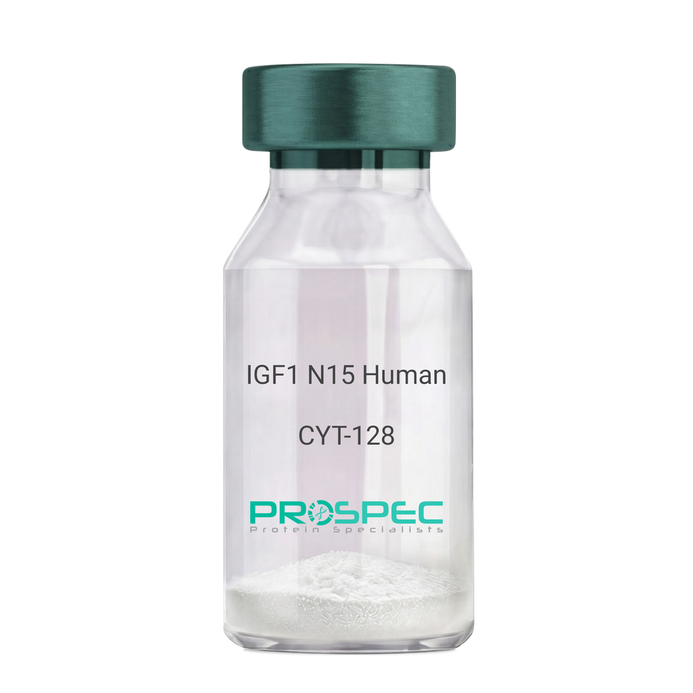
    IGF1 N15 Human

    Description:

    Insulin Like Growth Factor-1 N15 Labeled Human Recombinant

    Somatomedin C, IGF-I, IGFI, IGF1, IGF-IA, Mechano growth factor, MGF.

    Product # :

    CYT-128

    Price :

    Quantity :

    Shipping Method :

    Room Temp Icon

    Shipped at Room temp

    Add To Cart

    More Info

    • description
    • source
    • formulation
    • purity
    • biological activity
    • More Info
    • sds-page
    • Citations

    Description

    IGF1 N15 Human Recombinant produced in E.coli is a single, non-glycosylated polypeptide chain containing 70 amino acids and having a molecular mass of 7.74kDa. The N15 is stable isotope labeled. The IGF1 N15 is purified by proprietary chromatographic techniques.

    Source

    Escherichia Coli.

    Formulation

    IGF1 N15 protein was lyophilized from a 0.2µm filtered concentrated solution in PBS, pH 7.2.

    Purity

    Greater than 97.0% as determined by:
    (a) Analysis by RP-HPLC.
    (b) Analysis by SDS-PAGE.

    Biological Activity

    The ED50 as determined by a cell proliferation assay using serum free human MCF-7 cells is less than 2ng/ml, corresponding to a specific activity of > 5.0 × 10⁵ IU/mg.

    sds-page

    IGF-1 15N Human sds-page - Product image 1

    Citations

    More Info

    • Introduction

      The somatomedins, or insulin-like growth factors (IGFs), comprise a family of peptides that play important roles in mammalian growth and development. IGF1 mediates many of the growth-promoting effects of growth hormone (GH; MIM 139250). Early studies showed that growth hormone did not directly stimulate the incorporation of sulfate into cartilage, but rather acted through a serum factor, termed 'sulfation factor,' which later became known as 'somatomedin' (Daughaday et al., 1972). Three main somatomedins have been characterized: somatomedin C (IGF1), somatomedin A (IGF2; MIM 147470), and somatomedin B (MIM 193190) (Rotwein, 1986; Rosenfeld, 2003).

    • Synonyms

      Somatomedin C, IGF-I, IGFI, IGF1, IGF-IA, Mechano growth factor, MGF.

    • Physical Appearance

      Sterile Filtered White lyophilized (freeze-dried) powder.

    • Stability

      Lyophilized IGF1 N15 although stable at room temperature for 3 weeks, should be stored desiccated below -18°C. Upon reconstitution IGF1 N15 should be stored at 4°C between 2-7 days and for future use below -18°C. For long term storage it is recommended to add a carrier protein (0.1% HSA or BSA). Please prevent freeze-thaw cycles.

    • Solubility

      It is recommended to reconstitute the lyophilized IGF1 N15 in sterile 18M-cm H2O not less than 100µg/ml, which can then be further diluted to other aqueous solutions.

    • Amino Acid Sequence

      GPETLCGAEL VDALQFVCGD RGFYFNKPTG YGSSSRRAPQ TGIVDECCFR SCDLRRLEMY CAPLKPAKSA.

    • Background

      What is the molecular weight/Mw of IGF1 N15 HUMAN Protein?
      IGF1 N15 HUMAN Protein has a total Mw of 7.74kDa.

      What is the source or expression system of IGF1 N15 HUMAN Protein?
      Escherichia Coli.

      What is the Purity of IGF1 N15 HUMAN Protein?
      IGF1 N15 HUMAN Protein is >97% pure as determined by SDS-PAGE.

      What is the Biological Activity of IGF1 N15 HUMAN Protein?
      The ED50 as determined by a cell proliferation assay using serum free human MCF-7 cells is less than 2ng/ml, corresponding to a specific activity of > 5.0 × 10⁵ IU/mg.

      What is the amino acid sequence of IGF1 N15 HUMAN Protein?
      GPETLCGAEL VDALQFVCGD RGFYFNKPTG YGSSSRRAPQ TGIVDECCFR SCDLRRLEMY CAPLKPAKSA.

      What applications can IGF1 N15 HUMAN Protein be used in?
      IGF1 N15 HUMAN Protein can probably be used in western blot, ELISA and Lateral Flow.

      What is the endotoxin level for IGF1 N15 HUMAN Protein?
      The endotoxin level is minimal, IGF1 N15 HUMAN Protein was purified using conventional chromatography techniques.

    ProSpec's products are furnished for LABORATORY RESEARCH USE ONLY. The product may not be used as drugs, agricultural or pesticidal products, food additives or household chemicals.

    Igf1 N15 Human
  • View Data Sheet

    Name :

    Dengue Envelope-1 32kDa

    Description:

    Dengue Virus Subtype 1 Envelope 32kDa Recombinant

    Product # :

    DEN-014

    Price :

    Quantity :

    Shipping Method :

    Ice Icon

    Shipped with Ice Packs

    Add To Cart

    More Info

    • description
    • source
    • formulation
    • purity
    • More Info
    • Citations

    Description

    Recombinant Dengue Virus Subtype 1 Envelope 32kDa containing domains I+II of the dengue envelope, produced in E.coli and fused with a 6xHis Tag.

    Source

    E.coli.

    Formulation

    Phosphate buffered saline, pH-7.4.

    Purity

    Protein is >95% pure as determined by 12% PAGE (coomassie staining).

    Citations

    More Info

    • Introduction

      Dengue fever is affected by 1 of 4 closely linked virus serotypes of the genus Flavivirus, family Flaviviridae. One might have dengue fever infected by the different serotype virus after the primary infection. Detection of particular antibodies to dengue viruses is used for the diagnosis in clinic. Recently, lateral flow rapid test products have become a most suitable and known method in the clinical diagnosis. Though, there is difficulty for manufacturers to have the dengue antigens with a whole coverage for Dengue IgG & IgM recognition for all 4 serotype infections as well as with colloid gold binding ability. 8 dengue antigens have been established for the lateral flow products which are well defined for their coverage of dengue IgG & IgM recognition. The researcher can select the products below based on their own specific application.

    • Stability

      Dengue Envelope-1 32kDa although stable at 4°C for 1 week, should be stored below -18°C. Please prevent freeze thaw cycles.

    • Applications

      Immunoassay.

    • Purification Method

      Purified by proprietary chromatographic technique.

    ProSpec's products are furnished for LABORATORY RESEARCH USE ONLY. The product may not be used as drugs, agricultural or pesticidal products, food additives or household chemicals.

    Dengue Envelope 1 32Kda
  • View Data Sheet

    Name :

    S.typhi Paired Antibody

    Description:

    Mouse Anti Salmonella Typhi Paired

    Product # :

    ANT-665

    Price :

    Quantity :

    Shipping Method :

    Ice Icon

    Shipped with Ice Packs

    Add To Cart

    More Info

    • description
    • formulation
    • purity
    • More Info
    • Citations

    Description

    Typhi antibodies, capture and conjugating, target S. Typhi Outer Membrane Protein and S.Typhi specific polysaccharide respectively. This Antibody is used both as conjugate and capture.

    Formulation

    S.Typhi capture&conjugate antibody (2.7mg/1ml) in 0.01M phosphate buffered saline, pH 7.2 and 0.1% sodium azide.

    Purity

    Greater than 90%.

    Citations

    More Info

    • Introduction

      Salmonella Typhi is a pathogen causing typhoid fever, affecting over 17 million people with approximately 600,000 deaths annually worldwide. If untreated, typhoid fever cases result in mortality rates ranging from 12-30%.

    • Physical Appearance

      Sterile filtered clear colorless solution.

    • Stability

      S.typhi Paired antibody should be stored at 2-8°C.

    • Type

      Mouse antibody Monoclonal.

    • Purification Method

      Purified monoclonal IgG by protein A chromatography.

    ProSpec's products are furnished for LABORATORY RESEARCH USE ONLY. The product may not be used as drugs, agricultural or pesticidal products, food additives or household chemicals.

    Styphi Paired Antibody
  • View Data Sheet

    Name :

    IFN A Neut Antibody

    Description:

    Interferon-alpha Neutralizing, Mouse Anti-Human

    Product # :

    ANT-122

    Price :

    Quantity :

    Shipping Method :

    Room Temp Icon

    Shipped at Room temp

    Add To Cart

    More Info

    • formulation
    • More Info
    • Citations

    Formulation

    1mg/ml in PBS (after reconstitution).

    Citations

    More Info

    • Introduction

      IFN-alpha is produced by macrophages, IFN-alpha has antiviral activities and stimulates the production of two enzymes: a protein kinase and an oligoadenylate synthetase.

    • Solubility

      Reconstitute with sterile H20. Mix gently, wash the sides of the vial and wait 30-60 seconds before use.

    • Immunogen

      r.Human IFN Alpha.

    • Ig Subclass

      Mouse IgG1.

    • Clone

      NYRhIFN-A (neut).

    • Applications

      Direct ELISA (Does not work in Western Blotting). This is a Neutralizing antibody.

    • Note

      The antibody can be used for Capture ELISA together with ProSpec’s Mouse Anti Human IFN-alpha Western Blot antibody.

    • Titer

      By direct ELISA, 1:10,000 dilution will yield 0.5 O.D using alkaline phosphatase conjugated rabbit anti-mouse Ig (Jackson Laboratories).

    • Shipping Conditions

      Antibody is shipped lyophilized at ambient temperature.

    • Type

      Mouse Anti Human Monoclonal.

    • Storage Procedures

      In lyophilized form, for long periods, store at 4°C in a dry environment. After reconstitution, if not intended for use within a month, aliquot and store at -20°C.

    • Purification Method

      Ion exchange.

    ProSpec's products are furnished for LABORATORY RESEARCH USE ONLY. The product may not be used as drugs, agricultural or pesticidal products, food additives or household chemicals.

    Interferon Alpha Neutralizing Antibody
  • View Data Sheet

    Name :

    IFNW1 Human, HEK

    Description:

    Interferon-Omega 1 Human Recombinant, HEK

    IFN omega-1, IFN alpha-II-1, IFNW1.

    Product # :

    CYT-1225

    Price :

    Quantity :

    Shipping Method :

    Ice Icon

    Shipped with Ice Packs

    Add To Cart

    More Info

    • description
    • source
    • formulation
    • purity
    • biological activity
    • More Info
    • Citations

    Description

    IFNW1 Human Recombinant is a single, glycosylated, polypeptide chain (22-195 a.a) containing a total of 180 amino acids and having a molecular mass of 20.9 kDa. IFNW1 is fused to a 6 a.a his-Tag at C-terminus and is purified by proprietary chromatographic techniques.

    Source

    HEK293 Cells.

    Formulation

    The IFNW1 solution (1mg/ml) contains 10% Glycerol and Phosphate-Buffered Saline (pH 7.4).

    Purity

    Greater than 95.0% as determined by SDS-PAGE.

    Biological Activity

    The ED50 is ≤0.07 ng/ml, measured  in a cytotoxicity assay using TF-1 human erythroleukemic cells .

    Citations

    More Info

    • Synonyms

      IFN omega-1, IFN alpha-II-1, IFNW1.

    • Physical Appearance

      Sterile Filtered colorless solution.

    • Stability

      Store at 4°C if entire vial will be used within 2-4 weeks. Store, frozen at -20°C for longer periods of time. For long term storage it is recommended to add a carrier protein (0.1% HSA or BSA).Avoid multiple freeze-thaw cycles.

    • Amino Acid Sequence

      LGCDLPQNHG LLSRNTLVLL HQMRRISPFL CLKDRRDFRF PQEMVKGSQL QKAHVMSVLH EMLQQIFSLF HTERSSAAWN MTLLDQLHTG LHQQLQHLET CLLQVVGEGE SAGAISSPAL TLRRYFQGIR VYLKEKKYSD CAWEVVRMEI MKSLFLSTNM QERLRSKDRD LGSSHHHHHH.

    • Background

      Interferons, a family of signaling proteins, play a pivotal role in the immune system’s defense against viral infections and other threats. Among these, Interferon W1 (IFNW1), a member of the Type I interferon family, has emerged as a key player in orchestrating antiviral responses and modulating immune reactions. This research embarks on a detailed exploration of the IFNW1 protein, unveiling its structural intricacies, signaling pathways, and its broader implications in immune regulation and disease. By delving into IFNW1, scientists aim to comprehend the nuances of its functions, decipher its interactions within the complex interferon network, and explore its potential applications in therapeutic interventions and beyond.

      Structural Insights into IFNW1:

      IFNW1, like other Type I interferons, exhibits a unique tertiary structure that enables it to interact with specific cell surface receptors. This interaction triggers a cascade of events, leading to the activation of various antiviral genes and immune modulatory pathways. Understanding the structural basis of IFNW1 is crucial for elucidating its binding affinities, biological activities, and its significance in immune responses.

      Signaling Pathways and Antiviral Defense:

      IFNW1 engages with its cognate receptors, initiating Janus kinase (JAK)-Signal Transducer and Activator of Transcription (STAT) signaling pathways. This activation leads to the transcription of interferon-stimulated genes (ISGs) with potent antiviral properties. IFNW1’s ability to induce an antiviral state in infected and neighboring cells is fundamental for restricting viral replication and curtailing the spread of infections. Additionally, IFNW1 plays a role in modulating adaptive immune responses, contributing to the broader immune defense mechanisms.

      IFNW1 in Immunomodulation and Disease:

      Beyond its antiviral functions, IFNW1 is implicated in immunomodulation and disease pathogenesis. Dysregulation of IFNW1 signaling is associated with autoimmune disorders, including lupus and rheumatoid arthritis, highlighting its involvement in immune-related diseases. Moreover, IFNW1 is being explored in cancer immunotherapy, where its ability to modulate the tumor microenvironment and enhance immune surveillance presents opportunities for novel treatment strategies.

      Therapeutic Potential and Future Prospects:

      The unique properties of IFNW1, particularly its role in immune regulation and antiviral defense, position it as a potential therapeutic target. Research efforts are directed towards harnessing its immunomodulatory functions for developing therapies against infectious diseases, autoimmune disorders, and certain cancers. Additionally, understanding IFNW1’s interactions with other components of the immune system opens avenues for innovative approaches in personalized medicine and targeted immunotherapies.

      IFNW1 Protein, as an integral component of the interferon network, stands as a sentinel in the body’s defense against viral invasions and immune dysregulations. Its multifaceted roles in antiviral defense, immune modulation, and disease pathogenesis underscore its significance in biology and medicine. As researchers delve deeper into the intricacies of IFNW1, they pave the way for innovative therapies, immunomodulatory interventions, and a deeper understanding of immune responses. This research not only illuminates the pivotal role of IFNW1 but also holds the promise of transformative advancements in medicine, shaping the future of immunology and disease therapeutics.

      What is the molecular weight/Mw of IFNW1 HUMAN, HEK Protein?
      IFNW1 HUMAN, HEK Protein has a total Mw of 20.9kDa.

      What is the source or expression system of IFNW1 HUMAN, HEK Protein?
      HEK293 Cells.

      What is the Purity of IFNW1 HUMAN, HEK Protein?
      IFNW1 HUMAN, HEK Protein is >95% pure as determined by SDS-PAGE.

      What is the Biological Activity of IFNW1 HUMAN, HEK Protein?
      The ED50 is ≤0.07 ng/ml, measured in a cytotoxicity assay using TF-1 human erythroleukemic cells .

      What is the amino acid sequence of IFNW1 HUMAN, HEK Protein?
      LGCDLPQNHG LLSRNTLVLL HQMRRISPFL CLKDRRDFRF PQEMVKGSQL QKAHVMSVLH EMLQQIFSLF HTERSSAAWN MTLLDQLHTG LHQQLQHLET CLLQVVGEGE SAGAISSPAL TLRRYFQGIR VYLKEKKYSD CAWEVVRMEI MKSLFLSTNM QERLRSKDRD LGSSHHHHHH.

      What applications can IFNW1 HUMAN, HEK Protein be used in?
      IFNW1 HUMAN, HEK Protein can probably be used in western blot, ELISA and Lateral Flow.

      What is the endotoxin level for IFNW1 HUMAN, HEK Protein?
      The endotoxin level is minimal, IFNW1 HUMAN, HEK Protein was purified using conventional chromatography techniques.


    ProSpec's products are furnished for LABORATORY RESEARCH USE ONLY. The product may not be used as drugs, agricultural or pesticidal products, food additives or household chemicals.

    Ifn Omega Human
  • View Data Sheet

    Name :

    ASFV P72

    Description:

    African Swine Fever Virus P72 Recombinant

    Product # :

    ASFV-001

    Price :

    Quantity :

    Shipping Method :

    Ice Icon

    Shipped with Ice Packs

    Add To Cart

    More Info

    • description
    • source
    • formulation
    • purity
    • More Info
    • Citations

    Description

    Recombinant African Swine Fever Virus P72 (40-315 aa) produced in E.coli having a Mw of 33kDa.The African Swine Fever Virus P72 is fused to a 6xHis tag and purified by proprietary chromatographic technique.

    Source

    Escherichia Coli.

    Formulation

    African Swine Fever Virus P72 protein solution contains PBS and 25mM K2CO3.

    Purity

    Protein is >95% pure as determined by 10% PAGE (coomassie staining).

    Citations

    More Info

    • Introduction

      African swine fever virus (ASFV) causes African swine fever (ASF), a disease of swine characterized by high morbidity and mortality. ASFV is a large DNA virus which belongs to Asfarviridae family. ASFV’s multilayered virion consists from up to 54 polypeptides while p72 is the major capsid component. Middle part of p72 protein contains most epitopes tested by monoclonal antibodies, while C-terminal peptide of P72 isused for the genotyping of ASFV isolates.

    • Physical Appearance

      Sterile Filtered colorless solution.

    • Stability

      Store at 4°C if entire vial will be used within 2-4 weeks.Store, frozen at -20°C for longer periods of time.For long term storage it is recommended to add a carrier protein (0.1% HSA or BSA).Avoid multiple freeze-thaw cycles.

    • Applications

      ELISA, Western blot.

    ProSpec's products are furnished for LABORATORY RESEARCH USE ONLY. The product may not be used as drugs, agricultural or pesticidal products, food additives or household chemicals.

    product_image.jpg
  • View Data Sheet

    Name :

    CoV-2 S1 (1-681)

    Description:

    Coronavirus 2019-nCoV Spike Glycoprotein-S1 (1-681), Recombinant

    Product # :

    SARS-045

    Price :

    Quantity :

    Shipping Method :

    Ice Icon

    Shipped with Ice Packs

    Add To Cart

    More Info

    • description
    • source
    • formulation
    • purity
    • More Info
    • Citations

    Description

    The HEK293 derived recombinant protein contains the Coronavirus 2019-nCoV Spike Glycoprotein S1, Wuhan-Hu-1 strain, amino acids 1-681 fused to Fc tag at C-terminal having a Mw of 76 kDa.

    Source

    HEK293.

    Formulation

    CoV-2 S1protein solution is supplied PBS pH 7.4.

    Purity

    Protein is >95% pure as determined SDS-PAGE.

    Citations

    More Info

    • Introduction

      A human infecting coronavirus (viral pneumonia) called 2019 novel coronavirus, 2019-nCoV was found in the fish market at the city of Wuhan, Hubei province of China on December 2019.The 2019-nCoV shares an 87% identity to the 2 bat-derived severe acute respiratory syndrome 2018 SARS-CoV-2 located in Zhoushan of eastern China. 2019-nCoV has an analogous receptor-BD-structure to that of 2018 SARS-CoV, even though there is a.a. diversity so thus the 2019-nCoV might bind to ACE2 receptor protein (angiotensin-converting enzyme 2) in humans.While bats are probably the host of 2019-nCoV, researchers suspect that animal from the ocean sold at the seafood market was an intermediate host. RSCU analysis proposes that the 2019-nCoV is a recombinant within the viral spike glycoprotein between the bat coronavirus and an unknown coronavirus.

    • Physical Appearance

      Sterile Filtered clear solution.

    • Stability

      Protein is shipped on ice packs. Upon arrival, Store at -20°C.

    • Purification Method

      Purified by Protein-G chromatography technique.

    ProSpec's products are furnished for LABORATORY RESEARCH USE ONLY. The product may not be used as drugs, agricultural or pesticidal products, food additives or household chemicals.

    product_image.jpg
  • View Data Sheet

    Name :

    CoV-2 Spike (300-600)

    Description:

    Coronavirus 2019 Spike Receptor Binding Domain (300-600 a.a.) Recombinant

    Product # :

    SARS-018

    Price :

    Quantity :

    Shipping Method :

    Ice Icon

    Shipped with Ice Packs

    Add To Cart

    More Info

    • description
    • source
    • formulation
    • purity
    • More Info
    • Citations

    Description

    The E.Coli derived recombinant protein contains the Coronavirus 2019 Spike Receptor Binding Domain (300-600 a.a.) immunodominant region, fused to 6xHis tag at C-terminal

    Source

    Escherichia Coli.

    Formulation

    CoV 2019 Spike Receptor Binding Domain Protein 1mg/ml solution is supplied in 1x PBS.

    Purity

    CoV 2019 Spike Receptor Binding Domain Protein is >90% pure as determined SDS-PAGE.

    Citations

    More Info

    • Introduction

      A human infecting coronavirus (viral pneumonia) called 2019 novel coronavirus, 2019-nCoV was found in the fish market at the city of Wuhan, Hubei province of China on December 2019.

      The 2019-nCoV shares an 87% identity to the 2 bat-derived severe acute respiratory syndrome 2018 SARS-CoV-2 located in Zhoushan of eastern China. 2019-nCoV has an analogous receptor-BD-structure to that of 2018 SARS-CoV, even though there is a.a. diversity so thus the 2019-nCoV might bind to ACE2 receptor protein (angiotensin-converting enzyme 2) in humans.

      While bats are possibly the host of 2019-nCoV, researchers suspect that animal from the ocean sold at the seafood market was an intermediate host. RSCU analysis proposes that the 2019-nCoV is a recombinant within the viral spike glycoprotein between the bat coronavirus and an unknown coronavirus.

    • Physical Appearance

      Sterile Filtered clear solution.

    • Stability

      CoV 2019 Spike Receptor Binding Domain Protein is shipped on ice packs. Upon arrival, Store at -20°C.

    ProSpec's products are furnished for LABORATORY RESEARCH USE ONLY. The product may not be used as drugs, agricultural or pesticidal products, food additives or household chemicals.

    product_image.jpg
  • View Data Sheet

    Name :

    IFN a 2a Human

    Description:

    IFN-Alpha 2a Human Recombinant

    Leukocyte IFN, B cell IFN, Type I IFN, IFNA2, IFN-a 2a, Interferon-alpha-2a.

    Product # :

    CYT-204

    Price :

    Quantity :

    Shipping Method :

    Room Temp Icon

    Shipped at Room temp

    Add To Cart

    More Info

    • description
    • source
    • formulation
    • purity
    • biological activity
    • More Info
    • Citations

    Description

    IFN Alpha Human 2a Recombinant produced in E.Coli is a single, non-glycosylated, polypeptide chain containing 165 amino acids and having a molecular mass of 19241 Dalton. The difference between IFNA2A and IFNA2B is in the amino acid present at position 23. IFN-alpha 2a has a lysine at that position 23 while IFN-alpha 2b has arginine.The Interferon-alpha-2a gene was obtained from human leukocytes.The IFNA2A is purified by proprietary chromatographic techniques.

    Source

    Escherichia Coli.

    Formulation

    Lyophilized without additives.

    Purity

    Greater than 97.0% as determined by both:
    (a) Analysis by RP-HPLC.
    (b) Analysis by SDS-PAGE.

    Biological Activity

    The specific activity as determined in a viral resistance assay using bovine kidney MDBK cells was found to be 270,000,000 IU/mg.

    Citations

    More Info

    • Introduction

      IFN-alpha is produced by macrophages and has antiviral activities. IFN stimulates the production of two enzymes: protein kinase and an oligoadenylate synthetase.

    • Synonyms

      Leukocyte IFN, B cell IFN, Type I IFN, IFNA2, IFN-a 2a, Interferon-alpha-2a.

    • Physical Appearance

      Sterile Filtered White lyophilized (freeze-dried) powder.

    • Stability

      Lyophilized IFNA-2A although stable at room temperature for 3 weeks, should be stored desiccated below -18°C. Upon reconstitution IFN-alpha 2a should be stored at 4°C between 2-7 days and for future use below -18°C.For long term storage it is recommended to add a carrier protein (0.1% HSA or BSA).Please prevent freeze-thaw cycles.

    • Solubility

      It is recommended to reconstitute the lyophilized Interferon-alpha-2a in sterile 18MΩ-cm H2O not less than 100µg/ml, which can then be further diluted to other aqueous solutions.

    • Amino Acid Sequence

      CDLPQTHSLGSRRTLMLLAQMRKISLFSCLKDRHDFGFPQEEFGNQFQKAETIPVLHEM IQQIFNLFSTKDSSAAWDETLLDKFYTELYQQLNDLEACVIQGVGVTETPLMKEDSILAV RKYFQRITLYLKEKKYSPCAWEVVRAEIMRSFSLSTNLQESLRSKE.

      N-terminal methionine has been completely removed enzymatically.

    • Background

      What is the molecular weight/Mw of IFN A 2A HUMAN Protein?
      IFN A 2A HUMAN Protein has a total Mw of 19.2kDa.

      What is the source or expression system of IFN A 2A HUMAN Protein?
      Escherichia Coli.

      What is the Purity of IFN A 2A HUMAN Protein?
      IFN A 2A HUMAN Protein is >97% pure as determined by SDS-PAGE.

      What is the Biological Activity of IFN A 2A HUMAN Protein?
      The specific activity as determined in a viral resistance assay using bovine kidney MDBK cells was found to be 270,000,000 IU/mg.

      What is the amino acid sequence of IFN A 2A HUMAN Protein?
      CDLPQTHSLGSRRTLMLLAQMRKISLFSCLKDRHDFGFPQEEFGNQFQKAETIPVLHEM IQQIFNLFSTKDSSAAWDETLLDKFYTELYQQLNDLEACVIQGVGVTETPLMKEDSILAV RKYFQRITLYLKEKKYSPCAWEVVRAEIMRSFSLSTNLQESLRSKE.
      N-terminal methionine has been completely removed enzymatically.


      What applications can IFN A 2A HUMAN Protein be used in?
      IFN A 2A HUMAN Protein can probably be used in western blot, ELISA and Lateral Flow.

      What is the endotoxin level for IFN A 2A HUMAN Protein?
      The endotoxin level is minimal, IFN A 2A HUMAN Protein was purified using conventional chromatography techniques.


    • Protein content

      Protein quantitation was carried out by two independent methods:1. UV spectroscopy at 280 nm using the absorbency value of 0.924 as the extinction coefficient for a 0.1% (1mg/ml) solution. This value is calculated by the PC GENE computer analysis program of protein sequences (IntelliGenetics). 2. Analysis by RP-HPLC, using a calibrated solution of IFN-a 2a as a Reference Standard.

    ProSpec's products are furnished for LABORATORY RESEARCH USE ONLY. The product may not be used as drugs, agricultural or pesticidal products, food additives or household chemicals.

    Interferon Alpha 2A Human
  • View Data Sheet

    Name :

    CoV-2-S1 (319-541)

    Description:

    Coronavirus 2019 Spike Glycoprotein-S1 Receptor Binding Domain (319-541 a.a), Recombinant

    Product # :

    SARS-021

    Price :

    Quantity :

    Shipping Method :

    Ice Icon

    Shipped with Ice Packs

    Add To Cart

    More Info

    • description
    • source
    • formulation
    • purity
    • biological activity
    • More Info
    • Citations

    Description

    The HEK293 derived recombinant protein contains the Coronavirus 2019 Spike Glycoprotein S1 Receptor Binding Domain [ RBD ], Wuhan-Hu-1 strain, amino acids 319-541 fused to His tag at C-terminal.

    Source

    HEK293 Cells.

    Formulation

    CoV-S1 RBD protein is 1mg/ml contains 20mM sodium phosphate (NaPP), 300mM NaCl, pH7.2

    Purity

    Protein is >90% pure as determined SDS-PAGE.

    Biological Activity

    SARS CoV-2 Spike Glycoprotein S1 RBD was tested for its biological functionality by its binding to recombinant ACE2 receptor protein confirmed by structural complementation reporter assay (NanoBiT®).

    Citations

    More Info

    • Introduction

      A human infecting coronavirus (viral pneumonia) called 2019 novel coronavirus, 2019-nCoV was found in the fish market at the city of Wuhan, Hubei province of China on December 2019.

      The 2019-nCoV shares an 87% identity to the 2 bat-derived severe acute respiratory syndrome 2018 SARS-CoV-2 located in Zhoushan of eastern China. 2019-nCoV has an analogous receptor-BD-structure to that of 2018 SARS-CoV, even though there is a.a. diversity so thus the 2019-nCoV might bind to ACE2 receptor protein (angiotensin-converting enzyme 2) in humans.

      While bats are possibly the host of 2019-nCoV, researchers suspect that animal from the ocean sold at the seafood market was an intermediate host. RSCU analysis proposes that the 2019-nCoV is a recombinant within the viral spike glycoprotein between the bat coronavirus and an unknown coronavirus.

    • Physical Appearance

      Sterile Filtered clear solution.

    • Stability

      CoV-2 S1 RBD Protein is shipped on ice packs. Upon arrival, Store at -20°C.

    • Purification Method

      Purified by Metal-Afinity chromatographic technique.

    ProSpec's products are furnished for LABORATORY RESEARCH USE ONLY. The product may not be used as drugs, agricultural or pesticidal products, food additives or household chemicals.

    product_image.jpg
  • View Data Sheet

    Name :

    CoV-2 3CL

    Description:

    Coronavirus 2019 3CL Protease Recombinant

    Product # :

    SARS-058

    Price :

    Quantity :

    Shipping Method :

    Room Temp Icon

    Shipped at Room temp

    Add To Cart

    More Info

    • description
    • source
    • formulation
    • purity
    • biological activity
    • More Info
    • Citations

    Description

    Recombinant Coronavirus 2019 3CL Protease having a Mw of approximately 33.8 kDa was purified from E. coli. The CoV-2 3CL contains 306 amino acids and purified by proprietary chromatographic technique.

    Source

    Escherichia Coli.

    Formulation

    CoV-2 3CL solution contains 5 % Trehalose, 0.02 % Tween-20 and PBS, pH 7.4.

    Purity

    Protein is >98% pure as determined by SDS-PAGE and HPLC analyses.

    Biological Activity

    CoV-2 3CL is a cysteine protease that cleaves proteins with sequences including LQ(S/A/G).  The cleavage takes c-terminal to the glutamine residue. 

    Citations

    More Info

    • Introduction

      The SARS CoV-2 3CL protease (C30 Endopeptidase) derived from human SARS-CoV-2 is a C30-type cysteine protease situated in the NS3 segment. SARS CoV-2 3CL protease biological functionality is needed to process viral polyproteins to functional complete units. SARS CoV-2 3CL protease (main protease) is the core protease protein in CoV and corresponds to non-structural protein 5. SARS CoV-2 3CL cleaves the CoV polyprotein at 11 locations. SARS CoV-2 3CL protease has a cysteine-histidine catalytic dyad at its active site and cleaves a Gln–(Ser/Ala/Gly) peptide bond. SARS CoV-2 3CL protease at position P1 cleaves the bond between the glutamine and serine/alanine/glycine. SARS CoV-2 3CL protease is necessary in the processing of the CoV replicase polyprotein.

    • Physical Appearance

      Filtered White lyophilized (freeze-dried) powder.

    • Stability

      Store lyophilized protein at -20°C. Aliquot the product after reconstitution to avoid repeated freezing/thawing cycles. Reconstituted protein can be stored at 4°C for a limited period of time; it does not show any change after two weeks at 4°C.

    • Solubility

      It is recommended to add deionized water to prepare a working stock solution of approximately 0.5mg/ml and let the lyophilized pellet dissolve completely. CoV-2 3CL is not sterile! Please filter the product by an appropriate sterile filter before using it in the cell culture.

    • Amino Acid Sequence

      SGFRKMAFPS GKVEGCMVQV TCGTTTLNGL WLDDVVYCPR HVICTSEDML NPNYEDLLIR KSNHNFLVQA GNVQLRVIGH SMQNCVLKLK VDTANPKTPK YKFVRIQPGQ TFSVLACYNG SPSGVYQCAM RPNFTIKGSF LNGSCGSVGF NIDYDCVSFC YMHHMELPTG VHAGTDLEGN FYGPFVDRQT AQAAGTDTTI TVNVLAWLYA AVINGDRWFL NRFTTTLNDF NLVAMKYNYE PLTQDHVDIL GPLSAQTGIA VLDMCASLKE LLQNGMNGRT ILGSALLEDE FTPFDVVRQC SGVTFQ

    ProSpec's products are furnished for LABORATORY RESEARCH USE ONLY. The product may not be used as drugs, agricultural or pesticidal products, food additives or household chemicals.

    product_image.jpg
  • View Data Sheet

    Name :

    Dengue Control Antibody

    Description:

    Monoclonal Mouse Anti Dengue Control for Lateral Flow Test

    Product # :

    ANT-543

    Price :

    Quantity :

    Shipping Method :

    Ice Icon

    Shipped with Ice Packs

    Add To Cart

    More Info

    • description
    • formulation
    • purity
    • More Info
    • Citations

    Description

    Monoclonal Mouse Anti Dengue Control for Lateral Flow Test. Used to prepare a control line for dengue IgG/IgM rapid test, coating concentration is 0.4-0.5mg/ml, 1µl/cm.

    Formulation

    10mM phosphate buffered saline pH-7.2.

    Purity

    90% pure (12% SDS-PAGE and Coomassie staining).

    Citations

    More Info

    • Physical Appearance

      Sterile Filtered clear colorless solution.

    • Stability

      Store at 4°C if entire vial will be used within 2-4 weeks. Store, frozen at -20°C for longer periods of time. For long term storage it is recommended to add a carrier protein (0.1% HSA or BSA).Avoid multiple freeze-thaw cycles.

    • Applications

      Immunoassay.

    • Purification Method

      Purified monoclonal IgG by protein A chromatography.

    ProSpec's products are furnished for LABORATORY RESEARCH USE ONLY. The product may not be used as drugs, agricultural or pesticidal products, food additives or household chemicals.

    Dengue Control Antibody
  • View Data Sheet

    Name :

    Insulin Human, His

    Description:

    Insulin Human Recombinant, His Tag

    Insulin, Insulin-Dependent Diabetes Mellitus 2, Preproinsulin, Proinsulin, MODY10, IDDM1, IDDM2, IDDM, ILPR, IRDN.    

    Product # :

    CYT-1076

    Price :

    Quantity :

    Shipping Method :

    Ice Icon

    Shipped with Ice Packs

    Add To Cart

    More Info

    • description
    • source
    • formulation
    • purity
    • biological activity
    • More Info
    • Citations

    Description

    Insulin Recombinant produced in E.Coli is a single, non-glycosylated polypeptide chain containing 109 amino acids (25-110 a.a) and having a molecular mass of 11.8kDa. Insulin is fused to a 23 amino acid His-tag at N-terminus & purified by proprietary chromatographic techniques.

    Source

    Escherichia Coli.

    Formulation

    Insulin protein solution (0.5mg/ml) containing Phosphate-Buffered Saline (pH 7.4) and 10% Glycerol.

    Purity

    Greater than 95.0% as determined by SDS-PAGE.

    Biological Activity

    Measured in a cell proliferation assay using MCF7 human breast cancer cell. The ED50 for this effect is less or equal to 4 ug/ml.

    Citations

    More Info

    • Introduction

      Insulin decreases blood glucose concentration. it increases cell permeability to monosaccharides, amino acids and fatty acids. it accelerates glycolysis, the pentose phosphate cycle, and glycogen synthesis in liver.

    • Synonyms

      Insulin, Insulin-Dependent Diabetes Mellitus 2, Preproinsulin, Proinsulin, MODY10, IDDM1, IDDM2, IDDM, ILPR, IRDN.

    • Physical Appearance

      Sterile Filtered clear solution.

    • Stability

      Store at 4°C if entire vial will be used within 2-4 weeks. Store, frozen at -20°C for longer periods of time. For long term storage it is recommended to add a carrier protein (0.1% HSA or BSA).Avoid multiple freeze-thaw cycles.

    • Amino Acid Sequence

      MGSSHHHHHH SSGLVPRGSH MGSFVNQHLC GSHLVEALYL VCGERGFFYT PKTRREAEDL QVGQVELGGG PGAGSLQPLA LEGSLQKRGI VEQCCTSICS LYQLENYCN

    ProSpec's products are furnished for LABORATORY RESEARCH USE ONLY. The product may not be used as drugs, agricultural or pesticidal products, food additives or household chemicals.

    Insulin 2
  • View Data Sheet

    Name :

    Rabbit SARS CoV-2 IgG S1

    Description:

    Recombinant Anti Rabbit SARS CoV-2 IgG Spike S1

    Product # :

    ANT-762

    Price :

    Quantity :

    Shipping Method :

    Ice Icon

    Shipped with Ice Packs

    Add To Cart

    More Info

    • description
    • source
    • formulation
    • More Info
    • Citations

    Description

    Recombinant Anti Rabbit SARS CoV-2 IgG Kappa Spike S1 is a recombinant monoclonal antibody derived from HEK293 cells which recognizes the SARS-CoV and SARS-CoV-2 Spike glycoprotein. CoV-2 IgG S1 Rabbit Antibody binds to both SARS-CoV and SARS-CoV-2 with high affinity at amino acids 318-510 (RBD, Receptor Binding Domain) in the S1 subunit of the Spike protein. This chimeric rabbit antibody was made using the variable domain sequences of the original Human IgG1 format.

    Source

    HEK293 Cells.

    Formulation

    1mg/ml in PBS and 0.02% Proclin 300.

    Citations

    More Info

    • Introduction

      SARS Coronavirus is an enveloped virus containing three outer structural proteins, namely the membrane (M), envelope (E), and spike (S) proteins. Spike (S)-glycoprotein of the virus interacts with a cellular receptor and mediates membrane fusion to allow viral entry into susceptible target cells. Accordingly, S-protein plays an important role in virus infection cycle and is the primary target of neutralizing antibodies.It has recently been shown that SARS is caused by a human coronavirus. Human coronaviruses are the major cause of upper respiratory tract illness in humans, such as the common cold. Coronaviruses are positive-stranded RNA viruses, featuring the largest viral RNA genomes known to date (27-31 kb). The first step in coronavirus infection is binding of the viral spike protein, a 139-kDa protein, to certain receptors on host cells. The spike protein is the main surface antigen of the coronavirus. The most prominent protein in the culture supernatants infected with SARS virus is a 46 kDa nucleocapsid protein. This suggests that the nucleocapsid protein is a major immunogen that may be useful for early diagnostics.

    • Physical Appearance

      Sterile Filtered liquid formulation.

    • Stability

      Store at 4°C, stable for 2 weeks. For long-term storage, store at -20°C.

    • Immunogen

      The native monoclonal antibody was generated by sequencing peripheral blood lymphocytes of a patient exposed to the SARS-CoV

    • Applications

      ELISA.

    • Type

      Mouse antibody Monoclonal.

    • Purification Method

      Protein A affinity purified.

    ProSpec's products are furnished for LABORATORY RESEARCH USE ONLY. The product may not be used as drugs, agricultural or pesticidal products, food additives or household chemicals.

    Sars Anti Rabbit
  • View Data Sheet

    Name :

    S.Typhi OMP

    Description:

    Salmonella Typhi Outer Membrane Protein Recombinant

    Product # :

    STY-002

    Price :

    Quantity :

    Shipping Method :

    Ice Icon

    Shipped with Ice Packs

    Add To Cart

    More Info

    • description
    • source
    • formulation
    • purity
    • More Info
    • Citations

    Description

    Recombinant Salmonella Typhi Outer Membrane Protein produced in E.coli contains 315 amino acids, and fused to a 6 His Tag at C-terminus, migrating as a 33kDa band on SDS-PAGE.S. typhi outer membrane protein is a central pathogen in S. typhi infection, and is directly exposed to the outside to interact with the human immune system.

    Source

    Escherichia Coli.

    Formulation

    Sterile Filtered solution containing 10mM Tris-HCl, 1mM EDTA and 50mM arginine.

    Purity

    Protein is >95% pure as determined by 12% PAGE (coomassie staining).

    Citations

    More Info

    • Introduction

      Salmonella Typhi is a pathogen causing typhoid fever, affecting over 17 million people with approximately 600,000 deaths annually worldwide. If untreated, typhoid fever cases result in mortality rates ranging from 12-30%.

    • Physical Appearance

      Sterile Filtered solution.

    • Stability

      S.Typhi OMP although stable at 4°C for 1 week, should be stored below -18°C. Please prevent freeze thaw cycles.

    ProSpec's products are furnished for LABORATORY RESEARCH USE ONLY. The product may not be used as drugs, agricultural or pesticidal products, food additives or household chemicals.

    Styphi Omp
  • View Data Sheet

    Name :

    SARS MERS Spike Antibody

    Description:

    Mouse Anti MERS-CoV Spike

    Middle East respiratory syndrome coronavirus, Human betacoronavirus 2c EMC/2012, MERS-CoV, MERS, MERSCoV SP, Spike glycoprotein, S glycoprotein, S, Spike protein, E2, Peplomer protein.

    Product # :

    ANT-768

    Price :

    Quantity :

    Shipping Method :

    Ice Icon

    Shipped with Ice Packs

    Add To Cart

    More Info

    • formulation
    • More Info
    • Citations

    Formulation

    1mg/ml containing Phosphate-Buffered Saline (pH 7.4), 0.02% Sodium Azide and 10% Glycerol.

    Citations

    More Info

    • Introduction

      Since April 2012, cases of the Middle East Respiratory Syndrome Coronavirus (MERS-CoV) have been identified in various countries. Coronaviruses are the cause of the common cold, SARS (severe acute respiratory syndrome) and other severe illnesses with high mortality rates, all are classified into coronavirus family. MERS-CoV is a new type of SARS found in the coronavirus family causing severe pneumonia with sudden and serious respiratory illness with high mortality rates as well. Since January 27th 2015, the WHO has reported 956 human cases, including 351 deaths. More cases of the new coronavirus strain are expected. Like in other coronaviruses, large surface spike glycoprotein is a central structural protein of this virus; it is located above the virion surface to bind and enter into the target cell. Spike protein has 2 domains- S1 and S2. The S1 domain is responsible for cellular tropism and interaction with target cell, while the S2 domain is responsible for membrane fusion. The C-terminal of S1 domain contains a receptor binding domain, and is also a potential target for vaccine development and an antigen for diagnosis.

    • Synonyms

      Middle East respiratory syndrome coronavirus, Human betacoronavirus 2c EMC/2012, MERS-CoV, MERS, MERSCoV SP, Spike glycoprotein, S glycoprotein, S, Spike protein, E2, Peplomer protein.

    • Physical Appearance

      Sterile filtered colorless solution.

    • Immunogen

      Recombinant MERS-CoV Spike (18-1296aa) purified from Baculovirus.

    • Ig Subclass

      IgG1 kappa.

    • Clone

      PAT40G7AT

    • Applications

      SARS MERS Spike antibody has been tested by ELISA analysis to assure specificity and reactivity. Since application varies, however, each investigation should be titrated by the reagent to obtain optimal results.

    • Type

      Mouse antibody Monoclonal.

    • Storage Procedures

      For periods up to 1-month store at 4°C, for longer periods of time, store at -20°C. Prevent freeze thaw cycles.

    • Purification Method

      SARS MERS Spike antibody was purified from mouse ascitic fluids by protein-A affinity chromatography.

    ProSpec's products are furnished for LABORATORY RESEARCH USE ONLY. The product may not be used as drugs, agricultural or pesticidal products, food additives or household chemicals.

    Cov Mers Spike Antibody
  • View Data Sheet

    Name :

    Rubella E2

    Description:

    Rubella Virus E2 Recombinant

    Product # :

    RUB-292

    Price :

    Quantity :

    Shipping Method :

    Ice Icon

    Shipped with Ice Packs

    Add To Cart

    More Info

    • description
    • formulation
    • purity
    • More Info
    • Citations

    Description

    The E.Coli derived recombinant protein contains the Rubella Virus E2 regions, 31-105 amino acids.

    Formulation

    20mM imidazol, 8M urea and 0.3M NaCl.

    Purity

    Rubella protein is >95% pure as determined by 10% PAGE (coomassie staining).

    Citations

    More Info

    • Introduction

      The rubella virus (RV) structural proteins capsid, E2, and E1 are synthesized as a polyprotein precursor. The signal peptide that initiates translocation of E2 into the lumen of the endoplasmic reticulum remains attached to the carboxy terminus of the capsid protein after cleavage by signal peptidase.

    • Stability

      Rubella protein although stable at 4°C for 1 week, should be stored below -18°C. Please prevent freeze thaw cycles.

    • Applications

      Rubella antigen is suitable for ELISA and Western blots, excellent antigen for detection of Rubella Virus with minimal specificity problems.

    • Specificity

      Immunoreactive with sera of Rubella Virus infected individuals.

    • Purification Method

      Rubella protein was purified by proprietary chromatographic technique.

    ProSpec's products are furnished for LABORATORY RESEARCH USE ONLY. The product may not be used as drugs, agricultural or pesticidal products, food additives or household chemicals.

    product_image.jpg
  • View Data Sheet

    Name :

    CoV-2 S1 (16-685), Fc

    Description:

    Coronavirus 2019 Spike Glycoprotein-S1 (16-685 a.a.), Fc Recombinant

    Product # :

    SARS-027

    Price :

    Quantity :

    Shipping Method :

    Room Temp Icon

    Shipped at Room temp

    Add To Cart

    More Info

    • description
    • source
    • formulation
    • purity
    • More Info
    • Citations

    Description

    The HEK293 derived recombinant protein contains the Coronavirus 2019 CoV-2 Spike Glycoprotein S1, Wuhan-Hu-1 strain, amino acids 16-685 fused to Fc tag at C-terminal.

    Source

    HEK293 Cells.

    Formulation

    CoV-2 S1 protein is supplied in sodium chloride, glycine, arginine pH-7.5 & 5% trehalose.

    Purity

    Protein is >95% pure as determined SDS-PAGE.

    Citations

    More Info

    • Introduction

      A human infecting coronavirus (viral pneumonia) called 2019 novel coronavirus, 2019-nCoV was found in the fish market at the city of Wuhan, Hubei province of China on December 2019.

      The 2019-nCoV shares an 87% identity to the 2 bat-derived severe acute respiratory syndrome 2018 SARS-CoV-2 located in Zhoushan of eastern China. 2019-nCoV has an analogous receptor-BD-structure to that of 2018 SARS-CoV, even though there is a.a. diversity so thus the 2019-nCoV might bind to ACE2 receptor protein (angiotensin-converting enzyme 2) in humans.

      While bats are possibly the host of 2019-nCoV, researchers suspect that animal from the ocean sold at the seafood market was an intermediate host. RSCU analysis proposes that the 2019-nCoV is a recombinant within the viral spike glycoprotein between the bat coronavirus and an unknown coronavirus.

    • Physical Appearance

      Sterile Filtered White lyophilized (freeze-dried) powder.

    • Stability

      Lyophilized Cov-2 Spike S1 Glycoprotein although stable at room temperature for 3 weeks, should be stored desiccated below -18°C. Upon reconstitution CoV2 Spike protein should be stored at 4°C between 2-7 days and for future use below -18°C.For long term storage it is recommended to add a carrier protein (0.1% HSA or BSA).Please prevent freeze-thaw cycles.

    • Purification Method

      Purified by Protein-G chromatographic technique.

    ProSpec's products are furnished for LABORATORY RESEARCH USE ONLY. The product may not be used as drugs, agricultural or pesticidal products, food additives or household chemicals.

    product_image.jpg
  • View Data Sheet

    Name :

    SARS MERS, HEK

    Description:

    SARS MERS Spike Glycoprotein-S1, Recombinant

    Product # :

    SARS-023

    Price :

    Quantity :

    Shipping Method :

    Ice Icon

    Shipped with Ice Packs

    Add To Cart

    More Info

    • description
    • source
    • formulation
    • purity
    • More Info
    • Citations

    Description

    The HEK293 derived recombinant protein contains the SARS MERS Spike Glycoprotein S1, amino acids 18-725 fused to dimeric Fc tag at N-terminal having a total Mw of 215.7 kDa.

    Source

    HEK293

    Formulation

    SARS MERS S1 protein solution is supplied in PBS.

    Purity

    Protein is >85% pure as determined SDS-PAGE.

    Citations

    More Info

    • Introduction

      SARS Coronavirus is an enveloped virus containing three outer structural proteins, namely the membrane (M), envelope (E), and spike (S) proteins. Spike (S)-glycoprotein of the virus interacts with a cellular receptor and mediates membrane fusion to allow viral entry into susceptible target cells. Accordingly, S-protein plays an important role in virus infection cycle and is the primary target of neutralizing antibodies.

    • Physical Appearance

      Sterile Filtered clear solution.

    • Stability

      SARS MERS Protein is shipped on ice packs. Upon arrival, Store at -20°C.

    • Purification Method

      Purified by Protein-G chromatographic technique.

    ProSpec's products are furnished for LABORATORY RESEARCH USE ONLY. The product may not be used as drugs, agricultural or pesticidal products, food additives or household chemicals.

    product_image.jpg
  • 1
  • 2
  • 3
  • 4
  • 5
  • 6
  • …
  • 42
Your browser does not support the video tag.

Products

  • Cytokines
  • Growth Factors
  • Chemokines
  • CD Antigens
  • Neurotrophins
  • Hormones
  • Enzymes
  • Viral Antigens
  • Recombinant Proteins
  • Natural Proteins
  • Monoclonal Antibodies
  • Polyclonal Antibodies
  • Thermal Packaging

About

  • Company
  • Founder
  • Contribution
  • Contact Us
  • My Account

Ordering

  • Ordering Method
  • Ordering - Credit Card
  • Ordering PO
  • Shipping Fees
  • FAQ's

Legal

  • Terms & Conditions
  • Privacy
  • Site Map
  • info@prospecbio.com
  • Facebook
  • Instagram
  • Linkedin Profile

© 2026 ProSpec-Tany TechnoGene Ltd. All Rights Reserved.

  • Choosing a selection results in a full page refresh.
  • Opens in a new window.