Search results
1000 results found for “Stathmin”
Name
Description
Product #
Price
Quantity
Shipping Method
- View Data Sheet
Name :
SDF 1b HumanDescription:
Stromal Cell Derived Factor-1 Beta Human Recombinant (CXCL12)
SDF-1, CXCL12, Pre-B cell growth-stimulating factor, PBSF, hIRH, chemokine (C-X-C motif) ligand 12, SDF1, SDF1B, TPAR1, SCYB12, SDF-1b, TLSF-b.
Product # :
CHM-325Price :
Quantity :
Shipping Method :
Shipped at Room temp
More Info
- description
- source
- formulation
- purity
- biological activity
- More Info
Description
Stromal Cell-Derived Factor-1 beta Human Recombinant produced in E.Coli is a non-glycosylated, Polypeptide chain containing 72 amino acids and having a molecular mass of 8508 Dalton. The SDF-1b is purified by proprietary chromatographic techniques.
Source
Escherichia Coli.
Formulation
The CXCL12 protein was lyophilized from a concentrated (1mg/ml) sterile solution containing no additives.
Purity
Greater than 98.0% as determined by:
(a) Analysis by RP-HPLC.
(b) Analysis by SDS-PAGE.Biological Activity
The specific activity as determined by its ability to chemoattract human peripheral T cells activated with PHA and IL-2 using a concentation of 20-80ng/ml corresponding to a Specific Activity of 12,500-50,000IU/mg.More Info
-
Introduction
SDF-1 (stromal cell-derived factor-1) is small cytokine belonging to the chemokine family that is officially designated Chemokine (C-X-C motif) ligand 12 (CXCL12). It is produced in two forms, SDF-1?/CXCL12a and SDF-1?/CXCL12b, by alternate splicing of the same gene. Chemokines are characterized by the presence of four conserved cysteines, which form two disulfide bonds. The CXCL12 proteins belong to the group of CXC chemokines, whose initial pair of cysteines are separated by one intervening amino acid. CXCL12 is strongly chemotactic for lymphocytes and has been implicated as an important cell co-ordinator during development. During embryogenesis it directs the migration of hematopoietic cells from foetal liver to bone marrow. Mice which were knocked-out for CXCL12 gene were lethal before the birth or within just 1 hour of life. As another role, CXCL12a alters also the electrophysiology of neurons. CXCL12 was shown to be expressend in many tissues in mice (including brain, thymus, heart, lung, liver, kidney, spleen and bone marrow).
The receptor for this chemokine is CXCR4, which was previously called fusin. This CXCL12-CXCR4 interaction used to be considered exclusive (unlike for other chemokines and their receptors), but recently it was suggested that CXCL12 is also bound by CXCR7 receptor.
The gene for CXCL12 is located on human chromosome 10. In human and mouse both CXCL12 and CXCR4 show high identity of sequence: 99% and 90%, respectively. -
Synonyms
SDF-1, CXCL12, Pre-B cell growth-stimulating factor, PBSF, hIRH, chemokine (C-X-C motif) ligand 12, SDF1, SDF1B, TPAR1, SCYB12, SDF-1b, TLSF-b.
-
Physical Appearance
Sterile Filtered White lyophilized (freeze-dried) powder.
-
Stability
Lyophilized SDF-1b although stable at room temperature for 3 weeks, should be stored desiccated below -18°C. Upon reconstitution CXCL12 should be stored at 4°C between 2-7 days and for future use below -18°C. For long term storage it is recommended to add a carrier protein (0.1% HSA or BSA).Please prevent freeze-thaw cycles.
-
Solubility
It is recommended to reconstitute the lyophilized Stromal Cell-Derived Factor-1b in sterile 18MΩ-cm H2O not less than 100µg/ml, which can then be further diluted to other aqueous solutions.
-
Amino Acid Sequence
The sequence of the first five N-terminal amino acids was determined and was found to be Lys-Pro-Val-Ser-Leu.
-
Protein content
Protein quantitation was carried out by two independent methods: 1. UV spectroscopy at 280 nm using the absorbency value of 1.06 as the extinction coefficient for a 0.1% (1mg/ml) solution. This value is calculated by the PC GENE computer analysis program of protein sequences (IntelliGenetics). 2. Analysis by RP-HPLC, using a standard solution of SDF-1b as a Reference Standard.
ProSpec's products are furnished for LABORATORY RESEARCH USE ONLY. The product may not be used as drugs, agricultural or pesticidal products, food additives or household chemicals.
- View Data Sheet
Name :
Lymphotactin HumanDescription:
Lymphotactin Human Recombinant (XCL1)
XCL1, Cytokine SCM-1, ATAC, Lymphotaxin, SCM-1-alpha, Small inducible cytokine C1, XC chemokine ligand 1, LTN, LPTN, SCM1, SCM-1, SCYC1, SCM-1a.
Product # :
CHM-314Price :
Quantity :
Shipping Method :
Shipped at Room temp
More Info
- description
- source
- formulation
- purity
- biological activity
- More Info
Description
Lymphotactin Human Recombinant produced in E.Coli is a single, non-glycosylated, polypeptide chain containing 92 amino acids and having a molecular mass of 10007 Dalton. The Lymphotactin is purified by proprietary chromatographic techniques.
Source
Escherichia Coli.
Formulation
The XCL1 was lyophilized from a concentrated (1mg/ml) solution in water containing no additives.
Purity
Greater than 99.0% as determined by:
(a) Analysis by RP-HPLC.
(b) Analysis by SDS-PAGE.Biological Activity
The Biological activity is calculated by its ability to chemoattract human T cells at 10-100ng/ml corresponding to a Specific Activity of 10,000-100,000IU/mg.More Info
-
Introduction
Chemokine (C motif) ligand (XCL1) is a small cytokine belonging to the XC chemokine family that is also known as lymphotactin. It is found in high levels in spleen, thymus, intestine and peripheral blood leukocytes, and at lower levels in lung, prostate gland and ovary. Cellular sources for XCL1 include activated thymic and peripheral blood CD8+ T cells. This chemokine attracts T cells. In humans, XCL1 is closely related to another chemokine called XCL2, whose geneis found at the same locus on chromosome 1. XCL1 induces it chemotactic function by binding to a chemokine receptor called XCR1.
-
Synonyms
XCL1, Cytokine SCM-1, ATAC, Lymphotaxin, SCM-1-alpha, Small inducible cytokine C1, XC chemokine ligand 1, LTN, LPTN, SCM1, SCM-1, SCYC1, SCM-1a.
-
Physical Appearance
Sterile Filtered White lyophilized (freeze-dried) powder.
-
Stability
Lyophilized Lymphotactin although stable at room temperature for 3 weeks, should be stored desiccated below -18°C. Upon reconstitution XCL1 should be stored at 4°C between 2-7 days and for future use below -18°C. For long term storage it is recommended to add a carrier protein (0.1% HSA or BSA). Please avoid freeze-thaw cycles.
-
Solubility
It is recommended to reconstitute the lyophilized Lymphotactin in sterile 18MΩ-cm H2O not less than 100µg/ml, which can then be further diluted to other aqueous solutions.
-
Amino Acid Sequence
The sequence of the first five N-terminal amino acids was determined and was found to be Gln-Ser-Glu-Val-Ser.
ProSpec's products are furnished for LABORATORY RESEARCH USE ONLY. The product may not be used as drugs, agricultural or pesticidal products, food additives or household chemicals.
- View Data Sheet
Name :
ARTN HumanDescription:
Artemin Human Recombinant
ART, ARTN , EVN, NBN.
Product # :
CYT-306Price :
Quantity :
Shipping Method :
Shipped at Room temp
More Info
- description
- source
- formulation
- purity
- biological activity
- More Info
Description
Artemin Human Recombinant produced in E.Coli is a disulfide-linked homodimer, non-glycosylated, polypeptide chain containing 2 x 113 amino acids and having a total molecular mass of 24.2 kDa. Artemin is purified by proprietary chromatographic techniques.
Source
Escherichia Coli.
Formulation
Artemin was lyophilized after extensive dialysis against 10mM sodium citrate pH-4.5 and 25mM sodium chloride.
Purity
Greater than 98.0% as determined by:
(a) Analysis by RP-HPLC.
(b) Analysis by SDS-PAGE.Biological Activity
The activity is determined by the dose-dependent proliferation of the SH-SY5Y cell line and is typically 4-8 ng/mL. The activity can also be determined by its ability to promote survival and neurite outgrowth.More Info
-
Introduction
The protein encoded by this gene is a member of the glial cell line-derived neurotophic factor (GDNF) family of ligands which are a group of ligands within the TGF-beta superfamily of signaling molecules. GDNFs are unique in having neurotrophic properties and have potential use for gene therapy in neurodegenrative disease. Artemin has been shown in culture to support the survival of a number of periferal neuron populations and at least one population of dopaminergic CNS neurons. Its role in the PNS and CNS is further substantiated by its expression pattern in the proximity of these neurons. This protein is a ligand for the RET receptor and uses GFR-alpha 3 as a coreceptor. Four alternatively spliced transcripts have been described, two of which encode the same protein.
-
Synonyms
ART, ARTN , EVN, NBN.
-
Physical Appearance
Sterile Filtered White lyophilized (freeze-dried) powder.
-
Stability
Lyophilized Artemin although stable at room temperature for 3 weeks, should be stored desiccated below -18°C. Upon reconstitution Artemin Human Recombinant should be stored at 4°C between 2-7 days and for future use below -18°C.For long term storage it is recommended to add a carrier protein (0.1% HSA or BSA).Please prevent freeze-thaw cycles.
-
Solubility
It is recommended to reconstitute the lyophilized Artemin in sterile 18MΩ-cm H2O not less than 100µg/ml, which can then be further diluted to other aqueous solutions.
-
Amino Acid Sequence
AGGPGSRARA AGARGCRLRS QLVPVRALGL GHRSDELVRF RFCSGSCRRA RSPHDLSLAS LLGAGALRPP PGSRPVSQPC CRPTRYEAVS FMDVNSTWRT VDRLSATACG CLG.
-
Background
Artemin Human Recombinant: Unraveling its Role in Neurobiology and Therapeutic Applications
Abstract:
Artemin, a member of the glial cell line-derived neurotrophic factor (GDNF) family, holds significant potential in neurobiology and therapeutic interventions. This research paper provides an overview of Artemin human recombinant, elucidating its molecular characteristics, signaling pathways, and therapeutic implications in neurological disorders. Understanding the multifaceted role of Artemin offers new avenues for targeted therapies. This article offers a concise analysis of Artemin, highlighting its impact on neurobiology and its therapeutic applications.
Introduction:
Neurological disorders represent a major challenge in healthcare, necessitating innovative therapeutic strategies. Artemin, a member of the GDNF family, has emerged as a promising molecule in neurobiology. This paper provides an overview of Artemin, shedding light on its structure, function, and therapeutic potential.
Artemin Signaling and Mechanisms:
Artemin binds to its receptor, Ret tyrosine kinase, and activates downstream signaling pathways, including the PI3K/AKT and MAPK pathways. These signaling cascades play crucial roles in neuronal survival, growth, and differentiation, highlighting the significance of Artemin in neurodevelopment and neuroprotection.
Artemin in Neurological Disorders:
Artemin has been implicated in various neurological disorders, including peripheral neuropathies and neurodegenerative diseases. Its neuroprotective properties and ability to enhance neuronal survival and regeneration make it a promising target for therapeutic interventions. Furthermore, Artemin may play a role in pain modulation and sensory neuron function.
Therapeutic Potential of Artemin Human Recombinant:
Artemin human recombinant offers promising prospects in the field of neurotherapeutics. Strategies aimed at modulating Artemin signaling or delivering exogenous Artemin hold potential for promoting neuronal survival, regeneration, and functional recovery. Artemin-based therapies could be developed for a range of neurological disorders, including peripheral neuropathies, Parkinson's disease, and spinal cord injuries.
Challenges and Future Directions:
While the therapeutic targeting of Artemin shows promise, several challenges lie ahead. Further research is needed to understand the precise mechanisms underlying Artemin's effects and its interactions with other signaling pathways. Additionally, the development of effective delivery methods and the identification of patient subgroups that may benefit from Artemin-based therapies are important considerations for clinical translation.
Conclusion:
Artemin human recombinant represents a promising avenue for therapeutic interventions in neurological disorders. Understanding the molecular mechanisms and functional implications of Artemin in neurobiology offers new opportunities for developing innovative treatments. Continued research in this field has the potential to improve the lives of individuals affected by neurological conditions and advance the field of neurotherapeutics.
What is the molecular weight/Mw of ARTN Protein?
ARTN Protein has a total Mw of 24.2kDa.
What is the source or expression system of ARTN Protein?
Escherichia Coli.
What is the Purity of ARTN Protein?
ARTN Protein is >98% pure as determined by SDS-PAGE.
What is the Biological Activity of ARTN Protein?
The activity is determined by the dose-dependent proliferation of the SH-SY5Y cell line and is typically 4-8 ng/mL. The activity can also be determined by its ability to promote survival and neurite outgrowth.
What is the amino acid sequence of ARTN Protein?
AGGPGSRARA AGARGCRLRS QLVPVRALGL GHRSDELVRF RFCSGSCRRA RSPHDLSLAS LLGAGALRPP PGSRPVSQPC CRPTRYEAVS FMDVNSTWRT VDRLSATACG CLG.
What applications can ARTN Protein be used in?
ARTN Protein can probably be used in western blot, ELISA and Lateral Flow.
What is the endotoxin level for ARTN Protein?
The endotoxin level is minimal, ARTN Protein was purified using conventional chromatography techniques.
ProSpec's products are furnished for LABORATORY RESEARCH USE ONLY. The product may not be used as drugs, agricultural or pesticidal products, food additives or household chemicals.
- View Data Sheet
Name :
S100A8 Human, HisDescription:
S100 Calcium Binding Protein A8 Human Recombinant, His Tag
Calgranulin A, MRP8, CAGA, CGLA, CFAG, Protein S100-A8, S100 calcium-binding protein A8, Migration inhibitory factor-related protein 8, MRP-8, p8, Cystic fibrosis antigen, Leukocyte L1 complex light chain, Calprotectin L1L subunit, Urinary stone protein band A, S100A8, MIF, NIF, L1Ag, CP-10, MA387, 60B8AG.
Product # :
PRO-150Price :
Quantity :
Shipping Method :
Shipped at Room temp
More Info
- description
- source
- formulation
- More Info
Description
The Recombinant Human S100A8 produced in E.coli has a molecular mass of 12.08kDa containing 103 amino acid residues of the human S100A8 and fused to a 10 a.a. His tag at N-terminus.
Source
Escherichia Coli.
Formulation
S100A8 was filtered (0.4µm) and lyophilized in 0.5mg/ml in 20mM Tris and 100mM NaCl, pH 7.5.
More Info
-
Introduction
S100A8 is a part of the S100 family of proteins containing 2 EF-hand calcium-binding motifs. S100 proteins are localized in the cytoplasm and/or nucleus of a broad range of cells, and participate in the regulation of cellular processes such as cell cycle progression and differentiation. S100A8 plays a role in the inhibition of casein kinase and as a cytokine. S100A8 altered expression is related with cystic fibrosis disease. S100A8 is a calcium-binding protein that has antimicrobial activity against bacteria and fungi.S100A8 is crucial for resistance towards invasion by pathogenic bacteria. S100A8 up-regulates transcription of genes that are under the control of NF-kappa-B. S100A8 plays a role in the development of endotoxic shock in response to bacterial lipopolysaccharide. S100A8 endorses tubulin polymerization and promotes phagocyte migration and infiltration of granulocytes at sites of wounding. S100A8 takes part as a pro-inflammatory mediator in acute and chronic inflammation and up-regulates the release of IL8 and cell-surface expression of ICAM1.
-
Synonyms
Calgranulin A, MRP8, CAGA, CGLA, CFAG, Protein S100-A8, S100 calcium-binding protein A8, Migration inhibitory factor-related protein 8, MRP-8, p8, Cystic fibrosis antigen, Leukocyte L1 complex light chain, Calprotectin L1L subunit, Urinary stone protein band A, S100A8, MIF, NIF, L1Ag, CP-10, MA387, 60B8AG.
-
Stability
Store lyophilized protein at -20°C. Aliquot the product after reconstitution to avoid repeated freezing/thawing cycles. Reconstituted protein can be stored at 4°C for a limited period of time; it does not show any change after two weeks at 4°C.
-
Solubility
It is recommended to add deionized water to prepare a working stock solution of approximately 0.5 mg/ml and let the lyophilized pellet dissolve completely. Product is not sterile! Please filter the product by an appropriate sterile filter before using it in the cell culture.
-
Amino Acid Sequence
MKHHHHHHAS MLTELEKALN SIIDVYHKYS LIKGNFHAVY RDDLKKLLET ECPQYIRKKG ADVWFKELDI NTDGAVNFQEMLTELEKALN SIIDVYHKYS LIKGNFHAVY RDDLKKLLET ECPQYIRKKG ADVWFKELDI NTDGAVNFQE FLILVIKMGV AAHKKSHEES HKE.
ProSpec's products are furnished for LABORATORY RESEARCH USE ONLY. The product may not be used as drugs, agricultural or pesticidal products, food additives or household chemicals.
- View Data Sheet
Name :
Tamm HorsfallDescription:
Recombinant Human Tamm Horsfall Glycoprotein
Tamm-Horsfall urinary glycoprotein, THP, FJHN, HNFJ, THGP, MCKD2, ADMCKD2, UMOD, Uromodulin.
Product # :
ENZ-1206Price :
Quantity :
Shipping Method :
Shipped at Room temp
More Info
- description
- source
- formulation
- purity
- More Info
- SDS-PAGE
Description
Uromodulin Human Recombinant protein produced from HEK Cells, is a polypeptide chain containing 595 amino acids ( 25-613 a.a. ) and having a total Mw of 65 kDa.
Source
HEK293
Formulation
The UMOD protein was lyophilized from 0.4μm filtered solution containing 50mM NaCl, 0.02M TRIS, pH 7.5.
Purity
Greater than 90.0% as determined by SDS-PAGE analysis.
SDS-PAGE
More Info
-
Introduction
Uromodulin (Tamm–Horsfall protein) is produced mainly by cells in the kidney’s thick ascending limb and is the most abundant protein in normal urine.
Uromodulin takes part in salt and water regulation, helps prevent urinary tract infections and kidney stones, and influences inflammation and immune activity in the kidney.
Reduced Uromodulin levels is associated with chronic kidney disease.
UMOD mutations causes unproper protein folding and thus transported incorrectly, resulting in its accumulation inside kidney tubular cells that damages the tubules and cause kidney disease (ADTKD-UMOD), associated with high uric acid, gout, and progressive kidney failure.
-
Synonyms
Tamm-Horsfall urinary glycoprotein, THP, FJHN, HNFJ, THGP, MCKD2, ADMCKD2, UMOD, Uromodulin.
-
Physical Appearance
Filtered White lyophilized (freeze-dried) powder.
-
Stability
Lyophilized UMOD although stable at room temperature for 3 weeks, should be stored at -18C. Upon reconstitution UMOD should be stored at 4C between 2-7 days and for future use below -18C.
For long term storage it is recommended to add a carrier protein (0.1% HSA or BSA).
Please prevent freeze-thaw cycles.
-
Solubility
Add deionized water to prepare a working stock solution of approximately 0.5mg/ml and let the lyophilized pellet dissolve completely. Product is not sterile! Please filter the product by an appropriate sterile filter before using it in the cell culture.
-
Amino Acid Sequence
DTSEARWCSE CHSNATCTED EAVTTCTCQE GFTGDGLTCV DLDECAIPGA HNCSANSSCV NTPGSFSCVC PEGFRLSPGL GCTDVDECAE PGLSHCHALA TCVNVVGSYL CVCPAGYRGD GWHCECSPGS CGPGLDCVPE GDALVCADPC QAHRTLDEYW RSTEYGEGYA CDTDLRGWYR FVGQGGARMA ETCVPVLRCN TAAPMWLNGT HPSSDEGIVS RKACAHWSGH CCLWDASVQV KACAGGYYVY NLTAPPECHL AYCTDPSSVE GTCEECSIDE DCKSNNGRWH CQCKQDFNIT DISLLEHRLE CGANDMKVSL GKCQLKSLGF DKVFMYLSDS RCSGFNDRDN RDWVSVVTPA RDGPCGTVLT RNETHATYSN TLYLADEIII RDLNIKINFA CSYPLDMKVS LKTALQPMVS ALNIRVGGTG MFTVRMALFQ TPSYTQPYQG SSVTLSTEAF LYVGTMLDGG DLSRFALLMT NCYATPSSNA TDPLKYFIIQ DRCPHTRDST IQVVENGESS QGRFSVQMFR FAGNYDLVYL HCEVYLCDTM NEKCKPTCSG TRFRSGSVID QSRVLNLGPI TRKGVQATVH HHHHH
-
Background
Uromodulin is the most abundant protein in normal urine. Its secretion in urine follows proteolytic cleavage of the ectodomain of its glycosyl phosphatidylinosital-anchored counterpart that is situated on the luminal cell surface of the loop of Henle. Uromodulin plays a role as a constitutive inhibitor of calcium crystallization in renal fluids. Secretion of uromodulin in urine provides protection against urinary tract infections caused by uropathogenic bacteria. Defects in Uromodulin expression are associated with the autosomal dominant renal disorders medullary cystic kidney disease-2 (MCKD2) and familial juvenile hyperuricemic nephropathy (FJHN). These disorders are characterized by juvenile onset of hyperuricemia, gout, and progressive renal failure. While several transcript variants may exist for this gene, the full-length natures of only two have been described to date. UMOD is involved in regulating the circulating activity of cytokines as it binds to il-1, il-2 and tnf with high affinity.
What is the molecular weight/Mw of UMOD Protein?
UMOD Protein has a total Mw of 65kDa.
What is the source or expression system of UMOD Protein?
HEK293.
What is the Purity of UMOD Protein?
UMOD Protein is >90% pure as determined by SDS-PAGE.
What is the Biological Activity of UMOD Protein?
The biological functionality of UMOD Protein will be determined in the future.
What is the amino acid sequence of UMOD Protein?
UMOD Protein is composed from 595 amino acids.
What applications can UMOD Protein be used in?
UMOD Protein can probably be used in western blot, ELISA and Lateral Flow.
What is the endotoxin level for UMOD Protein?
The endotoxin level is minimal, UMOD Protein was purified using conventional chromatography techniques.
ProSpec's products are furnished for LABORATORY RESEARCH USE ONLY. The product may not be used as drugs, agricultural or pesticidal products, food additives or household chemicals.
- View Data Sheet
Name :
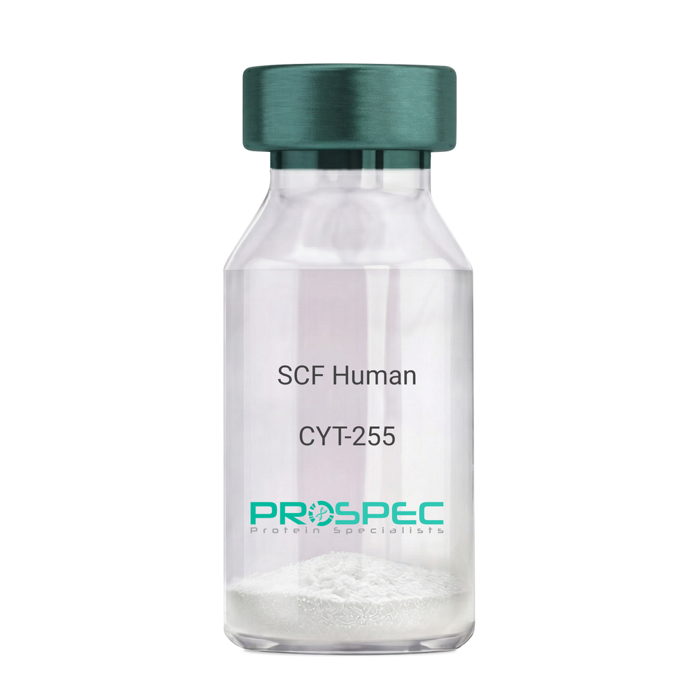
SCF HumanDescription:
Stem Cell Factor Human Recombinant
Kit ligand Precursor, C-kit ligand, SCF, Mast cell growth factor, MGF, SF, KL-1, Kitl, DKFZp686F2250.
Product # :
CYT-255Price :
Quantity :
Shipping Method :
Shipped at Room temp
More Info
- description
- source
- formulation
- purity
- biological activity
- More Info
Description
Stem Cell Factor Human Recombinant produced in E.Coli is a single, non-glycosylated polypeptide chain containing 165 amino acids and having a molecular mass of 18409 Dalton. The SCF is purified by proprietary chromatographic techniques.
Source
Escherichia Coli.
Formulation
Lyophilized from a concentrated (1mg/ml) solution in water containing 0.02% NaHCO3.
Purity
Greater than 98.0% as determined by:
(a) Analysis by SEC-HPLC.
(b) Analysis by SDS-PAGE.Biological Activity
The ED50 as determined by the dose-dependant stimulation of Human TF-1 cells is < 2 ng/ml, corresponding to a Specific Activity of 500,000 IU/mg.More Info
-
Introduction
Stem cell factor / KIT ligand (SCF) is a cytokine which binds CD117(c-Kit). SCF is also known as "steel factor" or "c-kit ligand". SCF exists in two forms, cell surface bound SCF and soluble (or free) SCF. Soluble SCF is produced by the cleavage of surface bound SCF by metalloproteases. SCF is a growth factor important for the survival, proliferation, and differentiation of hematopoietic stem cells and other hematopoietic progenitor cells. One of its roles is to change the BFU-E (burst-forming unit-erythroid) cells, which are the earliest erythrocyte precursors in the erythrocytic series, into the CFU-E (colony-forming unit-erythroid).
-
Synonyms
Kit ligand Precursor, C-kit ligand, SCF, Mast cell growth factor, MGF, SF, KL-1, Kitl, DKFZp686F2250.
-
Physical Appearance
Sterile Filtered White lyophilized (freeze-dried) powder.
-
Stability
Lyophilized KIT ligand although stable at room temperature for 3 weeks, should be stored desiccated below -18°C. Upon reconstitution SCF should be stored at 4°C between 2-7 days and for future use below -18°C.For long term storage it is recommended to add a carrier protein (0.1% HSA or BSA).Please prevent freeze-thaw cycles.
-
Solubility
It is recommended to reconstitute the lyophilized Stem Cell Factor in sterile 18MΩ-cm H2O not less than 100µg/ml, which can then be further diluted to other aqueous solutions.
-
Amino Acid Sequence
The sequence of the first five N-terminal amino acids was determined and was found to be Met-Glu-Gly-Ile-Cys.
-
Protein content
Protein quantitation was carried out by two independent methods1. UV spectroscopy at 280 nm using the absorbency value of 0.52 as the extinction coefficient for a 0.1% (1mg/ml) solution. This value is calculated by the PC GENE computer analysis program of protein sequences (IntelliGenetics). 2. Analysis by RP-HPLC, using a standard solution of Stem Cell Factor as a Reference Standard.
ProSpec's products are furnished for LABORATORY RESEARCH USE ONLY. The product may not be used as drugs, agricultural or pesticidal products, food additives or household chemicals.
- View Data Sheet
Name :
Stratifin HumanDescription:
Tyr-3/Trp- 5 Monooxygenase Activation Protein Sigma Human Recombinant
14-3-3 protein sigma, Epithelial cell marker protein 1, HME1, Stratifin, YWHAS, SFN, Stratifin.
Product # :
PKA-357Price :
Quantity :
Shipping Method :
Shipped with Ice Packs
More Info
- description
- source
- formulation
- purity
- More Info
Description
Stratifin Human Recombinant produced in E.Coli is a single, non-glycosylated, polypeptide chain containing 248 amino acids (1-248) and having a molecular mass of 27.7 kDa. Stratifin is purified by proprietary chromatographic techniques.
Source
Escherichia Coli.
Formulation
Stratifin solution containing 20mM Tris-HCl pH-8, 50mM NaCl and 10% Glycerol.
Purity
Greater than 95.0% as determined by SDS-PAGE.
More Info
-
Introduction
Stratifin is part of the 14-3-3 family. The 14-3-3 family of proteins plays an important regulatory function in signal transduction, checkpoint control, apoptotic and nutrient-sensing pathways. 14-3-3 proteins are highly conserved and ubiquitously expressed. There are 7 isoforms, beta, gamma, epsilon, sigma, zeta, tau and eta that have been identified in mammals. Stratifin is an epithelial cell marker that functions as a tumor suppressor whose expression can be down regulated via methylation. Failure of Stratifin expression results in a defective G2/M phase checkpoint and results in epithelial and non-epithelial tumorigenesis.
-
Synonyms
14-3-3 protein sigma, Epithelial cell marker protein 1, HME1, Stratifin, YWHAS, SFN, Stratifin.
-
Physical Appearance
Sterile filtered colorless solution.
-
Stability
Store at 4°C if entire vial will be used within 2-4 weeks. Store, frozen at -20°C for longer periods of time. For long term storage it is recommended to add a carrier protein (0.1% HSA or BSA).Avoid multiple freeze-thaw cycles.
-
Amino Acid Sequence
MERASLIQKA KLAEQAERYE DMAAFMKGAV EKGEELSCEE RNLLSVAYKN VVGGQRAAWR VLSSIEQKSN EEGSEEKGPE VREYREKVET ELQGVCDTVL GLLDSHLIKE AGDAESRVFY LKMKGDYYRY LAEVATGDDK KRIIDSARSA YQEAMDISKK EMPPTNPIRL GLALNFSVFH YEIANSPEEA ISLAKTTFDE AMADLHTLSE DSYKDSTLIM QLLRDNLTLW TADNAGEEGG EAPQEPQS.
ProSpec's products are furnished for LABORATORY RESEARCH USE ONLY. The product may not be used as drugs, agricultural or pesticidal products, food additives or household chemicals.
- View Data Sheet
Name :
Resistin Rat, HisDescription:
Resistin Rat Recombinant, His Tag
Cysteine-rich secreted protein FIZZ3, Adipose tissue-specific secretory factor, ADSF, C/EBP-epsilon-regulated myeloid-specific secreted cysteine-rich protein, Cysteine-rich secreted protein A12-alpha-like 2, RSTN, XCP1, RETN1, MGC126603, MGC126609.
Product # :
CYT-458Price :
Quantity :
Shipping Method :
Shipped at Room temp
More Info
- description
- source
- formulation
- purity
- More Info
Description
Resistin Rat Recombinant is manufactured with N-terminal fusion of His tag. Resistin Rat Recombinant His-Tagged Fusion Protein is an 11.9 kDa protein containing 94 amino acid residues of the Resistin Rat and 16 additional amino acid residues – His Tag (underlined).
Source
Escherichia Coli.
Formulation
Filtered (0.4µm) and lyophilized from 0.5mg/ml in 20mM Tris pH 8.0.
Purity
Greater than 95% as determined by SDS-PAGE.
More Info
-
Introduction
Resistin, a product of the RSTN gene, is a peptide hormone belonging to the class of cysteine-rich secreted proteins (monomeric peptide contains 11 cysteine residues) referred to as the RELM family, and is also described as ADSF (Adipose Tissue-Specific Secretory Factor) or FIZZ3 (Found in Inflammatory Zone 3). Mouse resistin is expressed as a 114 amino acid prepeptide; its hydrofobic Nterminal 20 amino acid signal peptide is cleaved before its secretion. Mouse resistin circulates in blood as a homodimeric protein consisting of two 94 amino acid polypeptides, which are disulfide-linked via Cys26.
Resistin may be an important link between obesity. Mouse resistin, specifically produced and secreted by adipocyte, acts on skeletal muscle myocytes, hepatocytes and adipocytes themselves so that it reduces their sensitivity. Steppan et al. have suggested that resistin suppressed the ability to stimulate glucose uptake. They have also suggested that resistin was present at elevated levels in blood of obese mice, and was down regulated by fasting and by antidiabetic drugs. Way et al., on the other hand, have found that resistin expression is severely suppressed in obesity.
Other studies have shown that mouse resistin increases during the differentiation of adipocytes, but it also seems to inhibit adipogenesis. In contrast, the human adipogenic differentiation is likely to be associated with a down regulation of resistin gene expression. -
Synonyms
Cysteine-rich secreted protein FIZZ3, Adipose tissue-specific secretory factor, ADSF, C/EBP-epsilon-regulated myeloid-specific secreted cysteine-rich protein, Cysteine-rich secreted protein A12-alpha-like 2, RSTN, XCP1, RETN1, MGC126603, MGC126609.
-
Physical Appearance
Filtered White lyophilized (freeze-dried) powder.
-
Stability
Store lyophilized protein at -20°C. Aliquot the product after reconstitution to avoid repeated freezing/thawing cycles. Reconstituted protein can be stored at 4°C for a limited period of time; it does not show any change after two weeks at 4°C.
-
Solubility
It is recommended to add deionized water to prepare a working stock solution of approximately 0.5mg/ml and let the lyophilized pellet dissolve completely. Product is not sterile! Please filter the product by an appropriate sterile filter before using it on cell culture.
-
Amino Acid Sequence
MRGSHHHHHH GMASHMPSMS LCPMDEAISK KINQDFSSLL PAAMKNTVLH CWSVSSRGRL ASCPEGTTVT SCSCGSGCGS WDVREDTMCH CQCGSIDWTA ARCCTLRVGS.
ProSpec's products are furnished for LABORATORY RESEARCH USE ONLY. The product may not be used as drugs, agricultural or pesticidal products, food additives or household chemicals.
- View Data Sheet
Name :
S100b Mouse, HisDescription:
S100 Calcium Binding Protein B Mouse Recombinant, His Tag
Protein S100-B, S-100 protein beta chain, S-100 protein subunit beta, S100 calcium-binding protein B, Bpb, AI850290, MGC74317, S100b.
Product # :
PRO-853Price :
Quantity :
Shipping Method :
Shipped with Ice Packs
More Info
- description
- source
- formulation
- purity
- More Info
Description
S100B Mouse Recombinant produced in E.Coli is a single, non-glycosylated polypeptide chain containing 92 amino acids (1-112 a.a.) and having a molecular mass of 12.8 kDa. The Mouse S100B is fused to a 20 amino acid His-Tag at N-terminus and purified by proprietary chromatographic techniques.
Source
Escherichia Coli.
Formulation
S100B Mouse solution containing 20mM Tris-HCl pH-8, 1mM DTT & 10% glycerol.
Purity
Greater than 85.0% as determined by SDS-PAGE.
More Info
-
Introduction
S100B faintly binds calcium but binds zinc extremely strongly-distinct binding sites with different affinities exist for both ions on each monomer. Physiological concentrations of potassium ion antagonize the binding of both divalent cations, especially affecting high-affinity calcium-binding sites. S100B binds to and initiates the activation of STK38 by releasing autoinhibitory intramolecular interactions within the kinase. Interaction with AGER after myocardial infarction participates in myocyte apoptosis by activating ERK1/2 and p53/TP53 signaling.
-
Synonyms
Protein S100-B, S-100 protein beta chain, S-100 protein subunit beta, S100 calcium-binding protein B, Bpb, AI850290, MGC74317, S100b.
-
Physical Appearance
Sterile filtered colorless solution.
-
Stability
Store at 4°C if entire vial will be used within 2-4 weeks. Store, frozen at -20°C for longer periods of time. For long term storage it is recommended to add a carrier protein (0.1% HSA or BSA).Avoid multiple freeze-thaw cycles.
-
Amino Acid Sequence
MGSSHHHHHH SSGLVPRGSH MSELEKAMVA LIDVFHQYSG REGDKHKLKK SELKELINNE LSHFLEEIKE QEVVDKVMET LDEDGDGECD FQEFMAFVAM VTTACHEFFE HE.
ProSpec's products are furnished for LABORATORY RESEARCH USE ONLY. The product may not be used as drugs, agricultural or pesticidal products, food additives or household chemicals.
- View Data Sheet
Name :
SDF 1a MouseDescription:
Stromal Cell-Derived Factor-1 Alpha Mouse Recombinant (CXCL12)
SDF-1, CXCL12, Pre-B cell growth-stimulating factor, PBSF, hIRH, chemokine (C-X-C motif) ligand 12, SDF1, SDF1A, TPAR1, SCYB12, SDF-1a, TLSF-a, 12-O-tetradecanoylphorbol 13-acetate repressed protein 1, Thymic lymphoma cell-stimulating factor, TLSF.
Product # :
CHM-324Price :
Quantity :
Shipping Method :
Shipped at Room temp
More Info
- description
- source
- formulation
- purity
- biological activity
- More Info
Description
Stromal Cell-Derived Factor-1 alpha Mouse Recombinant produced in E.Coli is a non-glycosylated, Polypeptide chain containing 68 amino acids and having a molecular mass of 8 kDa. The SDF-1a is purified by proprietary chromatographic techniques.
Source
Escherichia Coli.
Formulation
The protein was Lyophilized from a sterile filtered aqueous solution containing 0.1% Trifluoroacetic Acid (TFA).
Purity
Greater than 95% as determined by SDS-PAGE.
Biological Activity
The specific activity as determined by its ability to chemoattract human peripheral blood monocytes at 50-100 ng/ml corresponding to a Specific Activity of 10,000-20,000IU/mg.
More Info
-
Introduction
SDF-1 (stromal cell-derived factor-1) is small cytokine belonging to the chemokine family that is officially designated Chemokine (C-X-C motif) ligand 12 (CXCL12). It is produced in two forms, SDF-1?/CXCL12a and SDF-1?/CXCL12b, by alternate splicing of the same gene. Chemokines are characterized by the presence of four conserved cysteines, which form two disulfide bonds. The CXCL12 proteins belong to the group of CXC chemokines, whose initial pair of cysteines are separated by one intervening amino acid. CXCL12 is strongly chemotactic for lymphocytes and has been implicated as an important cell co-ordinator during development. During embryogenesis it directs the migration of hematopoietic cells from foetal liver to bone marrow. Mice which were knocked-out for CXCL12 gene were lethal before the birth or within just 1 hour of life. As another role, CXCL12a alters also the electrophysiology of neurons. CXCL12 was shown to be expressend in many tissues in mice (including brain, thymus, heart, lung, liver, kidney, spleen and bone marrow).
The receptor for this chemokine is CXCR4, which was previously called fusin. This CXCL12-CXCR4 interaction used to be considered exclusive (unlike for other chemokines and their receptors), but recently it was suggested that CXCL12 is also bound by CXCR7 receptor.
The gene for CXCL12 is located on human chromosome 10. In human and mouse both CXCL12 and CXCR4 show high identity of sequence: 99% and 90%, respectively. -
Synonyms
SDF-1, CXCL12, Pre-B cell growth-stimulating factor, PBSF, hIRH, chemokine (C-X-C motif) ligand 12, SDF1, SDF1A, TPAR1, SCYB12, SDF-1a, TLSF-a, 12-O-tetradecanoylphorbol 13-acetate repressed protein 1, Thymic lymphoma cell-stimulating factor, TLSF.
-
Physical Appearance
Sterile Filtered White lyophilized (freeze-dried) powder.
-
Stability
Lyophilized SDF-1a although stable at room temperature for 3 weeks, should be stored desiccated below -18°C. Upon reconstitution CXCL12 should be stored at 4°C between 2-7 days and for future use below -18°C. For long term storage it is recommended to add a carrier protein (0.1% HSA or BSA).Please prevent freeze-thaw cycles.
-
Solubility
It is recommended to reconstitute the lyophilized Stromal Cell-Derived Factor-1a in sterile 18MΩ-cm H2O not less than 100µg/ml, which can then be further diluted to other aqueous solutions.
-
Amino Acid Sequence
The sequence of the first five N-terminal amino acids was determined and was found to be Lys-Pro-Val-Ser-Leu.
ProSpec's products are furnished for LABORATORY RESEARCH USE ONLY. The product may not be used as drugs, agricultural or pesticidal products, food additives or household chemicals.
- View Data Sheet
Name :
STARD5 HumanDescription:
StAR-Related Lipid Transfer Domain Containing 5 Human Recombinant
StAR-Related Lipid Transfer (START) Domain Containing 5, START Domain-Containing Protein 5, StARD5.
Product # :
PRO-1775Price :
Quantity :
Shipping Method :
Shipped with Ice Packs
More Info
- description
- source
- formulation
- purity
- More Info
Description
STARD5 Human Recombinant produced in E.Coli is a single, non-glycosylated polypeptide chain topological domain containing 236 amino acids (1-213 a.a) and having a molecular mass of 26.2kDa. STARD5 is fused to a 23 amino acid His-tag at N-terminus & purified by proprietary chromatographic techniques.
Source
Escherichia Coli.
Formulation
STARD5 protein solution (1mg/ml) containing 20mM Tris-HCl buffer (pH8.0), 0.1M NaCl, 1mM DTT and 20% glycerol.
Purity
Greater than 95.0% as determined by SDS-PAGE.
More Info
-
Introduction
STARD5 is a member of the STARD family of proteins. This family of proteins has 15 different members, all of which hold the distinctive START domain and have a vital part in the metabolism and transport of lipids. The STARD family of proteins has six subfamilies based on their START domain sequences. STARD5 constitute one subfamily, sharing approximately 30% amino acid identity with each other. Even though STARD5 is not sterol-regulated it is induced by endomplasmic reticulum (ER) stress.
-
Synonyms
StAR-Related Lipid Transfer (START) Domain Containing 5, START Domain-Containing Protein 5, StARD5.
-
Physical Appearance
Sterile Filtered clear solution.
-
Stability
Store at 4°C if entire vial will be used within 2-4 weeks. Store, frozen at -20°C for longer periods of time.For long term storage it is recommended to add a carrier protein (0.1% HSA or BSA).Please avoid freeze thaw cycles.
-
Amino Acid Sequence
MGSSHHHHHH SSGLVPRGSH MGSMDPALAA QMSEAVAEKM LQYRRDTAGW KICREGNGVS VSWRPSVEFP GNLYRGEGIV YGTLEEVWDC VKPAVGGLRV KWDENVTGFE IIQSITDTLC VSRTSTPSAA MKLISPRDFV DLVLVKRYED GTISSNATHV EHPLCPPKPG FVRGFNHPCG CFCEPLPGEP TKTNLVTFFH TDLSGYLPQN VVDSFFPRSM TRFYANLQKA VKQFHE
ProSpec's products are furnished for LABORATORY RESEARCH USE ONLY. The product may not be used as drugs, agricultural or pesticidal products, food additives or household chemicals.
- View Data Sheet
Name :
STAR HumanDescription:
Steroidogenic Acute Regulatory Protein Human Recombinant
Steroidogenic acute regulatory protein, STARD1, StAR, START domain-containing protein 1, cholesterol trafficker, Mitochondrial steroid acute regulatory protein, StAR-related lipid transfer (START) domain containing 1.
Product # :
PRO-979Price :
Quantity :
Shipping Method :
Shipped with Ice Packs
More Info
- description
- source
- formulation
- purity
- More Info
Description
STAR Human Recombinant produced in E.coli is a single, non-glycosylated polypeptide chain containing 243 amino acids (64-285) and having a molecular mass of 27.1 kDa.STAR is fused to a 21 amino acid His-tag at N-terminus & purified by proprietary chromatographic techniques.
Source
E.coli.
Formulation
The STAR solution (1mg/1ml) contains 20mM Tris-HCl buffer (pH 8.0), 2mM DTT and 20% glycerol.
Purity
Greater than 90% as determined by SDS-PAGE.
More Info
-
Introduction
STAR facilitates the fast increase in pregnenolone synthesis stimulated by tropic hormones. STAR increases pregnenolone synthesis more than four-fold and a major STAR transcription of 1.6 kb is observed in ovary and testis. Throughout the growth and differentiation period of the ovary follicle, the immunoreactivity is likely to move from the granulosa cells of early antral follicles to the theca cell layers in the adult.
-
Synonyms
Steroidogenic acute regulatory protein, STARD1, StAR, START domain-containing protein 1, cholesterol trafficker, Mitochondrial steroid acute regulatory protein, StAR-related lipid transfer (START) domain containing 1.
-
Physical Appearance
Sterile Filtered colorless solution.
-
Stability
Store at 4°C if entire vial will be used within 2-4 weeks. Store, frozen at -20°C for longer periods of time. For long term storage it is recommended to add a carrier protein (0.1% HSA or BSA).Avoid multiple freeze-thaw cycles.
-
Amino Acid Sequence
MGSSHHHHHH SSGLVPRGSH MEETLYSDQE LAYLQQGEEA MQKALGILSN QEGWKKESQQ DNGDKVMSKV VPDVGKVFRL EVVVDQPMER LYEELVERME AMGEWNPNVK EIKVLQKIGK DTFITHELAA EAAGNLVGPR DFVSVRCAKR RGSTCVLAGM ATDFGNMPEQ KGVIRAEHGP TCMVLHPLAG SPSKTKLTWL LSIDLKGWLP KSIINQVLSQ TQVDFANHLR KRLESHPASE ARC
ProSpec's products are furnished for LABORATORY RESEARCH USE ONLY. The product may not be used as drugs, agricultural or pesticidal products, food additives or household chemicals.
- View Data Sheet
Name :
VSTM2L HumanDescription:
V-Set and Transmembrane Domain Containing 2 Like Human Recombinant
V-set and transmembrane domain-containing protein 2-like protein, VSTM2L, C20orf102, dJ1118M15.2.
Product # :
PRO-1018Price :
Quantity :
Shipping Method :
Shipped with Ice Packs
More Info
- description
- source
- formulation
- purity
- More Info
Description
VSTM2L Human Recombinant produced in E.Coli is a single, non-glycosylated polypeptide chain containing 205 amino acids (25-204 a.a.) and having a molecular mass of 22.6kDa. VSTM2L is fused to a 25 amino acid His-tag at N-terminus & purified by proprietary chromatographic techniques.
Source
E.coli.
Formulation
VSTM2L protein solution (0.25mg/ml) containing 20mM Tris-HCl buffer (pH8.0), 20% glycerol, 0.1M NaCl,1mM DTT and 0.1mM PMSF.
Purity
Greater than 90% as determined by SDS-PAGE.
More Info
-
Introduction
V-set and transmembrane domain containing 2 (VSTM2L) is a novel modulator of HN neuroprotective activity. VSTM2L colocalizes with HN in distinct brain areas as well as in primary cultured neurons, where it plays a part in the modulation of neuronal viability. When tested in HN neuroprotection bioassays, VSTM2L acts as a potent antagonist of HN neuroprotective activity. The VSTM2L protein is the first example of a secreted antagonist of HN and may have a role in the modulation of HN biological functions.
-
Synonyms
V-set and transmembrane domain-containing protein 2-like protein, VSTM2L, C20orf102, dJ1118M15.2.
-
Physical Appearance
Sterile Filtered colorless solution.
-
Stability
Store at 4°C if entire vial will be used within 2-4 weeks. Store, frozen at -20°C for longer periods of time. For long term storage it is recommended to add a carrier protein (0.1% HSA or BSA).Avoid multiple freeze-thaw cycles.
-
Amino Acid Sequence
MGSSHHHHHH SSGLVPRGSH MGSHMTRPAG HAPWDNHVSG HALFTETPHD MTARTGEDVE MACSFRGSGS PSYSLEIQWW YVRSHRDWTD KQAWASNQLK ASQQEDAGKE ATKISVVKVV GSNISHKLRL SRVKPTDEGS YECRVIDFSD GKARHHKVKA YLRVQPGENS VLHLPEAPPA APAPPPPKPG KELRKRSVDQ EACSL.
ProSpec's products are furnished for LABORATORY RESEARCH USE ONLY. The product may not be used as drugs, agricultural or pesticidal products, food additives or household chemicals.
- View Data Sheet
Name :
Lamin-A HumanDescription:
Lamin-A Human Recombinant
Prelamin-A/C, LMNA, LMN1, Lamin-A/C, 70 kDa lamin, Renal carcinoma antigen NY-REN-32, FPL, IDC, LFP, CDDC, EMD2, FPLD, HGPS, LDP1, LMNC, PRO1, CDCD1, CMD1A, FPLD2, LMNL1, CMT2B1, LGMD1B.
Product # :
PRO-690Price :
Quantity :
Shipping Method :
Shipped with Ice Packs
More Info
- description
- source
- formulation
- purity
- More Info
Description
Recombinant Human Lamin A produced in E.Coli is a single, non-glycosylated polypeptide chain containing 645 amino acids and having a molecular mass of 70 kDa. Lamin-A protein is fused to a 6xHis tag at N-terminus and purified by conventional chromatography techniques.
Source
Escherichia Coli.
Formulation
The Lamin-A Protein solution (0.9mg/ml) contains 20mM phosphate buffer pH 7.0, 500mM NaCl, 1mM DTT, 1.5mM EDTA and 20% (v/v) Glycerol.
Purity
Greater than 90.0% as determined by SDS-PAGE.
More Info
-
Introduction
Lamin-A is a major component of the nuclear lamina, a dynamic meshwork located just under the nuclear envelope and it is encoded by lamin A/C gene (LMNA).
Lamin-A is synthesized as Prelamin A, a longer precursor that in vivo goes through a serial post-translational modifications that lead to mature Lamin A.
Diverse mutations in the Lamin A/C gene are associated with different diseases that are collectively called laminophaties, including Emery-Dreifuss muscular dystrophy, familiar partial lipodystrophy, limb girdle muscular dystrophy, dilated cardiomyopathy, Charcot-Marie-Tooth disease, and Hutchinson-Gilford progeria syndrome. -
Synonyms
Prelamin-A/C, LMNA, LMN1, Lamin-A/C, 70 kDa lamin, Renal carcinoma antigen NY-REN-32, FPL, IDC, LFP, CDDC, EMD2, FPLD, HGPS, LDP1, LMNC, PRO1, CDCD1, CMD1A, FPLD2, LMNL1, CMT2B1, LGMD1B.
-
Physical Appearance
Sterile filtered colorless solution.
-
Stability
Store at 4°C if entire vial will be used within 2-4 weeks. Store, frozen at -20°C for longer periods of time. Avoid multiple freeze-thaw cycles.
-
Amino Acid Sequence
HHHHHH-METPSQRRATRSGAQASSTPLSPTRITRLQEKEDLQELNDRLAVYIDRVHSLETENAGLRLRITES
EEVVSREVSGIKAAYEAELGDARKTLDSVAKERARLQLELSKVREEFKELKARNTKKEGDLIAAQA
RLKDLEALLNSKEAALSTALSEKRTLEGELHDLRGQVAKLEAALGEAKKQLQDEMLRRVDAENRL
QTMKEELDFQKNIYSEELRETKRRHETRLVEIDNGKQREFESRLADALQELRAQHEDQVEQYKKE
LEKTYSAKLDNARQSAERNSNLVGAAHEELQQSRIRIDSLSAQLSQLQKQLAAKEAKLRDLEDSLA
RERDTSRRLLAEKEREMAEMRARMQQQLDEYQELLDIKLALDMEIHAYRKLLEGEEERLRLSPSP
TSQRSRGRASSHSSQTQGGGSVTKKRKLESTESRSSFSQHARTSGRVAVEEVDEEGKFVRLRN
KSNEDQSMGNWQIKRQNGDDPLLTYRFPPKFTLKAGQVVTIWAAGAGATHSPPTDLVWKAQNT
WGCGNSLRTALINSTGEEVAMRKLVRSVTVVEDDEDEDGDDLLHHHHGSHCSSSGDPAEYNLRS
RTVLCGTCGQPADKASASGSGAQVGGPISSGSSASSVTVTRSYRSVGGSGGGSFGDNLVTRS
ProSpec's products are furnished for LABORATORY RESEARCH USE ONLY. The product may not be used as drugs, agricultural or pesticidal products, food additives or household chemicals.
- View Data Sheet
Name :
Calumenin HumanDescription:
Calumenin Human Recombinant
CALU, Crocalbin, IEF SSP 9302, FLJ90608, Calumenin.
Product # :
PRO-2465Price :
Quantity :
Shipping Method :
Shipped with Ice Packs
More Info
- description
- source
- formulation
- purity
- biological activity
- More Info
Description
Calumenin Human Recombinant produced in E. coli is a single polypeptide chain containing 297 amino acids (20-315) and having a molecular mass of 35kDa. Calumenin is purified by proprietary chromatographic techniques.
Source
Escherichia Coli.
Formulation
The Calumenin solution (0.5mg/1ml) contains phosphate buffered saline (pH7.4) and 10% glycerol.
Purity
Greater than 90.0% as determined by SDS-PAGE.
Biological Activity
The IC50 value is < 2.0nM. The inhibitory function of Cystatin 5 on protease activity of papain was measured by a fluorometric assay using Z-FR-AMC at pH 7.5 at 25C.More Info
-
Introduction
Calumenin is a calcium-binding protein located in the endoplasmic reticulum (ER) and sarcoplasmic reticulum (SR) of mammalian tissues which plays a role in ER functions as protein folding and sorting. Calumenin belongs to a family of multiple EF-hand proteins (CERC) that include reticulocalbin, ERC-55, and Cab45 and the product of this gene. Calumenin binds 7 calcium ions having low affinity and takes part in such ER functions as protein folding and sorting.
-
Synonyms
CALU, Crocalbin, IEF SSP 9302, FLJ90608, Calumenin.
-
Physical Appearance
Sterile Filtered clear solution.
-
Stability
Store at 4°C if entire vial will be used within 2-4 weeks. Store, frozen at -20°C for longer periods of time. For long term storage it is recommended to add a carrier protein (0.1% HSA or BSA).Avoid multiple freeze-thaw cycles.
-
Amino Acid Sequence
MKPTEKKDRV HHEPQLSDKV HNDAQSFDYD HDAFLGAEEA KTFDQLTPEE SKERLGKIVS KIDGDKDGFV TVDELKDWIK FAQKRWIYED VERQWKGHDL NEDGLVSWEE YKNATYGYVL DDPDPDDGFN YKQMMVRDER RFKMADKDGD LIATKEEFTA FLHPEEYDYM KDIVVQETME DIDKNADGFI DLEEYIGDMY SHDGNTDEPE WVKTEREQFV EFRDKNRDGK MDKEETKDWI LPSDYDHAEA EARHLVYESD QNKDGKLTKE EIVDKYDLFV GSQATDFGEA LVRHDEF.
ProSpec's products are furnished for LABORATORY RESEARCH USE ONLY. The product may not be used as drugs, agricultural or pesticidal products, food additives or household chemicals.
- View Data Sheet
Name :
TIMP1 Human, Sf9Description:
Tissue Inhibitor of Metalloprotease 1 Human Recombinant, Sf9
TIMP Metallopeptidase Inhibitor 1, TIMP, EPA, Tissue Inhibitor Of Metalloproteinases 1, Fibroblast Collagenase Inhibitor, Erythroid-Potentiating Activity, Collagenase Inhibitor, TIMP-1, CLGI, Tissue Inhibitor Of Metalloproteinase 1 (Erythroid Potentiating Activity, Collagenase Inhibitor), Erythroid Potentiating Activity, Metalloproteinase Inhibitor 1, EPO, HCI, Metalloproteinase inhibitor 1.
Product # :
ENZ-876Price :
Quantity :
Shipping Method :
Shipped with Ice Packs
More Info
- description
- source
- formulation
- purity
- More Info
Description
TIMP1 produced in Sf9 Baculovirus cells is a single, glycosylated polypeptide chain containing 190 amino acids (24-207a.a.) and having a molecular mass of 21.5kDa. TIMP1 is expressed with a 6 amino acid His tag at C-Terminus and purified by proprietary chromatographic techniques.
Source
Sf9, Baculovirus cells.
Formulation
TIMP1 protein solution (0.2mg/ml) contains phosphate buffered saline (pH7.4).
Purity
Greater than 90.0% as determined by SDS-PAGE.
More Info
-
Introduction
TIMP1 is a member of the TIMP family. TIMP1 is an inducible glycoprotein produced by various cell types. The TIMP1 glycoprotein is a natural inhibitor of the matrix metalloproteinases, which a group of peptidases involved in degradation of the extracellular matrix. TIMP1 binds in a reversible mode to MMPs, with regions in the N-terminal domain binding to the MMP substrate-binding site. On top of its inhibitory function against most of the known MMPs, TIMP1 is able to promote cell proliferation in a broad range of cell types, and may also have an anti-apoptotic role. Furthermore, TIMP1 has erthyroid-potentiating activity via translocation to the nucleus and also inhibits apoptosis in B-cells. The TIMP1 gene is situated within intron 6 of the synapsin I gene and is transcribed in the opposite direction. TIMP1 activity is dependent on the existence of disulfide bonds. TIMP1 transcription is extremely inducible in reaction to many cytokines and hormones.
Increased TIMP1 levels are connected with squamous cell laryngeal carcinoma. TIMP1 overexpression is linked to gastric cancer. -
Synonyms
TIMP Metallopeptidase Inhibitor 1, TIMP, EPA, Tissue Inhibitor Of Metalloproteinases 1, Fibroblast Collagenase Inhibitor, Erythroid-Potentiating Activity, Collagenase Inhibitor, TIMP-1, CLGI, Tissue Inhibitor Of Metalloproteinase 1 (Erythroid Potentiating Activity, Collagenase Inhibitor), Erythroid Potentiating Activity, Metalloproteinase Inhibitor 1, EPO, HCI, Metalloproteinase inhibitor 1.
-
Physical Appearance
Sterile Filtered clear solution.
-
Stability
Store at 4°C if entire vial will be used within 2-4 weeks. Store, frozen at -20°C for longer periods of time. Please avoid freeze thaw cycles.
ProSpec's products are furnished for LABORATORY RESEARCH USE ONLY. The product may not be used as drugs, agricultural or pesticidal products, food additives or household chemicals.
- View Data Sheet
Name :
NPM1 HumanDescription:
Nucleophosmin Human Recombinant
Nucleophosmin, NPM, Nucleolar phosphoprotein B23, Nucleolar protein NO38, Numatrin, NPM1, NPM, B23.
Product # :
PRO-1120Price :
Quantity :
Shipping Method :
Shipped with Ice Packs
More Info
- description
- source
- formulation
- purity
- More Info
Description
NPM1 Human Recombinant produced in E.Coli is a single, non-glycosylated polypeptide chain containing 314 amino acids (1-294 a.a) and having a molecular mass of 34.7kDa.NPM1 is fused to a 20 amino acid His-tag at N-terminus & purified by proprietary chromatographic techniques.
Source
Escherichia Coli.
Formulation
NPM1 protein solution (0.5mg/ml) containing 20mM Tris-HCl buffer (pH8.0), 10% glycerol, 0.15M NaCl and 1mM DTT.
Purity
Greater than 85.0% as determined by SDS-PAGE.
More Info
-
Introduction
Nucleophosmin (NPM1) is a phosphoprotein which shuffles between the nucleus and the cytoplasm. NPM1 is assumed to be involved in a number of processes including regulation of the ARF/p53 pathway. Several genes fusion partners have been described, particularly the anaplastic lymphoma kinase gene on chromosome 2. NPM1 gene mutations are linked with acute myeloid leukemia.
-
Synonyms
Nucleophosmin, NPM, Nucleolar phosphoprotein B23, Nucleolar protein NO38, Numatrin, NPM1, NPM, B23.
-
Physical Appearance
Sterile Filtered colorless solution.
-
Stability
Store at 4°C if entire vial will be used within 2-4 weeks. Store, frozen at -20°C for longer periods of time. For long term storage it is recommended to add a carrier protein (0.1% HSA or BSA).Avoid multiple freeze-thaw cycles.
-
Amino Acid Sequence
MGSSHHHHHH SSGLVPRGSH MEDSMDMDMS PLRPQNYLFG CELKADKDYH FKVDNDENEH QLSLRTVSLG AGAKDELHIV EAEAMNYEGS PIKVTLATLK MSVQPTVSLG GFEITPPVVL RLKCGSGPVH ISGQHLVAVE EDAESEDEEE EDVKLLSISG KRSAPGGGSK VPQKKVKLAA
DEDDDDDDEE DDDEDDDDDD FDDEEAEEKA PVKKSIRDTP AKNAQKSNQN GKDSKPSSTP RSKGQESFKK QEKTPKTPKG PSSVEDIKAK MQASIEKGGS LPKVEAKFIN YVKNCFRMTD QEAIQDLWQW RKSL.
ProSpec's products are furnished for LABORATORY RESEARCH USE ONLY. The product may not be used as drugs, agricultural or pesticidal products, food additives or household chemicals.
- View Data Sheet
Name :
S100A9 HumanDescription:
S100 Calcium Binding Protein A9 Human Recombinant
Calgranulin B, 60B8AG, CAGB, CFAG, CGLB, L1AG, LIAG, MAC387, MIF, MRP14, NIF, P14, Protein S100-A9, S100 calcium-binding protein A9.
Product # :
PRO-814Price :
Quantity :
Shipping Method :
Shipped with Ice Packs
More Info
- description
- source
- formulation
- purity
- More Info
Description
S100A9 Human Recombinant produced in E.Coli is a single, non-glycosylated, polypeptide chain containing 122 amino acids (1-114 a.a.) and having a molecular mass of 14.3kDa. S100A9 protein is fused to an 8 amino acid His-Tag at C-terminus and purified by standard chromatography.
Source
Escherichia Coli.
Formulation
S100A9 Human solution containing 20mM Tris HCl pH-8, 0.1M NaCl and 10% glycerol.
Purity
Greater than 90% as determined by SDS-PAGE.
More Info
-
Introduction
S100A9 is part of the S100 family of proteins containing 2 EF-hand calcium-binding motifs. S100A9 protein is localized in the cytoplasm and/or nucleus of a wide range of cells, and participates in the regulation of several cellular processes such as cell cycle progression and differentiation. S100 genes include no less than 13 proteins which are localized as a cluster on chromosome 1q21. S100A9 is involved in the inhibition of casein kinase and altered expression of this protein is associated with the disease cystic fibrosis.
-
Synonyms
Calgranulin B, 60B8AG, CAGB, CFAG, CGLB, L1AG, LIAG, MAC387, MIF, MRP14, NIF, P14, Protein S100-A9, S100 calcium-binding protein A9.
-
Physical Appearance
Sterile filtered colorless solution.
-
Stability
Store at 4°C if entire vial will be used within 2-4 weeks. Store, frozen at -20°C for longer periods of time. For long term storage it is recommended to add a carrier protein (0.1% HSA or BSA).Avoid multiple freeze-thaw cycles.
-
Amino Acid Sequence
MTCKMSQLER NIETIINTFH QYSVKLGHPD TLNQGEFKEL VRKDLQNFLK KENKNEKVIE HIMEDLDTNA DKQLSFEEFI MLMARLTWAS HEKMHEGDEG PGHHHKPGLG EGTPLEHHHH HH.
ProSpec's products are furnished for LABORATORY RESEARCH USE ONLY. The product may not be used as drugs, agricultural or pesticidal products, food additives or household chemicals.
- View Data Sheet
Name :
FBLIM1 HumanDescription:
Filamin Binding LIM Protein 1 Human Recombinant
Filamin binding LIM protein 1, CAL, FBLP-1, FBLP1, RP11-169K16.5, Migfilin, Mitogen-inducible 2-interacting protein, MIG2-interacting protein.
Product # :
PRO-1473Price :
Quantity :
Shipping Method :
Shipped with Ice Packs
More Info
- description
- source
- formulation
- purity
- More Info
Description
FBLIM1 Human Recombinant produced in E.coli is a single, non-glycosylated polypeptide chain containing 396 amino acids (1-373) and having a molecular mass of 43.1 kDa. FBLIM1 is fused to a 23 amino acid His-tag at N-terminus & purified by proprietary chromatographic techniques.
Source
Escherichia Coli.
Formulation
The FBLIM1 solution (0.25mg/ml) contains 20mM Tris-HCl buffer (pH 8.0), 0.15M NaCl, 10% glycerol and 1mM DTT.
Purity
Greater than 85.0% as determined by SDS-PAGE.
More Info
-
Introduction
Filamin binding LIM protein 1 (FBLIM1) plays a role as an anchoring site for cell-ECM adhesion proteins and filamin-containing actin filaments. FBLIM1 is involved in cell shape spreading and motility. FBLIM1 participates in the regulation of filamin-mediated cross-linking and stabilization of actin filaments. FBLIM1 promotes stimulation of integrins and regulates integrin-mediated cell-cell adhesion.
-
Synonyms
Filamin binding LIM protein 1, CAL, FBLP-1, FBLP1, RP11-169K16.5, Migfilin, Mitogen-inducible 2-interacting protein, MIG2-interacting protein.
-
Physical Appearance
Sterile Filtered colorless solution.
-
Stability
Store at 4°C if entire vial will be used within 2-4 weeks. Store, frozen at -20°C for longer periods of time. For long term storage it is recommended to add a carrier protein (0.1% HSA or BSA).Avoid multiple freeze-thaw cycles.
-
Amino Acid Sequence
MGSSHHHHHH SSGLVPRGSH MGSMASKPEK RVASSVFITL APPRRDVAVA EEVRQAVCEA RRGRPWEAPA PMKTPEAGLA GRPSPWTTPG RAAATVPAAP MQLFNGGCPP PPPVLDGEDV LPDLDLLPPP PPPPPVLLPS EEEAPAPMGA SLIADLEQLH LSPPPPPPQA PAEGPSVQPG PLRPMEEELP PPPAEPVEKG ASTDICAFCH KTVSPRELAV EAMKRQYHAQ CFTCRTCRRQ LAGQSFYQKD GRPLCEPCYQ DTLERCGKCG EVVRDHIIRA LGQAFHPSCF TCVTCARCIG DESFALGSQN EVYCLDDFYR KFAPVCSICE NPIIPRDGKD AFKIECMGRN FHENCYRCED CRILLSVEPT DQGCYPLNNH LFCKPCHVKR SAAGCC.
ProSpec's products are furnished for LABORATORY RESEARCH USE ONLY. The product may not be used as drugs, agricultural or pesticidal products, food additives or household chemicals.
- View Data Sheet
Name :
SMAD2 HumanDescription:
SMAD Family Member 2 Human Recombinant
JV18, MADH2, MADR2, JV18-1, hMAD-2, hSMAD2, MGC22139, MGC34440, SMAD2, Mothers against decapentaplegic homolog 2, Mothers against DPP homolog 2, Mad-related protein 2, SMAD 2, SMAD family member 2.
Product # :
PRO-691Price :
Quantity :
Shipping Method :
Shipped with Ice Packs
More Info
- description
- source
- formulation
- purity
- More Info
Description
SMAD2 Human Recombinant produced in E.Coli is a single, non-glycosylated polypeptide chain containing 487 amino acids (1-467) and having a molecular mass of 54.4 kDa. SMAD2 is fused to 20 amino acid His-Tag at N-Terminus and purified by standard chromatography techniques.
Source
Escherichia Coli.
Formulation
The SMAD2 protein solution contains 20mM Tris-HCl pH-8, and 100mM NaCl.
Purity
Greater than 95.0% as determined by SDS-PAGE.
More Info
-
Introduction
SMAD2 is part of the SMAD family of proteins that mediate signal transduction by the TGF-beta/BMP-2/4 cytokine superfamily from receptor Ser/Thr protein kinases at the cell surface to the nucleus. Phosphorylated SMAD2 is then able to form a complex with SMAD4 or SARA. These complexes accumulate in the cell nucleus, where they are directly participating in the regulation of gene expression. SMAD2 mediates the signal of the TGF-beta, and therefore regulates multiple cellular processes, such as cell proliferation, apoptosis, and differentiation. SMAD2 is recruited to the TGF-beta receptors through its interaction with the SMAD anchor for receptor activation (SARA) protein.
TGF-beta 1 acts on adjacent stromal cells to turn on SMAD2 signalling that could lead to stromal decidualization. SMAD2 is the downstream signal transducers of TGF-beta-1 in human dental pulp cells. -
Synonyms
JV18, MADH2, MADR2, JV18-1, hMAD-2, hSMAD2, MGC22139, MGC34440, SMAD2, Mothers against decapentaplegic homolog 2, Mothers against DPP homolog 2, Mad-related protein 2, SMAD 2, SMAD family member 2.
-
Physical Appearance
Sterile Filtered colorless solution.
-
Stability
Store at 4°C if entire vial will be used within 2-4 weeks. Store, frozen at -20°C for longer periods of time. For long term storage it is recommended to add a carrier protein (0.1% HSA or BSA).Avoid multiple freeze-thaw cycles.
-
Amino Acid Sequence
MGSSHHHHHH SSGLVPRGSH MSSILPFTPP VVKRLLGWKK SAGGSGGAGG GEQNGQEEKW CEKAVKSLVK KLKKTGRLDE LEKAITTQNC NTKCVTIPSTCSEIWGLSTP NTIDQWDTTG LYSFSEQTRS LDGRLQVSHR KGLPHVIYCR LWRWPDLHSH HELKAIENCE YAFNLKKDEV CVNPYHYQRV ETPVLPPVLVPRHTEILTEL PPLDDYTHSI PENTNFPAGI EPQSNYIPET PPPGYISEDG ETSDQQLNQS MDTGSPAELS PTTLSPVNHS LDLQPVTYSE PAFWCSIAYYELNQRVGETF HASQPSLTVD GFTDPSNSER FCLGLLSNVN RNATVEMTRR HIGRGVRLYY IGGEVFAECL SDSAIFVQSP NCNQRYGWHP ATVCKIPPGCNLKIFNNQEF AALLAQSVNQ GFEAVYQLTR MCTIRMSFVK GWGAEYRRQT VTSTPCWIEL HLNGPLQWLD KVLTQMGSPS VRCSSMS.
ProSpec's products are furnished for LABORATORY RESEARCH USE ONLY. The product may not be used as drugs, agricultural or pesticidal products, food additives or household chemicals.
- View Data Sheet
Name :
SMAD3 HumanDescription:
Mothers Against Decapentaplegic Homolog 3 Human Recombinant
MADH3, JV15-2, HSPC193, HsT17436, MGC60396, DKFZp586N0721, DKFZp686J10186, SMAD3, Mothers against decapentaplegic homolog 3, Mothers against DPP homolog 3, MAD homolog 3, Mad3, hMAD-3, SMAD family member 3, SMAD 3, hSMAD3.
Product # :
PRO-751Price :
Quantity :
Shipping Method :
Shipped with Ice Packs
More Info
- description
- source
- formulation
- purity
- More Info
Description
SMAD3 Human Recombinant produced in E.Coli is a single, non-glycosylated polypeptide chain containing 445 amino acids (1-425) and having a molecular mass of 50.2 kDa. SMAD3 is fused to a 20 amino acid His-Tag at N-Terminus and purified by standard chromatography techniques.
Source
Escherichia Coli.
Formulation
The SMAD3 protein solution contains 20mM Tris-HCl pH-8, 0.1M NaCl, 0.1mM DTT and 20% glycerol.
Purity
Greater than 90.0% as determined by SDS-PAGE.
More Info
-
Introduction
SMAD3 is part of the SMAD family of proteins that mediate signal transduction by the TGF-beta/activin/BMP-2/4 cytokine superfamily from receptor Ser/Thr protein kinases at the cell surface to the nucleus. SMAD3 has a role as a transcriptional modulator activated by TGF-Beta and is involved in the regulation of carcinogenesis. SMAD3 mutations are related with the pathogenesis of human osteoarthritis. SMAD3 expression take part in tumor suppression in the premature stages of gastric carcinogenesis. SMAD3 is involved in nuclear translocation of beta-catenin.
-
Synonyms
MADH3, JV15-2, HSPC193, HsT17436, MGC60396, DKFZp586N0721, DKFZp686J10186, SMAD3, Mothers against decapentaplegic homolog 3, Mothers against DPP homolog 3, MAD homolog 3, Mad3, hMAD-3, SMAD family member 3, SMAD 3, hSMAD3.
-
Physical Appearance
Sterile Filtered colorless solution.
-
Stability
Store at 4°C if entire vial will be used within 2-4 weeks. Store, frozen at -20°C for longer periods of time. For long term storage it is recommended to add a carrier protein (0.1% HSA or BSA).Avoid multiple freeze-thaw cycles.
-
Amino Acid Sequence
MGSSHHHHHH SSGLVPRGSH MSSILPFTPP IVKRLLGWKK GEQNGQEEKW CEKAVKSLVK KLKKTGQLDE LEKAITTQNV NTKCITIPRS LDGRLQVSHR KGLPHVIYCR LWRWPDLHSH HELRAMELCE FAFNMKKDEV CVNPYHYQRV ETPVLPPVLV PRHTEIPAEF PPLDDYSHSI PENTNFPAGI EPQSNIPETP PPGYLSEDGE TSDHQMNHSM DAGSPNLSPN PMSPAHNNLD LQPVTYCEPA FWCSISYYEL NQRVGETFHA SQPSMTVDGF TDPSNSERFC LGLLSNVNRN AAVELTRRHI GRGVRLYYIG GEVFAECLSD SAIFVQSPNC NQRYGWHPAT VCKIPPGCNL KIFNNQEFAA LLAQSVNQGF EAVYQLTRMC TIRMSFVKGW GAEYRRQTVT STPCWIELHL NGPLQWLDKV LTQMGSPSIR CSSVS.
ProSpec's products are furnished for LABORATORY RESEARCH USE ONLY. The product may not be used as drugs, agricultural or pesticidal products, food additives or household chemicals.
- View Data Sheet
Name :
STIM1 HumanDescription:
Stromal Interaction Molecule 1 Human Recombinant
Stromal interaction molecule 1, GOK, D11S4896E, STIM-1, STIM1.
Product # :
PRO-620Price :
Quantity :
Shipping Method :
Shipped with Ice Packs
More Info
- description
- source
- formulation
- purity
- More Info
Description
Recombinant STIM1 produced in E.Coli is a single, non-glycosylated polypeptide chain (aa 23-213) containing a total of 343 amino acids and having a molecular mass of 38kDa. STIM1 is fused to a 152 aa Calmodulin tag at N-terminus and purified by conventional chromatography techniques.
Source
Escherichia Coli.
Formulation
The STIM1 protein solution (1mg/ml) contains 20mM Tris-HCl pH-7.5.
Purity
Greater than 90.0% as determined by SDS-PAGE.
More Info
-
Introduction
STIM1 is a type-1 transmembrane protein that is necessary for store-operated Ca(2+) entry, a process of extracellular Ca(2+) influx in response to the depletion of Ca(2+) stores in the endoplasmic reticulum (ER). STIM1 localizes predominantly to the ER; upon Ca(2+) release from the ER, STIM1 translocates to the ER-plasma membrane junctions and activates Ca(2+) channels. STIM1 is an adhesion molecule involved in early hematopoiesis by mediating attachment to stromal cells. STIM1 controls the survival and/or proliferation of b-cell precursors. STIM1 gene is one of several genes located in the imprinted gene domain of 11p15.5, a significant tumor-suppressor gene region. Alterations in this region have been associated with the Beckwith-Wiedemann syndrome, Wilms tumor, rhabdomyosarcoma, adrenocrotical carcinoma, and lung, ovarian, and breast cancer. STIM1 is involved in malignancies as well as early hematopoiesis, by mediating attachment to stromal cells. STIM1 is oriented in a head-to-tail configuration with the ribonucleotide reductase 1 gene (RRM1), with the 3' end of this gene situated 1.6 kb from the 5' end of the RRM1 gene.
-
Synonyms
Stromal interaction molecule 1, GOK, D11S4896E, STIM-1, STIM1.
-
Physical Appearance
Sterile filtered colorless solution.
-
Stability
Store at 4°C if entire vial will be used within 2-4 weeks. Store, frozen at -20°C for longer periods of time. For long term storage it is recommended to add a carrier protein (0.1% HSA or BSA). Avoid multiple freeze-thaw cycles.
-
Amino Acid Sequence
MADQLTEEQI AEFKEAFSLF DKDGDGTITT KELGTVMRSL GQNPTEAELQ DMINEVDADG NGTIDFPEFL TMMARKMKDT DSEEEIREAF RVFDKDGNGY ISAAELRHVM TNLGEKLTDE EVDEMIREAD IDGDGQVNYE EFVQMMTAKG SMLSHSHSEK ATGTSSGANS EESTAAEFCR IDKPLCHSED EKLSFEAVRN IHKLMDDDAN GDVDVEESDE FLREDLNYHD PTVKHSTFHG EDKLISVEDL WKAWKSSEVY NWTVDEVVQW LITYVELPQY EETFRKLQLS GHAMPRLAVT NTTMTGTVLK MTDRSHRQKL QLKALDTVLF GPPLLTRHNH LKD.
ProSpec's products are furnished for LABORATORY RESEARCH USE ONLY. The product may not be used as drugs, agricultural or pesticidal products, food additives or household chemicals.
- View Data Sheet
Name :
MINA HumanDescription:
MYC Induced Nuclear Antigen Human Recombinant
MYC Induced Nuclear Antigen, MINA53, MDIG, 60S Ribosomal Protein L27a Histidine Hydroxylase, Mineral Dust-Induced Gene Protein, Histone Lysine Demethylase MINA Ribosomal Oxygenase MINA, Nucleolar Protein 52, NO52, ROX, Bifunctional Lysine-Specific Demethylase And Histidyl-Hydroxylase MINA, Myc-Induced Nuclear Antigen, 53 KDa, Mineral Dust Induced Gene Protein, MYC-Induced Nuclear Antigen, EC 1.14.11.-, Bifunctional lysine-specific demethylase and histidyl-hydroxylase MINA.
Product # :
PRO-2078Price :
Quantity :
Shipping Method :
Shipped with Ice Packs
More Info
- description
- source
- formulation
- purity
- More Info
Description
MINA Human Recombinant produced in E.Coli is a single, non-glycosylated polypeptide chain containing 485 amino acids (1-465 a.a) and having a molecular mass of 54.9kDa. MINA is fused to a 20 amino acid His-tag at N-terminus & purified by proprietary chromatographic techniques.
Source
Escherichia Coli.
Formulation
MINA protein solution (0.25mg/ml) containing 20mM Tris-HCl buffer (pH 8.0), 0.15M NaCl, 10% glycerol and 1mM DTT.
Purity
Greater than 90.0% as determined by SDS-PAGE.
More Info
-
Introduction
MYC Induced Nuclear Antigen, also known as MINA is an oxygenase which can function both as a histone lysine demethylase and a ribosomal histidine hydroxylase. MINA is involved in the demethylation of trimethylated Lys-9 on histone H3 (H3K9me3), leading to an increase in ribosomal RNA expression. MINA also catalyzes the hydroxylation of 60S ribosomal protein L27a on His-39. In addition, MINA plays a significant role in cell growth and survival. MINA is implicated in ribosome biogenesis, probably in the duration of the assembly process of pre-ribosomal particles.
-
Synonyms
MYC Induced Nuclear Antigen, MINA53, MDIG, 60S Ribosomal Protein L27a Histidine Hydroxylase, Mineral Dust-Induced Gene Protein, Histone Lysine Demethylase MINA Ribosomal Oxygenase MINA, Nucleolar Protein 52, NO52, ROX, Bifunctional Lysine-Specific Demethylase And Histidyl-Hydroxylase MINA, Myc-Induced Nuclear Antigen, 53 KDa, Mineral Dust Induced Gene Protein, MYC-Induced Nuclear Antigen, EC 1.14.11.-, Bifunctional lysine-specific demethylase and histidyl-hydroxylase MINA.
-
Physical Appearance
Sterile Filtered clear solution.
-
Stability
Store at 4°C if entire vial will be used within 2-4 weeks. Store, frozen at -20°C for longer periods of time. For long term storage it is recommended to add a carrier protein (0.1% HSA or BSA).Avoid multiple freeze-thaw cycles.
-
Amino Acid Sequence
MGSSHHHHHH SSGLVPRGSH MPKKAKPTGS GKEEGPAPCK QMKLEAAGGP SALNFDSPSS LFESLISPIK TETFFKEFWE QKPLLIQRDD PALATYYGSL FKLTDLKSLC SRGMYYGRDV NVCRCVNGKK KVLNKDGKAH FLQLRKDFDQ KRATIQFHQP QRFKDELWRI QEKLECYFGS LVGSNVYITP AGSQGLPPHY DDVEVFILQL EGEKHWRLYH PTVPLAREYS VEAEERIGRP VHEFMLKPGD LLYFPRGTIH QADTPAGLAH STHVTISTYQ NNSWGDFLLD TISGLVFDTA KEDVELRTGI PRQLLLQVES TTVATRRLSG FLRTLADRLE GTKELLSSDM KKDFIMHRLP PYSAGDGAEL STPGGKLPRL DSVVRLQFKD HIVLTVLPDQ DQSDETQEKM VYIYHSLKNS RETHMMGNEE ETEFHGLRFP LSHLDALKQI WNSPAISVKD LKLTTDEEKE SLVLSLWTEC LIQVV.
ProSpec's products are furnished for LABORATORY RESEARCH USE ONLY. The product may not be used as drugs, agricultural or pesticidal products, food additives or household chemicals.
- View Data Sheet
Name :
ESM1 Human, SF9Description:
Endothelial Cell-Specific Molecule 1 Human Recombinant, Sf9
Endothelial Cell Specific Molecule 1, Endothelial Cell-Specific Molecule 1, ESM-1, Endocan.
Product # :
PRO-2588Price :
Quantity :
Shipping Method :
Shipped with Ice Packs
More Info
- description
- source
- formulation
- purity
- More Info
Description
ESM1 produced in Sf9 Baculovirus cells is a single, glycosylated polypeptide chain containing 174 amino acids (20-184a.a.) and having a molecular mass of 19.2kDa. (Molecular size on SDS-PAGE will appear at approximately 18-28kDa). ESM1 is expressed with a 9 amino acid His tag at C-Terminus and purified by proprietary chromatographic techniques.
Source
Sf9, Baculovirus cells.
Formulation
ESM1 protein solution (0.25mg/ml) contains Phosphate Buffered Saline (pH 7.4) and 10% glycerol.
Purity
Greater than 95.0% as determined by SDS-PAGE.
More Info
-
Introduction
ESM1, also knows as, Endothelial cell-specific molecule 1 is a protein encoded by a gene called ESM1 in humans. ESM1 is a secreted protein that is highly expressed in human kidney and lung tissues (in the endothelial cells). There are suggestions regarding the protein’s involvement in endothelium-dependent pathological diseases, due to his regulation that is performed by cytokines.
-
Synonyms
Endothelial Cell Specific Molecule 1, Endothelial Cell-Specific Molecule 1, ESM-1, Endocan.
-
Physical Appearance
Sterile Filtered clear solution.
-
Stability
Store at 4°C if entire vial will be used within 2-4 weeks. Store, frozen at -20°C for longer periods of time. For long term storage it is recommended to add a carrier protein (0.1% HSA or BSA).Avoid multiple freeze-thaw cycles.
-
Amino Acid Sequence
ADLWSNNYAV DCPQHCDSSE CKSSPRCKRT VLDDCGCCRV CAAGRGETCY RTVSGMDGMK CGPGLRCQPS NGEDPFGEEF GICKDCPYGT FGMDCRETCN CQSGICDRGT GKCLKFPFFQ YSVTKSSNRF VSLTEHDMAS GDGNIVREEV VKENAAGSPV MRKWLNPRHH HHHH.
ProSpec's products are furnished for LABORATORY RESEARCH USE ONLY. The product may not be used as drugs, agricultural or pesticidal products, food additives or household chemicals.