Search results
1000 results found for “Influenza”
Name
Description
Product #
Price
Quantity
Shipping Method
- View Data Sheet
Name :
ASFV P72Description:
African Swine Fever Virus P72 Recombinant
Product # :
ASFV-001Price :
Quantity :
Shipping Method :
Shipped with Ice Packs
More Info
- description
- source
- formulation
- purity
- More Info
- Citations
Description
Recombinant African Swine Fever Virus P72 (40-315 aa) produced in E.coli having a Mw of 33kDa.The African Swine Fever Virus P72 is fused to a 6xHis tag and purified by proprietary chromatographic technique.
Source
Escherichia Coli.
Formulation
African Swine Fever Virus P72 protein solution contains PBS and 25mM K2CO3.
Purity
Protein is >95% pure as determined by 10% PAGE (coomassie staining).
Citations
More Info
-
Introduction
African swine fever virus (ASFV) causes African swine fever (ASF), a disease of swine characterized by high morbidity and mortality. ASFV is a large DNA virus which belongs to Asfarviridae family. ASFV’s multilayered virion consists from up to 54 polypeptides while p72 is the major capsid component. Middle part of p72 protein contains most epitopes tested by monoclonal antibodies, while C-terminal peptide of P72 isused for the genotyping of ASFV isolates.
-
Physical Appearance
Sterile Filtered colorless solution.
-
Stability
Store at 4°C if entire vial will be used within 2-4 weeks.Store, frozen at -20°C for longer periods of time.For long term storage it is recommended to add a carrier protein (0.1% HSA or BSA).Avoid multiple freeze-thaw cycles.
-
Applications
ELISA, Western blot.
ProSpec's products are furnished for LABORATORY RESEARCH USE ONLY. The product may not be used as drugs, agricultural or pesticidal products, food additives or household chemicals.
- View Data Sheet
Name :
Rabbit SARS CoV-2 IgG S1Description:
Recombinant Anti Rabbit SARS CoV-2 IgG Spike S1
Product # :
ANT-762Price :
Quantity :
Shipping Method :
Shipped with Ice Packs
More Info
- description
- source
- formulation
- More Info
- Citations
Description
Recombinant Anti Rabbit SARS CoV-2 IgG Kappa Spike S1 is a recombinant monoclonal antibody derived from HEK293 cells which recognizes the SARS-CoV and SARS-CoV-2 Spike glycoprotein. CoV-2 IgG S1 Rabbit Antibody binds to both SARS-CoV and SARS-CoV-2 with high affinity at amino acids 318-510 (RBD, Receptor Binding Domain) in the S1 subunit of the Spike protein. This chimeric rabbit antibody was made using the variable domain sequences of the original Human IgG1 format.
Source
HEK293 Cells.
Formulation
1mg/ml in PBS and 0.02% Proclin 300.
Citations
More Info
-
Introduction
SARS Coronavirus is an enveloped virus containing three outer structural proteins, namely the membrane (M), envelope (E), and spike (S) proteins. Spike (S)-glycoprotein of the virus interacts with a cellular receptor and mediates membrane fusion to allow viral entry into susceptible target cells. Accordingly, S-protein plays an important role in virus infection cycle and is the primary target of neutralizing antibodies.It has recently been shown that SARS is caused by a human coronavirus. Human coronaviruses are the major cause of upper respiratory tract illness in humans, such as the common cold. Coronaviruses are positive-stranded RNA viruses, featuring the largest viral RNA genomes known to date (27-31 kb). The first step in coronavirus infection is binding of the viral spike protein, a 139-kDa protein, to certain receptors on host cells. The spike protein is the main surface antigen of the coronavirus. The most prominent protein in the culture supernatants infected with SARS virus is a 46 kDa nucleocapsid protein. This suggests that the nucleocapsid protein is a major immunogen that may be useful for early diagnostics.
-
Physical Appearance
Sterile Filtered liquid formulation.
-
Stability
Store at 4°C, stable for 2 weeks. For long-term storage, store at -20°C.
-
Immunogen
The native monoclonal antibody was generated by sequencing peripheral blood lymphocytes of a patient exposed to the SARS-CoV
-
Applications
ELISA.
-
Type
Mouse antibody Monoclonal.
-
Purification Method
Protein A affinity purified.
ProSpec's products are furnished for LABORATORY RESEARCH USE ONLY. The product may not be used as drugs, agricultural or pesticidal products, food additives or household chemicals.
- View Data Sheet
Name :
SARS MERS Spike AntibodyDescription:
Mouse Anti MERS-CoV Spike
Middle East respiratory syndrome coronavirus, Human betacoronavirus 2c EMC/2012, MERS-CoV, MERS, MERSCoV SP, Spike glycoprotein, S glycoprotein, S, Spike protein, E2, Peplomer protein.
Product # :
ANT-768Price :
Quantity :
Shipping Method :
Shipped with Ice Packs
More Info
- formulation
- More Info
- Citations
Formulation
1mg/ml containing Phosphate-Buffered Saline (pH 7.4), 0.02% Sodium Azide and 10% Glycerol.
Citations
More Info
-
Introduction
Since April 2012, cases of the Middle East Respiratory Syndrome Coronavirus (MERS-CoV) have been identified in various countries. Coronaviruses are the cause of the common cold, SARS (severe acute respiratory syndrome) and other severe illnesses with high mortality rates, all are classified into coronavirus family. MERS-CoV is a new type of SARS found in the coronavirus family causing severe pneumonia with sudden and serious respiratory illness with high mortality rates as well. Since January 27th 2015, the WHO has reported 956 human cases, including 351 deaths. More cases of the new coronavirus strain are expected. Like in other coronaviruses, large surface spike glycoprotein is a central structural protein of this virus; it is located above the virion surface to bind and enter into the target cell. Spike protein has 2 domains- S1 and S2. The S1 domain is responsible for cellular tropism and interaction with target cell, while the S2 domain is responsible for membrane fusion. The C-terminal of S1 domain contains a receptor binding domain, and is also a potential target for vaccine development and an antigen for diagnosis.
-
Synonyms
Middle East respiratory syndrome coronavirus, Human betacoronavirus 2c EMC/2012, MERS-CoV, MERS, MERSCoV SP, Spike glycoprotein, S glycoprotein, S, Spike protein, E2, Peplomer protein.
-
Physical Appearance
Sterile filtered colorless solution.
-
Immunogen
Recombinant MERS-CoV Spike (18-1296aa) purified from Baculovirus.
-
Ig Subclass
IgG1 kappa.
-
Clone
PAT40G7AT
-
Applications
SARS MERS Spike antibody has been tested by ELISA analysis to assure specificity and reactivity. Since application varies, however, each investigation should be titrated by the reagent to obtain optimal results.
-
Type
Mouse antibody Monoclonal.
-
Storage Procedures
For periods up to 1-month store at 4°C, for longer periods of time, store at -20°C. Prevent freeze thaw cycles.
-
Purification Method
SARS MERS Spike antibody was purified from mouse ascitic fluids by protein-A affinity chromatography.
ProSpec's products are furnished for LABORATORY RESEARCH USE ONLY. The product may not be used as drugs, agricultural or pesticidal products, food additives or household chemicals.
- View Data Sheet
Name :
Encephalitis Japanese 12kdaDescription:
Japanese Encephalitis Virus 12kda Recombinant
Product # :
TBE-288Price :
Quantity :
Shipping Method :
Shipped with Ice Packs
More Info
- description
- source
- formulation
- purity
- More Info
- Citations
Description
Recombinant Japanese Encephalitis Virus produced in E. coli contains 110 amino acids and having a Mw of 12kDa. Encephalitis Japanese is fused to a His tag at its N-terminus and purified by proprietary chromatographic technique.
Source
Escherichia Coli.
Formulation
Encephalitis Japanese solution contains PBS & 25mM K2CO3.
Purity
Protein is >95% pure as determined by 10% PAGE (coomassie staining).
Citations
More Info
-
Physical Appearance
Sterile Filtered clear solution.
-
Stability
Store at 4°C if entire vial will be used within 2-4 weeks. Store, frozen at -20°C for longer periods of time. For long term storage it is recommended to add a carrier protein (0.1% HSA or BSA). Avoid multiple freeze-thaw cycles.
-
Applications
ELISA.
-
Background
Recombinant JEV is designed to elicit a strong immune response, making it a valuable tool for vaccine development. Studies have shown that rJEV can induce robust neutralizing antibodies and cellular immune responses in animal models, providing protection against wild-type JEV infection. Pathogenicity and Virulence Factors: rJEV is used to dissect the roles of various viral proteins in disease pathogenesis. By mutating specific genes, researchers can identify virulence factors and understand how the virus interacts with host cells and evades the immune system.
ProSpec's products are furnished for LABORATORY RESEARCH USE ONLY. The product may not be used as drugs, agricultural or pesticidal products, food additives or household chemicals.
- View Data Sheet
Name :
CoV-2 N (329 a.a.)Description:
Coronavirus 2019 Nucleocapsid (329 a.a.), Recombinant
Product # :
SARS-044Price :
Quantity :
Shipping Method :
Shipped with Ice Packs
More Info
- description
- source
- formulation
- purity
- More Info
- Citations
Description
The E.Coli derived recombinant protein contains the Coronavirus 2019 C-terminal region 329 a.a. from the Nucleocapsid protein and fused to GST-6xHis tag at N-terminal and having a Mw. of 63.5 kDa.
Source
E.Coli.
Formulation
CoV-2 Nucleocapsid protein solution is supplied in 50mM Tris-HCl pH 8, 1M Urea, and 50% Glycerol.
Purity
CoV-2 Nucleocapsid protein is >95% pure as determined SDS-PAGE.
Citations
More Info
-
Introduction
A human infecting coronavirus (viral pneumonia) called 2019 novel coronavirus, 2019-nCoV was found in the fish market at the city of Wuhan, Hubei province of China on December 2019.
The 2019-nCoV shares an 87% identity to the 2 bat-derived severe acute respiratory syndrome 2018 SARS-CoV-2 located in Zhoushan of eastern China. 2019-nCoV has an analogous receptor-BD-structure to that of 2018 SARS-CoV, even though there is a.a. diversity so thus the 2019-nCoV might bind to ACE2 receptor protein (angiotensin-converting enzyme 2) in humans.
While bats are possibly the host of 2019-nCoV, researchers suspect that animal from the ocean sold at the seafood market was an intermediate host. RSCU analysis proposes that the 2019-nCoV is a recombinant within the viral spike glycoprotein between the bat coronavirus and an unknown coronavirus.
-
Physical Appearance
Sterile Filtered clear solution.
-
Stability
CoV-2 Spike Protein is shipped on ice packs. Upon arrival, Store at -20°C.
-
Purification Method
NTA Sepharose-Affinity Purification.
ProSpec's products are furnished for LABORATORY RESEARCH USE ONLY. The product may not be used as drugs, agricultural or pesticidal products, food additives or household chemicals.
- View Data Sheet
Name :
SARS MERS, Sf9Description:
SARS MERS Spike Recombinant
Middle East respiratory syndrome coronavirus, Human betacoronavirus 2c EMC/2012, MERS-CoV, MERS, MERSCoV SP, Spike glycoprotein, S glycoprotein, S, Spike protein, E2, Peplomer protein
Product # :
SARS-051Price :
Quantity :
Shipping Method :
Shipped with Ice Packs
More Info
- description
- source
- formulation
- purity
- More Info
- Citations
Description
SARS MERS Recombinant produced in Sf9 Baculovirus cells is a single, glycosylated polypeptide chain containing 1285 amino acids (18-1296aa) and having a molecular mass of 141.6kDa.SARS MERS is fused to a 6 amino acid His-tag at C-terminus & purified by proprietary chromatographic techniques.
Source
Sf9, Baculovirus cells.
Formulation
The SARS MERS solution (0.25mg/ml) contains 10% glycerol and Phosphate-Buffered Saline (pH 7.4).
Purity
Greater than 85.0% as determined by SDS-PAGE.
Citations
More Info
-
Introduction
Since April 2012, cases of the Middle East Respiratory Syndrome Coronavirus (MERS-CoV) have been identified in various countries. Coronaviruses are the cause of the common cold, SARS (severe acute respiratory syndrome) and other severe illnesses with high mortality rates, all are classified into coronavirus family. MERS-CoV is a new type of SARS found in the coronavirus family causing severe pneumonia with sudden and serious respiratory illness with high mortality rates as well. Since January 27th 2015, the WHO has reported 956 human cases, including 351 deaths. More cases of the new coronavirus strain are expected. Like in other coronaviruses, large surface spike glycoprotein is a central structural protein of this virus; it is located above the virion surface to bind and enter into the target cell. Spike protein has 2 domains- S1 and S2. The S1 domain is responsible for cellular tropism and interaction with target cell, while the S2 domain is responsible for membrane fusion. The C-terminal of S1 domain contains a receptor binding domain, and is also a potential target for vaccine development and an antigen for diagnosis.
-
Synonyms
Middle East respiratory syndrome coronavirus, Human betacoronavirus 2c EMC/2012, MERS-CoV, MERS, MERSCoV SP, Spike glycoprotein, S glycoprotein, S, Spike protein, E2, Peplomer protein
-
Physical Appearance
Sterile filtered colorless solution.
-
Stability
Store at 4°C if entire vial will be used within 2-4 weeks.Store, frozen at -20°C for longer periods of time.For long term storage it is recommended to add a carrier protein (0.1% HSA or BSA).Avoid multiple freeze-thaw cycles.
-
Amino Acid Sequence
YVDVGPDSVK SACIEVDIQQ TFFDKTWPRP IDVSKADGII YPQGRTYSNI TITYQGLFPY QGDHGDMYVY SAGHATGTTP QKLFVANYSQ DVKQFANGFV VRIGAAANST GTVIISPSTS ATIRKIYPAF MLGSSVGNFS DGKMGRFFNH TLVLLPDGCG TLLRAFYCIL EPRSGNHCPA GNSYTSFATY HTPATDCSDG NYNRNASLNS FKEYFNLRNC TFMYTYNITE DEILEWFGIT QTAQGVHLFS SRYVDLYGGN MFQFATLPVY DTIKYYSIIP HSIRSIQSDR KAWAAFYVYK LQPLTFLLDF SVDGYIRRAI DCGFNDLSQL HCSYESFDVE SGVYSVSSFE AKPSGSVVEQ AEGVECDFSP LLSGTPPQVY NFKRLVFTNC NYNLTKLLSL FSVNDFTCSQ ISPAAIASNC YSSLILDYFS YPLSMKSDLS VSSAGPISQF NYKQSFSNPT CLILATVPHN LTTITKPLKY SYINKCSRLL SDDRTEVPQL VNANQYSPCV SIVPSTVWED GDYYRKQLSP LEGGGWLVAS GSTVAMTEQL QMGFGITVQY GTDTNSVCPK LEFANDTKIA SQLGNCVEYS LYGVSGRGVF QNCTAVGVRQ QRFVYDAYQN LVGYYSDDGN YYCLRACVSV PVSVIYDKET KTHATLFGSV ACEHISSTMS QYSRSTRSML KRRDSTYGPL QTPVGCVLGL VNSSLFVEDC KLPLGQSLCA LPDTPSTLTP RSVRSVPGEM RLASIAFNHP IQVDQLNSSY FKLSIPTNFS FGVTQEYIQT TIQKVTVDCK QYVCNGFQKC EQLLREYGQF CSKINQALHG ANLRQDDSVR NLFASVKSSQ SSPIIPGFGG DFNLTLLEPV SISTGSRSAR SAIEDLLFDK VTIADPGYMQ GYDDCMQQGP ASARDLICAQ YVAGYKVLPP LMDVNMEAAY TSSLLGSIAG VGWTAGLSSF AAIPFAQSIF YRLNGVGITQ QVLSENQKLI ANKFNQALGA MQTGFTTTNE AFQKVQDAVN NNAQALSKLA SELSNTFGAI SASIGDIIQR LDVLEQDAQI DRLINGRLTT LNAFVAQQLV RSESAALSAQ LAKDKVNECV KAQSKRSGFC GQGTHIVSFV VNAPNGLYFM HVGYYPSNHI EVVSAYGLCD AANPTNCIAP VNGYFIKTNN TRIVDEWSYT GSSFYAPEPI TSLNTKYVAP QVTYQNISTN LPPPLLGNST GIDFQDELDE FFKNVSTSIP NFGSLTQINT TLLDLTYEML SLQQVVKALN ESYIDLKELG NYTYYNKWPH HHHHH
ProSpec's products are furnished for LABORATORY RESEARCH USE ONLY. The product may not be used as drugs, agricultural or pesticidal products, food additives or household chemicals.
- View Data Sheet
Name :
Zika EnvelopeDescription:
Zika Envelope Recombinant
Product # :
ZKV-001Price :
Quantity :
Shipping Method :
Shipped with Ice Packs
More Info
- description
- source
- formulation
- purity
- More Info
- Citations
Description
The E.Coli derived Recombinant Zika Envelope protein having a Mw of 19kDa .The Zika Envelope protein is fused to a 6xHis tag at C-terminus and purified by proprietary chromatographic technique.
Source
Escherichia Coli.
Formulation
Zika Envelope protein solution contains 25mM Tris-Cl and 25mM k2co3.
Purity
Zika Envelope protein is >95% pure as determined by SDS-PAGE.
Citations
More Info
-
Introduction
Zika virus (ZIKV) belongs to the family Flaviviridae and the genus Flavivirus, it is transmitted by daytime-active Aedes mosquitoes, such as A. aegypti and A. albopictus. The Zika virus is related to the dengue, yellow fever, Japanese encephalitis, and West Nile viruses. Much like the other flaviviruses, Zika virus is enveloped and icosahedral and has a nonsegmented, single-stranded, positive-sense RNA genome. Zika fever is an infection, which often causes no symptoms or only mild ones, like a mild form of dengue fever, and it is treated by rest. As of February 2016, there has been mounting evidence that Zika fever in pregnant women can cause abnormal brain development in their fetuses by mother-to-child transmission, which may result in miscarriage or microcephaly, however it is not yet known whether Zika virus causes microcephaly. Furthermore, a connection has been established with neurologic conditions in infected adults, including Guillain–Barre syndrome. Since the 1950s, Zika virus has been detected only within a narrow equatorial belt from Africa to Asia. Between the years 2013 and 2014, Zika virus has spread eastward across the Pacific Ocean to French Polynesia, New Caledonia, the Cook Islands, and Easter Island, and in 2015 to Mexico, Central America, the Caribbean, and South America, where the Zika outbreak has reached pandemic levels.
-
Physical Appearance
Sterile Filtered solution.
-
Stability
Zika Envelope protein although stable at 4°C for 1 week, should be stored below -18°C. Please prevent freeze thaw cycles.
-
Applications
Rapid test and Immunoassay.
ProSpec's products are furnished for LABORATORY RESEARCH USE ONLY. The product may not be used as drugs, agricultural or pesticidal products, food additives or household chemicals.
- View Data Sheet
Name :
CoV-2-S1Description:
Coronavirus 2019 Spike Glycoprotein-S1, Recombinant
Product # :
SARS-011Price :
Quantity :
Shipping Method :
Shipped with Ice Packs
More Info
- description
- source
- formulation
- purity
- More Info
- Citations
Description
The HEK293 derived recombinant protein contains the Novel Coronavirus 2019-nCoV Spike Glycoprotein S1, Wuhan-Hu-1 strain, amino acids 1-674 fused to Fc tag at C-terminal.
Source
HEK293
Formulation
nCoV-S1 protein solution is supplied in DPBS pH 7.4.DPBS pH 7.4.
Purity
Protein is >85% pure as determined SDS-PAGE.
Citations
More Info
-
Introduction
A human infecting coronavirus (viral pneumonia) called 2019 novel coronavirus, 2019-nCoV was found in the fish market at the city of Wuhan, Hubei province of China on December 2019.
The 2019-nCoV shares an 87% identity to the 2 bat-derived severe acute respiratory syndrome 2018 SARS-CoV-2 located in Zhoushan of eastern China. 2019-nCoV has an analogous receptor-BD-structure to that of 2018 SARS-CoV, even though there is a.a. diversity so thus the 2019-nCoV might bind to ACE2 receptor protein (angiotensin-converting enzyme 2) in humans.
While bats are possibly the host of 2019-nCoV, researchers suspect that animal from the ocean sold at the seafood market was an intermediate host. RSCU analysis proposes that the 2019-nCoV is a recombinant within the viral spike glycoprotein between the bat coronavirus and an unknown coronavirus.
-
Physical Appearance
Sterile Filtered clear solution.
-
Stability
Protein is shipped on ice packs. Upon arrival, Store at -20°C.
-
Purification Method
Purified by Protein-G chromatographic technique.
ProSpec's products are furnished for LABORATORY RESEARCH USE ONLY. The product may not be used as drugs, agricultural or pesticidal products, food additives or household chemicals.
- View Data Sheet
Name :
TBEV NEDescription:
Tick-Borne Encephalitis Virus NE Recombinant
Product # :
TBE-282Price :
Quantity :
Shipping Method :
Shipped with Ice Packs
More Info
- description
- formulation
- purity
- More Info
- Citations
Description
The E.coli derived recombinant 37 kDa protein NE contains the Tick-borne encephalitis virus N-terminus regions of glycoprotein E.
Formulation
20mM MES pH 6.5, 8M urea, 200 mM NaCl and 0.05% Tween-20.
Purity
Encephalitis protein is >95% pure as determined by 10% PAGE (coomassie staining).
Citations
More Info
-
Introduction
TBE is caused by tick-borne encephalitis virus (TBEV), a member of the family Flaviviridae.
A closely related virus in Far Eastern Eurasia, Russian spring-summer encephalitis virus (RSSEV).
The family Flaviviridae includes other tick-borne viruses are closely related to TBEV and RSSEV, such as Omsk hemorrhagic fever virus & Kyasanur Forest virus.
Louping ill virus is also a member of this family. -
Stability
Encephalitis protein although stable at 4°C for 1 week, should be stored below -18°C. Please prevent freeze thaw cycles.
-
Applications
Encephalitis antigen is suitable for ELISA and Western blots, excellent antigen for detection of Tick-Borne Encephalitis virus with minimal specificity problems.
-
Specificity
Immunoreactive with sera of encephalitis virus infected individuals.
-
Purification Method
Encephalitis protein was purified by proprietary chromatographic technique.
ProSpec's products are furnished for LABORATORY RESEARCH USE ONLY. The product may not be used as drugs, agricultural or pesticidal products, food additives or household chemicals.
- View Data Sheet
Name :
CoV-2 Spike (300-600)Description:
Coronavirus 2019 Spike Receptor Binding Domain (300-600 a.a.) Recombinant
Product # :
SARS-018Price :
Quantity :
Shipping Method :
Shipped with Ice Packs
More Info
- description
- source
- formulation
- purity
- More Info
- Citations
Description
The E.Coli derived recombinant protein contains the Coronavirus 2019 Spike Receptor Binding Domain (300-600 a.a.) immunodominant region, fused to 6xHis tag at C-terminal
Source
Escherichia Coli.
Formulation
CoV 2019 Spike Receptor Binding Domain Protein 1mg/ml solution is supplied in 1x PBS.
Purity
CoV 2019 Spike Receptor Binding Domain Protein is >90% pure as determined SDS-PAGE.
Citations
More Info
-
Introduction
A human infecting coronavirus (viral pneumonia) called 2019 novel coronavirus, 2019-nCoV was found in the fish market at the city of Wuhan, Hubei province of China on December 2019.
The 2019-nCoV shares an 87% identity to the 2 bat-derived severe acute respiratory syndrome 2018 SARS-CoV-2 located in Zhoushan of eastern China. 2019-nCoV has an analogous receptor-BD-structure to that of 2018 SARS-CoV, even though there is a.a. diversity so thus the 2019-nCoV might bind to ACE2 receptor protein (angiotensin-converting enzyme 2) in humans.
While bats are possibly the host of 2019-nCoV, researchers suspect that animal from the ocean sold at the seafood market was an intermediate host. RSCU analysis proposes that the 2019-nCoV is a recombinant within the viral spike glycoprotein between the bat coronavirus and an unknown coronavirus.
-
Physical Appearance
Sterile Filtered clear solution.
-
Stability
CoV 2019 Spike Receptor Binding Domain Protein is shipped on ice packs. Upon arrival, Store at -20°C.
ProSpec's products are furnished for LABORATORY RESEARCH USE ONLY. The product may not be used as drugs, agricultural or pesticidal products, food additives or household chemicals.
- View Data Sheet
Name :
IFNG Mouse, HisDescription:
Interferon-gamma Mouse Recombinant, His Tag
Immune Interferon, type II interferon, T cell interferon, MAF, IFNG, IFG, IFI, IFN-gamma.
Product # :
CYT-1001Price :
Quantity :
Shipping Method :
Shipped with Ice Packs
More Info
- description
- source
- formulation
- purity
- More Info
- SDS-PAGE
- Citations
Description
Interferon-gamma Mouse Recombinant produced in E.Coli is a single, non-glycosylated polypeptide chain containing 158 amino acids (23-155a.a.) and having a molecular mass of 18.2kDa.IFNG is fused to a 25 amino acid His-tag at N-terminus & purified by proprietary chromatographic techniques.
Source
Escherichia Coli.
Formulation
IFNG protein solution (0.5mg/ml) containing 20mM MES buffer (pH5.0), 1mM DTT, 0.2M NaCl & 40% glycerol.
Purity
Greater than 90% as determined by SDS-PAGE.
SDS-PAGE
Citations
More Info
-
Introduction
IFN-gamma produced by lymphocytes activated by specific antigens or mitogens.
IFN-gamma, in addition to having antiviral activity, has important immunoregulatory functions, it is a potent activator of macrophages, and has antiproliferative effects on transformed cells and it can potentiate the antiviral and antitumor effects of the type I interferons. -
Synonyms
Immune Interferon, type II interferon, T cell interferon, MAF, IFNG, IFG, IFI, IFN-gamma.
-
Physical Appearance
Sterile Filtered clear solution.
-
Stability
Store at 4°C if entire vial will be used within 2-4 weeks. Store, frozen at -20°C for longer periods of time.For long term storage it is recommended to add a carrier protein (0.1% HSA or BSA).Please avoid freeze thaw cycles.
-
Amino Acid Sequence
MGSSHHHHHH SSGLVPRGSH MGSHMHGTVI ESLESLNNYF NSSGIDVEEK SLFLDIWRNW QKDGDMKILQ SQIISFYLRL FEVLKDNQAI SNNISVIESH LITTFFSNSK AKKDAFMSIA KFEVNNPQVQ RQAFNELIRV VHQLLPESSL RKRKRSRC.
-
Background
What is the molecular weight/Mw of IFNG MOUSE, HIS Protein?
IFNG MOUSE, HIS Protein has a total Mw of 18.2kDa.
What is the source or expression system of IFNG MOUSE, HIS Protein?
Escherichia Coli.
What is the Purity of IFNG MOUSE, HIS Protein?
IFNG MOUSE, HIS Protein is >90% pure as determined by SDS-PAGE.
What is the Biological Activity of IFNG MOUSE, HIS Protein?
The biological functionality of IFNG MOUSE, HIS Protein will be determined in the future.
What is the amino acid sequence of IFNG MOUSE, HIS Protein?
MGSSHHHHHH SSGLVPRGSH MGSHMHGTVI ESLESLNNYF NSSGIDVEEK SLFLDIWRNW QKDGDMKILQ SQIISFYLRL FEVLKDNQAI SNNISVIESH LITTFFSNSK AKKDAFMSIA KFEVNNPQVQ RQAFNELIRV VHQLLPESSL RKRKRSRC.
What applications can IFNG MOUSE, HIS Protein be used in?
IFNG MOUSE, HIS Protein can probably be used in western blot, ELISA and Lateral Flow.
What is the endotoxin level for IFNG MOUSE, HIS Protein?
The endotoxin level is minimal, IFNG MOUSE, HIS Protein was purified using conventional chromatography techniques.
ProSpec's products are furnished for LABORATORY RESEARCH USE ONLY. The product may not be used as drugs, agricultural or pesticidal products, food additives or household chemicals.
- View Data Sheet
Name :
HIV-1 EnvelopeDescription:
HIV-1 Envelope Recombinant
Product # :
HIV-101Price :
Quantity :
Shipping Method :
Shipped with Ice Packs
More Info
- description
- source
- formulation
- purity
- More Info
- Citations
Description
HIV-1 envelope is an E.coli-derived recombinant protein that composes all of the reported immunogenic determinants found in gp41 and a small portion of gp120. The gene encoding this fusion protein was synthesized using codons optimized for E.coli expression and doesn’t represent a linear HIV-1 envelope sequence. HIV-1 is a non-glycosylated, 233 amino acid polypeptide chain, having a molecular mass of 27275.88 dalton and pI=9.68. HIV-1 envelope protein spans the C-Terminus of gp120 and most of gp41. Superior diagnostic reagent for HIV-1 and HIV type-O detection. Detects all HIV-1 and HIV-type O infected individuals responding to envelope proteins.
Source
Escherichia Coli.
Formulation
The HIV-1 contains 0.5X PBS & 6M urea.
Purity
Greater than 95.0% as determined by HPLC analysis and SDS-PAGE.
Citations
More Info
-
Introduction
Human immunodeficiency virus (HIV) is a retrovirusthat can lead to a condition in which the immune systembegins to fail, leading to opportunistic infections. HIV primarily infects vital cells in the humanimmune systemsuch as helper T cells(specifically CD4+ T cells), macrophagesand dendritic cells. HIV infection leads to low levels of CD4+ T cells through three main mechanisms: firstly, direct viral killing of infected cells; secondly, increased rates of apoptosisin infected cells; and thirdly, killing of infected CD4+ T cells by CD8 cytotoxic lymphocytesthat recognize infected cells. When CD4+ T cell numbers decline below a critical level, cell-mediated immunityis lost, and the body becomes progressively more susceptible to opportunistic infections. HIV was classified as a member of the genus Lentivirus, part of the family of Retroviridae. Lentiviruses have many common morphologies and biological properties. Many species are infected by lentiviruses, which are characteristically responsible for long-duration illnesses with a long incubation period. Lentiviruses are transmitted as single-stranded, positive-sense, enveloped RNA viruses. Upon entry of the target cell, the viral RNA genomeis converted to double-stranded DNAby a virally encoded reverse transcriptasethat is present in the virus particle. This viral DNA is then integrated into the cellular DNA by a virally encoded integraseso that the genome can be transcribed. Once the virus has infected the cell, two pathways are possible: either the virus becomes latentand the infected cell continues to function, or the virus becomes active and replicates, and a large number of virus particles are liberated that can then infect other cells.
-
Physical Appearance
Sterile filtered colorless clear solution.
-
Stability
HIV-1 Envelope although stable at 4°C for 1 week, should be stored below -18°C. Please prevent freeze thaw cycles.
-
Applications
HIV-1 Envelope antigen is suitable for ELISA and Western blots, excellent antigen for early detection of HIV seroconvertors with minimal specificity problems.
-
Specificity
Immunoreactive with all sera of HIV-1 and HIV-type O infected individuals and with 60-80% of HIV-2 infected individuals.
ProSpec's products are furnished for LABORATORY RESEARCH USE ONLY. The product may not be used as drugs, agricultural or pesticidal products, food additives or household chemicals.
- View Data Sheet
Name :
CoV-2-S1 (319-541)Description:
Coronavirus 2019 Spike Glycoprotein-S1 Receptor Binding Domain (319-541 a.a), Recombinant
Product # :
SARS-021Price :
Quantity :
Shipping Method :
Shipped with Ice Packs
More Info
- description
- source
- formulation
- purity
- biological activity
- More Info
- Citations
Description
The HEK293 derived recombinant protein contains the Coronavirus 2019 Spike Glycoprotein S1 Receptor Binding Domain [ RBD ], Wuhan-Hu-1 strain, amino acids 319-541 fused to His tag at C-terminal.
Source
HEK293 Cells.
Formulation
CoV-S1 RBD protein is 1mg/ml contains 20mM sodium phosphate (NaPP), 300mM NaCl, pH7.2
Purity
Protein is >90% pure as determined SDS-PAGE.
Biological Activity
SARS CoV-2 Spike Glycoprotein S1 RBD was tested for its biological functionality by its binding to recombinant ACE2 receptor protein confirmed by structural complementation reporter assay (NanoBiT®).
Citations
More Info
-
Introduction
A human infecting coronavirus (viral pneumonia) called 2019 novel coronavirus, 2019-nCoV was found in the fish market at the city of Wuhan, Hubei province of China on December 2019.
The 2019-nCoV shares an 87% identity to the 2 bat-derived severe acute respiratory syndrome 2018 SARS-CoV-2 located in Zhoushan of eastern China. 2019-nCoV has an analogous receptor-BD-structure to that of 2018 SARS-CoV, even though there is a.a. diversity so thus the 2019-nCoV might bind to ACE2 receptor protein (angiotensin-converting enzyme 2) in humans.
While bats are possibly the host of 2019-nCoV, researchers suspect that animal from the ocean sold at the seafood market was an intermediate host. RSCU analysis proposes that the 2019-nCoV is a recombinant within the viral spike glycoprotein between the bat coronavirus and an unknown coronavirus.
-
Physical Appearance
Sterile Filtered clear solution.
-
Stability
CoV-2 S1 RBD Protein is shipped on ice packs. Upon arrival, Store at -20°C.
-
Purification Method
Purified by Metal-Afinity chromatographic technique.
ProSpec's products are furnished for LABORATORY RESEARCH USE ONLY. The product may not be used as drugs, agricultural or pesticidal products, food additives or household chemicals.
- View Data Sheet
Name :
Dengue Envelope-1 32kDaDescription:
Dengue Virus Subtype 1 Envelope 32kDa Recombinant
Product # :
DEN-014Price :
Quantity :
Shipping Method :
Shipped with Ice Packs
More Info
- description
- source
- formulation
- purity
- More Info
- Citations
Description
Recombinant Dengue Virus Subtype 1 Envelope 32kDa containing domains I+II of the dengue envelope, produced in E.coli and fused with a 6xHis Tag.
Source
E.coli.
Formulation
Phosphate buffered saline, pH-7.4.
Purity
Protein is >95% pure as determined by 12% PAGE (coomassie staining).
Citations
More Info
-
Introduction
Dengue fever is affected by 1 of 4 closely linked virus serotypes of the genus Flavivirus, family Flaviviridae. One might have dengue fever infected by the different serotype virus after the primary infection. Detection of particular antibodies to dengue viruses is used for the diagnosis in clinic. Recently, lateral flow rapid test products have become a most suitable and known method in the clinical diagnosis. Though, there is difficulty for manufacturers to have the dengue antigens with a whole coverage for Dengue IgG & IgM recognition for all 4 serotype infections as well as with colloid gold binding ability. 8 dengue antigens have been established for the lateral flow products which are well defined for their coverage of dengue IgG & IgM recognition. The researcher can select the products below based on their own specific application.
-
Stability
Dengue Envelope-1 32kDa although stable at 4°C for 1 week, should be stored below -18°C. Please prevent freeze thaw cycles.
-
Applications
Immunoassay.
-
Purification Method
Purified by proprietary chromatographic technique.
ProSpec's products are furnished for LABORATORY RESEARCH USE ONLY. The product may not be used as drugs, agricultural or pesticidal products, food additives or household chemicals.
- View Data Sheet
Name :
Rubella Virus CapsidDescription:
Rubella Virus Capsid Recombinant
Product # :
RUB-294Price :
Quantity :
Shipping Method :
Shipped with Ice Packs
More Info
- description
- source
- formulation
- purity
- More Info
- Citations
Description
The E.Coli derived recombinant Rubella Virus Capsid is a 35kDa protein which is fused to a His tag in N-terminus.
Source
Escherichia Coli.
Formulation
25mM K2CO3 and PBS.
Purity
Protein is >95% pure as determined by 10% PAGE (coomassie staining).
Citations
More Info
-
Physical Appearance
Sterile Filtered clear solution.
-
Stability
Rubella Virus Capsid although stable at 4°C for 1 week, should be stored below -18°C. Please prevent freeze thaw cycles.
-
Background
The Rubella Capsid Protein is responsible for encapsidating the viral RNA genome, forming the nucleocapsid structure, which is crucial for viral assembly and replication. Studies using recombinant capsid protein have demonstrated its ability to self-assemble into virus-like particles (VLPs), which mimic the native structure of the virus and are valuable for studying viral assembly. The capsid protein is a major target of the host immune response during rubella infection. Recombinant Rubella Capsid Protein has been used to investigate the humoral and cellular immune responses it elicits. Understanding these immune responses is critical for improving vaccine design and developing diagnostic assays to detect rubella-specific antibodies.
-
Specificity
Immunoassay.
-
Purification Method
Purified by proprietary chromatographic technique.
ProSpec's products are furnished for LABORATORY RESEARCH USE ONLY. The product may not be used as drugs, agricultural or pesticidal products, food additives or household chemicals.
- View Data Sheet
Name :
CoV-2 S1 (1-681)Description:
Coronavirus 2019-nCoV Spike Glycoprotein-S1 (1-681), Recombinant
Product # :
SARS-045Price :
Quantity :
Shipping Method :
Shipped with Ice Packs
More Info
- description
- source
- formulation
- purity
- More Info
- Citations
Description
The HEK293 derived recombinant protein contains the Coronavirus 2019-nCoV Spike Glycoprotein S1, Wuhan-Hu-1 strain, amino acids 1-681 fused to Fc tag at C-terminal having a Mw of 76 kDa.
Source
HEK293.
Formulation
CoV-2 S1protein solution is supplied PBS pH 7.4.
Purity
Protein is >95% pure as determined SDS-PAGE.
Citations
More Info
-
Introduction
A human infecting coronavirus (viral pneumonia) called 2019 novel coronavirus, 2019-nCoV was found in the fish market at the city of Wuhan, Hubei province of China on December 2019.The 2019-nCoV shares an 87% identity to the 2 bat-derived severe acute respiratory syndrome 2018 SARS-CoV-2 located in Zhoushan of eastern China. 2019-nCoV has an analogous receptor-BD-structure to that of 2018 SARS-CoV, even though there is a.a. diversity so thus the 2019-nCoV might bind to ACE2 receptor protein (angiotensin-converting enzyme 2) in humans.While bats are probably the host of 2019-nCoV, researchers suspect that animal from the ocean sold at the seafood market was an intermediate host. RSCU analysis proposes that the 2019-nCoV is a recombinant within the viral spike glycoprotein between the bat coronavirus and an unknown coronavirus.
-
Physical Appearance
Sterile Filtered clear solution.
-
Stability
Protein is shipped on ice packs. Upon arrival, Store at -20°C.
-
Purification Method
Purified by Protein-G chromatography technique.
ProSpec's products are furnished for LABORATORY RESEARCH USE ONLY. The product may not be used as drugs, agricultural or pesticidal products, food additives or household chemicals.
- View Data Sheet
Name :
CoV-2 Spike E-MosaicDescription:
Coronavirus 2019 Spike E-Mosaic Recombinant
Product # :
SARS-014Price :
Quantity :
Shipping Method :
Shipped with Ice Packs
More Info
- description
- source
- formulation
- purity
- More Info
- Citations
Description
The E.Coli derived recombinant protein contains the Coronavirus 2019 spike Envelope Mosaic immunodominant regions having a Mwq of 40.5 kDa, fused to 6xHis tag at C-terminal.
Source
Escherichia Coli.
Formulation
CoV 2019 Spike E-Mosaic Protein 1mg/ml solution is supplied in 1x PBS.
Purity
CoV 2019 Spike E-Mosaic Protein is >90% pure as determined SDS-PAGE.
Citations
More Info
-
Introduction
A human infecting coronavirus (viral pneumonia) called 2019 novel coronavirus, 2019-nCoV was found in the fish market at the city of Wuhan, Hubei province of China on December 2019.
The 2019-nCoV shares an 87% identity to the 2 bat-derived severe acute respiratory syndrome 2018 SARS-CoV-2 located in Zhoushan of eastern China. 2019-nCoV has an analogous receptor-BD-structure to that of 2018 SARS-CoV, even though there is a.a. diversity so thus the 2019-nCoV might bind to ACE2 receptor protein (angiotensin-converting enzyme 2) in humans.
While bats are possibly the host of 2019-nCoV, researchers suspect that animal from the ocean sold at the seafood market was an intermediate host. RSCU analysis proposes that the 2019-nCoV is a recombinant within the viral spike glycoprotein between the bat coronavirus and an unknown coronavirus.
-
Physical Appearance
Sterile Filtered clear solution.
-
Stability
CoV 2019 Spike E-Mosaic Protein is shipped on ice packs. Upon arrival, Store at -20°C.
-
Amino Acid Sequence
S-part (20-210 a.a.)
RTQLPPAYTN SFTRGVYYPD KVFRSSVLHS TQDLFLPFFS NVTWFHAIHV SGTNGTKRFD NPVLPFNDGV YFASTEKSNI IRGWIFGTTL DSKTQSLLIV NNATNVVIKV CEFQFCNDPF LGVYYHKNNK SWMESEFRVY SSANNCTFEY VSQPFLMDLE GKQGNFKNLR EFVFKNIDGY FKIYSKHTPI
E-part (51-75 a.a.)
AYCCNIVNVS LVKPSFYVYS RVKNLNSSRV PDLLV
M part (100-222 a.a.)
RLFARTRSMW SFNPETNILL NVPLHGTILT RPLLESELVI GAVILRGHLR IAGHHLGRCD
IKDLPKEITV ATSRTLSYYK LGASQRVAGD SGFAAYSRYR IGNYKLNTDH SSSSDNIALL VQ
ProSpec's products are furnished for LABORATORY RESEARCH USE ONLY. The product may not be used as drugs, agricultural or pesticidal products, food additives or household chemicals.
- View Data Sheet
Name :
SARS MERS Spike S1Description:
Mouse Anti SARS MERS Spike S1
Product # :
ANT-766Price :
Quantity :
Shipping Method :
Shipped with Ice Packs
More Info
- description
- formulation
- purity
- More Info
- Citations
Description
SARS MERS Spike-S1 antibody is specific for the S1 domain of the spike protein of the MERS virus.
Formulation
1mg/ml in PBS and 0.1% NaN3.
Purity
Greater than 90% as determined by SDS-PAGE gel.
Citations
More Info
-
Introduction
SARS Coronavirus is an enveloped virus containing three outer structural proteins, namely the membrane (M), envelope (E), and spike (S) proteins. Spike (S)-glycoprotein of the virus interacts with a cellular receptor and mediates membrane fusion to allow viral entry into susceptible target cells. Accordingly, S-protein plays an important role in virus infection cycle and is the primary target of neutralizing antibodies.It has recently been shown that SARS is caused by a human coronavirus. Human coronaviruses are the major cause of upper respiratory tract illness in humans, such as the common cold. Coronaviruses are positive-stranded RNA viruses, featuring the largest viral RNA genomes known to date (27-31 kb). The first step in coronavirus infection is binding of the viral spike protein, a 139-kDa protein, to certain receptors on host cells. The spike protein is the main surface antigen of the coronavirus. The most prominent protein in the culture supernatants infected with SARS virus is a 46 kDa nucleocapsid protein. This suggests that the nucleocapsid protein is a major immunogen that may be useful for early diagnostics.
-
Physical Appearance
Sterile Filtered liquid formulation.
-
Stability
Store at 4°C, stable for 2 weeks. For long-term storage, store at -20°C.
-
Applications
ELISA.
-
Isotype
IgG1.
-
Type
Mouse antibody Monoclonal.
-
Purification Method
Protein A affinity purified.
ProSpec's products are furnished for LABORATORY RESEARCH USE ONLY. The product may not be used as drugs, agricultural or pesticidal products, food additives or household chemicals.
- View Data Sheet
Name :
Dengue Control AntibodyDescription:
Monoclonal Mouse Anti Dengue Control for Lateral Flow Test
Product # :
ANT-543Price :
Quantity :
Shipping Method :
Shipped with Ice Packs
More Info
- description
- formulation
- purity
- More Info
- Citations
Description
Monoclonal Mouse Anti Dengue Control for Lateral Flow Test. Used to prepare a control line for dengue IgG/IgM rapid test, coating concentration is 0.4-0.5mg/ml, 1µl/cm.
Formulation
10mM phosphate buffered saline pH-7.2.
Purity
90% pure (12% SDS-PAGE and Coomassie staining).
Citations
More Info
-
Physical Appearance
Sterile Filtered clear colorless solution.
-
Stability
Store at 4°C if entire vial will be used within 2-4 weeks. Store, frozen at -20°C for longer periods of time. For long term storage it is recommended to add a carrier protein (0.1% HSA or BSA).Avoid multiple freeze-thaw cycles.
-
Applications
Immunoassay.
-
Purification Method
Purified monoclonal IgG by protein A chromatography.
ProSpec's products are furnished for LABORATORY RESEARCH USE ONLY. The product may not be used as drugs, agricultural or pesticidal products, food additives or household chemicals.
- View Data Sheet
Name :
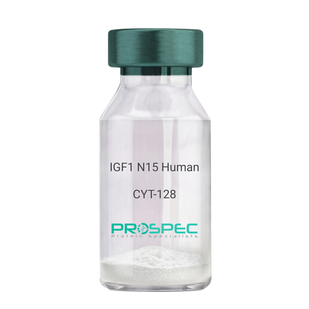
IGF1 N15 HumanDescription:
Insulin Like Growth Factor-1 N15 Labeled Human Recombinant
Somatomedin C, IGF-I, IGFI, IGF1, IGF-IA, Mechano growth factor, MGF.
Product # :
CYT-128Price :
Quantity :
Shipping Method :
Shipped at Room temp
More Info
- description
- source
- formulation
- purity
- biological activity
- More Info
- sds-page
- Citations
Description
IGF1 N15 Human Recombinant produced in E.coli is a single, non-glycosylated polypeptide chain containing 70 amino acids and having a molecular mass of 7.74kDa. The N15 is stable isotope labeled. The IGF1 N15 is purified by proprietary chromatographic techniques.
Source
Escherichia Coli.
Formulation
IGF1 N15 protein was lyophilized from a 0.2µm filtered concentrated solution in PBS, pH 7.2.
Purity
Greater than 97.0% as determined by:
(a) Analysis by RP-HPLC.
(b) Analysis by SDS-PAGE.Biological Activity
The ED50 as determined by a cell proliferation assay using serum free human MCF-7 cells is less than 2ng/ml, corresponding to a specific activity of > 5.0 × 10⁵ IU/mg.sds-page
Citations
More Info
-
Introduction
The somatomedins, or insulin-like growth factors (IGFs), comprise a family of peptides that play important roles in mammalian growth and development. IGF1 mediates many of the growth-promoting effects of growth hormone (GH; MIM 139250). Early studies showed that growth hormone did not directly stimulate the incorporation of sulfate into cartilage, but rather acted through a serum factor, termed 'sulfation factor,' which later became known as 'somatomedin' (Daughaday et al., 1972). Three main somatomedins have been characterized: somatomedin C (IGF1), somatomedin A (IGF2; MIM 147470), and somatomedin B (MIM 193190) (Rotwein, 1986; Rosenfeld, 2003).
-
Synonyms
Somatomedin C, IGF-I, IGFI, IGF1, IGF-IA, Mechano growth factor, MGF.
-
Physical Appearance
Sterile Filtered White lyophilized (freeze-dried) powder.
-
Stability
Lyophilized IGF1 N15 although stable at room temperature for 3 weeks, should be stored desiccated below -18°C. Upon reconstitution IGF1 N15 should be stored at 4°C between 2-7 days and for future use below -18°C. For long term storage it is recommended to add a carrier protein (0.1% HSA or BSA). Please prevent freeze-thaw cycles.
-
Solubility
It is recommended to reconstitute the lyophilized IGF1 N15 in sterile 18M-cm H2O not less than 100µg/ml, which can then be further diluted to other aqueous solutions.
-
Amino Acid Sequence
GPETLCGAEL VDALQFVCGD RGFYFNKPTG YGSSSRRAPQ TGIVDECCFR SCDLRRLEMY CAPLKPAKSA.
-
Background
What is the molecular weight/Mw of IGF1 N15 HUMAN Protein?
IGF1 N15 HUMAN Protein has a total Mw of 7.74kDa.
What is the source or expression system of IGF1 N15 HUMAN Protein?
Escherichia Coli.
What is the Purity of IGF1 N15 HUMAN Protein?
IGF1 N15 HUMAN Protein is >97% pure as determined by SDS-PAGE.
What is the Biological Activity of IGF1 N15 HUMAN Protein?
The ED50 as determined by a cell proliferation assay using serum free human MCF-7 cells is less than 2ng/ml, corresponding to a specific activity of > 5.0 × 10⁵ IU/mg.
What is the amino acid sequence of IGF1 N15 HUMAN Protein?
GPETLCGAEL VDALQFVCGD RGFYFNKPTG YGSSSRRAPQ TGIVDECCFR SCDLRRLEMY CAPLKPAKSA.
What applications can IGF1 N15 HUMAN Protein be used in?
IGF1 N15 HUMAN Protein can probably be used in western blot, ELISA and Lateral Flow.
What is the endotoxin level for IGF1 N15 HUMAN Protein?
The endotoxin level is minimal, IGF1 N15 HUMAN Protein was purified using conventional chromatography techniques.
ProSpec's products are furnished for LABORATORY RESEARCH USE ONLY. The product may not be used as drugs, agricultural or pesticidal products, food additives or household chemicals.
- View Data Sheet
Name :
Dengue Envelope-2 22kDaDescription:
Dengue Virus Subtype-2 Envelope 22kDa Recombinant
Product # :
DEN-007Price :
Quantity :
Shipping Method :
Shipped with Ice Packs
More Info
- description
- source
- formulation
- purity
- More Info
- Citations
Description
The E.coli derived recombinant 22 kDa protein is genetically engineered peptide which is derived from Dengue Type-2 Envelope. This region also contains a common antigen for Dengue IgG & IgM. This protein is fused to 6xHis tag.
Source
Escherichia Coli.
Formulation
Phosphorous buffered saline, pH-7.4.
Purity
Protein is >95% pure as determined by 12% PAGE (coomassie staining).
Citations
More Info
-
Introduction
Caused by one of four closely related virus serotypes of the genus Flavivirus, family Flaviviridae, each serotype is sufficiently different that there is no cross-protection and epidemics caused by multiple serotypes (hyperendemicity) can occur. In cell culture experiments and mice Morpholino antisense oligos have shown specific activity against Dengue virus.
-
Stability
Dengue Envelope ST2 although stable at 4°C for 1 week, should be stored below -18°C. Please prevent freeze thaw cycles.
-
Applications
Each laboratory should determine an optimum working titer for use in its particular application.
-
Purification Method
Purified by proprietary chromatographic technique.
ProSpec's products are furnished for LABORATORY RESEARCH USE ONLY. The product may not be used as drugs, agricultural or pesticidal products, food additives or household chemicals.
- View Data Sheet
Name :
WNV EnvelopeDescription:
West Nile Virus Envelope Recombinant
Product # :
WNV-001Price :
Quantity :
Shipping Method :
Shipped with Ice Packs
More Info
- description
- source
- formulation
- purity
- More Info
- Citations
Description
The E.Coli derived recombinant protein contains the West-Nile N-terminus Envelope Virus immunodominant regions. The protein migrates at 12KDa and is fused to a 6xHis tag at C terminus.
Source
Escherichia Coli.
Formulation
Tris base and 10Mm K2CO3.
Purity
Protein is >95% pure as determined by SDS-PAGE.
Citations
More Info
-
Introduction
West Nile virus (WNV) is a virus of the family Flaviviridae part of the Japanese encephalitis (JE) antigenic complex of viruses. Image reconstructions and cryoelectron microscopyreveal a 45-50 nm virion covered with a relatively smooth proteinsurface. This structure is similar to virus; both belong to the genus flavivirus within the family Flaviviridae. WNV is a positive-sense, single strand of RNA, it is between 11,000 and 12,000 nucleotides long which encode seven non-structural proteins and three structural proteins. The RNA strand is held within a nucleocapsid formed from 12 kDaprotein blocks; the capsid is contained within a host-derived membrane altered by two viral glycoproteins.
-
Stability
WNV Envelope although stable at 4°C for 1 week, should be stored below -18°C. Please prevent freeze thaw cycles.
-
Applications
ELISA and Lateral flow.
-
Purification Method
Purified by proprietary chromatographic technique.
ProSpec's products are furnished for LABORATORY RESEARCH USE ONLY. The product may not be used as drugs, agricultural or pesticidal products, food additives or household chemicals.
- View Data Sheet
Name :
SARS Spike MonoclonalDescription:
SARS-Spike protein, Monoclonal Antibody
Product # :
ANT-758Price :
Quantity :
Shipping Method :
Shipped with Ice Packs
More Info
- formulation
- More Info
- Citations
Formulation
0.5mg/ml, PBS, 0.2% gelatin and 0.05% sodium azide.
Citations
More Info
-
Introduction
SARS Coronavirus is an enveloped virus containing three outer structural proteins, namely the membrane (M), envelope (E), and spike (S) proteins. Spike (S)-glycoprotein of the virus interacts with a cellular receptor and mediates membrane fusion to allow viral entry into susceptible target cells. Accordingly, S-protein plays an important role in virus infection cycle and is the primary target of neutralizing antibodies.
It has recently been shown that SARS (severe acute respiratory syndrome) is caused by a human coronavirus. Human coronaviruses are the major cause of upper respiratory tract illness, such as the common cold, in humans. Coronaviruses are positive-stranded RNA viruses, featuring the largest viral RNA genomes known to date (27-31 kb). The first step in coronavirus infection is binding of the viral spike protein, a 139-kDa protein, to certain receptors on host cells. The spike protein is the main surface antigen of the coronavirus. The glycosilated spike protein (as well as the nucleocapsid protein) can be detected in infected cell culture supernatants with antisera from SARS patients. -
Stability
Store at 4C, stable for 2 weeks. For long-term storage, store at -20C.
-
Immunogen
The antibody was developed using a synthetic peptide from the Spike S2 glycoprotein for the Human SARS coronavirus (Genbank accession number NP_828851.1) corresponding to amino acids 1124-1140.
-
Applications
This monoclonal antibody can be used for detection of SARS Spike protein in western blot analysis at 0.5-2 ug/ml. Expected species cross reactivity based on immunogen sequence homology: COV-2 (100%)
-
Isotype
Mouse IgG3 kappa.
-
Type
Mouse antibody Monoclonal.
ProSpec's products are furnished for LABORATORY RESEARCH USE ONLY. The product may not be used as drugs, agricultural or pesticidal products, food additives or household chemicals.
- View Data Sheet
Name :
CoV-2 N-MosaicDescription:
Coronavirus 2019 Nucleocapsid Mosaic Recombinant
Product # :
SARS-015Price :
Quantity :
Shipping Method :
Shipped with Ice Packs
More Info
- description
- source
- formulation
- purity
- More Info
- Citations
Description
The E.Coli derived recombinant protein contains the Coronavirus 2019 full length nuclepocapsid Mosaic immunodominant regions [ full length N-antigen ], fused to 6xHis tag at C-terminal.
Source
Escherichia Coli.
Formulation
CoV 2019 Nucleocapsid-Mosaic Protein solution is supplied in 1x PBS.
Purity
CoV 2019 Nucleocapsid-Mosaic Protein ein is >90% pure as determined SDS-PAGE.
Citations
More Info
-
Introduction
A human infecting coronavirus (viral pneumonia) called 2019 novel coronavirus, 2019-nCoV was found in the fish market at the city of Wuhan, Hubei province of China on December 2019.
The 2019-nCoV shares an 87% identity to the 2 bat-derived severe acute respiratory syndrome 2018 SARS-CoV-2 located in Zhoushan of eastern China. 2019-nCoV has an analogous receptor-BD-structure to that of 2018 SARS-CoV, even though there is a.a. diversity so thus the 2019-nCoV might bind to ACE2 receptor protein (angiotensin-converting enzyme 2) in humans.
While bats are possibly the host of 2019-nCoV, researchers suspect that animal from the ocean sold at the seafood market was an intermediate host. RSCU analysis proposes that the 2019-nCoV is a recombinant within the viral spike glycoprotein between the bat coronavirus and an unknown coronavirus.
-
Physical Appearance
Sterile Filtered clear solution.
-
Stability
CoV 2019 Nucleocapsid-Mosaic Protein is shipped on ice packs. Upon arrival, Store at -20°C.
-
Specificity
Reactivity with SARS infected individuals not tested.
ProSpec's products are furnished for LABORATORY RESEARCH USE ONLY. The product may not be used as drugs, agricultural or pesticidal products, food additives or household chemicals.