Skip to content

High-quality research proteins.

  • 1-800-805-6531

  • Mon - Sat 8.00-18.00, Sun - Closed

  • info@prospecbio.com

  • PO Box 6591 East Brunswick NJ 08816

  • Products
    • Cytokines
    • Growth factors
    • Chemokines
    • Cd antigens
    • Neurotrophins
    • Hormones
    • Enzymes
    • Viral antigens
    • Recombinant proteins
    • Natural proteins
    • Monoclonal antibodies
    • Polyclonal antibodies
    • Thermal Packaging
  • About
    • Company
    • Founder
    • Contribution
  • Customers
    • Eastern Asia
    • Eastern Europe
    • North America
    • South America
    • South East Asia
    • Southern Asia
    • Western Europe
  • Ordering
    • Ordering Method
    • Ordering - Credit Card
    • Ordering - PO
    • Shipping Fees
    • FAQs
  • Services
    • DNA Cloning
    • Protein Expression
    • Monoclonal Antibody
    • Fermentation
    • Technology License
  • Contact us
Log in

Country/region

  • USD $ | Albania
  • USD $ | Andorra
  • USD $ | Argentina
  • USD $ | Armenia
  • USD $ | Australia
  • USD $ | Austria
  • USD $ | Azerbaijan
  • USD $ | Belarus
  • USD $ | Belgium
  • USD $ | Bolivia
  • USD $ | Brazil
  • USD $ | Bulgaria
  • USD $ | Cambodia
  • USD $ | Canada
  • USD $ | Chile
  • USD $ | China
  • USD $ | Colombia
  • USD $ | Costa Rica
  • USD $ | Croatia
  • USD $ | Cyprus
  • USD $ | Czechia
  • USD $ | Denmark
  • USD $ | Estonia
  • USD $ | Finland
  • USD $ | France
  • USD $ | Georgia
  • USD $ | Germany
  • USD $ | Greece
  • USD $ | Hong Kong SAR
  • USD $ | Hungary
  • USD $ | Iceland
  • USD $ | India
  • USD $ | Ireland
  • USD $ | Israel
  • USD $ | Italy
  • USD $ | Japan
  • USD $ | Kazakhstan
  • USD $ | Latvia
  • USD $ | Liechtenstein
  • USD $ | Lithuania
  • USD $ | Madagascar
  • USD $ | Malta
  • USD $ | Mexico
  • USD $ | Moldova
  • USD $ | Monaco
  • USD $ | Netherlands
  • USD $ | New Zealand
  • USD $ | North Macedonia
  • USD $ | Norway
  • USD $ | Panama
  • USD $ | Paraguay
  • USD $ | Peru
  • USD $ | Philippines
  • USD $ | Poland
  • USD $ | Portugal
  • USD $ | Romania
  • USD $ | Singapore
  • USD $ | Slovakia
  • USD $ | Slovenia
  • USD $ | South Africa
  • USD $ | South Korea
  • USD $ | Spain
  • USD $ | Sri Lanka
  • USD $ | Sweden
  • USD $ | Switzerland
  • USD $ | Taiwan
  • USD $ | Thailand
  • USD $ | Türkiye
  • USD $ | Ukraine
  • USD $ | United Kingdom
  • USD $ | United States
  • USD $ | Uruguay
  • USD $ | Vatican City
  • USD $ | Venezuela
  • USD $ | Vietnam
  • Facebook
  • Instagram
logoProSpec - Recombinant Protein Manufacturer for academic and R&D industry
Become a distributor
Account Log in
Wishlist
Cart Cart(0)
  • Products
    +
    • Cytokines
    • Growth factors
    • Chemokines
    • Cd antigens
    • Neurotrophins
    • Hormones
    • Enzymes
    • Viral antigens
    • Recombinant proteins
    • Natural proteins
    • Monoclonal antibodies
    • Polyclonal antibodies
    • Thermal Packaging
  • About
    +
    • Company
    • Founder
    • Contribution
  • Customers
    +
    • Eastern Asia
    • Eastern Europe
    • North America
    • South America
    • South East Asia
    • Southern Asia
    • Western Europe
  • Ordering
    +
    • Ordering Method
    • Ordering - Credit Card
    • Ordering - PO
    • Shipping Fees
    • FAQs
  • Services
    +
    • DNA Cloning
    • Protein Expression
    • Monoclonal Antibody
    • Fermentation
    • Technology License
  • Contact us

Item added to your cart

View cart
View All
  • Cytokines
  • Growth factors
  • Chemokines
  • Cd antigens
  • Neurotrophins
  • Hormones
  • Enzymes
  • Viral antigens
  • Recombinant proteins
  • Natural proteins
  • Monoclonal antibodies
  • Polyclonal antibodies
  • Thermal Packaging
  • Tumor Necrosis Factor

    Tumor Necrosis Factor

    View All
  • 4-1BB

    4-1BB

    View All
  • Adiponectin

    Adiponectin

    View All
  • AIF1

    AIF1

    View All
  • Other Cytokines

    Other Cytokines

    View All
  • AITRL

    AITRL

    View All
  • Angiopoietin

    Angiopoietin

    View All
  • Apolipoprotein

    Apolipoprotein

    View All
  • B-Cell Activating Factor

    B-Cell Activating Factor

    View All
  • Beta Defensin

    Beta Defensin

    View All
  • Bone Morphogenetic Protein

    Bone Morphogenetic Protein

    View All
  • B type Natriuretic Peptide

    B type Natriuretic Peptide

    View All
  • BST

    BST

    View All
  • Betacellulin

    Betacellulin

    View All
  • Cardiotrophin

    Cardiotrophin

    View All
  • Activin

    Activin

    View All
  • Fibroblast Growth Factor

    Fibroblast Growth Factor

    View All
  • Other Growth Factors

    Other Growth Factors

    View All
  • CSF

    CSF

    View All
  • CTGF

    CTGF

    View All
  • IGFBP

    IGFBP

    View All
  • VEGF Protein

    VEGF Protein

    View All
  • EGF

    EGF

    View All
  • Epigen

    Epigen

    View All
  • Erythropoietin

    Erythropoietin

    View All
  • Insulin-Like Growth Factor

    Insulin-Like Growth Factor

    View All
  • Growth Hormone

    Growth Hormone

    View All
  • HDGF

    HDGF

    View All
  • Hepatocyte Growth Factor

    Hepatocyte Growth Factor

    View All
  • Insulin

    Insulin

    View All
  • BCA-1/ BLC (CXCL13)

    BCA-1/ BLC (CXCL13)

    View All
  • BRAK (CXCL14)

    BRAK (CXCL14)

    View All
  • C-10 (CCL6)

    C-10 (CCL6)

    View All
  • Other Chemokines

    Other Chemokines

    View All
  • MEC (CCL28)

    MEC (CCL28)

    View All
  • LD78-beta (CCL3L1)

    LD78-beta (CCL3L1)

    View All
  • CTACK (CCL27)

    CTACK (CCL27)

    View All
  • CXCL16

    CXCL16

    View All
  • CXCL17

    CXCL17

    View All
  • Platelet Factor-4 (CXCL4)

    Platelet Factor-4 (CXCL4)

    View All
  • ENA-78 (CXCL5)

    ENA-78 (CXCL5)

    View All
  • Eotaxin (CCL11,24,26)

    Eotaxin (CCL11,24,26)

    View All
  • Exodus-2 (CCL21)

    Exodus-2 (CCL21)

    View All
  • Fractalkine (CX3CL1)

    Fractalkine (CX3CL1)

    View All
  • CXCL6

    CXCL6

    View All
  • Other CD Antigens

    Other CD Antigens

    View All
  • CD14

    CD14

    View All
  • CD164

    CD164

    View All
  • CD1

    CD1

    View All
  • CD2

    CD2

    View All
  • CD200

    CD200

    View All
  • CD207

    CD207

    View All
  • CD226

    CD226

    View All
  • CD23

    CD23

    View All
  • CD244

    CD244

    View All
  • CD247

    CD247

    View All
  • CD27

    CD27

    View All
  • CD274

    CD274

    View All
  • CD300

    CD300

    View All
  • CD33

    CD33

    View All
  • Other Neurotrophins

    Other Neurotrophins

    View All
  • Beta-NGF

    Beta-NGF

    View All
  • BDNF

    BDNF

    View All
  • CDNF

    CDNF

    View All
  • CNTF

    CNTF

    View All
  • GDNF

    GDNF

    View All
  • Glia Maturation Factor

    Glia Maturation Factor

    View All
  • MANF

    MANF

    View All
  • Midkine

    Midkine

    View All
  • Neuroglobin

    Neuroglobin

    View All
  • Neurotrophic factors

    Neurotrophic factors

    View All
  • Neuregulin

    Neuregulin

    View All
  • Neuropilin

    Neuropilin

    View All
  • Pigment Epithelium-Derived Factor

    Pigment Epithelium-Derived Factor

    View All
  • Pleiotrophin

    Pleiotrophin

    View All
  • Peptide Hormones

    Peptide Hormones

    View All
  • Other Hormones

    Other Hormones

    View All
  • HCG

    HCG

    View All
  • Endothelin

    Endothelin

    View All
  • Exendin

    Exendin

    View All
  • FSH

    FSH

    View All
  • Glucagon

    Glucagon

    View All
  • GHRP

    GHRP

    View All
  • GLP

    GLP

    View All
  • Thyrostimulin

    Thyrostimulin

    View All
  • Inhibin A

    Inhibin A

    View All
  • LHRH

    LHRH

    View All
  • Melanotan

    Melanotan

    View All
  • Procalcitonin

    Procalcitonin

    View All
  • PTH

    PTH

    View All
  • Peroxiredoxin

    Peroxiredoxin

    View All
  • Peptidase

    Peptidase

    View All
  • Synthetase

    Synthetase

    View All
  • Transferase

    Transferase

    View All
  • Hydrolase

    Hydrolase

    View All
  • Dehydrogenase

    Dehydrogenase

    View All
  • ACE2

    ACE2

    View All
  • Esterase

    Esterase

    View All
  • Protein Kinases

    Protein Kinases

    View All
  • Other Enzymes

    Other Enzymes

    View All
  • Phosphatase

    Phosphatase

    View All
  • Deaminase

    Deaminase

    View All
  • Oxygenase

    Oxygenase

    View All
  • Adenylate Kinase

    Adenylate Kinase

    View All
  • Lyase

    Lyase

    View All
  • Chagas

    Chagas

    View All
  • SARS

    SARS

    View All
  • Influenza

    Influenza

    View All
  • ASFV

    ASFV

    View All
  • Astrovirus

    Astrovirus

    View All
  • Borrelia

    Borrelia

    View All
  • Helicobacter Pylori

    Helicobacter Pylori

    View All
  • Chikungunya

    Chikungunya

    View All
  • Chlamydia

    Chlamydia

    View All
  • Cytomegalo

    Cytomegalo

    View All
  • Dengue

    Dengue

    View All
  • EBV

    EBV

    View All
  • Ebola

    Ebola

    View All
  • Feline Leukemia Virus

    Feline Leukemia Virus

    View All
  • Hepatitis A

    Hepatitis A

    View All
  • Other

    Other

    View All
  • Actin

    Actin

    View All
  • ADAM

    ADAM

    View All
  • Complement Component

    Complement Component

    View All
  • Ag85

    Ag85

    View All
  • Eukaryotic Translation Initiation Factor

    Eukaryotic Translation Initiation Factor

    View All
  • Anterior Gradient Protein

    Anterior Gradient Protein

    View All
  • Heat Shock Protein

    Heat Shock Protein

    View All
  • Alpha-2-HS-Glycoprotein

    Alpha-2-HS-Glycoprotein

    View All
  • Hemoglobin

    Hemoglobin

    View All
  • Allergy

    Allergy

    View All
  • Anaplasma

    Anaplasma

    View All
  • Angiogenin

    Angiogenin

    View All
  • Ankyrin Repeat Domain

    Ankyrin Repeat Domain

    View All
  • Annexin

    Annexin

    View All
  • Other Natural Proteins

    Other Natural Proteins

    View All
  • Aprotinin

    Aprotinin

    View All
  • Transferrin

    Transferrin

    View All
  • Avidin

    Avidin

    View All
  • Anti Coagulation Factors

    Anti Coagulation Factors

    View All
  • Natural Albumin

    Natural Albumin

    View All
  • Natural Coagulation Factors

    Natural Coagulation Factors

    View All
  • Fibronectin

    Fibronectin

    View All
  • Thrombin

    Thrombin

    View All
  • Anti Human Enzyme

    Anti Human Enzyme

    View All
  • Other Monoclonal Antibodies

    Other Monoclonal Antibodies

    View All
  • Anti Human Cytokine

    Anti Human Cytokine

    View All
  • Anti Human Heat Shock Protein

    Anti Human Heat Shock Protein

    View All
  • Anti Mouse Lymphocyte

    Anti Mouse Lymphocyte

    View All
  • Anti Human Chemokine

    Anti Human Chemokine

    View All
  • Anti Human Lymphocyte

    Anti Human Lymphocyte

    View All
  • Anti Viral Monoclonal

    Anti Viral Monoclonal

    View All
  • Anti Mouse Cytokine

    Anti Mouse Cytokine

    View All
  • Anti-GST Monoclonal

    Anti-GST Monoclonal

    View All
  • Other Polyclonal Antibodies

    Other Polyclonal Antibodies

    View All
  • Anti Viral

    Anti Viral

    View All
  • Anti-GST Monoclonal

    Anti-GST Monoclonal

    View All
Back

Search results

1000 results found for “Epiregulin”

Name

Description

Product #

Price

Quantity

Shipping Method

  • View Data Sheet

    Name :

    EIF1AY Human

    Description:

    Eukaryotic Translation Initiation Factor 1A Y-linked Recombinant Human

    Eukaryotic translation initiation factor 1A Y chromosome, Eukaryotic translation initiation factor 4C, eIF-4C, eIF-1A Y isoform.

    Product # :

    PRO-098

    Price :

    Quantity :

    Shipping Method :

    Ice Icon

    Shipped with Ice Packs

    Add To Cart

    More Info

    • description
    • source
    • formulation
    • purity
    • More Info
    • Citations

    Description

    EIF1AY produced in E.Coli is a single, non-glycosylated polypeptide chain containing 167 amino acids (1-144.a.a) and having a molecular mass of 18.8kDa. EIF1AY is fused to a 23 amino acid His-tag at N-terminus & purified by proprietary chromatographic techniques.

    Source

    Escherichia Coli.

    Formulation

    The EIF1AY protein solution (0.5mg/1ml) is formulated in 20 mM Tris-HCl buffer (pH8.0), 0.1M NaCl, 1mM DTT and 20% glycerol.

    Purity

    Greater than 95% as determined by SDS-PAGE.

    Citations

    More Info

    • Introduction

      EIF1AY is comparable to eukaryotic translation initiation factor 1A (EIF1A). EIF1AY protein is essential for highest rate of protein biosynthesis. EIF1AY increases ribosome dissociation into subunits and is obligatory for the binding of the 43S complex (a 40S subunit, eIF2/GTP/Met-tRNAi and eIF3) to the 5' end of capped RNA.

    • Synonyms

      Eukaryotic translation initiation factor 1A Y chromosome, Eukaryotic translation initiation factor 4C, eIF-4C, eIF-1A Y isoform.

    • Physical Appearance

      Sterile Filtered clear solution.

    • Stability

      Store at 4°C if entire vial will be used within 2-4 weeks. Store, frozen at -20°C for longer periods of time.Please avoid freeze thaw cycles.

    • Amino Acid Sequence

      MGSSHHHHHH SSGLVPRGSH MGSMPKNKGK GGKNRRRGKN ENESEKRELV FKEDGQEYAQ VIKMLGNGRL EALCFDGVKR LCHIRGKLRK KVWINTSDII LVGLRDYQDN KADVILKYNA DEARSLKAYG ELPEHAKINE TDTFGPGDDD EIQFDDIGDD DEDIDDI.

    ProSpec's products are furnished for LABORATORY RESEARCH USE ONLY. The product may not be used as drugs, agricultural or pesticidal products, food additives or household chemicals.

    Eif1Ay Human
  • View Data Sheet

    Name :

    EIF3J Human

    Description:

    Eukaryotic Translation Initiation Factor 3J Human Recombinant

    Eukaryotic translation initiation factor 3 subunit J, Eukaryotic translation initiation factor 3 subunit 1, eIF-3-alpha, eIF3 p35, EIF3J, EIF3S1, eIF3-alpha.

    Product # :

    PRO-970

    Price :

    Quantity :

    Shipping Method :

    Ice Icon

    Shipped with Ice Packs

    Add To Cart

    More Info

    • description
    • source
    • formulation
    • purity
    • More Info
    • Citations

    Description

    EIF3J Human Recombinant produced in E.coli is a single, non-glycosylated polypeptide chain containing 210 amino acids (70-258) and having a molecular mass of 24kDa (Molecular weight on SDS-PAGE will appear higher).EIF3J is fused to a 21 amino acid His-tag at N-terminus & purified by proprietary chromatographic techniques.

    Source

    Escherichia Coli.

    Formulation

    The EIF3J solution (0.5mg/ml) contains 20mM Tris-HCl buffer (pH 8.0), 2mM DTT, 10% glycerol and 200mM NaCl.

    Purity

    Greater than 90.0% as determined by SDS-PAGE.

    Citations

    More Info

    • Introduction

      Eukaryotic translation initiation factor 3 subunit J (EIF3J) is a member of the EIF-3 subunit J family. EIF3 has a crucial role in binding of initiator methionyl-tRNA and mRNA to the 40S ribosomal subunit to produce the 40S initiation complex. EIF3J binds to the aminoacyl (A) site and mRNA access channel of the 40S subunit, setting EIF3J directly in the ribosomal decoding center. In addition, EIF3J interacts with eIF1A and reduces 40S subunit affinity for mRNA. An elevated affinity for mRNA is restored upon enlistment of initiator tRNA, despite the fact EIF3J remains in the mRNA-binding cleft in the presence of tRNA.

    • Synonyms

      Eukaryotic translation initiation factor 3 subunit J, Eukaryotic translation initiation factor 3 subunit 1, eIF-3-alpha, eIF3 p35, EIF3J, EIF3S1, eIF3-alpha.

    • Physical Appearance

      Sterile Filtered colorless solution.

    • Stability

      Store at 4°C if entire vial will be used within 2-4 weeks. Store, frozen at -20°C for longer periods of time. For long term storage it is recommended to add a carrier protein (0.1% HSA or BSA).Avoid multiple freeze-thaw cycles.

    • Amino Acid Sequence

      MGSSHHHHHH SSGLVPRGSH MKISEKKKIA EKIKEKERQQ KKRQEEIKKR LEEPEEPKVL TPEEQLADKL RLKKLQEESD LELAKETFGV NNAVYGIDAM NPSSRDDFTE FGKLLKDKIT QYEKSLYYAS FLEVLVRDVC ISLEIDDLKK ITNSLTVLCS EKQKQEKQSK AKKKKKGVVP GGGLKATMKD DLADYGGYDG GYVQDYEDFM.

    ProSpec's products are furnished for LABORATORY RESEARCH USE ONLY. The product may not be used as drugs, agricultural or pesticidal products, food additives or household chemicals.

    Eif3J Human
  • View Data Sheet

    Name :

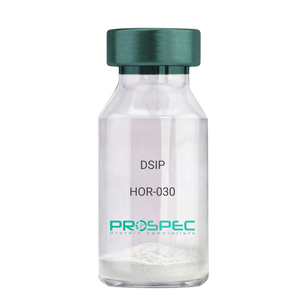
    DSIP

    Description:

    Delta Sleep Inducing Peptide

    Product # :

    HOR-030

    Price :

    Quantity :

    Shipping Method :

    Room Temp Icon

    Shipped at Room temp

    Add To Cart

    More Info

    • description
    • formulation
    • purity
    • More Info
    • Citations

    Description

    DSIP Synthetic is a single, non-glycosylated polypeptide chain containing 15 amino acids, having a molecular mass of 1419.55 Dalton and a Molecular formula of C62H98N16O22.

    Formulation

    The protein was lyophilized with no additives.

    Purity

    Greater than 97.0% as determined by analysis by RP-HPLC.

    Citations

    More Info

    • Physical Appearance

      Sterile Filtered White lyophilized (freeze-dried) powder.

    • Stability

      Lyophilized DSIP although stable at room temperature for 3 weeks, should be stored desiccated below -18°C. Upon reconstitution DSIP should be stored at 4°C between 2-7 days and for future use below -18°C. For long term storage it is recommended to add a carrier protein (0.1% HSA or BSA). Please prevent freeze-thaw cycles.

    • Solubility

      It is recommended to reconstitute the lyophilized DSIP in sterile 18MΩ-cm H2O not less than 100 µg/ml, which can then be further diluted to other aqueous solutions.

    • Amino Acid Sequence

      H-Gly-Glu-Pro-Pro-Pro-Gly-Lys-Pro-Ala-Asp-Asp-Ala-Gly-Leu-Val-OH

    • Background

      Delta Sleep-Inducing Peptide (DSIP), also known as Sleep-Promoting Peptide, is a neuropeptide that has been the subject of extensive research due to its potential role in sleep regulation, stress response, and neuroprotection. This nonapeptide, first isolated from the cerebral venous blood of rabbits during sleep, has been shown to induce slow-wave sleep, modulate pain perception, and exhibit potential antioxidant and immunomodulatory properties.

      DSIP's primary function is its interaction with the sleep regulatory system. By modulating the release of certain neurotransmitters, DSIP can influence sleep patterns, particularly promoting slow-wave sleep, the most restorative stage of sleep. Studies by Kovalzon et al. (2011) have demonstrated that DSIP can enhance sleep quality in rats, suggesting potential applications in sleep disorders and the promotion of healthy sleep patterns.

      In addition to its sleep-inducing effects, DSIP has been shown to possess neuroprotective properties. Research by Zolotarev et al. (2014) found that DSIP could protect neurons from oxidative stress, suggesting potential applications in the treatment of neurodegenerative diseases such as Alzheimer's and Parkinson's disease.

      Given its sleep-inducing and neuroprotective effects, DSIP has been proposed as a potential therapeutic agent for a variety of conditions, including sleep disorders, chronic pain, and neurodegenerative diseases. For instance, a study by Spong et al. (2016) found that DSIP could improve sleep quality in patients with chronic insomnia, indicating its potential as a therapeutic agent in the treatment of sleep disorders.

      While research on DSIP is promising, it is important to note that most studies have been conducted in animals or in vitro. More research is needed to fully understand the potential effects and applications of DSIP in humans. However, the existing body of research suggests that DSIP could be a promising tool in the treatment of sleep disorders, chronic pain, and neurodegenerative diseases.

    ProSpec's products are furnished for LABORATORY RESEARCH USE ONLY. The product may not be used as drugs, agricultural or pesticidal products, food additives or household chemicals.

    Dsip
  • View Data Sheet

    Name :

    Darbepoetin

    Description:

    Darbepoetin-Alpha Human Recombinant

    Erythropoietin-Mutant, EPO-mutant, NESP, Novel Erythropoiesis Stimulating Protein, EPO Mutant.

    Product # :

    CYT-1263

    Price :

    Quantity :

    Shipping Method :

    Room Temp Icon

    Shipped at Room temp

    Add To Cart

    More Info

    • description
    • source
    • formulation
    • purity
    • biological activity
    • More Info
    • Citations

    Description

    Darbepoetin-alpha Human Recombinant is produced in Chinese hamster ovary (CHO) cells by recombinant DNA technology is a glycosylated polypeptide chain containing 165 amino acids and having a predicted molecular mass of 18,200 Dalton and apparent glycosylated molecular mass of 37-40kDa. Darbepoetin is purified by proprietary chromatographic techniques.

    Source

    Chinese Hamster Ovary Cells(CHO).

    Formulation

    Each mg of lyophilized Darbepoetin powder contains 20mM Phosphate Buffer, 0.15M NaCl pH-6.2 & 0.005% Tween-80.

    Purity

    Greater than 97.0% as determined by SDS-PAGE.

    Biological Activity

    The Specific Activity was measured by Normocyth -aemic mice and was found to be 1,000,000 IU/mg.

    Citations

    More Info

    • Introduction

      Darbepoetin is a mutant recombinant human EPO-Alpha protein re-engineered, containing 5 amino acid changes at N30, T32, V87, N88, T90. Darbepoetin exhibits longer half-life stimulating red blood cell production than Erythropoietin. Darbepoetin maintains hemoglobin more effectively compared to EPO-Alpha.

    • Synonyms

      NESP, Novel Erythropoiesis Stimulating Protein, EPO Mutant.

    • Physical Appearance

      Sterile Filtered White lyophilized (freeze-dried) powder.

    • Stability

      Lyophilized Darbepoetin although stable at room temperature for 3 weeks, should be stored desiccated below -18°C. Upon reconstitution Darbepoetin should be stored at 4°C between 2-7 days and for future use below -18°C.For long term storage it is recommended to add a carrier protein (0.1% HSA or BSA).Please prevent freeze-thaw cycles.

    • Solubility

      It is recommended to reconstitute the lyophilized Darbepoetin in sterile 18MΩ-cm H2O not less than 100µg/ml, which can then be further diluted to other aqueous solutions.

    • Amino Acid Sequence

      APPRLICDSR VLERYLLEAK EAENITTGCN ETCSLNENIT VPDTKVNFYA WKRMEVGQQA VEVWQGLALL SEAVLRGQAL LVNSSQVNET LQLHVDKAVS GLRSLTTLLR ALGAQKEAIS PPDAASAAPL RTITADTFRK LFRVYSNFLR GKLKLYTGEA CRTGD.

    • Background

      What is the molecular weight/Mw of DARBEPOETIN Protein?
      DARBEPOETIN Protein has a total Mw of 38.5kDa.

      What is the source or expression system of DARBEPOETIN Protein?
      Chinese Hamster Ovary Cells(CHO).

      What is the Purity of DARBEPOETIN Protein?
      DARBEPOETIN Protein is >97% pure as determined by SDS-PAGE.

      What is the Biological Activity of DARBEPOETIN Protein?
      The Specific Activity was measured by Normocyth -aemic mice and was found to be 1,000,000 IU/mg.

      What is the amino acid sequence of DARBEPOETIN Protein?
      APPRLICDSR VLERYLLEAK EAENITTGCN ETCSLNENIT VPDTKVNFYA WKRMEVGQQA VEVWQGLALL SEAVLRGQAL LVNSSQVNET LQLHVDKAVS GLRSLTTLLR ALGAQKEAIS PPDAASAAPL RTITADTFRK LFRVYSNFLR GKLKLYTGEA CRTGD

      What applications can DARBEPOETIN Protein be used in?
      DARBEPOETIN Protein can probably be used in western blot, ELISA and Lateral Flow.

      What is the endotoxin level for DARBEPOETIN Protein?
      The endotoxin level is minimal, DARBEPOETIN Protein was purified using conventional chromatography techniques

    ProSpec's products are furnished for LABORATORY RESEARCH USE ONLY. The product may not be used as drugs, agricultural or pesticidal products, food additives or household chemicals.

    Darbepoetin
  • View Data Sheet

    Name :

    EPHA2 Human

    Description:

    EPH Receptor A2 Human Recombinant

    EPHA2, EPH Receptor A2, ECK, Tyrosine-Protein Kinase Receptor ECK, EC 2.7.10.1, CTRCT6, ARCC2, CTPP1, CTPA, Epithelial Cell Receptor Protein Tyrosine Kinase, Ephrin Type-A Receptor 2, Soluble EPHA2 Variant 1, Epithelial Cell Kinase, EC 2.7.10, EphA2.

    Product # :

    PRO-2032

    Price :

    Quantity :

    Shipping Method :

    Room Temp Icon

    Shipped at Room temp

    Add To Cart

    More Info

    • description
    • source
    • formulation
    • purity
    • More Info
    • Citations

    Description

    EPHA2 Human Recombinant produced in HEK cells is a single, glycosylated, polypeptide chain (Ala24-Glu530) containing a total of 515 amino acids, having a calculated molecular mass of 56.9kDa. The EPHA2 protein is fused to a 2 aa C-terminal linker and a 6 aa C-terminal His tag.

    Source

    HEK 293.

    Formulation

    EPHA2 was filtered (0.4µm) and lyophilized from 0.5mg/ml solution in phosphate buffered saline and 5% (w/v) trehalose.

    Purity

    Greater than 95.0% as determined by SDS-PAGE.

    Citations

    More Info

    • Introduction

      EPH Receptor A2 (EPHA2) is a member of the ephrin receptor subfamily of the protein-tyrosine kinase family. EPHA2 is a protein which binds ephrin-A ligands. EPH and EPH-related receptors are associated with mediating developmental events, particularly in the nervous system. Receptors in the EPH subfamily normally have a single kinase domain and an extracellular region containing a Cys-rich domain and 2 fibronectin type III repeats. The ephrin receptors are divided into two groups based on the similarity of their extracellular domain sequences and their affinities for binding ephrin-A and ephrin-B ligands. EPHA2 gene mutations are the cause of certain genetically-related cataract disorders.

    • Synonyms

      EPHA2, EPH Receptor A2, ECK, Tyrosine-Protein Kinase Receptor ECK, EC 2.7.10.1, CTRCT6, ARCC2, CTPP1, CTPA, Epithelial Cell Receptor Protein Tyrosine Kinase, Ephrin Type-A Receptor 2, Soluble EPHA2 Variant 1, Epithelial Cell Kinase, EC 2.7.10, EphA2.

    • Physical Appearance

      Filtered White lyophilized (freeze-dried) powder.

    • Stability

      Store lyophilized protein at -20°C. Aliquot the product after reconstitution to avoid repeated freezing/thawing cycles. Reconstituted protein can be stored at 4°C for a limited period of time; it does not show any change after two weeks at 4°C.

    • Solubility

      It is recommended to add deionized water to prepare a working stock solution of approximately 0.5 mg/ml and let the lyophilized pellet dissolve completely. EPHA2 is not sterile! Please filter the product by an appropriate sterile filter before using it in the cell culture.

    • Amino Acid Sequence

      AQGKEVVLLD FAAAGGELGW LTHPYGKGWD LMQNIMNDMP IYMYSVCNVM SGDQDNWLRT NWVYRGEAER IFIELKFTVR DCNSFPGGAS SCKETFNLYY AESDLDYGTN FQKRLFTKID TIAPDEITVS SDFEARHVKL NVEERSVGPL TRKGFYLAFQ DIGACVALLS VRVYYKKCPE LLQGLAHFPE TIAGSDAPSL ATVAGTCVDH AVVPPGGEEP RMHCAVDGEW LVPIGQCLCQ AGYEKVEDAC QACSPGFFKF EASESPCLEC PEHTLPSPEG ATSCECEEGF FRAPQDPASM PCTRPPSAPH YLTAVGMGAK VELRWTPPQD SGGREDIVYS VTCEQCWPES GECGPCEASV RYSEPPHGLT RTSVTVSDLE PHMNYTFTVE ARNGVSGLVT SRSFRTASVS INQTEPPKVR LEGRSTTSLS VSWSIPPPQQ SRVWKYEVTY RKKGDSNSYN VRRTEGFSVT LDDLAPDTTY LVQVQALTQE GQGAGSKVHE FQTLSPEKLH HHHHH.

    ProSpec's products are furnished for LABORATORY RESEARCH USE ONLY. The product may not be used as drugs, agricultural or pesticidal products, food additives or household chemicals.

    Epha2 Human
  • View Data Sheet

    Name :

    EGFR Human Sf9

    Description:

    Epidermal Growth Factor Receptor Sf9 Human Recombinant

    Epidermal growth factor receptor, EC 2.7.10.1, Receptor tyrosine-protein kinase ErbB-1, ERBB, mENA, ERBB1, EGFR.

    Product # :

    PKA-344

    Price :

    Quantity :

    Shipping Method :

    Room Temp Icon

    Shipped at Room temp

    Add To Cart

    More Info

    • description
    • source
    • formulation
    • purity
    • More Info
    • Citations

    Description

    The EGFR contains the extracellular domain of the human EGFR (25-647 a.a.) excluding the signal peptide which is cleaved by the insect cells having an approximate Mw of 85kDa. The EGFR is fused to a C-terminal Strep-tag and purified by proprietary chromatographic techniques.

    Source

    Sf9 Insect Cells.

    Formulation

    ErbB1 was lyophilized from a concentrated (1mg/ml) sterile solution containing 1x PBS pH-7.4.

    Purity

    Greater than 90.0% as determined by SDS-PAGE.

    Citations

    More Info

    • Introduction

      The epidermal growth factor receptor (EGF R) subfamily of receptor tyrosine kinases comprises four members: EGF R (also known as HER1, ErbB1 or ErbB), ErbB2 (Neu, HER-2), ErbB3 (HER-3), and ErbB4 (HER-4). All family members are type I transmembrane glycoprotein that has an extracellular domain which contains two cysteine-rich domains separated by a spacer region that is involved in ligand-binding, and a cytoplasmic domain which has a membrane-proximal tyrosine kinase domain and a C-terminal tail with multiple tyrosine autophosphorylation sites. The human EGF R gene encodes a 1210 amino acid (aa) residue precursor with a 24 aa putative signal peptide, a 621 aa extracellular domain, a 23 aa transmembrane domain, and a 542 aa cytoplasmic domain. EGF R has been shown to bind a subset of the EGF family ligands, including EGF, amphiregulin, TGF-a , betacellulin, epiregulin, HBEGF and neuregulin-2 in the absence of a co-receptor. Ligand binding induces EGF R homodimerization as well as heterdimerization with ErbB2, resulting in kinase activation, tyrosine phosphorylation and cell signaling. EGF R can also be recruited to form heterodimers with the ligand-activated ErbB3 or ErbB4. EGF R signaling has been shown to regulate multiple biological functions including cell proliferation, differentiation, motility and apoptosis. In addition, EGF R signaling has also been shown to play a role in carcinogenesis.

    • Synonyms

      Epidermal growth factor receptor, EC 2.7.10.1, Receptor tyrosine-protein kinase ErbB-1, ERBB, mENA, ERBB1, EGFR.

    • Physical Appearance

      Sterile Filtered White lyophilized (freeze-dried) powder.

    • Stability

      Lyophilized EGFR although stable at room temperature for 3 weeks, should be stored desiccated below -18°C. Upon reconstitution EGFR should be stored at 4°C between 2-7 days and for future use below -18°C.For long term storage it is recommended to add a carrier protein (0.1% HSA or BSA).Please prevent freeze-thaw cycles.

    • Solubility

      It is recommended to reconstitute the lyophilized EGFR in sterile PBS not less than 100µg/ml, which can then be further diluted to other aqueous solutions.

    • Amino Acid Sequence

      LEEKKV CQGTSNKLTQ LGTFEDHFLS LQRMFNNCEV VLGNLEITYV QRNYDLSFLK TIQEVAGYVL IALNTVERIP LENLQIIRGN MYYENSYALA VLSNYDANKT GLKELPMRNL QEILHGAVRF SNNPALCNVE SIQWRDIVSS DFLSNMSMDF QNHLGSCQKC DPSCPNGSCW GAGEENCQKL TKIICAQQCS GRCRGKSPSD CCHNQCAAGC TGPRESDCLV CRKFRDEATC KDTCPPLMLY NPTTYQMDVN PEGKYSFGAT CVKKCPRNYV VTDHGSCVRA CGADSYEMEE DGVRKCKKCE GPCRKVCNGI GIGEFKDSLS INATNIKHFK NCTSISGDLH ILPVAFRGDS FTHTPPLDPQ ELDILKTVKE ITGFLLIQAW PENRTDLHAF ENLEIIRGRT KQHGQFSLAV VSLNITSLGL RSLKEISDGD VIISGNKNLC YANTINWKKL FGTSGQKTKI ISNRGENSCK ATGQVCHALC SPEGCWGPEP RDCVSCRNVS RGRECVDKCK LLEGEPREFV ENSECIQCHP ECLPQAMNIT CTGRGPDNCI QCAHYIDGPH CVKTCPAGVM GENNTLVWKY ADAGHVCHLC HPNCTYGCTG PGLEGCPTNG PKIPSIAASW SHPQFEK.

    ProSpec's products are furnished for LABORATORY RESEARCH USE ONLY. The product may not be used as drugs, agricultural or pesticidal products, food additives or household chemicals.

    Egfr Human Sf9
  • View Data Sheet

    Name :

    Adiponectin Porcine, HEK

    Description:

    Adiponectin Porcine Recombinant, HEK derived

    Acrp30, AdipoQ, GBP-28, APM-1, ACDC.

    Product # :

    CYT-696

    Price :

    Quantity :

    Shipping Method :

    Room Temp Icon

    Shipped at Room temp

    Add To Cart

    More Info

    • description
    • source
    • formulation
    • purity
    • More Info
    • Citations

    Description

    The Acrp30 Porcine Recombinant contains a total of 238 amino acids having a molecular Mass of 26kDa. The Porcine Adiponectin is fused to a 13 amino acid long N-terminal FLAG tag.

    Source

    HEK293 (Human Embryonic Kidney cell line).

    Formulation

    Sterile filtered and lyophilized from 0.5mg/ml in 20mM Tris buffer and 50mM NaCl pH-7.5.

    Purity

    Greater than 90% as determined by SDS PAGE.

    Citations

    More Info

    • Introduction

      Adiponectin is a recently discovered 244 amino acid protein, the product of the apM1 gene, which is physiologically active and specifically and highly expressed in adipose cells (Adipokine). The protein belongs to the soluble defense collagen super family; it has a collagen-like domain structurally homologous with collagen VIII and X and complement factor C1q-like globular domain. APM-1 forms homotrimers, which are the building blocks for higher order complexes found circulating in serum.

    • Synonyms

      Acrp30, AdipoQ, GBP-28, APM-1, ACDC.

    • Stability

      For long term, store lyophilized AdipoQ at -20°C. Aliquot the product after reconstitution to avoid repeated freezing/thawing cycles. Reconstituted protein can be stored at 4°C for a limited period of time; it does not show any change after two weeks at 4°C.The lyophilized protein remains stable for 24 months when stored at -20°C.

    • Solubility

      Add deionized water and let the lyophilized pellet dissolve completely.

    • Amino Acid Sequence

      HVDYKDDDDK PAGETTEKPG ALLPMPKGAC AGWMAGIPGH PGHNGTPGRD GRDGVPGEKG EKGDTGLTGP KGDTGESGVT GVEGPRGFPG IPGRKGEPGE SAYVYRSAFS VGLETRVTVP NMPIRFTKIF YNQQNHYDVT TGKFHCNIPG LYYFSFHITV LKDVKVSLYK DKAVLFTYDQ QDKNVDQASG VLLYLEKGDQ WLQAYGDEEN GVYADNVNDS FTGFLLYHNIE.

    • Background

      What is the molecular weight/Mw of ADIPONECTIN Protein?
      ADIPONECTIN Protein has a total Mw of 26kDa.

      What is the source or expression system of ADIPONECTIN Protein?
      HEK293

      What is the Purity of ADIPONECTIN Protein?
      ADIPONECTIN Protein is >90% pure as determined by SDS-PAGE.

      What is the Biological Activity of ADIPONECTIN Protein?
      The biological functionality of ADIPONECTIN Protein will be determined in the future.

      What is the amino acid sequence of ADIPONECTIN Protein?
      HVDYKDDDDK PAGETTEKPG ALLPMPKGAC AGWMAGIPGH PGHNGTPGRD GRDGVPGEKG EKGDTGLTGP KGDTGESGVT GVEGPRGFPG IPGRKGEPGE SAYVYRSAFS VGLETRVTVP NMPIRFTKIF YNQQNHYDVT TGKFHCNIPG LYYFSFHITV LKDVKVSLYK DKAVLFTYDQ QDKNVDQASG VLLYLEKGDQ WLQAYGDEEN GVYADNVNDS FTGFLLYHNIE.

      What applications can ADIPONECTIN Protein be used in?
      ADIPONECTIN Protein can probably be used in western blot, ELISA and Lateral Flow.

      What is the endotoxin level for ADIPONECTIN Protein?
      The endotoxin level is minimal, ADIPONECTIN Protein was purified using conventional chromatography techniques.

    ProSpec's products are furnished for LABORATORY RESEARCH USE ONLY. The product may not be used as drugs, agricultural or pesticidal products, food additives or household chemicals.

    Adiponectin Porcine Hek
  • View Data Sheet

    Name :

    EGF Human

    Description:

    Epidermal Growth Factor Human Recombinant

    Urogastrone, URG, EGF.

    Product # :

    CYT-217

    Price :

    Quantity :

    Shipping Method :

    Room Temp Icon

    Shipped at Room temp

    Add To Cart

    More Info

    • description
    • source
    • formulation
    • purity
    • biological activity
    • More Info
    • Citations

    Description

    Epidermal Growth Factor Human Recombinant produced in E.Coli is a single, non-glycosylated, polypeptide chain containing 53 amino acids and having a molecular mass of 6.2kDa. The EGF is purified by proprietary chromatographic techniques.

    Source

    Escherichia Coli.

    Formulation

    EGF was lyophilized from a concentrated (1mg/ml) solution containing PBS pH-7.4.

    Purity

    Greater than 98.0% as determined by SDS-PAGE.

    Biological Activity

    The ED50 as determined by a cell proliferation assay using murine Balb/c 3T3 cells is less than 0.1 ng/ml, corresponding to a specific activity of >1.0x10⁷ IU/mg.

    Citations

    More Info

    • Introduction

      Epidermal growth factor has a profound effect on the differentiation of specific cells in vivo and is a potent mitogenic factor for a variety of cultured cells of both ectodermal and mesodermal origin. The EGF precursor is believed to exist as a membrane-bound molecule which is proteolytically cleaved to generate the 53-amino acid peptide hormone that stimulates cells to divide. EGF stimulates the growth of various epidermal and epithelial tissues in vivo and in vitro and of some fibroblasts in cell culture.

    • Synonyms

      Urogastrone, URG, EGF.

    • Physical Appearance

      Sterile Filtered White lyophilized (freeze-dried) powder.

    • Stability

      Lyophilized Epidermal Growth Factor Recombinant although stable at room temperature for 3 weeks, should be stored desiccated below -18°C. Upon reconstitution EGF should be stored at 4°C between 2-7 days and for future use below -18°C.For long term storage it is recommended to add a carrier protein (0.1% HSA or BSA).Please prevent freeze-thaw cycles.

    • Solubility

      It is recommended to reconstitute the lyophilized Epidermal Growth Factor in sterile 18MΩ-cm H2O not less than 100µg/ml, which can then be further diluted to other aqueous solutions.

    • Amino Acid Sequence

      NSDSECPLSH DGYCLHDGVC MYIEALDKYA CNCVVGYIGE RCQYRDLKWW ELR.

    • Background

      About EGF:

      In the sphere of biomedical studies, epidermal boom factor (EGF) is a cornerstone that gives precious insights into the mechanisms underlying tissue healing, differentiation, and mobile proliferation. In this article we will explore the characteristics and uses of epidermal growth factor (EGF).

      Description:

      Epidermal growth factor (EGF) is a 6-kDa protein consisting of 53 amino acid residues and 3 intramolecular disulfide linkages. Human tissues, such as platelets, the parotid gland, and the submandibular gland, are rich in EGF. EGF, which was first discovered in human urine and the submaxillary glands of mice, functions as a major modulator of cell proliferation by attaching to its receptor, EGFR, which is found on the cell membrane. EGF triggers autophosphorylation of transmembrane protein tyrosine kinase EGFR upon binding, hence initiating downstream signaling cascades through pathways such as phosphatidylinositol and ras. Beyond the cell membrane, EGF has a variety of roles as it also initiates cytoplasmic processes such actin depolymerization and membrane ruffle formation. Studies indicate that EGF and its receptor might possibly be important components of the nucleus, highlighting the complexity of EGF-mediated cellular responses.

      Function:

      By attaching to the epidermal growth factor receptor (EGFR), EGF promotes the survival, differentiation, and multiplication of cells. This connection is essential for boosting many physiological processes and stimulating cell proliferation. The preservation of oro-esophageal and stomach tissue integrity is greatly supported by salivary EGF, which is regulated by dietary inorganic iodine. Its actions include the healing of gastric and oral ulcers, the inhibition of gastric acid secretion, the stimulation of DNA synthesis, and the protection of mucosal surfaces against harmful substances such as bile acids, gastric acid, and bacteria. Salivary EGF's role extends to repairing gastric tissue and addressing oro-esophagal issues, showcasing its healing ability in resolving oral and gastrointestinal ailments, including ulcers.

      Mechanism:

      EGF functions by forming a strong bond with the cell surface's epidermal growth factor receptor (EGFR), which triggers ligand- induced dimerization. This incident sets off the intrinsic protein-tyrosine kinase activity of EGFR, which in turn initiates a signal transduction cascade inside the cell. Numerous biochemical changes are brought about by this cascade, such as increased intracellular calcium levels, increased glycolysis and protein synthesis, and increased expression of particular genes, most notably the EGFR gene. These carefully planned alterations eventually promote DNA synthesis and cell division, illuminating the complex process by which EGF directs basic biological functions and modulates cellular responses.

      What is the molecular weight/Mw of EGF Protein?
      EGF Protein has a total Mw of 6.2kDa.

      What is the source or expression system of EGF Protein?
      Escherichia Coli.

      What is the Purity of EGF Protein?
      EGF Protein is >98% pure as determined by SDS-PAGE.

      What is the Biological Activity of EGF Protein?
      The ED50 as determined by a cell proliferation assay using murine Balb/c 3T3 cells is less than 0.1 ng/ml, corresponding to a specific activity of >1.0x10⁷ IU/mg.

      What is the amino acid sequence of EGF Protein?
      NSDSECPLSH DGYCLHDGVC MYIEALDKYA CNCVVGYIGE RCQYRDLKWW ELR.

      What applications can EGF Protein be used in?
      EGF Protein can probably be used in western blot, ELISA and Lateral Flow.

      What is the endotoxin level for EGF Protein?
      The endotoxin level is minimal, EGF Protein was purified using conventional chromatography techniques.

    ProSpec's products are furnished for LABORATORY RESEARCH USE ONLY. The product may not be used as drugs, agricultural or pesticidal products, food additives or household chemicals.

    Egf Human
  • View Data Sheet

    Name :

    EPHB1 Human

    Description:

    EPH Receptor B1 Human Recombinant

    EPHB-1, Ephrin type-B receptor 1, Eph Receptor B1, ELK, EPH tyrosine kinase 2, EPH-like kinase 6, EK6, hEK6, HEK6, Neuronally-expressed EPH-related tyrosine kinase, NET, NETHek6, Tyrosine-protein kinase receptor EPH-2, EPHT2, soluble EPHB1 variant 1

    Product # :

    PRO-2728

    Price :

    Quantity :

    Shipping Method :

    Ice Icon

    Shipped with Ice Packs

    Add To Cart

    More Info

    • description
    • source
    • formulation
    • purity
    • biological activity
    • More Info
    • Citations

    Description

    EPHB1 Human Recombinant produced in HEK293 Cells is a single, glycosylated polypeptide chain containing 529 amino acids (18-540 a.a) and having a molecular mass of 59.2 kDa.EPHB1 is expressed with a 6 amino acid His tag at C-Terminus and purified by proprietary chromatographic techniques.

    Source

    HEK293 Cells.

    Formulation

    The EPHB1 solution (0.25mg/1ml) contains phosphate buffered saline (pH7.4) and 10% glycerol.

    Purity

    Greater than 95.0% as determined by SDS-PAGE.

    Biological Activity

    Measured by its binding ability in a functional ELISA with Human EFNB1 (cat# pro-2543).

    Citations

    More Info

    • Introduction

      EPH Receptor B1 (EPHB1) is a member of the ephrin receptor subfamily of the proteintyrosine kinase family which 16 receptors are known. EPHB1 binds ephrin-B2, ephrin-B1,ephrin‑A3,ephrin-A1,ephrin-B3 and ephrin-A4. EPHB1 binds tyrosine kinase and phosphorylates syndecan-2 and thisphosphorylation is necessary for syndecan-2 clustering and spine formation. Ephrin receptors and theirligands (the ephrins) mediate several developmental processes, mainly in the nervous system.

    • Synonyms

      EPHB-1, Ephrin type-B receptor 1, Eph Receptor B1, ELK, EPH tyrosine kinase 2, EPH-like kinase 6, EK6, hEK6, HEK6, Neuronally-expressed EPH-related tyrosine kinase, NET, NETHek6, Tyrosine-protein kinase receptor EPH-2, EPHT2, soluble EPHB1 variant 1

    • Physical Appearance

      Sterile filtered colorless solution.

    • Stability

      Store at 4°C if entire vial will be used within 2-4 weeks.Store, frozen at -20°C for longer periods of time.For long term storage it is recommended to add a carrier protein (0.1% HSA or BSA).Avoid multiple freeze-thaw cycles.

    • Amino Acid Sequence

      MEETLMDTRT ATAELGWTAN PASGWEEVSG YDENLNTIRT YQVCNVFEPN QNNWLLTTFI NRRGAHRIYT EMRFTVRDCS SLPNVPGSCK ETFNLYYYET DSVIATKKSA FWSEAPYLKV DTIAADESFS QVDFGGRLMK VNTEVRSFGP LTRNGFYLAF QDYGACMSLL SVRVFFKKCP SIVQNFAVFP ETMTGAESTS LVIARGTCIP NAEEVDVPIK LYCNGDGEWM VPIGRCTCKP GYEPENSVAC KACPAGTFKA SQEAEGCSHC PSNSRSPAEA SPICTCRTGY YRADFDPPEV ACTSVPSGPR NVISIVNETS IILEWHPPRE TGGRDDVTYN IICKKCRADR RSCSRCDDNV EFVPRQLGLT ECRVSISSLW AHTPYTFDIQ AINGVSSKSP FPPQHVSVNI TTNQAAPSTV PIMHQVSATM RSITLSWPQP EQPNGIILDY EIRYYEKEHN EFNSSMARSQ TNTARIDGLR PGMVYVVQVR ARTVAGYGKF SGKMCFQTLT DDDYKSELRE QLPHHHHHH

    ProSpec's products are furnished for LABORATORY RESEARCH USE ONLY. The product may not be used as drugs, agricultural or pesticidal products, food additives or household chemicals.

    Ephb1 Human
  • View Data Sheet

    Name :

    ACVR1 Human

    Description:

    Activin A Receptor Type 1 Human Recombinant

    ACVR1A, ALK2, ACVR1, ACTRI, ACTR-I, ACVRLK2, FOP, SKR1, TSRI, Activin receptor type I, Activin receptor-like kinase 2, ALK-2, TSR-I, Serine/threonine-protein kinase receptor R1, TGF-B superfamily receptor type I.

    Product # :

    CYT-1140

    Price :

    Quantity :

    Shipping Method :

    Ice Icon

    Shipped with Ice Packs

    Add To Cart

    More Info

    • description
    • source
    • formulation
    • purity
    • More Info
    • sds-page
    • Citations

    Description

    ACVR1 produced in Sf9 Baculovirus cells is a single, glycosylated polypeptide chain containing 342 amino acids (21-123a.a.) and having a molecular mass of 38.4kDa. ACVR1 is expressed with a 239 amino acid hIgG-His-Tag at C-Terminus and purified by proprietary chromatographic techniques.

    Source

    Sf9, Baculovirus cells.

    Formulation

    ACVR1 protein solution (0.25mg/ml) contains Phosphate Buffered Saline (pH 7.4) and 10% glycerol.

    Purity

    Greater than 90.0% as determined by SDS-PAGE.

    sds-page

    ACVR1 Human sds-page - Product image 1

    Citations

    More Info

    • Introduction

      Activin A Receptor Type 1 (ACVR1) is a member of TGF-beta serine/threonine kinase receptor family. ACVR1 forms a receptor complex contains2 type II and 2 type I transmembrane serine/threonine kinases. Type II receptors phosphorylate and activate type I receptors which autophosphorylate,bind and activate SMAD transcriptional regulators. ACVR1 takes part in left-right pattern formation during embryogenesis and is also essential in the BMP pathway which is responsible for the development and repair of the skeletal system.ACVR1 is linked to Fibrodysplasia Ossificans Progressiva which isknown for the formation of heterotopic bone throughout the body.

    • Synonyms

      ACVR1A, ALK2, ACVR1, ACTRI, ACTR-I, ACVRLK2, FOP, SKR1, TSRI, Activin receptor type I, Activin receptor-like kinase 2, ALK-2, TSR-I, Serine/threonine-protein kinase receptor R1, TGF-B superfamily receptor type I.

    • Physical Appearance

      Sterile filtered colorless solution.

    • Stability

      Store at 4°C if entire vial will be used within 2-4 weeks.Store, frozen at -20°C for longer periods of time. For long term storage it is recommended to add a carrier protein (0.1% HSA or BSA).Avoid multiple freeze-thaw cycles.

    • Amino Acid Sequence

      MEDEKPKVNP KLYMCVCEGL SCGNEDHCEG QQCFSSLSIN DGFHVYQKGC FQVYEQGKMT
      CKTPPSPGQA VECCQGDWCN RNITAQLPTK GKSFPGTQNF HLELEPKSCD KTHTCPPCPA
      PELLGGPSVF LFPPKPKDTL MISRTPEVTC VVVDVSHEDP EVKFNWYVDG VEVHNAKTKP
      REEQYNSTYR VVSVLTVLHQ DWLNGKEYKC KVSNKALPAP IEKTISKAKG QPREPQVYTL
      PPSRDELTKN QVSLTCLVKG FYPSDIAVEW ESNGQPENNY KTTPPVLDSD GSFFLYSKLT
      VDKSRWQQGN VFSCSVMHEA LHNHYTQKSL SLSPGKHHHH HH.

    • Background

      Functional Implications and Therapeutic Prospects of Activin A Receptor Type 1 Human Recombinant

      1. Abstract

      This study illuminates the functional roles and potential therapeutic applications of Activin A Receptor Type 1 Human Recombinant (ACVR1), a crucial protein in the TGF-beta superfamily signaling pathway. Through a comprehensive review of its structure, signaling mechanism, biological functions, and disease associations, this paper aims to elucidate the current understanding of ACVR1 and its potential therapeutic implications in various disease states.

      2. Introduction

      The Activin A Receptor Type 1 Human Recombinant, abbreviated as ACVR1, is a receptor protein vital for transmitting cellular signals in the Transforming Growth Factor-beta (TGF-beta) superfamily pathway. Known to play pivotal roles in organogenesis, bone growth, and cell differentiation, the ACVR1 and its functions present vast therapeutic potential.

      3. Structure and Signaling of ACVR1

      ACVR1 is a transmembrane serine/threonine kinase receptor, characterized by an extracellular ligand-binding domain and an intracellular kinase domain for signal transduction. Binding of ligands such as Activin A leads to the formation of heteromeric complexes with type II receptors, triggering phosphorylation events that activate downstream signaling pathways.

      4. Biological Functions of ACVR1

      Being a part of the TGF-beta superfamily signaling pathway, ACVR1 is implicated in a broad spectrum of biological processes. It is crucial for embryonic development, cellular proliferation, differentiation, apoptosis, and homeostasis. It also plays a significant role in bone morphogenesis, contributing to skeletal patterning and growth.

      5. ACVR1 in Disease Pathology

      The dysregulation of ACVR1 has been associated with various pathological conditions, including Fibrodysplasia Ossificans Progressiva (FOP), a rare genetic disorder characterized by progressive ossification of soft tissues. Mutations in ACVR1 lead to enhanced BMP signaling, causing aberrant bone formation. This highlights the critical role of ACVR1 in skeletal homeostasis and disease.

      6. Therapeutic Potential of ACVR1

      Given the central role of ACVR1 in cellular signaling and its association with disease, it presents a promising target for therapeutic intervention. Strategies to modulate ACVR1 signaling could potentially ameliorate symptoms of diseases like FOP, offering promising avenues for novel therapeutic approaches.

      7. Conclusion and Future Perspectives

      While our understanding of ACVR1's functional roles has expanded significantly over the years, much remains to be elucidated. Further research into the precise molecular mechanisms of ACVR1 and its pathway will pave the way for therapeutic advances, enhancing our capability to combat various diseases.

      What is the molecular weight / Mw of ACVR1 Protein?
      ACVR1 Protein has a total Mw of 38.4kDa.

      What is the source or expression system of ACVR1 Protein?
      Sf9, Baculovirus cells.

      What is the Purity of ACVR1 Protein?
      ACVR1 Protein is >90% pure as determined by SDS-PAGE.

      What is the Biological Activity of ACVR1 Protein?
      The biological functionality of ACVR1 Protein will be determined in the future.

      What is the endotoxin level for ACVR1 Protein?
      The endotoxin level is minimal, ACVR1 Protein was purified using conventional chromatography techniques.

      What is the amino acid sequence of ACVR1 Protein?
      MEDEKPKVNP KLYMCVCEGL SCGNEDHCEG QQCFSSLSIN DGFHVYQKGC FQVYEQGKMT
      CKTPPSPGQA VECCQGDWCN RNITAQLPTK GKSFPGTQNF HLELEPKSCD KTHTCPPCPA
      PELLGGPSVF LFPPKPKDTL MISRTPEVTC VVVDVSHEDP EVKFNWYVDG VEVHNAKTKP
      REEQYNSTYR VVSVLTVLHQ DWLNGKEYKC KVSNKALPAP IEKTISKAKG QPREPQVYTL
      PPSRDELTKN QVSLTCLVKG FYPSDIAVEW ESNGQPENNY KTTPPVLDSD GSFFLYSKLT
      VDKSRWQQGN VFSCSVMHEA LHNHYTQKSL SLSPGKHHHH HH.

      What applications can ACVR1 Protein be used in?
      ACVR1 Protein can probably be used in western blot, ELISA and Lateral Flow

    ProSpec's products are furnished for LABORATORY RESEARCH USE ONLY. The product may not be used as drugs, agricultural or pesticidal products, food additives or household chemicals.

    Acvr1 Human
  • View Data Sheet

    Name :

    Adiponectin Protein

    Description:

    Adiponectin Human Recombinant

    Acrp30, AdipoQ, GBP-28, APM-1, ACDC.

    Product # :

    CYT-280

    Price :

    Quantity :

    Shipping Method :

    Ice Icon

    Shipped with Ice Packs

    Add To Cart

    More Info

    • description
    • source
    • formulation
    • purity
    • More Info
    • sds-page
    • Citations

    Description

    The Adiponectin Human recombinant protein is a single, non-glycosilated polypeptide chain produced in E. coli, having a molecular weight of 25.1 kDa and containing 231 amino acids (15-244).

    Source

    Escherichia Coli.

    Formulation

    Acrp30 protein solution contains Phosphate buffered saline pH 7.4 and 1mM DTT.

    Purity

    Acrp30 purity is greater than 90% as determined by SDS-PAGE.

    sds-page

    Adiponectin-sds-page - Product image 1

    Citations

    More Info

    • Introduction

      The adipose tissue exclusively expresses and secretes Adiponectin (Acrp30). Acrp30 is involved in various physiological processes such as energy homeostasis, insulin sensitivity, hormonal processes, fatty acid metabolism and obesity.
      Adiponectin circulates in the plasma. Decreased levels of Adiponectin are associated with insulin resistance and hyperinsulinemia, as seen in people with obesity insulin resistance, and diabetes type 2, whose plasma levels of adiponectin are reduced.
      The modular structure of Acrp30 is comprised of N-terminal collagenous domain followed by a C-terminal globular domain.
      Acrp30 also acts as a significant negative regulator in hematopoiesis and immune systems; it may be involved in ending inflammatory responses through its inhibitory functions. Adiponectin inhibits endothelial NF-kappa-b signaling through a cAMP-dependent pathway, it also inhibits TNF-alpha- induced expression of endothelial adhesion molecules.

    • Synonyms

      Acrp30, AdipoQ, GBP-28, APM-1, ACDC.

    • Physical Appearance

      Sterile Filtered clear solution.

    • Stability

      Store at 4°C if entire vial will be used within 2-4 weeks. Store, frozen at -20°C for longer periods of time. For long term storage it is recommended to add a carrier protein (0.1% HSA or BSA).Avoid multiple freeze-thaw cycles.

    • Amino Acid Sequence

      MGHDQETTTQGPGVLLPLPKGACTGWMAGIPGHPGHNGAPGRDGRDGTPGE
      KGEKGDPGLIGPKGDIGETGVPGAEGPRGFPGIQGRKGEPGEGAYVYRSAFSV
      GLETYVTIPNMPIRFTKIFYNQQNHYDGSTGKFHCNIPGLYYFAYHITVYMKD
      VKVSLFKKDKAMLFTYDQYQENNVDQASGSVLLHLEVGDQVWLQVYGEGE
      RNGLYADNDNDSTFTGFLLYHDTN.

    • Background

      Adiponectin Human Recombinant: Unraveling its Potential in Therapeutic Applications

      1. Abstract

      This paper aims to deliver an extensive exploration into Adiponectin Human Recombinant, a vital adipokine implicated in a multitude of metabolic processes. By delving into the structure, biological roles, and signaling pathways of adiponectin, we elucidate its contribution to pathophysiological conditions. Moreover, we examine the potential therapeutic application of adiponectin in metabolic and cardiovascular diseases.

      2. Introduction

      Adiponectin, a protein predominantly secreted by adipose tissue, plays an integral part in regulating metabolic processes such as glucose regulation and fatty acid oxidation. Understanding the intricacies of adiponectin's actions could pave the way for innovative therapeutic interventions in diseases like obesity, diabetes, and cardiovascular disease.

      3. Structure and Signaling of Adiponectin

      Adiponectin is a 30kDa protein consisting of a collagen-like domain and a C-terminal globular domain. It signals through adiponectin receptors AdipoR1 and AdipoR2, which then activate several intracellular signaling pathways, including AMP-activated protein kinase (AMPK) and peroxisome proliferator-activated receptor-alpha (PPAR-α), regulating various metabolic processes.

      4. Biological Functions of Adiponectin

      Adiponectin has been shown to enhance insulin sensitivity, stimulate fatty acid oxidation, and exert anti-inflammatory effects. Additionally, it is involved in regulating energy homeostasis and has been linked to the regulation of food intake and body weight.

      5. Adiponectin in Disease Pathology

      Reduced levels of adiponectin have been associated with obesity, insulin resistance, type 2 diabetes, and cardiovascular disease. Moreover, adiponectin deficiency has been observed in metabolic syndrome, emphasizing the adipokine's crucial role in metabolic health.

      6. Therapeutic Potential of Adiponectin

      Given adiponectin's role in metabolic regulation, its potential as a therapeutic target is of considerable interest. Approaches to increase circulating adiponectin levels or enhance adiponectin signaling could offer potential therapeutic strategies for managing metabolic diseases and cardiovascular conditions.

      7. Conclusion and Future Perspectives

      While our understanding of adiponectin and its role in health and disease has greatly advanced in recent years, there is still much to uncover. Further research on the precise molecular mechanisms of adiponectin could pave the way for novel therapeutic approaches.

      What is the molecular weight / Mw of ADIPONECTIN Protein?
      ADIPONECTIN Protein has a total Mw of 25.1kDa.
      What is the source or expression system of ADIPONECTIN Protein?
      Escherichia Coli.

      What is the Purity of ADIPONECTIN Protein?
      ADIPONECTIN Protein is >90% pure as determined by SDS-PAGE.

      What is the Biological Activity of ADIPONECTIN Protein?
      The biological functionality of ADIPONECTIN Protein will be determined in the future.

      What is the amino acid sequence of ADIPONECTIN Protein?
      MGHDQETTTQGPGVLLPLPKGACTGWMAGIPGHPGHNGAPGRDGRDGTPGE
      KGEKGDPGLIGPKGDIGETGVPGAEGPRGFPGIQGRKGEPGEGAYVYRSAFSV
      GLETYVTIPNMPIRFTKIFYNQQNHYDGSTGKFHCNIPGLYYFAYHITVYMKD
      VKVSLFKKDKAMLFTYDQYQENNVDQASGSVLLHLEVGDQVWLQVYGEGE
      RNGLYADNDNDSTFTGFLLYHDTN..

      What applications can ADIPONECTIN Protein be used in?
      ADIPONECTIN Protein can probably be used in western blot, ELISA and Lateral Flow.

      What is the endotoxin level for ADIPONECTIN Protein?
      The endotoxin level is minimal, ADIPONECTIN Protein was purified using conventional chromatography techniques

    ProSpec's products are furnished for LABORATORY RESEARCH USE ONLY. The product may not be used as drugs, agricultural or pesticidal products, food additives or household chemicals.

    Adiponectin Human
  • View Data Sheet

    Name :

    Leptin qA Rat, PEG

    Description:

    Leptin Quadruple Antagonist, Pegylated Rat Recombinant

    OB Protein, Obesity Protein, OBS, Obesity factor.

    Product # :

    CYT-1241

    Price :

    Quantity :

    Shipping Method :

    Room Temp Icon

    Shipped at Room temp

    Add To Cart

    More Info

    • description
    • source
    • formulation
    • purity
    • biological activity
    • More Info
    • Citations

    Description

    Leptin Pegylated Quadruple Antagonist Rat Recombinant is a single non-glycosilated polypeptide chain containing 146 amino and additional Ala at N-terminus acids. The Rat Leptin antagonist is bound to 20 kDa mono-PEG at N-terminus, resulting in 35.6 kDa. The Rat Leptin Pegylated Quadruple Antagonist was mutated, resulting in D23L/L39A/D40A/F41A that was purified by proprietary chromatographic techniques.

    Source

    Escherichia coli.

    Formulation

    The Rat Leptin Pegylated Quadruple Antagonist was lyophilized from a concentrated (0.65mg/ml) solution with 0.003mM NaHCO3.

    Purity

    Greater than 98.0% as determined by:

    (a) Gel filtration analysis.

    (b) Analysis by SDS-PAGE.

    Biological Activity

    Rat Leptin Pegylated Quadruple Antagonist inhibits leptin-induced proliferation of BAF/3 cells stably transfected with the long form of human leptin receptor. Its in vitro.

    Citations

    More Info

    • Synonyms

      OB Protein, Obesity Protein, OBS, Obesity factor.

    • Physical Appearance

      White lyophilized (freeze-dried) powder.

    • Stability

      Lyophilized Rat Leptin Pegylated Quadruple Antagonist although stable at room temperature for several weeks, should be stored desiccated below -18°C. Upon reconstitution at > 0.1 and up to 2mM of rat pegylated leptin antagonist and filter sterilization rat pegylated leptin antagonist can be stored at 4°C or even room temperature for several weeks making it suitable for long term infusion studies using osmotic pumps. At lower concentration addition of a carrier protein (0.1% HSA or BSA) is suggested. Please prevent freeze-thaw cycles.

    • Solubility

      It is recommended to reconstitute the lyophilized Rat Leptin Pegylated Quadruple Antagonist in sterile water or sterile 0.4% NaHCO3adjusted to pH 8-9, not less than 100µg/ml, which can then be further diluted with other aqueous solutions.

    • Background

      Leptin is a hormone which mainly produced by adipocytes. Leptin’s main part is to regulate long-term energy balance. Leptin is encoded by the LEP gene. Leptin receptors are expressed by various brain and peripheral cell types. leptin levels influence satiety, appetite and triggers behaviors which lead to energy savings. High leptin levels are interpreted by the brain that energy reserves are high, whereas low leptin levels means that energy reserves are low, in the process adapting the organism to starvation through a variety of metabolic, neurobiochemical, endocrine and behavioral change.

    ProSpec's products are furnished for LABORATORY RESEARCH USE ONLY. The product may not be used as drugs, agricultural or pesticidal products, food additives or household chemicals.

    Leptin Rat Peg Qa
  • View Data Sheet

    Name :

    EIF3K Human

    Description:

    Eukaryotic Translation Initiation Factor 3K Human Recombinant

    Eukaryotic Translation Initiation Factor 3 Subunit K, PLAC-24, eIF3k, eIF-3 p25, eIF-3 p28, EIF3S12, HSPC029, M9, PRO1474, PTD001, Muscle-specific gene M9 protein, ARG134, MSTP001, muscle specific, Eukaryotic Translation Initiation Factor 3 subunit 12.

    Product # :

    PRO-179

    Price :

    Quantity :

    Shipping Method :

    Ice Icon

    Shipped with Ice Packs

    Add To Cart

    More Info

    • description
    • source
    • formulation
    • purity
    • More Info
    • Citations

    Description

    EIF3K produced in E.Coli is a single, non-glycosylated polypeptide chain containing 238 amino acids (1-218a.a.) and having a molecular mass of 27.2 kDa. EIF3K is fused to a 20 amino acid His-tag at N-terminus & purified by proprietary chromatographic techniques.

    Source

    Escherichia Coli.

    Formulation

    The EIF3K protein solution (1mg/1ml) is formulated in 20mM Tris-HCl buffer (pH8.0), 100mM NaCl, 1mM DTT and 20% glycerol.

    Purity

    Greater than 95% as determined by SDS-PAGE.

    Citations

    More Info

    • Introduction

      EIF3K is a member of the eIF3 subunit K family. EIF3K is the smallest subunit of eIF3 and it interacts with a number of other subunits of eIF3 and the 40S ribosomal subunit. EIF3K is conserved among high eukaryotes, including mammals, insects, and plants, and it is universally expressed in human tissues. EIF3K is distributed both in nucleus and cytoplasm and colocalized with cyclin D3, a regulatory subunit of cyclin-dependent kinase 4 (Cdk4).

    • Synonyms

      Eukaryotic Translation Initiation Factor 3 Subunit K, PLAC-24, eIF3k, eIF-3 p25, eIF-3 p28, EIF3S12, HSPC029, M9, PRO1474, PTD001, Muscle-specific gene M9 protein, ARG134, MSTP001, muscle specific, Eukaryotic Translation Initiation Factor 3 subunit 12.

    • Physical Appearance

      Sterile Filtered clear solution.

    • Stability

      Store at 4°C if entire vial will be used within 2-4 weeks. Store, frozen at -20°C for longer periods of time. For long term storage it is recommended to add a carrier protein (0.1% HSA or BSA).Avoid multiple freeze-thaw cycles.

    • Amino Acid Sequence

      MGSSHHHHHH SSGLVPRGSH MAMFEQMRAN VGKLLKGIDR YNPENLATLE RYVETQAKEN AYDLEANLAV LKLYQFNPAF FQTTVTAQIL LKALTNLPHT DFTLCKCMID QAHQEERPIR QILYLGDLLE TCHFQAFWQA LDENMDLLEG ITGFEDSVRK FICHVVGITY QHIDRWLLAE MLGDLSDSQL KVWMSKYGWS ADESGQIFIC SQEESIKPKN IVEKIDFDSV SSIMASSQ

    ProSpec's products are furnished for LABORATORY RESEARCH USE ONLY. The product may not be used as drugs, agricultural or pesticidal products, food additives or household chemicals.

    Eif3K Human
  • View Data Sheet

    Name :

    PPARG Human

    Description:

    Peroxisome Proliferator Activated Receptor Gamma Human Recombinant

    Peroxisome proliferator-activated receptor gamma, PPAR-gamma, PPARG, NR1C3, PPARG1, PPARG2.

    Product # :

    PKA-228

    Price :

    Quantity :

    Shipping Method :

    Ice Icon

    Shipped with Ice Packs

    Add To Cart

    More Info

    • description
    • source
    • formulation
    • purity
    • More Info
    • Citations

    Description

    Peroxisome Proliferator Activated Receptor Gamma Human Recombinant is expressed in E.coli having a molecular weight of 59.2 kDa and fused to an amino terminal hexahistidine tag.

    Source

    Escherichia Coli.

    Formulation

    The protein solution contains 20mM Tris-HCl and 50% glycerol.

    Purity

    Greater than 90.0% as determined by SDS-PAGE.

    Citations

    More Info

    • Introduction

      The peroxisome proliferator activated receptors (PPARs) are ligandactivated transcription factors within the nuclear receptor superfamily, which play important roles in adipogenesis, glucose homeostasis andinflammation. Three different isotypes can be distinguished; alpha, beta and gamma. PPARg, is mainly expressed in adipose tissue and to lesser extent in colon, the immune system and the retina. Whereas PPARg operates in the catabolism of fatty acids in the liver, PPARg influences the storage of fatty acids in the adipose tissue and plays a role in adipocyte differentiation. PPARg binds to DNA as a heterodimer with retinoid X receptor (RXR), activating expression of target genes after binding ligand.

    • Synonyms

      Peroxisome proliferator-activated receptor gamma, PPAR-gamma, PPARG, NR1C3, PPARG1, PPARG2.

    • Physical Appearance

      Sterile Filtered clear solution.

    • Stability

      Store at 4°C if entire vial will be used within 2-4 weeks.Store, frozen at -20°C for longer periods of time.Avoid multiple freeze-thaw cycles.

    ProSpec's products are furnished for LABORATORY RESEARCH USE ONLY. The product may not be used as drugs, agricultural or pesticidal products, food additives or household chemicals.

    Pparg Human
  • View Data Sheet

    Name :

    CHGA Human, His

    Description:

    Chromogranin-A Human Recombinant, His Tag

    CGA, CHGA, Vasostatin-2, Pituitary secretory protein I, SP-I.

    Product # :

    PRO-699

    Price :

    Quantity :

    Shipping Method :

    Ice Icon

    Shipped with Ice Packs

    Add To Cart

    More Info

    • description
    • source
    • formulation
    • purity
    • More Info
    • Citations

    Description

    Recombinant Human CHGA produced in E.Coli is a single, non-glycosylated polypeptide chain containing 460 amino acids (19-457 a.a) and having a molecular mass of 51.2kDa (Molecular weight on SDS-PAGE will appear higher). Chromgranin-A is fused to 21 amino acid His Tag at N-terminus and purified by proprietary chromatographic techniques.

    Source

    Escherichia Coli.

    Formulation

    The CHGA protein (0.5mg/ml) contains 20mM Tris-HCl buffer pH-7.5, 2mM EDTA, 0.1mM PMSF and 10% glycerol.

    Purity

    Greater than 80.0% as determined by Analysis by SDS-PAGE.

    Citations

    More Info

    • Introduction

      Chromgranin-A is part of the neuroendocrine secretory protein family. CHGA is located in secretory vesicles of neurons and endocrine cells. Chromgranin-A is a precursor to three biologically active peptides; vasostatin, pancreastatin, and parastatin. These peptides act as autocrine or paracrine negative modulators of the neuroendocrine system. Other peptides, including chromostatin, beta-granin, WE-14 and GE-25, are also derived from the full-length protein. Chromgranin-A has numerous biological activities on some tissues and organs and exerts a large spectrum of homeostatic actions, including antifungal and antimicrobial effect, modulation of cell adhesion, and inhibition of parathyroid hormone secretion.

    • Synonyms

      CGA, CHGA, Vasostatin-2, Pituitary secretory protein I, SP-I.

    • Physical Appearance

      Sterile filtered colorless solution.

    • Stability

      Store at 4°C if entire vial will be used within 2-4 weeks. Store, frozen at -20°C for longer periods of time. For long term storage it is recommended to add a carrier protein (0.1% HSA or BSA). Avoid multiple freeze-thaw cycles.

    • Amino Acid Sequence

      MGSSHHHHHH SSGLVPRGSH MLPVNSPMNK GDTEVMKCIV EVISDTLSKP SPMPVSQECF ETLRGDERIL SILRHQNLLK ELQDLALQGA KERAHQQKKH SGFEDELSEV LENQSSQAEL KEAVEEPSSK DVMEKREDSK EAEKSGEATD GARPQALPEP MQESKAEGNN QAPGEEEEEE EEATNTHPPA SLPSQKYPGP QAEGDSEGLS QGLVDREKGL SAEPGWQAKR EEEEEEEEEA EAGEEAVPEE EGPTVVLNPH PSLGYKEIRK GESRSEALAV DGAGKPGAEE AQDPEGKGEQ EHSQQKEEEE EMAVVPQGLF RGGKSGELEQ EEERLSKEWE DSKRWSKMDQ LAKELTAEKR LEGQEEEEDN RDSSMKLSFR ARAYGFRGPG PQLRRGWRPS SREDSLEAGL PLQVRGYPEE KKEEEGSANR RPEDQELESL SAIEAELEKV AHQLQALRRG.

    ProSpec's products are furnished for LABORATORY RESEARCH USE ONLY. The product may not be used as drugs, agricultural or pesticidal products, food additives or household chemicals.

    Chromogranin A Human His
  • View Data Sheet

    Name :

    DGCR6 Human

    Description:

    DiGeorge Syndrome Critical Region Gene 6 Human Recombinant

    DiGeorge syndrome critical region gene 6, Protein DGCR6, DGCR6.

    Product # :

    PRO-1372

    Price :

    Quantity :

    Shipping Method :

    Ice Icon

    Shipped with Ice Packs

    Add To Cart

    More Info

    • description
    • source
    • formulation
    • purity
    • More Info
    • Citations

    Description

    DGCR6 Human Recombinant produced in E.Coli is a single, non-glycosylated polypeptide chain containing 243 amino acids (1-220 a.a) and having a molecular mass of 27kDa. DGCR6 is fused to a 23 amino acid His-tag at N-terminus & purified by proprietary chromatographic techniques.

    Source

    E.coli.

    Formulation

    DGCR6 protein solution (0.5mg/ml) containing 20mM Tris-HCl buffer (pH 8.0), 0.2M NaCl and 20% glycerol.

    Purity

    Greater than 95% as determined by SDS-PAGE.

    Citations

    More Info

    • Introduction

      DiGeorge syndrome critical region gene 6 (DGCR6) shares homology with the Drosophila melanogaster gonadal protein, that participates in gonadal and germ cell development, and also with the gamma-1 subunit of human laminin. DGCR6 is involved in DiGeorge syndrome pathology and in schizophrenia.

    • Synonyms

      DiGeorge syndrome critical region gene 6, Protein DGCR6, DGCR6.

    • Physical Appearance

      Sterile Filtered colorless solution.

    • Stability

      Store at 4°C if entire vial will be used within 2-4 weeks. Store, frozen at -20°C for longer periods of time. For long term storage it is recommended to add a carrier protein (0.1% HSA or BSA).Avoid multiple freeze-thaw cycles.

    • Amino Acid Sequence

      MGSSHHHHHH SSGLVPRGSH MGSMERYAGA LEEVADGARQ QERHYQLLSA LQSLVKELPS SFQQRLSYTT LSDLALALLD GTVFEIVQGL LEIQHLTEKS LYNQRLRLQN EHRVLRQALR QKHQEAQQAC RPHNLPVLQA AQQRELEAVE HRIREEQRAM DQKIVLELDR KVADQQSTLE KAGVAGFYVT TNPQELMLQM NLLELIRKLQ QRGCWAGKAA LGLGGPWQLP AAQCDQKGSP VPP.

    ProSpec's products are furnished for LABORATORY RESEARCH USE ONLY. The product may not be used as drugs, agricultural or pesticidal products, food additives or household chemicals.

    Dgcr6 Human
  • View Data Sheet

    Name :

    PRDX2 Mouse

    Description:

    Eukaryotic Translation Initiation Factor 4E Mouse Recombinant

    PRDX2, Peroxiredoxin-2 (EC:1.11.1.15), TSA, Thioredoxin peroxidase 1, Thioredoxin-dependent peroxide reductase 1, Thiol-specific antioxidant protein, Prdx2, Tdpx1, Tpx.  

    Product # :

    ENZ-1061

    Price :

    Quantity :

    Shipping Method :

    Ice Icon

    Shipped with Ice Packs

    Add To Cart

    More Info

    • description
    • source
    • formulation
    • purity
    • biological activity
    • More Info
    • Citations

    Description

    PRDX2 Mouse Recombinant produced in E.Coli is a single, non-glycosylated polypeptide chain containing 222 amino acids (1-198 a.a) and having a molecular mass of 24.3kDa. PRDX2 is fused to a 24 amino acid His-tag at N-terminus & purified by proprietary chromatographic techniques.

    Source

    Escherichia Coli.

    Formulation

    PRDX2 protein solution (1mg/ml) containing 20mM Tris-HCl buffer (pH8.0) and 10% Glycerol 1mM DTT.

    Purity

    Greater than 90.0% as determined by SDS-PAGE.

    Biological Activity

    Specific activity is > 700 pmol/min/ug, Activity is defined as the amount of hydroperoxide that 1ug of enzyme can reduce at 25C for minute. 

    Citations

    More Info

    • Introduction

      PRDX2 is part of the peroxiredoxin family of antioxidant enzymes, which reduce hydrogen peroxide and alkyl hydroperoxides. PRDX2 takes part as an antioxidant protective role in cells, and contributes to the antiviral activity of CD8(+) T-cells. PRDX2 has proliferative effect in cancer development or progression.
      If PRDX2 protection is insufficient against peroxidases, the DNA damage results in neurological disease such as Alzheimer's or DNA damage leading to cancer.

    • Synonyms

      PRDX2, Peroxiredoxin-2 (EC:1.11.1.15), TSA, Thioredoxin peroxidase 1, Thioredoxin-dependent peroxide reductase 1, Thiol-specific antioxidant protein, Prdx2, Tdpx1, Tpx.

    • Physical Appearance

      Sterile Filtered clear solution.

    • Stability

      Store at 4°C if entire vial will be used within 2-4 weeks. Store, frozen at -20°C for longer periods of time. For long term storage it is recommended to add a carrier protein (0.1% HSA or BSA).Avoid multiple freeze-thaw cycles.

    • Amino Acid Sequence

      MGSSHHHHHH SSGLVPRGSH MGSHMASGNA QIGKSAPDFT ATAVVDGAFK EIKLSDYRGK YVVLFFYPLD FTFVCPTEII AFSDHAEDFR KLGCEVLGVS VDSQFTHLAW INTPRKEGGL GPLNIPLLAD VTKSLSQNYG VLKNDEGIAY RGLFIIDAKG VLRQITVNDL PVGRSVDEAL RLVQAFQYTD EHGEVCPAGW KPGSDTIKPN VDDSKEYFSK HN

    ProSpec's products are furnished for LABORATORY RESEARCH USE ONLY. The product may not be used as drugs, agricultural or pesticidal products, food additives or household chemicals.

    PRDX2 Mouse
  • View Data Sheet

    Name :

    Adiponectin Human (72-244)

    Description:

    Adiponectin (72-244) Human Recombinant

    Acrp30, AdipoQ, GBP-28, APM-1, ACDC.

    Product # :

    CYT-1231

    Price :

    Quantity :

    Shipping Method :

    Room Temp Icon

    Shipped at Room temp

    Add To Cart

    More Info

    • description
    • source
    • formulation
    • purity
    • More Info
    • Citations

    Description

    The Adiponectin Human is created as a recombinant protein with a 4kda N-terminal fusion of His Tag. The Adiponectin His-Tagged Fusion Protein, produced in E. coli, is a 24kDa protein containing 173 amino acid residues of the Acrp30 Human, 72-244 amino acids.

    Source

    Escherichia Coli.

    Formulation

    Each mg was lyophilized with 1xPBS, 0.4% SDS and 4mM DTT.

    Purity

    Greater than 90% as determined by SDS-PAGE.

    Citations

    More Info

    • Synonyms

      Acrp30, AdipoQ, GBP-28, APM-1, ACDC.

    • Physical Appearance

      Filtered White lyophilized (freeze-dried) powder.

    • Stability

      Store lyophilized Adiponectin at -20°C. Aliquot the product after reconstitution to avoid repeated freezing/thawing cycles. Reconstituted protein can be stored at 4°C for a limited period of time; it does not show any change after two weeks at 4°C.

    • Solubility

      It is recommended to add deionized water to prepare a working stock solution of approximately 0.5mg/ml and let the lyophilized pellet dissolve completely. Product is not sterile! Please filter the product by an appropriate sterile filter before using it on cell culture.

    • Amino Acid Sequence

      IGPKGDI GETGVPGAEG PRGFPGIQGR KGEPGEGAYV YRSAFSVGLE TYVTIPNMPI RFTKIFYNQQ NHYDGSTGKF HCNIPGLYYF AYHITVYMKD VKVSLFKKDK AMLFTYDQYQ ENNVDQASGS VLLHLEVGDQ VWLQVYGEGE RNGLYADNDN DSTFTGFLLY HDTN

    • Background

      Adiponectin is a protein produced and secreted by adipose tissue. Adiponectin takes part in regulating glucose levels as well as fatty acid breakdown.

      Adiponectin ‘s Functions:

      Anti-Inflammatory Effects - Adiponectin has anti-inflammatory properties that helps mitigate chronic inflammation.

      Regulation of Glucose and Lipid Metabolism - Adiponectin Enhances insulin sensitivity, helping in regulation of blood sugar levels and also promotes fatty acid oxidation, which helps reduce fat accumulation.

      Cardiovascular Health - It may influence vascular health and is associated with a lower risk of cardiovascular diseases.

      Levels and Health Implications:

      Normal Levels - usually, higher levels of adiponectin are associated with a lower risk of metabolic syndrome, cardiovascular diseases and type 2 diabetes.

      Low Levels - Reduced adiponectin levels are often linked with obesity, insulin resistance, and other metabolic disorders.

      Factors Influencing on the Adiponectin Levels:

      Weight - High body fat (especially visceral fat) can lower adiponectin levels.

      Diet and Exercise - Regular physical activity and a healthy diet can increase adiponectin levels.

      Genetics - Genetic factors might also be an influence on an individual adiponectin level.

      Adiponectin is an important component in metabolic health, therefore continuing the research of its functions and regulation keeps advance our understanding of its role in diseases like diabetes and cardiovascular conditions.

      What is the molecular weight / Mw of ADIPONECTIN Protein?
      ADIPONECTIN Protein has a total Mw of 24kDa.

      What is the source or expression system of ADIPONECTIN Protein?
      Escherichia Coli.

      What is the Purity of ADIPONECTIN Protein?
      ADIPONECTINProtein is >90% pure as determined by SDS-PAGE.

      What is the Biological Activity of ADIPONECTIN Protein?
      The biological functionality of ADIPONECTIN Protein will be determined in the future.

      What is the amino acid sequence of ADIPONECTIN Protein?
      IGPKGDI GETGVPGAEG PRGFPGIQGR KGEPGEGAYV YRSAFSVGLE TYVTIPNMPI RFTKIFYNQQ NHYDGSTGKF HCNIPGLYYF AYHITVYMKD VKVSLFKKDK AMLFTYDQYQ ENNVDQASGS VLLHLEVGDQ VWLQVYGEGE RNGLYADNDN DSTFTGFLLY HDTN.

      What applications can ADIPONECTIN Protein be used in ?
      ADIPONECTIN Protein can probably be used in western blot, ELISA and Lateral Flow.

      What is the endotoxin level for ADIPONECTIN Protein?
      The endotoxin level is minimal, ADIPONECTIN Protein was purified using conventional chromatography techniques.


    ProSpec's products are furnished for LABORATORY RESEARCH USE ONLY. The product may not be used as drugs, agricultural or pesticidal products, food additives or household chemicals.

    Adiponectin Human Protein
  • View Data Sheet

    Name :

    EGF (1-51), Human

    Description:

    Epidermal Growth Factor (1-51 a.a.)Human Recombinant

    Urogastrone, URG, EGF.

    Product # :

    CYT-1115

    Price :

    Quantity :

    Shipping Method :

    Room Temp Icon

    Shipped at Room temp

    Add To Cart

    More Info

    • description
    • source
    • formulation
    • purity
    • biological activity
    • More Info
    • Citations

    Description

    Epidermal Growth Factor (1-51 a.a.) Human Recombinant produced in yeast is a single, glycosylated polypeptide chain containing 51 amino acids and having a molecular mass of 6.0kDa. The EGF is purified by proprietary chromatographic techniques.

    Source

    Saccharomyces cerevisiae

    Formulation

    Lyophilized from a 0.2μm filtered concentrated solution in PBS, pH 7.4.

    Purity

    Greater than 98.0% as determined by:
    (a) Analysis by RP-HPLC.
    (b) Analysis by SDS-PAGE.

    Biological Activity

    The ED50 is determined by a cell proliferation assay using murine Balb/c 3T3 cells and is < than 0.1 ng/ml, corresponding to a specific activity of > 1.0 × 10⁷ IU/mg.

    Citations

    More Info

    • Introduction

      Epidermal growth factor has a profound effect on the differentiation of specific cells in vivo and is a potent mitogenic factor for a variety of cultured cells of both ectodermal and mesodermal origin. The EGF precursor is believed to exist as a membrane-bound molecule which is proteolytically cleaved to generate the 53-amino acid peptide hormone that stimulates cells to divide. EGF stimulates the growth of several epidermal and epithelial tissues in vivo and in vitro and of some fibroblasts in cell culture.

    • Synonyms

      Urogastrone, URG, EGF.

    • Physical Appearance

      Sterile Filtered White lyophilized (freeze-dried) powder.

    • Stability

      Lyophilized EGF although stable at room temperature for 3 weeks, should be stored desiccated below -18°C. Upon reconstitution Epidermal Growth Factor should be stored at 4°C between 2-7 days and for future use below -18°C.Please prevent freeze-thaw cycles.

    • Solubility

      It is recommended to reconstitute the lyophilized Epidermal Growth Factor in sterile 18MΩ-cm H2O not less than 100µg/ml, which can then be further diluted to other aqueous solutions.

    • Amino Acid Sequence

      NSDSECPLSH DGYCLHDGVC MYIEALDKYA CNCVVGYIGE RCQYRDLKWW E.

    • Background

      Exploring the Potential of Epidermal Growth Factor (1-51 a.a.) Human Recombinant: Novel Insights and Therapeutic Prospects

      Abstract:

      Epidermal Growth Factor (EGF) stands as a pivotal cytokine orchestrating essential cellular processes. This concise research paper delves into the unique realm of Epidermal Growth Factor (1-51 a.a.) Human Recombinant, unveiling its intricate molecular dynamics, signaling cascades, and therapeutic promise. Employing cutting-edge methodologies encompassing in vitro assays and animal models, this study elucidates the multifaceted cellular responses sparked by this truncated EGF variant, paving the way for potential clinical applications.

      Introduction:

      The truncated form of EGF, spanning amino acids 1 to 51 (a.a.), carries distinct attributes that set it apart from the full-length counterpart. This paper centers on exploring the intriguing dimensions of Epidermal Growth Factor (1-51 a.a.) Human Recombinant, offering new insights into its interactions and potential utility.

      Molecular Insights and Signaling Dynamics:

      At the heart of its function lies the interplay between EGF (1-51 a.a.) and the epidermal growth factor receptor (EGFR). High-resolution structural analyses unveil the nuances of their binding interface, initiating a cascade of phosphorylation events that trigger canonical and non-canonical signaling pathways. The MAPK pathway and the PI3K/Akt pathway, intricately modulated by EGF (1-51 a.a.), propel cellular processes like proliferation, migration, and evasion of apoptosis.

      In Vitro Profiling and Cellular Responses:

      In dissecting the cellular responses, diverse in vitro assays have been employed. These encompass cell viability assays, wound healing assays, and intricate fluorescence resonance energy transfer (FRET) studies. These assays converge to illuminate the dynamic orchestration of EGF-induced cellular behaviors, showcasing its role in promoting cellular migration, division, and wound closure.

      In Vivo Implications and Therapeutic Horizons:

      Translating these insights into tangible therapeutic possibilities, in vivo studies present a compelling narrative. In animal models, EGF (1-51 a.a.) emerges as a potent player in cutaneous wound healing, fostering accelerated tissue regeneration. Moreover, its potential extends to oncology, as it not only influences tumor microenvironments but also demonstrates anti-apoptotic effects, hinting at its role in tailored cancer interventions.

      Future Prospects and Challenges:

      While these discoveries hold immense promise, challenges persist. The intricate network of signaling events demands further scrutiny, considering potential cross-talk and off-target effects. Refining delivery mechanisms and dosing regimens is essential for realizing the clinical potential of EGF (1-51 a.a.).

      Conclusion:

      In a synthesis of complex molecular insights and tangible therapeutic potential, Epidermal Growth Factor (1-51 a.a.) Human Recombinant emerges as a captivating subject. Its truncated structure and distinctive signaling cascades paint a canvas of cellular orchestration. As research advances, harnessing its therapeutic benefits could usher in novel interventions for wound healing and cancer therapy.

      What is the molecular weight/Mw of EGF Protein?
      EGF Protein has a total Mw of 6kDa.

      What is the source or expression system of EGF Protein?
      Saccharomyces cerevisiae

      What is the Purity of EGF Protein?
      EGF Protein is >98% pure as determined by SDS-PAGE.

      What is the Biological Activity of EGF Protein?
      The ED50 is determined by a cell proliferation assay using murine Balb/c 3T3 cells and is < than 0.1 ng/ml, corresponding to a specific activity of > 1.0 × 10⁷ IU/mg.

      What is the amino acid sequence of EGF Protein?
      NSDSECPLSH DGYCLHDGVC MYIEALDKYA CNCVVGYIGE RCQYRDLKWW E.

      What applications can EGF Protein be used in?
      EGF Protein can probably be used in western blot, ELISA and Lateral Flow.

      What is the endotoxin level for EGF Protein?
      The endotoxin level is minimal, EGF Protein was purified using conventional chromatography techniques.

    ProSpec's products are furnished for LABORATORY RESEARCH USE ONLY. The product may not be used as drugs, agricultural or pesticidal products, food additives or household chemicals.

    Egf Protein
  • View Data Sheet

    Name :

    EIF4EBP1 Human

    Description:

    Eukaryotic translation initiation factor 4E-binding protein 1 Human Recombinant

    Eukaryotic translation initiation factor 4E-binding protein 1, eIF4E-binding protein 1, 4E-BP1, PHAS-I, EIF4EBP1, BP-1, 4EBP1, MGC4316.

    Product # :

    PRO-532

    Price :

    Quantity :

    Shipping Method :

    Ice Icon

    Shipped with Ice Packs

    Add To Cart

    More Info

    • description
    • source
    • formulation
    • purity
    • More Info
    • Citations

    Description

    EIF4EBP1 Human Recombinant fused with a 20 amino acid His tag at N-terminus produced in E.Coli is a single, non-glycosylated, polypeptide chain containing 138 amino acids (1-118 a.a.) and having a molecular mass of 14.7kDa (molecular weight on SDS-PAGE will appear higher).The EIF4EBP1 is purified by proprietary chromatographic techniques.

    Source

    Escherichia Coli.

    Formulation

    The EIF4EBP1 solution (1mg/ml) contains 20mM Tris-HCl buffer (pH8.0) and 10% glycerol.

    Purity

    Greater than 95.0% as determined by SDS-PAGE.

    Citations

    More Info

    • Introduction

      EIF4EBP1 (eukaryotic translation initiation factor 4E-binding protein 1) belongs to a family of translation repressor proteins. EIF4EBP1 regulates eIF4E (eukaryotic translation initiation factor 4E) activity by preventing its assembly into the eIF4F complex and mediates the regulation of protein translation by hormones, growth factors and other stimuli that signal through the MAP kinase and mTORC1 pathways. EIF4EBP1 is phosphorylated in response to various signals including UV irradiation, resulting in its dissociation from eIF4E and activation of mRNA translation. EIF4EBP1 C-terminus has domains which control function and phosphorylation. EIF4EBP1 has a role in progression of breast neoplasms through cell signaling.

    • Synonyms

      Eukaryotic translation initiation factor 4E-binding protein 1, eIF4E-binding protein 1, 4E-BP1, PHAS-I, EIF4EBP1, BP-1, 4EBP1, MGC4316.

    • Physical Appearance

      Sterile Filtered colorless solution.

    • Stability

      Store at 4°C if entire vial will be used within 2-4 weeks. Store, frozen at -20°C for longer periods of time. For long term storage it is recommended to add a carrier protein (0.1% HSA or BSA).Avoid multiple freeze-thaw cycles.

    • Amino Acid Sequence

      MGSSHHHHHH SSGLVPRGSH MSGGSSCSQT PSRAIPATRR VVLGDGVQLP PGDYSTTPGG TLFSTTPGGT RIIYDRKFLM ECRNSPVTKT PPRDLPTIPG VTSPSSDEPP MEASQSHLRN SPEDKRAGGE ESQFEMDI.

    ProSpec's products are furnished for LABORATORY RESEARCH USE ONLY. The product may not be used as drugs, agricultural or pesticidal products, food additives or household chemicals.

    Eif4Ebp1 Human
  • View Data Sheet

    Name :

    Leptin Rat, PEG

    Description:

    Pegylated Rat Leptin Recombinant

    OB Protein, Obesity Protein, OBS, Obesity factor.

    Product # :

    CYT-592

    Price :

    Quantity :

    Shipping Method :

    Room Temp Icon

    Shipped at Room temp

    Add To Cart

    More Info

    • description
    • source
    • formulation
    • purity
    • biological activity
    • More Info
    • Citations

    Description

    Mono-Pegylated Leptin Rat Recombinant produced in E.Coli is a single, non-glycosylated, polypeptide chain containing 146 amino acids and an additional Ala at N-terminus having a molecular mass of 35.6 kDa (with 20 kDa PEG) as determined by mass spectometry. However due to enlarged hydrodymanic volume it runs on the SDS-PAGE as 48 kDa protein and in gel-filtration on Superdex 200 as over 100 kDa protein. Its half-life in circulation after SC injection was over 20 hours. Rat Leptin was purified by proprietary chromatographic techniques according to Salomon et al (2006) Protein Expression and Purification 47, 128–136 and then pegylated.

    Source

    Escherichia Coli.

    Formulation

    The protein was lyophilized from a concentrated (0.65mg/ml) solution with 0.003mM NaHCO3.

    Purity

    Greater than 99.0% as determined by:
    (a) Analysis by Gel-Filtration.
    (b) Analysis by SDS-PAGE.

    Biological Activity

    Pegylated Rat Leptin is capable of stimulatng proliferation of BAF/3 cells stably transfected with the long form of human leptin receptor. Its in vitro activity is only slightly lower than the non-pegylated antagonist but in vivo it has profound weight reducing effect (as compared to the non-pegylated leptin), resulting mainly from reduced food intake.

    Citations

    More Info

    • Introduction

      A 16-kDa peptide hormone secreted from white adipocytes and implicated in the regulation of food intake and energy balance. Leptin provides the key afferent signal from fat cells in the feedback system that controls body fat stores.

    • Synonyms

      OB Protein, Obesity Protein, OBS, Obesity factor.

    • Physical Appearance

      Sterile Filtered White lyophilized (freeze-dried) powder.

    • Stability

      Lyophilized pegylated Rat Leptin Antagonist although stable at room temperature for several weeks, should be stored desiccated below -18°C. Upon reconstitution of pegylated Rat Leptin at > 0.1 mg/ml and up to 2 mg/ml and filter sterilization Rat leptin can be stored at 4C or even room temperature for several weeks making it suitable for long term infusion studies using osmotic pumps. At lower concentration addition of a carrier protein (0.1% HSA or BSA) is suggested. Please prevent freeze-thaw cycles.

    • Solubility

      It is recommended to reconstitute the lyophilized pegylated Rat Leptin in sterile water or in sterile 0.4% NaHCO3 adjusted to pH-8.5, not less than 100µg/ml, which can then be further diluted to other aqueous solutions.

    ProSpec's products are furnished for LABORATORY RESEARCH USE ONLY. The product may not be used as drugs, agricultural or pesticidal products, food additives or household chemicals.

    Leptin Rat Pegylated
  • View Data Sheet

    Name :

    EPO Mouse

    Description:

    Erythropoietin Mouse Recombinant

    Erythropoietin, erythropoietin isoform 1 precursor, Epo.

    Product # :

    CYT-1171

    Price :

    Quantity :

    Shipping Method :

    Ice Icon

    Shipped with Ice Packs

    Add To Cart

    More Info

    • description
    • source
    • formulation
    • purity
    • biological activity
    • More Info
    • sds-page
    • Citations

    Description

    EPO Mouse Recombinant produced in Baculovirus is a single glycosylated polypeptide chain containing 176 amino acids (27-192 aa) and having a molecular mass of 19.8kDa.EPO is fused to a 9 amino acid His tag at C-terminus and purified by proprietary chromatographic techniques.

    Source

    Sf9, Baculovirus cells.

    Formulation

    EPO Mouse protein (0.25mg/ml) contains 10% glycerol and Phosphate-Buffered Saline (pH 7.4).

    Purity

    Greater than 95.0% as determined by SDS-PAGE.

    Biological Activity

    Measured in a cell proliferation assay using TF-1 human erythroleukemic cells. The ED50 range ≤ 2ng/ml.

    sds-page

    EPO-sds-page - Product image 1

    Citations

    More Info

    • Introduction

      Erythropoietin or EPO is a hormone (glycoprotein), part of the type I cytokine group of proteins. EPO is found mainly in the kidney tissue, produced from fibroblast-like cortical interstitial cells near the proximal tubules. EPO is also present in the blood, where it acts as red cell production regulator, by the promotion of differentiation of erythroid and thereby starts hemoglobin synthesis. Furthermore, EPO has neuroprotective activity towards brain injuries & anti-apoptotic activity in different tissues.

    • Synonyms

      Erythropoietin, erythropoietin isoform 1 precursor, Epo.

    • Physical Appearance

      Sterile Filtered colorless solution.

    • Stability

      Store at 4°C if entire vial will be used within 2-4 weeks. Store, frozen at -20°C for longer periods of time. For long term storage it is recommended to add a carrier protein (0.1% HSA or BSA).Avoid multiple freeze-thaw cycles.

    • Amino Acid Sequence

      ADPMAPPRLI CDSRVLERYI LEAKEAENVT MGCAEGPRLS ENITVPDTKV NFYAWKRMEV EEQAIEVWQG LSLLSEAILQ AQALLANSSQ PPETLQLHID KAISGLRSLT SLLRVLGAQK ELMSPPDTTP PAPLRTLTVD TFCKLFRVYA NFLRGKLKLY TGEVCRRGDR HHHHHH.

    • Background

      What is the molecular weight/Mw of EPO Protein?
      EPO Protein has a total Mw of 19.8kDa.

      What is the source or expression system of EPO Protein?
      Sf9, Baculovirus cells.

      What is the Purity of EPO Protein?
      EPO Protein is >95% pure as determined by SDS-PAGE.

      What is the Biological Activity of EPO Protein?
      Measured in a cell proliferation assay using TF-1 human erythroleukemic cells. The ED50 range ≤ 2ng/ml.

      What is the amino acid sequence of EPO Protein?
      ADPMAPPRLI CDSRVLERYI LEAKEAENVT MGCAEGPRLS ENITVPDTKV NFYAWKRMEV EEQAIEVWQG LSLLSEAILQ AQALLANSSQ PPETLQLHID KAISGLRSLT SLLRVLGAQK ELMSPPDTTP PAPLRTLTVD TFCKLFRVYA NFLRGKLKLY TGEVCRRGDR HHHHHH.

      What applications can EPO Protein be used in?
      EPO Protein can probably be used in western blot, ELISA and Lateral Flow.

      What is the endotoxin level for EPO Protein?
      The endotoxin level is minimal, EPO Protein was purified using conventional chromatography techniques.

    ProSpec's products are furnished for LABORATORY RESEARCH USE ONLY. The product may not be used as drugs, agricultural or pesticidal products, food additives or household chemicals.

    Epo Mouse
  • View Data Sheet

    Name :

    PDPN P56F7AT Antibody

    Description:

    Podoplanin clone P56F7AT, Mouse Anti Human

    Podoplanin, Glycoprotein 36, PA2.26 antigen, T1A, GP36, GP40, Gp38, OTS8, T1A2, HT1A-1, PA2.26, T1-alpha, PDPN.

    Product # :

    ANT-641

    Price :

    Quantity :

    Shipping Method :

    Ice Icon

    Shipped with Ice Packs

    Add To Cart

    More Info

    • formulation
    • More Info
    • Citations

    Formulation

    1mg/ml containing PBS, pH-7.4, 10% Glycerol and 0.02% Sodium Azide.

    Citations

    More Info

    • Introduction

      Podoplanin is a small mucin-like type-1 transmembrane protein, typically expressed in various specialized cell types throughout the body. Podoplanin is a type-I integral membrane glycoprotein with diverse distribution in human tissues. PDPN physiological function is related to its mucin-type character. The homologous protein in other species has been described as a differentiation antigen and influenza-virus receptor.
      PDPN is expressed in lymphatic progenitor cells and afterwards during mouse development in lymphatic endothelial cells. Podoplanin is a specific marker for lymph vessel endothelial cells. Over-expression of podoplanin significantly elevates endothelial cell adhesion, migration, and tube formation. Inhibition of Podoplanin expression decreases cell adhesion in human dermal lymphatic endothelial cells. Podoplanin is used as a specific marker for lymphatic endothelium in histopathology.
      Podoplanin expression is increased in nearly all human colon, rectum, and small intestine tumors. AGGRUS may serve as a diagnostic marker that distinguishes seminomas, the majority of which over express the protein, from embryonal carcinoma in testicular germ cell tumors.

    • Synonyms

      Podoplanin, Glycoprotein 36, PA2.26 antigen, T1A, GP36, GP40, Gp38, OTS8, T1A2, HT1A-1, PA2.26, T1-alpha, PDPN.

    • Physical Appearance

      Sterile filtered colorless solution.

    • Immunogen

      Anti-human PDPN mAb, is derived from hybridization of mouse F0 myeloma cells with spleen cells from BALB/c mice immunized with recombinant human PDPN amino acids 99-207 purified from E. coli.

    • Ig Subclass

      Mouse IgG1 heavy chain and k light chain.

    • Clone

      P56F7AT.

    • Applications

      PDPN antibody has been tested by ELISA, Western blot analysis, Flow cytometry and ICC/IF to assure specificity and reactivity. Since application varies, however, each investigation should be titrated by the reagent to obtain optimal results.

    • Type

      Mouse Anti Human Monoclonal.

    • Storage Procedures

      For periods up to 1 month store at 4°C, for longer periods of time, store at -20°C. Prevent freeze thaw cycles.

    • Purification Method

      PDPN antibody was purified from mouse ascitic fluids by protein-A affinity chromatography.

    ProSpec's products are furnished for LABORATORY RESEARCH USE ONLY. The product may not be used as drugs, agricultural or pesticidal products, food additives or household chemicals.

    Pdpn P56F7At Antibody
  • View Data Sheet

    Name :

    Terlipressin

    Description:

    Terlipressin

    Product # :

    HOR-287

    Price :

    Quantity :

    Shipping Method :

    Room Temp Icon

    Shipped at Room temp

    Add To Cart

    More Info

    • description
    • formulation
    • purity
    • More Info
    • Citations

    Description

    Terlipressin contains 12 amino acids Gly-Gly-Gly-c[Cys-Tyr-Phe-Gln-Asn-Cys]-Pro-Lys-Gly-NH2 and having a molecular weight of 1227.37 Dalton.

    Formulation

    The protein (1 mg/ml) was lyophilized with no additives.

    Purity

    Greater than 98.0% as determined by:
    (a) Analysis by RP-HPLC.
    (b) Analysis by SDS-PAGE.

    Citations

    More Info

    • Introduction

      Terlipressin is similar to a naturally occurring hormone present in the body, known as antidiuretic hormone (ADH) or vasopressin. ADH has two main effects in the body. Firstly, it causes narrowing of blood vessels (vasoconstriction), thereby limiting blood flow to a particular area of the body. It also acts on receptors in the kidney to retain water in the body, which helps to prevent excessive loss of water in the urine. Terlipressin is commonly used to stop bleeding of varices in the food pipe (oesophagus). Varices are fragile distended veins that can occur in various parts of the body such as the oesophagus. This is caused by an increase in blood pressure in certain diseases such as severe liver disease. These fragile varices can rupture and lead to life threatening bleeding. Terlipressin is therefore given to narrow blood vessels, and so restricting blood flow to the varices and stopping the bleeding.

    • Physical Appearance

      Sterile Filtered White lyophilized (freeze-dried) powder.

    • Stability

      Lyophilized Terlipressin although stable at room temperature for 3 weeks, should be stored desiccated below -18°C. Upon reconstitution Terlipressin should be stored at 4°C between 2-7 days and for future use below -18°C.For long term storage it is recommended to add a carrier protein (0.1% HSA or BSA).Please prevent freeze-thaw cycles.

    • Solubility

      It is recommended to reconstitute the lyophilized Terlipressin18MΩ-cm H2O not less than 100 µg/ml, which can then be further diluted to other aqueous solutions.

    ProSpec's products are furnished for LABORATORY RESEARCH USE ONLY. The product may not be used as drugs, agricultural or pesticidal products, food additives or household chemicals.

    Terlipressin
  • 1
  • 2
  • 3
  • 4
  • 5
  • 6
  • …
  • 42
Your browser does not support the video tag.

Products

  • Cytokines
  • Growth Factors
  • Chemokines
  • CD Antigens
  • Neurotrophins
  • Hormones
  • Enzymes
  • Viral Antigens
  • Recombinant Proteins
  • Natural Proteins
  • Monoclonal Antibodies
  • Polyclonal Antibodies
  • Thermal Packaging

About

  • Company
  • Founder
  • Contribution
  • Contact Us
  • My Account

Ordering

  • Ordering Method
  • Ordering - Credit Card
  • Ordering PO
  • Shipping Fees
  • FAQ's

Legal

  • Terms & Conditions
  • Privacy
  • Site Map
  • info@prospecbio.com
  • Facebook
  • Instagram
  • Linkedin Profile

© 2026 ProSpec-Tany TechnoGene Ltd. All Rights Reserved.

  • Choosing a selection results in a full page refresh.
  • Opens in a new window.