Skip to content

High-quality research proteins.

  • 1-800-805-6531

  • Mon - Sat 8.00-18.00, Sun - Closed

  • info@prospecbio.com

  • PO Box 6591 East Brunswick NJ 08816

  • Products
    • Cytokines
    • Growth factors
    • Chemokines
    • Cd antigens
    • Neurotrophins
    • Hormones
    • Enzymes
    • Viral antigens
    • Recombinant proteins
    • Natural proteins
    • Monoclonal antibodies
    • Polyclonal antibodies
    • Thermal Packaging
  • About
    • Company
    • Founder
    • Contribution
  • Customers
    • Eastern Asia
    • Eastern Europe
    • North America
    • South America
    • South East Asia
    • Southern Asia
    • Western Europe
  • Ordering
    • Ordering Method
    • Ordering - Credit Card
    • Ordering - PO
    • Shipping Fees
    • FAQs
  • Services
    • DNA Cloning
    • Protein Expression
    • Monoclonal Antibody
    • Fermentation
    • Technology License
  • Contact us
Log in

Country/region

  • USD $ | Albania
  • USD $ | Andorra
  • USD $ | Argentina
  • USD $ | Armenia
  • USD $ | Australia
  • USD $ | Austria
  • USD $ | Azerbaijan
  • USD $ | Belarus
  • USD $ | Belgium
  • USD $ | Bolivia
  • USD $ | Brazil
  • USD $ | Bulgaria
  • USD $ | Cambodia
  • USD $ | Canada
  • USD $ | Chile
  • USD $ | China
  • USD $ | Colombia
  • USD $ | Costa Rica
  • USD $ | Croatia
  • USD $ | Cyprus
  • USD $ | Czechia
  • USD $ | Denmark
  • USD $ | Estonia
  • USD $ | Finland
  • USD $ | France
  • USD $ | Georgia
  • USD $ | Germany
  • USD $ | Greece
  • USD $ | Hong Kong SAR
  • USD $ | Hungary
  • USD $ | Iceland
  • USD $ | India
  • USD $ | Ireland
  • USD $ | Israel
  • USD $ | Italy
  • USD $ | Japan
  • USD $ | Kazakhstan
  • USD $ | Latvia
  • USD $ | Liechtenstein
  • USD $ | Lithuania
  • USD $ | Madagascar
  • USD $ | Malta
  • USD $ | Mexico
  • USD $ | Moldova
  • USD $ | Monaco
  • USD $ | Netherlands
  • USD $ | New Zealand
  • USD $ | North Macedonia
  • USD $ | Norway
  • USD $ | Panama
  • USD $ | Paraguay
  • USD $ | Peru
  • USD $ | Philippines
  • USD $ | Poland
  • USD $ | Portugal
  • USD $ | Romania
  • USD $ | Singapore
  • USD $ | Slovakia
  • USD $ | Slovenia
  • USD $ | South Africa
  • USD $ | South Korea
  • USD $ | Spain
  • USD $ | Sri Lanka
  • USD $ | Sweden
  • USD $ | Switzerland
  • USD $ | Taiwan
  • USD $ | Thailand
  • USD $ | Türkiye
  • USD $ | Ukraine
  • USD $ | United Kingdom
  • USD $ | United States
  • USD $ | Uruguay
  • USD $ | Vatican City
  • USD $ | Venezuela
  • USD $ | Vietnam
  • Facebook
  • Instagram
logoProSpec - Recombinant Protein Manufacturer for academic and R&D industry
Become a distributor
Account Log in
Wishlist
Cart Cart(0)
  • Products
    +
    • Cytokines
    • Growth factors
    • Chemokines
    • Cd antigens
    • Neurotrophins
    • Hormones
    • Enzymes
    • Viral antigens
    • Recombinant proteins
    • Natural proteins
    • Monoclonal antibodies
    • Polyclonal antibodies
    • Thermal Packaging
  • About
    +
    • Company
    • Founder
    • Contribution
  • Customers
    +
    • Eastern Asia
    • Eastern Europe
    • North America
    • South America
    • South East Asia
    • Southern Asia
    • Western Europe
  • Ordering
    +
    • Ordering Method
    • Ordering - Credit Card
    • Ordering - PO
    • Shipping Fees
    • FAQs
  • Services
    +
    • DNA Cloning
    • Protein Expression
    • Monoclonal Antibody
    • Fermentation
    • Technology License
  • Contact us

Item added to your cart

View cart
View All
  • Cytokines
  • Growth factors
  • Chemokines
  • Cd antigens
  • Neurotrophins
  • Hormones
  • Enzymes
  • Viral antigens
  • Recombinant proteins
  • Natural proteins
  • Monoclonal antibodies
  • Polyclonal antibodies
  • Thermal Packaging
  • Tumor Necrosis Factor

    Tumor Necrosis Factor

    View All
  • 4-1BB

    4-1BB

    View All
  • Adiponectin

    Adiponectin

    View All
  • AIF1

    AIF1

    View All
  • Other Cytokines

    Other Cytokines

    View All
  • AITRL

    AITRL

    View All
  • Angiopoietin

    Angiopoietin

    View All
  • Apolipoprotein

    Apolipoprotein

    View All
  • B-Cell Activating Factor

    B-Cell Activating Factor

    View All
  • Beta Defensin

    Beta Defensin

    View All
  • Bone Morphogenetic Protein

    Bone Morphogenetic Protein

    View All
  • B type Natriuretic Peptide

    B type Natriuretic Peptide

    View All
  • BST

    BST

    View All
  • Betacellulin

    Betacellulin

    View All
  • Cardiotrophin

    Cardiotrophin

    View All
  • Activin

    Activin

    View All
  • Fibroblast Growth Factor

    Fibroblast Growth Factor

    View All
  • Other Growth Factors

    Other Growth Factors

    View All
  • CSF

    CSF

    View All
  • CTGF

    CTGF

    View All
  • IGFBP

    IGFBP

    View All
  • VEGF Protein

    VEGF Protein

    View All
  • EGF

    EGF

    View All
  • Epigen

    Epigen

    View All
  • Erythropoietin

    Erythropoietin

    View All
  • Insulin-Like Growth Factor

    Insulin-Like Growth Factor

    View All
  • Growth Hormone

    Growth Hormone

    View All
  • HDGF

    HDGF

    View All
  • Hepatocyte Growth Factor

    Hepatocyte Growth Factor

    View All
  • Insulin

    Insulin

    View All
  • BCA-1/ BLC (CXCL13)

    BCA-1/ BLC (CXCL13)

    View All
  • BRAK (CXCL14)

    BRAK (CXCL14)

    View All
  • C-10 (CCL6)

    C-10 (CCL6)

    View All
  • Other Chemokines

    Other Chemokines

    View All
  • MEC (CCL28)

    MEC (CCL28)

    View All
  • LD78-beta (CCL3L1)

    LD78-beta (CCL3L1)

    View All
  • CTACK (CCL27)

    CTACK (CCL27)

    View All
  • CXCL16

    CXCL16

    View All
  • CXCL17

    CXCL17

    View All
  • Platelet Factor-4 (CXCL4)

    Platelet Factor-4 (CXCL4)

    View All
  • ENA-78 (CXCL5)

    ENA-78 (CXCL5)

    View All
  • Eotaxin (CCL11,24,26)

    Eotaxin (CCL11,24,26)

    View All
  • Exodus-2 (CCL21)

    Exodus-2 (CCL21)

    View All
  • Fractalkine (CX3CL1)

    Fractalkine (CX3CL1)

    View All
  • CXCL6

    CXCL6

    View All
  • Other CD Antigens

    Other CD Antigens

    View All
  • CD14

    CD14

    View All
  • CD164

    CD164

    View All
  • CD1

    CD1

    View All
  • CD2

    CD2

    View All
  • CD200

    CD200

    View All
  • CD207

    CD207

    View All
  • CD226

    CD226

    View All
  • CD23

    CD23

    View All
  • CD244

    CD244

    View All
  • CD247

    CD247

    View All
  • CD27

    CD27

    View All
  • CD274

    CD274

    View All
  • CD300

    CD300

    View All
  • CD33

    CD33

    View All
  • Other Neurotrophins

    Other Neurotrophins

    View All
  • Beta-NGF

    Beta-NGF

    View All
  • BDNF

    BDNF

    View All
  • CDNF

    CDNF

    View All
  • CNTF

    CNTF

    View All
  • GDNF

    GDNF

    View All
  • Glia Maturation Factor

    Glia Maturation Factor

    View All
  • MANF

    MANF

    View All
  • Midkine

    Midkine

    View All
  • Neuroglobin

    Neuroglobin

    View All
  • Neurotrophic factors

    Neurotrophic factors

    View All
  • Neuregulin

    Neuregulin

    View All
  • Neuropilin

    Neuropilin

    View All
  • Pigment Epithelium-Derived Factor

    Pigment Epithelium-Derived Factor

    View All
  • Pleiotrophin

    Pleiotrophin

    View All
  • Peptide Hormones

    Peptide Hormones

    View All
  • Other Hormones

    Other Hormones

    View All
  • HCG

    HCG

    View All
  • Endothelin

    Endothelin

    View All
  • Exendin

    Exendin

    View All
  • FSH

    FSH

    View All
  • Glucagon

    Glucagon

    View All
  • GHRP

    GHRP

    View All
  • GLP

    GLP

    View All
  • Thyrostimulin

    Thyrostimulin

    View All
  • Inhibin A

    Inhibin A

    View All
  • LHRH

    LHRH

    View All
  • Melanotan

    Melanotan

    View All
  • Procalcitonin

    Procalcitonin

    View All
  • PTH

    PTH

    View All
  • Peroxiredoxin

    Peroxiredoxin

    View All
  • Peptidase

    Peptidase

    View All
  • Synthetase

    Synthetase

    View All
  • Transferase

    Transferase

    View All
  • Hydrolase

    Hydrolase

    View All
  • Dehydrogenase

    Dehydrogenase

    View All
  • ACE2

    ACE2

    View All
  • Esterase

    Esterase

    View All
  • Protein Kinases

    Protein Kinases

    View All
  • Other Enzymes

    Other Enzymes

    View All
  • Phosphatase

    Phosphatase

    View All
  • Deaminase

    Deaminase

    View All
  • Oxygenase

    Oxygenase

    View All
  • Adenylate Kinase

    Adenylate Kinase

    View All
  • Lyase

    Lyase

    View All
  • Chagas

    Chagas

    View All
  • SARS

    SARS

    View All
  • Influenza

    Influenza

    View All
  • ASFV

    ASFV

    View All
  • Astrovirus

    Astrovirus

    View All
  • Borrelia

    Borrelia

    View All
  • Helicobacter Pylori

    Helicobacter Pylori

    View All
  • Chikungunya

    Chikungunya

    View All
  • Chlamydia

    Chlamydia

    View All
  • Cytomegalo

    Cytomegalo

    View All
  • Dengue

    Dengue

    View All
  • EBV

    EBV

    View All
  • Ebola

    Ebola

    View All
  • Feline Leukemia Virus

    Feline Leukemia Virus

    View All
  • Hepatitis A

    Hepatitis A

    View All
  • Other

    Other

    View All
  • Actin

    Actin

    View All
  • ADAM

    ADAM

    View All
  • Complement Component

    Complement Component

    View All
  • Ag85

    Ag85

    View All
  • Eukaryotic Translation Initiation Factor

    Eukaryotic Translation Initiation Factor

    View All
  • Anterior Gradient Protein

    Anterior Gradient Protein

    View All
  • Heat Shock Protein

    Heat Shock Protein

    View All
  • Alpha-2-HS-Glycoprotein

    Alpha-2-HS-Glycoprotein

    View All
  • Hemoglobin

    Hemoglobin

    View All
  • Allergy

    Allergy

    View All
  • Anaplasma

    Anaplasma

    View All
  • Angiogenin

    Angiogenin

    View All
  • Ankyrin Repeat Domain

    Ankyrin Repeat Domain

    View All
  • Annexin

    Annexin

    View All
  • Other Natural Proteins

    Other Natural Proteins

    View All
  • Aprotinin

    Aprotinin

    View All
  • Transferrin

    Transferrin

    View All
  • Avidin

    Avidin

    View All
  • Anti Coagulation Factors

    Anti Coagulation Factors

    View All
  • Natural Albumin

    Natural Albumin

    View All
  • Natural Coagulation Factors

    Natural Coagulation Factors

    View All
  • Fibronectin

    Fibronectin

    View All
  • Thrombin

    Thrombin

    View All
  • Anti Human Enzyme

    Anti Human Enzyme

    View All
  • Other Monoclonal Antibodies

    Other Monoclonal Antibodies

    View All
  • Anti Human Cytokine

    Anti Human Cytokine

    View All
  • Anti Human Heat Shock Protein

    Anti Human Heat Shock Protein

    View All
  • Anti Mouse Lymphocyte

    Anti Mouse Lymphocyte

    View All
  • Anti Human Chemokine

    Anti Human Chemokine

    View All
  • Anti Human Lymphocyte

    Anti Human Lymphocyte

    View All
  • Anti Viral Monoclonal

    Anti Viral Monoclonal

    View All
  • Anti Mouse Cytokine

    Anti Mouse Cytokine

    View All
  • Anti-GST Monoclonal

    Anti-GST Monoclonal

    View All
  • Other Polyclonal Antibodies

    Other Polyclonal Antibodies

    View All
  • Anti Viral

    Anti Viral

    View All
  • Anti-GST Monoclonal

    Anti-GST Monoclonal

    View All
Back

Search results

1000 results found for “Epiregulin”

Name

Description

Product #

Price

Quantity

Shipping Method

  • View Data Sheet

    Name :

    CKS2 Human

    Description:

    CDC28 Protein Kinase 2 Human Recombinant

    CDC28, CKSHS2, CKS2, CDC28 Protein Kinase 2.

    Product # :

    PKA-258

    Price :

    Quantity :

    Shipping Method :

    Ice Icon

    Shipped with Ice Packs

    Add To Cart

    More Info

    • description
    • source
    • formulation
    • purity
    • More Info
    • Citations

    Description

    CKS2 Human Recombinant fused to T7-Tag produced in E.Coli is a single, non- glycosylated polypeptide chain containing 94 amino acids and having a molecular mass of 11 kDa.

    Source

    Escherichia Coli.

    Formulation

    The CKS2 protein solution contains 20mM Tris-HCl pH-7.5 and 20% glycerol.

    Purity

    Greater than 95.0% as determined by SDS-PAGE.

    Citations

    More Info

    • Introduction

      CKS2 protein binds to the catalytic subunit of the cyclin dependent kinases and is necessary for their biological function. The CKS2 mRNA is expressed in various patterns through the cell cycle in HeLa cells, which shows spcific role for the encoded protein. CKS2 (CDC28) Protein kinase 2 binds to the catalytic subunit of cyclin-dependent kinases and has thus in involved in cell cycle regulation. CKS2 is required for the first metaphase/anaphase transition during the meiosis. An increase in expression of CKS2 protects the cells from apoptosis. CKS2 is essential during early embryogenesis and cell cycle progression in somatic cells.

    • Synonyms

      CDC28, CKSHS2, CKS2, CDC28 Protein Kinase 2.

    • Physical Appearance

      Sterile Filtered colorless solution.

    • Stability

      Store at 4°C if entire vial will be used within 2-4 weeks. Store, frozen at -20°C for longer periods of time. For long term storage it is recommended to add a carrier protein (0.1% HSA or BSA).Avoid multiple freeze-thaw cycles.

    • Amino Acid Sequence

      MASMTGGQQM GRGSHMAHKQ IYYSDKYFDE HYEYRHVMLP RELSKQVPKT HLMSEEEWRRLGVQQSLGWV HYMIHEPEPH ILLFRRPLPK DQQ.

    ProSpec's products are furnished for LABORATORY RESEARCH USE ONLY. The product may not be used as drugs, agricultural or pesticidal products, food additives or household chemicals.

    Cks2 Human
  • View Data Sheet

    Name :

    pykF E.Coli

    Description:

    Pyruvate Kinase I E.Coli Recombinant

    Pyruvate kinase I, PK-1, pykF, b1676, JW1666.

    Product # :

    PKA-028

    Price :

    Quantity :

    Shipping Method :

    Ice Icon

    Shipped with Ice Packs

    Add To Cart

    More Info

    • description
    • source
    • formulation
    • purity
    • More Info
    • Citations

    Description

    pykF E.Coli Recombinant produced in E.Coli is a single, non-glycosylated polypeptide chain containing 494 amino acids (1-470 a.a) and having a molecular mass of 53.3kDa.pykF is fused to a 24 amino acid His-tag at N-terminus & purified by proprietary chromatographic techniques.

    Source

    E.coli.

    Formulation

    pykF protein solution (1mg/ml) containing 20mM Tris-HCl buffer (pH8.0) and 10% glycerol.

    Purity

    Greater than 95% as determined by SDS-PAGE.

    Citations

    More Info

    • Introduction

      pykF (pyruvate kinase I) is a member of the pyruvate kinase family. pykF is an enzyme involved in glycolysis. pykF catalyzes the transfer of a phosphate group from phosphoenolpyruvate (PEP) to ADP, yielding one molecule of pyruvate and one molecule of ATP, a process which also requires a Magnesium ion. This is the final step in the glycolytic pathway, which produces pyruvate molecules, the ultimate product of aerobic glycolysis.

    • Synonyms

      Pyruvate kinase I, PK-1, pykF, b1676, JW1666.

    • Physical Appearance

      Sterile Filtered colorless solution.

    • Stability

      Store at 4°C if entire vial will be used within 2-4 weeks. Store, frozen at -20°C for longer periods of time. For long term storage it is recommended to add a carrier protein (0.1% HSA or BSA).Avoid multiple freeze-thaw cycles.

    • Amino Acid Sequence

      MGSSHHHHHH SSGLVPRGSH MGSHMKKTKI VCTIGPKTES EEMLAKMLDA GMNVMRLNFS HGDYAEHGQR IQNLRNVMSK TGKTAAILLD TKGPEIRTMK LEGGNDVSLK AGQTFTFTTD KSVIGNSEMV AVTYEGFTTD LSVGNTVLVD DGLIGMEVTA IEGNKVICKV LNNGDLGENK GVNLPGVSIA LPALAEKDKQ DLIFGCEQGV DFVAASFIRK RSDVIEIREH LKAHGGENIH IISKIENQEG LNNFDEILEA SDGIMVARGD LGVEIPVEEV IFAQKMMIEK CIRARKVVIT ATQMLDSMIK NPRPTRAEAG DVANAILDGT DAVMLSGESA KGKYPLEAVS IMATICERTD RVMNSRLEFN NDNRKLRITE AVCRGAVETA EKLDAPLIVV ATQGGKSARA VRKYFPDATI LALTTNEKTA HQLVLSKGVV PQLVKEITST DDFYRLGKEL ALQSGLAHKG DVVVMVSGAL VPSGTTNTAS VHVL.

    ProSpec's products are furnished for LABORATORY RESEARCH USE ONLY. The product may not be used as drugs, agricultural or pesticidal products, food additives or household chemicals.

    Pykf
  • View Data Sheet

    Name :

    HDAC2 Antibody

    Description:

    Histone Deacetylase 2, Mouse Anti Human

    Histone deacetylase 2, YAF1, HD2, YY1-associated factor 1, transcriptional regulator homolog RPD3, RPD3, EC 3.5.1.98.

    Product # :

    ANT-742

    Price :

    Quantity :

    Shipping Method :

    Ice Icon

    Shipped with Ice Packs

    Add To Cart

    More Info

    • formulation
    • More Info
    • Citations

    Formulation

    1mg/ml containing PBS, pH-7.4, 10% Glycerol and 0.02% Sodium Azide.

    Citations

    More Info

    • Introduction

      HDAC2 is a member of the histone deacetylase family that performs through the construction of large multiprotein complexes and are in charge of the deacetylation of lysine residues on the N-terminal region of the core histones. HDAC2 forms transcriptional repressor complexes by relating to a diversity of proteins, like YY1- a mammalian zinc-finger transcription factor.
      In addition, HDAC2 has a vital part in transcriptional regulation, cell cycle progression and developmental procedures.

    • Synonyms

      Histone deacetylase 2, YAF1, HD2, YY1-associated factor 1, transcriptional regulator homolog RPD3, RPD3, EC 3.5.1.98.

    • Physical Appearance

      Sterile filtered colorless solution.

    • Immunogen

      Anti-human HDAC2 mAb, is derived from hybridization of mouse F0 myeloma cells with spleen cells from BALB/c mice immunized with recombinant human HDAC2 amino acids 1-488 purified from E. coli.

    • Ig Subclass

      Mouse IgG2b heavy chain and κ light chain.

    • Clone

      PAT3B1AT.

    • Applications

      HDAC2 antibody has been tested by ELISA, Western blot analysis, ICC/IF and Flow cytometry to assure specificity and reactivity. Since application varies, however, each investigation should be titrated by the reagent to obtain optimal results.

    • Type

      Mouse Anti Human Monoclonal.

    • Storage Procedures

      For periods up to 1 month store at 4°C, for longer periods of time, store at -20°C. Prevent freeze thaw cycles.

    • Purification Method

      HDAC2 antibody was purified from mouse ascitic fluids by protein-A affinity chromatography.

    ProSpec's products are furnished for LABORATORY RESEARCH USE ONLY. The product may not be used as drugs, agricultural or pesticidal products, food additives or household chemicals.

    Hdac2 Antibody
  • View Data Sheet

    Name :

    SERPINA3 Human

    Description:

    Alpha-1 AntiChymotrypsin Human

    Alpha-1-antichymotrypsin, ACT, Cell growth-inhibiting gene 24/25 protein, SERPINA3, AACT, A1ACT, GIG24, GIG25, MGC88254.

    Product # :

    PRO-378

    Price :

    Quantity :

    Shipping Method :

    Room Temp Icon

    Shipped at Room temp

    Add To Cart

    More Info

    • description
    • source
    • formulation
    • purity
    • More Info
    • Citations

    Description

    Human Alpha-1 AntiChymotrypsin produced from normal human serum having a molecular mass of 68kDa.

    Source

    Human Serum.

    Formulation

    Lyophilized from 0.02M Tris-buffer,pH-7.5 and 0.15M Nacl.

    Purity

    Greater than 90.0%.

    Citations

    More Info

    • Introduction

      Alpha 1 ACT is an early-stage acute-phase plasma protein and a serpin that preferentially inactivates chymotrypsin, cathepsin G, and chymase. Alpha-1- ACT, a serine protease inhibitor, is tightly associated with amyloid plaques in Alzheimer's disease (AD) and in normal aged human and monkey brain.
      Regulation of the serine proteases and serine protease inhibitors plays an important role in neuromuscular differentiation. Prostate specific antigen (PSA), a chymotrypsin-like serine protease, is predominantly complexed to Alpha-1-ACT.

    • Synonyms

      Alpha-1-antichymotrypsin, ACT, Cell growth-inhibiting gene 24/25 protein, SERPINA3, AACT, A1ACT, GIG24, GIG25, MGC88254.

    • Physical Appearance

      Sterile Filtered White lyophilized (freeze-dried) powder.

    • Stability

      Human SERPINA3 although stable at room temperature for 3 weeks, should be stored between 2-8°C. Do not freeze.

    • Solubility

      It is recommended to reconstitute the lyophilized human A1ACT in sterile 18MΩ-cm H2O at 1mg/ml, which can then be further diluted to other aqueous solutions.

    • Human Virus Test

      Starting material tested and found negative for HIV I & II antibodies, Hepatitis B surface antigen, and Hepatitis C antibodies.

    ProSpec's products are furnished for LABORATORY RESEARCH USE ONLY. The product may not be used as drugs, agricultural or pesticidal products, food additives or household chemicals.

    Serpina3 Human
  • View Data Sheet

    Name :

    PDPN Antibody

    Description:

    Podoplanin, Mouse Anti Human

    Podoplanin, Glycoprotein 36, PA2.26 antigen, T1A, GP36, GP40, Gp38, OTS8, T1A2, HT1A-1, PA2.26, T1-alpha, PDPN.

    Product # :

    ANT-343

    Price :

    Quantity :

    Shipping Method :

    Ice Icon

    Shipped with Ice Packs

    Add To Cart

    More Info

    • formulation
    • More Info
    • Citations

    Formulation

    1mg/ml containing PBS, pH-7.4, & 0.1% Sodium Azide.

    Citations

    More Info

    • Introduction

      Podoplanin is a small mucin-like type-1 transmembrane protein, typically expressed in various specialized cell types throughout the body. Podoplanin is a type-I integral membrane glycoprotein with diverse distribution in human tissues. PDPN physiological function is related to its mucin-type character. The homologous protein in other species has been described as a differentiation antigen and influenza-virus receptor.
      PDPN is expressed in lymphatic progenitor cells and afterwards during mouse development in lymphatic endothelial cells. Podoplanin is a specific marker for lymph vessel endothelial cells. Over-expression of podoplanin significantly elevates endothelial cell adhesion, migration, and tube formation. Inhibition of Podoplanin expression decreases cell adhesion in human dermal lymphatic endothelial cells. Podoplanin is used as a specific marker for lymphatic endothelium in histopathology.
      Podoplanin expression is increased in nearly all human colon, rectum, and small intestine tumors. AGGRUS may serve as a diagnostic marker that distinguishes seminomas, the majority of which over express the protein, from embryonal carcinoma in testicular germ cell tumors.

    • Synonyms

      Podoplanin, Glycoprotein 36, PA2.26 antigen, T1A, GP36, GP40, Gp38, OTS8, T1A2, HT1A-1, PA2.26, T1-alpha, PDPN.

    • Physical Appearance

      Sterile filtered colorless solution.

    • Immunogen

      Anti-human PDPN mAb is derived from hybridization of mouse FO myeloma cells with spleen cells from BALB/c mice immunized with recombinant human PDPN amino acids 1-206 purified from E. coli.

    • Ig Subclass

      Mouse IgG2b heavy chain and κ light chain.

    • Clone

      P5E2AT.

    • Applications

      PDPN antibody has been tested by ELISA and Western blot analysis to assure specificity and reactivity. Since application varies, however, each investigation should be titrated by the reagent to obtain optimal results. Recommended dilution range for Western blot analysis is 1:500 ~ 1,000. Recommended starting dilution is 1:500.

    • Type

      Mouse Anti Human Monoclonal.

    • Storage Procedures

      For periods up to 1 month store at 4°C, for longer periods of time, store at -20°C. Prevent freeze thaw cycles.

    • Purification Method

      PDPN antibody was purified from mouse ascitic fluids by protein-G affinity chromatography.

    ProSpec's products are furnished for LABORATORY RESEARCH USE ONLY. The product may not be used as drugs, agricultural or pesticidal products, food additives or household chemicals.

    Pdpn Antibody
  • View Data Sheet

    Name :

    Leptin Rat

    Description:

    Leptin Rat Recombinant

    OB Protein, Obesity Protein, OBS, Obesity factor.

    Product # :

    CYT-227

    Price :

    Quantity :

    Shipping Method :

    Room Temp Icon

    Shipped at Room temp

    Add To Cart

    More Info

    • description
    • source
    • formulation
    • purity
    • biological activity
    • More Info
    • Citations

    Description

    Leptin Rat Recombinant produced in E.Coli is a single, non-glycosylated, polypeptide chain containing 147 amino acids and having a molecular mass of 16 kDa.The Leptin is purified by proprietary chromatographic techniques.

    Source

    Escherichia Coli.

    Formulation

    The protein was lyophilized from a concentrated (1mg/ml) solution with 0.0045mM NaHCO3.

    Purity

    Greater than 95.0% as determined by:
    (a) Analysis by SEC-HPLC.
    (b) Analysis by SDS-PAGE.

    Biological Activity

    Biological activity of Leptin Rat is evidenced by inducing proliferation of BAF/3 cells stably transfected with the long form of human leptin receptor.

    Citations

    More Info

    • Introduction

      A 16-kDa peptide hormone secreted from white adipocytes and implicated in the regulation of food intake and energy balance. Leptin provides the key afferent signal from fat cells in the feedback system that controls body fat stores.

    • Synonyms

      OB Protein, Obesity Protein, OBS, Obesity factor.

    • Physical Appearance

      Sterile Filtered White lyophilized (freeze-dried) powder.

    • Stability

      Lyophilized Leptin although stable at room temperature for 3 weeks, should be stored desiccated below -18°C. Upon reconstitution Leptin should be stored at 4°C between 2-7 days and for future use below -18°C.For long term storage it is recommended to add a carrier protein (0.1% HSA or BSA).Please prevent freeze-thaw cycles.

    • Solubility

      It is recommended to reconstitute the lyophilized Leptin in sterile 18MΩ-cm H2O not less than 100µg/ml, which can then be further diluted to other aqueous solutions.

    • Amino Acid Sequence

      The sequence of the first five N-terminal amino acids was determined and was found to be Ala-Val-Pro-Ile-His.

    • Protein content

      Protein quantitation was carried out by two independent methods:1. UV spectroscopy at 280 nm using the absorbency value of 0.201 as the extinction coefficient for a 0.1% (1mg/ml) solution. This value is calculated by the PC GENE computer analysis program of protein sequences (IntelliGenetics).2. Analysis by RP-HPLC, using a calibrated solution of Leptin rat as a Reference Standard.

    ProSpec's products are furnished for LABORATORY RESEARCH USE ONLY. The product may not be used as drugs, agricultural or pesticidal products, food additives or household chemicals.

    Leptin Rat
  • View Data Sheet

    Name :

    TUBG1 Antibody

    Description:

    Tubulin Gamma 1, Mouse Anti Human

    Tubulin gamma 1, TUBG, TUBGCP1, tubulin gamma polypeptide, Gamma-tubulin complex component 1, GCP-1, gamma-1-tubulin, Tubulin gamma-1 chain.

    Product # :

    ANT-570

    Price :

    Quantity :

    Shipping Method :

    Ice Icon

    Shipped with Ice Packs

    Add To Cart

    More Info

    • formulation
    • More Info
    • Citations

    Formulation

    1mg/ml containing PBS, pH-7.4, 10% Glycerol and 0.02% Sodium Azide.

    Citations

    More Info

    • Introduction

      TUBG1 belongs to the tubulin superfamily. TUBG1 is localizes to the centrosome and binds to microtubules to create the gamma-tubulin ring complex. TUBG1 facilitates the microtubule nucleation and is essential for microtubule formation and progression of the cell cycle.

    • Synonyms

      Tubulin gamma 1, TUBG, TUBGCP1, tubulin gamma polypeptide, Gamma-tubulin complex component 1, GCP-1, gamma-1-tubulin, Tubulin gamma-1 chain.

    • Physical Appearance

      Sterile filtered colorless solution.

    • Immunogen

      Anti-human TUBG1 mAb, is derived from hybridization of mouse F0 myeloma cells with spleen cells from BALB/c mice immunized with a recombinant human TUBG1 protein 1-451 amino acids purified from E. coli.

    • Ig Subclass

      Mouse IgG2b heavy chain and k light chain.

    • Clone

      PAT9D6AT.

    • Applications

      TUBG1 antibody has been tested by ELISA, Western blot analysis and ICC/IF to assure specificity and reactivity. Since application varies, however, each investigation should be titrated by the reagent to obtain optimal results.

    • Type

      Mouse Anti Human Monoclonal.

    • Storage Procedures

      For periods up to 1 month store at 4°C, for longer periods of time, store at -20°C. Prevent freeze thaw cycles.

    • Purification Method

      TUBG1 antibody was purified from mouse ascitic fluids by protein-A affinity chromatography.

    ProSpec's products are furnished for LABORATORY RESEARCH USE ONLY. The product may not be used as drugs, agricultural or pesticidal products, food additives or household chemicals.

    Tubg1 Antibody
  • View Data Sheet

    Name :

    TGFBR1 Human, Active

    Description:

    Transforming Growth Factor Beta Receptor 1 Human Recombinant, Active

    TGFBR1, AAT5, ACVRLK4, ALK-5, ALK5, ESS1, LDS1, LDS1A, LDS2A, MSSE, SKR4, tbetaR-I, TGFR-1.

    Product # :

    PKA-135

    Price :

    Quantity :

    Shipping Method :

    Ice Icon

    Shipped with Ice Packs

    Add To Cart

    More Info

    • description
    • source
    • formulation
    • purity
    • biological activity
    • More Info
    • Citations

    Description

    TGFBR1 produced in Sf9 insect cells is a single, glycosylated polypeptide chain containing 342 amino acids (27-126a.a.) and having a molecular mass of 38kDa. TGFBR1 is expressed with 242 amino acid hIgG-His-Tag at C-Terminus and purified by proprietary chromatographic techniques.

    Source

    Sf9, Baculovirus cells.

    Formulation

    TGFBR1 protein solution (0.5mg/ml) Phosphate Buffered Saline (pH 7.4) containing 10% glycerol.

    Purity

    Greater than 90.0% as determined by SDS-PAGE.

    Biological Activity

    ≤ 2 ug/ml, measured by its binding ability in a functional ELISA with Mouse CD105. 

    Citations

    More Info

    • Synonyms

      TGFBR1, AAT5, ACVRLK4, ALK-5, ALK5, ESS1, LDS1, LDS1A, LDS2A, MSSE, SKR4, tbetaR-I, TGFR-1.

    • Physical Appearance

      Sterile filtered colorless solution.

    • Stability

      Store at 4°C if entire vial will be used within 2-4 weeks. Store, frozen at -20°C for longer periods of time. For long term storage it is recommended to add a carrier protein (0.1% HSA or BSA).Avoid multiple freeze-thaw cycles.

    • Amino Acid Sequence

      ADLLLPGATA LQCFCHLCTK DNFTCVTDGL CFVSVTETTD KVIHNSMCIA EIDLIPRDRP FVCAPSSKTG SVTTTYCCNQ DHCNKIELPT TVKSSPGLGP VELVEPKSCD KTHTCPPCPA PELLGGPSVF LFPPKPKDTL MISRTPEVTC VVVDVSHEDP EVKFNWYVDG VEVHNAKTKP REEQYNSTYR VVSVLTVLHQ DWLNGKEYKC KVSNKALPAP IEKTISKAKG QPREPQVYTL
      PPSRDELTKN QVSLTCLVKG FYPSDIAVEW ESNGQPENNY KTTPPVLDSD GSFFLYSKLT VDKSRWQQGN VFSCSVMHEA LHNHYTQKSL SLSPGKHHHH HH.

    • Background

      Transforming Growth Factor Beta Receptor 1 (TGFBR1), a transmembrane protein crucial in the TGF-β signaling pathway, holds a paramount position in regulating diverse cellular processes. Its intricate involvement in development, immune responses, tissue homeostasis, and disease has elevated TGFBR1 to a central role in biology and medicine. This research embarks on a comprehensive exploration of the TGFBR1 protein, unraveling its structural complexities, signaling mechanisms, and its far-reaching implications in various physiological and pathological contexts. By dissecting the intricacies of TGFBR1, scientists aim to decode the fundamental cellular processes it governs and explore potential therapeutic avenues in the domains of cancer, fibrosis, and immunology.

      Structural Complexity of TGFBR1:

      TGFBR1 is a serine/threonine kinase receptor with an extracellular ligand-binding domain, a transmembrane domain, and an intracellular kinase domain. Its structure allows it to interact with TGF-β ligands and initiate downstream signaling cascades. Understanding the three-dimensional architecture of TGFBR1 is pivotal for deciphering its interactions with ligands, co-receptors, and intracellular signaling partners, shedding light on the molecular intricacies of its function.

      Signaling Pathways and Physiological Functions:

      Upon ligand binding, TGFBR1 phosphorylates downstream effectors, regulating processes like cell proliferation, differentiation, apoptosis, and immune responses. TGF-β signaling mediated by TGFBR1 is vital in embryogenesis, tissue repair, and immune tolerance. Dysregulation of this pathway is implicated in numerous diseases, including cancer, fibrosis, and autoimmune disorders, underscoring the significance of TGFBR1 in maintaining cellular and tissue homeostasis.

      TGFBR1 in Cancer Biology:

      TGFBR1's dual role as a tumor suppressor and a promoter of cancer progression reflects its complexity in cancer biology. In early stages, TGFBR1 signaling suppresses cell growth and promotes apoptosis, acting as a defense against tumorigenesis. However, in advanced stages, cancer cells exploit TGFBR1 signaling to facilitate invasion, metastasis, and immune evasion. Understanding the context-dependent nature of TGFBR1's functions in cancer is pivotal for developing targeted therapies.

      Targeting TGFBR1 in Therapeutics:

      Given its critical roles in various diseases, TGFBR1 has emerged as an attractive target for therapeutic interventions. In cancer, efforts are underway to develop small molecule inhibitors and monoclonal antibodies that modulate TGFBR1 signaling, aiming to curb tumor progression. Additionally, in fibrotic disorders, targeting TGFBR1 offers hope for halting the pathological tissue remodeling characteristic of these diseases, providing potential treatments for conditions such as pulmonary fibrosis and liver cirrhosis.

      TGFBR1 Protein, with its intricate signaling mechanisms and diverse physiological roles, stands at the crossroads of fundamental cellular processes and disease pathogenesis. Its involvement in development, immune regulation, cancer, and tissue homeostasis underscores its significance in biology and medicine. As researchers delve deeper into the complexities of TGFBR1, they pave the way for innovative therapies and a deeper understanding of diseases, ultimately shaping the future of healthcare and scientific exploration. This research not only illuminates the pivotal role of TGFBR1 but also holds the promise of transformative advancements in medicine and our understanding of cellular signaling pathways.

    ProSpec's products are furnished for LABORATORY RESEARCH USE ONLY. The product may not be used as drugs, agricultural or pesticidal products, food additives or household chemicals.

    Tgfbr1 Protein
  • View Data Sheet

    Name :

    KIR3DL2 Human

    Description:

    Killer Cell Immunoglobulin-Like Receptor, 3 Domains Long Cytoplasmic Tail 2 Human Recombinant, Sf9

    Killer Cell Immunoglobulin Like Receptor, Three Ig Domains And Long Cytoplasmic Tail 2, Killer Cell Immunoglobulin-Like Receptor, Three Domains, Long Cytoplasmic Tail, 2, P70 Natural Killer Cell Receptor Clone CL-5, Natural Killer-Associated Transcript 4, CD158 Antigen-Like Family Member K, MHC Class I NK Cell Receptor, P70 NK Receptor CL-5, CD158K, NKAT-4, NKAT4, Killer Cell Immunoglobulin-Like Receptor 3DL2, Killer-Cell Immunoglobulin-Like Receptor, P70 Killer Cell Inhibitory Receptor, Killer Ig Receptor, KIR Antigen 3DL2, CD158k Antigen, KIR-3DL2, NKAT4B, 3DL2, P140.

    Product # :

    PRO-2466

    Price :

    Quantity :

    Shipping Method :

    Ice Icon

    Shipped with Ice Packs

    Add To Cart

    More Info

    • description
    • source
    • formulation
    • purity
    • More Info
    • Citations

    Description

    KIR3DL2 Human Recombinant produced in Sf9 Baculovirus cells is a single, glycosylated polypeptide chain containing 561 amino acids (22-340a.a.) and having a molecular mass of 62.2kDa (Molecular size on SDS-PAGE will appear at approximately 70-100kDa). KIR3DL2 is expressed with a 239 amino acids hIgG-His tag at C-Terminus and purified by proprietary chromatographic techniques.

    Source

    Sf9, Baculovirus cells.

    Formulation

    KIR3DL2 protein solution (0.5mg/ml) contains Phosphate Buffered Saline (pH 7.4) and 10% glycerol.

    Purity

    Greater than 85.0% as determined by SDS-PAGE.

    Citations

    More Info

    • Introduction

      Killer Cell Immunoglobulin-Like Receptor, 3 Domains Long Cytoplasmic Tail 2 (KIR3DL2) is a type 1 transmembrane protein of the p70 family of killer cell Ig-like receptors. KIR3DL2 is a receptor on NK cells for HLA-A alleles which Inhibits their activity consequently preventing cell lysis.

    • Synonyms

      Killer Cell Immunoglobulin Like Receptor, Three Ig Domains And Long Cytoplasmic Tail 2, Killer Cell Immunoglobulin-Like Receptor, Three Domains, Long Cytoplasmic Tail, 2, P70 Natural Killer Cell Receptor Clone CL-5, Natural Killer-Associated Transcript 4, CD158 Antigen-Like Family Member K, MHC Class I NK Cell Receptor, P70 NK Receptor CL-5, CD158K, NKAT-4, NKAT4, Killer Cell Immunoglobulin-Like Receptor 3DL2, Killer-Cell Immunoglobulin-Like Receptor, P70 Killer Cell Inhibitory Receptor, Killer Ig Receptor, KIR Antigen 3DL2, CD158k Antigen, KIR-3DL2, NKAT4B, 3DL2, P140.

    • Physical Appearance

      Sterile Filtered colorless solution.

    • Stability

      Store at 4°C if entire vial will be used within 2-4 weeks. Store, frozen at -20°C for longer periods of time. For long term storage it is recommended to add a carrier protein (0.1% HSA or BSA).Avoid multiple freeze-thaw cycles.

    • Amino Acid Sequence

      ADPLMGGQDK PFLSARPSTV VPRGGHVALQ CHYRRGFNNF MLYKEDRSHV PIFHGRIFQE SFIMGPVTPA HAGTYRCRGS RPHSLTGWSA PSNPLVIMVT GNHRKPSLLA HPGPLLKSGE TVILQCWSDV MFEHFFLHRE GISEDPSRLV GQIHDGVSKA NFSIGPLMPV LAGTYRCYGS VPHSPYQLSA PSDPLDIVIT GLYEKPSLSA QPGPTVQAGE NVTLSCSSWS SYDIYHLSRE GEAHERRLRA VPKVNRTFQA DFPLGPATHG GTYRCFGSFR ALPCVWSNSS DPLLVSVTGN PSSSWPSPTE PSSKSGICRH LHVEPKSCDK THTCPPCPAP ELLGGPSVFL FPPKPKDTLM ISRTPEVTCV VVDVSHEDPE VKFNWYVDGV EVHNAKTKPR EEQYNSTYRV VSVLTVLHQD WLNGKEYKCK VSNKALPAPI EKTISKAKGQ PREPQVYTLP PSRDELTKNQ VSLTCLVKGF YPSDIAVEWE SNGQPENNYK TTPPVLDSDG SFFLYSKLTV DKSRWQQGNV FSCSVMHEAL HNHYTQKSLS LSPGKHHHHH H.

    ProSpec's products are furnished for LABORATORY RESEARCH USE ONLY. The product may not be used as drugs, agricultural or pesticidal products, food additives or household chemicals.

    Kir3Dl2 Human
  • View Data Sheet

    Name :

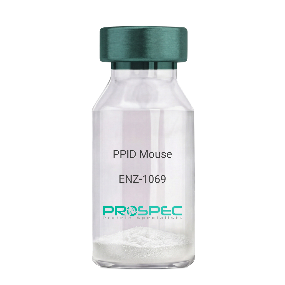
    PPID Mouse

    Description:

    Peptidylprolyl Isomerase D Mouse Recombinant

    Peptidyl-prolyl cis-trans isomerase D, PPIase D, 40 kDa peptidyl-prolyl cis-trans isomerase, Cyclophilin-40, CYP-40, Cyclophilin-related protein, CYP40, CYPD, PPID, Peptidylprolyl Isomerase D.

    Product # :

    ENZ-1069

    Price :

    Quantity :

    Shipping Method :

    Ice Icon

    Shipped with Ice Packs

    Add To Cart

    More Info

    • description
    • source
    • formulation
    • purity
    • biological activity
    • More Info
    • Citations

    Description

    PPID Mouse Recombinant produced in E.Coli is a single, non-glycosylated polypeptide chain containing 395 amino acids (1-370a.a.) and having a molecular mass of 43.4kDa. PPID is fused to a 25 amino acid His-tag at N-terminus & purified by proprietary chromatographic techniques.

    Source

    Escherichia Coli.

    Formulation

    PPID protein solution (1mg/ml) containing 20mM Tris-Hcl buffer (pH8.0), 10% glycerol and 1mM DTT.

    Purity

    Greater than 90.0% as determined by SDS-PAGE.

    Biological Activity

    Specific activity is > 700nmol/min/mg, and is defined as the amount of enzyme that cleaves 1umole of suc-AAFP-PNA per minute at 37°C in Tris–HCl pH 8.0 using chymotrypsin.

    Citations

    More Info

    • Introduction

      Cyclophilin-D is a member of the peptidyl-prolyl cis-trans isomerase (PPIase) family. PPIases catalyze the cis-trans isomerization of proline imidic peptide bonds in oligopeptides and speeds up the protein folding. Cyclophilin-D possess PPIase activity and binds to the immunosuppressant cyclosporin-A. Cyclophilin-D is very well known that its overexpression suppresses the apoptosis in cancer cell. Cyclophilin-D suppresses apoptotic cell death by the use of mitochondrial hexokinase-2 dependent mechanism in cancer cells.

    • Synonyms

      Peptidyl-prolyl cis-trans isomerase D, PPIase D, 40 kDa peptidyl-prolyl cis-trans isomerase, Cyclophilin-40, CYP-40, Cyclophilin-related protein, CYP40, CYPD, PPID, Peptidylprolyl Isomerase D.

    • Physical Appearance

      Sterile Filtered colorless solution.

    • Stability

      Store at 4°C if entire vial will be used within 2-4 weeks. Store, frozen at -20°C for longer periods of time. For long term storage it is recommended to add a carrier protein (0.1% HSA or BSA). Please prevent freeze-thaw cycles.

    • Amino Acid Sequence

      MGSSHHHHHH SSGLVPRGSH MGSEFMSHAS PAAKPSNSKN PRVFFDVDIG GERVGRIVLE LFADIVPKTA ENFRALCTGE KGTGSTTGKP LHFKGCPFHR IIKKFMIQGG DFSNQNGTGG ESIYGEKFED ENFHYKHDRE GLLSMANAGP NTNGSQFFIT TVPTPHLDGK HVVFGQVIKG LGVARTLENV EVNGEKPAKL CVIAECGELK EGDDWGIFPK DGSGDSHPDF PEDADIDLKD VDKILLISED LKNIGNTFFK SQNWEMAIKK YAKVLRYVDS SKAVIEKADR SRLQPIALSC VLNIGACKLK MSNWQGAIDS CLEALEMDPS NTKALYRKAQ GWQGLKEYDQ ALADLKKAQE IAPGDKAIQA ELLKVKQMIK AQKDKEKAVY AKMFA.

    ProSpec's products are furnished for LABORATORY RESEARCH USE ONLY. The product may not be used as drugs, agricultural or pesticidal products, food additives or household chemicals.

    Ppid Mouse
  • View Data Sheet

    Name :

    LGALS1 Human

    Description:

    Galectin-1 Human Recombinant

    Galectin-1, GAL1, GAL-1, Lectin galactoside-binding soluble 1, Beta-galactoside- binding lectin L-14-I, Lactose-binding lectin 1, S-Lac lectin 1, Galaptin, 14 kDa lectin, HPL, HBL, Putative MAPK-activating protein PM12, GBP, DKFZp686E23103.

    Product # :

    CYT-544

    Price :

    Quantity :

    Shipping Method :

    Room Temp Icon

    Shipped at Room temp

    Add To Cart

    More Info

    • description
    • source
    • formulation
    • purity
    • biological activity
    • More Info
    • Citations

    Description

    LGALS1 Human Recombinant produced in E.Coli is a single, non-glycosylated, polypeptide chain containing 135 amino acids and having a molecular mass of 14.7kDa.The LGALS1 is purified by proprietary chromatographic techniques.

    Source

    Escherichia Coli.

    Formulation

    The Galectin-1 protein was lyophilized from a concentrated (1mg/ml) containing 10mM sodium phosphate, pH-7.5.

    Purity

    Greater than 95.0% as determined by SDS-PAGE.

    Biological Activity

    The activity of Human Galectin-1 which is determined by the ability to induce chemotaxis of human THP-1 cells is detectable starting at 100ng/ml, corresponding to a specific activity of 1.0x10⁴ units/mg.

    Citations

    More Info

    • Introduction

      The galectins are a family of beta-galactoside-binding proteins implicated in modulating cell-cell and cell-matrix interactions. Galectin-1 is an autocrine negative growth factor that regulates cell proliferation. Galectin-1 regulates cell apoptosis and cell differentiation. Galectin-1 binds CD45, CD3 and CD4 & inhibits CD45 protein phosphatase activity and therefore the dephosphorylation of lyn kinase. Galectin-1 and its ligands are one of the master regulators of immune responses as T-cell homeostasis and survival, T-cell immune disorders, inflammation and allergies as well as host–pathogen interactions. Galectin-1 expression or overexpression in tumors and/or the tissue surrounding them must be considered as a sign of the malignant tumor progression that is often related to the long-range dissemination of tumoral cells (metastasis), to their dissemination into the surrounding normal tissue, and to tumor immune-escape. Galectin-1 in its oxidized form plays a number of important roles in the regeneration of the central nervous system after injury. The targeted overexpression (or delivery) of Galectin-1 should be considered as a method of choice for the treatment of some kinds of inflammation-related diseases, neurodegenerative pathologies and muscular dystrophies. In contrast, the targeted inhibition of Galectin-1 expression is what should be developed for therapeutic applications against cancer progression. Galectin-1 is thus a promising molecular target for the development of new and original therapeutic tools. There is 88% homology between the human and mouse galectin-1.

    • Synonyms

      Galectin-1, GAL1, GAL-1, Lectin galactoside-binding soluble 1, Beta-galactoside- binding lectin L-14-I, Lactose-binding lectin 1, S-Lac lectin 1, Galaptin, 14 kDa lectin, HPL, HBL, Putative MAPK-activating protein PM12, GBP, DKFZp686E23103.

    • Physical Appearance

      Sterile Filtered White lyophilized (freeze-dried) powder.

    • Stability

      Lyophilized Galectin-1 Recombinant although stable at room temperature for 3 weeks, should be stored desiccated below -18°C. Upon reconstitution Galectin-1 should be stored at 4°C between 2-7 days and for future use below -18°C.For long term storage it is recommended to add a carrier protein (0.1% HSA or BSA).Please prevent freeze-thaw cycles.

    • Solubility

      It is recommended to reconstitute the lyophilized LGALS1 in sterile 18MΩ-cm H2O not less than 100µg/ml, which can then be further diluted to other aqueous solutions.

    • Amino Acid Sequence

      MACGLVASNL NLKPGECLRV RGEVAPDAKS FVLNLGKDSN NLCLHFNPRF NAHGDANTIV CNSKDGGAWG TEQREAVFPF QPGSVAEVCI TFDQANLTVK LPDGYEFKFP NRLNLEAINY MAADGDFKIK CVAFD.

    • Background

      What is the molecular weight/Mw of LGALS1 HUMAN Protein?
      LGALS1 HUMAN Protein has a total Mw of 14.7kDa.

      What is the source or expression system of LGALS1 HUMAN Protein?
      Escherichia Coli.

      What is the Purity of LGALS1 HUMAN Protein?
      LGALS1 HUMAN Protein is >95% pure as determined by SDS-PAGE.

      What is the Biological Activity of LGALS1 HUMAN Protein?
      The activity of Human Galectin-1 which is determined by the ability to induce chemotaxis of human THP-1 cells is detectable starting at 100ng/ml, corresponding to a specific activity of 1.0x10⁴ units/mg.

      What is the amino acid sequence of LGALS1 HUMAN Protein?
      MACGLVASNL NLKPGECLRV RGEVAPDAKS FVLNLGKDSN NLCLHFNPRF NAHGDANTIV CNSKDGGAWG TEQREAVFPF QPGSVAEVCI TFDQANLTVK LPDGYEFKFP NRLNLEAINY MAADGDFKIK CVAFD.

      What applications can LGALS1 HUMAN Protein be used in?
      LGALS1 HUMAN Protein can probably be used in western blot, ELISA and Lateral Flow.

      What is the endotoxin level for LGALS1 HUMAN Protein?
      The endotoxin level is minimal, LGALS1 HUMAN Protein was purified using conventional chromatography techniques.


    • Protein content

      UV spectroscopy at 280 nm using the absorbency value of 0.59 as the extinction coefficient for a 0.1% (1mg/ml) solution. This value is calculated by the PC GENE computer analysis program of protein sequences (IntelliGenetics).

    ProSpec's products are furnished for LABORATORY RESEARCH USE ONLY. The product may not be used as drugs, agricultural or pesticidal products, food additives or household chemicals.

    Galectin 1 Human
  • View Data Sheet

    Name :

    LIF Human, Sf9

    Description:

    Leukemia Inhibitory Factor Human Recombinant, Sf9

    Leukemia Inhibitory Factor, Differentiation Inhibitory Activity, Cholinergic Differentiation Factor, Differentiation-Stimulating Factor, Hepatocyte-Stimulating Factor III, Differentiation-Inducing Factor, Melanoma-Derived LPL Inhibitor, Human Interleukin In DA Cells, D Factor, HILDA, MLPLI, Emfilermin, DIA, CDF, Leukemia inhibitory factor, LIF, Differentiation-stimulating factor, D factor, Melanoma-derived LPL inhibitor, MLPLI, Emfilermin.

    Product # :

    CYT-1003

    Price :

    Quantity :

    Shipping Method :

    Ice Icon

    Shipped with Ice Packs

    Add To Cart

    More Info

    • description
    • source
    • formulation
    • purity
    • biological activity
    • More Info
    • Citations

    Description

    LIF Human Recombinant produced in Sf9 Baculovirus cells is a single, glycosylated polypeptide chain containing 189 amino acids (23-202a.a.) and having a molecular mass of 20.8kDa (Molecular size on SDS-PAGE will appear at approximately 18-40kDa). LIF is expressed with a 6 amino acid His tag at C-Terminus and purified by proprietary chromatographic techniques.

    Source

    Sf9, Baculovirus cells.

    Formulation

    LIF protein solution (0.25mg/ml) contains Phosphate Buffered Saline (pH 7.4) and 10% glycerol.

    Purity

    Greater than 90.0% as determined by SDS-PAGE.

    Biological Activity

    Measured in a cell proliferation assay using TF-1 human erythroleukemic cell. The ED50 for this effects is less or equal to 0.5 ng/ml.

    Citations

    More Info

    • Introduction

      Leukemia Inhibitory Factor also called LIF is a lymphoid factor that promotes long-term maintenance of embryonic stem cells by suppressing spontaneous differentiation. Leukemia Inhibitory Factor has several functions such as cholinergic neuron differentiation, control of stem cell pluripotency, bone & fat metabolism, mitogenesis of factor dependent cell lines & promotion of megakaryocyte production in vivo. Human and mouse LIF exhibit a 78% identity in its amino acid sequence.

    • Synonyms

      Leukemia Inhibitory Factor, Differentiation Inhibitory Activity, Cholinergic Differentiation Factor, Differentiation-Stimulating Factor, Hepatocyte-Stimulating Factor III, Differentiation-Inducing Factor, Melanoma-Derived LPL Inhibitor, Human Interleukin In DA Cells, D Factor, HILDA, MLPLI, Emfilermin, DIA, CDF, Leukemia inhibitory factor, LIF, Differentiation-stimulating factor, D factor, Melanoma-derived LPL inhibitor, MLPLI, Emfilermin.

    • Physical Appearance

      Sterile filtered colorless solution.

    • Stability

      Store at 4°C if entire vial will be used within 2-4 weeks. Store, frozen at -20°C for longer periods of time. For long term storage it is recommended to add a carrier protein (0.1% HSA or BSA). Avoid multiple freeze-thaw cycles.

    • Amino Acid Sequence

      ADPSPLPITP VNATCAIRHP CHNNLMNQIR SQLAQLNGSA NALFILYYTA QGEPFPNNLD KLCGPNVTDF PPFHANGTEK AKLVELYRIV VYLGTSLGNI TRDQKILNPS ALSLHSKLNA TADILRGLLS NVLCRLCSKY HVGHVDVTYG PDTSGKDVFQ KKKLGCQLLG KYKQIIAVLA
      QAFHHHHHH.

    ProSpec's products are furnished for LABORATORY RESEARCH USE ONLY. The product may not be used as drugs, agricultural or pesticidal products, food additives or household chemicals.

    Lif Human Sf9
  • View Data Sheet

    Name :

    SPARC Human

    Description:

    Secreted Protein Acidic & Rich in Cysteine Recombinant Human

    Osteonectin, ON, Basement-membrane protein 40, BM-40, SPARC, Secreted Protein acidic and Rich in Cysteine.

    Product # :

    PRO-2602

    Price :

    Quantity :

    Shipping Method :

    Room Temp Icon

    Shipped at Room temp

    Add To Cart

    More Info

    • description
    • source
    • formulation
    • purity
    • biological activity
    • More Info
    • Citations

    Description

    Secreted Protein Acidic & Rich in Cysteine Human Recombinant produced in E.Coli is a single, non- glycosylated polypeptide chain containing 286 amino acids and having a molecular mass of 32.7kDa.SPARC is purified by proprietary chromatographic techniques.

    Source

    Escherichia Coli.

    Formulation

    Lyophilized from a 0.2μm filtered concentrated solution in PBS, pH 7.4.

    Purity

    Greater than 98.0% as determined by:
    (a) Analysis by RP-HPLC.
    (b) Analysis by SDS-PAGE.

    Biological Activity

    The ED50 as determined by its ability to inhibit the cell growth of Mv1Lu mink lung epithelial cells is < 3.0 µg/mL, corresponding to a specific activity of > 333 IU/mg.

    Citations

    More Info

    • Introduction

      SPARC, an acronym for “secreted protein, acidic and rich in cysteine”, is also known as osteonectin or BM-40. It is the founding member of a family of secreted matricellular proteins with similar domain structure. The 303 amino acid, 43 kDa protein contains a 17 aa signal sequence, an N-terminal acidic region that binds calcium, a follistatin domain containing Kazal-like sequences, and a C-terminal extracellular calcium (EC) binding domain with two EF-hand motifs. SPARC is produced by fibroblasts, capillary endothelial cells, platelets and macrophages, especially in areas of tissue morphogenesis and remodeling. SPARC shows context-specific effects, but generally inhibits adhesion, spreading and proliferation, and promotes collagen matrix formation. For endothelial cells, SPARC disrupts focal adhesions and binds and sequesters PDGF and VEGF. SPARC is abundantly expressed in bone, where it promotes osteoblast differentiation and inhibits adipogenesis.

    • Synonyms

      Osteonectin, ON, Basement-membrane protein 40, BM-40, SPARC, Secreted Protein acidic and Rich in Cysteine.

    • Physical Appearance

      Sterile Filtered White lyophilized (freeze-dried) powder.

    • Stability

      Lyophilized SPARC although stable at room temperature for 3 weeks, should be stored desiccated below -18°C. Upon reconstitution Secreted Protein Acidic & Rich in Cysteine should be stored at 4°C between 2-7 days and for future use below -18°C.Please prevent freeze-thaw cycles.

    • Solubility

      It is recommended to reconstitute the lyophilized SPARC in sterile 18MΩ-cm H2O not less than 100µg/ml, which can then be further diluted to other aqueous solutions.

    • Amino Acid Sequence

      APQQEALPDE TEVVEETVAE VTEVSVGANP VQVEVGEFDD GAEETEEEVV AENPCQNHHC KHGKVCELDE NNTPMCVCQD PTSCPAPIGE FEKVCSNDNK TFDSSCHFFA TKCTLEGTKK GHKLHLDYIG PCKYIPPCLD SELTEFPLRM RDWLKNVLVT LYERDEDNNL LTEKQKLRVK KIHENEKRLE AGDHPVELLA RDFEKNYNMY IFPVHWQFGQ LDQHPIDGYL SHTELAPLRA PLIPMEHCTT RFFETCDLDN DKYIALDEWA GCFGIKQKDI DKDLVI.

    ProSpec's products are furnished for LABORATORY RESEARCH USE ONLY. The product may not be used as drugs, agricultural or pesticidal products, food additives or household chemicals.

    Sparc Protein
  • View Data Sheet

    Name :

    CCN1 Human

    Description:

    Cysteine-Rich Angiogenic Inducer 61 Human Recombinant

    CYR61, Protein CYR61, Cysteine-rich angiogenic inducer 61, IGF-binding protein 10, IGFBP-10, IBP-10, Protein GIG1, CCN family member 1, CCN1, GIG1, IGFBP10.

    Product # :

    CYT-164

    Price :

    Quantity :

    Shipping Method :

    Room Temp Icon

    Shipped at Room temp

    Add To Cart

    More Info

    • description
    • source
    • formulation
    • purity
    • biological activity
    • More Info
    • Citations

    Description

    CYR61 Human Recombinant produced in E.Coli is a single, non-glycosylated, polypeptide chain containing 357 amino acids and having a molecular mass of 39.5kDa.The CYR61 is purified by proprietary chromatographic techniques.

    Source

    Escherichia Coli.

    Formulation

    Lyophilized from a 0.2m filtered concentrated solution in PBS, pH 7.4.

    Purity

    Greater than 95.0% as determined by SDS-PAGE.

    Biological Activity

    The ED50 was determined by the proliferation of mouse 3T3 cells is < 2.0 ug/ml, corresponding to a specific activity of > 500 units/mg.

    Citations

    More Info

    • Introduction

      CYR61 is a growth factor-inducible, immediate-early gene that has multifaceted activities in various cancers. CYR61 is a secreted, cysteine-rich, binding protein which is encoded by a growth factor-inducible immediate-early gene. Acting as an extracellular, matrix-associated signaling molecule, CYR61 promotes the adhesion of endothelial cells through interaction with integrin and enhances growth factor-induced DNA synthesis in the same cell type.

    • Synonyms

      CYR61, Protein CYR61, Cysteine-rich angiogenic inducer 61, IGF-binding protein 10, IGFBP-10, IBP-10, Protein GIG1, CCN family member 1, CCN1, GIG1, IGFBP10.

    • Physical Appearance

      Sterile Filtered White lyophilized (freeze-dried) powder.

    • Stability

      Lyophilized CYR61 Human although stable at room temperature for 3 weeks, should be stored desiccated below -18°C. Upon reconstitution CYR61 should be stored at 4°C between 2-7 days and for future use below -18°C.Please prevent freeze-thaw cycles.

    • Solubility

      It is recommended to reconstitute the lyophilized CYR61 in sterile 18M-cm H2O not less than 100µg/ml, which can then be further diluted to other aqueous solutions.

    • Amino Acid Sequence

      TCPAACHCPL EAPKCAPGVG LVRDGCGCCK VCAKQLNEDC SKTQPCDHTK GLECNFGASS TALKGICRAQ SEGRPCEYNS RIYQNGESFQ PNCKHQCTCI DGAVGCIPLC PQELSLPNLG CPNPRLVKVT GQCCEEWVCD EDSIKDPMED QDGLLGKELG FDASEVELTR NNELIAVGKG SSLKRLPVFG MEPRILYNPL QGQKCIVQTT SWSQCSKTCG TGISTRVTND NPECRLVKET RICEVRPCGQ PVYSSLKKGK KCSKTKKSPE PVRFTYAGCL SVKKYRPKYC GSCVDGRCCT
      PQLTRTVKMR FRCEDGETFS KNVMMIQSCK CNYNCPHANE AAFPFYRLFN DIHKFRD

    • Background

      Title: Cysteine-Rich Angiogenic Inducer 61 Human Recombinant: A Potential Regulator of Angiogenesis

      Abstract:


      Cysteine-rich angiogenic inducer 61 (CYR61) is an important extracellular matrix-associated protein that plays a significant role in angiogenesis and cell adhesion. This research paper provides a comprehensive analysis of human recombinant CYR61, focusing on its production, characterization, and potential applications in regulating angiogenesis. The paper discusses the significance of CYR61 in physiological and pathological angiogenesis, including wound healing, tumor development, and cardiovascular diseases. Furthermore, it explores ongoing research and clinical trials investigating the therapeutic potential of recombinant CYR61 in various pathological conditions. The information presented in this paper aims to enhance our understanding of human recombinant CYR61 and its utility as a research tool and a potential regulator of angiogenesis.

      Introduction:


      Cysteine-rich angiogenic inducer 61 (CYR61) is an extracellular matrix-associated protein that plays a crucial role in angiogenesis, the formation of new blood vessels from pre-existing ones. Human recombinant CYR61, produced through genetic engineering techniques, provides researchers with a valuable tool to study its biological functions and explore its therapeutic potential.

      Production and Characterization:


      Recombinant CYR61 is typically generated using expression systems such as mammalian cells or bacteria. The protein is then purified and characterized to ensure its structural integrity and functional activity. Quality control measures are implemented to confirm the specificity and bioactivity of the recombinant CYR61.

      Role in Angiogenesis:


      CYR61 is involved in various aspects of angiogenesis, including endothelial cell proliferation, migration, and tube formation. It interacts with integrins and other cell surface receptors to modulate signaling pathways involved in angiogenic processes. Recombinant CYR61 serves as a valuable tool for studying the mechanisms underlying angiogenesis and exploring its potential as a therapeutic target.

      Therapeutic Implications:


      The dysregulation of angiogenesis is associated with several pathological conditions, including cancer, cardiovascular diseases, and chronic wounds. Recombinant CYR61 has shown promise as a potential regulator of angiogenesis and a therapeutic agent. It can be used to promote or inhibit angiogenesis, depending on the specific context. Ongoing research and clinical trials are investigating the therapeutic applications of recombinant CYR61 in various diseases, including cancer and ischemic disorders.

      Conclusion:


      Human recombinant CYR61 is a valuable research tool and a potential regulator of angiogenesis. Its production, characterization, and applications in modulating angiogenic processes contribute to our understanding of angiogenesis and the development of novel therapeutic interventions. Continued research and clinical trials exploring the therapeutic potential of recombinant CYR61 offer promising prospects for improving outcomes in cancer, cardiovascular diseases, and wound healing.

      What is the molecular weight/Mw of CCN1 Protein?
      CCN1 Protein has a total Mw of 39.5kDa.

      What is the source or expression system of CCN1 Protein?
      Escherichia Coli.

      What is the Purity of CCN1 Protein?
      CCN1 Protein is >95% pure as determined by SDS-PAGE.

      What is the Biological Activity of CCN1 Protein?
      The ED50 was determined by the proliferation of mouse 3T3 cells is < 2.0 ug/ml, corresponding to a specific activity of > 500 units/mg.

      What is the amino acid sequence of CCN1 Protein?
      TCPAACHCPL EAPKCAPGVG LVRDGCGCCK VCAKQLNEDC SKTQPCDHTK GLECNFGASS TALKGICRAQ SEGRPCEYNS RIYQNGESFQ PNCKHQCTCI DGAVGCIPLC PQELSLPNLG CPNPRLVKVT GQCCEEWVCD EDSIKDPMED QDGLLGKELG FDASEVELTR NNELIAVGKG SSLKRLPVFG MEPRILYNPL QGQKCIVQTT SWSQCSKTCG TGISTRVTND NPECRLVKET RICEVRPCGQ PVYSSLKKGK KCSKTKKSPE PVRFTYAGCL SVKKYRPKYC GSCVDGRCCT
      PQLTRTVKMR FRCEDGETFS KNVMMIQSCK CNYNCPHANE AAFPFYRLFN DIHKFRD

      What applications can CCN1 Protein be used in?
      CCN1 Protein can probably be used in western blot, ELISA and Lateral Flow.

      What is the endotoxin level for CCN1 Protein?
      The endotoxin level is minimal, CCN1 Protein was purified using conventional chromatography techniques.

    ProSpec's products are furnished for LABORATORY RESEARCH USE ONLY. The product may not be used as drugs, agricultural or pesticidal products, food additives or household chemicals.

    Cyr61 Human
  • View Data Sheet

    Name :

    Transferrin Human

    Description:

    Transferrin Human Recombinant

    Serotransferrin, Transferrin, Siderophilin, Beta-1-metal-binding globulin, TF, PRO1557, PRO2086, DKFZp781D0156, HTF.

    Product # :

    PRO-747

    Price :

    Quantity :

    Shipping Method :

    Room Temp Icon

    Shipped at Room temp

    Add To Cart

    More Info

    • description
    • source
    • formulation
    • purity
    • biological activity
    • More Info
    • Citations

    Description

    Recombinant Human Transferrin produced in Plant is a non-glycosylated, polypeptide chain containing 679 amino acids and having a molecular mass of 76 kDa. The Recombinant Human Transferrin is purified by proprietary chromatographic techniques.

    Source

    Oryza sativa (rice).

    Formulation

    The protein (1mg/ml) was lyophilized with no additives.

    Purity

    Purity as determined by SDS-PAGE is 97%.

    Biological Activity

    One mg of Recombinant Human Transferrin will bind to approximately 2 micrograms of Fe.

    Citations

    More Info

    • Introduction

      Transferrin is the iron-transport protein of vertebrate serum and donates iron to cells through interaction with a specific membrane receptor, CD71. Transferrin appears to be indispensable for most cells growing in tissue culture.
      It is referred to frequently as a growth factor because, in analogy to other growth factor-receptor interactions, proliferating cells express high numbers of transferrin receptors, and the binding of transferrin to their receptors is needed for cells to initiate and maintain their DNA synthesis. Apart from its role as an iron transport protein transferrin acts as a cytokine and has functions that may not be related to its iron-carrying capacity.

    • Synonyms

      Serotransferrin, Transferrin, Siderophilin, Beta-1-metal-binding globulin, TF, PRO1557, PRO2086, DKFZp781D0156, HTF.

    • Physical Appearance

      Sterile Filtered lyophilized (freeze-dried) powder.

    • Stability

      Lyophilized Transferrin although stable at room temperature for 3 weeks, should be stored desiccated below -18°C. Upon reconstitution Transferrin Human Recombinant should be stored at 4°C between 2-7 days and for future use below -18°C.For long term storage it is recommended to add a carrier protein (0.1% HSA or BSA).Please prevent freeze-thaw cycles.

    • Solubility

      Stock solutions can be prepared by dissolving gently into PBS for several minutes. Recommended stock concentrations are 5mg/ml to 20 mg/ml in PBS, though others can be used as well. Please try to avoid the formation of bubbles when dissolving the protein. Sterile filter through 0.2µm filter.

    ProSpec's products are furnished for LABORATORY RESEARCH USE ONLY. The product may not be used as drugs, agricultural or pesticidal products, food additives or household chemicals.

    Transferrin Human
  • View Data Sheet

    Name :

    SERPINA7 Human

    Description:

    Serpin Peptidase Inhibitor, Clade A Member 7 Human

    Product # :

    PRO-2739

    Price :

    Quantity :

    Shipping Method :

    Room Temp Icon

    Shipped at Room temp

    Add To Cart

    More Info

    • description
    • source
    • formulation
    • purity
    • More Info
    • Citations

    Description

    Human Serpin Peptidase Inhibitor, Clade A Member 7 Protein produced in Human plasma having a molecular mass of approximately 55kD.

    Source

    Human serum.

    Formulation

    The protein was lyophilized (0.2 µm filtered) from 20mM NH4HCO3.

    Purity

    Greater than 98.0%.

    Citations

    More Info

    • Introduction

      Thyroxine binding globulin, also known as SERPINA7, is synthesized in the liver and carried in the blood.
      SERPINA7 may be decreased or increased in a number of diseases. Its main value in diagnosis is to assess thyroid function by determining the amount of free T3/T4.
      Thyroxine binding globulin, binds and transports thyroid hormones in the circulation. SERPINA7 carries the majority of the T3/T4 in the blood. SERPINA7 has only one binding site for T3/T4 and about 25% of Thyroxine binding globulin is usually saturated.

    • Physical Appearance

      Sterile Filtered White lyophilized (freeze-dried) powder.

    • Stability

      SERPINA7 Human although stable at room temperature for 3 weeks, should be stored between 2-8°C.

    • Solubility

      It is recommended to reconstitute the lyophilized SERPINA7 Human in phosphate buffer containing 0.15M NaCl.

    • Human Virus Test

      Starting material donor has been tested and certified negative for antibodies to HIV-1, HIV-2, HCV, HBSAG, Parvovirus B19, Syphilis and HIV/HBV/HCV (PCR).

    ProSpec's products are furnished for LABORATORY RESEARCH USE ONLY. The product may not be used as drugs, agricultural or pesticidal products, food additives or household chemicals.

    Serpina7 Human
  • View Data Sheet

    Name :

    C3 Rat

    Description:

    Complement C3 Rat

    Complement C3, C3 and PZP-like alpha-2-macroglobulin domain-containing protein 1, C3, CPAMD1.

    Product # :

    PRO-2706

    Price :

    Quantity :

    Shipping Method :

    Ice Icon

    Shipped with Ice Packs

    Add To Cart

    More Info

    • description
    • source
    • formulation
    • purity
    • More Info
    • Citations

    Description

    Rat Complement C3 produced in Rat plasma having a molecular weight of 187kDa.

    Source

    Rat Plasma.

    Formulation

    C3 solution contains phosphate buffer saline.

    Purity

    Greater than 95.0% as determined by SDS-PAGE.

    Citations

    More Info

    • Introduction

      C3 is central to the activation of all 3 pathways of complement activation. Initiation of each pathway generates proteolytic enzyme complexes which binds the target surface. These enzymes cleave a peptide bond in C3 releasing the anaphylatoxin C3a and activating C3b. Most of the C3 activated during complement activation never attaches to the surface due to its thioester reaction with water forming fluid phase C3b which is rapidly inactivated by factors H and I forming iC3b. Surface-bound C3b is necessary in all 3 pathways for efficient activation of C5 and formation of C5b-9 complexes that lyse the target cell membrane.

    • Synonyms

      Complement C3, C3 and PZP-like alpha-2-macroglobulin domain-containing protein 1, C3, CPAMD1.

    • Physical Appearance

      Sterile filtered solution.

    • Stability

      C3 Mouse is stable at 4°C if entire vial will be used within 2-4 weeks.Store, frozen below -20°C for longer periods of time.For long term storage it is recommended to add a carrier protein (0.1% HSA or BSA).Avoid multiple freeze-thaw cycles.

    ProSpec's products are furnished for LABORATORY RESEARCH USE ONLY. The product may not be used as drugs, agricultural or pesticidal products, food additives or household chemicals.

    C3 Rat
  • View Data Sheet

    Name :

    VEGF Human

    Description:

    Vascular Endothelial Growth Factor Human Recombinant

    Vascular endothelial growth factor A, VEGF-A, Vascular permeability factor, VPF, VEGF, MGC70609.

    Product # :

    CYT-241

    Price :

    Quantity :

    Shipping Method :

    Room Temp Icon

    Shipped at Room temp

    Add To Cart

    More Info

    • description
    • source
    • formulation
    • purity
    • biological activity
    • More Info
    • Citations

    Description

    Vascular Endothelial Growth Factor Human Recombinant produced in E.Coli is a double, non-glycosylated, polypeptide chain containing 165 amino acids and having a molecular mass of 38.2kDa.The VEGF is purified by proprietary chromatographic techniques.

    Source

    Escherichia Coli.

    Formulation

    The VEGF protein was lyophilized from a concentrated (1mg/ml) solution with no additives.

    Purity

    Greater than 95.0% as determined by:
    (a) Analysis by RP-HPLC.
    (b) Analysis by SDS-PAGE.

    Biological Activity

    Determined by the dose-dependent stimulation of the proliferation of human umbilical vein endothelial cells (HUVEC) using a concentration range of 3.7-5.6 ng/ml, corresponding to a Specific Activity of 178,570-270,270IU/mg.

    Citations

    More Info

    • Introduction

      Vascular endothelial growth factor (VEGF) is an important signaling protein involved in vessel formation As its name implies, VEGF activity has been mostly studied on cells of the vascular endothelium, although it does have effects on a number of other cell types (e.g. stimulation monocyte/macrophage migration, neurons, cancer cells, kidney epithelial cells ). VEGF mediates increased vascular permeability, induces vasculogenesis and endothelial cell production, promotes cell migration, and inhibits apoptosis. In vitro, VEGF has been shown to stimulate endothelial cell mitogenesis and cell migration. VEGF is also a vasodilator and increases microvascular permeability and was originally referred to as vascular permeability factor. VEGF is located in normal cartilage though only osteoarthritic cartilage expresses the VEGF receptors, NP1, VEGFR1 and VEGFR2. The VEGF level in the culture media from OA chondrocytes was more than 3 folds higher than in media from normal chondrocytes

    • Synonyms

      Vascular endothelial growth factor A, VEGF-A, Vascular permeability factor, VPF, VEGF, MGC70609.

    • Physical Appearance

      Sterile Filtered White lyophilized (freeze-dried) powder.

    • Stability

      Lyophilized VEGF although stable at room temperature for 3 weeks, should be stored desiccated below -18°C. Upon reconstitution VEGF should be stored at 4°C between 2-7 days and for future use below -18°C.For long term storage it is recommended to add a carrier protein (0.1% HSA or BSA).Please prevent freeze-thaw cycles.

    • Solubility

      It is recommended to reconstitute the lyophilized VEGF in sterile 18M-cm H2O not less than 100µg/ml, which can then be further diluted to other aqueous solutions.

    • Amino Acid Sequence

      APMAEGGGQN HHEVVKFMDV YQRSYCHPIE TLVDIFQEYP DEIEYIFKPS CVPLMRCGGC CNDEGLECVP TEESNITMQI MRIKPHQGQH IGEMSFLQHN KCECRPKKDR ARQENPCGPC SERRKHLFVQ DPQTCKCSCK NTDSRCKARQ LELNERTCRC DKPRR

    • Protein content

      VEGF protein quantitation was carried out by two independent methods:1. UV spectroscopy at 280 nm using the absorbency value of 0.2875 as the extinction coefficient for a 0.1% (1mg/ml) solution. This value is calculated by the PC GENE computer analysis program of protein sequences (IntelliGenetics). 2. Analysis by RP-HPLC, using a calibrated solution of VEGF as a Reference Standard.

    ProSpec's products are furnished for LABORATORY RESEARCH USE ONLY. The product may not be used as drugs, agricultural or pesticidal products, food additives or household chemicals.

    Vegf Human
  • View Data Sheet

    Name :

    SPA-Cys Long

    Description:

    Staphylococcal Protein-A Cys Long Form Recombinant

    Immunoglobulin G-binding protein A, IgG-binding protein A, Staphylococcal protein A, SPA.

    Product # :

    PRO-1924

    Price :

    Quantity :

    Shipping Method :

    Room Temp Icon

    Shipped at Room temp

    Add To Cart

    More Info

    • description
    • source
    • formulation
    • purity
    • More Info
    • Citations

    Description

    SPA-Cys long Recombinant produced in E.Coli is a single non-glycosylated polypeptide chain with a Cys on C-terminus. SPA-Cys is comprised of 5 IgG-binding domains E-D-A-B-C aligned in series containing 423 amino acids and having a molecular mass of 46.7kDa containing little or no carbohydrate.

    Source

    Escherichia Coli.

    Formulation

    SPA protein was lyophilized with no additives.

    Purity

    Greater than 97.0% as determined by analysis by SDS-PAGE.

    Citations

    More Info

    • Introduction

      Protein A is a cell wall component produced by several strains of Staphylococcus aureus. The recombinant Protein A is genetically engineered protein and holds 5 IgG-binding regions of protein A. Recombinant Protein A functions basically the same as native Protein A and is perfect for purification of polyclonal or monoclonal IgG antibodies. Protein A binds to human IgG1, IgG2 and IgG4, mouse IgG2a, IgG2b and IgG3 and rat IgG2c. Protein A also binds to total IgG from rabbit, pig, dog, cat, and guinea pig.

    • Synonyms

      Immunoglobulin G-binding protein A, IgG-binding protein A, Staphylococcal protein A, SPA.

    • Physical Appearance

      Sterile Filtered White lyophilized (freeze-dried) powder.

    • Stability

      Lyophilized SPA although stable at room temperature for 3 weeks, should be stored desiccated below -18°C. Upon reconstitution SPA should be stored at 4°C between 2-7 days and for future use below -18°C. For long term storage it is recommended to add a carrier protein (0.1% HSA or BSA). Please prevent freeze-thaw cycles.

    • Solubility

      It is recommended to reconstitute the lyophilized SPA in sterile 18MΩ-cm H2O not less than 0.1mg/ml, which can then be further diluted to other aqueous solutions.

    • Amino Acid Sequence

      AQHDEAQQNA FYQVLNMPNL NADQRNGFIQ SLKDDPSQSA NVLGEAQKLN DSQAPKADAQ QNNFNKDQQS AFYEILNMPN LNEAQRNGFI QSLKDDPSQS TNVLGEAKKL NESQAPKADN NFNKEQQNAF YEILNMPNLN EEQRNGFIQS LKDDPSQSAN LLSEAKKLNE SQAPKADNKF NKEQQNAFYE ILHLPNLNEE QRNGFIQSLK DDPSQSANLL AEAKKLNDAQ APKADNKFNK EQQNAFYEIL HLPNLTEEQR NGFIQSLKDD PSVSKEILAE AKKLNDAQAP KEEDNKKPGK EDGNKPGKED GNKPGKEDNK KPGKEDGNKP GKEDNNKPGK EDGNKPGKED NNKPGKEDGN KPGKEDGNKP GKEDGNGVHV VKPGDTVNDI AKANGTTADK IAADNKLADK NMIKPGQELV VDC

    ProSpec's products are furnished for LABORATORY RESEARCH USE ONLY. The product may not be used as drugs, agricultural or pesticidal products, food additives or household chemicals.

    Spa Cys Long
  • View Data Sheet

    Name :

    AIMP1 Antibody

    Description:

    Aminoacyl tRNA Synthetase Complex-Interacting Multifunctional Protein 1, Mouse Anti Human

    Aminoacyl tRNA synthase complex-interacting multifunctional protein 1, Multisynthase complex auxiliary component p43, AIMP1, EMAP2, SCYE1, p43, EMAPII.

    Product # :

    ANT-619

    Price :

    Quantity :

    Shipping Method :

    Ice Icon

    Shipped with Ice Packs

    Add To Cart

    More Info

    • formulation
    • More Info
    • Citations

    Formulation

    1mg/ml containing PBS, pH-7.4, 10% Glycerol and 0.02% Sodium Azide.

    Citations

    More Info

    • Introduction

      AIMP1 (EMPA2 or p43) is a cytokine that is specifically induced by apoptosis, and it is involved in the control of angiogenesis, inflammation, and wound healing. The release of the AIMP1 cytokine renders the tumor-associated vasculature sensitive to tumor necrosis factor. Furthermore, AIMP1 is involved in the stimulation of inflammatory responses after proteolytic cleavage in tumor cells.

    • Synonyms

      Aminoacyl tRNA synthase complex-interacting multifunctional protein 1, Multisynthase complex auxiliary component p43, AIMP1, EMAP2, SCYE1, p43, EMAPII.

    • Physical Appearance

      Sterile filtered colorless solution.

    • Immunogen

      Anti-human AIMP1 mAb, is derived from hybridization of mouse F0 myeloma cells with spleen cells from BALB/c mice immunized with a recombinant human AIMP1 protein 1-336 amino acids purified from E. coli.

    • Ig Subclass

      Mouse IgG2a heavy chain and Kappa light chain.

    • Clone

      PAT6D10AT.

    • Applications

      The antibody has been tested by ELISA, Western blot analysis, Flow cytometry and ICC/IF to assure specificity and reactivity. Since application varies, however, each investigation should be titrated by the reagent to obtain optimal results.

    • Type

      Mouse Anti Human Monoclonal.

    • Storage Procedures

      For periods up to 1 month store at 4°C, for longer periods of time, store at -20°C. Prevent freeze thaw cycles.

    • Purification Method

      AIMP1 antibody was purified by protein-A affinity chromatography.

    ProSpec's products are furnished for LABORATORY RESEARCH USE ONLY. The product may not be used as drugs, agricultural or pesticidal products, food additives or household chemicals.

    Aimp1 Antibody
  • View Data Sheet

    Name :

    Activin-A Human Plant-Active

    Description:

    Activin-A Human Recombinant, Plant-Active

    Inhba, Inhibin beta A, FSH releasing protein.

    Product # :

    CYT-414

    Price :

    Quantity :

    Shipping Method :

    Room Temp Icon

    Shipped at Room temp

    Add To Cart

    More Info

    • description
    • source
    • formulation
    • purity
    • biological activity
    • More Info
    • Citations

    Description

    Active form Activin-A Human Recombinant produced in Plant is a homodimeric, glycosylated, polypeptide chain containing 2 x 116 amino acids and having a molecular weight of 27.4kDa.The Active form Activin-A is fused to a 6-His tag at N-terminus and purified by standard chromatographic techniques.

    Source

    Nicotiana benthamiana.

    Formulation

    Active form Activin-A was lyophilized from a concentrated 1mg/ml protein solution containing 50mM Tris-HCl pH-7.4

    Purity

    Greater than 98% as obsereved by SDS-PAGE.

    Biological Activity

    The biological activity of INHBA is measured by its ability to inhibit mouse plasmacytoma cell line (MPC-11) cells proliferation ([3H]thymidine incorporation). ED50<5ng/ml.

    Citations

    More Info

    • Introduction

      Activins are homodimers or heterodimers of the different β subunit isoforms, part of the TGFβ family. Mature Activin A has two 116 amino acids residues βA subunits (βA-βA). Activin displays an extensive variety of biological activities, including mesoderm induction, neural cell differentiation, bone remodelling, haematopoiesis, and reproductive physiology. Activins takes part in the production and regulation of hormones such as FSH, LH, GnRH and ACTH. Cells that are identified to express Activin A include fibroblasts, endothelial cells, hepatocytes, vascular smooth muscle cells, macrophages, keratinocytes, osteoclasts, bone marrow monocytes, prostatic epithelium, neurons, chondrocytes, osteoblasts, Leydig cells, Sertoli cells, and ovarian granulosa cells.

    • Synonyms

      Inhba, Inhibin beta A, FSH releasing protein.

    • Physical Appearance

      Lyophilized freeze dried powder.

    • Stability

      For long term storage it is recommended to add a carrier protein (0.1% HSA or BSA). Repeated freezing and thawing is not recommended.

    • Solubility

      INHBA protein should be reconstituted in distilled water to a concentration of 50 ug /ml. Due to the protein nature, dimmers and multimers may be observed.

    • Amino Acid Sequence

      HHHHHHGLECDGKVNICCKKQFFVSFKDIGWNDWIIAPSG
      YHANYCEGECPSHIAGTSGSSLSFHSTVINHYRMRGHSPFA
      NLKSCCVPTKLRPMSMLYYDDGQNIIKKDIQNMIVEECGCS.

    • Background

      What is the molecular weight / Mw of Activin A Protein?
      Activin A Protein has a total Mw of 27.4 kDa.

      What is the source or expression system of Activin A Protein?
      Nicotiana benthamiana.

      What is the Purity of Activin A Protein?
      Activin A Protein is >98% pure as determined by SDS-PAGE.

      What is the Biological Activity of Activin A Protein?
      The biological activity of INHBA is measured by its ability to inhibit mouse plasmacytoma cell line (MPC-11) cells proliferation ([3H]thymidine incorporation). ED50<5ng/ml.

      What is the endotoxin level for Activin A Protein?
      The endotoxin level is minimal, ACTIVIN A Protein was purified using conventional chromatography techniques.

      What is the amino acid sequence of ACTIVIN A Protein?HHHHHHGLECDGKVNICCKKQFFVSFKDIGWNDWIIAPSGYHANYCEGECPSHIAGTSGSSLSFHSTVINHYRMRGHSPFANLKSCCVPTKLRPMSMLYYDDGQNIIKKDIQNMIVEECGCS

      What applications can ACTIVIN-A Protein be used in?

      ACTIVIN A Protein can probably be used in western blot, ELISA and Lateral Flow.


    ProSpec's products are furnished for LABORATORY RESEARCH USE ONLY. The product may not be used as drugs, agricultural or pesticidal products, food additives or household chemicals.

    Activin A Active
  • View Data Sheet

    Name :

    IGF1 Rat

    Description:

    IGF-1 Rat Recombinant

    Somatomedin C, IGF-I, IGFIA, IGF1.

    Product # :

    CYT-289

    Price :

    Quantity :

    Shipping Method :

    Room Temp Icon

    Shipped at Room temp

    Add To Cart

    More Info

    • description
    • source
    • formulation
    • purity
    • biological activity
    • More Info
    • Citations

    Description

    IGF-1 Rat Recombinant produced in E.Coli is a single, non-glycosylated, polypeptide chain containing 70 amino acids and having a molecular mass of 7.7kDa. IGF-I is purified by proprietary chromatographic techniques.

    Source

    Escherichia Coli.

    Formulation

    The protein was lyophilized with a 0.2µm filtered concentrated solution in 20mM PBS, pH 7.0.

    Purity

    Greater than 98.0% as determined by SDS-PAGE.

    Biological Activity

    The ED50 as determined by a cell proliferation assay using FDC-P1 cells is less than 2.0ng/ml, corresponding to a specific activity of >500,000units/mg.

    Citations

    More Info

    • Introduction

      The somatomedins, or IGFs, comprise a family of peptides that play important roles in mammalian growth and development. IGF1 mediates many of the growth-promoting effects of GH. Early studies showed that GH did not directly stimulate the incorporation of sulfate into cartilage, but rather acted through a serum factor, termed 'sulfation factor,' which later became known as 'somatomedin' (Daughaday et al., 1972). Three main somatomedins have been characterized: somatomedin C (IGF1), somatomedin A (IGF2; MIM 147470), and somatomedin B (MIM 193190) (Rotwein, 1986; Rosenfeld, 2003).

    • Synonyms

      Somatomedin C, IGF-I, IGFIA, IGF1.

    • Physical Appearance

      Sterile Filtered White lyophilized (freeze-dried) powder.

    • Stability

      Lyophilized IGF-1 although stable at room temperature for 3 weeks, should be stored desiccated below -18°C. Upon reconstitution IGFI should be stored at 4°C between 2-7 days and for future use below -18°C.For long term storage it is recommended to add a carrier protein (0.1% HSA or BSA).Please prevent freeze-thaw cycles.

    • Solubility

      It is recommended to reconstitute the lyophilized IGF1 in sterile 18MΩ-cm H2O not less than 100µg/ml, which can then be further diluted to other aqueous solutions.

    • Amino Acid Sequence

      GPETLCGAEL VDALQFVCGP RGFYFNKPTG YGSSIRRAPQ TGIVDECCFR SCDLRRLEMY CAPLKPTKSA.

    • Background

      What is the molecular weight/Mw of IGF1 RAT Protein?
      IGF1 RAT Protein has a total Mw of 7.7kDa.

      What is the source or expression system of IGF1 RAT Protein?
      Escherichia Coli.

      What is the Purity of IGF1 RAT Protein?
      IGF1 RAT Protein is >98% pure as determined by SDS-PAGE.

      What is the Biological Activity of IGF1 RAT Protein?
      The ED50 as determined by a cell proliferation assay using FDC-P1 cells is less than 2.0ng/ml, corresponding to a specific activity of >500,000units/mg.

      What is the amino acid sequence of IGF1 RAT Protein?
      GPETLCGAEL VDALQFVCGP RGFYFNKPTG YGSSIRRAPQ TGIVDECCFR SCDLRRLEMY CAPLKPTKSA.

      What applications can IGF1 RAT Protein be used in?
      IGF1 RAT Protein can probably be used in western blot, ELISA and Lateral Flow.

      What is the endotoxin level for IGF1 RAT Protein?
      The endotoxin level is minimal, IGF1 RAT Protein was purified using conventional chromatography techniques.

    ProSpec's products are furnished for LABORATORY RESEARCH USE ONLY. The product may not be used as drugs, agricultural or pesticidal products, food additives or household chemicals.

    Igf 1 Rat
  • View Data Sheet

    Name :

    BSND Human

    Description:

    Bartter Syndrome Infantile with Sensorineural Deafness Human Recombinant

    Bartter Syndrome Infantile With Sensorineural Deafness (Barttin) , Deafness Autosomal Recessive 73, DFNB73, BART, barttin.

    Product # :

    PRO-1551

    Price :

    Quantity :

    Shipping Method :

    Ice Icon

    Shipped with Ice Packs

    Add To Cart

    More Info

    • description
    • source
    • formulation
    • purity
    • More Info
    • Citations

    Description

    BSND Human Recombinant produced in E.coli is a single, non-glycosylated polypeptide chain containing 290 amino acids (54-320) and having a molecular mass of 31.7kDa.BSND is fused to a 23 amino acid His-tag at N-terminus & purified by proprietary chromatographic techniques.

    Source

    Escherichia Coli.

    Formulation

    The BSND solution (0.25mg/ml) contains 20mM Tris-HCl buffer (pH 8.0), 0.15M NaCl, 1mM DTT and 10% glycerol.

    Purity

    Greater than 90% as determined by SDS-PAGE.

    Citations

    More Info

    • Introduction

      BSND is a vital beta subunit for CLC chloride channels. These heteromeric channels are restricted to basolateral membranes of renal tubules and of potassium-secreting epithelia of the inner ear. BSND gene mutations are linked with Bartter syndrome with sensorineural deafness.

    • Synonyms

      Bartter Syndrome Infantile With Sensorineural Deafness (Barttin) , Deafness Autosomal Recessive 73, DFNB73, BART, barttin.

    • Physical Appearance

      Sterile Filtered clear solution.

    • Stability

      Store at 4°C if entire vial will be used within 2-4 weeks. Store, frozen at -20°C for longer periods of time. For long term storage it is recommended to add a carrier protein (0.1% HSA or BSA).Avoid multiple freeze-thaw cycles.

    • Amino Acid Sequence

      MGSSHHHHHH SSGLVPRGSH MGSCQCYPKI TFVPADSDFQ GILSPKAMGL LENGLAAEMK SPSPQPPYVR LWEEAAYDQS LPDFSHIQMK VMSYSEDHRS LLAPEMGQPK LGTSDGGEGG PGDVQAWMEA AVVIHKGSDE SEGERRLTQS WPGPLACPQG PAPLASFQDD LDMDSSEGSS PNASPHDREE ACSPQQEPQG CRCPLDRFQD FALIDAPTLE DEPQEGQQWE IALPNNWQRY PRTKVEEKEA SDTGGEEPEK EEEDLYYGLP DGAGDLLPDK ELGFEPDTQG

    ProSpec's products are furnished for LABORATORY RESEARCH USE ONLY. The product may not be used as drugs, agricultural or pesticidal products, food additives or household chemicals.

    Bsnd Human
  • View Data Sheet

    Name :

    NANOGP8 Human

    Description:

    Nanog Homeobox Pseudogene 8 Human Recombinant

    Nanog Homeobox Pseudogene 8, NANOG, NANOGP1, PN8.

    Product # :

    PRO-1553

    Price :

    Quantity :

    Shipping Method :

    Ice Icon

    Shipped with Ice Packs

    Add To Cart

    More Info

    • description
    • source
    • formulation
    • purity
    • More Info
    • Citations

    Description

    NANOGP8 Human Recombinant produced in E.coli is a single, non-glycosylated polypeptide chain containing 305 amino acids (1-305) and having a molecular mass of 34.6kDa.NANOGP8 is purified by proprietary chromatographic techniques.

    Source

    Escherichia Coli.

    Formulation

    The NANOGP8 solution (1mg/ml) contains 20mM Tris-HCl buffer (pH 8.0), 0.4M Urea and 5% glycerol.

    Purity

    Greater than 85% as determined by SDS-PAGE.

    Citations

    More Info

    • Introduction

      NANOGP8 belongs to the homeobox family of DNA binding transcription factors which is known to conserve pluripotency of embryonic stem cells. One change only separate NANOGP8 from NANOG – instead of 'Gln-253' in the inferred amino acid sequence in NANOG there is a ‘His-253’ in NANOGP8.

    • Synonyms

      Nanog Homeobox Pseudogene 8, NANOG, NANOGP1, PN8.

    • Physical Appearance

      Sterile Filtered clear solution.

    • Stability

      Store at 4°C if entire vial will be used within 2-4 weeks. Store, frozen at -20°C for longer periods of time. For long term storage it is recommended to add a carrier protein (0.1% HSA or BSA).Avoid multiple freeze-thaw cycles.

    • Amino Acid Sequence

      MSVDPACPQS LPCFEASDCK ESSPMPVICG PEENYPSLQM SSAEMPHTET VSPLPSSMDL LIQDSPDSST SPKGKQPTSA ENSVAKKEDK VPVKKQKTRT VFSSTQLCVL NDRFQRQKYL SLQQMQELSN ILNLSYKQVK TWFQNQRMKS KRWQKNNWPK NSNGVTQKAS APTYPSLYSS YHQGCLVNPT GNLPMWSNQT WNNSTWSNQT QNIQSWSNHS WNTQTWCTQS WNNQAWNSPF YNCGEESLQS CMHFQPNSPA SDLEAALEAA GEGLNVIQQT TRYFSTPQTM DLFLNYSMNM QPEDV

    ProSpec's products are furnished for LABORATORY RESEARCH USE ONLY. The product may not be used as drugs, agricultural or pesticidal products, food additives or household chemicals.

    Nanogp8 Human
  • 1
  • …
  • 40
  • 41
  • 42
Your browser does not support the video tag.

Products

  • Cytokines
  • Growth Factors
  • Chemokines
  • CD Antigens
  • Neurotrophins
  • Hormones
  • Enzymes
  • Viral Antigens
  • Recombinant Proteins
  • Natural Proteins
  • Monoclonal Antibodies
  • Polyclonal Antibodies
  • Thermal Packaging

About

  • Company
  • Founder
  • Contribution
  • Contact Us
  • My Account

Ordering

  • Ordering Method
  • Ordering - Credit Card
  • Ordering PO
  • Shipping Fees
  • FAQ's

Legal

  • Terms & Conditions
  • Privacy
  • Site Map
  • info@prospecbio.com
  • Facebook
  • Instagram
  • Linkedin Profile

© 2026 ProSpec-Tany TechnoGene Ltd. All Rights Reserved.

  • Choosing a selection results in a full page refresh.
  • Opens in a new window.