Search results
1000 results found for “gaba(a) receptor-associated protein”
Name
Description
Product #
Price
Quantity
Shipping Method
- View Data Sheet
Name :
TNFRSF14 Human, HisDescription:
HVEM Human Recombinant, His Tag
Tumor Necrosis Factor Receptor Superfamily Member 14, HVEM, TR2, Herpes Virus Entry Mediator A, Tumor Necrosis Factor Receptor-Like 2, Herpesvirus Entry Mediator, HVEA, ATAR, CD270, LIGHTR, CD40-Like Protein, Tumor Necrosis Factor Receptor-Like Gene2.
Product # :
CYT-800Price :
Quantity :
Shipping Method :
Shipped with Ice Packs
More Info
- description
- source
- formulation
- purity
- More Info
Description
TNFRSF14 Human Recombinant produced in E. Coli is a single, glycosylated polypeptide chain containing 187 amino acids (39-202) and having a molecular mass of 19.7kDa.TNFRSF14 is fused to a 23 amino acid His-tag at N-terminus & purified by proprietary chromatographic techniques.
Source
E.coli.
Formulation
The TNFRSF14 solution (0.5mg/ml) containing 20mM Tris-HCl (pH 8.0), 0.1M NaCl and 10% glycerol.
Purity
Greater than 85% as determined by SDS-PAGE.
More Info
-
Introduction
TNFRSF14, a member of the TNF receptor superfamily, is a type I transmembrane protein. TNFRSF14 is expressed in peripheral blood T cells, B cells, monocytes and in various tissues enriched in lymphoid cells. TNFRSF14 operates as a co-stimulatory factor for the activation of lymphoid cells and as a deterrent to infection by herpesvirus. Additionally, TNFRSF14 encourages the proliferation of T cells, and triggers apoptosis of various tumor cells.
-
Synonyms
Tumor Necrosis Factor Receptor Superfamily Member 14, HVEM, TR2, Herpes Virus Entry Mediator A, Tumor Necrosis Factor Receptor-Like 2, Herpesvirus Entry Mediator, HVEA, ATAR, CD270, LIGHTR, CD40-Like Protein, Tumor Necrosis Factor Receptor-Like Gene2.
-
Physical Appearance
Sterile filtered colorless solution.
-
Stability
Store at 4°C if entire vial will be used within 2-4 weeks. Store, frozen at -20°C for longer periods of time. For long term storage it is recommended to add a carrier protein (0.1% HSA or BSA).Avoid multiple freeze-thaw cycles.
-
Amino Acid Sequence
MGSSHHHHHH SSGLVPRGSH MGSLPSCKED EYPVGSECCP KCSPGYRVKE ACGELTGTVC EPCPPGTYIA HLNGLSKCLQ CQMCDPAMGL RASRNCSRTE NAVCGCSPGH FCIVQDGDHC AACRAYATSS PGQRVQKGGT ESQDTLCQNC PPGTFSPNGT LEECQHQTKC SWLVTKAGAG TSSSHWV.
ProSpec's products are furnished for LABORATORY RESEARCH USE ONLY. The product may not be used as drugs, agricultural or pesticidal products, food additives or household chemicals.
- View Data Sheet
Name :
SMARCA4 HumanDescription:
SMARCA4 Human Recombinant
SWI/SNF Related, Matrix Associated, Actin Dependent Regulator Of Chromatin, Subfamily A, Member 4, Mitotic Growth And Transcription Activator, ATP-Dependent Helicase SMARCA4, BRG1, Global Transcription Activator Homologous Sequence Sucrose Nonfermenting-Like 4, BRG1-Associated Factor 190A, Protein Brahma Homolog 1, BRM/SWI2-Related Gene 1, Homeotic Gene Regulator, Brahma Protein-Like 1 Nuclear Protein GRB1, Protein BRG-1, SNF2-Like 4, SNF2-Beta, BAF190A, SNF2L4, MRD16, RTPS2, SWI/SNF-Related Matrix-Associated Actin-Dependent Regulator Of Chromatin Subfamily A Member 4, Transcription Activator BRG1, EC 3.6.4.-, EC 3.6.1, BAF190, SNF2LB, HSNF2b, SNF2B, SNF2, SWI2, Transcription activator BRG1.
Product # :
PRO-2071Price :
Quantity :
Shipping Method :
Shipped with Ice Packs
More Info
- description
- source
- formulation
- purity
- More Info
Description
SMARCA4 Human Recombinant produced in E.Coli is a single, non-glycosylated polypeptide chain containing 211 amino acids (362-549aa) and having a molecular mass of 25kDa. SMARCA4 is fused to a 23 amino acid His-tag at N-terminus & purified by proprietary chromatographic techniques.
Source
Escherichia Coli.
Formulation
SMARCA4 protein solution (1mg/ml) containing 20mM Tris-HCl buffer (pH 8.0), 0.4M UREA and 10% glycerol.
Purity
Greater than 80.0% as determined by SDS-PAGE.
More Info
-
Introduction
SMARCA4 belongs to the SWI/SNF family of proteins and is similar to the brahma protein of Drosophila. Proteins in this family have helicase and ATPase activities and are considered to regulate transcription ofcertain genes by altering the chromatin structure around those genes. SMARCA4 is part of the large ATP-dependent chromatin remodeling complex SNF/SWI, which is essential for transcriptional activation of genesnormally repressed by chromatin. In addition, SMARCA4 is able to bind BRCA1, as well as regulating the expression ofthe tumorigenic protein CD44. Moreover, mutations in SMARCA4 cause rhabdoid tumor predisposition syndrome type 2.
-
Synonyms
SWI/SNF Related, Matrix Associated, Actin Dependent Regulator Of Chromatin, Subfamily A, Member 4, Mitotic Growth And Transcription Activator, ATP-Dependent Helicase SMARCA4, BRG1, Global Transcription Activator Homologous Sequence Sucrose Nonfermenting-Like 4, BRG1-Associated Factor 190A, Protein Brahma Homolog 1, BRM/SWI2-Related Gene 1, Homeotic Gene Regulator, Brahma Protein-Like 1 Nuclear Protein GRB1, Protein BRG-1, SNF2-Like 4, SNF2-Beta, BAF190A, SNF2L4, MRD16, RTPS2, SWI/SNF-Related Matrix-Associated Actin-Dependent Regulator Of Chromatin Subfamily A Member 4, Transcription Activator BRG1, EC 3.6.4.-, EC 3.6.1, BAF190, SNF2LB, HSNF2b, SNF2B, SNF2, SWI2, Transcription activator BRG1.
-
Physical Appearance
Sterile Filtered clear solution.
-
Stability
Store at 4°C if entire vial will be used within 2-4 weeks. Store, frozen at -20°C for longer periods of time. For long term storage it is recommended to add a carrier protein (0.1% HSA or BSA).Avoid multiple freeze-thaw cycles.
-
Amino Acid Sequence
MGSSHHHHHH SSGLVPRGSH MGSDPVEILQ EREYRLQARI AHRIQELENL PGSLAGDLRT KATIELKALR LLNFQRQLRQ EVVVCMRRDT ALETALNAKA YKRSKRQSLR EARITEKLEK QQKIEQERKR RQKHQEYLNS ILQHAKDFKE YHRSVTGKIQ KLTKAVATYH ANTEREQKKE NERIEKERMR RLMAEDEEGY RKLIDQKKDK R.
ProSpec's products are furnished for LABORATORY RESEARCH USE ONLY. The product may not be used as drugs, agricultural or pesticidal products, food additives or household chemicals.
- View Data Sheet
Name :
CFB-b HumanDescription:
Complement Factor B Fragment b Human
Complement factor B (EC:3.4.21.47), C3/C5 convertase, Glycine-rich beta glycoprotein, GBG, PBF2, Properdin factor B, Complement factor B Ba fragment, Complement factor B Bb fragment, CFB, Complement Factor B, BFD, AHUS4, BF, BFD, CFAB, FB, FBI12, H2-Bf.
Product # :
PRO-2736Price :
Quantity :
Shipping Method :
Shipped with Ice Packs
More Info
- description
- source
- formulation
- purity
- More Info
Description
CFB-b Human produced in Human Plasma having a molecular mass of 33 kDa.
Source
Human Plasma.
Formulation
CFB-b solution (1mg/ml) contains Phosphate-buffered saline, pH 7.3.
Purity
Greater than 95.0% as determined by SDS-PAGE.
More Info
-
Introduction
Complement Factor B, also known as CFB, encodes complement factor B which is a component of the alternative pathway of complement activation. Factor B circulates in the blood as a single chain polypeptide. Once the alternative pathway is activated it is cleaved by complement factor D yielding the noncatalytic chain Ba and the catalytic subunit Bb. The active subunit Bb is a serine protease which connects with C3b to form the alternative pathway C3 convertase. Also, Bb is involved in the proliferation of preactivated B lymphocytes, while Ba inhibits their proliferation.
-
Synonyms
Complement factor B (EC:3.4.21.47), C3/C5 convertase, Glycine-rich beta glycoprotein, GBG, PBF2, Properdin factor B, Complement factor B Ba fragment, Complement factor B Bb fragment, CFB, Complement Factor B, BFD, AHUS4, BF, BFD, CFAB, FB, FBI12, H2-Bf.
-
Physical Appearance
Sterile filtered solution.
-
Stability
CFB-b Human is stable at 4°C if entire vial will be used within 2-4 weeks. Store, frozen below -20°C for longer periods of time.For long term storage it is recommended to add a carrier protein (0.1% HSA or BSA).Avoid multiple freeze-thaw cycles.
-
Human Virus Test
Plasma from each donor has been tested and found negative for antibody to HIV-1, HIV-2, HCV and HBSAG.
ProSpec's products are furnished for LABORATORY RESEARCH USE ONLY. The product may not be used as drugs, agricultural or pesticidal products, food additives or household chemicals.
- View Data Sheet
Name :
MAGOHB HumanDescription:
Mago-Nashi Homolog B Human Recombinant
mago-nashi homolog B (Drosophila), mago, magoh, MGN2, MAGOHB, MAGOH2.
Product # :
PRO-1488Price :
Quantity :
Shipping Method :
Shipped with Ice Packs
More Info
- description
- source
- formulation
- purity
- More Info
Description
MAGOHB Human Recombinant produced in E.Coli is a single, non-glycosylated polypeptide chain containing 171 amino acids (1-148 a.a.) and having a molecular mass of 19.7kDa.MAGOHB is fused to a 23 amino acid His-tag at N-terminus & purified by proprietary chromatographic techniques.
Source
Escherichia Coli.
Formulation
MAGOHB protein solution (0.25mg/ml) contains 20mM Tris-HCl buffer, (pH 8.0), 1mM DTT, 30% glycerol and 0.15M NaCl.
Purity
Greater than 90.0% as determined by SDS-PAGE.
More Info
-
Introduction
Mago-Nashi Homolog B (MAGOHB) is a member of the mago nashi family. MAGOHB involved in mRNA splicing and in the nonsense-mediated decay (NMD) pathway. MAGOHB interacts with RBM8A and is a part of the exon junction complex (EJC) containing NCBP1, NCBP2, RNPS1, RBM8A, SRRM1, NXF1, UPF3B, UPF2 and ALYREF/THOC4.
-
Synonyms
mago-nashi homolog B (Drosophila), mago, magoh, MGN2, MAGOHB, MAGOH2.
-
Physical Appearance
Sterile Filtered clear solution.
-
Stability
Store at 4°C if entire vial will be used within 2-4 weeks. Store, frozen at -20°C for longer periods of time. For long term storage it is recommended to add a carrier protein (0.1% HSA or BSA).Avoid multiple freeze-thaw cycles.
-
Amino Acid Sequence
MGSSHHHHHH SSGLVPRGSH MGSMAVASDF YLRYYVGHKG KFGHEFLEFE FRPDGKLRYA NNSNYKNDVM IRKEAYVHKS VMEELKRIID DSEITKEDDA LWPPPDRVGR QELEIVIGDE HISFTTSKIG SLIDVNQSKD PEGLRVFYYL VQDLKCLVFS LIGLHFKIKP I.
ProSpec's products are furnished for LABORATORY RESEARCH USE ONLY. The product may not be used as drugs, agricultural or pesticidal products, food additives or household chemicals.
- View Data Sheet
Name :
LILRB1 HumanDescription:
Leukocyte Immunoglobulin Like Receptor B1 Human Recombinant
Leukocyte immunoglobulin-like receptor subfamily B member 1,Leukocyte immunoglobulin-like receptor 1, leucocyte Ig-like receptor B1, CD85 antigen-like family member J, Immunoglobulin-like transcript 2, myeloid inhibitory receptor 7, Monocyte/macrophage immunoglobulin-like receptor 7, Ig-like transcript 2, MIR-7, LILRB1, ILT2, LIR1, MIR7, LIR-1, ILT-2, PIRB, PIR-B
Product # :
PRO-2668Price :
Quantity :
Shipping Method :
Shipped with Ice Packs
More Info
- description
- source
- formulation
- purity
- biological activity
- More Info
Description
LILRB1 Human Recombinant produced in Sf9 Baculovirus cells is a single, glycosylated polypeptide chain containing 446 amino acids (24-461 a.a) and having a molecular mass of 48.5kDa.LILRB1 is fused to an 8 amino acid His-tag at C-terminus & purified by proprietary chromatographic techniques.
Source
Sf9, Baculovirus cells.
Formulation
The LILRB1 solution (0.25mg/ml) contains 10% glycerol and Phosphate-Buffered Saline (pH 7.4).
Purity
Greater than 95.0% as determined by SDS-PAGE.
Biological Activity
Biological activity is > 50%. It is defined by the ability of the immobilized protein to support the adhesion of HSB2 human peripheral blood acute lymphoblastic leukemia cells, while the cells are added to LILRB1 coated plates at 5ug/ml.
More Info
-
Introduction
Leukocyte Immunoglobulin Like Receptor B1 (LILRB1) is a member of the leukocyteimmunoglobulinlike receptor (LIR) family. LILRB1is expressed on immune cells, binds to MHC classI molecules on antigen-presenting cells and transduces a negative signal that inhibits stimulation of an immuneresponse. LILRB1 controls inflammatory responses and cytotoxicity to help focus the immune responseand limit autoreactivity. LILRB1 is also expressed on the surface of B cells and monocytes, subsets of NKcells, gamma δ T cells,memory/effector CD8+ T cells and monocyte-derived dendritic cells.
-
Synonyms
Leukocyte immunoglobulin-like receptor subfamily B member 1,
Leukocyte immunoglobulin-like receptor 1, leucocyte Ig-like receptor B1, CD85 antigen-like family member J, Immunoglobulin-like transcript 2, myeloid inhibitory receptor 7, Monocyte/macrophage immunoglobulin-like receptor 7, Ig-like transcript 2, MIR-7, LILRB1, ILT2, LIR1, MIR7, LIR-1, ILT-2, PIRB, PIR-B -
Physical Appearance
Sterile Filtered colorless solution.
-
Stability
Store at 4°C if entire vial will be used within 2-4 weeks. Store, frozen at -20°C for longer periods of time. For long term storage it is recommended to add a carrier protein (0.1% HSA or BSA).Avoid multiple freeze-thaw cycles.
-
Amino Acid Sequence
GHLPKPTLWA EPGSVITQGS PVTLRCQGGQ ETQEYRLYRE KKTAPWITRI PQELVKKGQF PIPSITWEHT GRYRCYYGSD TAGRSESSDP LELVVTGAYI KPTLSAQPSP VVNSGGNVTL QCDSQVAFDG FILCKEGEDE HPQCLNSQPH ARGSSRAIFS VGPVSPSRRW WYRCYAYDSN SPYEWSLPSD LLELLVLGVS KKPSLSVQPG PIVAPEETLT LQCGSDAGYN RFVLYKDGER DFLQLAGAQP QAGLSQANFT LGPVSRSYGG QYRCYGAHNL SSEWSAPSDP LDILIAGQFY DRVSLSVQPG PTVASGENVT LLCQSQGWMQ TFLLTKEGAA DDPWRLRSTY QSQKYQAEFP MGPVTSAHAG TYRCYGSQSS KPYLLTHPSD PLELVVSGPS GGPSSPTTGP TSTSGPEDQP LTPTGSDPQS GLGRHLGVLE HHHHHH
ProSpec's products are furnished for LABORATORY RESEARCH USE ONLY. The product may not be used as drugs, agricultural or pesticidal products, food additives or household chemicals.
- View Data Sheet
Name :
DAG1 HumanDescription:
Dystroglycan 1 Human Recombinant
Dystroglycan, Dystrophin-associated glycoprotein 1, DAG1, A3a, DAG, AGRNR, 156DAG, MDDGC7, MDDGC9.
Product # :
PRO-1571Price :
Quantity :
Shipping Method :
Shipped at Room temp
More Info
- description
- source
- formulation
- purity
- More Info
Description
DAG1 Human Recombinant produced in E.Coli is a single, non-glycosylated, polypeptide chain (a.a 30-312) containing 293 amino acids including a 10 a.a N-terminal His tag. The total molecular mass is 31.87kDa (calculated).
Source
Escherichia Coli.
Formulation
DAG1 filtered (0.4 µm) and lyophilized from 0.5mg/ml in 0.05M phosphate buffer and 0.075M NaCl, pH 7.4.
Purity
Greater than 90.0% as determined by SDS-PAGE.
More Info
-
Introduction
Dystroglycan 1 (DAG1) is a laminin binding component of the dystrophin-glycoprotein complex which provides a connection between the subsarcolemmal cytoskeleton and the extracellular matrix. The N-terminal domain of alpha-dystroglycan is secreted into the cerebrospinal fluid. The effect of DAG1 on the nervous system remains vague. The complete dystroglycan complex is expressed in a various tissues and has a role in processes such as laminin and basement membrane assembly, sarcolemmal stability, cell survival, peripheral nerve myelination, nodal structure, cell migration, and epithelial polarization. DAG1 is a candidate gene for the site of the mutation in autosomal recessive muscular dystrophies. The dramatic decrease of DAG1 in Duchenne muscular dystrophy leads to a loss of linkage between the sarcolemma and extracellular matrix, making muscle fibers more susceptible to necrosis.
-
Synonyms
Dystroglycan, Dystrophin-associated glycoprotein 1, DAG1, A3a, DAG, AGRNR, 156DAG, MDDGC7, MDDGC9.
-
Physical Appearance
Filtered White lyophilized (freeze-dried) powder.
-
Stability
Store lyophilized protein at -20°C. Aliquot the product after reconstitution to avoid repeated freezing/thawing cycles. Reconstituted protein can be stored at 4°C for a limited period of time; it does not show any change after two weeks at 4°C.
-
Solubility
It is recommended to add deionized water to prepare a working stock solution of approximately 0.5mg/ml and let the lyophilized pellet dissolve completely. DAG1 is not sterile! Please filter the product by an appropriate sterile filter before using it in the cell culture.
-
Amino Acid Sequence
MKHHHHHHASHWPSEPSEAV RDWENQLEAS MHSVLSDLHE AVPTVVGIPD GTAVVGRSFR VTIPTDLIAS SGDIIKVSAA GKEALPSWLH WDSQSHTLEG LPLDTDKGVH YISVSATRLG ANGSHIPQTS SVFSIEVYPE DHSELQSVRT ASPDPGEVVS SACAADEPVT VLTVILDADL TKMTPKQRID LLHRMRSFSE VELHNMKLVP VVNNRLFDMS AFMAGPGNAK KVVENGALLS WKLGCSLNQN SVPDIHGVEA PAREGAMSAQ LGYPVVGWHI ANKKPPLPKR VRR.
ProSpec's products are furnished for LABORATORY RESEARCH USE ONLY. The product may not be used as drugs, agricultural or pesticidal products, food additives or household chemicals.
- View Data Sheet
Name :
CTBP1 HumanDescription:
C-Terminal Binding Protein 1 Human Recombinant
EC 1.1.1, BARS, MGC104684, CTBP1, C-terminal-binding protein 1, CTBP.
Product # :
PRO-796Price :
Quantity :
Shipping Method :
Shipped with Ice Packs
More Info
- description
- source
- formulation
- purity
- More Info
Description
CTBP1 produced in E.Coli is a single, non-glycosylated polypeptide chain containing 440 amino acids (1-440 a.a.) and having a molecular mass of 47.5kDa.CTBP1 is purified by proprietary chromatographic techniques.
Source
Escherichia Coli.
Formulation
The CTBP1 protein solution contains 20mM Tris-HCl, pH-8, 100mM NaCl and 10% glycerol.
Purity
Greater than 90.0% as determined by SDS-PAGE.
More Info
-
Introduction
CTBP1 participates in controlling the equilibrium between tubular and stacked structures in the Golgi complex. CTBP1 functions in brown adipose tissue differentiation. CTBP1 has dehydrogenase activity. CTBP1 binds to the C-terminus of adenovirus E1A proteins. CTBP1 phosphoprotein is a transcriptional repressor and that takes part during cellular proliferation. CTBP1 & CTBP2 can dimerize and interact with a polycomb group protein complex which is involved in regulation of gene expression during development.
-
Synonyms
EC 1.1.1, BARS, MGC104684, CTBP1, C-terminal-binding protein 1, CTBP.
-
Physical Appearance
Sterile filtered colorless solution.
-
Stability
Store at 4°C if entire vial will be used within 2-4 weeks. Store, frozen at -20°C for longer periods of time. For long term storage it is recommended to add a carrier protein (0.1% HSA or BSA). Avoid multiple freeze-thaw cycles.
-
Amino Acid Sequence
MGSSHLLNKG LPLGVRPPIM NGPLHPRPLV ALLDGRDCTV EMPILKDVAT VAFCDAQSTQ EIHEKVLNEA VGALMYHTIT LTREDLEKFK ALRIIVRIGS GFDNIDIKSA GDLGIAVCNV PAASVEETAD STLCHILNLY RRATWLHQAL REGTRVQSVE QIREVASGAA RIRGETLGII GLGRVGQAVA LRAKAFGFNV LFYDPYLSDG VERALGLQRV STLQDLLFHS DCVTLHCGLN EHNHHLINDF TVKQMRQGAF LVNTARGGLV DEKALAQALK EGRIRGAALD VHESEPFSFS QGPLKDAPNL ICTPHAAWYS EQASIEMREE AAREIRRAIT GRIPDSLKNC VNKDHLTAAT HWASMDPAVV HPELNGAAYR YPPGVVGVAP TGIPAAVEGI VPSAMSLSHG LPPVAHPPHA PSPGQTVKPE ADRDHASDQL.
ProSpec's products are furnished for LABORATORY RESEARCH USE ONLY. The product may not be used as drugs, agricultural or pesticidal products, food additives or household chemicals.
- View Data Sheet
Name :
GLOD4 HumanDescription:
Glyoxalase Domain Containing 4 Human Recombinant
Glyoxalase domain containing protein 4, HC71, C17orf25, CGI-150, Chromosome 17 open reading frame 25.
Product # :
PRO-894Price :
Quantity :
Shipping Method :
Shipped with Ice Packs
More Info
- description
- source
- formulation
- purity
- More Info
Description
GLOD4 Human Recombinant produced in E.coli is a single, non-glycosylated polypeptide chain containing 318 amino acids (1-298) and having a molecular mass of 35.3 kDa.The GLOD4 is fused to a 20 amino acid His-Tag at N-terminus and purified by proprietary chromatographic techniques.
Source
E.coli.
Formulation
The GLOD4 solution (1mg/ml) contains 20mM Tris-HCl buffer (pH 8.0), 1mM DTT, 0.1M NaCl and 20% glycerol.
Purity
Greater than 95% as determined by SDS-PAGE.
More Info
-
Introduction
GLOD4 is a part of the glyoxalase system which is a group of enzymes that is in charge of the detoxification of methylglyoxal and other reactive aldehydes that are usually produced in metabolism. The mitochondrial protein GLOD4 is known to interact with NUDT9.
-
Synonyms
Glyoxalase domain containing protein 4, HC71, C17orf25, CGI-150, Chromosome 17 open reading frame 25.
-
Physical Appearance
Sterile Filtered colorless solution.
-
Stability
Store at 4°C if entire vial will be used within 2-4 weeks. Store, frozen at -20°C for longer periods of time. For long term storage it is recommended to add a carrier protein (0.1% HSA or BSA).Avoid multiple freeze-thaw cycles.
-
Amino Acid Sequence
MGSSHHHHHH SSGLVPRGSH MAARRALHFV FKVGNRFQTA RFYRDVLGMK VLRHEEFEEG CKAACNGPYD GKWSKTMVGF GPEDDHFVAE LTYNYGVGDY KLGNDFMGIT LASSQAVSNA RKLEWPLTEV AEGVFETEAP GGYKFYLQNR SLPQSDPVLK VTLAVSDLQK SLNYWCNLLG MKIYEKDEEK QRALLGYADN QCKLELQGVK GGVDHAAAFG RIAFSCPQKE LPDLEDLMKR ENQKILTPLV SLDTPGKATV QVVILADPDG HEICFVGDEA FRELSKMDPE GSKLLDDAMA ADKSDEWFAK HNKPKASG
ProSpec's products are furnished for LABORATORY RESEARCH USE ONLY. The product may not be used as drugs, agricultural or pesticidal products, food additives or household chemicals.
- View Data Sheet
Name :
C8 HumanDescription:
Complement C8 Human
Product # :
PRO-2695Price :
Quantity :
Shipping Method :
Shipped with Ice Packs
More Info
- description
- source
- formulation
- purity
- More Info
Description
Human Complement C8 produced in Human plasma is glycosylated polypeptide chain conteining 3 chains and having a total molecular mass of 151kDa.
Source
Human Plasma.
Formulation
C8 protein solution contains PBS pH 7.2.
Purity
Greater than 95% as determined by SDS-PAGE.
More Info
-
Introduction
Native human C8 is a glycosylated protein consist of 3 polypeptide chains: The alpha chain and the gamma chain are disulfide linkedwhereas, The beta chain is non-covalently bound to the a-g complex. C8 is necessary for formation of the membrane attack complex and is activated by binding on the cell membrane newly-formed C5b,C6,C7 complexes. Each pathway of complement activation generates proteolytic enzyme complexes which bind the target surface. These enzymes cleave a peptide bond in the larger alpha chain of C5 releasing C5a and activating C5b. Although C5b is unstable it remains bound to the activating complex for a few minutes during which it binds a single C6 from the surrounding fluid or it decays and is no longer capable of forming MAC.The C5b,6 complex may also remain connected to the C3/C5 convertase where the binding of a single C7 exposes a membrane-binding region and C5b,6,7 can enter into the bilipid layer of the target cell. Each C5b-7 complex can bind 1 molecule of C8 causing the complex to enter more firmly into the membrane.
-
Physical Appearance
Sterile filtered solution.
-
Stability
C8 Human is stable at 4°C if entire vial will be used within 2-4 weeks.Store, frozen below -20°C for longer periods of time.For long term storage it is recommended to add a carrier protein (0.1% HSA or BSA).Avoid multiple freeze-thaw cycles.
-
Human Virus Test
Plasma from each donor has been tested and found negative for antibody to HIV-1, HIV-2, HCV and HBSAG.
ProSpec's products are furnished for LABORATORY RESEARCH USE ONLY. The product may not be used as drugs, agricultural or pesticidal products, food additives or household chemicals.
- View Data Sheet
Name :
GOPC HumanDescription:
Golgi-Associated PDZ and Coiled-Coil Motif Containing Human Recombinant
CAL, dJ94G16.2, FIG, GOPC1, PIST, Golgi-associated PDZ and coiled-coil motif-containing protein, PDZ protein interacting specifically with TC10, CFTR-associated ligand, Fused in glioblastoma, GOPC.
Product # :
PRO-793Price :
Quantity :
Shipping Method :
Shipped with Ice Packs
More Info
- description
- source
- formulation
- purity
- More Info
Description
GOPC Human Recombinant produced in E.Coli is a single, non-glycosylated polypeptide chain containing 198 amino acids (278-454 a.a.) and having a molecular mass of 21.5 kDa. The GOPC is fused to a 20 amino acid His Tag and purified by conventional chromatography.
Source
Escherichia Coli.
Formulation
The GOPC protein solution contains 20mM Tris-HCl pH-8, 1mM DTT and 10% glycerol.
Purity
Greater than 90.0% as determined by SDS-PAGE.
More Info
-
Introduction
GOPC is a PDZ domain-containing Golgi protein which binds the extreme C terminus of proteins in a sequence-specific manner. GOPC participates in intracellular protein trafficking and degradation. GOPC regulates CFTR chloride currents and acid-induced ACCN3 currents by modulating cell surface expression of both channels. GOPC regulates the intracellular trafficking of the ADR1B receptor. GOPC takes part in autophagy. GOPC over expression results in CFTR intracellular retention and degradation in the lysosomes.
-
Synonyms
CAL, dJ94G16.2, FIG, GOPC1, PIST, Golgi-associated PDZ and coiled-coil motif-containing protein, PDZ protein interacting specifically with TC10, CFTR-associated ligand, Fused in glioblastoma, GOPC.
-
Physical Appearance
Sterile Filtered colorless solution.
-
Stability
Store at 4°C if entire vial will be used within 2-4 weeks. Store, frozen at -20°C for longer periods of time. For long term storage it is recommended to add a carrier protein (0.1% HSA or BSA).Avoid multiple freeze-thaw cycles.
-
Amino Acid Sequence
MGSSHHHHHH SSGLVPRGSH MIRKVLLLKE DHEGLGISIT GGKEHGVPIL ISEIHPGQPA DRCGGLHVGD AILAVNGVNL RDTKHKEAVT ILSQQRGEIE FEVVYVAPEV DSDDENVEYE DESGHRYRLY LDELEGGGNP GASCKDTSGE IKVLQGFNKK AVTDTHENGD LGTASETPLD DGASKLDDLH TLYHKKSY.
ProSpec's products are furnished for LABORATORY RESEARCH USE ONLY. The product may not be used as drugs, agricultural or pesticidal products, food additives or household chemicals.
- View Data Sheet
Name :
GROEL (27-573) HumanDescription:
GroEL (HSP60) (27-573 a.a.) Human Recombinant
CPN60, GROEL, HLD4, HSP-60, HSP60, HSP65, HuCHA60, SPG13, Chaperonin 60, 60 kDa chaperonin, P60 lymphocyte protein, 60 kDa heat shock protein, mitochondrial.
Product # :
HSP-055Price :
Quantity :
Shipping Method :
Shipped with Ice Packs
More Info
- description
- source
- formulation
- purity
- More Info
Description
GROEL Human Recombinant produced in E.Coli is a single, non-glycosylated polypeptide chain containing 572 amino acids (27-573 a.a.) and having a molecular mass of 60kDa.GROEL is fused to a 25 amino acid His-tag at N-terminus & purified by proprietary chromatographic techniques.
Source
Escherichia Coli.
Formulation
GROEL protein solution (0.5mg/ml) contains 20mM Tris-HCl buffer, (pH 8.0) 0.1M NaCl and 10% glycerol.
Purity
Greater than 90.0% as determined by SDS-PAGE.
More Info
-
Introduction
GroEL, HSP60 is a chaperonin located in the mitochondria which is responsible for the transportation & refolding of proteins from the cytoplasm directly into the mitochondrial matrix. GroEL is regulated by the HSP10 cochaperonin, which is a single heptameric protein ring having a molecular mass of 10 kDa which form a unique complex with HSP60. HSP10, GroES coordinates the ATPase activity of the HSP60 subunits in order to allow the release of bound polypeptide in a manner that is productive for its correct folding.
-
Synonyms
CPN60, GROEL, HLD4, HSP-60, HSP60, HSP65, HuCHA60, SPG13, Chaperonin 60, 60 kDa chaperonin, P60 lymphocyte protein, 60 kDa heat shock protein, mitochondrial.
-
Physical Appearance
Sterile filtered colorless solution.
-
Stability
Store at 4°C if entire vial will be used within 2-4 weeks.Store, frozen at -20°C for longer periods of time. For long term storage it is recommended to add a carrier protein (0.1% HSA or BSA). Avoid multiple freeze-thaw cycles.
-
Amino Acid Sequence
MGSSHHHHHH SSGLVPRGSH MGSHMAKDVK FGADARALML QGVDLLADAV AVTMGPKGRT VIIEQSWGSP KVTKDGVTVA KSIDLKDKYK NIGAKLVQDV ANNTNEEAGD GTTTATVLAR SIAKEGFEKI SKGANPVEIR RGVMLAVDAV IAELKKQSKP VTTPEEIAQV ATISANGDKE IGNIISDAMK KVGRKGVITV KDGKTLNDEL EIIEGMKFDR GYISPYFINT SKGQKCEFQD AYVLLSEKKI SSIQSIVPAL EIANAHRKPL VIIAEDVDGE ALSTLVLNRL KVGLQVVAVK APGFGDNRKN QLKDMAIATG GAVFGEEGLT LNLEDVQPHD LGKVGEVIVT KDDAMLLKGK GDKAQIEKRI QEIIEQLDVT TSEYEKEKLN ERLAKLSDGV AVLKVGGTSD VEVNEKKDRV TDALNATRAA VEEGIVLGGG CALLRCIPAL DSLTPANEDQ KIGIEIIKRT LKIPAMTIAK NAGVEGSLIV EKIMQSSSEV GYDAMAGDFV NMVEKGIIDP TKVVRTALLD AAGVASLLTT AEVVVTEIPK EEKDPGMGAM GGMGGGMGGG MF.
ProSpec's products are furnished for LABORATORY RESEARCH USE ONLY. The product may not be used as drugs, agricultural or pesticidal products, food additives or household chemicals.
- View Data Sheet
Name :
ASGR2 HumanDescription:
Asialoglycoprotein Receptor 2 Human Recombinant
Asialoglycoprotein receptor 2, ASGP-R 2, ASGPR 2, C-type lectin domain family 4 member H2, Hepatic lectin H2, HL-2, ASGR2, CLEC4H2, HBXBP.
Product # :
PRO-1261Price :
Quantity :
Shipping Method :
Shipped with Ice Packs
More Info
- description
- source
- formulation
- purity
- More Info
Description
ASGR2 Human Recombinant produced in E.Coli is a single, non-glycosylated polypeptide chain containing 255 amino acids (80-311 a.a) and having a molecular mass of 28.9kDa.ASGR2 is fused to a 23 amino acid His-tag at N-terminus & purified by proprietary chromatographic techniques.
Source
Escherichia Coli.
Formulation
ASGR2 protein solution (0.5mg/ml) containing 20mM Tris-HCl buffer (pH 8.0), 0.2M NaCl, 30% glycerol and 1mM DTT.
Purity
Greater than 90.0% as determined by SDS-PAGE.
More Info
-
Introduction
Asialoglycoprotein Receptor 2 (ASGR2) is a hetero-oligomeric protein comprised of major and minor subunits, encoded by different genes. ASGR2 is a subunit of the asialoglycoprotein receptor; it is the less abundant minor subunit. The ASGR2 receptor is a transmembrane protein, which has a crucial role in serum glycoprotein homeostasis by mediating the endocytosis and lysosomal degradation of glycoproteins with exposed terminal galactose or N-acetylgalactosamine residues.
-
Synonyms
Asialoglycoprotein receptor 2, ASGP-R 2, ASGPR 2, C-type lectin domain family 4 member H2, Hepatic lectin H2, HL-2, ASGR2, CLEC4H2, HBXBP.
-
Physical Appearance
Sterile Filtered colorless solution.
-
Stability
Store at 4°C if entire vial will be used within 2-4 weeks. Store, frozen at -20°C for longer periods of time. For long term storage it is recommended to add a carrier protein (0.1% HSA or BSA).Avoid multiple freeze-thaw cycles.
-
Amino Acid Sequence
MGSSHHHHHH SSGLVPRGSH MGSQSEGHRG AQLQAELRSL KEAFSNFSSS TLTEVQAIST HGGSVGDKIT SLGAKLEKQQ QDLKADHDAL LFHLKHFPVD LRFVACQMEL LHSNGSQRTC CPVNWVEHQG SCYWFSHSGK AWAEAEKYCQ LENAHLVVIN SWEEQKFIVQ HTNPFNTWIG LTDSDGSWKW VDGTDYRHNY KNWAVTQPDN WHGHELGGSE DCVEVQPDGR WNDDFCLQVY RWVCEKRRNA TGEVA.
ProSpec's products are furnished for LABORATORY RESEARCH USE ONLY. The product may not be used as drugs, agricultural or pesticidal products, food additives or household chemicals.
- View Data Sheet
Name :
FGFR1 Human, HisDescription:
Fibroblast Growth Factor Receptor-1 Human Recombinant, His Tag
FGFR-1, bFGF-R, C-FGR, CD331, fms-related tyrosine kinase 2, Pfeiffer syndrome, CEK, FLG, FLT2, KAL2, BFGFR, FGFBR, HBGFR, FGFR1/FGFR1OP2 FUSION GENE, FGFR1/ZNF198 FUSION GENE, FLG FGFR1/BCR FUSION GENE, FLG protein, FMS-LIKE GENE, N-sam tyrosine kinase, basic fibroblast growth factor receptor 1.
Product # :
PKA-100Price :
Quantity :
Shipping Method :
Shipped with Ice Packs
More Info
- description
- source
- formulation
- purity
- More Info
Description
FGFR Human Recombinant produced in Sf9 Baculovirus cells is a single, glycosylated polypeptide chain containing 363 amino acids (22-376) and having a molecular mass of 40.4kDa. (Molecular size on SDS-PAGE will appear at approximately 40-57kDa). FGFR is fused to a 8 amino acid His-tag at C-terminus & purified by proprietary chromatographic techniques.
Source
Sf9, Baculovirus cells.
Formulation
The FGFR solution (0.5mg/1ml) contains phosphate buffered Saline (pH7.4), and 10% glycerol.
Purity
Greater than 95.0% as determined by SDS-PAGE.
More Info
-
Introduction
Fibroblast Growth Factors (FGFs) comprise a family of at least eighteen structurally realted proteins that are involved in a multitude of physiological and pathological cellular processes, including cell growth, differentation, angiogenesis, wound healing and tumorgenesis. The biological activities of the FGFs are mediated by a family if type I transmembrane tyrosine kinases which undergo dimerization and autophosphorylation after ligand binding. Four distinct genes encoding closely related FGF receptors, FGFR-1to -4 are known. Multiple forms of FGFR-1 to -3 are generated by alternative splicing of the mRNAs. A frequent splicing event involving FGFR-1 and -2 results in receptors containing all three Ig domains, referred to as the alpha isoform, or only IgII and IgIII, referred to as the ? isoform. Only the alpha isoform has been identified for FGFR-3 and FGFR-4. Additional splicing events for FGFR-1 to -3, involving the C-terminal half of the IgIII domain encoded by two mutually exclusive alternative exons, generate FGF receptors with alternative IgIII domains (IIIb and IIIc). A IIIa isoform which is a secreted FGF binding protein containing only the N-terminal half of the IgIII domain plus some intron sequences has also been reported for FGFR-1. Mutations in FGFR-1 to -3 have been found in patients with birth defects involving craniosynostosis.
-
Synonyms
FGFR-1, bFGF-R, C-FGR, CD331, fms-related tyrosine kinase 2, Pfeiffer syndrome, CEK, FLG, FLT2, KAL2, BFGFR, FGFBR, HBGFR, FGFR1/FGFR1OP2 FUSION GENE, FGFR1/ZNF198 FUSION GENE, FLG FGFR1/BCR FUSION GENE, FLG protein, FMS-LIKE GENE, N-sam tyrosine kinase, basic fibroblast growth factor receptor 1.
-
Physical Appearance
Sterile Filtered colorless solution.
-
Stability
Store at 4°C if entire vial will be used within 2-4 weeks.Store, frozen at -20°C for longer periods of time. For long term storage it is recommended to add a carrier protein (0.1% HSA or BSA).Avoid multiple freeze-thaw cycles.
-
Amino Acid Sequence
RPSPTLPEQA QPWGAPVEVE SFLVHPGDLL QLRCRLRDDV QSINWLRDGV QLAESNRTRI TGEEVEVQDS VPADSGLYAC VTSSPSGSDT TYFSVNVSDA LPSSEDDDDD DDSSSEEKET DNTKPNRMPV APYWTSPEKM EKKLHAVPAA KTVKFKCPSS GTPNPTLRWL KNGKEFKPDH RIGGYKVRYA TWSIIMDSVV PSDKGNYTCI VENEYGSINH TYQLDVVERS PHRPILQAGL PANKTVALGS NVEFMCKVYS DPQPHIQWLK HIEVNGSKIG PDNLPYVQIL KTAGVNTTDK EMEVLHLRNV SFEDAGEYTC LAGNSIGLSH HSAWLTVLEA LEERPAVMTS PLYLELEHHH HHH.
ProSpec's products are furnished for LABORATORY RESEARCH USE ONLY. The product may not be used as drugs, agricultural or pesticidal products, food additives or household chemicals.
- View Data Sheet
Name :
Borrelia NapADescription:
Borrelia Burgdorferi Neutrophil Activating Protein A Recombinant
Product # :
BOR-014Price :
Quantity :
Shipping Method :
Shipped with Ice Packs
More Info
- description
- source
- formulation
- purity
- More Info
Description
Recombinant Borrelia Burgdorferi NapA produced in E.coli is a non-glycosylated, full-length polypeptide chain having a calculated molecular mass of 21 kDa. Borrelia NapA is expressed with a -6x His tag at N-terminus and purified by proprietary chromatographic techniques.
Source
Escherichia Coli.
Formulation
Borrelia NapA is supplied in 16mM HEPES buffer pH-7.8, 400mM NaCl and 20% glycerol.
Purity
Greater than 95.0% as determined by SDS-PAGE.
More Info
-
Introduction
Borrelia belongs to a genus of bacteria of the spirochete phylum. Borrelia causes borreliosis, which is a zoonotic, vector-borne disease transmitted mainly by ticks and some by lice, depending on the species. Of the 36 known species of Borrelia, 12 are distinguished to cause Lyme disease or borreliosis and are transmitted by ticks. The main Borrelia species causing Lyme disease are Borrelia burgdorferi, Borrelia afzelii, and Borrelia garinii. The Borrelia genus members have a linear chromosome which is about 900 kbp in length as well as an excess of both linear and circular plasmids in the 5-220 kbp size range. The plasmids are atypical, as compared to most bacterial plasmids, since they contain many paralogous sequences, a large number of pseudogenes and, in some cases, essential genes. Moreover, a number of the plasmids have features suggesting that they are prophages.
-
Physical Appearance
Sterile Filtered clear solution.
-
Stability
Store at 4°C if entire vial will be used within 2-4 weeks. Store, frozen at -20°C for longer periods of time. Avoid multiple freeze-thaw cycles.
ProSpec's products are furnished for LABORATORY RESEARCH USE ONLY. The product may not be used as drugs, agricultural or pesticidal products, food additives or household chemicals.
- View Data Sheet
Name :
EPHB2 HumanDescription:
EPH Receptor B2 Human Recombinant
EPHB2, CAPB, DRT, EK5, EPHT3, ERK, Hek5, PCBC, Tyro5, Developmentally-regulated Eph-related tyrosine kinase, ELK-related tyrosine kinase, EPH tyrosine kinase 3, EPH-like kinase 5, hEK5, Renal carcinoma antigen NY-REN-47, Tyrosine-protein kinase TYRO5, Tyrosine-protein kinase receptor EPH-3.
Product # :
PRO-2379Price :
Quantity :
Shipping Method :
Shipped with Ice Packs
More Info
- description
- source
- formulation
- purity
- More Info
Description
EPHB2 Human Recombinant produced in Sf9 Baculovirus cells is a single, glycosylated polypeptide chain containing 533 amino acids (19-543a.a) and having a molecular mass of 59.1kDa (Molecular size on SDS-PAGE will appear at approximately 50-70kDa). EPHB2 is fused to a 8 amino acid His-tag at C-terminus & purified by proprietary chromatographic techniques.
Source
Sf9, Baculovirus cells.
Formulation
EPHB2 protein solution (0.5mg/ml) containing Phosphate Buffered Saline (pH 7.4), 1mM DTT and 20% glycerol.
Purity
Greater than 90% as determined by SDS-PAGE.
More Info
-
Introduction
EPH Receptor B2 (EPHB2) is a part of the transmembrane Eph receptor tyrosine kinase family (RTKs) which binds proteins of the Ephrin family on adjacent cells. The interaction leads to contact-dependent bidirectional signaling into neighboring cells. Hippocampal neurons can release vesicles containing full length EPHB2, and these are taken up by neighboring glial cells. EPHB2 takes part in the guidance of commissural axons through the embryonic midline and regulates dendritic spines development and maturation and stimulates the formation of excitatory synapses.
-
Synonyms
EPHB2, CAPB, DRT, EK5, EPHT3, ERK, Hek5, PCBC, Tyro5, Developmentally-regulated Eph-related tyrosine kinase, ELK-related tyrosine kinase, EPH tyrosine kinase 3, EPH-like kinase 5, hEK5, Renal carcinoma antigen NY-REN-47, Tyrosine-protein kinase TYRO5, Tyrosine-protein kinase receptor EPH-3.
-
Physical Appearance
Sterile Filtered colorless solution.
-
Stability
Store at 4°C if entire vial will be used within 2-4 weeks. Store, frozen at -20°C for longer periods of time. For long term storage it is recommended to add a carrier protein (0.1% HSA or BSA).Avoid multiple freeze-thaw cycles.
-
Amino Acid Sequence
VEETLMDSTT ATAELGWMVH PPSGWEEVSG YDENMNTIRT YQVCNVFESS QNNWLRTKFI RRRGAHRIHV EMKFSVRDCS SIPSVPGSCK ETFNLYYYEA DFDSATKTFP NWMENPWVKV DTIAADESFS QVDLGGRVMK INTEVRSFGP VSRSGFYLAF QDYGGCMSLI AVRVFYRKCP RIIQNGAIFQ ETLSGAESTS LVAARGSCIA NAEEVDVPIK LYCNGDGEWL VPIGRCMCKA GFEAVENGTV CRGCPSGTFK ANQGDEACTH CPINSRTTSE GATNCVCRNG YYRADLDPLD MPCTTIPSAP QAVISSVNET SLMLEWTPPR DSGGREDLVY NIICKSCGSG RGACTRCGDN VQYAPRQLGL TEPRIYISDL LAHTQYTFEI QAVNGVTDQS PFSPQFASVN ITTNQAAPSA VSIMHQVSRT VDSITLSWSQ PDQPNGVILD YELQYYEKEL SEYNATAIKS PTNTVTVQGL KAGAIYVFQV RARTVAGYGR YSGKMYFQTM TEAEYQTSIQ EKLPLLEHHH HHH.
ProSpec's products are furnished for LABORATORY RESEARCH USE ONLY. The product may not be used as drugs, agricultural or pesticidal products, food additives or household chemicals.
- View Data Sheet
Name :
AZGP1 Human, Sf9Description:
Alpha-2-Glycoprotein 1 Zinc-Binding Human Recombinant, Sf9
Alpha-2-Glycoprotein 1, Zinc-Binding, Zinc-Alpha-2-Glycoprotein, Zn-Alpha-2-Glycoprotein, Zn-Alpha-2-GP, ZAG, Testicular Tissue Protein Li 227, Alpha-2-Glycoprotein 1, Zinc, Alpha-2-Glycoprotein, Zinc, Zn-Alpha2-Glycoprotein, ZNGP1, ZA2G.
Product # :
PRO-2643Price :
Quantity :
Shipping Method :
Shipped with Ice Packs
More Info
- description
- source
- formulation
- purity
- More Info
Description
AZGP1 Human Recombinant produced in Sf9 Baculovirus cells is a single, glycosylated polypeptide chain containing 286 amino acids (21-298 a.a) and having a molecular mass of 33.2kDa. AZGP1 is fused to an 8 amino acid His-tag at C-terminus & purified by proprietary chromatographic techniques.
Source
Sf9, Baculovirus cells.
Formulation
The AZGP1 solution (0.5mg/ml) contains 10% glycerol and Phosphate-Buffered Saline (pH 7.4).
Purity
Greater than 95.0% as determined by SDS-PAGE.
More Info
-
Introduction
Alpha-2-Glycoprotein 1 Zinc-Binding or AZGP1 is a protein, part of the MHC class 1 protein family. AZGP1 is thought to connect to prolactin-inducible protein (PIP), and not to , B 2-microglobulin (MHC light chain). AZGP1 is secreted from epithelia and can be found in nearly all fluids of the body. In adipocytes, AZGP1 triggers lipid degradation, that leads to a broad fat lost that can be linked to progressive type of cancers.
-
Synonyms
Alpha-2-Glycoprotein 1, Zinc-Binding, Zinc-Alpha-2-Glycoprotein, Zn-Alpha-2-Glycoprotein, Zn-Alpha-2-GP, ZAG, Testicular Tissue Protein Li 227, Alpha-2-Glycoprotein 1, Zinc, Alpha-2-Glycoprotein, Zinc, Zn-Alpha2-Glycoprotein, ZNGP1, ZA2G.
-
Physical Appearance
Sterile Filtered colorless solution.
-
Stability
Store at 4°C if entire vial will be used within 2-4 weeks. Store, frozen at -20°C for longer periods of time. For long term storage it is recommended to add a carrier protein (0.1% HSA or BSA).Avoid multiple freeze-thaw cycles.
-
Amino Acid Sequence
QENQDGRYSL TYIYTGLSKH VEDVPAFQAL GSLNDLQFFR YNSKDRKSQP MGLWRQVEGM EDWKQDSQLQ KAREDIFMET LKDIVEYYND SNGSHVLQGR FGCEIENNRS SGAFWKYYYD GKDYIEFNKE IPAWVPFDPA AQITKQKWEA EPVYVQRAKA YLEEECPATL RKYLKYSKNI LDRQDPPSVV VTSHQAPGEK KKLKCLAYDF YPGKIDVHWT RAGEVQEPEL RGDVLHNGNG TYQSWVVVAV PPQDTAPYSC HVQHSSLAQP LVVPWEASLE HHHHHH.
ProSpec's products are furnished for LABORATORY RESEARCH USE ONLY. The product may not be used as drugs, agricultural or pesticidal products, food additives or household chemicals.
- View Data Sheet
Name :
ACVR2A HumanDescription:
Actv Receptor Type 2A Human Recombinant
ACVR2A, ACTRIIA, ACTR-IIA,
Product # :
CYT-976Price :
Quantity :
Shipping Method :
Shipped with Ice Packs
More Info
- description
- source
- formulation
- purity
- More Info
- sds-page
Description
ACVR2A Human Recombinant produced in Sf9 Baculovirus cells is a single, glycosylated polypeptide chain containing 124 amino acids (20-135a.a.) and having a molecular mass of 14.4kDa (Molecular size on SDS-PAGE will appear at approximately 28kDa.ACVR2A is fused to 6 amino acid His-tag at C-terminus & purified by proprietary chromatographic techniques.
Source
Sf9, Baculovirus cells.
Formulation
ACVR2A protein solution (0.5mg/ml) containing Phosphate Buffered Saline (pH 7.4) and 10% glycerol.
Purity
Greater than 95% as determined by SDS-PAGE.
sds-page
More Info
-
Introduction
ACVR2A takes part in various biological processes including mesoderm induction, neural cell differentiation, bone remodeling, hematopoiesis, carcinogenesis, and inflammation. ACVR2A which is a receptor for Actv A, Actv B and inhibin A mediates induction of adipogenesis by GDF6.
-
Synonyms
ACVR2A, ACTRIIA, ACTR-IIA,
-
Physical Appearance
Sterile Filtered colorless solution.
-
Stability
Store at 4°C if entire vial will be used within 2-4 weeks. Store, frozen at -20°C for longer periods of time. For long term storage it is recommended to add a carrier protein (0.1% HSA or BSA).Avoid multiple freeze-thaw cycles.
-
Amino Acid Sequence
AILGRSETQE CLFFNANWEK DRTNQTGVEP CYGDKDKRRH CFATWKNISG SIEIVKQGCW LDDINCYDRT DCVEKKDSPE VYFCCCEGNM CNEKFSYFPE MEVTQPTSNP VTPKPPLEHH HHHH.
-
Background
A Comprehensive Examination of the Activin Receptor Type 2A Human Recombinant: Biological Functions and Therapeutic Possibilities
1. Abstract
This paper delves into the complex world of Activin Receptor Type 2A Human Recombinant (ACVR2A), a crucial element of the Transforming Growth Factor-beta (TGF-beta) signaling pathway. The structure, biological implications, and signaling pathway of ACVR2A are all extensively reviewed. The potential for ACVR2A as a therapeutic target in various pathological conditions is also explored.
2. Introduction
The ACVR2A, a receptor protein vital to the TGF-beta signaling pathway, plays a significant role in a multitude of biological processes, including embryogenesis, cell differentiation, and homeostasis. Understanding the intricate operations of ACVR2A could open the door to innovative therapeutic strategies.
3. Structure and Signaling of ACVR2A
As a transmembrane serine/threonine kinase receptor, ACVR2A is characterized by a ligand-binding extracellular domain and an intracellular domain responsible for signal transduction. Upon binding of specific ligands like activin, ACVR2A interacts with type I receptors to trigger phosphorylation events, leading to the activation of downstream SMAD signaling pathways.
4. Biological Functions of ACVR2A
ACVR2A plays a substantial role in a wide range of biological processes. These include embryonic development, cell differentiation, bone growth, immune responses, and homeostasis. Furthermore, ACVR2A is instrumental in follicle-stimulating hormone (FSH) regulation, highlighting its importance in reproduction.
5. ACVR2A in Disease Pathology
Impairments in ACVR2A signaling have been linked to several diseases, including various cancers and reproductive disorders. Mutations in the ACVR2A gene have been implicated in tumor progression, underscoring the receptor's role in cell proliferation and differentiation.
6. Therapeutic Potential of ACVR2A
The centrality of ACVR2A in critical biological processes and disease pathology suggests its therapeutic potential. By modulating ACVR2A signaling, it may be possible to intervene in diseases characterized by aberrant TGF-beta signaling. Additionally, ACVR2A antagonists are being studied for their potential in cancer treatment.
7. Conclusion and Future Perspectives
Our comprehension of ACVR2A's functions has substantially increased in recent years, yet much remains to be discovered. Further research into ACVR2A's precise molecular mechanisms and involvement in disease will undoubtedly yield new therapeutic strategies.
What is the molecular weight / Mw of ACVR2A Protein?
ACVR2A Protein has a total Mw of 14.4kDa.
What is the source or expression system of ACVR2A Protein?
Sf9, Baculovirus cells.
What is the Purity of ACVR2A Protein?
ACVR2A Protein is >95% pure as determined by SDS-PAGE.
What is the Biological Activity of ACVR2A Protein?
The biological functionality of ACVR2A Protein will be determined in the future.
What is the amino acid sequence of ACVR2A Protein?
AILGRSETQE CLFFNANWEK DRTNQTGVEP CYGDKDKRRH CFATWKNISG SIEIVKQGCW LDDINCYDRT DCVEKKDSPE VYFCCCEGNM CNEKFSYFPE MEVTQPTSNP VTPKPPLEHH HHHH.
What applications can ACVR2A Protein be used in?
ACVR2A Protein can probably be used in western blot, ELISA and Lateral Flow.
What is the endotoxin level for ACVR2A Protein?
The endotoxin level is minimal, ACVR2A Protein was purified using conventional chromatography techniques.
ProSpec's products are furnished for LABORATORY RESEARCH USE ONLY. The product may not be used as drugs, agricultural or pesticidal products, food additives or household chemicals.
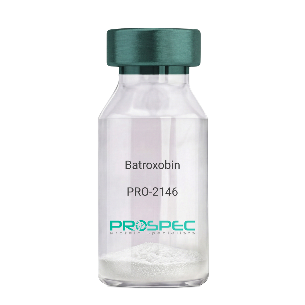
- View Data Sheet
Name :
BatroxobinDescription:
Batroxobin
Thrombin-like enzyme batroxobin, EC 3.4.21.74, BX, Bothrops atrox serine proteinase, Venombin-A, Batroxobin.
Product # :
PRO-2146Price :
Quantity :
Shipping Method :
Shipped at Room temp
More Info
- description
- formulation
- More Info
Description
Batroxobin, isolated from Bothrops atrox snake venom, has an Mw of approximately 43kDa.
Formulation
The Batroxobin protein was lyophilized from a concentrated (1mg/ml) solution with no additives.
More Info
-
Introduction
Batroxobin is a serin protease that reduces fibronogen levels and is originally extracted from snake venom of Bothrops Atrox. Batroxobin is used in defibrinogenation and thrombolysis and also has an effect on c-fos gene and growth factor.
Batroxobin can efficiently restrain proliferation of VSMCs, by blocking the release and uptake of Ca2+, thus influencing [Ca2+]i.
Batroxobin converts fibrinogen to fibrin through the restricted release of fibrinopeptide-A from fibrinogen to promote blood to clot. Unlike thrombin, it is not affected by heparin and hirudin. -
Synonyms
Thrombin-like enzyme batroxobin, EC 3.4.21.74, BX, Bothrops atrox serine proteinase, Venombin-A, Batroxobin.
-
Physical Appearance
Sterile Filtered white lyophilized powder.
-
Stability
Store the lyophilized Batroxobin between 2-8°C. Do not freeze!
-
Solubility
It is recommended to reconstitute the lyophilized Batroxobin in sterile 18MΩ-cm H2O not less than 100µg/ml or more than 10mg/ml solutions.
-
Unit Definition
100BU [Batroxobin Units]=1mg.
ProSpec's products are furnished for LABORATORY RESEARCH USE ONLY. The product may not be used as drugs, agricultural or pesticidal products, food additives or household chemicals.
- View Data Sheet
Name :
Tpc1808 RatDescription:
Tropic 1808 Rat Recombinant
Tropic 1808, Tpc1808.
Product # :
PRO-587Price :
Quantity :
Shipping Method :
Shipped at Room temp
More Info
- description
- source
- formulation
- purity
- More Info
Description
Tropic-1808 Rat Recombinant protein fused to N-terminal His-Tag produced in E.Coli is a single, non-glycosylated polypeptide chain containing 285 amino acids and having a molecular mass of 29.1 kDa.The Tpc1808 is purified by proprietary chromatographic techniques.
Source
Escherichia Coli.
Formulation
The Tropic-1808 was lyophilized from 1X PBS, pH 7.4.
Purity
Greater than 95.0% as determined by: (a) Analysis by RP-HPLC.
(b) Analysis by SDS-PAGE.More Info
-
Introduction
Tropic 1808 is a candidate chemotropic factor induced by nerve injury. Tpc1808 protein, similar to NGF, could promote the expression of NF-H in a time-dependent manner. Tpc1808 is the gene related to promotion of nerve growth, and both the Tpc1808 gene and the Tpc1808 recombinant protein up-regulate the expression of NF-H in PC12 cells.
-
Synonyms
Tropic 1808, Tpc1808.
-
Physical Appearance
Sterile Filtered White lyophilized (freeze-dried) powder.
-
Stability
Lyophilized Tpc1808 although stable 10°C for 1 week, should be stored desiccated below -18°C.Please prevent freeze-thaw cycles.
-
Amino Acid Sequence
MSYYHHHHHHMNLAQIAALNQISNLNAIRVGQVLKVSNAAGSNNTQNTTQPS
AGVPTNTASSTTGYTVKSGDTLSAIAAANGVSLANLLSWNNLSLQAIIYPGQKL
TIQNANNATVTTPNAPTSTPTVMPSTNGSYTVKSGDTLYGIAAKLGTNVQTLLS
LNGLQLSSTIYVGQVLKTTGAVAGAGTATSTPTPVTPTVSKPAAANGVSTAGLS
AAQAAWLRTAVVDAQAATAGTGVLASVTVAQAILESGWGQSALASAPYHNF
NLYLIKVKNTWKLMTLLLS.
ProSpec's products are furnished for LABORATORY RESEARCH USE ONLY. The product may not be used as drugs, agricultural or pesticidal products, food additives or household chemicals.
- View Data Sheet
Name :
SAA Paired AntibodyDescription:
Anti Human Serum Amyloid A (APO-SAA) Paired Antibody Mouse
Product # :
ANT-785Price :
Quantity :
Shipping Method :
Shipped with Ice Packs
More Info
- description
- formulation
- purity
- More Info
Description
SAA Paired monoclonal antibodies are used to develop rapid test for serum amyloid A protein rapid test. Please note that when ordering for example: 100µg paired antibody, you receive 50µg from each antibody (100µg in total).
Formulation
*SAA conjugation antibody in PBS, NaCl and 0.095% NaN3.
* SAA coating antibody in PBS, NaCl and 0.095% NaN3.
Purity
Greater than 95%.
More Info
-
Physical Appearance
2 vials of sterile Filtered clear colorless solution.
-
Stability
For periods up to 1month SAA Paired Antibody should be stored at 4°C, for longer periods of time, store at -20°C. Prevent freeze thaw cycles.
-
Applications
Lateral flow immunoassay.
-
Background
Serum amyloid A protein which is encoded by the SAA gene in humans, is a member of the SAA family. SAA is expressed by the liver and secreted in plasma. Serum amyloid A is the main acute phase reactant and apolipoprotein of the HDL complex. Elevated serum SAA protein levels may be linked with lung cancer. The level of Apo-SAA in plasma increases within 24 hours of an inflammatory stimulus and under these conditions is the most abundant HDL Apolipoprotein.
-
Type
Mouse antibody Monoclonal.
-
Purification Method
Purified monoclonal IgG1 by protein A chromatography.
ProSpec's products are furnished for LABORATORY RESEARCH USE ONLY. The product may not be used as drugs, agricultural or pesticidal products, food additives or household chemicals.
- View Data Sheet
Name :
MINA HumanDescription:
MYC Induced Nuclear Antigen Human Recombinant
MYC Induced Nuclear Antigen, MINA53, MDIG, 60S Ribosomal Protein L27a Histidine Hydroxylase, Mineral Dust-Induced Gene Protein, Histone Lysine Demethylase MINA Ribosomal Oxygenase MINA, Nucleolar Protein 52, NO52, ROX, Bifunctional Lysine-Specific Demethylase And Histidyl-Hydroxylase MINA, Myc-Induced Nuclear Antigen, 53 KDa, Mineral Dust Induced Gene Protein, MYC-Induced Nuclear Antigen, EC 1.14.11.-, Bifunctional lysine-specific demethylase and histidyl-hydroxylase MINA.
Product # :
PRO-2078Price :
Quantity :
Shipping Method :
Shipped with Ice Packs
More Info
- description
- source
- formulation
- purity
- More Info
Description
MINA Human Recombinant produced in E.Coli is a single, non-glycosylated polypeptide chain containing 485 amino acids (1-465 a.a) and having a molecular mass of 54.9kDa. MINA is fused to a 20 amino acid His-tag at N-terminus & purified by proprietary chromatographic techniques.
Source
Escherichia Coli.
Formulation
MINA protein solution (0.25mg/ml) containing 20mM Tris-HCl buffer (pH 8.0), 0.15M NaCl, 10% glycerol and 1mM DTT.
Purity
Greater than 90.0% as determined by SDS-PAGE.
More Info
-
Introduction
MYC Induced Nuclear Antigen, also known as MINA is an oxygenase which can function both as a histone lysine demethylase and a ribosomal histidine hydroxylase. MINA is involved in the demethylation of trimethylated Lys-9 on histone H3 (H3K9me3), leading to an increase in ribosomal RNA expression. MINA also catalyzes the hydroxylation of 60S ribosomal protein L27a on His-39. In addition, MINA plays a significant role in cell growth and survival. MINA is implicated in ribosome biogenesis, probably in the duration of the assembly process of pre-ribosomal particles.
-
Synonyms
MYC Induced Nuclear Antigen, MINA53, MDIG, 60S Ribosomal Protein L27a Histidine Hydroxylase, Mineral Dust-Induced Gene Protein, Histone Lysine Demethylase MINA Ribosomal Oxygenase MINA, Nucleolar Protein 52, NO52, ROX, Bifunctional Lysine-Specific Demethylase And Histidyl-Hydroxylase MINA, Myc-Induced Nuclear Antigen, 53 KDa, Mineral Dust Induced Gene Protein, MYC-Induced Nuclear Antigen, EC 1.14.11.-, Bifunctional lysine-specific demethylase and histidyl-hydroxylase MINA.
-
Physical Appearance
Sterile Filtered clear solution.
-
Stability
Store at 4°C if entire vial will be used within 2-4 weeks. Store, frozen at -20°C for longer periods of time. For long term storage it is recommended to add a carrier protein (0.1% HSA or BSA).Avoid multiple freeze-thaw cycles.
-
Amino Acid Sequence
MGSSHHHHHH SSGLVPRGSH MPKKAKPTGS GKEEGPAPCK QMKLEAAGGP SALNFDSPSS LFESLISPIK TETFFKEFWE QKPLLIQRDD PALATYYGSL FKLTDLKSLC SRGMYYGRDV NVCRCVNGKK KVLNKDGKAH FLQLRKDFDQ KRATIQFHQP QRFKDELWRI QEKLECYFGS LVGSNVYITP AGSQGLPPHY DDVEVFILQL EGEKHWRLYH PTVPLAREYS VEAEERIGRP VHEFMLKPGD LLYFPRGTIH QADTPAGLAH STHVTISTYQ NNSWGDFLLD TISGLVFDTA KEDVELRTGI PRQLLLQVES TTVATRRLSG FLRTLADRLE GTKELLSSDM KKDFIMHRLP PYSAGDGAEL STPGGKLPRL DSVVRLQFKD HIVLTVLPDQ DQSDETQEKM VYIYHSLKNS RETHMMGNEE ETEFHGLRFP LSHLDALKQI WNSPAISVKD LKLTTDEEKE SLVLSLWTEC LIQVV.
ProSpec's products are furnished for LABORATORY RESEARCH USE ONLY. The product may not be used as drugs, agricultural or pesticidal products, food additives or household chemicals.
- View Data Sheet
Name :
APP Human, HEKDescription:
Amyloid beta (A4) Precursor Protein Human Recombinant, HEK
ABPP, APPI, Alzheimer disease amyloid A4 protein homolog, Alzheimer disease amyloid protein, Amyloid precursor protein, Amyloid-beta precursor protein, Amyloid-beta A4 protein, Cerebral vascular amyloid peptide, CVAP, PreA4, Protease nexin-II, PN-II, A4, AD1, beta-amyloid peptide, beta-amyloid precursor protein, testicular tissue protein Li 2, AAA, ABETA, alpha-sApp, CTF gamma, PN2.
Product # :
PRO-2777Price :
Quantity :
Shipping Method :
Shipped with Ice Packs
More Info
- description
- source
- formulation
- purity
- biological activity
- More Info
Description
APP Human Recombinant is a single, glycosylated, polypeptide chain (18-701 a.a) containing a total of 690 amino acids, having a molecular mass of 78.2 kDa. APP is fused to a 6 amino acid His-tag at C-terminus and is purified by proprietary chromatographic techniques.
Source
HEK293 Cells.
Formulation
The APP solution (0.5mg/ml) contains 10% Glycerol and Phosphate-Buffered Saline (pH 7.4).
Purity
> 90% by SDS-PAGE.
Biological Activity
The inhibitory function of APP on activity of trypsin was measured by a fluorometric
assay using Mca-RPKPVE-Nval-WRK(Dnp)-NH2 at pH 7.5 at 37C. The IC50 ≤ 1 nM.
More Info
-
Synonyms
ABPP, APPI, Alzheimer disease amyloid A4 protein homolog, Alzheimer disease amyloid protein, Amyloid precursor protein, Amyloid-beta precursor protein, Amyloid-beta A4 protein, Cerebral vascular amyloid peptide, CVAP, PreA4, Protease nexin-II, PN-II, A4, AD1, beta-amyloid peptide, beta-amyloid precursor protein, testicular tissue protein Li 2, AAA, ABETA, alpha-sApp, CTF gamma, PN2.
-
Physical Appearance
Sterile Filtered colorless solution.
-
Stability
Store at 4°C if entire vial will be used within 2-4 weeks. Store, frozen at -20°C for longer periods of time. For long term storage it is recommended to add a carrier protein (0.1% HSA or BSA). Avoid multiple freeze-thaw cycles.
-
Amino Acid Sequence
LEVPTDGNAG LLAEPQIAMF CGRLNMHMNV QNGKWDSDPS GTKTCIDTKE GILQYCQEVY PELQITNVVE ANQPVTIQNW CKRGRKQCKT HPHFVIPYRC LVGEFVSDAL LVPDKCKFLH QERMDVCETH LHWHTVAKET CSEKSTNLHD YGMLLPCGID KFRGVEFVCC PLAEESDNVD SADAEEDDSD VWWGGADTDY ADGSEDKVVE VAEEEEVAEV EEEEADDDED DEDGDEVEEE AEEPYEEATE RTTSIATTTT TTTESVEEVV REVCSEQAET GPCRAMISRW YFDVTEGKCA PFFYGGCGGN RNNFDTEEYC MAVCGSAMSQ SLLKTTQEPL ARDPVKLPTT AASTPDAVDK YLETPGDENE HAHFQKAKER LEAKHRERMS QVMREWEEAE RQAKNLPKAD KKAVIQHFQE KVESLEQEAA NERQQLVETH MARVEAMLND RRRLALENYI TALQAVPPRP RHVFNMLKKY VRAEQKDRQH TLKHFEHVRM VDPKKAAQIR SQVMTHLRVI YERMNQSLSL LYNVPAVAEE IQDEVDELLQ KEQNYSDDVL ANMISEPRIS YGNDALMPSL TETKTTVELL PVNGEFSLDD LQPWHSFGAD SVPANTENEV EPVDARPAAD RGLTTRPGSG LTNIKTEEIS EVKMDAEFRH DSGYEVHHQK LVFFAEDVGS NKGA HHHHHH.
-
Background
Alzheimer's disease (AD) is a neurodegenerative disorder characterized by the accumulation of amyloid plaques and neurofibrillary tangles in the brain, leading to cognitive decline and memory loss. The amyloid beta (Aβ) peptide, derived from the amyloid beta (A4) precursor protein, has been identified as a key player in the pathogenesis of AD. This research aims to explore the significance of the amyloid beta precursor protein, its processing, and the implications it holds for understanding and treating Alzheimer's disease.
The amyloid beta precursor protein (APP) is a transmembrane protein widely expressed in various tissues, with higher concentrations found in the brain. APP undergoes sequential proteolytic processing by enzymes known as secretases, leading to the generation of Aβ peptides of different lengths. Of particular importance is the production of the Aβ42 peptide, which has a propensity to aggregate and form the characteristic amyloid plaques in AD.
Understanding the processing and metabolism of APP is crucial for unraveling the mechanisms underlying AD pathology. Mutations in the APP gene and dysregulation of its processing have been associated with familial forms of AD, highlighting the pivotal role of APP in disease development. Investigating the function of APP and its proteolytic fragments can provide valuable insights into the molecular events leading to AD and potentially lead to the identification of therapeutic targets.
This research will delve into the processing of the amyloid beta precursor protein, shedding light on the different cleavage pathways mediated by α-, β-, and γ-secretases. The paper will discuss the impact of these proteolytic events on the generation of Aβ peptides and how alterations in these pathways contribute to AD pathogenesis. Furthermore, it will explore the aggregation properties of Aβ peptides and their role in the formation of amyloid plaques, as well as their impact on neuronal function and viability.
The study will also examine the potential of APP and Aβ as biomarkers for AD diagnosis and progression monitoring. Investigating the levels of APP and Aβ peptides in biological fluids and utilizing imaging techniques to detect amyloid plaques could enhance early diagnosis and facilitate the development of novel therapeutic interventions.
By elucidating the molecular mechanisms involving APP and Aβ in AD, this research aims to contribute to the understanding of the disease pathogenesis and identify potential therapeutic targets. Additionally, it underscores the importance of ongoing research in this field to develop effective strategies for early diagnosis, disease modification, and improved patient outcomes.
ProSpec's products are furnished for LABORATORY RESEARCH USE ONLY. The product may not be used as drugs, agricultural or pesticidal products, food additives or household chemicals.
- View Data Sheet
Name :
SOD Human, 15NDescription:
Superoxide Dismutase, 15N Human Recombinant
Superoxide dismutase [Cu-Zn], EC 1.15.1.1, SOD1, SOD, ALS, ALS1, IPOA.
Product # :
PRO-2613Price :
Quantity :
Shipping Method :
Shipped at Room temp
More Info
- description
- source
- formulation
- purity
- More Info
Description
Recombinant Human Superoxide Dismutase, 15N produced in E.Coli is a single non-glycosylated polypeptide chain containing 153 amino acids and having a total molecular mass of 15.8kDa.
Source
Escherichia Coli.
Formulation
Lyophilized from a 0.2μm filtered concentrated (1mg/ml) solution in PBS, 0.1mM CuCl2 and 0.2mM ZnCl2.
Purity
Greater than 95.0% as determined by SDS-PAGE Analysis.
More Info
-
Introduction
Human Cu/Zn Superoxide Dismutase (SOD) catalyzes the reaction between superoxide anions and hydrogen to yield molecular oxygen and hydrogen peroxide. SOD protects the cell against dangerous levels of superoxide. SOD binds copper and zinc ions and is 1 of 3 isozymes accountable for destroying free superoxide radicals in the body. The encoded protein neutralizes supercharged oxygen molecules, which can damage cells if their levels are not controlled.
-
Synonyms
Superoxide dismutase [Cu-Zn], EC 1.15.1.1, SOD1, SOD, ALS, ALS1, IPOA.
-
Physical Appearance
Sterile Filtered White lyophilized (freeze-dried) powder.
-
Stability
Lyophilized SOD although stable at room temperature for 3 weeks, should be stored desiccated below -18°C. Upon reconstitution SOD should be stored at 4°C between 2-7 days and for future use below -18°C.For long term storage it is recommended to add a carrier protein (0.1% HSA or BSA).Please prevent freeze-thaw cycles.
-
Solubility
It is recommended to reconstitute the lyophilized SOD in sterile 18MΩ-cm H2O not less than 100µg/ml, which can then be further diluted to other aqueous solutions.
-
Amino Acid Sequence
ATKAVCVLKGDGPVQGIINFEQKESNGPVKVWGSIKGLTEGLHGFHVHEFGD NTAGCTSAGPHFNPLSRKHGGPKDEERHVGDLGNVTADKDGVADVSIEDSV ISLSGDHCIIGRTLVVHEKADDLGKGGNEESTKTGNAGSRLACGVIGIAQ.
ProSpec's products are furnished for LABORATORY RESEARCH USE ONLY. The product may not be used as drugs, agricultural or pesticidal products, food additives or household chemicals.
- View Data Sheet
Name :
RBP2 HumanDescription:
Retinol Binding Protein-2 Human Recombinant
CRABP-II, CRBP2, CRBPII, RBPC2, Retinol-binding protein 2, Cellular retinol-binding protein II, RBP2.
Product # :
CYT-751Price :
Quantity :
Shipping Method :
Shipped with Ice Packs
More Info
- description
- source
- formulation
- purity
- More Info
Description
RBP2 Human Recombinant produced in E.Coli is a single, non-glycosylated polypeptide chain containing 158 amino acids (1-134 a.a.) and having a molecular mass of 18kDa. RBP2 is fused to a 24 amino acid His-tag at N-terminus & purified by proprietary chromatographic techniques.
Source
E.coli.
Formulation
RBP2 protein solution (1mg/ml) contains 20mM Tris-HCl buffer (pH 8.0), 0.1M NaCl and 10% glycerol.
Purity
Greater than 95% as determined by SDS-PAGE.
More Info
-
Introduction
Retinol Binding Protein-2 (RBP2) which is present in the small intestinal epithelium takes part in the uptake and intracellular metabolism of vitamin A. Vitamin A is a fat-soluble vitamin essential for growth, reproduction, differentiation of epithelial tissues, and vision. RBP2 moderates the supply of retinoic acid to the nuclei of endometrial cells throughout the menstrual cycle.
-
Synonyms
CRABP-II, CRBP2, CRBPII, RBPC2, Retinol-binding protein 2, Cellular retinol-binding protein II, RBP2.
-
Physical Appearance
Sterile Filtered colorless solution.
-
Stability
Store at 4°C if entire vial will be used within 2-4 weeks. Store, frozen at -20°C for longer periods of time. For long term storage it is recommended to add a carrier protein (0.1% HSA or BSA).Avoid multiple freeze-thaw cycles.
-
Amino Acid Sequence
MGSSHHHHHH SSGLVPRGSH MGSHMTRDQN GTWEMESNEN FEGYMKALDI DFATRKIAVR LTQTKVIDQD GDNFKTKTTS TFRNYDVDFT VGVEFDEYTK SLDNRHVKAL VTWEGDVLVC VQKGEKENRG WKQWIEGDKL YLELTCGDQV CRQVFKKK.
ProSpec's products are furnished for LABORATORY RESEARCH USE ONLY. The product may not be used as drugs, agricultural or pesticidal products, food additives or household chemicals.