Skip to content

High-quality research proteins.

  • 1-800-805-6531

  • Mon - Sat 8.00-18.00, Sun - Closed

  • info@prospecbio.com

  • PO Box 6591 East Brunswick NJ 08816

  • Products
    • Cytokines
    • Growth factors
    • Chemokines
    • Cd antigens
    • Neurotrophins
    • Hormones
    • Enzymes
    • Viral antigens
    • Recombinant proteins
    • Natural proteins
    • Monoclonal antibodies
    • Polyclonal antibodies
    • Thermal Packaging
  • About
    • Company
    • Founder
    • Contribution
  • Customers
    • Eastern Asia
    • Eastern Europe
    • North America
    • South America
    • South East Asia
    • Southern Asia
    • Western Europe
  • Ordering
    • Ordering Method
    • Ordering - Credit Card
    • Ordering - PO
    • Shipping Fees
    • FAQs
  • Services
    • DNA Cloning
    • Protein Expression
    • Monoclonal Antibody
    • Fermentation
    • Technology License
  • Contact us
Log in

Country/region

  • USD $ | Albania
  • USD $ | Andorra
  • USD $ | Argentina
  • USD $ | Armenia
  • USD $ | Australia
  • USD $ | Austria
  • USD $ | Azerbaijan
  • USD $ | Belarus
  • USD $ | Belgium
  • USD $ | Bolivia
  • USD $ | Brazil
  • USD $ | Bulgaria
  • USD $ | Cambodia
  • USD $ | Canada
  • USD $ | Chile
  • USD $ | China
  • USD $ | Colombia
  • USD $ | Costa Rica
  • USD $ | Croatia
  • USD $ | Cyprus
  • USD $ | Czechia
  • USD $ | Denmark
  • USD $ | Estonia
  • USD $ | Finland
  • USD $ | France
  • USD $ | Georgia
  • USD $ | Germany
  • USD $ | Greece
  • USD $ | Hong Kong SAR
  • USD $ | Hungary
  • USD $ | Iceland
  • USD $ | India
  • USD $ | Ireland
  • USD $ | Israel
  • USD $ | Italy
  • USD $ | Japan
  • USD $ | Kazakhstan
  • USD $ | Latvia
  • USD $ | Liechtenstein
  • USD $ | Lithuania
  • USD $ | Madagascar
  • USD $ | Malta
  • USD $ | Mexico
  • USD $ | Moldova
  • USD $ | Monaco
  • USD $ | Netherlands
  • USD $ | New Zealand
  • USD $ | North Macedonia
  • USD $ | Norway
  • USD $ | Panama
  • USD $ | Paraguay
  • USD $ | Peru
  • USD $ | Philippines
  • USD $ | Poland
  • USD $ | Portugal
  • USD $ | Romania
  • USD $ | Singapore
  • USD $ | Slovakia
  • USD $ | Slovenia
  • USD $ | South Africa
  • USD $ | South Korea
  • USD $ | Spain
  • USD $ | Sri Lanka
  • USD $ | Sweden
  • USD $ | Switzerland
  • USD $ | Taiwan
  • USD $ | Thailand
  • USD $ | Türkiye
  • USD $ | Ukraine
  • USD $ | United Kingdom
  • USD $ | United States
  • USD $ | Uruguay
  • USD $ | Vatican City
  • USD $ | Venezuela
  • USD $ | Vietnam
  • Facebook
  • Instagram
logoProSpec - Recombinant Protein Manufacturer for academic and R&D industry
Become a distributor
Account Log in
Wishlist
Cart Cart(0)
  • Products
    +
    • Cytokines
    • Growth factors
    • Chemokines
    • Cd antigens
    • Neurotrophins
    • Hormones
    • Enzymes
    • Viral antigens
    • Recombinant proteins
    • Natural proteins
    • Monoclonal antibodies
    • Polyclonal antibodies
    • Thermal Packaging
  • About
    +
    • Company
    • Founder
    • Contribution
  • Customers
    +
    • Eastern Asia
    • Eastern Europe
    • North America
    • South America
    • South East Asia
    • Southern Asia
    • Western Europe
  • Ordering
    +
    • Ordering Method
    • Ordering - Credit Card
    • Ordering - PO
    • Shipping Fees
    • FAQs
  • Services
    +
    • DNA Cloning
    • Protein Expression
    • Monoclonal Antibody
    • Fermentation
    • Technology License
  • Contact us

Item added to your cart

View cart
View All
  • Cytokines
  • Growth factors
  • Chemokines
  • Cd antigens
  • Neurotrophins
  • Hormones
  • Enzymes
  • Viral antigens
  • Recombinant proteins
  • Natural proteins
  • Monoclonal antibodies
  • Polyclonal antibodies
  • Thermal Packaging
  • Tumor Necrosis Factor

    Tumor Necrosis Factor

    View All
  • 4-1BB

    4-1BB

    View All
  • Adiponectin

    Adiponectin

    View All
  • AIF1

    AIF1

    View All
  • Other Cytokines

    Other Cytokines

    View All
  • AITRL

    AITRL

    View All
  • Angiopoietin

    Angiopoietin

    View All
  • Apolipoprotein

    Apolipoprotein

    View All
  • B-Cell Activating Factor

    B-Cell Activating Factor

    View All
  • Beta Defensin

    Beta Defensin

    View All
  • Bone Morphogenetic Protein

    Bone Morphogenetic Protein

    View All
  • B type Natriuretic Peptide

    B type Natriuretic Peptide

    View All
  • BST

    BST

    View All
  • Betacellulin

    Betacellulin

    View All
  • Cardiotrophin

    Cardiotrophin

    View All
  • Activin

    Activin

    View All
  • Fibroblast Growth Factor

    Fibroblast Growth Factor

    View All
  • Other Growth Factors

    Other Growth Factors

    View All
  • CSF

    CSF

    View All
  • CTGF

    CTGF

    View All
  • IGFBP

    IGFBP

    View All
  • VEGF Protein

    VEGF Protein

    View All
  • EGF

    EGF

    View All
  • Epigen

    Epigen

    View All
  • Erythropoietin

    Erythropoietin

    View All
  • Insulin-Like Growth Factor

    Insulin-Like Growth Factor

    View All
  • Growth Hormone

    Growth Hormone

    View All
  • HDGF

    HDGF

    View All
  • Hepatocyte Growth Factor

    Hepatocyte Growth Factor

    View All
  • Insulin

    Insulin

    View All
  • BCA-1/ BLC (CXCL13)

    BCA-1/ BLC (CXCL13)

    View All
  • BRAK (CXCL14)

    BRAK (CXCL14)

    View All
  • C-10 (CCL6)

    C-10 (CCL6)

    View All
  • Other Chemokines

    Other Chemokines

    View All
  • MEC (CCL28)

    MEC (CCL28)

    View All
  • LD78-beta (CCL3L1)

    LD78-beta (CCL3L1)

    View All
  • CTACK (CCL27)

    CTACK (CCL27)

    View All
  • CXCL16

    CXCL16

    View All
  • CXCL17

    CXCL17

    View All
  • Platelet Factor-4 (CXCL4)

    Platelet Factor-4 (CXCL4)

    View All
  • ENA-78 (CXCL5)

    ENA-78 (CXCL5)

    View All
  • Eotaxin (CCL11,24,26)

    Eotaxin (CCL11,24,26)

    View All
  • Exodus-2 (CCL21)

    Exodus-2 (CCL21)

    View All
  • Fractalkine (CX3CL1)

    Fractalkine (CX3CL1)

    View All
  • CXCL6

    CXCL6

    View All
  • Other CD Antigens

    Other CD Antigens

    View All
  • CD14

    CD14

    View All
  • CD164

    CD164

    View All
  • CD1

    CD1

    View All
  • CD2

    CD2

    View All
  • CD200

    CD200

    View All
  • CD207

    CD207

    View All
  • CD226

    CD226

    View All
  • CD23

    CD23

    View All
  • CD244

    CD244

    View All
  • CD247

    CD247

    View All
  • CD27

    CD27

    View All
  • CD274

    CD274

    View All
  • CD300

    CD300

    View All
  • CD33

    CD33

    View All
  • Other Neurotrophins

    Other Neurotrophins

    View All
  • Beta-NGF

    Beta-NGF

    View All
  • BDNF

    BDNF

    View All
  • CDNF

    CDNF

    View All
  • CNTF

    CNTF

    View All
  • GDNF

    GDNF

    View All
  • Glia Maturation Factor

    Glia Maturation Factor

    View All
  • MANF

    MANF

    View All
  • Midkine

    Midkine

    View All
  • Neuroglobin

    Neuroglobin

    View All
  • Neurotrophic factors

    Neurotrophic factors

    View All
  • Neuregulin

    Neuregulin

    View All
  • Neuropilin

    Neuropilin

    View All
  • Pigment Epithelium-Derived Factor

    Pigment Epithelium-Derived Factor

    View All
  • Pleiotrophin

    Pleiotrophin

    View All
  • Peptide Hormones

    Peptide Hormones

    View All
  • Other Hormones

    Other Hormones

    View All
  • HCG

    HCG

    View All
  • Endothelin

    Endothelin

    View All
  • Exendin

    Exendin

    View All
  • FSH

    FSH

    View All
  • Glucagon

    Glucagon

    View All
  • GHRP

    GHRP

    View All
  • GLP

    GLP

    View All
  • Thyrostimulin

    Thyrostimulin

    View All
  • Inhibin A

    Inhibin A

    View All
  • LHRH

    LHRH

    View All
  • Melanotan

    Melanotan

    View All
  • Procalcitonin

    Procalcitonin

    View All
  • PTH

    PTH

    View All
  • Peroxiredoxin

    Peroxiredoxin

    View All
  • Peptidase

    Peptidase

    View All
  • Synthetase

    Synthetase

    View All
  • Transferase

    Transferase

    View All
  • Hydrolase

    Hydrolase

    View All
  • Dehydrogenase

    Dehydrogenase

    View All
  • ACE2

    ACE2

    View All
  • Esterase

    Esterase

    View All
  • Protein Kinases

    Protein Kinases

    View All
  • Other Enzymes

    Other Enzymes

    View All
  • Phosphatase

    Phosphatase

    View All
  • Deaminase

    Deaminase

    View All
  • Oxygenase

    Oxygenase

    View All
  • Adenylate Kinase

    Adenylate Kinase

    View All
  • Lyase

    Lyase

    View All
  • Chagas

    Chagas

    View All
  • SARS

    SARS

    View All
  • Influenza

    Influenza

    View All
  • ASFV

    ASFV

    View All
  • Astrovirus

    Astrovirus

    View All
  • Borrelia

    Borrelia

    View All
  • Helicobacter Pylori

    Helicobacter Pylori

    View All
  • Chikungunya

    Chikungunya

    View All
  • Chlamydia

    Chlamydia

    View All
  • Cytomegalo

    Cytomegalo

    View All
  • Dengue

    Dengue

    View All
  • EBV

    EBV

    View All
  • Ebola

    Ebola

    View All
  • Feline Leukemia Virus

    Feline Leukemia Virus

    View All
  • Hepatitis A

    Hepatitis A

    View All
  • Other

    Other

    View All
  • Actin

    Actin

    View All
  • ADAM

    ADAM

    View All
  • Complement Component

    Complement Component

    View All
  • Ag85

    Ag85

    View All
  • Eukaryotic Translation Initiation Factor

    Eukaryotic Translation Initiation Factor

    View All
  • Anterior Gradient Protein

    Anterior Gradient Protein

    View All
  • Heat Shock Protein

    Heat Shock Protein

    View All
  • Alpha-2-HS-Glycoprotein

    Alpha-2-HS-Glycoprotein

    View All
  • Hemoglobin

    Hemoglobin

    View All
  • Allergy

    Allergy

    View All
  • Anaplasma

    Anaplasma

    View All
  • Angiogenin

    Angiogenin

    View All
  • Ankyrin Repeat Domain

    Ankyrin Repeat Domain

    View All
  • Annexin

    Annexin

    View All
  • Other Natural Proteins

    Other Natural Proteins

    View All
  • Aprotinin

    Aprotinin

    View All
  • Transferrin

    Transferrin

    View All
  • Avidin

    Avidin

    View All
  • Anti Coagulation Factors

    Anti Coagulation Factors

    View All
  • Natural Albumin

    Natural Albumin

    View All
  • Natural Coagulation Factors

    Natural Coagulation Factors

    View All
  • Fibronectin

    Fibronectin

    View All
  • Thrombin

    Thrombin

    View All
  • Anti Human Enzyme

    Anti Human Enzyme

    View All
  • Other Monoclonal Antibodies

    Other Monoclonal Antibodies

    View All
  • Anti Human Cytokine

    Anti Human Cytokine

    View All
  • Anti Human Heat Shock Protein

    Anti Human Heat Shock Protein

    View All
  • Anti Mouse Lymphocyte

    Anti Mouse Lymphocyte

    View All
  • Anti Human Chemokine

    Anti Human Chemokine

    View All
  • Anti Human Lymphocyte

    Anti Human Lymphocyte

    View All
  • Anti Viral Monoclonal

    Anti Viral Monoclonal

    View All
  • Anti Mouse Cytokine

    Anti Mouse Cytokine

    View All
  • Anti-GST Monoclonal

    Anti-GST Monoclonal

    View All
  • Other Polyclonal Antibodies

    Other Polyclonal Antibodies

    View All
  • Anti Viral

    Anti Viral

    View All
  • Anti-GST Monoclonal

    Anti-GST Monoclonal

    View All
Back

Search results

1000 results found for “calpain”

Name

Description

Product #

Price

Quantity

Shipping Method

  • View Data Sheet

    Name :

    Resistin Human, Antagonist

    Description:

    Resistin Antagonist Human Recombinant

    Cysteine-rich secreted protein FIZZ3, Adipose tissue-specific secretory factor, ADSF, C/EBP-epsilon-regulated myeloid-specific secreted cysteine-rich protein, Cysteine-rich secreted protein A12-alpha-like 2, RSTN, XCP1, RETN1, MGC126603, MGC126609.

    Product # :

    CYT-1255

    Price :

    Quantity :

    Shipping Method :

    Room Temp Icon

    Shipped at Room temp

    Add To Cart

    More Info

    • description
    • source
    • formulation
    • purity
    • biological activity
    • More Info

    Description

    Resistin Human antagonist is a monomeric C7A mutant that does not form covalent dimers. Resistin Human antagonist is purified by proprietary chromatographic techniques.

    Source

    Escherichia coli.

    Formulation

    Resistin was lyophilized from a concentrated (1mg/ml) solution with 0.03% NaHCO3.

    Purity

    Greater than 98.0% as determined by:

    (a) Analysis by Gel Filtration.

    (b) Analysis by SDS-PAGE.

    (c) Analysis by RP-HPLC.

    Biological Activity

    The biological activity was evidenced by resistin antagonist activity to inhibit resistin-induced Akt phosphorylation in two cell lines. It also reduced the weight (mainly the visceral fat) and normalized GTT and ITT inHFD-fed mice.

    More Info

    • Synonyms

      Cysteine-rich secreted protein FIZZ3, Adipose tissue-specific secretory factor, ADSF, C/EBP-epsilon-regulated myeloid-specific secreted cysteine-rich protein, Cysteine-rich secreted protein A12-alpha-like 2, RSTN, XCP1, RETN1, MGC126603, MGC126609.

    • Physical Appearance

      Sterile Filtered White lyophilized (freeze-dried) powder.

    • Stability

      Lyophilized Resistin although stable at room temperature for 3 weeks, should be stored desiccated below -18°C. Upon reconstitution Resistin Mouse should be stored at 4°C between 2-7 days and for future use below -18°C. For long term storage it is recommended to add a carrier protein (0.1% HSA or BSA). Please prevent freeze-thaw cycles.

    • Solubility

      It is recommended to reconstitute the lyophilized Resistin in sterile 18MΩ-cm H2O at a concentration of 100µg/ml, which can then be further diluted to other aqueous solutions.

    • Amino Acid Sequence

      The sequence of the first seven N-terminal amino acids was determined and was found to be Ala-Ser-Ser-Lys-Thr-Leu-Ala.

    • Background

      Resistin, also known as adipose tissue-specific secretory factor (ADSF) is a cysteine-rich peptide derived from adipose tissue. Resistin takes part in the inflammatory response, glucose metabolism, and angiogenesis. Resistin blocks insulin stimulated uptake of glucose by adipocytes and promote glucose release by hepatocytes. As such,Resistin considered to participate in diet‑induced insulin-sensitivity. Resistin causes high levels of low-density lipoprotein (LDL), increasing the risk of heart disease.

    ProSpec's products are furnished for LABORATORY RESEARCH USE ONLY. The product may not be used as drugs, agricultural or pesticidal products, food additives or household chemicals.

    Resistin Antagonist
  • View Data Sheet

    Name :

    HBsAg adw

    Description:

    Hepatitis B Surface Antigen, adw Recombinant

    Product # :

    HBS-872

    Price :

    Quantity :

    Shipping Method :

    Ice Icon

    Shipped with Ice Packs

    Add To Cart

    More Info

    • description
    • source
    • formulation
    • purity
    • More Info

    Description

    HbsAg adw produced Pichia Pastoris, having a molecular weight of approximately 24.0 kDa as shown on SDS-PAGE.

    Source

    Pichia Pastoris.

    Formulation

    Sterile Filtered solution containing 20mM Phosphate Buffer, 154mM sodium chloride, pH 7.1.

    Purity

    Greater than 95.0% as determined by SDS-PAGE.

    More Info

    • Introduction

      HBsAg is the surface antigenof the Hepatitis-B-Virus (HBV). The capsidof a virus has different surface proteins from the rest of the virus. The antigen is a protein that binds specifically on one of these surface proteins. It is commonly referred to as the Australian Antigen.

    • Physical Appearance

      Sterile Filtered pale solution.

    • Stability

      HBsAg Should be stored at 4°C.DO NOT FREEZE.

    ProSpec's products are furnished for LABORATORY RESEARCH USE ONLY. The product may not be used as drugs, agricultural or pesticidal products, food additives or household chemicals.

    Hbsag Adw
  • View Data Sheet

    Name :

    Angiotensin

    Description:

    Angiotensin

    Angiotensinogen, Serpin A8, ANHU, SERPINA8.

    Product # :

    ENZ-283

    Price :

    Quantity :

    Shipping Method :

    Room Temp Icon

    Shipped at Room temp

    Add To Cart

    More Info

    • description
    • source
    • purity
    • More Info

    Description

    Angiotensin contains a total of 8 amino acids having a molecular weight of 1031.2 Dalton and a molecular formula of C49H70N14O11.

    Source

    Synthetic.

    Purity

    Greater than 98.0% as determined by RP-HPLC.

    More Info

    • Introduction

      Angiotensin is an oligopeptide in the blood that causes vasoconstriction, increased blood pressure, and release of aldosterone from the adrenal cortex. It is a powerful dipsogen. It is derived from the precursor molecule angiotensinogen, a serum globulin produced in the liver. It plays an important role in the renin-angiotensin system.
      The protein encoded by this gene, pre-angiotensinogen or angiotensinogen precursor, is expressed in the liver and is cleaved by the enzyme renin in response to lowered blood pressure. The resulting product, angiotensin I is then cleaved by angiotensin converting enzyme (ACE) to generate the physiologically active enzyme angiotensin II. The protein is involved in maintaining blood pressure and in the pathogenesis of essential hypertension and preeclampsia.

    • Synonyms

      Angiotensinogen, Serpin A8, ANHU, SERPINA8.

    • Physical Appearance

      Sterile Filtered white lyophilized powder.

    • Stability

      Lyophilized Angiotensin although stable at room temperature for 3 weeks, should be stored desiccated below -18°C. Upon reconstitution Serpin A8 should be stored at 4°C between 2-7 days and for future use below -18°C.Please prevent freeze-thaw cycles.

    • Solubility

      It is recommended to reconstitute the lyophilized Angiotensin in sterile 18MΩ-cm H2O not less than 100 µg/ml or more than 10 mg/ml solutions.

    • Amino Acid Sequence

      Asn-Arg-Val-Tyr-Val-His-Pro-Phe-OH.

    ProSpec's products are furnished for LABORATORY RESEARCH USE ONLY. The product may not be used as drugs, agricultural or pesticidal products, food additives or household chemicals.

    Angiotensin
  • View Data Sheet

    Name :

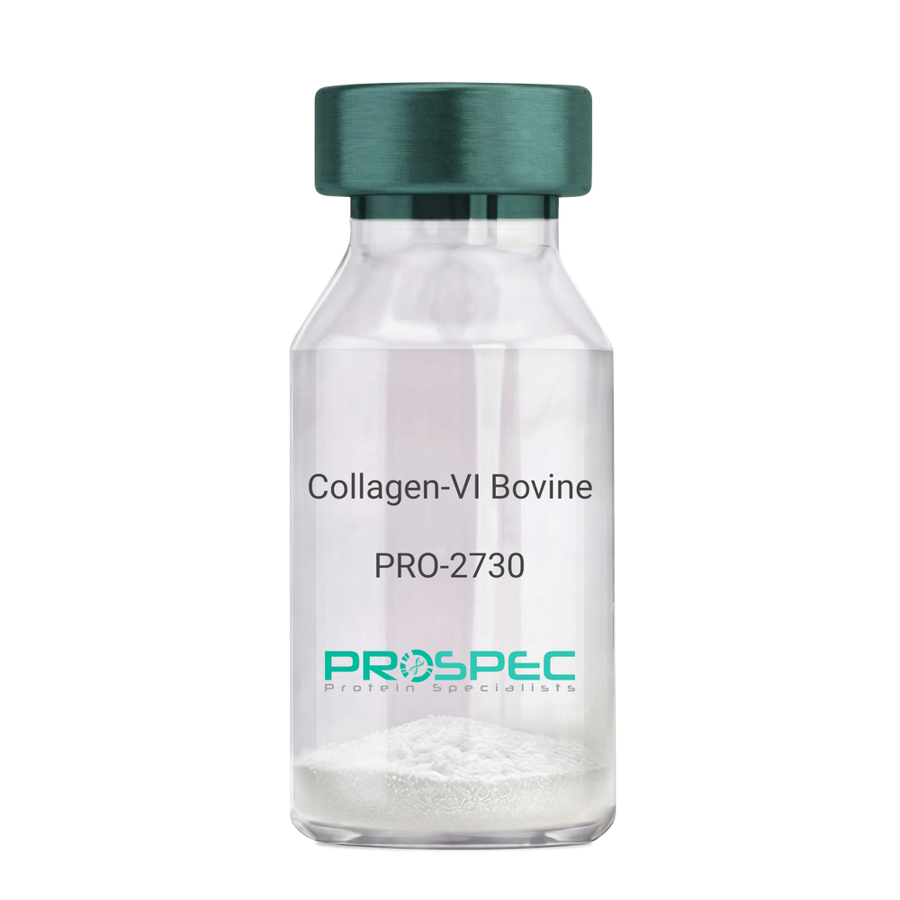
    Collagen-VI Bovine

    Description:

    Bovine Collagen-VI

    Product # :

    PRO-2730

    Price :

    Quantity :

    Shipping Method :

    Room Temp Icon

    Shipped at Room temp

    Add To Cart

    More Info

    • description
    • source
    • formulation
    • purity
    • More Info

    Description

    Bovine Collagen-VI is a natural protein purified from Bovine placenta. Bovine Collagen-VI is purified by proprietary chromatographic techniques.

    Source

    Bovine placenta.

    Formulation

    Collagen-VI was lyophilized without additives.

    Purity

    > 90.0%.

    More Info

    • Introduction

      Collagen, a major component of the extracellular matrix, is a fibrous protein that provides tensile strength to tissues giving them structural integrity. Collagen and its derivative, gelatin, have been widely used in medical, pharmaceutical and consumer products for more than 100 years. The supply of these materials, created from animal remains, is both abundant and inexpensive. However, most formulations are not highly purified and have the potential to cause an inflammatory reaction in some product users. In addition, concerns have been raised over the last several years about the potential for contamination of bovine products with the agent that causes mad cow disease and its human variant, Creutzfeldt-Jakob Disease. Animal collagens are subject to extensive modifications that continue over the life of the molecule in the extracellular space. These differences influence both the extractability of collagens from tissue and the biophysical characteristics of these collagens. As a result, collagens isolated from tissues exhibit significant lot-to-lot variability and, as bulk materials, are often analytically intractable. Products that contain animal-derived collagen can induce potentially harmful inflammatory or immune responses in humans and pose risk of contamination with viruses or prions, potentially life-threatening pathogens. Recombinant collagens are essentially identical to the native collagen protein thereby reducing the risk of inflammation, immune response, and disease as compared to animal-sourced collagen.

    • Physical Appearance

      Filtered White lyophilized (freeze-dried) powder.

    • Stability

      Lyophilized Collagen-VI although stable at room temperature for 3 weeks, should be stored desiccated below -18C. Upon reconstitution Collagen-VI should be stored at 4C between 2-7 days and for future use below -18C.For long term storage it is recommended to add a carrier protein (0.1% HSA or BSA).Please prevent freeze-thaw cycles.

    • Solubility

      It is recommended to Add 0.5 M acetic acid, pH 2.5 to prepare a working stock solution not less than 100µg/ml, which can then be further diluted to other aqueous solutions.

    ProSpec's products are furnished for LABORATORY RESEARCH USE ONLY. The product may not be used as drugs, agricultural or pesticidal products, food additives or household chemicals.

    Collagen Vi Bovine
  • View Data Sheet

    Name :

    SARS Spike (14-667)

    Description:

    SARS Spike (14-667 a.a.), Recombinant

    Product # :

    SARS-031

    Price :

    Quantity :

    Shipping Method :

    Ice Icon

    Shipped with Ice Packs

    Add To Cart

    More Info

    • description
    • source
    • formulation
    • purity
    • biological activity
    • More Info

    Description

    The HEK293 derived recombinant protein contains the SARS Coronavirus Spike S1 Gycoprotein, amino acids 14-667 fused to His tag at C-terminal.

    Source

    HEK293

    Formulation

    SARS Spike S1 glycoprotein is lyophilized from 1x PBS pH-7.4 + 10% Glycerol.

    Purity

    Protein is >90% pure as determined SDS-PAGE.

    Biological Activity

    Measured by its binding ability in a functional ELISA with Human ACE-2 (CAT# enz-1125)

    More Info

    • Introduction

      SARS Coronavirus is an enveloped virus containing three outer structural proteins, namely the membrane (M), envelope (E), and spike (S) proteins. Spike (S)-glycoprotein of the virus interacts with a cellular receptor and mediates membrane fusion to allow viral entry into susceptible target cells. Accordingly, S-protein plays an important role in virus infection cycle and is the primary target of neutralizing antibodies.

    • Physical Appearance

      Sterile Filtered clear solution.

    • Stability

      Protein is shipped on ice packs. Upon arrival, Store at -20°C.

    • Purification Method

      Purified by immobilized metal affinity chromatographic technique.

    ProSpec's products are furnished for LABORATORY RESEARCH USE ONLY. The product may not be used as drugs, agricultural or pesticidal products, food additives or household chemicals.

    product_image.jpg
  • View Data Sheet

    Name :

    LGALS4 Human

    Description:

    Galectin-4 Human Recombinant

    Galectin-4, Gal-4, Lactose-binding lectin 4, L-36 lactose-binding protein, L36LBP, Antigen NY-CO-27, LGALS4, lectin galactoside-binding soluble 4, GAL4.

    Product # :

    CYT-686

    Price :

    Quantity :

    Shipping Method :

    Ice Icon

    Shipped with Ice Packs

    Add To Cart

    More Info

    • description
    • source
    • formulation
    • purity
    • More Info
    • sds-page

    Description

    Galectin-4 produced in E.Coli is a single, non-glycosylated polypeptide chain containing 343 amino acids (1-323 a.a.) and having a molecular mass of 38.1kDa.Galectin-4 is fused to 20 amino acid His-Tag at N-terminus and purified by proprietary chromatographic techniques.

    Source

    Escherichia Coli.

    Formulation

    The LGALS4 protein solution contains 20mM Tris-HCl, pH-8, 1mM DTT and 10% glycerol.

    Purity

    Greater than 95.0% as determined by SDS-PAGE.

    sds-page

    LGALS4 Human-SDS-PAGE - Product image 1

    More Info

    • Introduction

      Galectin-4 is a member of the subfamily of galectins composed of two carbohydrate recognition domains having similar peptide chains. The galectins are a family of beta-galactoside-binding proteins having a role in modulating cell-cell and cell-matrix interactions, which inhibits chronic inflammations, GVHD, and allergic responses. LGALS4 expression is limited to small intestine, colon, and rectum, and it is underexpressed in colorectal cancer. LGALS4 binds as an endogenous ligand to glycosphingolipids having 3-O-sulfated Gal residues and bind as well to cholesterol-3-sulfate. LGALS4 takes part in cell adhesion. LGALS4 plays a role in crosslinking the lateral cell membranes of the surface-lining epithelial cells, thus supporting epithelial integrity against mechanical stress exerted by the bowel lume. LGALS4 is in charge of intestinal inflammation via selective regulation of peripheral and mucosal T-cell cell cycle, in addition to cell death by apoptosis of T-cells by a pathway independent of the activation of caspases. LGALS4 blockade decreases TNF-alpha inhibitor induced T-cell death. LGALS4 decreases pro-inflammatory cytokine secretion including IL-6 & IL-17.

    • Synonyms

      Galectin-4, Gal-4, Lactose-binding lectin 4, L-36 lactose-binding protein, L36LBP, Antigen NY-CO-27, LGALS4, lectin galactoside-binding soluble 4, GAL4.

    • Physical Appearance

      Sterile filtered colorless solution.

    • Stability

      Store at 4°C if entire vial will be used within 2-4 weeks. Store, frozen at -20°C for longer periods of time. For long term storage it is recommended to add a carrier protein (0.1% HSA or BSA).Avoid multiple freeze-thaw cycles.

    • Amino Acid Sequence

      MGSSHHHHHH SSGLVPRGSH MAYVPAPGYQ PTYNPTLPYY QPIPGGLNVG MSVYIQGVAS EHMKRFFVNF VVGQDPGSDV AFHFNPRFDG WDKVVFNTLQ GGKWGSEERK RSMPFKKGAA FELVFIVLAE HYKVVVNGNP FYEYGHRLPL QMVTHLQVDG DLQLQSINFI GGQPLRPQGP PMMPPYPGPG HCHQQLNSLP TMEGPPTFNP PVPYFGRLQG GLTARRTIII KGYVPPTGKS FAINFKVGSS GDIALHINPR MGNGTVVRNS LLNGSWGSEE KKITHNPFGP GQFFDLSIRC GLDRFKVYAN GQHLFDFAHR LSAFQRVDTL EIQGDVTLSY VQI.

    • Background

      What is the molecular weight/Mw of LGALS4 HUMAN Protein?
      LGALS4 HUMAN Protein has a total Mw of 38.1kDa.

      What is the source or expression system of LGALS4 HUMAN Protein?
      Escherichia Coli.

      What is the Purity of LGALS4 HUMAN Protein?
      LGALS4 HUMAN Protein is >95% pure as determined by SDS-PAGE.

      What is the Biological Activity of LGALS4 HUMAN Protein?
      The biological functionality of LGALS4 HUMAN Protein will be determined in the future.

      What is the amino acid sequence of LGALS4 HUMAN Protein?
      MGSSHHHHHH SSGLVPRGSH MAYVPAPGYQ PTYNPTLPYY QPIPGGLNVG MSVYIQGVAS EHMKRFFVNF VVGQDPGSDV AFHFNPRFDG WDKVVFNTLQ GGKWGSEERK RSMPFKKGAA FELVFIVLAE HYKVVVNGNP FYEYGHRLPL QMVTHLQVDG DLQLQSINFI GGQPLRPQGP PMMPPYPGPG HCHQQLNSLP TMEGPPTFNP PVPYFGRLQG GLTARRTIII KGYVPPTGKS FAINFKVGSS GDIALHINPR MGNGTVVRNS LLNGSWGSEE KKITHNPFGP GQFFDLSIRC GLDRFKVYAN GQHLFDFAHR LSAFQRVDTL EIQGDVTLSY VQI.

      What applications can LGALS4 HUMAN Protein be used in?
      LGALS4 HUMAN Protein can probably be used in western blot, ELISA and Lateral Flow.

      What is the endotoxin level for LGALS4 HUMAN Protein?
      The endotoxin level is minimal, LGALS4 HUMAN Protein was purified using conventional chromatography techniques.


    ProSpec's products are furnished for LABORATORY RESEARCH USE ONLY. The product may not be used as drugs, agricultural or pesticidal products, food additives or household chemicals.

    Lgals4 Human
  • View Data Sheet

    Name :

    CTSZ Human, Sf9

    Description:

    Cathepsin-Z Human Recombinant, Sf9

    Cathepsin Z, Cathepsin X, Cysteine-Type Carboxypeptidase, Lysosomal Carboxypeptidase B, Carboxypeptidase LB, Cathepsin B2, Cathepsin IV, Cathepsin Z1, Cathepsin P, Cathepsin Y, EC 3.4.18.1, Preprocathepsin P, CTSX.

    Product # :

    ENZ-1097

    Price :

    Quantity :

    Shipping Method :

    Ice Icon

    Shipped with Ice Packs

    Add To Cart

    More Info

    • description
    • source
    • formulation
    • purity
    • biological activity
    • More Info

    Description

    CTSZ produced in Sf9 Baculovirus cells is a single, glycosylated polypeptide chain containing 288 amino acids (24-303.a.) and having a molecular mass of 32.5kDa. (Molecular size on SDS-PAGE will appear at approximately 28-40kDa).CTSZ is expressed with an 8 amino acid His tag at C-Terminus and purified by proprietary chromatographic techniques.

    Source

    Sf9, Baculovirus cells.

    Formulation

    CTSZ protein solution (0.5mg/ml) contains 10% glycerol & Phosphate Buffered Saline (pH 7.4).

    Purity

    Greater than 95% as determined by SDS-PAGE.

    Biological Activity

    The specific activity is determent as the ability of 1 unit to convert 1.0 pmole of Mca-PLGL-Dpa-AR-NH2 to MCA- Pro-Leu-OH per minute at pH 3.5 at 25C and is > 1,400 pmol/min/ug.

    More Info

    • Introduction

      Cathepsin-Z (CTSZ) is a lysosomal cysteine proteinase and a member of the peptidase C1 family. CTSZ, which is known also as cathepsin X and cathepsin P, exhibits carboxy-monopeptidase and carboxy-dipeptidase activities. CTSZ is expressed ubiquitously in cancer cell lines and primary tumors and as other members of this family, takes part in tumorigenesis.

    • Synonyms

      Cathepsin Z, Cathepsin X, Cysteine-Type Carboxypeptidase, Lysosomal Carboxypeptidase B, Carboxypeptidase LB, Cathepsin B2, Cathepsin IV, Cathepsin Z1, Cathepsin P, Cathepsin Y, EC 3.4.18.1, Preprocathepsin P, CTSX.

    • Physical Appearance

      Sterile Filtered clear solution.

    • Stability

      Store at 4°C if entire vial will be used within 2-4 weeks. Store, frozen at -20°C for longer periods of time.For long term storage it is recommended to add a carrier protein (0.1% HSA or BSA).Please avoid freeze thaw cycles.

    • Amino Acid Sequence

      GLYFRRGQTC YRPLRGDGLA PLGRSTYPRP HEYLSPADLP KSWDWRNVDG VNYASITRNQ
      HIPQYCGSCW AHASTSAMAD RINIKRKGAW PSTLLSVQNV IDCGNAGSCE GGNDLSVWDY
      AHQHGIPDET CNNYQAKDQE CDKFNQCGTC NEFKECHAIR NYTLWRVGDY GSLSGREKMM
      AEIYANGPIS CGIMATERLA NYTGGIYAEY QDTTYINHVV SVAGWGISDG TEYWIVRNSW
      GEPWGERGWL RIVTSTYKDG KGARYNLAIE EHCTFGDPIV LEHHHHHH

    ProSpec's products are furnished for LABORATORY RESEARCH USE ONLY. The product may not be used as drugs, agricultural or pesticidal products, food additives or household chemicals.

    Cathepsin Z Protein
  • View Data Sheet

    Name :

    SORBS3 Human

    Description:

    Sorbin And SH3 Domain Containing 3 Human Recombinant

    Sorbin And SH3 Domain Containing 3, SCAM1, Vinexin Beta (SH3-Containing Adaptor Molecule-1), vinexin, SH3-Containing Adapter Molecule 1.

    Product # :

    PRO-1829

    Price :

    Quantity :

    Shipping Method :

    Ice Icon

    Shipped with Ice Packs

    Add To Cart

    More Info

    • description
    • source
    • formulation
    • purity
    • More Info

    Description

    SORBS3 Human Recombinant produced in E.coli is a single, non-glycosylated polypeptide chain containing 352 amino acids (1-329) and having a molecular mass of 39.1 kDa. SORBS3 is fused to a 23 amino acid His-tag at N-terminus.

    Source

    Escherichia Coli.

    Formulation

    The SORBS3 solution (0.25mg/ml) contains 20mM Tris-HCl buffer (pH 8.0), 1mM DTT and 20% glycerol.

    Purity

    Greater than 90% as determined by SDS-PAGE.

    More Info

    • Introduction

      SORBS3 is an SH3 domain-containing adaptor protein. The existence of SH3 domains in the SORBS3 protein have a role in its capability to attach to other cytoplasmic molecules and contribute to cystoskeletal organization, cell adhesion and migration, signaling, and gene expression. Various transcript variants encoding different isoforms are known for this gene.

    • Synonyms

      Sorbin And SH3 Domain Containing 3, SCAM1, Vinexin Beta (SH3-Containing Adaptor Molecule-1), vinexin, SH3-Containing Adapter Molecule 1.

    • Physical Appearance

      Sterile Filtered clear solution.

    • Stability

      Store at 4°C if entire vial will be used within 2-4 weeks. Store, frozen at -20°C for longer periods of time. For long term storage it is recommended to add a carrier protein (0.1% HSA or BSA).Avoid multiple freeze-thaw cycles.

    • Amino Acid Sequence

      MGSSHHHHHH SSGLVPRGSH MGSMADGGSP FLGRRDFVYP SSTRDPSASN GGGSPARREE KKRKAARLKF DFQAQSPKEL TLQKGDIVYI HKEVDKNWLE GEHHGRLGIF PANYVEVLPA DEIPKPIKPP TYQVLEYGEA VAQYTFKGDL EVELSFRKGE HICLIRKVNE NWYEGRITGT GRQGIFPASY VQVSREPRLR LCDDGPQLPT SPRLTAAARS ARHPSSPSAL RSPADPIDLG GQTSPRRTGF SFPTQEPRPQ TQNLGTPGPA LSHSRGPSHP LDLGTSSPNT SQIHWTPYRA MYQYRPQNED ELELREGDRV DVMQQCDDGW FVGVSRRTQK FGTFPGNYVA PV

    ProSpec's products are furnished for LABORATORY RESEARCH USE ONLY. The product may not be used as drugs, agricultural or pesticidal products, food additives or household chemicals.

    Sorbs3 Human
  • View Data Sheet

    Name :

    BLOC1S5 Human

    Description:

    Biogenesis of Lysosomal Organelles Complex-1, Subunit 5 Human Recombinant

    Biogenesis of lysosome-related organelles complex 1 subunit 5, BLOC-1 subunit 5, Protein Muted homolog, BLOC1S5, MUTED.

    Product # :

    PRO-1155

    Price :

    Quantity :

    Shipping Method :

    Ice Icon

    Shipped with Ice Packs

    Add To Cart

    More Info

    • description
    • source
    • formulation
    • purity
    • More Info

    Description

    BLOC1S5 Human Recombinant produced in E.Coli is a single, non-glycosylated polypeptide chain containing 210 amino acids (1-187 a.a) and having a molecular mass of 24kDa.BLOC1S5 is fused to a 23 amino acid His-tag at N-terminus & purified by proprietary chromatographic techniques.

    Source

    Escherichia Coli.

    Formulation

    BLOC1S5 protein solution (0.25mg/ml) containing 20mM Tris-HCl buffer (pH 8.0), 0.1M NaCl, 40% glycerol, 2mM DTT, 0.1mM PMSF and 1mM EDTA.

    Purity

    Greater than 90.0% as determined by SDS-PAGE.

    More Info

    • Introduction

      Muted homolog (MUTED) is a component of the BLOC-1 complex, a complex which is necessary for normal biogenesis of lysosome-related organelles (LRO), such as platelet dense granules and melanosomes. The BLOC-1 complex is needed to steer membrane protein cargos into vesicles assembled at cell bodies for release into neurites and nerve terminals. In addition, this complex, along with SNARE proteins, is suggested to be involved in neurite extension. MUTED also interacts with pallidin, dystrobrevin binding protein 1 and CNO/cappuccino. MUTED is ubiquitously expressed with higher levels in the brain, bone marrow, kidney, and liver and lower levels in the skeletal muscle.

    • Synonyms

      Biogenesis of lysosome-related organelles complex 1 subunit 5, BLOC-1 subunit 5, Protein Muted homolog, BLOC1S5, MUTED.

    • Physical Appearance

      Sterile Filtered colorless solution.

    • Stability

      Store at 4°C if entire vial will be used within 2-4 weeks. Store, frozen at -20°C for longer periods of time. For long term storage it is recommended to add a carrier protein (0.1% HSA or BSA).Avoid multiple freeze-thaw cycles.

    • Amino Acid Sequence

      MGSSHHHHHH SSGLVPRGSH MGSMSGGGTE TPVGCEAAPG GGSKKRDSLG TAGSAHLIIK DLGEIHSRLL DHRPVIQGET RYFVKEFEEK RGLREMRVLE NLKNMIHETN EHTLPKCRDT MRDSLSQVLQ RLQAANDSVC RLQQREQERK KIHSDHLVAS EKQHMLQWDN FMKEQPNKRA EVDEEHRKAM ERLKEQYAEM EKDLAKFSTF.

    ProSpec's products are furnished for LABORATORY RESEARCH USE ONLY. The product may not be used as drugs, agricultural or pesticidal products, food additives or household chemicals.

    Muted Human
  • View Data Sheet

    Name :

    FCAR Human

    Description:

    Fc Fragment Of IgA Receptor Human Recombinant

    Fc Fragment Of IgA Receptor, Fc Fragment Of IgA, Receptor For, CD89, Immunoglobulin Alpha Fc Receptor, Fc Alpha Receptor, FCAR Variant 14, IgA Fc Receptor, CD89 Antigen, CTB-61M7.2, FcalphaRI, Immunoglobulin alpha Fc receptor, IgA Fc receptor.

    Product # :

    PRO-2471

    Price :

    Quantity :

    Shipping Method :

    Ice Icon

    Shipped with Ice Packs

    Add To Cart

    More Info

    • description
    • source
    • formulation
    • purity
    • More Info

    Description

    FCAR produced in Sf9 Baculovirus cells is a single, glycosylated polypeptide chain containing 215 amino acids (22-227a.a.) and having a molecular mass of 24.5kDa. (Molecular size on SDS-PAGE will appear at approximately 28-40kDa).FCAR is expressed with a 9 amino acid His tag at C-Terminus and purified by proprietary chromatographic techniques.

    Source

    Sf9, Baculovirus cells.

    Formulation

    FCAR protein solution (0.25mg/ml) contains Phosphate Buffered Saline (pH 7.4) and 10% glycerol.

    Purity

    Greater than 85.0% as determined by SDS-PAGE.

    More Info

    • Introduction

      Fc Fragment Of IgA Receptor, also known as FCAR, is part of the multichain immune recognition receptor family which is the most abundant immunoglobulin in mucosal areas however it is only the second most common antibody isotype in serum. FCAR participates in both pro-and anti-inflammatory responses depending on the state of IgA bound. FCAR is also a vital Fc receptor for neutrophil killing of tumor cells. Once FCAR expressing neutrophils interact with IgA-opsonized tumor cells, the neutrophils not only show antibody-dependent cell-mediated cytotoxicity, but also release the cytokines TNF-α and IL-1β which cause increased neutrophil migration to the site.

    • Synonyms

      Fc Fragment Of IgA Receptor, Fc Fragment Of IgA, Receptor For, CD89, Immunoglobulin Alpha Fc Receptor, Fc Alpha Receptor, FCAR Variant 14, IgA Fc Receptor, CD89 Antigen, CTB-61M7.2, FcalphaRI, Immunoglobulin alpha Fc receptor, IgA Fc receptor.

    • Physical Appearance

      Sterile Filtered colorless solution.

    • Stability

      Store at 4°C if entire vial will be used within 2-4 weeks. Store, frozen at -20°C for longer periods of time. For long term storage it is recommended to add a carrier protein (0.1% HSA or BSA).Avoid multiple freeze-thaw cycles.

    • Amino Acid Sequence

      ADPQEGDFPM PFISAKSSPV IPLDGSVKIQ CQAIREAYLT QLMIIKNSTY REIGRRLKFW NETDPEFVID HMDANKAGRY QCQYRIGHYR FRYSDTLELV VTGLYGKPFL SADRGLVLMP GENISLTCSS AHIPFDRFSL AKEGELSLPQ HQSGEHPANF SLGPVDLNVS GIYRCYGWYN RSPYLWSFPS NALELVVTDS IHQDYTTQNH HHHHH.

    ProSpec's products are furnished for LABORATORY RESEARCH USE ONLY. The product may not be used as drugs, agricultural or pesticidal products, food additives or household chemicals.

    Fcar Human
  • View Data Sheet

    Name :

    S.Typhi OMP

    Description:

    Salmonella Typhi Outer Membrane Protein Recombinant

    Product # :

    STY-002

    Price :

    Quantity :

    Shipping Method :

    Ice Icon

    Shipped with Ice Packs

    Add To Cart

    More Info

    • description
    • source
    • formulation
    • purity
    • More Info

    Description

    Recombinant Salmonella Typhi Outer Membrane Protein produced in E.coli contains 315 amino acids, and fused to a 6 His Tag at C-terminus, migrating as a 33kDa band on SDS-PAGE.S. typhi outer membrane protein is a central pathogen in S. typhi infection, and is directly exposed to the outside to interact with the human immune system.

    Source

    Escherichia Coli.

    Formulation

    Sterile Filtered solution containing 10mM Tris-HCl, 1mM EDTA and 50mM arginine.

    Purity

    Protein is >95% pure as determined by 12% PAGE (coomassie staining).

    More Info

    • Introduction

      Salmonella Typhi is a pathogen causing typhoid fever, affecting over 17 million people with approximately 600,000 deaths annually worldwide. If untreated, typhoid fever cases result in mortality rates ranging from 12-30%.

    • Physical Appearance

      Sterile Filtered solution.

    • Stability

      S.Typhi OMP although stable at 4°C for 1 week, should be stored below -18°C. Please prevent freeze thaw cycles.

    ProSpec's products are furnished for LABORATORY RESEARCH USE ONLY. The product may not be used as drugs, agricultural or pesticidal products, food additives or household chemicals.

    Styphi Omp
  • View Data Sheet

    Name :

    HCV NS4 (1916-1947 a.a.)

    Description:

    Hepatitis C Virus NS4 (1916-1947 a.a.) Recombinant

    Product # :

    HCV-202

    Price :

    Quantity :

    Shipping Method :

    Ice Icon

    Shipped with Ice Packs

    Add To Cart

    More Info

    • description
    • formulation
    • purity
    • More Info

    Description

    E.coli derived 30 kDa recombinant protein. Artificial mosaic polypeptide composite constructed from diagnostically relevant antigenic regions derived from the NS4 region.

    Formulation

    1.5M urea, 25mM Tris-HCl pH-8, 0.2% Triton-X & 50% Glycerol.

    Purity

    HCV NS4 protein is >95% pure as determined by 10% PAGE (coomassie staining).

    More Info

    • Introduction

      HCV is a small 50nm, enveloped, single-stranded, positive sense RNAvirus in the family Flaviviridae.
      HCV has a high rate of replication with approximately one trillion particles produced each day in an infected individual. Due to lack of proofreading by the HCV RNA polymerase, the HCV has an exceptionally high mutation rate, a factor that may help it elude the host's immune response. Hepatitis C virus is classified into six genotypes(1-6) with several subtypes within each genotype. The preponderance and distribution of HCV genotypes varies globally. Genotype is clinically important in determining potential response to IFN-based therapy and the required duration of such therapy. Genotypes 1 and 4 are less responsive to IFN-based treatment than are the other genotypes (2, 3, 5 and 6).

    • Stability

      HCV NS4 although stable at 4°C for 1 week, should be stored below -18°C. Please prevent freeze thaw cycles.

    • Applications

      HCV NS4 antigen is suitable for ELISA and Western blots, excellent antigen for detection of HCV with minimal specificity problems.

    • Specificity

      Immunoreactive with sera of HCV-infected individuals.

    • Purification Method

      HCV NS4 protein was purified by proprietary chromatographic technique.

    ProSpec's products are furnished for LABORATORY RESEARCH USE ONLY. The product may not be used as drugs, agricultural or pesticidal products, food additives or household chemicals.

    Hcv Ns4
  • View Data Sheet

    Name :

    CDKN1A Human (20-149)

    Description:

    Cyclin-Dependent Kinase Inhibitor 1A (20-149 a.a.) Human Recombinant

    Cyclin-Dependent Kinase Inhibitor 1A (P21, Cip1), Melanoma Differentiation Associated Protein 6, Wild-Type P53-Activated Fragment 1, CDK-Interaction Protein 1, DNA Synthesis Inhibitor, CDK-Interacting Protein 1, CDKN1, P21Cip1/Waf1, MDA6, P21CIP1, CAP20, WAF1, CIP1, PIC1, P21, SDI1.

    Product # :

    PKA-137

    Price :

    Quantity :

    Shipping Method :

    Room Temp Icon

    Shipped at Room temp

    Add To Cart

    More Info

    • description
    • source
    • formulation
    • purity
    • More Info

    Description

    The CDKN1A Human is created as a recombinant protein with a 4kda N-terminal fusion of His Tag. The CDKN1A His-Tagged Fusion Protein, produced in E. coli, is a 20kDa protein containing 130 amino acid residues of the CDKN1A Human, 20-149 amino acids.

    Source

    Escherichia Coli.

    Formulation

    Each mg was lyophilized with 1xPBS, 0.4% SDS and 4mM DTT.

    Purity

    Greater than 90% as determined by SDS-PAGE.

    More Info

    • Synonyms

      Cyclin-Dependent Kinase Inhibitor 1A (P21, Cip1), Melanoma Differentiation Associated Protein 6, Wild-Type P53-Activated Fragment 1, CDK-Interaction Protein 1, DNA Synthesis Inhibitor, CDK-Interacting Protein 1, CDKN1, P21Cip1/Waf1, MDA6, P21CIP1, CAP20, WAF1, CIP1, PIC1, P21, SDI1.

    • Physical Appearance

      Filtered White lyophilized (freeze-dried) powder.

    • Stability

      Store lyophilized CDKN1A at -20°C. Aliquot the product after reconstitution to avoid repeated freezing/thawing cycles. Reconstituted protein can be stored at 4°C for a limited period of time; it does not show any change after two weeks at 4°C.

    • Solubility

      It is recommended to add deionized water to prepare a working stock solution of approximately 0.5mg/ml and let the lyophilized pellet dissolve completely. Product is not sterile! Please filter the product by an appropriate sterile filter before using it on cell culture.

    • Amino Acid Sequence

      LFGPVDSEQL SRDCDALMAG CIQEARERWN FDFVTETPLEGDFAWERVRG LGLPKLYLPT GPRRGRDELG GGRRPGTSPA LLQGTAEEDH VDLSLSCTLVPRSGEQAEGS PGGPGDSQGR KRRQTSMTDF

    • Background

      CDKN1A gene in humans plays an important role in maintaining genomic stability which is critical to cancer prevention and proper function of regular cellular processes. CDKN1A plays a main role in the cellular response to DNA mutation. CDKN1A overexpression causes cell cycle to stop. CDKN1A, along with CDK2 complexes aids to prevent kinase activity and inhibits progression through G1/S. CDKN1A can additionally increase assembly and activity in complexes of CDK4 or CDK6 and cyclin D.

    ProSpec's products are furnished for LABORATORY RESEARCH USE ONLY. The product may not be used as drugs, agricultural or pesticidal products, food additives or household chemicals.

    Cdkn1A Protein
  • View Data Sheet

    Name :

    BMP 7 Human, HEK

    Description:

    Bone Morphogenetic protein-7 Human Recombinant, HEK

    Osteogenic Protein 1, BMP-7.

    Product # :

    CYT-082

    Price :

    Quantity :

    Shipping Method :

    Room Temp Icon

    Shipped at Room temp

    Add To Cart

    More Info

    • description
    • source
    • formulation
    • purity
    • biological activity
    • More Info

    Description

    BMP-7 Human Recombinant produced in HEK cells is a glycosylated disulfide-linked homodimer, having a molecular weight range of 30-38kDa due to glycosylation.The BMP7 corresponds to amino acid residues 315 to 431 of the full-length BMP-7 precursor and is purified by proprietary chromatographic techniques.

    Source

    HEK.

    Formulation

    The BMP7 was lyophilized from 1mg/ml in 1xPBS.

    Purity

    Greater than 95% as obsereved by SDS-PAGE.

    Biological Activity

    The specific activity was determined by the dose dependent induction of alkaline phosphatase production in the ATDC-5 cell line (Mouse chondrogenic cell line) and is typically 50-250ng/ml.

    More Info

    • Introduction

      The bone morphogenetic proteins (BMPs) are a family of secreted signaling molecules that can induce ectopic bone growth. Many BMPs are part of the transforming growth factor-beta (TGFB) superfamily. BMPs were originally identified by an ability of demineralized bone extract to induce endochondral osteogenesis in vivo in an extraskeletal site. Based on its expression early in embryogenesis, the BMP encoded by this gene has a proposed role in early development. In addition, the fact that this BMP is closely related to BMP5 and BMP7 has lead to speculation of possible bone inductive activity.

    • Synonyms

      Osteogenic Protein 1, BMP-7.

    • Physical Appearance

      Sterile Filtered White lyophilized (freeze-dried) powder.

    • Stability

      Lyophilized BMP7 although stable at room temperature for 3 weeks, should be stored desiccated below -18°C. Upon reconstitution BMP-7 should be stored at 4°C between 2-7 days and for future use below -18°C.For long term storage it is recommended to add a carrier protein (0.1% HSA or BSA).Please prevent freeze-thaw cycles.

    • Solubility

      It is recommended to reconstitute the lyophilized BMP-7 in sterile water not less than 100µg/ml, which can then be further diluted to other aqueous solutions.

    • Amino Acid Sequence

      DFSLDNEVHSSFIHRRLRSQERREMQREILSILGLPHRPRPHLQGKHNSAPMFMLDLYNAM AVEEGGGPGGQGFSYPYKAVFSTQGPPLASLQDSHFLTDADMVMSFVNLVEHDKEFFHPR YHHREFRFDLSKIPEGEAVTAAEFRIYKDYIRERFDNETFRISVYQVLQEHLGRESDLFLDSRTLWASE EGWLVFDITATSNHWVVNPRHNLGLQLSVETLDGQSINPKLAGLIGRHGPQNKQPFMVAFFKAT.

    • Background

      BMP-7 Bone Morphogenetic Protein-7 Human Recombinant: A Key Regulator of Osteogenesis and Beyond

      Abstract:

      BMP-7 (Bone Morphogenetic Protein-7), also known as Osteogenic Protein 1 or BMP-7, is a potent growth factor that plays a crucial role in various biological processes, particularly in osteogenesis and tissue regeneration.

      This research paper aims to comprehensively explore the molecular characteristics, signaling pathways, and diverse physiological functions of BMP-7.

      Additionally, it investigates the therapeutic implications of BMP-7 in different disorders. Synonyms such as Osteogenic Protein 1 and BMP-7 associated with the protein are discussed throughout the paper to highlight their relevance in scientific literature.

      Introduction:

      BMP-7, also known as Osteogenic Protein 1 or BMP-7, is a growth factor with multifaceted roles in osteogenesis, tissue regeneration, and disease. This section introduces BMP-7 and its synonyms, highlighting their significance and relevance in scientific research.

      Molecular Characteristics of BMP-7:

      This section explores the molecular characteristics of BMP-7, including its primary amino acid sequence, protein structure, post-translational modifications, and binding partners. The importance of these factors in determining BMP-7's biological activity and receptor specificity is discussed.

      Signaling Pathways Activated by BMP-7 :

      BMP-7 activates specific signaling pathways upon binding to its receptors, leading to diverse cellular responses. This section focuses on the canonical BMP signaling pathway, highlighting the activation of Smad-dependent and Smad-independent pathways. The downstream effectors and transcriptional regulators involved in mediating BMP-7's cellular responses are also discussed.

      Physiological Functions of BMP-7 :

      BMP-7 plays critical roles in various physiological processes, particularly in osteogenesis and tissue regeneration. This section provides an in-depth analysis of BMP-7's contributions to these processes, emphasizing its role in promoting bone formation, cartilage development, renal function, and wound healing.

      Therapeutic Implications of BMP-7 :

      The unique properties of BMP-7 make it a promising therapeutic candidate for various disorders. This section discusses the potential applications of BMP-7 in bone regeneration, cartilage repair, kidney disease, and tissue engineering. The challenges and future directions in utilizing BMP-7 as a therapeutic agent are also explored.

      BMP-7 in Disease Progression:

      BMP-7 is implicated in the progression of certain diseases, including fibrosis, cancer, and cardiovascular disorders. This section examines the role of BMP-7 in tissue fibrosis, tumor progression, angiogenesis, and cardiac remodeling. The therapeutic implications and targeting of BMP-7 in disease management are also discussed.

      Conclusion:

      BMP-7, also known as Osteogenic Protein 1 or BMP-7, is a critical growth factor involved in osteogenesis, tissue regeneration, and disease progression. Understanding the molecular characteristics, signaling pathways, and physiological functions of BMP-7 contributes to the exploration of its therapeutic potential in various disorders.

      What is the molecular weight/Mw of BMP7 Protein?
      BMP7 Protein has a total Mw of 38kDa.

      What is the source or expression system of BMP7 Protein?
      HEK.

      What is the Purity of BMP7 Protein?
      BMP7 Protein is >95% pure as determined by SDS-PAGE.

      What is the Biological Activity of BMP7 Protein?
      The specific activity was determined by the dose dependent induction of alkaline phosphatase production in the ATDC-5 cell line (Mouse chondrogenic cell line) and is typically 50-250ng/ml.

      What is the amino acid sequence of BMP7 Protein?
      DFSLDNEVHSSFIHRRLRSQERREMQREILSILGLPHRPRPHLQGKHNSAPMFMLDLYNAM AVEEGGGPGGQGFSYPYKAVFSTQGPPLASLQDSHFLTDADMVMSFVNLVEHDKEFFHPR YHHREFRFDLSKIPEGEAVTAAEFRIYKDYIRERFDNETFRISVYQVLQEHLGRESDLFLDSRTLWASE EGWLVFDITATSNHWVVNPRHNLGLQLSVETLDGQSINPKLAGLIGRHGPQNKQPFMVAFFKAT.


      What applications can BMP7 Protein be used in?
      BMP7 Protein can probably be used in western blot, ELISA and Lateral Flow.

      What is the endotoxin level for BMP7 Protein?
      The endotoxin level is minimal, BMP7 Protein was purified using conventional chromatography techniques.

    ProSpec's products are furnished for LABORATORY RESEARCH USE ONLY. The product may not be used as drugs, agricultural or pesticidal products, food additives or household chemicals.

    Bmp 7 Human Hek
  • View Data Sheet

    Name :

    LYVE1 Human 25-235 a.a.

    Description:

    Lymphatic Vessel Endothelial Hyaluronic Acid Receptor 1 (25-235 a.a) Human Recombinant

    HAR, XLKD1, LYVE-1, CRSBP-1, LYVE1, Lymphatic vessel endothelial hyaluronic acid receptor 1 precursor, Cell surface retention sequence-binding protein 1, Hyaluronic acid receptor, Extracellular link domain-containing protein.

    Product # :

    PKA-349

    Price :

    Quantity :

    Shipping Method :

    Ice Icon

    Shipped with Ice Packs

    Add To Cart

    More Info

    • description
    • source
    • formulation
    • purity
    • More Info

    Description

    LYVE1 Human Recombinant produced in insect cells is a single, glycosylated polypeptide chain containing 229 amino acids and having a molecular mass of 24.8 kDa. As a result of glycosylation, the LYVE1 migrates on SDS-PAGE at approximately 50 kDa. LYVE1 is expressed with 15 amino acids His tag at C-Terminus and purified by proprietary chromatographic techniques.

    Source

    High Five insect cells.

    Formulation

    The LYVE1 protein solution contains 20mM Tris buffer pH-7.5 and 10% Glycerol.

    Purity

    Greater than 90.0% as determined by:(a) Analysis by RP-HPLC.(b) Analysis by SDS-PAGE.

    More Info

    • Introduction

      LYVE1 is a selective marker of the lymphatic endothelium & a surface endocytic receptor for both soluble and immobilized hyaluronan, LYVE1 is an extracellular glycosaminoglycan that plays a role in cell adhesion and migration. LYVE1 functions in lympathic hyaluronan transport and is involved in tumor metastasis. Recombinant human LYVE1 was expressed in and purified by conventional chromatography techniques. The normal adult human choroid is endowed with a significant number of LYVE-1 positive macrophages. LYVE-1 is expressed in a reticulum cell neoplasm in an axillary lymph node. This reticulum cell sarcoma is a lymphatic sinus lining cell sarcoma which might represent another subtype of reticulum cell sarcomas.
      LYVE-1 immunohistochemistry is a functional method for detecting lymphatics invaded by cancer cells, and detailed examination of the submucosa around the tumor is important for predicting LN metastasis.

    • Synonyms

      HAR, XLKD1, LYVE-1, CRSBP-1, LYVE1, Lymphatic vessel endothelial hyaluronic acid receptor 1 precursor, Cell surface retention sequence-binding protein 1, Hyaluronic acid receptor, Extracellular link domain-containing protein.

    • Physical Appearance

      Sterile Filtered colorless solution.

    • Stability

      Store at 4°C if entire vial will be used within 2-4 weeks. Store, frozen at -20°C for longer periods of time. For long term storage it is recommended to add a carrier protein (0.1% HSA or BSA).Avoid multiple freeze-thaw cycles.

    • Amino Acid Sequence

      DPLRAEELS IQVSCRIMGI TLVSKKANQQ LNFTEAKEAC RLLGLSLAGK DQVETALKAS FETCSYGWVG DGFVVISRIS PNPKCGKNGV GVLIRKVPVS RQFAAYCYNS SDTWTNSCIP EIITTKDPIF NTQTATQTTE FIVSDSTYSV ASPYSTIPAP TTTPPAPAST SIPRRKKLIC VTEVFMETST MSTETEPFVE NKAAFKNEAA GFGGSGRLVP RGSHHHHHH.

    ProSpec's products are furnished for LABORATORY RESEARCH USE ONLY. The product may not be used as drugs, agricultural or pesticidal products, food additives or household chemicals.

    Lyve1 Human 25 235 Aa
  • View Data Sheet

    Name :

    Protein-A/G Cys

    Description:

    Protein A/G Cys Recombinant

    Product # :

    PRO-1928

    Price :

    Quantity :

    Shipping Method :

    Room Temp Icon

    Shipped at Room temp

    Add To Cart

    More Info

    • description
    • source
    • formulation
    • purity
    • More Info
    • sds-page, HPLC

    Description

    Protein-A/G Recombinant produced in E.Coli is a single non-glycosylated polypeptide chain fused with a Cys at C-terminus. Protein-A/G is comprised of 5 IgG-binding regions of protein A (E-D-A-B-C) and 2 of protein G (C1-C3) containing 430 amino acids in total and having a molecular mass of 47.8kDa. Cell wall binding region, cell membrane binding region and albumin binding region have been eliminated from the recombinant Protein A/G to guarantee the maximum specific IgG binding.

    Source

    Escherichia Coli.

    Formulation

    Protein-A/G was lyophilized without any additives.

    Purity

    Greater than 96.0% as determined by:
    (a) Analysis by HPLC.
    (b) Analysis by SDS-PAGE.

    sds-page, HPLC

    protein a/g cys hplc - Product image 1
    protein a/g cys sds-page - Product image 2

    More Info

    • Introduction

      The recombinant Protein A/G is a genetically engineered protein comprised of 7 IgG-binding domains EDABC-C1C3, corresponding to the Protein A and G domains which are included in the recombinant sequence. The Protein A part is from Staphylococcus aureus segments E, D, A, B and C. The Protein G part is from Streptococcus segments C1 and C3. The recombinant Protein A/G has a broader binding capacity than either Protein A or Protein G alone. The recombinant Protein A/G is ideal for purification of monoclonal or polyclonal IgG antibodies. Protein A/G binds to various human, mouse and rat IgG subclasses such as the human IgG1, IgG2, IgG3, IgG4; mouse IgG2a, IgG2b, IgG3 and rat IgG2a, IgG2c. In addition, Protein A/G binds to total IgG from cow, goat, sheep, horse, rabbit, guinea pig, pig, dog and cat.

    • Physical Appearance

      Sterile Filtered White lyophilized (freeze-dried) powder.

    • Stability

      Lyophilized Protein-A/G although stable at room temperature for 3 weeks, should be stored desiccated below -18°C. Upon reconstitution Protein-A/G should be stored at 4°C between 2-7 days and for future use below -18°C. For long term storage it is recommended to add a carrier protein (0.1% HSA or BSA). Please prevent freeze-thaw cycles.

    • Solubility

      It is recommended to reconstitute the lyophilized Protein-A/G in sterile 18M-cm H2O not less than 0.1mg/ml, which can then be further diluted to other aqueous solutions.

    • Amino Acid Sequence

      NAAQHDEAQQ NAFYQVLNMP NLNADQRNGF IQSLKDDPSQ SANVLGEAQK LNDSQAPKAD AQQNNFNKDQ QSAFYEILNM PNLNEAQRNG FIQSLKDDPS QSTNVLGEAK KLNESQAPKA DNNFNKEQQN AFYEILNMPN LNEEQRNGFI QSLKDDPSQS ANLLSEAKKL NESQAPKADN KFNKEQQNAF YEILHLPNLN EEQRNGFIQS LKDDPSQSAN LLAEAKKLND AQAPKADNKF NKEQQNAFYE ILHLPNLTEE QRNGFIQSLK DDPSVSKEIL AEAKKLNDAQ APKEEDSLEG SGSGTYKLIL NGKTLKGETT TEAVDAATAE KVFKQYANDN GVDGEWTYDD ATKTFTVTEK PEVIDASELT PAVTTYKLVI NGKTLKGETT TKAVDAETAE KAFKQYANDN GVDGVWTYDD ATKTFTVTEC.

    ProSpec's products are furnished for LABORATORY RESEARCH USE ONLY. The product may not be used as drugs, agricultural or pesticidal products, food additives or household chemicals.

    Protein A G Cys
  • View Data Sheet

    Name :

    CST3 Rat

    Description:

    Cystatin C Rat Recombinant

    Cystatin-C, Cystatin-3, CYSC, MGC105556.

    Product # :

    PRO-438

    Price :

    Quantity :

    Shipping Method :

    Room Temp Icon

    Shipped at Room temp

    Add To Cart

    More Info

    • description
    • source
    • formulation
    • purity
    • More Info

    Description

    Total 134 AA, Mw: 14.93 kDa (calculated). N-terminal His-tag (14AA).

    Source

    Escherichia Coli.

    Formulation

    Filtered (0.4 micron) and lyophilized from 0.5 mg/ml in 0.03M Acetate buffer, pH 4.

    Purity

    Greater than 96% as determined by SDS-PAGE.

    More Info

    • Introduction

      Cystatins are a superfamily of cysteine proteinase inhibitors found in both plants and animals. They comprise a group of proteinase inhibitors, widely distributed in tissues and body fluids, and form tight complexes with cysteine proteases such as cathepsin B, H, L and S. Cystatin C, a secreted molecule of this family, is of interest from biochemical, medicine and evolutionary points of view. Cystatin C, with molecular weight of 13260 Da, is composed of 120 amino acids, lacks carbohydrate and has two disulfide bridges located near the carboxyl terminus. Cystatin C is increased in patients with malignant diseases, and is related to the insufficiency of renal function and appears to be a better marker than creatinine. On the other hand, low levels of cystatin C involve cause the breakdown of the elastic laminae and, subsequently, the atherosclerosis and abdominal aortic aneurysm.

    • Synonyms

      Cystatin-C, Cystatin-3, CYSC, MGC105556.

    • Physical Appearance

      Sterile Filtered White lyophilized (freeze-dried) powder.

    • Stability

      Store lyophilized protein at -20°C. Aliquot the product after reconstitution to avoid repeated freezing/ thawing cycles. Reconstituted protein can be stored at 4°C for a limited period of time; it does not show any change after two weeks at 4°C.

    • Solubility

      Add 0.2 ml of 0.1M Acetate buffer pH4 and let the lyophilized pellet dissolve completely. For conversion into higher pH value, we recommend intensive dilution by relevant buffer to a concentration of 0.1mg/ml. In higher concentrations the solubility of this antigen is limited.

    • Amino Acid Sequence

      MRGSHHHHHH GMASGTSRPP PRLLGAPQEA DASEEGVQRA LDFAVSEYNK GSNDAYHSRA IQVVRARKQLVAGINYYLDV EMGRTTCTKS QTNLTNCPFH DQPHLMRKAL CSFQIYSVPW KGTHTLTKSS.

    ProSpec's products are furnished for LABORATORY RESEARCH USE ONLY. The product may not be used as drugs, agricultural or pesticidal products, food additives or household chemicals.

    Cystatin C Rat
  • View Data Sheet

    Name :

    Adiponectin Human, Trimeric

    Description:

    Adiponectin Human Recombinant, Trimeric form

    Acrp30, AdipoQ, GBP-28, APM-1, ACDC.

    Product # :

    CYT-233

    Price :

    Quantity :

    Shipping Method :

    Room Temp Icon

    Shipped at Room temp

    Add To Cart

    More Info

    • description
    • source
    • formulation
    • purity
    • biological activity
    • More Info

    Description

    Trimeric form of Adiponectin Human trimeric form was expressed in HEK293 cells. The cysteine 39 was replaced with Alanine (C39A) 9. hAd-C39A can only form a trimer, but not a hexamer or an HMW form.

    Source

    HEK293 (Human embryonic kidney cell line).

    Formulation

    Filtered (0.4µm) and lyophilized from 0.5mg/ml in 0.05M phosphate buffer, 0.075M NaCl, pH 7.4.

    Purity

    Greater than 95% as determined by SDS-PAGE.

    Biological Activity

    ED50= 3-8.5 μg/ml, as determined by its ability to inhibit proliferation of HASMCs induced by HB EGF.

    More Info

    • Introduction

      Adiponectin is a hormone exclusively expressed from adipose tissue.
      Many studies demonstrate that Adiponectin has direct anti-diabetic, anti-atherogenic and anti-inflammatory functions. APM-1 can increase insulin sensitivity of skeletal muscle. Attenuate hepatic lipogenesis and gluconeogenesis, regulate NO production in endothelial cells, inhibit proliferation of smooth muscle cells and prevent lipid accumulation of macrophage cells.
      In the circulation, Adiponectin is present as three different oligomeric complexes, including the high molecular weight (HMW), the middle molecular weight (MMW, also called hexamer) and low molecular weigh (MMW, also called trimer) forms 8. Different oligomeric complex of Adiponectin activates different signaling pathways and exerts distinct functions.

    • Synonyms

      Acrp30, AdipoQ, GBP-28, APM-1, ACDC.

    • Physical Appearance

      Filtered White lyophilized (freeze-dried) powder.

    • Stability

      Store lyophilized Adiponectin at -20°C. Aliquot the product after reconstitution to avoid repeated freezing/thawing cycles. Reconstituted protein can be stored at 4°C for a limited period of time; it does not show any change after two weeks at 4°C.

    • Solubility

      Add deionized water to a working volume of 0.5mg/ml and let the lyophilized pellet dissolve completely. Product is not sterile! Please filter the product by an appropriate sterile filter before using it in the cell culture.

    • Amino Acid Sequence

      ETTTQGPGVL LPLPKGAATG WMAGIPGHPG HNGAPGRDGR DGTPGEKGEK GDPGLIGPKG DIGETGVPGA EGPRGFPGIQ GRKGEPGEGA YVYRSAFSVG LETYVTIPNM PIRFTKIFYN QQNHYDGSTG KFHCNIPGLY YFAYHIVYMK DVKVSLFKKD KAMLFTYDQY QENNVDQASG SVLLHLEVGD QVWLQVYGEG ERNGLYADND NDSTFTGFLL YHDTNDYKDD DDK.

    • Background

      What is the molecular weight/Mw of ADIPONECTIN Protein?
      ADIPONECTIN Protein has a total Mw of 25 kDa.

      What is the source or expression system of ADIPONECTIN Protein?
      HEK293.

      What is the Purity of ADIPONECTIN Protein?
      ADIPONECTIN Protein is >95% pure as determined by SDS-PAGE.

      What is the Biological Activity of ADIPONECTIN Protein?
      ED50= 3-8.5 μg/ml, as determined by its ability to inhibit proliferation of HASMCs induced by HB EGF.

      What is the amino acid sequence of ADIPONECTIN Protein?
      ETTTQGPGVL LPLPKGAATG WMAGIPGHPG HNGAPGRDGR DGTPGEKGEK GDPGLIGPKG DIGETGVPGA EGPRGFPGIQ GRKGEPGEGA YVYRSAFSVG LETYVTIPNM PIRFTKIFYN QQNHYDGSTG KFHCNIPGLY YFAYHIVYMK DVKVSLFKKD KAMLFTYDQY QENNVDQASG SVLLHLEVGD QVWLQVYGEG ERNGLYADND NDSTFTGFLL YHDTNDYKDD DDK.

      What applications can ADIPONECTIN Protein be used in?
      ADIPONECTIN Protein can probably be used in western blot, ELISA and Lateral Flow.

      What is the endotoxin level for ADIPONECTIN Protein?
      The endotoxin level is minimal, ADIPONECTIN Protein was purified using conventional chromatography techniques.

    ProSpec's products are furnished for LABORATORY RESEARCH USE ONLY. The product may not be used as drugs, agricultural or pesticidal products, food additives or household chemicals.

    Adiponectin Human Trimeric
  • View Data Sheet

    Name :

    Activin-A Mouse

    Description:

    Activin-A Mouse Recombinant

    Inhba, Inhibin beta A, FSH releasing protein.

    Product # :

    CYT-146

    Price :

    Quantity :

    Shipping Method :

    Room Temp Icon

    Shipped at Room temp

    Add To Cart

    More Info

    • description
    • source
    • formulation
    • purity
    • biological activity
    • More Info

    Description

    Active form Activin-A Murine Recombinant produced in e.coli is a homodimeric, non-glycosylated, polypeptide chain containing 2 x 117 amino acids and having a molecular weight of 26.2kDa.The Active form Activin-A is purified by standard chromatographic techniques.

    Source

    E.Coli.

    Formulation

    Mouse Activin-A lyophilized from a concentrated 1mg/ml protein solution containing 0.1% TFA.

    Purity

    Greater than 95% as obsereved by SDS-PAGE.

    Biological Activity

    Biological activity is assessed by the ability to induce cytoxicity of MPC-11 cells and was found to be 8.8ng/ml corresponding to a specific activity of 1.1 x 10⁵ units/mg.

    More Info

    • Introduction

      Activins are homodimers or heterodimers of the different β subunit isoforms, part of the TGFβ family. Mature Activin A has two 116 amino acids residues βA subunits (βA-βA). Activin displays an extensive variety of biological activities, including mesoderm induction, neural cell differentiation, bone remodelling, haematopoiesis, and reproductive physiology. Activins takes part in the production and regulation of hormones such as FSH, LH, GnRH and ACTH. Cells that are identified to express Activin A include fibroblasts, endothelial cells, hepatocytes, vascular smooth muscle cells, macrophages, keratinocytes, osteoclasts, bone marrow monocytes, prostatic epithelium, neurons, chondrocytes, osteoblasts, Leydig cells, Sertoli cells, and ovarian granulosa cells.

    • Synonyms

      Inhba, Inhibin beta A, FSH releasing protein.

    • Physical Appearance

      Lyophilized freeze dried powder.

    • Stability

      Lyophilized Activin-A although stable at room temperature for 3 weeks, should be stored desiccated below -18°C. Upon reconstitution Activin-A should be stored at 4°C between 2-7 days and for future use below -18°C.For long term storage it is recommended to add a carrier protein (0.1% HSA or BSA).Please prevent freeze-thaw cycles.

    • Solubility

      Murine INHBA protein should be reconstituted in distilled pyrogen free water to a concentration of 100ug /ml which can then be further diluted to other aqueous solutions.

    • Amino Acid Sequence

      MGLECDGKVN ICCKKQFFVS FKDIGWNDWI IAPSGYHANY CEGECPSHIA GTSGSSLSFH STVINHYRMR GHSPFANLKS CCVPTKLRPM SMLYYDDGQN IIKKDIQNMI VEECGCS.

    • Background

      What is the molecular weight / Mw of Activin A Protein?
      Activin A Protein has a total Mw of 26.2 kDa.

      What is the source or expression system of Activin A Protein?
      Ecoli

      What is the Purity of Activin A Protein?
      Activin A Protein is >95% pure as determined by SDS-PAGE.

      What is the Biological Activity of Activin A Protein?
      Biological activity is assessed by the ability to induce cytoxicity of MPC-11 cells and was found to be 8.8ng/ml corresponding to a specific activity of 110,000 units/mg.

      What is the endotoxin level for Activin A Protein?
      The endotoxin level is minimal, ACTIVIN A Protein was purified using conventional chromatography techniques.

      What is the amino acid sequence of ACTIVIN A Protein?
      MGLECDGKVN ICCKKQFFVS FKDIGWNDWI IAPSGYHANY CEGECPSHIA GTSGSSLSFH STVINHYRMR GHSPFANLKS CCVPTKLRPM SMLYYDDGQN IIKKDIQNMI VEECGCS.

      What applications can ACTIVIN A Protein be used in?
      ACTIVIN A Protein can probably be used in western blot, ELISA and Lateral Flow.

    ProSpec's products are furnished for LABORATORY RESEARCH USE ONLY. The product may not be used as drugs, agricultural or pesticidal products, food additives or household chemicals.

    Inhba Mouse
  • View Data Sheet

    Name :

    ALCAM (CD166) Mouse

    Description:

    Activated Leukocyte Cell Adhesion Molecule (CD166) Mouse Recombinant

    Activated leukocyte cell adhesion molecule, ALCAM, Alcam, CD166 antigen, CD166 antigen isoform1, CD166, Protein DM-GRASP, AI853494 Protein, MuSC, SC1, BEN.

    Product # :

    PRO-2749

    Price :

    Quantity :

    Shipping Method :

    Ice Icon

    Shipped with Ice Packs

    Add To Cart

    More Info

    • description
    • source
    • formulation
    • purity
    • biological activity
    • More Info

    Description

    ALCAM mouse produced in Sf9 Baculovirus cells is a single, glycosylated polypeptide chain containing 739 amino acids (28-527a.a.) and having a molecular mass of 83.1kDa. ALCAM is expressed with a 239 amino acid hIgG-His-Tag at C-Terminus and purified by proprietary chromatographic techniques.

    Source

    Sf9, Baculovirus cells.

    Formulation

    ALCAM protein solution (0.25mg/ml) contains 50% glycerol and Phosphate-Buffered Saline (pH 7.4).

    Purity

    Greater than 90.0% as determined by SDS-PAGE.

    Biological Activity

    Measured by its ability to block the adhesion of Jurkat human acute T cell leukemia cells to immobilized Recombinant Human CD6. When cells are added to 1 ug/ml of mouse ALCAM binded to CD6, this effect is more to 50%.

    More Info

    • Introduction

      CD166 antigen isoform 2 (ALCAM) is a type 1 membrane glycoprotein and belongs to the immunoglobulin superfamily. ALCAM is expressed on thymic epithelium, microvascular endothelium, activated lymphocytes and monocytes, and monocyte-derived dendritic cells. CD166 and CD6 interaction plays a role in T cell development and T cell regulation, as well as in the binding of T cells and B cells to activated leukocytes.

    • Synonyms

      Activated leukocyte cell adhesion molecule, ALCAM, Alcam, CD166 antigen, CD166 antigen isoform1, CD166, Protein DM-GRASP, AI853494 Protein, MuSC, SC1, BEN.

    • Physical Appearance

      Sterile filtered colorless solution.

    • Stability

      Store at 4°C if entire vial will be used within 2-4 weeks.Store, frozen at -20°C for longer periods of time.For long term storage it is recommended to add a carrier protein (0.1% HSA or BSA).Avoid multiple freeze-thaw cycles.

    • Amino Acid Sequence

      WYTVNSAYGD TIVMPCRLDV PQNLMFGKWK YEKPDGSPVF IAFRSSTKKS VQYDDVPEYK DRLSLSENYT LSIANAKISD EKRFVCMLVT EDNVFEAPTL VKVFKQPSKP EIVNKAPFLE TDQLKKLGDC ISRDSYPDGN ITWYRNGKVL QPVEGEVAIL FKKEIDPGTQ LYTVTSSLEY KTTRSDIQMP FTCSVTYYGP SGQKTIYSEQ EIFDIYYPTE QVTIQVLPPK NAIKEGDNIT
      LQCLGNGNPP PEEFMFYLPG QPEGIRSSNT YTLTDVRRNA TGDYKCSLID KRNMAASTTI TVHYLDLSLN PSGEVTKQIG DTLPVSCTIS ASRNATVVWM KDNIRLRSSP SFSSLHYQDA GNYVCETALQ EVEGLKKRES LTLIVEGKPQ IKMTKKTDPS GLSKTIICHV EGFPKPAIHW TITGSGSVIN QTEESPYING RYYSKIIISP EENVTLTCTA ENQLERTVNS LNVSAISIPE
      HDEADDISDE NREKVNDQAK LEPKSCDKTH TCPPCPAPEL LGGPSVFLFP PKPKDTLMIS RTPEVTCVVV DVSHEDPEVK FNWYVDGVEV HNAKTKPREE QYNSTYRVVS VLTVLHQDWL NGKEYKCKVS NKALPAPIEK TISKAKGQPR EPQVYTLPPS RDELTKNQVS LTCLVKGFYP SDIAVEWESN GQPENNYKTT PPVLDSDGSF FLYSKLTVDK SRWQQGNVFS CSVMHEALHN HYTQKSLSLS PGK HHHHHH

    ProSpec's products are furnished for LABORATORY RESEARCH USE ONLY. The product may not be used as drugs, agricultural or pesticidal products, food additives or household chemicals.

    Cd166 Mouse
  • View Data Sheet

    Name :

    DCUN1D4 Human

    Description:

    DCN1 Defective in Cullin Neddylation 1 Domain Containing 4 Human Recombinant

    DCN1 Defective In Cullin Neddylation 1 Domain Containing 4, Defective In Cullin Neddylation Protein 1-Like Protein 4, DCUN1 Domain-Containing Protein 4, KIAA0276, DCN1-Like Protein 4.

    Product # :

    PRO-1715

    Price :

    Quantity :

    Shipping Method :

    Ice Icon

    Shipped with Ice Packs

    Add To Cart

    More Info

    • description
    • source
    • formulation
    • purity
    • More Info

    Description

    DCUN1D4 Human Recombinant produced in E.coli is a single, non-glycosylated polypeptide chain containing 315 amino acids (1-292) and having a molecular mass of 36.5kDa.DCUN1D4 is fused to a 23 amino acid His-tag at N-terminus & purified by proprietary chromatographic techniques.

    Source

    Escherichia Coli.

    Formulation

    The DCUN1D4 solution (0.25mg/ml) contains 20mM Tris-HCl buffer (pH8.0), 0.2M NaCl, 2mM DTT, 2mM EDTA and 50% glycerol.

    Purity

    Greater than 85.0% as determined by SDS-PAGE.

    More Info

    • Introduction

      DCUN1D4 holds 1 DCUN1 domain.

    • Synonyms

      DCN1 Defective In Cullin Neddylation 1 Domain Containing 4, Defective In Cullin Neddylation Protein 1-Like Protein 4, DCUN1 Domain-Containing Protein 4, KIAA0276, DCN1-Like Protein 4.

    • Physical Appearance

      Sterile Filtered clear solution.

    • Stability

      Store at 4°C if entire vial will be used within 2-4 weeks. Store, frozen at -20°C for longer periods of time. For long term storage it is recommended to add a carrier protein (0.1% HSA or BSA).Avoid multiple freeze-thaw cycles.

    • Amino Acid Sequence

      MGSSHHHHHH SSGLVPRGSH MGSMHSDAAA VNFQLNSHLS TLANIHKIYH TLNKLNLTED IGQDDHQTGS LRSCSSSDCF NKVMPPRKKR RPASGDDLSA KKSRHDSMYR KYDSTRIKTE EEAFSSKRCL EWFYEYAGTD DVVGPEGMEK FCEDIGVEPE NVVMLVLAWK LDAQNMGYFT LQEWLKGMTS LQCDTTEKLR NTLDYLRSFL NDSTNFKLIY RYAFDFAREK DQRSLDINTA KCMLGLLLGK IWPLFPVFHQ FLEQSKYKVI NKDQWCNVLE FSRTINLDLS NYDEDGAWPV LLDEFVEWYK DKQMS

    ProSpec's products are furnished for LABORATORY RESEARCH USE ONLY. The product may not be used as drugs, agricultural or pesticidal products, food additives or household chemicals.

    Dcun1D4 Human
  • View Data Sheet

    Name :

    SEPW1 Human

    Description:

    Selenoprotein W 1 Human Recombinant

    Selenoprotein W, 1, SelW, Selenoprotein W.

    Product # :

    PRO-2082

    Price :

    Quantity :

    Shipping Method :

    Ice Icon

    Shipped with Ice Packs

    Add To Cart

    More Info

    • description
    • source
    • formulation
    • purity
    • More Info

    Description

    SEPW1 Human Recombinant produced in E.Coli is a single, non-glycosylated polypeptide chain containing 110 amino acids (1-87 a.a) and having a molecular mass of 11.8kDa. SEPW1 is fused to a 23 amino acid His-tag at N-terminus & purified by proprietary chromatographic techniques.

    Source

    Escherichia Coli.

    Formulation

    SEPW1 protein solution (0.25mg/ml) containing 20mM Tris-HCl buffer (pH 8.0), 0.15M NaCl, 20% glycerol and 1mM DTT.

    Purity

    Greater than 95.0% as determined by SDS-PAGE.

    More Info

    • Introduction

      Selenoprotein W 1, also known as SEPW1 is a selenoprotein, which has a selenocysteine (Sec) residue at its active site. The selenocysteine is encoded through the UGA codon which normally signals translation termination. The 3' UTR of selenoprotein genes share a common stem-loop structure, the sec insertion sequence (SECIS), which is essential for the recognition of UGA as a Sec codon instead of as a stop signal. SEPW1 shows highest expression in skeletal muscle and heart, and also involved in oxidation-reduction reactions.

    • Synonyms

      Selenoprotein W, 1, SelW, Selenoprotein W.

    • Physical Appearance

      Sterile Filtered clear solution.

    • Stability

      Store at 4°C if entire vial will be used within 2-4 weeks. Store, frozen at -20°C for longer periods of time. For long term storage it is recommended to add a carrier protein (0.1% HSA or BSA).Avoid multiple freeze-thaw cycles.

    • Amino Acid Sequence

      MGSSHHHHHH SSGLVPRGSH MGSMALAVRV VYCGACGYKS KYLQLKKKLE DEFPGRLDIC GEGTPQATGF FEVMVAGKLI HSKKKGDGYV DTESKFLKLV AAIKAALAQG.

    ProSpec's products are furnished for LABORATORY RESEARCH USE ONLY. The product may not be used as drugs, agricultural or pesticidal products, food additives or household chemicals.

    Sepw1 Human
  • View Data Sheet

    Name :

    MYL6 Human

    Description:

    Myosin Light Chain 6 Human Recombinant

    Myosin light polypeptide 6, 17 kDa myosin light chain, LC17, Myosin light chain 3, MLC-3, Myosin light chain alkali 3, Myosin light chain A3, Smooth muscle and nonmuscle myosin light chain alkali 6, MYL6, ESMLC, LC17A, LC17B, MLC1SM, MLC3NM, MLC3SM, LC17-GI, LC17-NM.

    Product # :

    PRO-171

    Price :

    Quantity :

    Shipping Method :

    Ice Icon

    Shipped with Ice Packs

    Add To Cart

    More Info

    • description
    • source
    • formulation
    • purity
    • More Info

    Description

    MYL6 Human Recombinant fused with a 20 amino acid His tag at N-terminus produced in E.Coli is a single, non-glycosylated, polypeptide chain containing 171 amino acids (1-151 a.a.) and having a molecular mass of 19.1kDa. The MYL6 is purified by proprietary chromatographic techniques.

    Source

    Escherichia Coli.

    Formulation

    The MYL6 solution (0.5 mg/ml) contains 20mM Tris-HCl buffer (pH 8.0), 1mM DTT and 10% glycerol.

    Purity

    Greater than 90.0% as determined by SDS-PAGE.

    More Info

    • Introduction

      Myosin light polypeptide 6 (MYL6) is a hexameric ATPase cellular motor protein. MYL6 is composed of 2 heavy chains, 2 nonphosphorylatable alkali light chains, and 2o phosphorylatable regulatory light chains. MYL6 is a myosin alkali light chain which is expressed in smooth muscle and non-muscle tissues. MYL6 partakes in generating the force for cellular movements, thus having an imperative role in overall cellular function.

    • Synonyms

      Myosin light polypeptide 6, 17 kDa myosin light chain, LC17, Myosin light chain 3, MLC-3, Myosin light chain alkali 3, Myosin light chain A3, Smooth muscle and nonmuscle myosin light chain alkali 6, MYL6, ESMLC, LC17A, LC17B, MLC1SM, MLC3NM, MLC3SM, LC17-GI, LC17-NM.

    • Physical Appearance

      Sterile Filtered colorless solution.

    • Stability

      Store at 4°C if entire vial will be used within 2-4 weeks. Store, frozen at -20°C for longer periods of time. For long term storage it is recommended to add a carrier protein (0.1% HSA or BSA).Avoid multiple freeze-thaw cycles.

    • Amino Acid Sequence

      MGSSHHHHHH SSGLVPRGSH MCDFTEDQTA EFKEAFQLFD RTGDGKILYS QCGDVMRALG QNPTNAEVLK VLGNPKSDEM NVKVLDFEHF LPMLQTVAKN KDQGTYEDYV EGLRVFDKEG NGTVMGAEIR HVLVTLGEKM TEEEVEMLVA GHEDSNGCIN YEELVRMVLN G.

    ProSpec's products are furnished for LABORATORY RESEARCH USE ONLY. The product may not be used as drugs, agricultural or pesticidal products, food additives or household chemicals.

    Myl6 Human
  • View Data Sheet

    Name :

    Myostatin Propeptide Human, HEK

    Description:

    Myostatin Propeptide Human Recombinant, HEK

    GDF-8, MSTN, Growth Differentiation Factor 8, MSTN Muscle Hypertrophy.

    Product # :

    CYT-936

    Price :

    Quantity :

    Shipping Method :

    Ice Icon

    Shipped with Ice Packs

    Add To Cart

    More Info

    • description
    • source
    • formulation
    • purity
    • More Info

    Description

    Myostatin Propetide Human Recombinant produced in HEK cells is a single, glycosylated, polypeptide chain (Asn24-Arg266) containing a total of 253 amino acids, having a calculated molecular mass of 29.1kDa. Myostatin Propetide is fused to a 10 aa C-terminal His tag.

    Source

    HEK 293.

    Formulation

    Myostatin Propetide solution at a concentration of 0.25mg/ml in phosphate buffered saline (PBS) pH 8.0 and 20% (w/v) glycerol.

    Purity

    Greater than 80.0% as determined by SDS-PAGE.

    More Info

    • Introduction

      GDF8 is a member of the bone morphogenetic protein (BMP) family and the TGF-beta superfamily. This group of proteins is characterized by a polybasic proteolytic processing site which is cleaved to produce a mature protein containing seven conserved cysteine residues. The members of this family are regulators of cell growth and differentiation in both embryonic and adult tissues. This gene is thought to encode a secreted protein which negatively regulates skeletal muscle growth.

    • Synonyms

      GDF-8, MSTN, Growth Differentiation Factor 8, MSTN Muscle Hypertrophy.

    • Physical Appearance

      Filtered colorless solution.

    • Stability

      Store at 4°C if entire vial will be used within 2-4 weeks. Store, frozen at -20°C for longer periods of time. For long term storage it is recommended to add a carrier protein (0.1% HSA or BSA).Avoid multiple freeze-thaw cycles.

    • Amino Acid Sequence

      NENSEQKENV EKEGLCNACT WRQNTKSSRI EAIKIQILSK LRLETAPNIS KDVIRQLLPK APPLRELIDQ YDVQRDDSSD GSLEDDDYHA TTETIITMPT ESDFLMQVDG KPKCCFFKFS SKIQYNKVVK AQLWIYLRPV ETPTTVFVQI LRLIKPMKDG TRYTGIRSLK LDMNPGTGIW QSIDVKTVLQ NWLKQPESNL GIEIKALDEN GHDLAVTFPG PGEDGLNPFL EVKVTDTPKR SRR HHHHHHH HHH.

    ProSpec's products are furnished for LABORATORY RESEARCH USE ONLY. The product may not be used as drugs, agricultural or pesticidal products, food additives or household chemicals.

    Myostatin Propeptide Human Hek
  • 1
  • …
  • 37
  • 38
  • 39
  • 40
  • 41
  • 42
Your browser does not support the video tag.

Products

  • Cytokines
  • Growth Factors
  • Chemokines
  • CD Antigens
  • Neurotrophins
  • Hormones
  • Enzymes
  • Viral Antigens
  • Recombinant Proteins
  • Natural Proteins
  • Monoclonal Antibodies
  • Polyclonal Antibodies
  • Thermal Packaging

About

  • Company
  • Founder
  • Contribution
  • Contact Us
  • My Account

Ordering

  • Ordering Method
  • Ordering - Credit Card
  • Ordering PO
  • Shipping Fees
  • FAQ's

Legal

  • Terms & Conditions
  • Privacy
  • Site Map
  • info@prospecbio.com
  • Facebook
  • Instagram
  • Linkedin Profile

© 2026 ProSpec-Tany TechnoGene Ltd. All Rights Reserved.

  • Choosing a selection results in a full page refresh.
  • Opens in a new window.