Search results
1000 results found for “htlv”
Name
Description
Product #
Price
Quantity
Shipping Method
- View Data Sheet
Name :
ZNHIT1 HumanDescription:
Zinc Finger HIT-Type Containing 1 Human Recombinant
Zinc Finger HIT-Type Containing 1, ZNHIT1, ZNFN4A1, Zinc Finger Protein, Subfamily 4A (HIT Domain Containing) Member 1, Zinc Finger HIT Domain Containing 1, Putative Cyclin G1 Interacting Protein, Cyclin-G1-Binding Protein 1, Zinc Finger Protein Subfamily 4A Member 1, P18 Hamlet, CG1I, H_DJ0747G18.14, p18Hamlet, Zinc Finger HIT Domain-Containing Protein 1, Zinc Finger HIT Type 1, CGBP1.
Product # :
PRO-1448Price :
Quantity :
Shipping Method :
Shipped with Ice Packs
More Info
- description
- source
- formulation
- purity
- More Info
- Citations
Description
ZNHIT1 Human Recombinant produced in E.coli is a single, non-glycosylated polypeptide chain containing 177 amino acids (1-154) and having a molecular mass of 19.9kDa. ZNHIT1 is fused to a 23 amino acid His-tag at N-terminus.
Source
Escherichia Coli.
Formulation
The ZNHIT1 solution (1mg/ml) contains 20mM Tris-HCl buffer (pH 8.0), 10% glycerol and 0.4M Urea.
Purity
Greater than 95.0% as determined by SDS-PAGE.
Citations
More Info
-
Introduction
Zinc Finger HIT-Type Containing 1 (ZNHIT1) is a member of the ZNHIT1 family and contains 1 HIT-type zinc finger. The ZNHIT1 protein is induced by DNA damage and appears to have a role in p53-mediated apoptosis induction. ZNHIT1 interacts with MAPK11 and MAPK14 and is a component of the chromatin-remodeling SRCAP complex composed of at least SRCAP, DMAP1, RUVBL1, RUVBL2, ACTL6A, YEATS4, ACTR6 and ZNHIT1. ZNHIT1 binds to NR1D2 and discharges it of its inhibitory effect on the transcription of APOC3 without affecting its DNA-binding activity.
-
Synonyms
Zinc Finger HIT-Type Containing 1, ZNHIT1, ZNFN4A1, Zinc Finger Protein, Subfamily 4A (HIT Domain Containing) Member 1, Zinc Finger HIT Domain Containing 1, Putative Cyclin G1 Interacting Protein, Cyclin-G1-Binding Protein 1, Zinc Finger Protein Subfamily 4A Member 1, P18 Hamlet, CG1I, H_DJ0747G18.14, p18Hamlet, Zinc Finger HIT Domain-Containing Protein 1, Zinc Finger HIT Type 1, CGBP1.
-
Physical Appearance
Sterile Filtered colorless solution.
-
Stability
Store at 4°C if entire vial will be used within 2-4 weeks. Store, frozen at -20°C for longer periods of time. For long term storage it is recommended to add a carrier protein (0.1% HSA or BSA).Avoid multiple freeze-thaw cycles.
-
Amino Acid Sequence
MGSSHHHHHH SSGLVPRGSH MGSMVEKKTS VRSQDPGQRR VLDRAARQRR INRQLEALEN DNFQDDPHAG LPQLGKRLPQ FDDDADTGKK KKKTRGDHFK LRFRKNFQAL LEEQNLSVAE GPNYLTACAG PPSRPQRPFC AVCGFPSPYT CVSCGARYCT VRCLGTHQET RCLKWTV.
ProSpec's products are furnished for LABORATORY RESEARCH USE ONLY. The product may not be used as drugs, agricultural or pesticidal products, food additives or household chemicals.
- View Data Sheet
Name :
ATF1 HumanDescription:
Activating Transcription Factor-1 Human Recombinant
Activating transcription factor 1, cyclic AMP-dependent transcription factor ATF-1, Protein TREB36, EWS-ATF1, FUS/ATF-1.
Product # :
PKA-019Price :
Quantity :
Shipping Method :
Shipped with Ice Packs
More Info
- description
- source
- formulation
- purity
- More Info
- Citations
Description
ATF1 Human Recombinant produced in E.coli is a single, non-glycosylated polypeptide chain containing 295 amino acids (1-271 and having a molecular mass of 31.8kDa.ATF1 is fused to a 24 amino acid His-tag at N-terminus & purified by proprietary chromatographic techniques.
Source
E.coli.
Formulation
The ATF1 solution (0.25mg/ml) contains 20mM Tris-HCl buffer (pH 8.0), 200mM NaCl, 5mM DTT, 2mM EDTA and 50% glycerol.
Purity
Greater than 85% as determined by SDS-PAGE.
Citations
More Info
-
Introduction
ATF1, a cyclic-AMP dependent transcription factor, is expressed in a large selection of cell types and can dimerize with CREB. MSK1 and MSK2 protein kinases are essential for the stress-induced phosphorylation of transcription factors CREB and ATF1 in primary embryonic fibroblasts. Epidermal growth factor induction of c-jun expression needs ATF1 and MEF2 sites in the c-jun promoter.
-
Synonyms
Activating transcription factor 1, cyclic AMP-dependent transcription factor ATF-1, Protein TREB36, EWS-ATF1, FUS/ATF-1.
-
Physical Appearance
Sterile Filtered colorless solution.
-
Stability
Store at 4°C if entire vial will be used within 2-4 weeks. Store, frozen at -20°C for longer periods of time. For long term storage it is recommended to add a carrier protein (0.1% HSA or BSA).Avoid multiple freeze-thaw cycles.
-
Amino Acid Sequence
MGSSHHHHHH SSGLVPRGSH MGSHMEDSHK STTSETAPQP GSAVQGAHIS HIAQQVSSLS ESEESQDSSD SIGSSQKAHG ILARRPSYRK ILKDLSSEDT RGRKGDGENS GVSAAVTSMS VPTPIYQTSS GQYIAIAPNG ALQLASPGTD GVQGLQTLTM TNSGSTQQGT TILQYAQTSD GQQILVPSNQ VVVQTASGDM QTYQIRTTPS ATSLPQTVVM TSPVTLTSQT TKTDDPQLKR EIRLMKNREA ARECRRKKKE YVKCLENRVA VLENQNKTLI EELKTLKDLY SNKSV
ProSpec's products are furnished for LABORATORY RESEARCH USE ONLY. The product may not be used as drugs, agricultural or pesticidal products, food additives or household chemicals.
- View Data Sheet
Name :
ANGPTL2 HumanDescription:
Angiopoietin-like Protein 2 Human Recombinant
Angiopoietin-related protein 2, Angiopoietin-like protein 2, ANGPTL2, ARP2, HARP.
Product # :
CYT-765Price :
Quantity :
Shipping Method :
Shipped with Ice Packs
More Info
- description
- source
- formulation
- purity
- More Info
- sds-page
- Citations
Description
ANGPTL2 Human Recombinant produced in E.Coli is a single, non-glycosylated, polypeptide chain containing 493 amino acids ( 22-493 a.a.) including a 20 a.a N-terminal His tag. The total molecular mass is 57.1kDa (calculated).
Source
Escherichia Coli.
Formulation
ANGPTL2 protein solution (0.5mg/ml) contains 20mM Tris HCL (pH7-8) and 10% glycerol.
Purity
Greater than 85.0% as determined by SDS-PAGE.
sds-page
Citations
More Info
-
Introduction
Angiopoietins belong to the vascular endothelial growth factor family and the only known growth factors largely specific for vascular endothelium. Angiopoietins-1, 2 and 4 partake in the formation of blood vessels. ANGPTL2 displays angiogenic effects. Angiopoietin-like Protein 2 (ANGPTL2) is an anti-diabetic factor. ANGPTL2 induces sprouting in endothelial cells through an autocrine and paracrine action. ANGPTL2 increases insulin sensitivity in adipocytes. In addition, ANGPTL2 is a mediator of chronic adipose tissue inflammation. ANGPTL2 is widely expressed in the heart, small intestine, spleen and stomach. ANGPTL2 is also found in lower levels in the colon, ovary, adrenal gland, skeletal muscle and in prostate.
-
Synonyms
Angiopoietin-related protein 2, Angiopoietin-like protein 2, ANGPTL2, ARP2, HARP.
-
Physical Appearance
Sterile filtered colorless solution.
-
Stability
Store at 4°C if entire vial will be used within 2-4 weeks.Store, frozen at -20°C for longer periods of time.For long term storage it is recommended to add a carrier protein (0.1% HSA or BSA).Avoid multiple freeze-thaw cycles.
-
Amino Acid Sequence
MGSSHHHHHH SSGLVPRGSH MGQEDGFEGT EEGSPREFIY LNRYKRAGES QDKCTYTFIV PQQRVTGAIC VNSKEPEVLL ENRVHKQELE LLNNELLKQK RQIETLQQLV EVDGGIVSEV KLLRKESRNM NSRVTQLYMQ LLHEIIRKRD NALELSQLEN RILNQTADML QLASKYKDLE HKYQHLATLA HNQSEIIAQL EEHCQRVPSA RPVPQPPPAA PPRVYQPPTY NRIINQISTN EIQSDQNLKV LPPPLPTMPT LTSLPSSTDK PSGPWRDCLQ ALEDGHDTSS IYLVKPENTN RLMQVWCDQR HDPGGWTVIQ RRLDGSVNFF RNWETYKQGF GNIDGEYWLG LENIYWLTNQ GNYKLLVTME DWSGRKVFAE YASFRLEPES EYYKLRLGRY HGNAGDSFTW HNGKQFTTLD RDHDVYTGNC AHYQKGGWWY NACAHSNLNG VWYRGGHYRS RYQDGVYWAE FRGGSYSLKK VVMMIRPNPN TFH
-
Background
Angiopoietin-like Protein 2 Human Recombinant: A Potential Therapeutic Target for Metabolic and Cardiovascular Disorders
Abstract:
Angiopoietin-like protein 2 (ANGPTL2) has emerged as a crucial regulator in metabolic and cardiovascular disorders. This multifunctional protein is involved in various biological processes, including angiogenesis, adipose tissue function, and inflammation. The availability of human recombinant ANGPTL2 protein has provided researchers with a valuable tool to explore its therapeutic potential. This concise review provides an overview of the role of ANGPTL2 in metabolic and cardiovascular health and discusses the potential of ANGPTL2 human recombinant protein as a therapeutic target.
Introduction:
Metabolic disorders, such as obesity and type 2 diabetes, are closely associated with cardiovascular diseases and pose significant global health challenges. ANGPTL2, a member of the angiopoietin-like protein family, has gained attention for its involvement in metabolic regulation and cardiovascular homeostasis. Through interactions with various receptors and signaling pathways, ANGPTL2 influences lipid metabolism, insulin sensitivity, inflammation, and vascular integrity.
Mechanisms of ANGPTL2 Action:
ANGPTL2 acts through binding to integrins, toll-like receptors (TLRs), and other receptors on different cell types, including adipocytes, endothelial cells, and immune cells. By influencing angiogenesis, inflammation, and extracellular matrix remodeling, ANGPTL2 affects adipose tissue function, lipid metabolism, and insulin signaling.
Role of ANGPTL2 in Metabolic Regulation:
ANGPTL2 plays a critical role in metabolic regulation and the development of metabolic disorders. It promotes adipose tissue inflammation, impairs adipogenesis, and alters adipokine secretion, contributing to metabolic dysfunction. Furthermore, ANGPTL2 modulates lipid metabolism by regulating lipoprotein lipase activity, affecting triglyceride clearance, and promoting hepatic lipid accumulation.
ANGPTL2 in Cardiovascular Health and Disease:
Increasing evidence suggests that ANGPTL2 is implicated in cardiovascular diseases, including atherosclerosis and heart failure. ANGPTL2 promotes vascular inflammation, endothelial dysfunction, and smooth muscle cell proliferation, which contribute to atherosclerotic plaque progression and vascular remodeling. Additionally, ANGPTL2 influences cardiac remodeling and fibrosis, impacting heart failure development.
Therapeutic Potential of ANGPTL2 Human Recombinant Protein:
The availability of ANGPTL2 human recombinant protein opens avenues for therapeutic interventions targeting metabolic and cardiovascular disorders. Preclinical studies employing ANGPTL2 blockade or supplementation have shown promising results in improving metabolic parameters, reducing atherosclerosis, and preserving cardiac function. However, further research is needed to optimize the clinical application of ANGPTL2 human recombinant protein, including dosage, timing, and delivery methods.
Conclusion:
ANGPTL2 holds promise as a therapeutic target for metabolic and cardiovascular disorders. Its involvement in key biological processes makes it an attractive candidate for interventions aiming to improve metabolic health and prevent cardiovascular complications. The development of ANGPTL2 human recombinant protein provides a valuable tool for investigating its therapeutic potential further.
What is the molecular weight/Mw of ANGPTL2 Protein?
ANGPTL2 Protein has a total Mw of 57.1kDa.
What is the source or expression system of ANGPTL2 Protein?
Escherichia Coli.
What is the Purity of ANGPTL2 Protein?
ANGPTL2 Protein is >85% pure as determined by SDS-PAGE.
What is the Biological Activity of ANGPTL2 Protein?
The biological functionality of ANGPTL2 Protein will be determined in the future.
What is the amino acid sequence of ANGPTL2 Protein?
MGSSHHHHHH SSGLVPRGSH MGQEDGFEGT EEGSPREFIY LNRYKRAGES QDKCTYTFIV PQQRVTGAIC VNSKEPEVLL ENRVHKQELE LLNNELLKQK RQIETLQQLV EVDGGIVSEV KLLRKESRNM NSRVTQLYMQ LLHEIIRKRD NALELSQLEN RILNQTADML QLASKYKDLE HKYQHLATLA HNQSEIIAQL EEHCQRVPSA RPVPQPPPAA PPRVYQPPTY NRIINQISTN EIQSDQNLKV LPPPLPTMPT LTSLPSSTDK PSGPWRDCLQ ALEDGHDTSS IYLVKPENTN RLMQVWCDQR HDPGGWTVIQ RRLDGSVNFF RNWETYKQGF GNIDGEYWLG LENIYWLTNQ GNYKLLVTME DWSGRKVFAE YASFRLEPES EYYKLRLGRY HGNAGDSFTW HNGKQFTTLD RDHDVYTGNC AHYQKGGWWY NACAHSNLNG VWYRGGHYRS RYQDGVYWAE FRGGSYSLKK VVMMIRPNPN TFH
What applications can ANGPTL2 Protein be used in?
ANGPTL2 Protein can probably be used in western blot, ELISA and Lateral Flow.
What is the endotoxin level for ANGPTL2 Protein?
The endotoxin level is minimal, ANGPTL2 Protein was purified using conventional chromatography techniques.
ProSpec's products are furnished for LABORATORY RESEARCH USE ONLY. The product may not be used as drugs, agricultural or pesticidal products, food additives or household chemicals.
- View Data Sheet
Name :
HA Monoclonal AntibodyDescription:
HA Mouse Antibody
Product # :
ANT-225Price :
Quantity :
Shipping Method :
Shipped with Ice Packs
More Info
- description
- formulation
- More Info
- Citations
Description
Monoclonal antibodies are produced by immunizing mouse with synthetic peptide (YPYDVPDYA) coupled to KLH.
Formulation
1×PBS & 50% glycerol.
Citations
More Info
-
Physical Appearance
Sterile filtered colorless solution.
-
Ig Subclass
Mouse IgG1.
-
Clone
PHASHG.
-
Applications
Western Blot, Immunoprecipitation.
-
Titer
Western Blotting: 1µg/ml.
-
Type
Mouse Antibody Monoclonal.
-
Storage Procedures
For periods up to 1 month store at 4°C, for longer periods of time, store at -20°C. Prevent freeze thaw cycles.
ProSpec's products are furnished for LABORATORY RESEARCH USE ONLY. The product may not be used as drugs, agricultural or pesticidal products, food additives or household chemicals.
- View Data Sheet
Name :
TNF a Rat, HisDescription:
Tumor Necrosis Factor-alpha Rat Recombinant, His Tag
Tumor Necrosis Factor-alpha, TNF a His, Cachectin, TNF-alpha, Tumor necrosis factor ligand superfamily member 2, TNF-a, N-terminal fragment, NTF, Intracellular domain 1, Intracellular domain 2, ICD2, C-domain 1, C-domain 2, Tumor necrosis factor, soluble form, Tnfa, Tnfsf2, RATTNF, Tnfa.
Product # :
CYT-900Price :
Quantity :
Shipping Method :
Shipped with Ice Packs
More Info
- description
- source
- formulation
- purity
- More Info
- Citations
Description
TNF a Rat Recombinant produced in E.Coli is a single, non-glycosylated polypeptide chain containing 181 amino acids (80-235a.a.) and having a molecular mass of 19.9kDa.TNF a is fused to a 25 amino acid His-tag at N-terminus & purified by proprietary chromatographic techniques.
Source
Escherichia Coli.
Formulation
TNF a protein solution (1mg/ml) containing Phosphate Buffer Saline (pH7.4), 10% glycerol and 1mM DTT.
Purity
Greater than 90.0% as determined by analysis by SDS-PAGE.
Citations
More Info
-
Introduction
Tumor necrosis factor is a cytokine involved in systemic inflammation and is a member of a group of cytokines that all stimulate the acute phase reaction. TNF is mainly secreted by macrophages.
TNF causes apoptotic cell death, cellular proliferation, differentiation, inflammation, tumorigenesis and viral replication, TNF is also involved in lipid metabolism, and coagulation. TNF's primary role is in the regulation of immune cells.
Dysregulation and, in particular, overproduction of TNF have been implicated in a variety of human diseases- autoimmune diseases, insulin resistance, and cancer. -
Synonyms
Tumor Necrosis Factor-alpha, TNF a His, Cachectin, TNF-alpha, Tumor necrosis factor ligand superfamily member 2, TNF-a, N-terminal fragment, NTF, Intracellular domain 1, Intracellular domain 2, ICD2, C-domain 1, C-domain 2, Tumor necrosis factor, soluble form, Tnfa, Tnfsf2, RATTNF, Tnfa.
-
Physical Appearance
Sterile Filtered clear solution.
-
Stability
Store at 4°C if entire vial will be used within 2-4 weeks. Store, frozen at -20°C for longer periods of time. For long term storage it is recommended to add a carrier protein (0.1% HSA or BSA).Avoid multiple freeze-thaw cycles.
-
Amino Acid Sequence
MGSSHHHHHH SSGLVPRGSH MGSHMLRSSS QNSSDKPVAH VVANHQAEEQ LEWLSQRANA LLANGMDLKD NQLVVPADGL YLIYSQVLFK GQGCPDYVLL THTVSRFAIS YQEKVSLLSA IKSPCPKDTP EGAELKPWYE PMYLGGVFQL EKGDLLSAEV NLPKYLDITE SGQVYFGVIA L.
ProSpec's products are furnished for LABORATORY RESEARCH USE ONLY. The product may not be used as drugs, agricultural or pesticidal products, food additives or household chemicals.
- View Data Sheet
Name :
TNFRSF17 Human, HisDescription:
B-Cell Maturation Antigen Human Recombinant, His Tag
BCMA, CD269, Tumor Necrosis Factor Receptor Superfamily Member 17, BCM, TNFRSF17, B-cell maturation protein, CD269 antigen.
Product # :
CYT-190Price :
Quantity :
Shipping Method :
Shipped with Ice Packs
More Info
- description
- source
- formulation
- purity
- More Info
- Citations
Description
TNFRSF17 Human Recombinant produced in E.Coli is a single, non-glycosylated polypeptide chain containing 130 amino acids (78-184 a.a) and having a molecular mass of 14.1kDa.TNFRSF17 is fused to a 23 amino acid His-tag at N-terminus & purified by proprietary chromatographic techniques.
Source
Escherichia Coli.
Formulation
TNFRSF17 protein solution (0.5mg/ml) containing 20mM Tris-HCl buffer (pH 8.0), 0.15M NaCl, 10% glycerol and 1mM DTT.
Purity
Greater than 90.0% as determined by SDS-PAGE.
Citations
More Info
-
Introduction
TNFRSF17 is a receptor for tnfsf13b/blys/baff and tnfsf13/april. TNFRSF17 promotes b-cell survival and plays a role in the regulation of humoral immunity. TNFRSF17 activates nf-kappa-b and jnk. TNFRSF17 is a member of the TNF-receptor superfamily. TNFRSF17 is expressed in mature B lymphocytes, and is invloved in B cell development and autoimmune response. TNFRSF17 specifically binds to the tumor TNFSF13B/TALL-1/BAFF, which causes NF-kappaB and MAPK8/JNK activation. TNFRSF17 binds to a variety of TRAF family members, and therefore transduces signals for cell survival and proliferation. TNFRSF17 is a type III membrane protein having 1 extracellular cysteine rich domain. Within the TNFRSF, it shares the highest homology with TACI. BCMA and TACI have both been shown to bind to APRIL and BAFF, members of the TNF ligand superfamily. BCMA expression has been found in immune organs. TNFRSF17 appears to be localized to the Golgi compartment. The binding of BCMA to APRIL or BAFF has been shown to stimulate IgM production in peripheral blood B cells and increase the survival of cultured B cells.
-
Synonyms
BCMA, CD269, Tumor Necrosis Factor Receptor Superfamily Member 17, BCM, TNFRSF17, B-cell maturation protein, CD269 antigen.
-
Physical Appearance
Sterile Filtered colorless liquid.
-
Stability
Store at 4°C if entire vial will be used within 2-4 weeks. Store, frozen at -20°C for longer periods of time. For long term storage it is recommended to add a carrier protein (0.1% HSA or BSA).Avoid multiple freeze-thaw cycles.
-
Amino Acid Sequence
MGSSHHHHHH SSGLVPRGSH MGSRKINSEP LKDEFKNTGS GLLGMANIDL EKSRTGDEII LPRGLEYTVE ECTCEDCIKS KPKVDSDHCF PLPAMEEGAT ILVTTKTNDY CKSLPAALSA TEIEKSISAR.
ProSpec's products are furnished for LABORATORY RESEARCH USE ONLY. The product may not be used as drugs, agricultural or pesticidal products, food additives or household chemicals.
- View Data Sheet
Name :
YWHAG Human, HisDescription:
Tyr-3/Trp-5 Monooxygenase Activation Protein Gamma Human Recombinant, His Tag
14-3-3 protein gamma, Protein kinase C inhibitor protein 1, KCIP-1, YWHAG, Tyrosine 3-monooxygenase/tryptophan 5-monooxygenase activation protein, gamma polypeptide.
Product # :
PKA-262Price :
Quantity :
Shipping Method :
Shipped with Ice Packs
More Info
- description
- source
- formulation
- purity
- More Info
- Citations
Description
YWHAG Human Recombinant produced in E.Coli is a single, non-glycosylated, polypeptide chain containing 246 amino acids (2-247) and having a molecular mass of 36 kDa. YWHAG is fused to His Tag and purified by proprietary chromatographic techniques.
Source
Escherichia Coli.
Formulation
YWHAG solution containing 1x PBS and 50% glycerol.
Purity
Greater than 95.0% as determined by SDS-PAGE.
Citations
More Info
-
Introduction
The 14-3-3 family of proteins plays a key regulatory role in signal transduction, checkpoint control, apoptotic and nutrient-sensing pathways. 14-3-3 proteins are highly conserved and ubiquitously expressed. There are at least seven isoforms that have been identified in mammals. The 14-3-3gamma, a subtype of the 14-3-3 family of proteins, was thought to be brain and neuron-specific. It has been shown to interact with RAF1 and protein kinase C, proteins involved in various signal transduction pathways.
-
Synonyms
14-3-3 protein gamma, Protein kinase C inhibitor protein 1, KCIP-1, YWHAG, Tyrosine 3-monooxygenase/tryptophan 5-monooxygenase activation protein, gamma polypeptide.
-
Physical Appearance
Sterile filtered colorless solution.
-
Stability
Store at 4°C if entire vial will be used within 2-4 weeks. Store, frozen at -20°C for longer periods of time. Avoid multiple freeze-thaw cycles.
ProSpec's products are furnished for LABORATORY RESEARCH USE ONLY. The product may not be used as drugs, agricultural or pesticidal products, food additives or household chemicals.
- View Data Sheet
Name :
AMTN HumanDescription:
Amelotin Human Recombinant
UNQ689, Amelotin, PRO1329.
Product # :
PRO-1301Price :
Quantity :
Shipping Method :
Shipped with Ice Packs
More Info
- description
- source
- formulation
- purity
- More Info
- Citations
Description
AMTN Human Recombinant produced in E.Coli is a single, non-glycosylated polypeptide chain containing 216 amino acids (17-209 a.a.) and having a molecular mass of 22.2kDa.AMTN is fused to a 23 amino acid His-tag at N-terminus & purified by proprietary chromatographic techniques.
Source
Escherichia Coli.
Formulation
AMTN protein solution (0.5mg/ml) contains mM Tris-HCl buffer (pH 8.0), 0.2M NaCl, 20% glycerol and 2mM DTT.
Purity
Greater than 90.0% as determined by SDS-PAGE.
Citations
More Info
-
Introduction
AMTN is a member of the amelotin family. AMTN is a lately discovered secreted enamel protein. AMTN mainly expressed during the maturation stage of enamel formation. AMTN gathers in a basal lamina-like structure at the interface between ameloblasts and enamel mineral and it co-localizes with one more lately described enamel protein, odontogenic ameloblast- related protein (ODAM(.
-
Synonyms
UNQ689, Amelotin, PRO1329.
-
Physical Appearance
Sterile Filtered clear solution.
-
Stability
Store at 4°C if entire vial will be used within 2-4 weeks. Store, frozen at -20°C for longer periods of time. For long term storage it is recommended to add a carrier protein (0.1% HSA or BSA).Avoid multiple freeze-thaw cycles.
-
Amino Acid Sequence
MGSSHHHHHH SSGLVPRGSH MGSLPQLKPA LGLPPTKLAP DQGTLPNQQQ SNQVFPSLSL IPLTQMLTLG PDLHLLNPAA GMTPGTQTHP LTLGGLNVQQ QLHPHVLPIF VTQLGAQGTI LSSEELPQIF TSLIIHSLFP GGILPTSQAG ANPDVQDGSL PAGGAGVNPA TQGTPAGRLP TPSGTDDDFA VTTPAGIQRS THAIEEATTE SANGIQ.
ProSpec's products are furnished for LABORATORY RESEARCH USE ONLY. The product may not be used as drugs, agricultural or pesticidal products, food additives or household chemicals.
- View Data Sheet
Name :
IGFBP1 Human, HEKDescription:
Insulin-Like Growth Factor Binding Protein-1 Human Recombinant, HEK
IBP-1, IGF-Binding Protein 1, AFBP, PP12, IGF-BP25, hIGFBP-1, IGFBP-1.
Product # :
CYT-1214Price :
Quantity :
Shipping Method :
Shipped with Ice Packs
More Info
- description
- source
- formulation
- purity
- biological activity
- More Info
- Citations
Description
IGFBP1 Human Recombinant is a single, glycosylated, polypeptide chain (26-259 a.a) containing a total of 234 amino acids, having a molecular mass of 25.2 kDa. IGFBP1 is purified by proprietary chromatographic techniques.
Source
HEK293 Cells.
Formulation
The IGFBP1 solution (0.5mg/ml) contains 10% Glycerol and Phosphate-Buffered Saline (pH 7.4).
Purity
Greater than 95.0% as determined by SDS-PAGE.
Biological Activity
The ED50 is ≤3 ug/ml, measured by its ability to inhibit proliferation using MCF-7 human breast cancer cells in the presence of Human IGF-1.
Citations
More Info
-
Synonyms
IBP-1, IGF-Binding Protein 1, AFBP, PP12, IGF-BP25, hIGFBP-1, IGFBP-1.
-
Physical Appearance
Sterile Filtered colorless solution.
-
Stability
Store at 4°C if entire vial will be used within 2-4 weeks. Store, frozen at -20°C for longer periods of time. For long term storage it is recommended to add a carrier protein (0.1% HSA or BSA).Avoid multiple freeze-thaw cycles.
-
Amino Acid Sequence
APWQCAPCSA EKLALCPPVS ASCSEVTRSA GCGCCPMCAL PLGAACGVAT ARCARGLSCR ALPGEQQPLH ALTRGQGACV QESDASAPHA AEAGSPESPE STEITEEELL DNFHLMAPSE EDHSILWDAI STYDGSKALH VTNIKKWKEP CRIELYRVVE SLAKAQETSG EEISKFYLPN CNKNGFYHSR QCETSMDGEA GLCWCVYPWN GKRIPGSPEI RGDPNCQIYF NVQN.
-
Background
IGFBP-1 (Insulin-like Growth Factor Binding Protein-1) is a vital protein that regulates the actions of insulin-like growth factors (IGFs) in various physiological processes. This research paper aims to investigate the structure, function, and potential therapeutic applications of IGFBP-1, shedding light on its diverse roles in growth regulation and its therapeutic potential.
IGFBP-1 belongs to the IGFBP family and is primarily synthesized and secreted by the liver. It acts as a carrier protein, binding to IGFs in the bloodstream and modulating their availability and distribution to target tissues. By binding to IGFs, IGFBP-1 regulates IGF signaling pathways, influencing cellular growth, differentiation, and metabolism.
The structure of IGFBP-1 comprises an N-terminal domain responsible for IGF binding, followed by linker regions and a C-terminal domain involved in protein-protein interactions. Post-translational modifications, including phosphorylation and glycosylation, further regulate the activity and stability of IGFBP-1.
IGFBP-1 plays a pivotal role in modulating IGF actions in various tissues and physiological contexts. It is involved in fetal development, skeletal growth, and tissue repair. Additionally, IGFBP-1 has been implicated in metabolic regulation, insulin sensitivity, and the pathogenesis of metabolic disorders such as diabetes and obesity.
Therapeutically, IGFBP-1 holds significant promise. Its ability to modulate IGF activity opens avenues for targeted therapies in conditions associated with dysregulated IGF signaling, including cancer. The dysregulation of the IGF pathway is frequently observed in cancer, making IGFBP-1 an attractive candidate for novel therapeutic approaches. Manipulating IGFBP-1 levels or developing IGFBP-1-derived peptides may offer innovative strategies for inhibiting tumor growth or enhancing the effectiveness of existing cancer therapies.
The availability of IGFBP-1 human recombinant proteins has greatly facilitated research and development endeavors. Recombinant IGFBP-1 proteins provide invaluable tools for investigating the interactions between IGFBP-1, IGFs, and other regulatory molecules. They enable detailed exploration of the molecular mechanisms underlying IGFBP-1 function and offer opportunities to unlock its full therapeutic potential.
What is the molecular weight/Mw of IGFBP1 HUMAN, HEK Protein?
IGFBP1 HUMAN, HEK Protein has a total Mw of 25.2kDa.
What is the source or expression system of IGFBP1 HUMAN, HEK Protein?
HEK293 Cells.
What is the Purity of IGFBP1 HUMAN, HEK Protein?
IGFBP1 HUMAN, HEK Protein is >95% pure as determined by SDS-PAGE.
What is the Biological Activity of IGFBP1 HUMAN, HEK Protein?
The ED50 is ≤3 ug/ml, measured by its ability to inhibit proliferation using MCF-7 human breast cancer cells in the presence of Human IGF-1.
What is the amino acid sequence of IGFBP1 HUMAN, HEK Protein?
APWQCAPCSA EKLALCPPVS ASCSEVTRSA GCGCCPMCAL PLGAACGVAT ARCARGLSCR ALPGEQQPLH ALTRGQGACV QESDASAPHA AEAGSPESPE STEITEEELL DNFHLMAPSE EDHSILWDAI STYDGSKALH VTNIKKWKEP CRIELYRVVE SLAKAQETSG EEISKFYLPN CNKNGFYHSR QCETSMDGEA GLCWCVYPWN GKRIPGSPEI RGDPNCQIYF NVQN.
What applications can IGFBP1 HUMAN, HEK Protein be used in?
IGFBP1 HUMAN, HEK Protein can probably be used in western blot, ELISA and Lateral Flow.
What is the endotoxin level for IGFBP1 HUMAN, HEK Protein?
The endotoxin level is minimal, IGFBP1 HUMAN, HEK Protein was purified using conventional chromatography techniques.
ProSpec's products are furnished for LABORATORY RESEARCH USE ONLY. The product may not be used as drugs, agricultural or pesticidal products, food additives or household chemicals.
- View Data Sheet
Name :
BATF3 HumanDescription:
Basic Leucine Zipper Transcription Factor ATF-Like 3 Human Recombinant
JDP1, JUNDM1, SNFT, Basic leucine zipper transcriptional factor ATF-like 3, B-ATF-3, 21 kDa small nuclear factor isolated from T-cells, Jun dimerization protein p21SNFT, BATF3.
Product # :
PRO-1871Price :
Quantity :
Shipping Method :
Shipped with Ice Packs
More Info
- description
- source
- formulation
- purity
- More Info
- Citations
Description
BATF3 Human Recombinant produced in E.Coli is a single, non-glycosylated polypeptide chain containing 150 amino acids (1-127 a.a) and having a molecular mass of 16.9kDa. BATF3 is fused to a 23 amino acid His-tag at N-terminus & purified by proprietary chromatographic techniques.
Source
Escherichia Coli.
Formulation
BATF3 protein solution (0.25mg/ml) containing 20mM Tris-HCl buffer (pH 8.0), 0.2M NaCl and 30% glycerol.
Purity
Greater than 90.0% as determined by SDS-PAGE.
Citations
More Info
-
Introduction
Basic Leucine Zipper Transcription Factor ATF-Like 3 (BATF3) which is localizes to the nucleus, contains 1 bZIP domain. BATF3 functions as a negative regulator of AP-1-mediated transcription when interacting with c-Jun, particularly by heterodimerizing with c-Jun and binding to DNA response elements. BATF3 also takes part in repression of interleukin-2.
-
Synonyms
JDP1, JUNDM1, SNFT, Basic leucine zipper transcriptional factor ATF-like 3, B-ATF-3, 21 kDa small nuclear factor isolated from T-cells, Jun dimerization protein p21SNFT, BATF3.
-
Physical Appearance
Sterile Filtered colorless solution.
-
Stability
Store at 4°C if entire vial will be used within 2-4 weeks. Store, frozen at -20°C for longer periods of time. For long term storage it is recommended to add a carrier protein (0.1% HSA or BSA).Avoid multiple freeze-thaw cycles.
-
Amino Acid Sequence
MGSSHHHHHH SSGLVPRGSH MGSMSQGLPA AGSVLQRSVA APGNQPQPQP QQQSPEDDDR KVRRREKNRV AAQRSRKKQT QKADKLHEEY ESLEQENTML RREIGKLTEE LKHLTEALKE HEKMCPLLLC PMNFVPVPPR PDPVAGCLPR.
ProSpec's products are furnished for LABORATORY RESEARCH USE ONLY. The product may not be used as drugs, agricultural or pesticidal products, food additives or household chemicals.
- View Data Sheet
Name :
BLVRB MouseDescription:
Biliverdin Reductase B Mouse Recombinant
Flavin reductase (NADPH), FR, Biliverdin reductase B, BVR-B, Biliverdin-IX beta-reductase, NADPH-dependent diaphorase, NADPH-flavin reductase, FLR.
Product # :
ENZ-1074Price :
Quantity :
Shipping Method :
Shipped with Ice Packs
More Info
- description
- source
- formulation
- purity
- More Info
- Citations
Description
BLVRB Mouse Recombinant produced in E. coli is a single, non-glycosylated polypeptide chain containing 229 amino acids (1-206 a.a) and having a molecular mass of 24.6kDa.BLVRB is fused to a 23 amino acid His-tag at N-terminus & purified by proprietary chromatographic techniques.
Source
Escherichia Coli.
Formulation
BLVRB protein solution (0.5mg/ml) containing 20mM Tris-HCl buffer (pH8.0) containing 10% glycerol.
Purity
Greater than 95.0% as determined by SDS-PAGE.
Citations
More Info
-
Introduction
BLVRB (EC 1.3.1.24) catalyzes electron transfer from reduced pyridine nucleotides to flavins as well as methylene blue, pyrroloquinoline quinone, riboflavin, or methemoglobin. BLVRB is involved in protecting cells from oxidative damage or in regulating iron metabolism. BLVRB converts biliverdin to bilirubin in the liver, converting a double-bond between the second and third pyrrole ring into a single-bond. BLVRB plays a role as in human erythrocytic heme catabolic pathway and most mammalian species. Biliverdin reductase is abundantly expressed in kidney, spleen, liver and brain as well as at lower levels in the thymus and minimal levels being detected in testis.
-
Synonyms
Flavin reductase (NADPH), FR, Biliverdin reductase B, BVR-B, Biliverdin-IX beta-reductase, NADPH-dependent diaphorase, NADPH-flavin reductase, FLR.
-
Physical Appearance
Sterile filtered colorless solution.
-
Stability
Store at 4°C if entire vial will be used within 2-4 weeks. Store, frozen at -20°C for longer periods of time. For long term storage it is recommended to add a carrier protein (0.1% HSA or BSA).Avoid multiple freeze-thaw cycles.
-
Amino Acid Sequence
MGSSHHHHHH SSGLVPRGSH MGSMTVKKIA IFGATGRTGL TTLAQAVQAG YEVTVLVRDS SRLPSEGPQP AHVVVGDVRQ AADVDKTVAG QEAVIVLLGT GNDLSPTTVM SEGTRNIVTA MKAHGVDKVV ACTSAFLLWD PTKVPPRLQD VTDDHIRMHK ILQESGLKYV AVMPPHIGDQ PLTGAYTVTL DGRGPSRVIS KHDLGHFMLR CLTTNEYDGH TTYPSHQYD.
ProSpec's products are furnished for LABORATORY RESEARCH USE ONLY. The product may not be used as drugs, agricultural or pesticidal products, food additives or household chemicals.
- View Data Sheet
Name :
CHI3L1 HumanDescription:
Chitinase 3-Like 1 Human Recombinant
Chitinase 3-like 1, CHI3L1, Chitinase-3-like protein 1, 39 kDa synovial protein, Cartilage glycoprotein 39, CGP-39, GP-39, hCGP-39.
Product # :
ENZ-688Price :
Quantity :
Shipping Method :
Shipped with Ice Packs
More Info
- description
- source
- formulation
- purity
- More Info
- Citations
Description
CHI3L1 Human Recombinant produced in CHO is a single, glycosylated polypeptide chain containing 408 amino acids (1-383) and having a molecular mass of 45.5kDa. CHI3L1 is fused to a 25 amino acid myc-His-tag at C-terminus & purified by proprietary chromatographic techniques.
Source
CHO (chinese hamster ovary cells).
Formulation
The CHI3L1 solution (0.25mg/ml) contains Phosphate-Buffered Saline (pH 7.4).
Purity
Greater than 90.0% as determined by SDS-PAGE.
Citations
More Info
-
Introduction
Chitinase 3-Like 1 (CHI3L1) catalyze the hydrolysis of chitin that is an abundant glycopolymer present in insect exoskeletons and fungal cell walls. The glycoside hydrolase 18 family of chitinases comprises 8 human family members. CHI3L1 belongs to the glycosyl hydrolase 18 family. CHI3L1 lacks chitinase activity and is secreted by activated macrophages, chondrocytes, neutrophils and synovial cells. CHI3L1 takes part in the process of inflammation and tissue remodeling.
-
Synonyms
Chitinase 3-like 1, CHI3L1, Chitinase-3-like protein 1, 39 kDa synovial protein, Cartilage glycoprotein 39, CGP-39, GP-39, hCGP-39.
-
Physical Appearance
Sterile Filtered colorless solution.
-
Stability
Store at 4°C if entire vial will be used within 2-4 weeks. Store, frozen at -20°C for longer periods of time. For long term storage it is recommended to add a carrier protein (0.1% HSA or BSA).Avoid multiple freeze-thaw cycles.
-
Amino Acid Sequence
MGVKASQTGF VVLVLLQCCS AYKLVCYYTS WSQYREGDGS CFPDALDRFL CTHIIYSFAN ISNDHIDTWE WNDVTLYGML NTLKNRNPNL KTLLSVGGWN FGSQRFSKIA SNTQSRRTFI KSVPPFLRTH GFDGLDLAWL YPGRRDKQHF TTLIKEMKAE FIKEAQPGKK QLLLSAALSA GKVTIDSSYD IAKISQHLDF ISIMTYDFHG AWRGTTGHHS PLFRGQEDAS PDRFSNTDYA VGYMLRLGAP ASKLVMGIPT FGRSFTLASS ETGVGAPISG PGIPGRFTKE AGTLAYYEIC DFLRGATVHR ILGQQVPYAT KGNQWVGYDD QESVKSKVQY LKDRQLAGAM VWALDLDDFQ GSFCGQDLRF PLTNAIKDAL AATKLGPEQK LISEEDLNSA VDHHHHHH .
ProSpec's products are furnished for LABORATORY RESEARCH USE ONLY. The product may not be used as drugs, agricultural or pesticidal products, food additives or household chemicals.
- View Data Sheet
Name :
CTLA4 AntibodyDescription:
Cytotoxic T-Lymphocyte Associated Antigen-4, Mouse Anti Human
GSE, CD152, IDDM12, CELIAC3, CTLA-4.
Product # :
ANT-481Price :
Quantity :
Shipping Method :
Shipped with Ice Packs
More Info
- formulation
- More Info
- Citations
Formulation
1mg/ml containing PBS, pH-7.4, 10% Glycerol and 0.02% Sodium Azide.
Citations
More Info
-
Introduction
CTLA-4 is a member of the immunoglobulin superfamily and encodes a protein which transmits an inhibitory signal to T cells. The protein contains a V domain, a transmembrane domain, and a cytoplasmic tail. Alternate transcriptional splice variants, encoding different isoforms, have been characterized. The membrane-bound isoform functions as a homodimer interconnected by a disulfide bond, while the soluble isoform functions as a monomer. Mutations in this gene have been associated with insulin-dependent diabetes mellitus, Graves disease, Hashimoto thyroiditis, celiac disease, systemic lupus erythematosus, thyroid-associated orbitopathy, and other autoimmune diseases.
-
Synonyms
GSE, CD152, IDDM12, CELIAC3, CTLA-4.
-
Physical Appearance
Sterile filtered colorless solution.
-
Immunogen
Anti-human CTLA4 mAb, clone PAT4F2AT, is derived from hybridization of mouse F0 myeloma cells with spleen cells from BALB/c mice immunized with a recombinant human CTLA4 protein 36-161 amino acids purified from E. coli.
-
Ig Subclass
Mouse IgG1 heavy chain and Kappa light chain.
-
Clone
PAT4F2AT.
-
Applications
The antibody has been tested by ELISA, Western blot analysis to assure specificity and reactivity. Since application varies, however, each investigation should be titrated by the reagent to obtain optimal results. Recommended starting dilution is 1:3000.
-
Type
Mouse Anti Human Monoclonal.
-
Storage Procedures
For periods up to 1 month store at 4°C, for longer periods of time, store at -20°C. Prevent freeze thaw cycles.
-
Purification Method
CTLA4 antibody was purified by protein-A affinity chromatography.
ProSpec's products are furnished for LABORATORY RESEARCH USE ONLY. The product may not be used as drugs, agricultural or pesticidal products, food additives or household chemicals.
- View Data Sheet
Name :
HSPA13 AntibodyDescription:
Mouse Anti Human Heat Shock 70kDa protein 13
Heat shock protein 70kDa family member 13, STCH, Stress 70 protein chaperone microsome-associated 60kD, Microsomal stress-70 protein ATPase core.
Product # :
ANT-708Price :
Quantity :
Shipping Method :
Shipped with Ice Packs
More Info
- formulation
- More Info
- Citations
Formulation
1mg/ml containing PBS, pH-7.4, 10% Glycerol and 0.02% Sodium Azide.
Citations
More Info
-
Introduction
HSPA13 belongs to the heat shock protein 70 family and is related to microsomes. Members of this protein family take part in the processing of cytosolic and secretory proteins, in addition to the exclusion of denatured or incorrectly-folded proteins. HSPA13 is known to cooperate with PLIC-1 and PLIC-2, proteins which have a role in the signaling connection between the membrane receptors for thrombospondin and the cytoskeleton
-
Synonyms
Heat shock protein 70kDa family member 13, STCH, Stress 70 protein chaperone microsome-associated 60kD, Microsomal stress-70 protein ATPase core.
-
Physical Appearance
Sterile filtered colorless solution.
-
Immunogen
Anti-human HSPA13 mAb, is derived from hybridization of mouse F0 myeloma cells with spleen cells from BALB/c mice immunized with a recombinant human HSPA13 protein 23-471 amino acids purified from E. coli.
-
Ig Subclass
Mouse IgG1 heavy chain and k light chain.
-
Clone
PAT2F6AT.
-
Applications
HSPA13 antibody has been tested by ELISA and Western blot analysis to assure specificity and reactivity. Since application varies, however, each investigation should be titrated by the reagent to obtain optimal results.
-
Type
Mouse Anti Human Monoclonal.
-
Storage Procedures
For periods up to 1 month store at 4°C, for longer periods of time, store at -20°C. Prevent freeze thaw cycles.
-
Purification Method
HSPA13 antibody was purified from mouse ascitic fluids by protein-A affinity chromatography.
ProSpec's products are furnished for LABORATORY RESEARCH USE ONLY. The product may not be used as drugs, agricultural or pesticidal products, food additives or household chemicals.
- View Data Sheet
Name :
ST13 AntibodyDescription:
HSP70 Interacting Protein, Mouse Anti Human
AAG2, SNC6, HSPABP, FAM10A1, FAM10A4, HSPABP1, ST-13, Hsc70-interacting protein, Suppression of tumorigenicity protein 13, Putative tumor suppressor ST13, Protein FAM10A1, Progesterone receptor-associated p48 protein, Renal carcinoma antigen NY-REN-33, ST13, HIP, HOP, P48, PRO0786, FLJ27260, MGC129952.
Product # :
ANT-330Price :
Quantity :
Shipping Method :
Shipped with Ice Packs
More Info
- formulation
- More Info
- Citations
Formulation
1mg/ml containing PBS, pH-7.4, & 0.1% Sodium Azide.
Citations
More Info
-
Introduction
ST13 is an adaptor protein (co-chaperone) that mediates the association of HSP70 & HSP90 and appears in early receptor complexes. ST13 plays a role in the assembly process of glucocorticoid receptor, which requires the assistance of multiple molecular chaperones. The expression of ST13 is downregulated in colorectal carcinoma tissue signifying that is candidate tumor suppressor gene. Through common binding to both Hsp70 and Hsp90, ST13 functions as an adaptor that can integrate Hsp70 and Hsp90 interactions. The expression of ST13 decreases in colorectal cancer tissue compared with that in adjacent normal tissue. ST13 is mostly expressed in colorectal epithelia and adenocarcinoma cells. ST13 functions to promote the efficiency of glucocorticoid receptor maturation in cells. The expression levels of the ST13 gene were significantly decreased in primary tumors compared with adjacent mucosa.
-
Synonyms
AAG2, SNC6, HSPABP, FAM10A1, FAM10A4, HSPABP1, ST-13, Hsc70-interacting protein, Suppression of tumorigenicity protein 13, Putative tumor suppressor ST13, Protein FAM10A1, Progesterone receptor-associated p48 protein, Renal carcinoma antigen NY-REN-33, ST13, HIP, HOP, P48, PRO0786, FLJ27260, MGC129952.
-
Immunogen
Anti-human ST13 mAb, is derived from hybridization of mouse F0 myeloma cells with spleen cells from BALB/c mice immunized with recombinant human ST13 amino acids 1-369 purified from E. coli.
-
Ig Subclass
Mouse IgG1 heavy chain and κ light chain.
-
Clone
PAT1F1AT.
-
Applications
ST13 antibody has been tested by ELISA and Western blot analysis to assure specificity and reactivity. Since application varies, however, each investigation should be titrated by the reagent to obtain optimal results. Recommended dilution range for Western blot analysis is 1:1000 ~ 3000. Recommended starting dilution is 1:1000.
-
Type
Mouse Anti Human Monoclonal.
-
Storage Procedures
For periods up to 1 month store at 4°C, for longer periods of time, store at -20°C. Prevent freeze thaw cycles.
-
Purification Method
ST13 antibody was purified from mouse ascitic fluids by protein-G affinity chromatography.
ProSpec's products are furnished for LABORATORY RESEARCH USE ONLY. The product may not be used as drugs, agricultural or pesticidal products, food additives or household chemicals.
- View Data Sheet
Name :
TBCEL AntibodyDescription:
Tubulin Folding Cofactor E-Like, Mouse Anti Human
Tubulin Folding Cofactor E-Like, E-Like, LRRC351, Leucine Rich Repeat Containing Catastrophin, Tubulin-Specific Chaperone E-Like.
Product # :
ANT-576Price :
Quantity :
Shipping Method :
Shipped with Ice Packs
More Info
- formulation
- More Info
- Citations
Formulation
1mg/ml containing PBS, pH-7.4, 10% Glycerol and 0.02% Sodium Azide.
Citations
More Info
-
Introduction
TBCEL, is a factor that is in charge of the microtubule cytoskeleton in determining cell behavior. TBCEL plays a role as a regulator of tubulin stability. While widely expressed in testis, TBCEL is also present in several tissues at a much lower level. TBCEL comprises of seven LRR (leucine-rich) repeats, one LRRCT domain and one ubiquitin-like domain. The gene that translates TBCEL consists of 66,704 bases and maps to human chromosome 11q23.3. Chromosome 11 houses over 1,400 genes and consist of nearly 4% of the human genome. Jervell and Lange-Nielsen syndrome, Jacobsen syndrome, Niemann-Pick disease, hereditary angioedema and Smith-Lemli-Opitz syndrome are associated with defects in genes that map to chromosome 11.
-
Synonyms
Tubulin Folding Cofactor E-Like, E-Like, LRRC351, Leucine Rich Repeat Containing Catastrophin, Tubulin-Specific Chaperone E-Like.
-
Physical Appearance
Sterile filtered colorless solution.
-
Immunogen
Anti-human TBCEL mAb, is derived from hybridization of mouse F0 myeloma cells with spleen cells from BALB/c mice immunized with a recombinant human TBCEL protein 1-424 amino acids purified from E. coli.
-
Ig Subclass
Mouse IgG2b heavy chain and k light chain.
-
Clone
PAT1B10AT.
-
Applications
TBCEL antibody has been tested by ELISA, Western blot analysis, ICC/IF and Flow cytometry to assure specificity and reactivity. Since application varies, however, each investigation should be titrated by the reagent to obtain optimal results.
-
Type
Mouse Anti Human Monoclonal.
-
Storage Procedures
For periods up to 1 month store at 4°C, for longer periods of time, store at -20°C. Prevent freeze thaw cycles.
-
Purification Method
KRAS antibody was purified from mouse ascitic fluids by protein-A affinity chromatography.
ProSpec's products are furnished for LABORATORY RESEARCH USE ONLY. The product may not be used as drugs, agricultural or pesticidal products, food additives or household chemicals.
- View Data Sheet
Name :
TPD52L1 AntibodyDescription:
Tumor Protein D52 L1, Mouse Anti Human
Tumor protein D53, Tumor protein D52-like 1, TPD52L1, hD53, D53, MGC8556, TPD52L2.
Product # :
ANT-302Price :
Quantity :
Shipping Method :
Shipped with Ice Packs
More Info
- formulation
- More Info
- Citations
Formulation
1mg/ml containing PBS, pH-7.4, & 0.1% Sodium Azide.
Citations
More Info
-
Introduction
TPD52L1 (hD53, hD53L1) is a member of D52 gene family and overexpressed in human breast carcinoma. TPD52L1 contains coiled-coil motif, which is responsible for homo- and heteromeric interaction, and may be a regulator of cell proliferation and calcium-mediated signal transduction. Moreover, TPD52L1 interacts with the mitogen-activated protein kinase kinase kinase 5 (MAP3K5/ASK1) and positively regulates MAP3K5-induced apoptosis.
-
Synonyms
Tumor protein D53, Tumor protein D52-like 1, TPD52L1, hD53, D53, MGC8556, TPD52L2.
-
Immunogen
Anti-human TPD52L1 mAb, is derived from hybridization of mouse SP2/O myeloma cells with spleen cells from BALB/c mice immunized with recombinant human TPD52L1 amino acids 1-131 purified from E. coli.
-
Ig Subclass
Mouse IgG2b heavy chain and κ light chain.
-
Clone
Pd1C5AT.
-
Applications
TPD52L1 antibody has been tested by ELISA, Western blot and immunohistochemistry analysis to assure specificity. Since application varies, however, each investigation should be titrated by the reagent to obtain optimal results. Recommended dilution range for Western blot analysis is 1:500 ~ 1:3,000. Recommended starting dilution is 1:1000.
-
Type
Mouse Anti Human Monoclonal.
-
Storage Procedures
For periods up to 1 month store at 4°C, for longer periods of time, store at -20°C. Prevent freeze thaw cycles.
-
Purification Method
TPD52L1 antibody was purified from mouse ascitic fluids by protein-G affinity chromatography.
ProSpec's products are furnished for LABORATORY RESEARCH USE ONLY. The product may not be used as drugs, agricultural or pesticidal products, food additives or household chemicals.
- View Data Sheet
Name :
LDHA AntibodyDescription:
Lactate Dehydrogenase A, Mouse Anti Human
LDH-A, GSD11, LDH1, LDHM, PIG19, EC 1.1.1.27, lactate dehydrogenase M, LDH-M, LDH-1, L-lactate dehydrogenase A chain, LDH muscle subunit, Renal carcinoma antigen NY-REN-59, Cell proliferation-inducing gene 19 protein, LDHA.
Product # :
ANT-004Price :
Quantity :
Shipping Method :
Shipped with Ice Packs
More Info
- formulation
- More Info
- Citations
Formulation
1mg/ml containing PBS, pH-7.4, & 0.1% Sodium Azide.
Citations
More Info
-
Introduction
LDHA catalyzes the conversion of L-lactate and NAD to pyruvate and NADH in the final step of anaerobic glycolysis. LDHA is localized primarily in muscle tissue and is part of the lactate dehydrogenase family. Mutations in LDHA have been linked to exertional myoglobinuria. LDH1 is decreased in essential thrombocythemia. LDHA is induced through a non-genomic pathway of estrogen action. Reduction in LDH-A activity results in stimulation of mitochondrial respiration and decrease of mitochondrial membrane potential.
-
Synonyms
LDH-A, GSD11, LDH1, LDHM, PIG19, EC 1.1.1.27, lactate dehydrogenase M, LDH-M, LDH-1, L-lactate dehydrogenase A chain, LDH muscle subunit, Renal carcinoma antigen NY-REN-59, Cell proliferation-inducing gene 19 protein, LDHA.
-
Immunogen
Anti-human LDHA mAb, is derived from hybridization of mouse F0 myeloma cells with spleen cells from BALB/c mice immunized with recombinant human LDHA amino acids 1-332 purified from E. coli.
-
Ig Subclass
Mouse IgG2b heavy chain and κ light chain.
-
Clone
PAT1A4AT.
-
Applications
LDHA antibody has been tested by ELISA, Western blot and Immunofluorescence analysis to assure specificity and reactivity. Since application varies, however, each investigation should be titrated by the reagent to obtain optimal results. Recommended dilution range for Western blot analysis and Immunofluorescence is 1:500 ~ 8,000.
Recommended starting dilution is 1:1,000. -
Type
Mouse Anti Human Monoclonal.
-
Storage Procedures
For periods up to 1 month store at 4°C, for longer periods of time, store at -20°C. Prevent freeze thaw cycles.
-
Purification Method
LDHA antibody was purified from mouse ascitic fluids by protein-G affinity chromatography.
ProSpec's products are furnished for LABORATORY RESEARCH USE ONLY. The product may not be used as drugs, agricultural or pesticidal products, food additives or household chemicals.
- View Data Sheet
Name :
CFLAR AntibodyDescription:
CASP8 and FADD-like apoptosis regulator, Mouse Anti Human
CASP8 and FADD-like apoptosis regulator, Cellular FLICE-like inhibitory protein, Caspase-eight-related protein, Caspase-like apoptosis regulatory protein, MACH-related inducer of toxicity, Caspase homolog, Inhibitor of FLICE, FADD-like antiapoptotic molecule 1, Usurpin, c-FLIP, Casper, CLARP, MRIT, CASH, I-FLICE, FLAME-1, CFLAR, CASP8AP1.
Product # :
ANT-433Price :
Quantity :
Shipping Method :
Shipped with Ice Packs
More Info
- formulation
- More Info
- Citations
Formulation
1mg/ml containing PBS, pH-7.4, & 0.1% Sodium Azide.
Citations
More Info
-
Introduction
CFLAR contains two death effector domains (DEDs) and a caspase-like domain. CFLAR may play a critical role between cell survival and cell death pathway in mammalian cells. CFLAR also interacts with adapter protein FADD and caspase-8 and -10, and potently inhibits apoptosis induced by all known death receptors DR3 (death receptor 3), TRAIL-R (TNF-related apoptosis-inducing ligand receptor) and TNFR1 (tumor necrosis factor receptor 1).
-
Synonyms
CASP8 and FADD-like apoptosis regulator, Cellular FLICE-like inhibitory protein, Caspase-eight-related protein, Caspase-like apoptosis regulatory protein, MACH-related inducer of toxicity, Caspase homolog, Inhibitor of FLICE, FADD-like antiapoptotic molecule 1, Usurpin, c-FLIP, Casper, CLARP, MRIT, CASH, I-FLICE, FLAME-1, CFLAR, CASP8AP1.
-
Immunogen
Anti-human CFLAR mAb is derived from hybridization of mouse F0 myeloma cells with spleen cells from BALB/c mice immunized with recombinant human CFLAR amino acids 1-376 purified from E. coli.
-
Ig Subclass
Mouse IgG3 heavy chain and κ light chain.
-
Clone
P5D8AT.
-
Applications
CFLAR antibody has been tested by ELISA and Western blot analysis to assure specificity and reactivity. Since application varies, however, each investigation should be titrated by the reagent to obtain optimal results. Recommended dilution range for Western blot analysis is 1:500 ~ 1,000. Recommended starting dilution is 1:500.
-
Type
Mouse Anti Human Monoclonal.
-
Storage Procedures
For periods up to 1 month store at 4°C, for longer periods of time, store at -20°C. Prevent freeze thaw cycles.
-
Purification Method
CFLAR antibody was purified from mouse ascitic fluids by protein-G affinity chromatography.
ProSpec's products are furnished for LABORATORY RESEARCH USE ONLY. The product may not be used as drugs, agricultural or pesticidal products, food additives or household chemicals.
- View Data Sheet
Name :
LDHBDescription:
Lactate Dehydrogenase B Recombinant
Lactate Dehydrogenase, EC 1.1.1.27, LDH.
Product # :
ENZ-279Price :
Quantity :
Shipping Method :
Shipped at Room temp
More Info
- description
- source
- formulation
- purity
- biological activity
- More Info
- Citations
Description
The DNA encoding chicken LDH-B is cloned from cDNA library of chicken heart.
Source
Escherichia Coli.
Formulation
The protein (1 mg/ml) was lyophilized with 0.1mg potassium phosphate.
Purity
Greater than 95.0% as determined by:(a) Analysis by RP-HPLC.
(b) Analysis by SDS-PAGE.Biological Activity
The specific activity was found to be 258 U/mg protein.Citations
More Info
-
Introduction
Lactate dehydrogenase (LDH) is an enzyme(EC1.1.1.27) present in a wide variety of organisms, including plants and animals.
A tetrameric enzyme that catalyses the interconversion of pyruvateand lactate with concomitant interconversion of NADH and NAD+. At high concentrations of pyruvate, the enzyme exhibits feedback inhibition and the rate of conversion of pyruvate to lactate is decreased. In vertebrates, genes for three different subunits (LDH-A, LDH-B and LDH-C) exist. -
Synonyms
Lactate Dehydrogenase, EC 1.1.1.27, LDH.
-
Physical Appearance
Sterile lyophilized powder.
-
Stability
Lyophilized Lactate Dehydrogenase although stable at room temperature for 3 weeks, should be stored desiccated below -18°C. Upon reconstitution LDH should be stored at 4°C between 2-7 days and for future use below -18°C.Please prevent freeze-thaw cycles.
-
Solubility
It is recommended to reconstitute the lyophilized LDH in sterile 18MΩ-cm H2O not less than 100 µg/ml, which can then be further diluted to other aqueous solutions.
-
Unit Definition
One unit is defined as 1umol of NAD+ production per minute under the assay conditions (25°C, pH 7.0). Both transaminase activities include a-hydroxyglutarate dehydrogenase activity.
ProSpec's products are furnished for LABORATORY RESEARCH USE ONLY. The product may not be used as drugs, agricultural or pesticidal products, food additives or household chemicals.
- View Data Sheet
Name :
Prolactin Mouse, HisDescription:
Prolactin Mouse Recombinant, His Tag
AV290867, Gha1, Prl1a1, Growth hormone a1, Mammotropin, Luterotropic hormone, Lutetropin, PRL.
Product # :
CYT-1060Price :
Quantity :
Shipping Method :
Shipped with Ice Packs
More Info
- description
- source
- formulation
- purity
- More Info
- Citations
Description
Prolactin Mouse produced in E.Coli is a single, non-glycosylated, polypeptide chain containing 222 amino acids (30-228a.a.) and having a molecular mass of 25 kDa. Prolactin Mouse protein is fused to a 23 amino acid His tag at N-terminus and is purified by standard chromatography.
Source
Escherichia Coli.
Formulation
The Prolactin Mouse solution (0.25mg/ml) contains Phosphate Buffered Saline (pH 7.4) and 20% glycerol.
Purity
Greater than 90% as determined by SDS-PAGE.
Citations
More Info
-
Introduction
Prolactin is a neuroendocrine hormone synthesized primarily by the pituitary gland but also a variety of other cell types including the placenta, brain and uterus. Its primary function is to promote and maintain lactation but has also been shown to have a role in breast cancer development, regulation of reproductive function and immunoregulation.
-
Synonyms
AV290867, Gha1, Prl1a1, Growth hormone a1, Mammotropin, Luterotropic hormone, Lutetropin, PRL.
-
Physical Appearance
Sterile filtered colorless solution.
-
Stability
Store at 4°C if entire vial will be used within 2-4 weeks. Store, frozen at -20°C for longer periods of time. For long term storage it is recommended to add a carrier protein (0.1% HSA or BSA). Avoid multiple freeze-thaw cycles.
-
Amino Acid Sequence
MGSSHHHHHH SSGLVPRGSH MGSQPLPICS AGDCQTSLRE LFDRVVILSH YIHTLYTDMF IEFDKQYVQD REFMVKVIND CPTSSLATPE DKEQALKVPP EVLLNLILSL VQSSSDPLFQ LITGVGGIQE APEYILSRAK EIEEQNKQLL EGVEKIISQA YPEAKGNGIY VWSQLPSLQ GVDEESKILS LRNTIRCLRR DSHKVDNFLK VLRCQIAHQN NC.
ProSpec's products are furnished for LABORATORY RESEARCH USE ONLY. The product may not be used as drugs, agricultural or pesticidal products, food additives or household chemicals.
- View Data Sheet
Name :
TNFRSF14 HumanDescription:
HVEM-Fc Human Recombinant
Tumor Necrosis Factor Receptor Superfamily Member 14, HVEM, TR2, Herpes Virus Entry Mediator A, Tumor Necrosis Factor Receptor-Like 2, Herpesvirus Entry Mediator, HVEA, ATAR, CD270, LIGHTR, CD40-Like Protein, Tumor Necrosis Factor Receptor-Like Gene2.
Product # :
CYT-821Price :
Quantity :
Shipping Method :
Shipped at Room temp
More Info
- description
- source
- formulation
- purity
- biological activity
- More Info
- Citations
Description
TNFRSF14 Human Recombinant produced in Pichia Pastoris is a single, glycosylated, polypeptide chain containing 396 amino acids and having a molecular mass of 58.0kDa. However, TNFRSF14 migrates with an apparent molecular mass of 70 kDa in SDS-PAGE under reducing conditions.
Source
Pichia Pastoris.
Formulation
TNFRSF14 protein was lyophilized from a 0.2 µm filtered concentrated solution in PBS, pH7.4, with 3 % Trehalose.
Purity
Greater than 95.0% as determined by: (a) Analysis by RP-HPLC.(b) Analysis by SDS-PAGE.
Biological Activity
Fully biologically active when compared to standard. The biologically active as determined by its ability to inhibit TNF-beta -mediated cytotoxicity using murine L929 cells.Citations
More Info
-
Introduction
TNFRSF14, a member of the TNF receptor superfamily, is a type I transmembrane protein. TNFRSF14 is expressed in peripheral blood T cells, B cells, monocytes and in various tissues enriched in lymphoid cells. TNFRSF14 operates as a co-stimulatory factor for the activation of lymphoid cells and as a deterrent to infection by herpesvirus. Additionally, TNFRSF14 encourages the proliferation of T cells, and triggers apoptosis of various tumor cells.
-
Synonyms
Tumor Necrosis Factor Receptor Superfamily Member 14, HVEM, TR2, Herpes Virus Entry Mediator A, Tumor Necrosis Factor Receptor-Like 2, Herpesvirus Entry Mediator, HVEA, ATAR, CD270, LIGHTR, CD40-Like Protein, Tumor Necrosis Factor Receptor-Like Gene2.
-
Physical Appearance
Sterile Filtered White lyophilized (freeze-dried) powder.
-
Stability
Lyophilized TNFRSF14 although stable at room temperature for 3 weeks, should be stored desiccated below -18°C. Upon reconstitution TNFRSF14 should be stored at 4°C between 2-7 days and for future use below -18°C. For long term storage it is recommended to add a carrier protein (0.1% HSA or BSA). Please prevent freeze-thaw cycles.
-
Solubility
It is recommended to reconstitute the lyophilized TNFRSF14 in sterile 18MΩ-cm H2O not less than 100µg/ml, which can then be further diluted to other aqueous solutions.
-
Amino Acid Sequence
LPSCKEDEYP VGSECCPKCS PGYRVKEACG ELTGTVCEPC PPGTYIAHLN GLSKCLQCQM CDPAMGLRAS RNCSRTENAV CGCSPGHFCI VQDGDHCAAC RAYATSSPGQ RVQKGGTESQ DTLCQNCPPG TFSPNGTLEE CQHQTKCSWL VTKAGAGTSS SHWVEPKSSD KTHTCPPCPA PEFEGAPSVF LFPPKPKDTL MISRTPEVTC VVVDVSHEDP EVKFNWYVDG VEVHNAKTKP REEQYNSTYR VVSVLTVLHQ DWLNGKEYKC KVSNKALPTP IEKTISKAKG QPREPQVYTL PPSRDELTKN QVSLTCLVKG FYPSDIAVEW ESNGQPENNY KTTPPVLDSD GSFFLYSKLT VDKSRWQQGN VFSCSVMHEA LHNHYTQKSL SLSPGK
ProSpec's products are furnished for LABORATORY RESEARCH USE ONLY. The product may not be used as drugs, agricultural or pesticidal products, food additives or household chemicals.
- View Data Sheet
Name :
RANK HumanDescription:
RANK Human Recombinant
TNFRSF11A, ODFR, RANK, Tumor Necrosis Factor Receptor Superfamily, Member 11a, Activator Of NFKB, Receptor Activator Of Nuclear Factor-Kappa B, CD265 Antigen, LOH18CR1, TRANCER, CD265
Product # :
CYT-734Price :
Quantity :
Shipping Method :
Shipped at Room temp
More Info
- description
- source
- formulation
- purity
- biological activity
- More Info
- Citations
Description
RANK Human Recombinant produced in E.Coli is a single, non-glycosylated polypeptide chain containing 174 amino acids and having a molecular mass of 19.1kDa. The RANK is purified by proprietary chromatographic techniques.
Source
Escherichia Coli.
Formulation
Lyophilized from a 0.2 µm filtered concentrated solution in 20mM Tris-HCl, pH 8.0 and 150mM NaCl.
Purity
Greater than 98.0% as determined by:
(a) Analysis by RP-HPLC.
(b) Analysis by SDS-PAGE.Biological Activity
The ED50 as determined by its ability to inhibit sRANK Ligand induced nuclear factor kappa B (NFkappaB) in RAW 264.7 cells is less than 50 ng/ml, corresponding to a specific activity of
> 2.0 × 10⁴ IU/mg in the presence of 15 ng/ml of recombinant sRANK Ligand.Citations
More Info
-
Introduction
sRANK Receptor is a part of of the TNF superfamily of ligands and receptors which participates in the regulation of specific immunity and bone turnover. sRANK Receptor was originally acknowledged as a dendritic-cell-membrane protein, which by interacting with RANKL augments the capacity of dendritic cells to stimulate naive T cell proliferation and to endorse the survival of RANK and T cells. The full length human RANK cDNA encodes a type I transmembrane protein of 616 amino acids with a predicted 183 amino acid extracellular domain and a 383 amino acid cytoplasmic domain. sRANK Receptor is also expressed in a various tissues including skeletal muscle, thymus, liver, colon, small intestine and adrenal gland.
-
Synonyms
TNFRSF11A, ODFR, RANK, Tumor Necrosis Factor Receptor Superfamily, Member 11a, Activator Of NFKB, Receptor Activator Of Nuclear Factor-Kappa B, CD265 Antigen, LOH18CR1, TRANCER, CD265
-
Physical Appearance
Sterile Filtered White lyophilized (freeze-dried) powder.
-
Stability
Lyophilized RANK although stable at room temperature for 3 weeks, should be stored desiccated below -18°C. Upon reconstitution sRANK Receptor should be stored at 4°C between 2-7 days and for future use below -18°C. For long term storage it is recommended to add a carrier protein (0.1% HSA or BSA).Please prevent freeze-thaw cycles.
-
Solubility
It is recommended to reconstitute the lyophilized RANK in sterile 18M-cm H2O not less than 100µg/ml, which can then be further diluted to other aqueous solutions.
-
Amino Acid Sequence
QIAPPCTSEK HYEHLGRCCN KCEPGKYMSS KCTTTSDSVC LPCGPDEYLD SWNEEDKCLL HKVCDTGKAL VAVVAGNSTT PRRCACTAGY HWSQDCECCR RNTECAPGLG AQHPLQLNKD TVCKPCLAGY FSDAFSSTDK CRPWTNCTFL GKRVEHHGTE KSDAVCSSSL PARK.
ProSpec's products are furnished for LABORATORY RESEARCH USE ONLY. The product may not be used as drugs, agricultural or pesticidal products, food additives or household chemicals.
- View Data Sheet
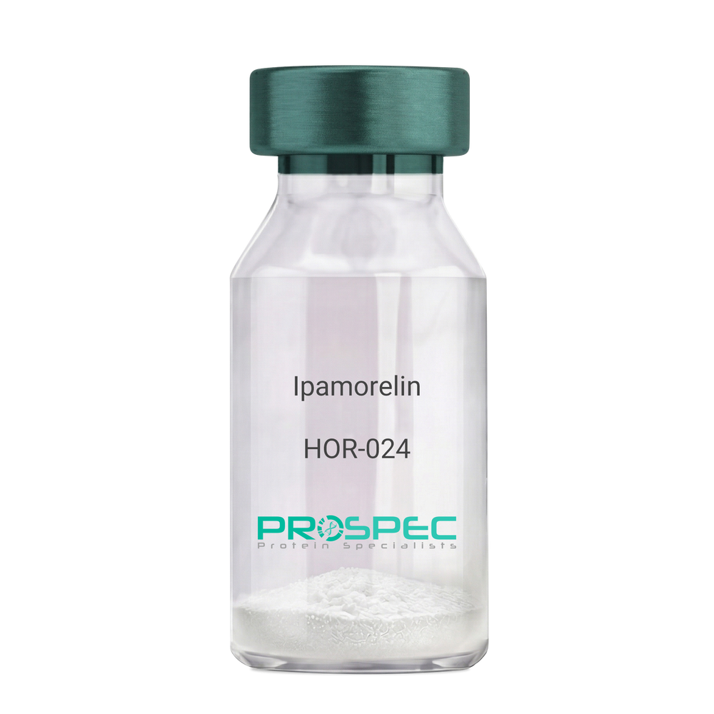
Name :
IpamorelinDescription:
Ipamorelin
Ipamorelin
Product # :
HOR-024Price :
Quantity :
Shipping Method :
Shipped at Room temp
More Info
- description
- formulation
- purity
- More Info
- Citations
Description
Ipamorelin Synthetic is a single, non-glycosylated polypeptide chain containing 4 amino acids, having a molecular mass of 711.85 Dalton and a Molecular formula of C38H49N9O5.
Formulation
The protein was lyophilized with no additives.
Purity
Greater than 97.0% as determined by analysis by RP-HPLC.
Citations
More Info
-
Introduction
Ipamorelin is a peptide selective agonist of the ghrelin/growth hormone secretagogue receptor and a growth hormone secretagogue. Ipamorelin is a pentapeptide that was derived from GHRP1. Ipamorelin significantly increases plasma growth hormone levels in both animals and humans. Like pralmorelin and GHRP-6, ipamorelin does not affect prolactin, FSH, LH or TSH levels. However, unlike GHRP2 and GHRP6, but as growth hormone-releasing hormone (GHRH), ipamorelin does not stimulate the secretion of adrenocorticotropic hormone (ACTH) or cortisol, and is highly selective for inducing the secretion only of GH.
-
Synonyms
Ipamorelin
-
Physical Appearance
Sterile Filtered White lyophilized (freeze-dried) powder.
-
Stability
Lyophilized Ipamorelin although stable at room temperature for 3 weeks, should be stored desiccated below -18°C. Upon reconstitution Ipamorelin should be stored at 4°C between 2-7 days and for future use below -18°C. For long term storage it is recommended to add a carrier protein (0.1% HSA or BSA). Please prevent freeze-thaw cycles.
-
Solubility
It is recommended to reconstitute the lyophilized Ipamorelin in sterile 18MΩ-cm H2O not less than 100 µg/ml, which can then be further diluted to other aqueous solutions.
-
Amino Acid Sequence
Aib-His-D-2-Nal-D-Phe-Lys-NH2.
ProSpec's products are furnished for LABORATORY RESEARCH USE ONLY. The product may not be used as drugs, agricultural or pesticidal products, food additives or household chemicals.