Search results
1000 results found for “Malaria”
Name
Description
Product #
Price
Quantity
Shipping Method
- View Data Sheet
Name :
GOT1 MouseDescription:
Glutamic-Oxaloacetic Transaminase 1 Mouse Recombinant
Aspartate aminotransferase, cytoplasmic, cAspAT, Cysteine aminotransferase, cytoplasmic, Cysteine transaminase, cytoplasmic, cCAT, Glutamate oxaloacetate transaminase 1, Transaminase A.
Product # :
ENZ-872Price :
Quantity :
Shipping Method :
Shipped with Ice Packs
More Info
- description
- source
- formulation
- purity
- More Info
Description
GOT1 Mouse Recombinant produced in E.Coli is a single, non-glycosylated polypeptide chain containing 436 amino acids (1-413a.a) and having a molecular mass of 48.6kDa.GOT1 is fused to a 23 amino acid His-tag at N-terminus & purified by proprietary chromatographic techniques.
Source
Escherichia Coli.
Formulation
GOT1 protein solution (0.5mg/ml) containing Phosphate buffered saline (pH7.4), 10% glycerol and 1mM DTT.
Purity
Greater than 95.0% as determined by SDS-PAGE.
More Info
-
Introduction
GOT1 is a pyridoxal phosphate-dependent enzyme which exists in cytoplasmic and mitochondrial forms, GOT1 and GOT2, which participate in amino acid metabolism and the urea and tricarboxylic acid cycles. Both enzymes are homodimeric and show close homology.
-
Synonyms
Aspartate aminotransferase, cytoplasmic, cAspAT, Cysteine aminotransferase, cytoplasmic, Cysteine transaminase, cytoplasmic, cCAT, Glutamate oxaloacetate transaminase 1, Transaminase A.
-
Physical Appearance
Sterile filtered colorless solution.
-
Stability
Store at 4°C if entire vial will be used within 2-4 weeks.Store, frozen at -20°C for longer periods of time.For long term storage it is recommended to add a carrier protein (0.1% HSA or BSA).Avoid multiple freeze-thaw cycles.
-
Amino Acid Sequence
MGSSHHHHHH SSGLVPRGSH MGSMAPPSVF AQVPQAPPVL VFKLTADFRD DPDPRKVNLG VGAYRTDESQ PWVLPVVRKV EQKIANDNSL NHEYLPILGL AEFRSCASRL VLGDNSPAIR ENRVGGVQSL GGTGALRIGA DFLGRWYNGT DNKNTPIYVS SPTWENHNAV FSAAGFKDIR PYCYWDAEKR GLDLQGFLND LENAPEFSIF VLHACAHNPT GTDPTPEQWK QIAAVMQRRF LFPFFDSAYQ GFASGDLEKD AWAIRYFVSE GFELFCAQSF SKNFGLYNER VGNLTVVGKE SDSVLRVLSQ MEKIVRITWS NPPAQGARIV AATLSDPELF KEWKGNVKTM ADRILTMRSE LRARLEALKT PGTWSHITEQ IGMFSFTGLN PKQVEYLVNE KHIYLLPSGR INMCGLTTKN LDYVATSIHE AVTKIQ.
ProSpec's products are furnished for LABORATORY RESEARCH USE ONLY. The product may not be used as drugs, agricultural or pesticidal products, food additives or household chemicals.
- View Data Sheet
Name :
GPC4 511 aa HumanDescription:
Glypican-4 511 aa Human Recombinant
Glypican 4, Glypican Proteoglycan 4, K-glypican, DJ900E8.1 (Glypican 4), glypican-4.
Product # :
PRO-2034Price :
Quantity :
Shipping Method :
Shipped with Ice Packs
More Info
- description
- source
- formulation
- purity
- More Info
Description
Glypican-4 Human Recombinant produced in E.Coli is a single, non-glycosylated, polypeptide chain (Ala19-Ser529) containing 521 amino acids including a 10 aa His tag at N-terminus. The total calculated molecular mass is 58.7kDa.
Source
Escherichia Coli.
Formulation
Glypican-4 filtered (0.4µm) solution at a concentration of 0.2mg/ml in 20mM Tris buffer, 50mM NaCl, pH 8.0 and 5mM DTT.
Purity
Greater than 90.0% as determined by SDS-PAGE.
More Info
-
Introduction
Glypican 4, also known as GPC4, is part of a family of glycosylphosphatidylinositol (GPI)-anchored heparan sulphate proteoglycans (HSPGs) which take part in the control of cell division and growth regulation. GPC4 is broadly expressed in human tissues, including lung, kidney, heart, placenta, skeletal muscle, and pancreas. In addition, GPC4 has been shown to be present in astrocytes, haematopoietic-progenitor and bone-marrow-stromal cells.
-
Synonyms
Glypican 4, Glypican Proteoglycan 4, K-glypican, DJ900E8.1 (Glypican 4), glypican-4.
-
Physical Appearance
Filtered colorless solution.
-
Stability
Store at 4°C if entire vial will be used within 2-4 weeks. Store, frozen at -20°C for longer periods of time. For long term storage it is recommended to add a carrier protein (0.1% HSA or BSA).Avoid multiple freeze-thaw cycles.
-
Amino Acid Sequence
MKHHHHHHASALLAAELKSK SCSEVRRLYV SKGFNKNDAP LHEINGDHLK ICPQGSTCCS QEMEEKYSLQ SKDDFKSVVS EQCNHLQAVF ASRYKKFDEF FKELLENAEK SLNDMFVKTY GHLYMQNSEL FKDLFVELKR YYVVGNVNLE EMLNDFWARL LERMFRLVNS QYHFTDEYLE CVSKYTEQLK PFGDVPRKLK LQVTRAFVAA RTFAQGLAVA GDVVSKVSVV NPTAQCTHAL LKMIYCSHCR GLVTVKPCYN YCSNIMRGCL ANQGDLDFEW NNFIDAMLMV AERLEGPFNI ESVMDPIDVK ISDAIMNMQD NSVQVSQKVF QGCGPPKPLP AGRISRSISE SAFSARFRPH HPEERPTTAA GTSLDRLVTD VKEKLKQAKK FWSSLPSNVC NDERMAAGNG NEDDCWNGKG KSRYLFAVTG NGLANQGNNP EVQVDTSKPD ILILRQIMAL RVMTSKMKNA YNGNDVDFFD ISDESSGEGS GSGCEYQQCP SEFDYNATDH AGKSANEKAD S.
ProSpec's products are furnished for LABORATORY RESEARCH USE ONLY. The product may not be used as drugs, agricultural or pesticidal products, food additives or household chemicals.
- View Data Sheet
Name :
MAP1LC3B MouseDescription:
Microtubule-Associated Protein 1 Light Chain 3 Beta Mouse Recombinant
Microtubule-Associated Protein 1 Light Chain 3 beta, ATG8F, Autophagy-related ubiquitin-like modifier LC3 B, MAP1 light chain 3-like protein 2, MAP1A/MAP1B LC3 B, LC3B, MAP1A/1BLC3, MAP1ALC3.
Product # :
PRO-2482Price :
Quantity :
Shipping Method :
Shipped with Ice Packs
More Info
- description
- source
- formulation
- purity
- More Info
Description
MAP1LC3B Mouse Recombinant produced in E.Coli is a single, non-glycosylated polypeptide chain containing 144 amino acids (1-120 a.a.) and having a molecular mass of 16.7kDa. MAP1LC3B is fused to a 24 amino acid His-tag at N-terminus & purified by proprietary chromatographic techniques.
Source
Escherichia Coli.
Formulation
MAP1LC3B protein solution (0.25mg/ml) containing 20mM MES(pH6.0), 0.1M NaCl, 1mM DTT and 30% glycerol.
Purity
Greater than 95.0% as determined by SDS-PAGE.
More Info
-
Introduction
MAP1LC3B is a member of the MAP1 LC3 family. MAP1LC3B is a subunit of neuronal microtubule-associated MAP1A and MAP1B proteins, that are involved in microtubule assembly and important for neurogenesis. In addition, MAP1LC3B takes part in formation of autophagosomal vacuoles and is expressed mainly in heart, testis, brain and skeletal muscle.
-
Synonyms
Microtubule-Associated Protein 1 Light Chain 3 beta, ATG8F, Autophagy-related ubiquitin-like modifier LC3 B, MAP1 light chain 3-like protein 2, MAP1A/MAP1B LC3 B, LC3B, MAP1A/1BLC3, MAP1ALC3.
-
Physical Appearance
Sterile filtered colorless solution.
-
Stability
Store at 4°C if entire vial will be used within 2-4 weeks. Store, frozen at -20°C for longer periods of time. For long term storage it is recommended to add a carrier protein (0.1% HSA or BSA).Avoid multiple freeze-thaw cycles.
-
Amino Acid Sequence
MGSSHHHHHH SSGLVPRGSH MGSHMPSEKT FKQRRSFEQR VEDVRLIREQ HPTKIPVIIE RYKGEKQLPV LDKTKFLVPD HVNMSELIKI IRRRLQLNAN QAFFLLVNGH SMVSVSTPIS EVYESERDED GFLYMVYASQ ETFG.
ProSpec's products are furnished for LABORATORY RESEARCH USE ONLY. The product may not be used as drugs, agricultural or pesticidal products, food additives or household chemicals.
- View Data Sheet
Name :
MAPT HumanDescription:
Microtubule-Associated Protein Tau Human Recombinant
TAU, DDPAC, FTDP-17, MAPTL, MSTD, MTBT1, MTBT2, PPND.
Product # :
PRO-295Price :
Quantity :
Shipping Method :
Shipped with Ice Packs
More Info
- description
- source
- formulation
- purity
- More Info
Description
MAPT Recombinant Human (Isoform-4) produced in E.Coli is a single, non-glycosylated polypeptide chain containing 372 amino acids (1-352 a.a.) and having a molecular mass of 38.9 kDa (Real molecular weight on SDS-PAGE will be shift up). The MAPT is fused to a 20 amino acid His-Tag at N-terminus and purified by proprietary chromatographic techniques.
Source
Escherichia Coli.
Formulation
The MAPT solution (0.5mg/ml) contains 20mM Tris-HCl pH-8, 1mM DTT, 0.2M NaCl & 10% glycerol.
Purity
Greater than 85.0% as determined by SDS-PAGE.
More Info
-
Introduction
MAPT is a neuronal microtubule associated protein localized mostly on axons.
MAPT promotes tubulin polymerisation and stabilizes microtubules, however it also serves to connect certain signalling pathways to the cytoskeleton. MAPT, in its hyperphosphorylated form, is the main part of paired helical filaments (PHF) and neurofibrillary lesions in Alzheimer''s disease (AD) brain. -
Synonyms
TAU, DDPAC, FTDP-17, MAPTL, MSTD, MTBT1, MTBT2, PPND.
-
Physical Appearance
Sterile filtered colorless solution.
-
Stability
Store at 4°C if entire vial will be used within 2-4 weeks.Store, frozen at -20°C for longer periods of time. For long term storage it is recommended to add a carrier protein (0.1% HSA or BSA).Avoid multiple freeze-thaw cycles.
-
Amino Acid Sequence
MGSSHHHHHH SSGLVPRGSH MAEPRQEFEV MEDHAGTYGL GDRKDQGGYT MHQDQEGDTD AGLKAEEAGI GDTPSLEDEA AGHVTQARMV SKSKDGTGSD DKKAKGADGK TKIATPRGAA PPGQKGQANA TRIPAKTPPA PKTPPSSGEP PKSGDRSGYS SPGSPGTPGS RSRTPSLPTP PTREPKKVAV VRTPPKSPSS AKSRLQTAPV PMPDLKNVKS KIGSTENLKH QPGGGKVQIV YKPVDLSKVT SKCGSLGNIH HKPGGGQVEV KSEKLDFKDR VQSKIGSLDN ITHVPGGGNK KIETHKLTFR ENAKAKTDHG AEIVYKSPVV SGDTSPRHLS NVSSTGSIDM VDSPQLATLA DEVSASLAKQ GL.
ProSpec's products are furnished for LABORATORY RESEARCH USE ONLY. The product may not be used as drugs, agricultural or pesticidal products, food additives or household chemicals.
- View Data Sheet
Name :
MCP 1 RatDescription:
Monocyte Chemotactic Protein-1 Rat Recombinant (CCL2)
Small inducible cytokine A2, CCL2, Monocyte chemotactic protein 1, MCP-1, Monocyte chemoattractant protein 1, Monocyte chemotactic and activating factor, MCAF, Monocyte secretory protein JE, HC11, chemokine (C-C motif) ligand 2, MCP1, SCYA2, GDCF-2, SMC-CF, HSMCR30, MGC9434, GDCF-2 HC11, Immediate-early serum-responsive JE protein.
Product # :
CHM-315Price :
Quantity :
Shipping Method :
Shipped at Room temp
More Info
- description
- source
- formulation
- purity
- biological activity
- More Info
Description
Monocyte Chemotactic Protein-1 Rat Recombinant produced in E.Coli is a non-glycosylated, Polypeptide chain containing 125 amino acids and having a molecular mass of 14.1 kDa. The MCP-1 is purified by proprietary chromatographic techniques.
Source
Escherichia Coli.
Formulation
The protein was lyophilized from a concentrated (1mg/ml) sterile solution containing no additives.
Purity
Greater than 98.0% as determined by SDS-PAGE.
Biological Activity
ED50 =1-10ng/ml corresponding to a Specific Activity of 100,000-1,000,000IU/mg. The biological activity was determined by measuring the dose dependent chemotaxis with human THP-1 cells. The optimal concentration should be determined for each specific application by an initial dose-response assay.More Info
-
Introduction
Chemokine (C-C motif) ligand 2 (CCL2) is a small cytokine belonging to the CC chemokine family that is also known as monocyte chemotactic protein-1 (MCP-1). It is found at the site of tooth eruption and bone degradation. In the bone, CCL2 is expressed by mature osteoclasts and osteoblasts and is under the control of nuclear factor ?B (NF?B). CCL2 recruits immune cells, such as monocytes, to sites of tissue injury and infection. This chemokine is produced as a protein precursor containing signal peptide of 23 amino acids and a mature peptide of 76 amino acids. It is a monomeric polypeptide, with a molecular weightof approximately 13kDa. As with many other CC chemokines, CCL2 is located on chromosome 17 in humans. The cell surface receptors that bind CCL2 are CCR2 and CCR5.
-
Synonyms
Small inducible cytokine A2, CCL2, Monocyte chemotactic protein 1, MCP-1, Monocyte chemoattractant protein 1, Monocyte chemotactic and activating factor, MCAF, Monocyte secretory protein JE, HC11, chemokine (C-C motif) ligand 2, MCP1, SCYA2, GDCF-2, SMC-CF, HSMCR30, MGC9434, GDCF-2 HC11, Immediate-early serum-responsive JE protein.
-
Physical Appearance
Sterile Filtered White lyophilized (freeze-dried) powder.
-
Stability
Lyophilized MCP-1 although stable at room temperature for 3 weeks, should be stored desiccated below -18°C. Upon reconstitution CCL2 should be stored at 4°C between 2-7 days and for future use below -18°C.For long term storage it is recommended to add a carrier protein (0.1% HSA or BSA).Please prevent freeze-thaw cycles.
-
Solubility
It is recommended to reconstitute the lyophilized Monocyte Chemotactic Protein-1 in sterile 18MΩ-cm H2O not less than 100µg/ml, which can then be further diluted to other aqueous solutions.
-
Amino Acid Sequence
QPDAVNAPLT CCYSFTGKMI PMSRLENYKR ITSSRCPKEA VVFVTKLKRE ICADPNKEWV QKYIRKLDQN QVRSETTVFY KIASTLRTSA PLNVNLTHKS EANASTLFST TTSSTSVEVT SMTEN.
ProSpec's products are furnished for LABORATORY RESEARCH USE ONLY. The product may not be used as drugs, agricultural or pesticidal products, food additives or household chemicals.
- View Data Sheet
Name :
RARRES2 HumanDescription:
Retinoic Acid Receptor Responder 2 Human Recombinant
Chemerin, TIG2, Retinoic acid receptor responder protein 2, RAR-responsive protein TIG2, RARRES2, HP10433.
Product # :
PRO-788Price :
Quantity :
Shipping Method :
Shipped at Room temp
More Info
- description
- source
- formulation
- purity
- More Info
Description
RARRES2 Human Recombinant produced in E.Coli is a single, non-glycosylated, polypeptide chain containing 138 amino acids and having a total molecular mass of 16 kDa. RARRES2 Human Recombinant is purified by proprietary chromatographic techniques.
Source
Escherichia Coli.
Formulation
The RARRES2 protein was lyophilized from 0.02% TFA.
Purity
Greater than 98.0% as determined by: (a) Analysis by RP-HPLC. (b) Analysis by SDS-PAGE.
More Info
-
Introduction
RARRES2 is a secreted chemotactic protein that initiates chemotaxis through the ChemR23 G protein-coupled seven-transmembrane domain ligand. RARRES2 acts as an adipokine, and is truncated on both termini from the proprotein. RARRES2 is structurally related to the cathelicidin precursors, cystatin C and kininogens. RARRES2 promotes calcium mobilization and chemotaxis of immature dendritic cells and macrophages. RARRES2 is secreted as a precursor of little biological activity, which requires proteolytic cleavage of its COOH-terminal domain to be exchangeed into a potent and highly specific agonist of ChemR23. RARRES2 signals via its receptor, ChemR23 (CMKLR1), as a positive regulator of adipocyte differentiation and metabolic function. The Chemerin receptor acts as a coreceptor for SIV and some primary HIV-1 strains.
-
Synonyms
Chemerin, TIG2, Retinoic acid receptor responder protein 2, RAR-responsive protein TIG2, RARRES2, HP10433.
-
Physical Appearance
Sterile Filtered White lyophilized (freeze-dried) powder.
-
Stability
Lyophilized Chemerin although stable at room temperature for 3 weeks, should be stored desiccated below -18°C. Upon reconstitution Chemerin should be stored at 4°C between 2-7 days and for future use below -18°C. For long term storage it is recommended to add a carrier protein (0.1% HSA or BSA). Please prevent freeze-thaw cycles.
-
Solubility
It is recommended to reconstitute the lyophilized Chemerin in sterile 18MΩ-cm H2O not less than 100µg/ml, which can then be further diluted to other aqueous solutions.
-
Amino Acid Sequence
MELTEAQRRG LQVALEEFHK HPPVQWAFQE TSVESAAVDTP FPAGIFVRLE FKLQQTSRK RDWKKPECKV RPNGRKRKCL ACIKLGSEDK VLGRLVHCPI ETQVLREAEE HQETQCLRVQ RAGEDPHSFY FPGQFAFS.
ProSpec's products are furnished for LABORATORY RESEARCH USE ONLY. The product may not be used as drugs, agricultural or pesticidal products, food additives or household chemicals.
- View Data Sheet
Name :
IL6ST HumanDescription:
Interleukin-6 Signal Transducer Human Recombinant
Interleukin 6 signal transducer, oncostatin M receptor, IL6ST, CD130, CDw130, GP130, GP130-RAPS, IL6R-beta
Product # :
CYT-1156Price :
Quantity :
Shipping Method :
Shipped with Ice Packs
More Info
- description
- source
- formulation
- purity
- More Info
Description
IL6ST Human produced in Sf9 Baculovirus cells is a single, glycosylated polypeptide chain containing 605 amino acids (23-619 a.a) and having a molecular mass of 68.9kDa.IL6ST is fused to an 8 amino acid His tag at C-terminus and purified by proprietary chromatographic techniques.
Source
Sf9, Baculovirus cells.
Formulation
The IL6ST solution (0.25mg/ml) contains 10% Glycerol and Phosphate-Buffered Saline (pH 7.4).
Purity
Greater than 95% as determined by SDS-PAGE.
More Info
-
Introduction
Interleukin-6 Signal Transducer or IL6ST is a receptor, part of the family of class 1 cytokine receptor. IL6ST binds to IL-6 through membrane-anchored or soluble IL-6R starts a connection between another complex of IL6ST and IL-6 , thus forming a homo-dimer and a signal transduction occurs.
-
Synonyms
Interleukin 6 signal transducer, oncostatin M receptor, IL6ST, CD130, CDw130, GP130, GP130-RAPS, IL6R-beta
-
Physical Appearance
Sterile Filtered colorless solution.
-
Stability
Store at 4°C if entire vial will be used within 2-4 weeks. Store, frozen at -20°C for longer periods of time. For long term storage it is recommended to add a carrier protein (0.1% HSA or BSA).Avoid multiple freeze-thaw cycles.
-
Amino Acid Sequence
ELLDPCGYIS PESPVVQLHS NFTAVCVLKE KCMDYFHVNA NYIVWKTNHF TIPKEQYTII NRTASSVTFT DIASLNIQLT CNILTFGQLE QNVYGITIIS GLPPEKPKNL SCIVNEGKKM RCEWDRGRET HLETNFTLKS EWATHKFADC KAKRDTPTSC TVDYSTVYFV NIEVWVEAEN ALGKVTSDHI NFDPVYKVKP NPPHNLSVIN SEELSSILKL TWTNPSIKSV IILKYNIQYR TKDASTWSQI PPEDTASTRS SFTVQDLKPF TEYVFRIRCM KEDGKGYWSD WSEEASGITY EDRPSKAPSF WYKIDPSHTQ GYRTVQLVWK TLPPFEANGK ILDYEVTLTR WKSHLQNYTV NATKLTVNLT NDRYVATLTV RNLVGKSDAA VLTIPACDFQ ATHPVMDLKA FPKDNMLWVE WTTPRESVKK YILEWCVLSD KAPCITDWQQ EDGTVHRTYL RGNLAESKCY LITVTPVYAD GPGSPESIKA YLKQAPPSKG PTVRTKKVGK NEAVLEWDQL PVDVQNGFIR NYTIFYRTII GNETAVNVDS SHTEYTLSSL TSDTLYMVRM AAYTDEGGKD GPEFTFTTPK FAQGEIELEH HHHHH
-
Background
Significance of Human Recombinant Interleukin-6 Signal Transducer: Insights and Implications
Abstract:
The Interleukin-6 (IL-6) signal transducer holds a pivotal role in the complex IL-6 signaling pathway, orchestrating crucial cellular responses. This paper delves into the significance of Human Recombinant IL-6 Signal Transducer in unraveling IL-6-mediated cellular communication and highlights its potential applications in research and therapeutic development. The review sheds light on the methodology of producing this transducer and its relevance in advancing our understanding of cytokine signaling.
Introduction:
IL-6, a pleiotropic cytokine, is known to exert its diverse effects through a complex signaling cascade, wherein the signal transducer plays a crucial role. The availability of Human Recombinant IL-6 Signal Transducer enables the investigation of its role in health and disease. This transducer is vital for transmitting IL-6 signals, influencing processes like immune response, inflammation, and cell differentiation.
IL-6 Signal Transduction Pathway:
The IL-6 signal transduction pathway involves binding of IL-6 to its receptor, leading to the recruitment of the signal transducer and subsequent activation of downstream signaling molecules such as JAK/STAT pathway. This orchestrated response modulates gene expression, thereby impacting cellular behavior.
Methods of Production:
Human Recombinant IL-6 Signal Transducer is produced by expressing the corresponding gene in a suitable expression system, often utilizing bacterial or mammalian cells. Proper post-translational modifications are necessary to ensure its biological activity and appropriate functioning within the signaling cascade.
Applications in Research:
Human Recombinant IL-6 Signal Transducer serves as a fundamental tool in elucidating IL-6-mediated signaling mechanisms. Its availability facilitates the exploration of how aberrant signaling contributes to diseases such as autoimmune disorders, inflammatory conditions, and certain cancers. The transducer's interaction with other signaling pathways is also of interest for comprehensive pathway analysis.
Therapeutic Implications:
Understanding the IL-6 signal transduction pathway has led to the development of targeted therapies for IL-6-related diseases. Modulation of this pathway presents potential opportunities for novel therapeutic interventions, thereby offering a new dimension in precision medicine.
Challenges and Future Directions:
While the availability of Human Recombinant IL-6 Signal Transducer has significantly advanced our knowledge, challenges remain in deciphering the intricate nuances of IL-6 signaling. Developing strategies to selectively intervene in this pathway without disturbing physiological homeostasis presents an ongoing challenge.
Conclusion:
The Human Recombinant IL-6 Signal Transducer serves as a cornerstone in unraveling the complexities of IL-6 signaling, shedding light on its roles in health and disease. Its applications span from fundamental research to therapeutic development, showcasing its potential to shape the future of precision medicine.
ProSpec's products are furnished for LABORATORY RESEARCH USE ONLY. The product may not be used as drugs, agricultural or pesticidal products, food additives or household chemicals.
- View Data Sheet
Name :
IFNAR2 HumanDescription:
Interferon Alpha And Beta Receptor Subunit 2 Human Recombinant
Interferon alpha/beta receptor 2, IFNAR2, IFN-alpha binding protein, Interferon alpha binding protein, Type I interferon receptor 2, interferon alpha/beta receptor 2 isoform a, IFN-R-2, IFNABR, IFNARB, IFN-alpha-REC, IFN-R, IMD45.
Product # :
CYT-1167Price :
Quantity :
Shipping Method :
Shipped with Ice Packs
More Info
- description
- source
- formulation
- purity
- biological activity
- More Info
- SDS-PAGE
Description
IFNAR2 Human Recombinant produced in Baculovirus is a single glycosylated polypeptide chain containing 456 amino acids (27-243 a.a.) and having a molecular mass of 51.7kDa. IFNAR2 is fused to an 239 amino acid hIgG-His tag at C-terminus and purified by proprietary chromatographic techniques.
Source
Sf9, Baculovirus cells.
Formulation
IFNAR2 protein (0.25mg/ml) contains 10% glycerol and Phosphate-Buffered Saline (pH 7.4).
Purity
Greater than 90.0% as determined by SDS-PAGE.
Biological Activity
The ED50 range ≤ 500 ng/ml and is measured by its binding ability in a functional ELISA with Human IFN-alpha 2 in the presence of Human IFN-alpha/beta R1.
SDS-PAGE
More Info
-
Introduction
Interferon Alpha and Beta Receptor Subunit 2 or IFNAR2 is a protein, part of the class II cytokine receptor family. When different innate immune signaling transduction are activated, there is a rapid induction of type I IFNs because it has an intronless gene source. IFNAR2 is the main ligand and binds to the receptor. Once bind, stabilization occurs and a signaling compelx recptor is formed. IFNAR2 also takes part in the activation process of STAT proteins.
-
Synonyms
Interferon alpha/beta receptor 2, IFNAR2, IFN-alpha binding protein, Interferon alpha binding protein, Type I interferon receptor 2, interferon alpha/beta receptor 2 isoform a, IFN-R-2, IFNABR, IFNARB, IFN-alpha-REC, IFN-R, IMD45.
-
Physical Appearance
Sterile Filtered colorless solution.
-
Stability
Store at 4°C if entire vial will be used within 2-4 weeks. Store, frozen at -20°C for longer periods of time. For long term storage it is recommended to add a carrier protein (0.1% HSA or BSA).Avoid multiple freeze-thaw cycles.
-
Amino Acid Sequence
ISYDSPDYTD ESCTFKISLR NFRSILSWEL KNHSIVPTHY TLLYTIMSKP EDLKVVKNCA NTTRSFCDLT DEWRSTHEAY VTVLEGFSGN TTLFSCSHNF WLAIDMSFEP PEFEIVGFTN HINVMVKFPS IVEEELQFDL SLVIEEQSEG IVKKHKPEIK GNMSGNFTYI IDKLIPNTNY CVSVYLEHSD EQAVIKSPLK CTLLPPGQES ESAESAKLEP KSCDKTHTCP PCPAPELLGG PSVFLFPPKP KDTLMISRTP EVTCVVVDVS HEDPEVKFNW YVDGVEVHNA KTKPREEQYN STYRVVSVLT VLHQDWLNGK EYKCKVSNKA LPAPIEKTIS KAKGQPREPQ VYTLPPSRDE LTKNQVSLTC LVKGFYPSDI AVEWESNGQP ENNYKTTPPV LDSDGSFFLY SKLTVDKSRW QQGNVFSCSV MHEALHNHYT QKSLSLSPGK HHHHHH.
-
Background
What is the molecular weight/Mw of IFNAR2 HUMAN Protein?
IFNAR2 HUMAN Protein has a total Mw of 51.7kDa.
What is the source or expression system of IFNAR2 HUMAN Protein?
Sf9, Baculovirus cells.
What is the Purity of IFNAR2 HUMAN Protein?
IFNAR2 HUMAN Protein is >90% pure as determined by SDS-PAGE.
What is the Biological Activity of IFNAR2 HUMAN Protein?
The ED50 range ≤ 500 ng/ml and is measured by its binding ability in a functional ELISA with Human IFN-alpha 2 in the presence of Human IFN-alpha/beta R1.
What is the amino acid sequence of IFNAR2 HUMAN Protein?
ISYDSPDYTD ESCTFKISLR NFRSILSWEL KNHSIVPTHY TLLYTIMSKP EDLKVVKNCA NTTRSFCDLT DEWRSTHEAY VTVLEGFSGN TTLFSCSHNF WLAIDMSFEP PEFEIVGFTN HINVMVKFPS IVEEELQFDL SLVIEEQSEG IVKKHKPEIK GNMSGNFTYI IDKLIPNTNY CVSVYLEHSD EQAVIKSPLK CTLLPPGQES ESAESAKLEP KSCDKTHTCP PCPAPELLGG PSVFLFPPKP KDTLMISRTP EVTCVVVDVS HEDPEVKFNW YVDGVEVHNA KTKPREEQYN STYRVVSVLT VLHQDWLNGK EYKCKVSNKA LPAPIEKTIS KAKGQPREPQ VYTLPPSRDE LTKNQVSLTC LVKGFYPSDI AVEWESNGQP ENNYKTTPPV LDSDGSFFLY SKLTVDKSRW QQGNVFSCSV MHEALHNHYT QKSLSLSPGK HHHHHH.
What applications can IFNAR2 HUMAN Protein be used in?
IFNAR2 HUMAN Protein can probably be used in western blot, ELISA and Lateral Flow.
What is the endotoxin level for IFNAR2 HUMAN Protein?
The endotoxin level is minimal, IFNAR2 HUMAN Protein was purified using conventional chromatography techniques.
ProSpec's products are furnished for LABORATORY RESEARCH USE ONLY. The product may not be used as drugs, agricultural or pesticidal products, food additives or household chemicals.
- View Data Sheet
Name :
NRIP3 HumanDescription:
Nuclear Receptor-Interacting Protein 3 Human Recombinant
C11orf14, NY-SAR-105, Nuclear receptor-interacting protein 3, Sarcoma antigen NY-SAR-105, NRIP3.
Product # :
PRO-1384Price :
Quantity :
Shipping Method :
Shipped with Ice Packs
More Info
- description
- source
- formulation
- purity
- More Info
Description
NRIP3 Human Recombinant produced in E.Coli is a single, non-glycosylated polypeptide chain containing 264 amino acids (1-241 a.a.) and having a molecular mass of 29.4kDa. NRIP3 is fused to a 23 amino acid His-tag at N-terminus & purified by proprietary chromatographic techniques.
Source
E.coli.
Formulation
NRIP3 protein solution (0.5mg/ml) contains 20mM Tris-HCl buffer (pH 8.0), 0.15M NaCl, 20% glycerol and 1mM DTT.
Purity
Greater than 85% as determined by SDS-PAGE.
More Info
-
Introduction
Nuclear Receptor-Interacting Protein (NRIP3)is a 241 amino acid protein which is encoded by a gene that maps to human chromosome 11.Diseases associated with NRIP3 include sarcoma, and acute myeloid leukemia.
-
Synonyms
C11orf14, NY-SAR-105, Nuclear receptor-interacting protein 3, Sarcoma antigen NY-SAR-105, NRIP3.
-
Physical Appearance
Sterile Filtered colorless solution.
-
Stability
Store at 4°C if entire vial will be used within 2-4 weeks. Store, frozen at -20°C for longer periods of time. For long term storage it is recommended to add a carrier protein (0.1% HSA or BSA).Avoid multiple freeze-thaw cycles.
-
Amino Acid Sequence
MGSSHHHHHH SSGLVPRGSH MGSMFYSGLL TEGGRKETDM REAASLRQQR RMKQAVQFIH KDSADLLPLD GLKKLGSSKD MQPHNILQRR LMETNLSKLR SGPRVPWASK TNKLNQAKSE GLKKSEEDDM ILVSCQCAGK DVKALVDTGC LYNLISLACV DRLGLKEHVK SHKHEGEKLS LPRHLKVVGQ IEHLVITLGS LRLDCPAAVV DDNEKNLSLG LQTLRSLKCI INLDKHRLIM GKTDKEEIPF VETVSLNEDN TSEA.
ProSpec's products are furnished for LABORATORY RESEARCH USE ONLY. The product may not be used as drugs, agricultural or pesticidal products, food additives or household chemicals.
- View Data Sheet
Name :
IL 22 MouseDescription:
Interleukin-22 Mouse Recombinant
IL-TIF, TIFa, IL-10-related T-cell-derived-inducible factor, IL-22, ILTIF, IL-D110, zcyto18, MGC79382, MGC79384, TIFIL-23.
Product # :
CYT-539Price :
Quantity :
Shipping Method :
Shipped at Room temp
More Info
- description
- source
- formulation
- purity
- biological activity
- More Info
Description
Interleukin-22 Mouse Recombinant produced in E.Coli is a single, non-glycosylated polypeptide chain containing 147 amino acids and having a molecular mass of 16.7 kDa. The Murine IL-22 is purified by proprietary chromatographic techniques.
Source
Escherichia Coli.
Formulation
Lyophilized from 1X PBS.
Purity
Greater than 97.0% as determined by:
(a) Analysis by RP-HPLC.
(b) Analysis by SDS-PAGE.Biological Activity
The ED50 as determined by its ability to induce IL-10 secretion in Colo205 cells is less than 0.5ng/ml, corresponding to a Specific Activity of 2,000,000IU/mg.More Info
-
Introduction
Interleukin-22 (IL-22), also known as IL-10-related T cell-derived inducible factor (IL-TIF) was initially identified as a gene induced by IL-9 in mouse T cells and mast cells. Mouse IL-22 cDNA encodes a 179 amino acid residue protein with a putative 33 amino acids signal peptide that is cleaved to generate a 147 aa mature protein that shares approximately 79% and 22% aa sequence identity with human IL-22 and IL-10, respectively. IL-22 has been shown to activate STAT-1 and STAT-3 in several hepatoma cell lines and upregulate the production of acute phase proteins. IL-22 is produced by normal mouse T cells upon Con A activation. Mouse IL-22 expression is also induced in various organs upon lipopolysaccharide injection, suggesting that IL-22 may be involved in inflammatory responses. The functional IL-22 receptor complex consists of two receptor subunits, IL-22R (previously an orphan receptor named CRF2-9) and IL-10R? (previously known as CRF2-4), belonging to the class II cytokine receptor family.
-
Synonyms
IL-TIF, TIFa, IL-10-related T-cell-derived-inducible factor, IL-22, ILTIF, IL-D110, zcyto18, MGC79382, MGC79384, TIFIL-23.
-
Physical Appearance
Sterile Filtered White lyophilized (freeze-dried) powder.
-
Stability
Lyophilized Interleukin-22 although stable at room temperature for 3 weeks, should be stored desiccated below -18°C. Upon reconstitution IL22 should be stored at 4°C between 2-7 days and for future use below -18°C.Please prevent freeze-thaw cycles.
-
Solubility
It is recommended to reconstitute the lyophilized Interleukin -22 in sterile 18MΩ-cm H2O not less than 100µg/ml, which can then be further diluted to other aqueous solutions.
-
Amino Acid Sequence
The sequence of the first five N-terminal amino acids was determined and was found to be Met-Leu-Pro-Val-Asn.
ProSpec's products are furnished for LABORATORY RESEARCH USE ONLY. The product may not be used as drugs, agricultural or pesticidal products, food additives or household chemicals.
- View Data Sheet
Name :
PAK4 HumanDescription:
p21 Activated Kinase 4 Human Recombinant
PAK-4, p21 (CDKN1A)-Activated Kinase 4, p21-Activated Kinase 4, Serine/threonine-protein kinase PAK 4, KIAA1142, PAK4.
Product # :
PKA-259Price :
Quantity :
Shipping Method :
Shipped with Ice Packs
More Info
- description
- source
- formulation
- purity
- More Info
Description
PAK4 Human Recombinant produced in E.Coli is a single, non-glycosylated polypeptide chain containing 628 amino acids (1-591 a.a.) and having a molecular mass of 68.3 kDa.PAK4 is fused to 37 amino acid His-Tag at N-terminus and is purified by standard chromatography techniques.
Source
Escherichia Coli.
Formulation
The PAK4 protein (0.5mg/ml) contains 25mM HEPES pH-7.5, 150mM NaCl, 5mM MgCl2 and 30% glycerol.
Purity
Greater than 90% as determined by SDS-PAGE.
More Info
-
Introduction
PAK4 is part of the group B family of p21-activated kinases (PAK). It's known as an effector protein for cell division cycle 42(Cdc42) and protein that plays an important role in regulating cytoskeletal organization and cell morphology. PAK4 expression is eminent in many cancer cell lines, and is involved in tumorigenesis. PAK proteins are significant effectors that associate Rho GTPases to cytoskeleton reorganization and nuclear signaling. PAK proteins are targets for the small GTP binding proteins Cdc42 and Rac and have been implicated in a wide range of biological activities. PAK4 cooperates specifically with the GTP-bound form of Cdc42Hs and weakly activates the JNK family of MAP kinases. PAK4 is a mediator of filopodia formation and is involved in the reorganization of the actin cytoskeleton. PAK4 activates the JNK pathway. PAK4 phosphorylates and inactivates the protein phosphatase SSH1, leading to increased inhibitory phosphorylation of the actin binding/depolymerizing factor cofilin. Decreased cofilin activity may lead to stabilization of actin filaments.
-
Synonyms
PAK-4, p21 (CDKN1A)-Activated Kinase 4, p21-Activated Kinase 4, Serine/threonine-protein kinase PAK 4, KIAA1142, PAK4.
-
Physical Appearance
Sterile filtered colorless solution.
-
Stability
Store at 4°C if entire vial will be used within 2-4 weeks. Store, frozen at -20°C for longer periods of time. For long term storage it is recommended to add a carrier protein (0.1% HSA or BSA). Avoid multiple freeze-thaw cycles.
-
Amino Acid Sequence
MRGSHHHHHH GMASMTGGQQ MGRDLYDDDD KDRWGSHMFG KRKKRVEISA PSNFEHRVHT GFDQHEQKFT GLPRQWQSLI EESARRPKPL VDPACITSIQ PGAPKTIVRG SKGAKDGALT LLLDEFENMS VTRSNSLRRD SPPPPARARQ ENGMPEEPAT TARGGPGKAG SRGRFAGHSE AGGGSGDRRR AGPEKRPKSS REGSGGPQES SRDKRPLSGP DVGTPQPAGL ASGAKLAAGR PFNTYPRADT DHPSRGAQGE PHDVAPNGPS AGGLAIPQSS SSSSRPPTRA RGAPSPGVLG PHASEPQLAP PACTPAAPAV PGPPGPRSPQ REPQRVSHEQ FRAALQLVVD PGDPRSYLDN FIKIGEGSTG IVCIATVRSS GKLVAVKKMD LRKQQRRELL FNEVVIMRDY QHENVVEMYN
SYLVGDELWV VMEFLEGGAL TDIVTHTRMN EEQIAAVCLA VLQALSVLHA QGVIHRDIKS DSILLTHDGR VKLSDFGFCA QVSKEVPRRK SLVGTPYWMA PELISRLPYG PEVDIWSLGI MVIEMVDGEP PYFNEPPLKA MKMIRDNLPP RLKNLHKVSP SLKGFLDRLL VRDPAQRATA AELLKHPFLA KAGPPASIVP LMRQNRTR.
ProSpec's products are furnished for LABORATORY RESEARCH USE ONLY. The product may not be used as drugs, agricultural or pesticidal products, food additives or household chemicals.
- View Data Sheet
Name :
LGALS3 MouseDescription:
Galectin-3 Mouse Recombinant
CBP35, GAL3, GALBP, GALIG, LGALS2, MAC2, Galectin-3, Lectin, galactose binding, soluble 3, Lectin, galactose binding, soluble 3, isoform CRA_a, Lectin, galactose binding, soluble 3, isoform CRA_d, Lgals3.
Product # :
CYT-188Price :
Quantity :
Shipping Method :
Shipped with Ice Packs
More Info
- description
- source
- formulation
- purity
- More Info
- sds-page
Description
LGALS3 Mouse Recombinant produced in E. coli is a single polypeptide chain containing 287 amino acids (1-264) and having a molecular mass of 29.8 kDa. LGALS3 is fused to a 23 amino acid His-tag at N-terminus & purified by proprietary chromatographic techniques.
Source
E.coli.
Formulation
The LGALS3 solution (0.5mg/1ml) contains 20mM Tris-HCl buffer (pH 8.0), 0.15M NaCl, 50% glycerol, 1mM DTT and 2mM EDTA.
Purity
Greater than 95% as determined by SDS-PAGE.
sds-page
More Info
-
Introduction
Galectin-3 mediates with the alpha-3, beta-1 integrin the stimulation by cspg4 of endothelial cells migration. Galectin-3 plays an necessary part during the acquisition of vasculogenic mimicry and angiogenic properties associated with melanoma progression. LGALS3 overexpression is highly expressed in early stages of papillary carcinoma, and its expression intensity declines during tumor progression. Serum levels of LGALS3 are high in patients with thyroid malignancy but there is considerable overlap in serum LGALS3 concentrations between those with benign and malignant nodular thyroid disease. LGLAS3 takes part as an immune regulator to inhibit T-cell immune responses and promote tumor growth, as a result providing a new mechanism for tumor immune tolerance.
-
Synonyms
CBP35, GAL3, GALBP, GALIG, LGALS2, MAC2, Galectin-3, Lectin, galactose binding, soluble 3, Lectin, galactose binding, soluble 3, isoform CRA_a, Lectin, galactose binding, soluble 3, isoform CRA_d, Lgals3.
-
Physical Appearance
Sterile Filtered colorless solution.
-
Stability
Store at 4°C if entire vial will be used within 2-4 weeks. Store, frozen at -20°C for longer periods of time. For long term storage it is recommended to add a carrier protein (0.1% HSA or BSA).Avoid multiple freeze-thaw cycles.
-
Amino Acid Sequence
MGSSHHHHHH SSGLVPRGSH MGSMADSFSL NDALAGSGNP NPQGYPGAWG NQPGAGGYPG AAYPGAYPGQ APPGAYPGQA PPGAYPGQAP PSAYPGPTAP GAYPGPTAPG AYPGSTAPGA FPGQPGAPGA YPSAPGGYPA AGPYGVPAGP LTVPYDLPLP GGVMPRMLIT IMGTVKPNAN RIVLDFRRGN DVAFHFNPRF NENNRRVIVC NTKQDNNWGK EERQSAFPFE SGKPFKIQVL VEADHFKVAV NDAHLLQYNH RMKNLREISQ LGISGDITLT SANHAMI.
-
Background
What is the molecular weight/Mw of LGALS3 Protein?
LGALS3 Protein has a total Mw of 29.8kDa.
What is the source or expression system of LGALS3 Protein?
Escherichia Coli.
What is the Purity of LGALS3 Protein?
LGALS3 Protein is >95% pure as determined by SDS-PAGE.
What is the Biological Activity of LGALS3 Protein?
The biological functionality of LGALS3 Protein will be determined in the future.
What is the amino acid sequence of LGALS3 Protein?
MGSSHHHHHH SSGLVPRGSH MGSMADSFSL NDALAGSGNP NPQGYPGAWG NQPGAGGYPG AAYPGAYPGQ APPGAYPGQA PPGAYPGQAP PSAYPGPTAP GAYPGPTAPG AYPGSTAPGA FPGQPGAPGA YPSAPGGYPA AGPYGVPAGP LTVPYDLPLP GGVMPRMLIT IMGTVKPNAN RIVLDFRRGN DVAFHFNPRF NENNRRVIVC NTKQDNNWGK EERQSAFPFE SGKPFKIQVL VEADHFKVAV NDAHLLQYNH RMKNLREISQ LGISGDITLT SANHAMI.
What applications can LGALS3 Protein be used in?
LGALS3 Protein can probably be used in western blot, ELISA and Lateral Flow.
What is the endotoxin level for LGALS3 Protein?
The endotoxin level is minimal, LGALS3 Protein was purified using conventional chromatography techniques.
ProSpec's products are furnished for LABORATORY RESEARCH USE ONLY. The product may not be used as drugs, agricultural or pesticidal products, food additives or household chemicals.
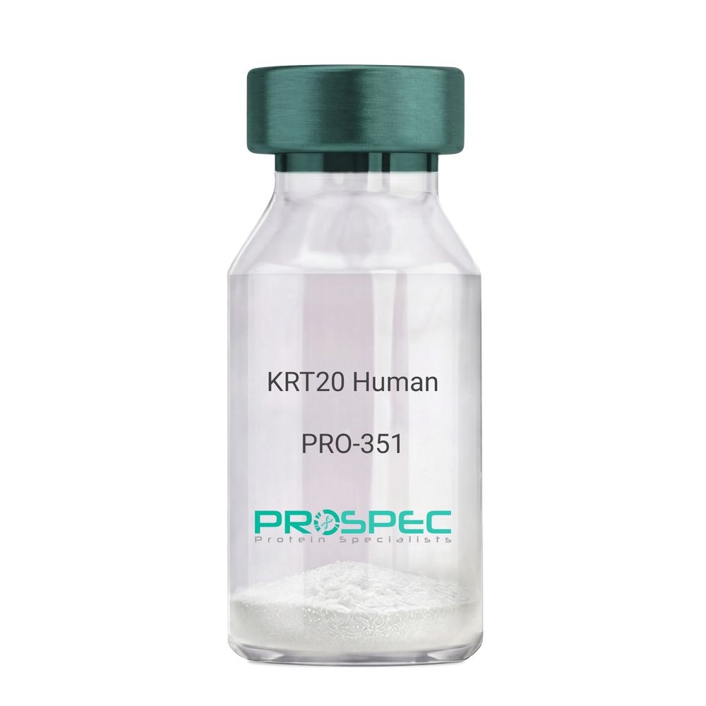
- View Data Sheet
Name :
KRT20 HumanDescription:
Cytokeratin 20 Human Recombinant
Keratin type I cytoskeletal 20, Cytokeratin-20, CK-20, Keratin-20, K20, Protein IT, KRT20, CD20, CK20, KRT21, MGC35423.
Product # :
PRO-351Price :
Quantity :
Shipping Method :
Shipped at Room temp
More Info
- description
- source
- formulation
- purity
- More Info
Description
Cytokeratin 20 Human Recombinant produced in E.Coli is a single,non-glycosylated polypeptide chain having a molecular mass of 48,553 Dalton. The KRT20 is purified by proprietary chromatographic techniques.
Source
Escherichia Coli.
Formulation
The protein (1mg/1ml) was lyophilized after from a sterile solution containing 30mM Tris-HCL pH-8, 9.5M urea, 2mM DDT, 2mM EDTA and 10mM methylammonium chloride.
Purity
Greater than 95.0% as determined by SDS-PAGE.
More Info
-
Introduction
KRT20 is a member of the keratin family. The keratins are intermediate filament proteins responsible for the structural integrity of epithelial cells and are subdivided into cytokeratins and hair keratins. The type I cytokeratins consist of acidic proteins which are arranged in pairs of heterotypic keratin chains. This cytokeratin is a major cellular protein of mature enterocytes and goblet cells and is specifically expressed in the gastric and intestinal mucosa. The type I cytokeratin genes are clustered in a region of chromosome 17q12-q21.
-
Synonyms
Keratin type I cytoskeletal 20, Cytokeratin-20, CK-20, Keratin-20, K20, Protein IT, KRT20, CD20, CK20, KRT21, MGC35423.
-
Physical Appearance
Sterile Filtered White lyophilized (freeze-dried) powder.
-
Stability
Lyophilized KRT20 although stable at room temperature for 3 weeks, should be stored desiccated below -18°C. Upon reconstitution KRT20 should be stored at 4°C between 2-7 days and for future use below -18°C.For long term storage it is recommended to add a carrier protein (0.1% HSA or BSA).Please prevent freeze-thaw cycles.
-
Solubility
It is recommended to reconstitute the lyophilized KRT20 in sterile 18MΩ-cm H2O not less than 100µg/ml, which can then be further diluted to other aqueous solutions.
-
Reconstitution to filaments
Performed by mixing equimolar amounts of cytokeratins of type I and type II at concentrations of approx. 0.5 mg/ml, both dissolved in 9.5 M urea buffer (see above). Protofilaments and filament complexes are obtained by dialyzing the resulting polypeptide solution stepwise to a concentration of 4 M urea and then to low salt condition (50 mM NaCI, 2 mM dithiothreitol, 10 mM Tris-HCI, pH 7.4). For immunization purposes, the solution can be further dialyzed against PBS (phosphate buffered saline, e.g. Dulbecco's PBS).
ProSpec's products are furnished for LABORATORY RESEARCH USE ONLY. The product may not be used as drugs, agricultural or pesticidal products, food additives or household chemicals.
- View Data Sheet
Name :
HIV-1 gp41, HRPDescription:
HIV-1 gp41 Recombinant, HRP labeled
Product # :
HIV-118Price :
Quantity :
Shipping Method :
Shipped with Ice Packs
More Info
- description
- source
- formulation
- purity
- More Info
Description
HIV-1 gp41 HRP labeledis a non-glycosylated polypeptide chain, containing 288 amino acids (466-753 a.a.) and having a Mw of 32kDa. HIV1 gp41 HRP labeled is fused to an 114kDa beta-galactosidase tag at N-terminus having a total Mw of 146kDa.
Source
Escherichia Coli.
Formulation
(1mg/ml) 8M urea, 20mM Tris-HCl pH-8.0 and 10mM b-mercaptoethanol.
Purity
Greater than 95.0% as determined by SDS-PAGE.
More Info
-
Introduction
Human immunodeficiency virus (HIV) is a retrovirusthat can lead to a condition in which the immune systembegins to fail, leading to opportunistic infections. HIV primarily infects vital cells in the humanimmune systemsuch as helper T cells(specifically CD4+ T cells), macrophagesand dendritic cells. HIV infection leads to low levels of CD4+ T cells through three main mechanisms: firstly, direct viral killing of infected cells; secondly, increased rates of apoptosisin infected cells; and thirdly, killing of infected CD4+ T cells by CD8 cytotoxic lymphocytesthat recognize infected cells. When CD4+ T cell numbers decline below a critical level, cell-mediated immunityis lost, and the body becomes progressively more susceptible to opportunistic infections. HIV was classified as a member of the genus Lentivirus, part of the family of Retroviridae. Lentiviruses have many common morphologies and biological properties. Many species are infected by lentiviruses, which are characteristically responsible for long-duration illnesses with a long incubation period. Lentiviruses are transmitted as single-stranded, positive-sense, enveloped RNA viruses. Upon entry of the target cell, the viral RNA genomeis converted to double-stranded DNAby a virally encoded reverse transcriptasethat is present in the virus particle. This viral DNA is then integrated into the cellular DNA by a virally encoded integraseso that the genome can be transcribed. Once the virus has infected the cell, two pathways are possible: either the virus becomes latentand the infected cell continues to function, or the virus becomes active and replicates, and a large number of virus particles are liberated that can then infect other cells.
-
Physical Appearance
Sterile filtered colorless clear solution.
-
Stability
HIV-1 horseradish peroxidase although stable at 4°C for 1 week, should be stored below -18°C. Please prevent freeze thaw cycles.
-
Specificity
Reacts strongly with human HIV positive serum.
ProSpec's products are furnished for LABORATORY RESEARCH USE ONLY. The product may not be used as drugs, agricultural or pesticidal products, food additives or household chemicals.
- View Data Sheet
Name :
HIV-2 gp32, HRPDescription:
HIV-2 gp32 Recombinant, HRP Labeled
Product # :
HIV-137Price :
Quantity :
Shipping Method :
Shipped with Ice Packs
More Info
- description
- source
- formulation
- purity
- More Info
Description
HIV-2 gp32 HRP Labeled recombinant- contains the full-length sequence of HIV-2 envelope immunodominant regions gp32 having a Mw of 32kDa and fused to a beta-galactosidase at N-terminus.
Source
Escherichia Coli.
Formulation
0.01M Na2CO3, 10mM EDTA, 14mM beta-ME and 0.02% Sarcosyl.
Purity
Greater than 95.0% as determined by SDS-PAGE.
More Info
-
Introduction
HIV-1 and HIV-2 appear to package their RNA differently. HIV-1 binds to any appropriate RNA whereas HIV-2 preferentially binds to mRNA which creates the Gag protein itself. This means that HIV-1 is better able to mutate. HIV-2 is transmitted in the same ways as HIV-1: Through exposure to bodily fluids such as blood, semen, tears and vaginal fluids.
Immunodeficiency develops more slowly with HIV-2.
HIV-2 is less infectious in the early stages of the virus than with HIV-1.
The infectiousness of HIV-2 increases as the virus progresses.
Major differences include reduced pathogenicity of HIV-2 relative to HIV-1, enhanced immune control of HIV-2 infection and often some degree of CD4-independence. Despite considerable sequence and phenotypic differences between HIV-1 and 2 envelopes, structurally they are quite similar. Both membrane-anchored proteins eventually form the 6-helix bundles from the N-terminal and C-terminal regions of the ectodomain, which is common to many viral and cellular fusion proteins and which seems to drive fusion.
HIV-1 gp41 helical regions can form more stable 6-helix bundles than
HIV-2 gp41 helical regions however HIV-2 fusion occurs at a lower threshold temperature (25°C), does not require Ca2+ in the medium, is insensitive to treatment of target cells with cytochalasin B, and is not affected by target membrane glycosphingolipid composition. -
Physical Appearance
Sterile filtered colorless clear solution.
-
Stability
HIV-2 gp-32 although stable at room temperature for 3 weeks, should be stored at 4°C.
ProSpec's products are furnished for LABORATORY RESEARCH USE ONLY. The product may not be used as drugs, agricultural or pesticidal products, food additives or household chemicals.
- View Data Sheet
Description:
SARS-Associated Coronavirus Spike (1-53, 90-115, 171-205 a.a.), Recombinant
Product # :
SARS-008Price :
Quantity :
Shipping Method :
Shipped with Ice Packs
More Info
- description
- source
- formulation
- purity
- More Info
Description
The E.coli derived recombinant protein contains the Spike (1-53, 90-115, 171-205 a.a.) protein immunodominant regions fused to 6xHis tag at C-terminal.
Source
Escherichia Coli.
Formulation
SARS Spike protein solution is supplied in PBS.
Purity
Protein is >90% pure as determined SDS-PAGE.
More Info
-
Introduction
SARS Associated Coronavirus Spike is a causative agent of SARS virus. Diseases caused by Coronaviruses are usually controlled by CD8 T cells. The spike antigen is one of the main surface proteins on Coronavirus, therefore one of the main proteins that are candidates for vaccines.
-
Physical Appearance
Sterile Filtered clear solution.
-
Stability
Protein is shipped on ice packs. Upon arrival, Store at -20°C.
-
Specificity
Immunoreactive with sera of SARS-infected individuals.
-
Purification Method
Purified by proprietary chromatographic technique.
ProSpec's products are furnished for LABORATORY RESEARCH USE ONLY. The product may not be used as drugs, agricultural or pesticidal products, food additives or household chemicals.
- View Data Sheet
Name :
GM-CSF Poricne, HisDescription:
Granulocyte Macrophage-Colony Stimulating Factor Porcine Recombinant, His Tag
CSF-2, MGI-1GM, GM-CSF, Pluripoietin-alpha, Molgramostin, Sargramostim
Product # :
CYT-1160Price :
Quantity :
Shipping Method :
Shipped with Ice Packs
More Info
- description
- source
- formulation
- purity
- biological activity
- More Info
Description
GMCSF Poricne Recombinant produced in E.Coli is a single, non-glycosylated polypeptide chain containing 148 amino acids (18-144a.a.) and having a molecular mass of 16.6kDa.GMCSF is fused to a 21 amino acid His tag at N-Terminus and purified by proprietary chromatographic techniques.
Source
Escherichia Coli.
Formulation
GMCSF protein solution (0.5mg/ml) containing Phosphate-Buffered Saline (pH7.4).
Purity
Greater than 95.0% as determined by SDS-PAGE.
Biological Activity
The ED50 range is ≤ 40 ng/ml, and is measured in a cell proliferation assay using TF-1 human erythroleukemic cells.
More Info
-
Introduction
The hematopoietic growth factor GM-CSF or granulocyte macrophage colony-stimulating factor, stimulates the development of neutrophils & macrophages, enhance proliferation and development of early erythroid megakaryocytic & eosinophilic progenitor cells. GM-CSF is secreted from the fibroblasts, monocytes, T-lymphocytes & endothelial cells. This protein blocks the migration of neutrophils & induces the biological activity of mature end-cells.
-
Synonyms
CSF-2, MGI-1GM, GM-CSF, Pluripoietin-alpha, Molgramostin, Sargramostim
-
Physical Appearance
Sterile Filtered colorless solution.
-
Stability
Store at 4°C if entire vial will be used within 2-4 weeks. Store, frozen at -20°C for longer periods of time. For long term storage it is recommended to add a carrier protein (0.1% HSA or BSA).Avoid multiple freeze-thaw cycles.
-
Amino Acid Sequence
MGSSHHHHHH SSGLVPRGSH MAPTRPPSPV TRPWQHVDAI KEALSLLNNS NDTAAVMNET VDVVCEMFDP QEPTCVQTRL NLYKQGLRGS LTRLKSPLTL LAKHYEQHCP LTEETSCETQ SITFKSFKDS LNKFLFTIPF DCWGPVKK
-
Background
What is the molecular weight/Mw of GM-CSF PORCINE, HIS Protein?
GM-CSF PORCINE, HIS Protein has a total Mw of 16.6kDa.
What is the source or expression system of GM-CSF PORCINE, HIS Protein?
Escherichia Coli.
What is the Purity of GM-CSF PORCINE, HIS Protein?
GM-CSF PORCINE, HIS Protein is >95% pure as determined by SDS-PAGE.
What is the Biological Activity of GM-CSF PORCINE, HIS Protein?
The ED50 range is ≤ 40 ng/ml, and is measured in a cell proliferation assay using TF-1 human erythroleukemic cells.
What is the amino acid sequence of GM-CSF PORCINE, HIS Protein?
MGSSHHHHHH SSGLVPRGSH MAPTRPPSPV TRPWQHVDAI KEALSLLNNS NDTAAVMNET VDVVCEMFDP QEPTCVQTRL NLYKQGLRGS LTRLKSPLTL LAKHYEQHCP LTEETSCETQ SITFKSFKDS LNKFLFTIPF DCWGPVKK
What applications can GM-CSF PORCINE, HIS Protein be used in?
GM-CSF PORCINE, HIS Protein can probably be used in western blot, ELISA and Lateral Flow.
What is the endotoxin level for GM-CSF PORCINE, HIS Protein?
The endotoxin level is minimal, GM-CSF PORCINE, HIS Protein was purified using conventional chromatography techniques.
ProSpec's products are furnished for LABORATORY RESEARCH USE ONLY. The product may not be used as drugs, agricultural or pesticidal products, food additives or household chemicals.
- View Data Sheet
Name :
S100A9 HumanDescription:
S100 Calcium Binding Protein A9 Human Recombinant
Calgranulin B, 60B8AG, CAGB, CFAG, CGLB, L1AG, LIAG, MAC387, MIF, MRP14, NIF, P14, Protein S100-A9, S100 calcium-binding protein A9.
Product # :
PRO-814Price :
Quantity :
Shipping Method :
Shipped with Ice Packs
More Info
- description
- source
- formulation
- purity
- More Info
Description
S100A9 Human Recombinant produced in E.Coli is a single, non-glycosylated, polypeptide chain containing 122 amino acids (1-114 a.a.) and having a molecular mass of 14.3kDa. S100A9 protein is fused to an 8 amino acid His-Tag at C-terminus and purified by standard chromatography.
Source
Escherichia Coli.
Formulation
S100A9 Human solution containing 20mM Tris HCl pH-8, 0.1M NaCl and 10% glycerol.
Purity
Greater than 90% as determined by SDS-PAGE.
More Info
-
Introduction
S100A9 is part of the S100 family of proteins containing 2 EF-hand calcium-binding motifs. S100A9 protein is localized in the cytoplasm and/or nucleus of a wide range of cells, and participates in the regulation of several cellular processes such as cell cycle progression and differentiation. S100 genes include no less than 13 proteins which are localized as a cluster on chromosome 1q21. S100A9 is involved in the inhibition of casein kinase and altered expression of this protein is associated with the disease cystic fibrosis.
-
Synonyms
Calgranulin B, 60B8AG, CAGB, CFAG, CGLB, L1AG, LIAG, MAC387, MIF, MRP14, NIF, P14, Protein S100-A9, S100 calcium-binding protein A9.
-
Physical Appearance
Sterile filtered colorless solution.
-
Stability
Store at 4°C if entire vial will be used within 2-4 weeks. Store, frozen at -20°C for longer periods of time. For long term storage it is recommended to add a carrier protein (0.1% HSA or BSA).Avoid multiple freeze-thaw cycles.
-
Amino Acid Sequence
MTCKMSQLER NIETIINTFH QYSVKLGHPD TLNQGEFKEL VRKDLQNFLK KENKNEKVIE HIMEDLDTNA DKQLSFEEFI MLMARLTWAS HEKMHEGDEG PGHHHKPGLG EGTPLEHHHH HH.
ProSpec's products are furnished for LABORATORY RESEARCH USE ONLY. The product may not be used as drugs, agricultural or pesticidal products, food additives or household chemicals.
- View Data Sheet
Name :
CFP Human, NativeDescription:
Complement Factor Properdin Human
Properdin, Complement factor P, CFP, PFC, Complement factor properdin, BFD, PFD, Properdin.
Product # :
PRO-2702Price :
Quantity :
Shipping Method :
Shipped with Ice Packs
More Info
- description
- source
- formulation
- purity
- More Info
Description
CFP Human produced in Human plasma having a molecular weight of 53kDa.
Source
Human Plasma.
Formulation
CFP solution contains PBS, pH 7.2.
Purity
Greater than 95.0% as determined by SDS-PAGE.
More Info
-
Introduction
Complement Factor Properdin (CFP) which is a plasma glycoprotein, is a positive regulator of the alternative complement pathway of the innate immune system. CFP binds and stabilizes the C3- and C5-convertase enzyme complexes in a feedback loop that eventually ends with formation of the membrane attack complex and lysis of the target cell. Mutations in CFP lead to 2 forms of properdin deficiency that cause high susceptibility to meningococcal infections.
-
Synonyms
Properdin, Complement factor P, CFP, PFC, Complement factor properdin, BFD, PFD, Properdin.
-
Physical Appearance
Sterile filtered solution.
-
Stability
CFP Human is stable at 4°C if entire vial will be used within 2-4 weeks.Store, frozen below -20°C for longer periods of time.For long term storage it is recommended to add a carrier protein (0.1% HSA or BSA).Avoid multiple freeze-thaw cycles.
-
Human Virus Test
Plasma from each donor has been tested and found negative for antibody to HIV-1, HIV-2, HCV, HTLV-I &II, STS and HBSAG.
ProSpec's products are furnished for LABORATORY RESEARCH USE ONLY. The product may not be used as drugs, agricultural or pesticidal products, food additives or household chemicals.
- View Data Sheet
Name :
NPL HumanDescription:
N-acetylneuraminate Pyruvate Lyase Human Recombinant
N-acetylneuraminate lyase, NALase, N-acetylneuraminate pyruvate-lyase, N-acetylneuraminic acid aldolase, Sialate lyase, Sialate-pyruvate lyase, Sialic acid aldolase, Sialic acid lyase, NPL, C1orf13, NAL, C112, NPL1.
Product # :
ENZ-125Price :
Quantity :
Shipping Method :
Shipped with Ice Packs
More Info
- description
- source
- formulation
- purity
- More Info
Description
NPL produced in E.Coli is a single, non-glycosylated polypeptide chain containing 340 amino acids (1-320 a.a.) and having a molecular mass of 37.3kDa.NPL is fused to a 20 amino acid His-tag at N-terminus & purified by proprietary chromatographic techniques.
Source
Escherichia Coli.
Formulation
NPL protein solution (1mg/ml) containing 20mM Tris-HCl buffer (pH8.0), 20% glycerol, 0.1M NaCl and 1mM DTT.
Purity
Greater than 95.0% as determined by SDS-PAGE.
More Info
-
Introduction
N-acetylneuraminate lyase (NPL) is an enzyme which catalyzes the chemical reaction (N-acetylneuraminate ->N-acetyl-D-mannosamine + pyruvate). NPL is a member of a family of lyases, specifically the oxo-acid-lyases, which cleave carbon-carbon bonds. NPL participates in amino sugars metabolism.
-
Synonyms
N-acetylneuraminate lyase, NALase, N-acetylneuraminate pyruvate-lyase, N-acetylneuraminic acid aldolase, Sialate lyase, Sialate-pyruvate lyase, Sialic acid aldolase, Sialic acid lyase, NPL, C1orf13, NAL, C112, NPL1.
-
Physical Appearance
Sterile Filtered colorless solution.
-
Stability
Store at 4°C if entire vial will be used within 2-4 weeks. Store, frozen at -20°C for longer periods of time. For long term storage it is recommended to add a carrier protein (0.1% HSA or BSA).Avoid multiple freeze-thaw cycles.
-
Amino Acid Sequence
MGSSHHHHHH SSGLVPRGSH MAFPKKKLQG LVAATITPMT ENGEINFSVI GQYVDYLVKE QGVKNIFVNG TTGEGLSLSV SERRQVAEEW VTKGKDKLDQ VIIHVGALSL KESQELAQHA AEIGADGIAV IAPFFLKPWT KDILINFLKE VAAAAPALPF YYYHIPALTG VKIRAEELLD GILDKIPTFQ GLKFSDTDLL DFGQCVDQNR QQQFAFLFGV DEQLLSALVM GATGAVGSTY NYLGKKTNQM LEAFEQKDFS LALNYQFCIQ RFINFVVKLG FGVSQTKAIM TLVSGIPMGP PRLPLQKASR EFTDSAEAKL KSLDFLSFTD LKDGNLEAGS.
ProSpec's products are furnished for LABORATORY RESEARCH USE ONLY. The product may not be used as drugs, agricultural or pesticidal products, food additives or household chemicals.
- View Data Sheet
Name :
HMOX2 HumanDescription:
Heme Oxygenase-2 Human Recombinant
EC 1.14.99.3, HO2, Heme oxygenase 2, HO-2, HMOX2.
Product # :
ENZ-478Price :
Quantity :
Shipping Method :
Shipped with Ice Packs
More Info
- description
- source
- formulation
- purity
- More Info
Description
HMOX2 Human Recombinant produced in E.Coli is a single, non-glycosylated, polypeptide chain containing 264 amino acids (1-264 a.a.) and having a molecular mass of 30.5 kDa. HMOX2 is purified by proprietary chromatographic techniques.
Source
Escherichia Coli.
Formulation
HMOX2 solution containing 20mM Tris pH-8, 1mM DTT and 10% glycerol.
Purity
Greater than 90% as determined by SDS-PAGE.
More Info
-
Introduction
HMOX2 cleaves the heme ring at the alpha methene bridge to form biliverdin. Biliverdin is subsequently transferred to bilirubin by biliverdin reductase. Under physiological conditions, the activity of HMOX2 is highest in the spleen, where senescent erythrocytes are sequestrated and destroyed. HMOX2 participates in the production of carbon monoxide in the brain where it operates as a neurotransmitter. HMOX2 is an essential enzyme in heme catabolism and is involved in cellular response to oxidative stress.
-
Synonyms
EC 1.14.99.3, HO2, Heme oxygenase 2, HO-2, HMOX2.
-
Physical Appearance
Sterile filtered colorless solution.
-
Stability
HMOX2 Human Recombinant although stable at 4°C for 1 week, should be stored below -18°C. Please prevent freeze thaw cycles.
-
Amino Acid Sequence
SAEVETSEG VDESEKKNSG ALEKENQMRM ADLSELLKEG TKEAHDRAEN TQFVKDFLKG NIKKELFKLA TTALYFTYSA LEEEMERNKD HPAFAPLYFP MELHRKEALT KDMEYFFGEN WEEQVQCPKA AQKYVERIHY IGQNEPELLV AHAYTRYMGD LSGGQVLKKV AQRALKLPST GEGTQFYLFE NVDNAQQFKQ LYRARMNALD LNMKTKERIV EEANKAFEYN MQIFNELDQA GSTLARETLE DGFPVHDGKG DMRK.
ProSpec's products are furnished for LABORATORY RESEARCH USE ONLY. The product may not be used as drugs, agricultural or pesticidal products, food additives or household chemicals.
- View Data Sheet
Name :
ANGPTL4 Human, HEKDescription:
Angiopoietin-like Protein 4 Human Recombinant, HEK
ANGPTL4, NL2, ARP4, FIAF, PGAR, HFARP, pp1158, ANGPTL2, Fasting- Induced Adipose Factor, Hepatic Fibrinogen/Angiopoietin-Related Protein, PPARG Angiopoietin-Related Protein.
Product # :
CYT-698Price :
Quantity :
Shipping Method :
Shipped at Room temp
More Info
- description
- source
- formulation
- purity
- More Info
Description
The ANGPTL4 Human Recombinant is manufactured with C-terminal fusion of 11 amino acid FLAG Tag. The ANGPTL4 Flag -Tagged Fusion Protein is a 44.2kDa protein containing 392 amino acid residues of the Angiopoietin-like Protein 4 and 11 additional amino acid residues - Flag Tag (underlined).
Source
HEK293.
Formulation
Filtered and lyophilized from 0.5mg/ml in 20mM Tris buffer and 50mM NaCl pH-7.5.
Purity
Greater than 90% as determined by SDS-PAGE.
More Info
-
Introduction
The fasting-induced adipose factor (FIAF, ANGPTL4, PGAR, HFARP) was identified as an adipocytokine up-regulated by fasting, by peroxisome proliferator-activated receptor agonists, and by hypoxia. At the protein level, in human and mouse blood plasma, FIAF was found to be present both as a native protein and in a truncated form. Differentiation of mouse 3T3-L1 adipocytes was associated with the production of truncated FIAF, whereas in human white adipose tissue and SGBS adipocytes, only the native FIAF could be detected. Interestingly, the truncated FIAF was produced by human liver.
Experimental data suggest that FIAF is mainly presented in human blood plasma in a truncated form (FIAF-S2), whose level is increased by fenofibrate treatment. Levels of both truncated and native FIAF showed marked inter individual variation but were not associated with body mass index and were not influenced by prolonged semistarvation. -
Synonyms
ANGPTL4, NL2, ARP4, FIAF, PGAR, HFARP, pp1158, ANGPTL2, Fasting- Induced Adipose Factor, Hepatic Fibrinogen/Angiopoietin-Related Protein, PPARG Angiopoietin-Related Protein.
-
Physical Appearance
Filtered White lyophilized (freeze-dried) powder.
-
Stability
Store lyophilized Angiopoietin-like Protein 4 Human recombinant at -20°C. Aliquot the product after reconstitution to avoid repeated freezing/thawing cycles. Reconstituted protein can be stored at 4°C for a limited period of time; it does not show any change after two weeks at 4°C.
-
Solubility
Add pyrogen free water to a working concentration of 0.5mg/ml and let the lyophilized pellet dissolve completely. Product is not sterile! Please filter the product by an appropriate sterile filter before using it in the cell culture.
-
Amino Acid Sequence
GPVQSKSPRF ASWDEMNVLA HGLLQLGQGL REHAERTRSQ LSALERRLSA CGSACQGTEG STDLPLAPES RVDPEVLHSL QTQLKAQNSR IQQLFHKVAQ QQRHLEKQHL RIQHLQSQFG LLDHKHLDHE VAKPARRKRL PEMAQPVDPA HNVSRLHRLP RDCQELFQVG ERQSGLFEIQ PQGSPPFLVN CKMTSDGGWT VIQRRHDGSV DFNRPWEAYK AGFGDPHGEF WLGLEKVHSI TGDRNSRLAV QLRDWDGNAE LLQFSVHLGG EDTAYSLQLT APVAGQLGAT TVPPSGLSVP FSTWDQDHDL RRDKNCAKSL SGGWWFGTCS HSNLNGQYFR SIPQQRQKLK KGIFWKTWRG RYYPLQATTM LIQPMAAEAA SAAADYKDDDDK.
-
Applications
Western blotting.
-
Background
What is the molecular weight/Mw of ANGPTL4 Protein?
ANGPTL4 Protein has a total Mw of 44.2kDa.
What is the source or expression system of ANGPTL4 Protein?
HEK293.
What is the Purity of ANGPTL4 Protein?
ANGPTL4 Protein is >90% pure as determined by SDS-PAGE.
What is the Biological Activity of ANGPTL4 Protein?
The biological functionality of ANGPTL4 Protein will be determined in the future.
What is the amino acid sequence of ANGPTL4 Protein?
GPVQSKSPRF ASWDEMNVLA HGLLQLGQGL REHAERTRSQ LSALERRLSA CGSACQGTEG STDLPLAPES RVDPEVLHSL QTQLKAQNSR IQQLFHKVAQ QQRHLEKQHL RIQHLQSQFG LLDHKHLDHE VAKPARRKRL PEMAQPVDPA HNVSRLHRLP RDCQELFQVG ERQSGLFEIQ PQGSPPFLVN CKMTSDGGWT VIQRRHDGSV DFNRPWEAYK AGFGDPHGEF WLGLEKVHSI TGDRNSRLAV QLRDWDGNAE LLQFSVHLGG EDTAYSLQLT APVAGQLGAT TVPPSGLSVP FSTWDQDHDL RRDKNCAKSL SGGWWFGTCS HSNLNGQYFR SIPQQRQKLK KGIFWKTWRG RYYPLQATTM LIQPMAAEAA SAAADYKDDDDK.
What applications can ANGPTL4 Protein be used in?
ANGPTL4 Protein can probably be used in western blot, ELISA and Lateral Flow.
What is the endotoxin level for ANGPTL4 Protein?
The endotoxin level is minimal, ANGPTL4 Protein was purified using conventional chromatography techniques.
ProSpec's products are furnished for LABORATORY RESEARCH USE ONLY. The product may not be used as drugs, agricultural or pesticidal products, food additives or household chemicals.
- View Data Sheet
Name :
IL 13 RatDescription:
Interleukin-13 Rat Recombinant
NC300, ALRH, BHR1, P600, IL-13.
Product # :
CYT-391Price :
Quantity :
Shipping Method :
Shipped at Room temp
More Info
- description
- source
- formulation
- purity
- biological activity
- More Info
Description
Interleukin-13 Rat Recombinant produced in E.Coli is a single, non-glycosylated polypeptide chain containing 113 amino acids and having a molecular mass of 12.7 kDa.The IL-13 is purified by proprietary chromatographic techniques.
Source
Escherichia Coli.
Formulation
The protein (1mg/ml) was lyophilized with no additives.
Purity
Greater than 95% as determined by SDS-PAGE.
Biological Activity
ED50 range = 2-6 ng/mL as determined by the dose dependent proliferation of human TF-1 cells. Optimal concentration for individual application should be determined by a dose response assay.More Info
-
Introduction
IL13 is an immunoregulatory cytokine produced primarily by activated Th2 cells. IL-13 is involved in several stages of B-cell maturation and differentiation. It up-regulates CD23 and MHC class II expression, and promotes IgE isotype switching of B cells. This cytokine down-regulates macrophage activity, thereby inhibits the production of pro-inflammatory cytokines and chemokines. This cytokine is found to be critical to the pathogenesis of allergen-induced asthma but operates through mechanisms independent of IgE and eosinophils. This gene, IL3, IL5, IL4, and CSF2 form a cytokine gene cluster on chromosome 5q, with this gene particularly close to IL4.
-
Synonyms
NC300, ALRH, BHR1, P600, IL-13.
-
Physical Appearance
Sterile Filtered White lyophilized (freeze-dried) powder.
-
Stability
Lyophilized Interleukin-13 although stable at room temperature for 3 weeks, should be stored desiccated below -18°C. Upon reconstitution IL13 should be stored at 4°C between 2-7 days and for future use below -18°C.For long term storage it is recommended to add a carrier protein (0.1% HSA or BSA).Please prevent freeze-thaw cycles.
-
Solubility
It is recommended to reconstitute the lyophilized Interleukin 13 in sterile 18MΩ-cm H2O not less than 100µg/ml, which can then be further diluted to other aqueous solutions.
-
Protein content
Protein quantitation was carried out by two independent methods:1. UV spectroscopy at 280 nm using the absorbency value of 0.69 as the extinction coefficient for a 0.1% (1mg/ml) solution. This value is calculated by the PC GENE computer analysis program of protein sequences (IntelliGenetics). 2. Analysis by RP-HPLC, using a calibrated solution of IL-13 as a Reference Standard.
ProSpec's products are furnished for LABORATORY RESEARCH USE ONLY. The product may not be used as drugs, agricultural or pesticidal products, food additives or household chemicals.
- View Data Sheet
Name :
CD320 HumanDescription:
CD320 Human Recombinant
CD320 antigen isoform 1, CD320, 8D6, 8D6A, TCBLR, FDC-SM-8D6, FDC-signaling molecule 8D6.
Product # :
PRO-2428Price :
Quantity :
Shipping Method :
Shipped with Ice Packs
More Info
- description
- source
- formulation
- purity
- More Info
Description
CD320 produced in Sf9 Baculovirus cells is a single, glycosylated polypeptide chain containing 438 amino acids (31-229a.a.) and having a molecular mass of 47.4kDa. (Molecular size on SDS-PAGE will appear at approximately 57-70kDa). CD320 is expressed with a 239 amino acid hIgG-His tag at C-Terminus and purified by proprietary chromatographic techniques.
Source
Sf9, Baculovirus cells.
Formulation
CD320 protein solution (0.5mg/ml) containing Phosphate Buffered Saline (pH 7.4), and 10% glycerol.
Purity
Greater than 90% as determined by SDS-PAGE.
More Info
-
Introduction
CD320 is a part of the low-density lipoprotein receptor family, having 2 low-density lipoprotein receptor type A domains divided by a complement-like cysteine-rich region. CD320 participates in cobalamin uptake and internalizes the complex by endocytosis. CD320 is also plays a role as costimulator to promote B cell and follicular dendritic cells responses. CD320 is also known to augment the proliferation of PC precursors generated by IL-10.
-
Synonyms
CD320 antigen isoform 1, CD320, 8D6, 8D6A, TCBLR, FDC-SM-8D6, FDC-signaling molecule 8D6.
-
Physical Appearance
Sterile Filtered colorless solution.
-
Stability
Store at 4°C if entire vial will be used within 2-4 weeks. Store, frozen at -20°C for longer periods of time. For long term storage it is recommended to add a carrier protein (0.1% HSA or BSA).Avoid multiple freeze-thaw cycles.
-
Amino Acid Sequence
LEAAASPLST PTSAQAAGPS SGSCPPTKFQ CRTSGLCVPL TWRCDRDLDC SDGSDEEECR IEPCTQKGQC PPPPGLPCPC TGVSDCSGGT DKKLRNCSRL ACLAGELRCT LSDDCIPLTW RCDGHPDCPD SSDELGCGTN EILPEGDATT MGPPVTLESV TSLRNATTMG PPVTLESVPS VGNATSSSAG DQSGSPTAYL EPKSCDKTHT CPPCPAPELL GGPSVFLFPP KPKDTLMISR TPEVTCVVVD VSHEDPEVKF NWYVDGVEVH NAKTKPREEQ YNSTYRVVSV LTVLHQDWLN GKEYKCKVSN KALPAPIEKT ISKAKGQPRE PQVYTLPPSR DELTKNQVSL TCLVKGFYPS DIAVEWESNG QPENNYKTTP PVLDSDGSFF LYSKLTVDKS RWQQGNVFSC SVMHEALHNH YTQKSLSLSP GKHHHHHH.
ProSpec's products are furnished for LABORATORY RESEARCH USE ONLY. The product may not be used as drugs, agricultural or pesticidal products, food additives or household chemicals.