Search results
1000 results found for “interleukin 8 (cxcl8)”
Name
Description
Product #
Price
Quantity
Shipping Method
- View Data Sheet
Name :
IFN a 1b HumanDescription:
Interferon-Alpha 1b Human Recombinant
Product # :
CYT-283Price :
Quantity :
Shipping Method :
Shipped at Room temp
More Info
- description
- source
- formulation
- purity
- biological activity
- More Info
- Citations
Description
Interferon-alpha 1b Human Recombinant produced in E.Coli is a single, non-glycosylated, polypeptide chain containing 167 amino acids and having a molecular mass of 19.5 kDa. The Interferon-alpha 1b gene was obtained from human leukocytes. The IFN-a 1b is purified by proprietary chromatographic techniques.
Source
Escherichia Coli.
Formulation
Lyophilized from a 0.2µm filtered solution (50ug/ml) in PBS, pH 7.4, containing 4% mannitol and 1% HSA.
Purity
Greater than 96.0% as determined by:
(a) Analysis by RP-HPLC.
(b) Analysis by SDS-PAGE.Biological Activity
The specific activity as determined in a viral resistance assay using bovine kidney MDBK cells was found to be 1 x 10⁸ IU/ mg.Citations
More Info
-
Introduction
IFN-alpha 1b is produced by macrophages, IFN-alpha 1b has antiviral activities and stimulates the production of two enzymes: a protein kinase and an oligoadenylate synthetase.
-
Physical Appearance
Sterile Filtered White lyophilized (freeze-dried) powder.
-
Stability
Lyophilized Interferon alpha 1b although stable at room temperature for 3 weeks, should be stored desiccated below -18°C. Upon reconstitution IFN-alpha 1b should be stored at 4°C between 2-7 days and for future use below -18°C.For long term storage it is recommended to add a carrier protein (0.1% HSA or BSA).Please prevent freeze-thaw cycles.
-
Solubility
It is recommended to reconstitute the lyophilized IFN alpha 1b in sterile 18MΩ-cm H2O not less than 100µg/ml, which can then be further diluted to other aqueous solutions.
-
Amino Acid Sequence
The sequence of the first five N-terminal amino acids was determined and was found to be Cys-Asp-Leu-Pro-Glu.
-
Background
What is the molecular weight/Mw of IFNA1 B Protein?
IFNA1 B Protein has a total Mw of 19.5kDa.
What is the source or expression system of IFNA1 B Protein?
Escherichia Coli.
What is the Purity of IFNA1 B Protein?
IFNA1 B Protein is >96% pure as determined by SDS-PAGE.
What is the Biological Activity of IFNA1 B Protein?
The specific activity as determined in a viral resistance assay using bovine kidney MDBK cells was found to be 1 x 10⁸ IU/ mg.
What is the amino acid sequence of IFNA1 B Protein?
The sequence of the first five N-terminal amino acids was determined and was found to be Cys-Asp-Leu-Pro-Glu.
What applications can IFNA1 B Protein be used in?
IFNA1 B Protein can probably be used in western blot, ELISA and Lateral Flow.
What is the endotoxin level for IFNA1 B Protein?
The endotoxin level is minimal, IFNA1 B Protein was purified using conventional chromatography techniques.
ProSpec's products are furnished for LABORATORY RESEARCH USE ONLY. The product may not be used as drugs, agricultural or pesticidal products, food additives or household chemicals.
- View Data Sheet
Name :
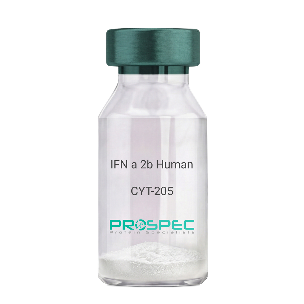
IFN a 2b HumanDescription:
IFN-Alpha 2b Human Recombinant
IFN alpha 2b, IFNA, INFA2, MGC125764, MGC125765.
Product # :
CYT-205Price :
Quantity :
Shipping Method :
Shipped at Room temp
More Info
- description
- source
- formulation
- purity
- biological activity
- More Info
- Citations
Description
IFN-a 2b Human Recombinant produced in E.Coli is a single, non-glycosylated, polypeptide chain containing 166 amino acids and having a molecular mass of 19400 Dalton. The difference between IFNA2A and IFNA2B is in the amino acid present at position 23. IFN-alpha 2a has a lysine at that position 23 while IFN-alpha 2b has arginine. The IFN-alpha 2b gene was obtained from human leukocytes. The IFN-a 2b is purified by proprietary chromatographic techniques.
Source
Escherichia Coli.
Formulation
Lyophilized from a (1 mg/ml) solution in containing 2.3 mg Sodium phosphate dibasic and 0.55 mg sodium phosphate monobasic buffer.
Purity
Greater than 98.0% as determined by(a) Analysis by RP-HPLC.
(b) Analysis by SDS-PAGE.Biological Activity
The specific activity as determined in a viral resistance assay using bovine kidney MDBK cells was found to be 260,000,000 IU/ mg.Citations
More Info
-
Introduction
IFN-alpha is produced by macrophages and has antiviral activities. IFN stimulates the production of two enzymes: protein kinase and an oligoadenylate synthetase.
-
Synonyms
IFN alpha 2b, IFNA, INFA2, MGC125764, MGC125765.
-
Physical Appearance
Sterile Filtered White lyophilized (freeze-dried) powder.
-
Stability
Lyophilized IFN although stable at room temperature for 3 weeks, should be stored desiccated below -18°C. Upon reconstitution IFN alpha 2b should be stored at 4°C between 2-7 days and for future use below -18°C.For long term storage it is recommended to add a carrier protein (0.1% HSA or BSA).Please prevent freeze-thaw cycles.
-
Solubility
It is recommended to reconstitute the lyophilized IFN-alpha 2b in sterile 18MΩ-cm H2O not less than 100µg/ml, which can then be further diluted to other aqueous solutions.
-
Amino Acid Sequence
MCDLPQTHSL GSRRTLMLLA QMRRISLFSC LKDRHDFGFP QEEFGNQFQK AETIPVLHEM IQQIFNLFST KDSSAAWDET LLDKFYTELY QQLNDLEACV IQGVGVTETP LMKEDSILAV RKYFQRITLY LKEKKYSPCA WEVVRAEIMR SFSLSTNLQE SLRSKE
-
Background
IFN-alpha forms are produced by monocytes/macrophages, lymphoblastoid cells, fibroblasts, and a number of different cell types following induction by viruses, nucleic acids, glucocorticoid hormones, and low-molecular weight substances (n-butyrate, 5-bromodeoxy uridine). At least 23 different variants of IFN-alpha are known. The individual proteins have molecular masses between 19-26 kDa and consist of proteins with lengths of 156-166 and 172 amino acids. All IFN-alpha subtypes possess a common conserved sequence region between amino acid positions 115-151 while the amino-terminal ends are variable. Many IFN-alpha subtypes differ in their sequences at only one or two positions. Naturally occurring variants also include proteins truncated by 10 amino acids at the carboxy-terminal end. Disulfide bonds are formed between cysteines at positions 1/98 and 29/138. The disulfide bond 29/138 is essential for biological activity while the 1/98 bond can be reduces without affecting biological activity. All IFN-alpha forms contain a potential glycosylation site but most subtypes are not glycosylated. In contrast to IFN-gamma IFN-alpha proteins are stable at pH2.
There are at least 23 different IFN-alpha genes. They have a length of 1-2 kb and are clustered on human chromosome 9p22. It is not known whether all these genes are expressed following stimulation of the cells. In some cell systems expression of some subtypes (IFN-alpha-1, IFN-alpha-2, IFN-alpha-4) is stronger than those of others. IFN-alpha genes do not contain intron sequences found in many other eukaryotic genes (see also: gene expression). Based upon the structures two types of IFN-alpha genes, designated class 1 and II, are distinguished. They encode proteins of 156-166 and172 amino acids, respectively. Deletions covering 9p22 are observed frequently in cells of lymphoblastoid leukemias. It is not known to date whether this is of significance with respect to IFN expression.
The gene encoding the IFN-alpha receptor maps to human chromosome 21q22.1. IFN-alpha and IFN-beta are thought to bind to the same IFN binding subunit which is expressed in 100-5000 copies in IFN-alpha sensitive and -resistant cells and is associated with other as yet unidentified proteins. The IFN-omega (Omega IFN) also binds to the IFN-alpha/IFN-beta receptor. Another receptor expressed on B-lymphocytes is identical with CD21. This receptor also binds Epstein-Barr virus through its gp350/220 coat protein.Signal transduction mechanisms elicited after binding of IFN-alpha to its receptors involves tyrosine phosphorylation (see also: PTK; protein tyrosine kinase) of various non-receptor tyrosine kinases belonging to the Janus kinases. Soluble forms of the IFN-alpha receptor, corresponding to truncated forms of the extracellular domain of the cell surface IFN-alpha receptor, have been found in human serum and in normal human urine.
All known subtypes of IFN-alpha show the same antiviral antiparasitic, antiproliferative activities in suitable bioassays although they may differ in relative activities. Human IFN-alpha is also a potent antiviral substance in murine, porcine, and bovine cell systems. Human IFN-alpha is less active in rodent cells. Site-directed mutagenesis techniques have been used to create some variants of certain subtypes (IFN-alpha-2) that display approximately 100-fold enhanced antiviral activities in mouse cells.
What is the molecular weight/Mw of IFN A 2B HUMAN Protein?
IFN A 2B HUMAN Protein has a total Mw of 19.4kDa.
What is the source or expression system of IFN A 2B HUMAN Protein?
Escherichia Coli.
What is the Purity of IFN A 2B HUMAN Protein?
IFN A 2B HUMAN Protein is >98% pure as determined by SDS-PAGE.
What is the Biological Activity of IFN A 2B HUMAN Protein?
The specific activity as determined in a viral resistance assay using bovine kidney MDBK cells was found to be 260,000,000 IU/ mg.
What is the amino acid sequence of IFN A 2B HUMAN Protein?
MCDLPQTHSL GSRRTLMLLA QMRRISLFSC LKDRHDFGFP QEEFGNQFQK AETIPVLHEM IQQIFNLFST KDSSAAWDET LLDKFYTELY QQLNDLEACV IQGVGVTETP LMKEDSILAV RKYFQRITLY LKEKKYSPCA WEVVRAEIMR SFSLSTNLQE SLRSKE
What applications can IFN A 2B HUMAN Protein be used in?
IFN A 2B HUMAN Protein can probably be used in western blot, ELISA and Lateral Flow.
What is the endotoxin level for IFN A 2B HUMAN Protein?
The endotoxin level is minimal, IFN A 2B HUMAN Protein was purified using conventional chromatography techniques.
-
Protein content
Protein quantitation was carried out by two independent methods1. UV spectroscopy at 280 nm using the absorbency value of 0.924 as the extinction coefficient for a 0.1% (1mg/ml) solution. This value is calculated by the PC GENE computer analysis program of protein sequences (IntelliGenetics). 2. Analysis by RP-HPLC, using a calibrated solution of IFN alpha 2b as a Reference Standard.
ProSpec's products are furnished for LABORATORY RESEARCH USE ONLY. The product may not be used as drugs, agricultural or pesticidal products, food additives or household chemicals.
- View Data Sheet
Name :
CSDC2 HumanDescription:
Cold Shock Domain Containing C2 Human Recombinant
Cold shock domain-containing protein C2, RNA-binding protein PIPPin, PIPPIN, DJ347H13.2.
Product # :
PRO-1439Price :
Quantity :
Shipping Method :
Shipped with Ice Packs
More Info
- description
- source
- formulation
- purity
- More Info
- Citations
Description
CSDC2 Human Recombinant produced in E.coli is a single, non-glycosylated polypeptide chain containing 176 amino acids (1-153) and having a molecular mass of 19.2kDa. CSDC2 is fused to a 23 amino acid His-tag at N-terminus.
Source
Escherichia Coli.
Formulation
The CSDC2 solution (0.5mg/ml) contains 20mM Tris-HCl buffer (pH8.0), 10% glycerol and 0.1M NaCl.
Purity
Greater than 90.0% as determined by SDS-PAGE.
Citations
More Info
-
Introduction
Cold shock domain-containing protein C2 (CSDC2) is RNA-binding factor that binds specifically to the very 3'-UTR ends of both histone H1 and H3. 3 mRNAs encompass the polyadenylation signal. The CSDPs (cold shock domain containing proteins) are one group of the evolutionarily conserved nucleic acid-binding proteins extensively distributed in bacteria, plants, animals, and involved in a variety of cellular processes, including adaptation to low temperature, cellular growth, nutrient stress and stationary phase. CSDC2 has a central role in the negative regulation of histone variant synthesis in the developing brain.
-
Synonyms
Cold shock domain-containing protein C2, RNA-binding protein PIPPin, PIPPIN, DJ347H13.2.
-
Physical Appearance
Sterile Filtered colorless solution.
-
Stability
Store at 4°C if entire vial will be used within 2-4 weeks. Store, frozen at -20°C for longer periods of time. For long term storage it is recommended to add a carrier protein (0.1% HSA or BSA).Avoid multiple freeze-thaw cycles.
-
Amino Acid Sequence
MGSSHHHHHH SSGLVPRGSH MGSMTSESTS PPVVPPLHSP KSPVWPTFPF HREGSRVWER GGVPPRDLPS PLPTKRTRTY SATARASAGP VFKGVCKQFS RSQGHGFITP ENGSEDIFVH VSDIEGEYVP VEGDEVTYKM CPIPPKNQKF QAVEVVLTQL APHTPHETWS GQVVGS.
ProSpec's products are furnished for LABORATORY RESEARCH USE ONLY. The product may not be used as drugs, agricultural or pesticidal products, food additives or household chemicals.
- View Data Sheet
Name :
LIF Human, GSTDescription:
Leukemia Inhibitory Factor, GST tag Human Recombinant
D factor, MLPLI, HILDA, Emfilermin, Leukemia Inhibitory factor, Differentiation-stimulating factor, Melanoma-derived LPL inhibitor.
Product # :
CYT-001Price :
Quantity :
Shipping Method :
Shipped with Ice Packs
More Info
- description
- source
- formulation
- purity
- More Info
- Citations
Description
LIF produced in E.Coli is a single, non-glycosylated polypeptide chain containing 415 amino acids (23-202a.a.) and having a molecular mass of 47.2kDa.LIF is fused to a 236 amino acid His-GST tag at N-terminus & purified by proprietary chromatographic techniques.
Source
Escherichia Coli.
Formulation
The LIF GST protein solution (1mg/1ml) is formulated in 20mM Tris-HCl buffer (pH 8.0) 1mM DTT, 50mM NaCl and 20% glycerol.
Purity
Greater than 90% as determined by SDS-PAGE.
Citations
More Info
-
Introduction
Leukemia inhibitory factor, is a pleiotropic cytokine which is expressed by numerous cells including activated T lymphocytes, monocytes, mast cells and neuronal cells. LIF takes part in the induction of hematopoietic differentiation in normal and myeloid leukemia cells, induction of neuronal cell differentiation, regulator of mesenchymal to epithelial conversion during kidney development, and is a key player in immune tolerance at the maternal-fetal interface.
-
Synonyms
D factor, MLPLI, HILDA, Emfilermin, Leukemia Inhibitory factor, Differentiation-stimulating factor, Melanoma-derived LPL inhibitor.
-
Physical Appearance
Sterile Filtered clear solution.
-
Stability
Store at 4°C if entire vial will be used within 2-4 weeks. Store, frozen at -20°C for longer periods of time.Please avoid freeze thaw cycles.
-
Amino Acid Sequence
MHHHHHHMSP ILGYWKIKGL VQPTRLLLEY LEEKYEEHLY ERDEGDKWRN KKFELGLEFP NLPYYIDGDV KLTQSMAIIR YIADKHNMLG GCPKERAEIS MLEGAVLDIR YGVSRIAYSK DFETLKVDFL SKLPEMLKMF EDRLCHKTYL NGDHVTHPDF MLYDALDVVL YMDPMCLDAF PKLVCFKKRI EAIPQIDKYL KSSKYIAWPL QGWQATFGGG DHPPKSDLVP RGSHMSPLPI TPVNATCAIR HPCHNNLMNQ IRSQLAQLNG SANALFILYY TAQGEPFPNN LDKLCGPNVT DFPPFHANGT EKAKLVELYR IVVYLGTSLG NITRDQKILN PSALSLHSKL NATADILRGL LSNVLCRLCS KYHVGHVDVT YGPDTSGKDV FQKKKLGCQL LGKYKQIIAV LAQAF
ProSpec's products are furnished for LABORATORY RESEARCH USE ONLY. The product may not be used as drugs, agricultural or pesticidal products, food additives or household chemicals.
- View Data Sheet
Name :
Visfatin Human, HisDescription:
Visfatin Recombinant Human, His Tag
PBEF, Pre-B cell colony-enhancing factor, Nicotinamide phosphoribosyltransferase NAmPRTase, Nampt, MGC117256, DKFZP666B131, 1110035O14Rik.
Product # :
CYT-563Price :
Quantity :
Shipping Method :
Shipped with Ice Packs
More Info
- description
- source
- formulation
- purity
- More Info
- Citations
Description
Visfatin Human Recombinant produced in E.Coli is a single, non-glycosylated, polypeptide chain containing 511 amino acids and having a molecular mass of 57 kDa. The recombinant human Visfatin is fused to His tag at N-Terminus.
Source
Escherichia Coli.
Formulation
Visfatin His tag protein contains 20mM Tris pH-8, 0.1mM DTT & 10% glycerol.
Purity
Greater than 95.0% as determined by(a) Analysis by RP-HPLC.
(b) Analysis by SDS-PAGE.Citations
More Info
-
Introduction
Excess adiposity is the most important risk in the development of insulin resistance and type 2 diabetes mellitus (T2DM). Adipose tissue produces several proteins (adipocytokines) such as leptin, adiponectin, resistin, tumor necrosis factor-?, and IL-6, that modulate insulin sensitivity and appear to play an important role in the pathogenesis of insulin resistance, diabetes, dyslipidemia, inflammation, and atherosclerosis. However, the mechanisms by which fat tissue induces insulin resistance and the role of adipocytokines in the pathogenesis of T2DM have not been well established.
Visfatin, also known as pre-B cell colony-enhancing factor (PBEF), is a cytokine that is highly expressed in visceral fat and was originally isolated as a secreted factor that synergizes with IL-7 and stem cell factors to promote the growth of B cell precursors. Visfatin homologs have been identified in carp, invertebrate mollusks, and bacteria, as well as in vertebrates, including humans and the mouse. It has been postulated to play a role in innate immunity.
Visfatin exerts insulin-mimetic effects that are dose-dependent and quantitatively similar to those of insulin in stimulating muscle and adipocyte glucose transport, and in inhibiting hepatocyte glucose production. Intravenous injection of recombinant visfatin in mice decreased plasma glucose in a dose-dependent fashion. In keeping with its insulin-mimetic effects, visfatin was as effective as insulin in reducing hyperglycemia in insulin-deficient diabetic mice. Visfatin was also found to be bound to and activate insulin receptor, causing receptor phosphorylation and the activation of downstream signaling molecules. However, visfatin and insulin did not compete for binding to the insulin receptor, indicating that the two proteins were recognized by different regions of the receptor. Thus, visfatin might play a role in glucose homeostasis and dysregulation in biosynthesis or signal transduction, and might contribute to the pathogenesis of diabetes. -
Synonyms
PBEF, Pre-B cell colony-enhancing factor, Nicotinamide phosphoribosyltransferase NAmPRTase, Nampt, MGC117256, DKFZP666B131, 1110035O14Rik.
-
Physical Appearance
Sterile Filtered solution at a concentration of 1mg/ml.
-
Stability
Store at 4°C if entire vial will be used within 2-4 weeks. Store, frozen at -20°C for longer periods of time. Please avoid freeze thaw cycles.
-
Amino Acid Sequence
MGSSHHHHHH SSGLVPRGSH MNPAAEAEFN ILLATDSYKV THYKQYPPNT SKVYSYFECR EKKTENSKLR KVKYEETVFY GLQYILNKYL KGKVVTKEKI QEAKDVYKEH FQDDVFNEKG WNYILEKYDG HLPIEIKAVP EGFVIPRGNV LFTVENTDPE CYWLTNWIET ILVQSWYPIT VATNSREQKK ILAKYLLETS GNLDGLEYKL HDFGYRGVSS QETAGIGASA HLVNFKGTDT VAGLALIKKY YGTKDPVPGY SVPAAEHSTI TAWGKDHEKD AFEHIVTQFS SVPVSVVSDS YDIYNACEKI WGEDLRHLIV SRSTQAPLII RPDSGNPLDT VLKVLEILGK KFPVTENSKG YKLLPPYLRV IQGDGVDINT LQEIVEGMKQ KMWSIENIAF GSGGGLLQKL TRDLLNCSFK CSYVVTNGLG INVFKDPVAD PNKRSKKGRL SLHRTPAGNF VTLEEGKGDL EEYGQDLLHT VFKNGKVTKS YSFDEIRKNA QLNIELEAAH H.
ProSpec's products are furnished for LABORATORY RESEARCH USE ONLY. The product may not be used as drugs, agricultural or pesticidal products, food additives or household chemicals.
- View Data Sheet
Name :
C3b Inactivated MouseDescription:
Complement C3b Inactivated Mouse
Complement C3, C3 and PZP-like alpha-2-macroglobulin domain-containing protein 1, C3, CPAMD1.
Product # :
PRO-2813Price :
Quantity :
Shipping Method :
Shipped with Ice Packs
More Info
- description
- source
- formulation
- purity
- More Info
- Citations
Description
Mouse Complement C3b Inactivated produced in Mouse serum having a molecular weight of 175kDa.
Source
Mouse serum.
Formulation
C3b Mouse Inactivated solution contains PBS.
Purity
Greater than 95.0% as determined by SDS-PAGE.
Citations
More Info
-
Synonyms
Complement C3, C3 and PZP-like alpha-2-macroglobulin domain-containing protein 1, C3, CPAMD1.
-
Physical Appearance
Sterile Filtered solution.
-
Stability
C3b Mouse Inactivated is stable at 4°C if entire vial will be used within 2-4 weeks. Store, frozen below -20°C for longer periods of time. For long term storage it is recommended to add a carrier protein (0.1% HSA or BSA). Avoid multiple freeze-thaw cycles.
-
Background
Complement component 3b (C3b) is a key protein in the complement system, a crucial component of the innate immune response. C3b plays a pivotal role in opsonization, inflammation, and clearance of pathogens and damaged cells. In mouse models, research on C3b has provided valuable insights into its mechanism of action, activity, and implications in immune function. This paper aims to delve into the dynamics of mouse C3b, shedding light on its functional attributes and potential applications in immunological research. C3b Inactivated protein has lost its biological capabilities due to structural changes compared to C3b that has binding sites to factor P, factor H, factor B, factorC5, and factor-I and therefore it destroys these sites.
Significantly, C3b inactivated protein cannot bind factor B and is therefore it cannot participate in complement activation.
C3b inactivated protein still binds tC50 properdin and CR3 receptor.
ProSpec's products are furnished for LABORATORY RESEARCH USE ONLY. The product may not be used as drugs, agricultural or pesticidal products, food additives or household chemicals.
- View Data Sheet
Name :
CAMP HumanDescription:
Cathelicidin Antimicrobial Peptide Human Recombinant
CAP-18, CAP18, CRAMP, FALL-39, FALL39, HSD26, LL37, 18 kDa cationic antimicrobial protein, FALL-39 peptide antibiotic, Cathelicidin antimicrobial peptide.
Product # :
PRO-1405Price :
Quantity :
Shipping Method :
Shipped with Ice Packs
More Info
- description
- source
- formulation
- purity
- More Info
- Citations
Description
CAMP Human Recombinant produced in E.Coli is a single, non-glycosylated polypeptide chain containing 163 amino acids (34-173 a.a.) and having a molecular mass of 18.4kDa.CAMP is fused to a 23 amino acid His-tag at N-terminus.
Source
Escherichia Coli.
Formulation
CAMP protein solution (0.5mg/ml) contains 20mM Tris-HCl buffer (pH 8.0), 0.4M UREA and 10% glycerol.
Purity
Greater than 85.0% as determined by SDS-PAGE.
Citations
More Info
-
Introduction
CAMP belongs to the antimicrobial peptide family, contains highly conserved N-terminal signal peptide, a cathelin domain and a structurally variable cationic antimicrobial peptide that produced by extracellular proteolysis from the C-terminus. CAMP has numerous functions besides the antimicrobial activity such as: cell chemotaxis, immune mediator induction and inflammatory response regulation.
-
Synonyms
CAP-18, CAP18, CRAMP, FALL-39, FALL39, HSD26, LL37, 18 kDa cationic antimicrobial protein, FALL-39 peptide antibiotic, Cathelicidin antimicrobial peptide.
-
Physical Appearance
Sterile Filtered clear solution.
-
Stability
Store at 4°C if entire vial will be used within 2-4 weeks. Store, frozen at -20°C for longer periods of time. For long term storage it is recommended to add a carrier protein (0.1% HSA or BSA).Avoid multiple freeze-thaw cycles.
-
Amino Acid Sequence
MGSSHHHHHH SSGLVPRGSH MGSQVLSYKE AVLRAIDGIN QRSSDANLYR LLDLDPRPTM DGDPDTPKPV SFTVKETVCP RTTQQSPEDC DFKKDGLVKR CMGTVTLNQA RGSFDISCDK DNKRFALLGD FFRKSKEKIG KEFKRIVQRI KDFLRNLVPR TES.
ProSpec's products are furnished for LABORATORY RESEARCH USE ONLY. The product may not be used as drugs, agricultural or pesticidal products, food additives or household chemicals.
- View Data Sheet
Name :
LIF Human, YeastDescription:
LIF Human Recombinant, Yeast
CDF, HILDA, D-FACTOR, Differentiation- stimulating factor, Melanoma-derived LPL inhibitor, MLPLI, Emfilermin, Leukemia inhibitory factor, LIF, DIA.
Product # :
CYT-191Price :
Quantity :
Shipping Method :
Shipped at Room temp
More Info
- description
- source
- formulation
- purity
- biological activity
- More Info
- Citations
Description
LIF Human Recombinant produced in yeast is a single, glycosylated polypeptide chain containing 180 amino acids and having a molecular mass of 58.5 kDa. The LIF is purified by proprietary chromatographic techniques.
Source
Pichia pastoris.
Formulation
The protein was lyophilized from a 0.2 µm filtered PBS.
Purity
Greater than 98.0% as determined by:
(a) Analysis by RP-HPLC.
(b) Analysis by SDS-PAGE.Biological Activity
The biological activity of recombinant human LIF was measured by the ability to induce differentiation of murine M1 myeloid leukemic cells. The minimal detectable concentration of human LIF in this assay is <0.05 ng/mL. The specific activity is > 1 x 10⁸ units/mg.Citations
More Info
-
Introduction
Leukemia Inhibitory Factor also called LIF is a lymphoid factor that promotes long-term maintenance of embryonic stem cells by suppressing spontaneous differentiation. Leukemia Inhibitory Factor has several functions such as cholinergic neuron differentiation, control of stem cell pluripotency, bone & fat metabolism, mitogenesis of factor dependent cell lines & promotion of megakaryocyte production in vivo. Human and mouse LIF exhibit a 78% identity in its amino acid sequence.
-
Synonyms
CDF, HILDA, D-FACTOR, Differentiation- stimulating factor, Melanoma-derived LPL inhibitor, MLPLI, Emfilermin, Leukemia inhibitory factor, LIF, DIA.
-
Physical Appearance
Sterile Filtered White lyophilized (freeze-dried) powder.
-
Stability
Lyophilized LIF although stable at room temperature for 3 weeks, should be stored desiccated below -18°C. Upon reconstitution LIF should be stored at 4°C between 2-7 days and for future use below -18°C. Please prevent freeze-thaw cycles.
-
Solubility
It is recommended to reconstitute the lyophilized LIF in sterile 18M-cm H2O not less than 100µg/ml, which can then be further diluted to other aqueous solutions.
-
Amino Acid Sequence
S P L P I T P V N A T C A I R H P C H N N L M N Q I R S Q L A Q L N G S A N A L F I L Y Y T A Q G E P F P N N L D K L C G P N V T D F P P F H A N G T E K A K L V E L Y R I V V Y L G T S L G N I T R D Q K I L N P S A L S L H S K L N A T A D I L R G L L S N V L C R L C S K Y H V G H V D V T Y G P D T S G K D V F Q K K K L G C Q L L G K Y K Q I I A V L A Q A F.
ProSpec's products are furnished for LABORATORY RESEARCH USE ONLY. The product may not be used as drugs, agricultural or pesticidal products, food additives or household chemicals.
- View Data Sheet
Name :
CD105 Human, HisDescription:
Endoglin Human Recombinant, His-Tag
CD105, ENG, END, ORW, HHT1, ORW1, FLJ41744, Endoglin.
Product # :
CYT-823Price :
Quantity :
Shipping Method :
Shipped with Ice Packs
More Info
- description
- source
- formulation
- purity
- More Info
- sds-page
- Citations
Description
Endoglin Human Recombinant produced in E.coli is a single, non-glycosylated polypeptide chain containing 594 amino acids (26-586) and having a molecular mass of 64.9 kDa. Endoglin is fused to a 36 amino acid His-tag at N-terminus.
Source
Escherichia Coli.
Formulation
The Endoglin solution (0.25mg/ml) contains 20mM Tris-HCl buffer (pH 8.0), 1mM DTT, 150mM NaCl and 10% glycerol.
Purity
Greater than 85% as determined by SDS-PAGE.
sds-page
Citations
More Info
-
Introduction
Endoglin is a type I membrane glycoprotein located on cell surfaces and is part of the TGF beta receptor complex.The Endoglin protein consists of a homodimer of 180 kDA with disulfide links. Endoglin has been found on endothelial cells, activated macrophages, fibroblasts, and smooth muscle cells. Furthermore, Endoglin has been found to be part of the TGF-beta1 receptor complex. Endoglin thus may be involved in the binding of TGF-beta1, TGF-beta3, activin-A, BMP-2, and BMP-7. Beside TGF-beta signaling endoglin may have other functions. It has been postulated that endoglin is involved in the cytoskeletal organization affecting cell morphology and migration. Endoglin has a role in the development of the cardiovascular system and in vascular remodeling. Endoglin expression is regulated during heart development . Experimental mice without the endoglin gene die due to cardiovascular abnormalities.
-
Synonyms
CD105, ENG, END, ORW, HHT1, ORW1, FLJ41744, Endoglin.
-
Physical Appearance
Sterile Filtered clear solution.
-
Stability
Store at 4°C if entire vial will be used within 2-4 weeks. Store, frozen at -20°C for longer periods of time. For long term storage it is recommended to add a carrier protein (0.1% HSA or BSA).Avoid multiple freeze-thaw cycles.
-
Amino Acid Sequence
MRGSHHHHHH GMASMTGGQQ MGRDLYDDDD KDRWGSETVH CDLQPVGPER DEVTYTTSQV SKGCVAQAPN AILEVHVLFL EFPTGPSQLE LTLQASKQNG TWPREVLLVL SVNSSVFLHL QALGIPLHLA YNSSLVTFQE PPGVNTTELP SFPKTQILEW AAERGPITSA AELNDPQSIL LRLGQAQGSL SFCMLEASQD MGRTLEWRPR TPALVRGCHL EGVAGHKEAH ILRVLPGHSA GPRTVTVKVE LSCAPGDLDA VLILQGPPYV SWLIDANHNM QIWTTGEYSF KIFPEKNIRG FKLPDTPQGL LGEARMLNAS IVASFVELPL ASIVSLHASS CGGRLQTSPA PIQTTPPKDT CSPELLMSLI QTKCADDAMT LVLKKELVAH LKCTITGLTF WDPSCEAEDR GDKFVLRSAY SSCGMQVSAS MISNEAVVNI LSSSSPQRKK VHCLNMDSLS FQLGLYLSPH FLQASNTIEP GQQSFVQVRV SPSVSEFLLQ LDSCHLDLGP EGGTVELIQG RAAKGNCVSL LSPSPEGDPR FSFLLHFYTV PIPKTGTLSC TVALRPKTGS QDQEVHRTVF MRLNIISPDL SGCTSKG
-
Background
What is the molecular weight/Mw of CD105 Protein?
CD105 Protein has a total Mw of 64.9kDa.
What is the source or expression system of CD105 Protein?
Escherichia Coli.
What is the Purity of CD105 Protein?
CD105 Protein is >85% pure as determined by SDS-PAGE.
What is the Biological Activity of CD105 Protein?
The biological functionality of CD105 Protein will be determined in the future.
What is the amino acid sequence of CD105 Protein?
MRGSHHHHHH GMASMTGGQQ MGRDLYDDDD KDRWGSETVH CDLQPVGPER DEVTYTTSQV SKGCVAQAPN AILEVHVLFL EFPTGPSQLE LTLQASKQNG TWPREVLLVL SVNSSVFLHL QALGIPLHLA YNSSLVTFQE PPGVNTTELP SFPKTQILEW AAERGPITSA AELNDPQSIL LRLGQAQGSL SFCMLEASQD MGRTLEWRPR TPALVRGCHL EGVAGHKEAH ILRVLPGHSA GPRTVTVKVE LSCAPGDLDA VLILQGPPYV SWLIDANHNM QIWTTGEYSF KIFPEKNIRG FKLPDTPQGL LGEARMLNAS IVASFVELPL ASIVSLHASS CGGRLQTSPA PIQTTPPKDT CSPELLMSLI QTKCADDAMT LVLKKELVAH LKCTITGLTF WDPSCEAEDR GDKFVLRSAY SSCGMQVSAS MISNEAVVNI LSSSSPQRKK VHCLNMDSLS FQLGLYLSPH FLQASNTIEP GQQSFVQVRV SPSVSEFLLQ LDSCHLDLGP EGGTVELIQG RAAKGNCVSL LSPSPEGDPR FSFLLHFYTV PIPKTGTLSC TVALRPKTGS QDQEVHRTVF MRLNIISPDL SGCTSKG
What applications can CD105 Protein be used in?
CD105 Protein can probably be used in western blot, ELISA and Lateral Flow.
What is the endotoxin level for CD105 Protein?
The endotoxin level is minimal, CD105 Protein was purified using conventional chromatography techniques.
ProSpec's products are furnished for LABORATORY RESEARCH USE ONLY. The product may not be used as drugs, agricultural or pesticidal products, food additives or household chemicals.
- View Data Sheet
Name :
MIF Human His CDescription:
Macrophage Migration Inhibitory Factor Human Recombinant, His Tag C-Terminus
Phenylpyruvate tautomerase, Glycosylation-inhibiting factor, GIF, MMIF, MIF.
Product # :
CYT-521Price :
Quantity :
Shipping Method :
Shipped at Room temp
More Info
- description
- source
- formulation
- purity
- biological activity
- More Info
- Citations
Description
MIF human Recombinant, fused to His-tag at C-terminus, was cloned into an E. coli expression vector and was purified to apparent homogeneity by using conventional column chromatography techniques.Macrophage Inducing Factor Human Recombinant is a single, non-glycosylated, polypeptide chaincontaining 123 amino acidsand having a molecular mass of 13.5 kDa.
Source
Escherichia Coli.
Formulation
Human MIF was lyophilized from a 1mg/ml solution containing PBS pH-7.4.
Purity
Greater than 95.0% as determined by:
(a) Analysis by RP-HPLC.
(b) Analysis by SDS-PAGE.Biological Activity
Measured by its ability to bind rhCD74 in a functional ELISA.Citations
More Info
-
Introduction
The cytokine Macrophage migration inhibitory factor (MIF) has been identified to be secreted by the pituitary gland and the monocyte/macrophage and to play an important role in endotoxic shock. MIF has the unique property of being released from macrophages and T cells in response to physiological concentrations of glucocorticoids. The secretion of MIF is tightly regulated and decreases at high, anti-inflammatory steroid concentration.
-
Synonyms
Phenylpyruvate tautomerase, Glycosylation-inhibiting factor, GIF, MMIF, MIF.
-
Physical Appearance
Sterile Filtered lyophilized powder.
-
Stability
Lyophilized MIF although stable at room temperature for 3 weeks, should be stored desiccated below -18°C. Upon reconstitution MIF should be stored at 4°C between 2-7 days and for future use below -18°C.For long term storage it is recommended to add a carrier protein (0.1% HSA or BSA).Please prevent freeze-thaw cycles.
-
Solubility
It is recommended to reconstitute the lyophilized MIF in sterile 18MΩ-cm H2O not less than 100µg/ml, which can then be further diluted to other aqueous solutions.
-
Amino Acid Sequence
MPMFIVNTNVPRASVPDGFLSELTQQLAQATGKPPQYIAVHVVPDQLMAFGGSSEPC
ALCSLHSIGKIGGAQNRSYSKLLCGLLAERLRISPDRVYINYYDMNAANVGWNNSTF
ALEHHHHHH.
ProSpec's products are furnished for LABORATORY RESEARCH USE ONLY. The product may not be used as drugs, agricultural or pesticidal products, food additives or household chemicals.
- View Data Sheet
Name :
ESAT6-CFP10Description:
ESAT6-CFP10 Chimeric Recombinant
Early Secretory Target Mycobacterium Tuberculosis, ESAT-6
Product # :
PRO-2737Price :
Quantity :
Shipping Method :
Shipped with Ice Packs
More Info
- description
- source
- formulation
- purity
- More Info
- Citations
Description
Recombinant Chimeric ESAT6-CFP10 produced in E. coli is fused to a 6 amino acid his tag at its C-terminus and purified by proprietary chromatographic technique.
Source
Escherichia Coli.
Formulation
ESAT6-CFP10 solution contains 25mM K2CO3, 0.02% NaN3 and PBS.
Purity
Protein is >95% pure as determined by 12% PAGE (coomassie staining).
Citations
More Info
-
Introduction
Mycobacterium antigen ESAT6 has been isolated from low molecular weight fractions of the shot-term-culture filtrate (ST-CF) and it can easily be detected in tuberculosis patients. The export of ESAT-6 which is a potent T-cell antigen, and related proteins requires a dedicated secretory apparatus that is encoded by a group of genes, several of which also code for proteins that are recognized strongly by T cells. The ESAT-6 systems can consequently be considered as immunogenicity islands and there is mounting evidence that the equivalent genes are subject to selective pressure imposed by the immune system of the host. This antigen includes many epitopes detectable in the serum of most patients with tuberculosis (more than 90%). By the attempts to obtain the vaccine on the basis of ESAT-6 it was demonstrated that the optimization of adjuvant is very important when using the combination of dioctadecylammonium bromide and monophosphoryllipide. ESAT-6 is very potential as diagnostic for differentiation between the mycobacterial infection and BCG vaccination. The main topic in ESAT-6 using is in antibody production and in test-systems for tuberculosis elaboration. Early secretory antigen 6 (ESAT6) and cell filtrate protein 10 (CFP10) are 2 antigens secreted as a complex by the replicating Mycobacterium tuberculosis complex (MTC).
-
Synonyms
Early Secretory Target Mycobacterium Tuberculosis, ESAT-6
-
Physical Appearance
Sterile Filtered clear solution.
-
Stability
Store at 4°C if entire vial will be used within 2-4 weeks.Store, frozen at -20°C for longer periods of time.For long term storage it is recommended to add a carrier protein (0.1% HSA or BSA).Avoid multiple freeze-thaw cycles.
-
Applications
Immunoassay.
ProSpec's products are furnished for LABORATORY RESEARCH USE ONLY. The product may not be used as drugs, agricultural or pesticidal products, food additives or household chemicals.
- View Data Sheet
Name :
LECT1 (214-333) HumanDescription:
Leukocyte Cell Derived Chemotaxin 1 (214-333 a.a.) Human Recombinant
BRICD3, CHM-I, CHM1, MYETS1, Leukocyte cell-derived chemotaxin 1, Chondrosurfactant protein, CH-SP, Chondromodulin-1, ChM-I, LECT1.
Product # :
PRO-1857Price :
Quantity :
Shipping Method :
Shipped with Ice Packs
More Info
- description
- source
- formulation
- purity
- More Info
- Citations
Description
LECT1 Human Recombinant produced in E. coli is. a single polypeptide chain containing 143 amino acids (214-333) and having a molecular mass of 16.2kDa. LECT1 is fused to a 23 amino acid His-tag at N-terminus & purified by proprietary chromatographic techniques.
Source
Escherichia Coli.
Formulation
The LECT1 solution (1mg/ml) contains 20mM Tris-HCl buffer (pH 8.0), 0.4M UREA and 10% glycerol.
Purity
Greater than 85% as determined by SDS-PAGE.
Citations
More Info
-
Introduction
Leukocyte Cell Derived Chemotaxin 1 (214-333 a.a.), also known as LECT1, is a glycosylated transmembrane protein which is cleaved to form a mature, secreted protein. The mature protein encourages chondrocyte growth and inhibits angiogenesis. The mature protein takes part in endochondral bone development by permitting cartilaginous anlagen to be vascularized and replaced by bone. LECT1 is expressed in the avascular area of prehypertrophic cartilage and its expression reduces during vascular invasion and chondrocyte hypertrophy.
-
Synonyms
BRICD3, CHM-I, CHM1, MYETS1, Leukocyte cell-derived chemotaxin 1, Chondrosurfactant protein, CH-SP, Chondromodulin-1, ChM-I, LECT1.
-
Physical Appearance
Sterile Filtered clear solution.
-
Stability
Store at 4°C if entire vial will be used within 2-4 weeks. Store frozen at -20°C for longer periods of time. For long term storage it is recommended to add a carrier protein (0.1% HSA or BSA).Avoid multiple freeze-thaw cycles.
-
Amino Acid Sequence
MGSSHHHHHH SSGLVPRGSH MGSREVVRKI VPTTTKRPHS GPRSNPGAGR LNNETRPSVQ EDSQAFNPDN PYHQEGESMT FDPRLDHEGI CCIECRRSYT HCQKICEPLG GYYPWPYNYQ GCRSACRVIM PCSWWVARIL GMV.
ProSpec's products are furnished for LABORATORY RESEARCH USE ONLY. The product may not be used as drugs, agricultural or pesticidal products, food additives or household chemicals.
- View Data Sheet
Name :
IRF2 HumanDescription:
IFN Regulatory Factor-2 Human Recombinant
IRF-2, IRF2, MAR, DKFZp686F0244, IFN regulatory factor 2.
Product # :
CYT-534Price :
Quantity :
Shipping Method :
Shipped with Ice Packs
More Info
- description
- source
- formulation
- purity
- More Info
- SDS-PAGE
- Citations
Description
IFN Regulatory Factor-2 Human Recombinant produced in E.Coli is a single, non-glycosylated, polypeptide chain containing 133 amino acids (1-113) with a His Tag of 20 aa, and having a molecular mass of 15 kDa.The IRF2 is purified by proprietary chromatographic techniques.
Source
Escherichia Coli.
Formulation
1 mg/ml in 20mM Tris pH-8, 1mM DTT and 10% Glycerol.
Purity
Greater than 95.0% as determined by SDS-PAGE.
SDS-PAGE
Citations
More Info
-
Introduction
IFN regulatory factor 2 is a member of the IFN regulatory transcription factor (IRF) family. IRF2 competitively inhibits the IRF1-mediated transcriptional activation of IFNs alpha and beta, and presumably other genes that employ IRF1 for transcription activation. However, IRF2 also functions as a transcriptional activator of histone H4. IRF2 binds to the upstream regulatory region of type-1 IFN and IFN-inducible MHC class-1 genes (the IFN consensus sequence (ics) and represses those genes.
-
Synonyms
IRF-2, IRF2, MAR, DKFZp686F0244, IFN regulatory factor 2.
-
Physical Appearance
Sterile Filtered colorless solution.
-
Stability
Store at 4°C if entire vial will be used within 2-4 weeks. Store, frozen at -20°C for longer periods of time. For long term storage it is recommended to add a carrier protein (0.1% HSA or BSA).Avoid multiple freeze-thaw cycles.
-
Amino Acid Sequence
MGSSHHHHHH SSGLVPRGSH MPVERMRMRP WLEEQINSNT IPGLKWLNKE KKIFQIPWMHAARHGWDVEK DAPLFRNWAI HTGKHQPGVD KPDPKTWKAN FRCAMNSLPD IEEVKDKSIKKGNNAFRVYR MLP.
-
Background
What is the molecular weight/Mw of IRF2 HUMAN Protein?
IRF2 HUMAN Protein has a total Mw of 15kDa.
What is the source or expression system of IRF2 HUMAN Protein?
Escherichia Coli.
What is the Purity of IRF2 HUMAN Protein?
IRF2 HUMAN Protein is >95% pure as determined by SDS-PAGE.
What is the Biological Activity of IRF2 HUMAN Protein?
The biological functionality of IRF2 HUMAN Protein will be determined in the future.
What is the amino acid sequence of IRF2 HUMAN Protein?
MGSSHHHHHH SSGLVPRGSH MPVERMRMRP WLEEQINSNT IPGLKWLNKE KKIFQIPWMHAARHGWDVEK DAPLFRNWAI HTGKHQPGVD KPDPKTWKAN FRCAMNSLPD IEEVKDKSIKKGNNAFRVYR MLP.
What applications can IRF2 HUMAN Protein be used in?
IRF2 HUMAN Protein can probably be used in western blot, ELISA and Lateral Flow.
What is the endotoxin level for IRF2 HUMAN Protein?
The endotoxin level is minimal, IRF2 HUMAN Protein was purified using conventional chromatography techniques.
ProSpec's products are furnished for LABORATORY RESEARCH USE ONLY. The product may not be used as drugs, agricultural or pesticidal products, food additives or household chemicals.
- View Data Sheet
Name :
TACI HumanDescription:
Tumor Necrosis Factor Receptor 13B Human Recombinant
CD267, CVID, CVID2, TACI, TNFRSF14B, Tumor necrosis factor receptor superfamily, member 13B, Tumor necrosis factor receptor superfamily, member 13B, isoform CRA_a, TNFRSF13B.
Product # :
CYT-819Price :
Quantity :
Shipping Method :
Shipped at Room temp
More Info
- description
- source
- formulation
- purity
- biological activity
- More Info
- Citations
Description
TACI Human Recombinant produced in E.Coli is a single, non-glycosylated, polypeptide chain containing 160 amino acids and having a molecular mass of 18.0kDa.
Source
Escherichia Coli.
Formulation
TACI protein was lyophilized from a 0.2 µm filtered concentrated solution in PBS, pH 7.4.
Purity
Greater than 95.0% as determined by: (a) Analysis by RP-HPLC.(b) Analysis by SDS-PAGE.
Biological Activity
Fully biologically active when compared to standard. The biological activity is determined by its ability to block human BAFF induced T2B cell survival using a concentration range of 1.0-3.0 µg/ml corresponding to a specific activity of 334-1000 IU/mg.Citations
More Info
-
Introduction
TNFRSF13B (TACI) is a transmembrane receptor protein found predominantly on the surface of B cells (a significant part of the immune system). TACI was at first discovered owing to its ability to interact with calcium-modulator and cyclophilin ligand (CAML). Later on, it was found that TACI plays a key role in humoral immunity by interacting with two members of the TNF family. Also, TACI controls T cell-independent B cell antibody responses, isotype switching, and B cell homeostasis.
-
Synonyms
CD267, CVID, CVID2, TACI, TNFRSF14B, Tumor necrosis factor receptor superfamily, member 13B, Tumor necrosis factor receptor superfamily, member 13B, isoform CRA_a, TNFRSF13B.
-
Physical Appearance
Sterile Filtered White lyophilized (freeze-dried) powder.
-
Stability
Lyophilized TACI although stable at room temperature for 3 weeks, should be stored desiccated below -18°C. Upon reconstitution TACI should be stored at 4°C between 2-7 days and for future use below -18°C. For long term storage it is recommended to add a carrier protein (0.1% HSA or BSA). Please prevent freeze-thaw cycles.
-
Solubility
It is recommended to reconstitute the lyophilized TACI in sterile 18MΩ-cm H2O not less than 100µg/ml, which can then be further diluted to other aqueous solutions.
-
Amino Acid Sequence
MSGLGRSRRG GRSRVDQEER FPQGLWTGVA MRSCPEEQYW DPLLGTCMSC KTICNHQSQR TCAAFCRSLS CRKEQGKFYD HLLRDCISCA SICGQHPKQC AYFCENKLRS PVNLPPELRR QRSGEVENNS DNSGRYQGLE HRGSEASPAL PGLKLSADQV
ProSpec's products are furnished for LABORATORY RESEARCH USE ONLY. The product may not be used as drugs, agricultural or pesticidal products, food additives or household chemicals.
- View Data Sheet
Name :
LAIR1 MouseDescription:
Leukocyte-Associated Ig-Like Receptor 1 Mouse Recombinant
Leukocyte-associated immunoglobulin-like receptor 1, LAIR-1, mLAIR1, CD305, LAIR1.
Product # :
PRO-2356Price :
Quantity :
Shipping Method :
Shipped with Ice Packs
More Info
- description
- source
- formulation
- purity
- More Info
- Citations
Description
LAIR1 Mouse Recombinant produced in Sf9 Baculovirus cells is a single, glycosylated polypeptide chain containing 132 amino acids (22-144 aa) and having a molecular mass of 15kDa (Migrates at 18-28kDa on SDS-PAGE under reducing conditions).LAIR1 is expressed with a 6 amino acid His tag at C-Terminus and purified by proprietary chromatographic techniques.
Source
Sf9, Baculovirus cells.
Formulation
LAIR1 protein solution (0.5mg/ml) contains Phosphate buffered saline (pH7.4) and 10% glycerol.
Purity
Greater than 90.0% as determined by SDS-PAGE.
Citations
More Info
-
Introduction
LAIR1 is a glycoprotein expressed on the surface of the majority of human peripheral blood mononuclear leukocytes including T cells, B cells, NK cells, macrophages and dendritic cells. LAIR-1 functions as an inhibitory receptor in NK cells, T cells and B cells. Inhibitory receptors control the immune response to prevent lysis of cells recognized as self. Lair1 consists of a leader sequence, extracellular domain, transmembrane domain, cytoplasmic region.LAIR1 is a member of both the immunoglobulin superfamily and the leukocyte-associated inhibitory receptor family. LAIR1 maps to a region of 19q13.4 labeled the leukocyte receptor cluster, which contains at least 29 genes encoding leukocyte-expressed receptors of the immunoglobulin superfamily.
-
Synonyms
Leukocyte-associated immunoglobulin-like receptor 1, LAIR-1, mLAIR1, CD305, LAIR1.
-
Physical Appearance
Sterile filtered colorless solution.
-
Stability
Store at 4°C if entire vial will be used within 2-4 weeks. Store, frozen at -20°C for longer periods of time. For long term storage it is recommended to add a carrier protein (0.1% HSA or BSA).Avoid multiple freeze-thaw cycles.
-
Amino Acid Sequence
ADPQEGSLPD ITIFPNSSLM ISQGTFVTVV CSYSDKHDLY NMVRLEKDGS TFMEKSTEPY KTEDEFEIGP VNETITGHYS CIYSKGITWS ERSKTLELKV IKENVIQTPA PGPTSDTSWL KTYSIYHHHH HH
ProSpec's products are furnished for LABORATORY RESEARCH USE ONLY. The product may not be used as drugs, agricultural or pesticidal products, food additives or household chemicals.
- View Data Sheet
Name :
Rabbit SARS CoV-2 IgG S1Description:
Recombinant Anti Rabbit SARS CoV-2 IgG Spike S1
Product # :
ANT-762Price :
Quantity :
Shipping Method :
Shipped with Ice Packs
More Info
- description
- source
- formulation
- More Info
- Citations
Description
Recombinant Anti Rabbit SARS CoV-2 IgG Kappa Spike S1 is a recombinant monoclonal antibody derived from HEK293 cells which recognizes the SARS-CoV and SARS-CoV-2 Spike glycoprotein. CoV-2 IgG S1 Rabbit Antibody binds to both SARS-CoV and SARS-CoV-2 with high affinity at amino acids 318-510 (RBD, Receptor Binding Domain) in the S1 subunit of the Spike protein. This chimeric rabbit antibody was made using the variable domain sequences of the original Human IgG1 format.
Source
HEK293 Cells.
Formulation
1mg/ml in PBS and 0.02% Proclin 300.
Citations
More Info
-
Introduction
SARS Coronavirus is an enveloped virus containing three outer structural proteins, namely the membrane (M), envelope (E), and spike (S) proteins. Spike (S)-glycoprotein of the virus interacts with a cellular receptor and mediates membrane fusion to allow viral entry into susceptible target cells. Accordingly, S-protein plays an important role in virus infection cycle and is the primary target of neutralizing antibodies.It has recently been shown that SARS is caused by a human coronavirus. Human coronaviruses are the major cause of upper respiratory tract illness in humans, such as the common cold. Coronaviruses are positive-stranded RNA viruses, featuring the largest viral RNA genomes known to date (27-31 kb). The first step in coronavirus infection is binding of the viral spike protein, a 139-kDa protein, to certain receptors on host cells. The spike protein is the main surface antigen of the coronavirus. The most prominent protein in the culture supernatants infected with SARS virus is a 46 kDa nucleocapsid protein. This suggests that the nucleocapsid protein is a major immunogen that may be useful for early diagnostics.
-
Physical Appearance
Sterile Filtered liquid formulation.
-
Stability
Store at 4°C, stable for 2 weeks. For long-term storage, store at -20°C.
-
Immunogen
The native monoclonal antibody was generated by sequencing peripheral blood lymphocytes of a patient exposed to the SARS-CoV
-
Applications
ELISA.
-
Type
Mouse antibody Monoclonal.
-
Purification Method
Protein A affinity purified.
ProSpec's products are furnished for LABORATORY RESEARCH USE ONLY. The product may not be used as drugs, agricultural or pesticidal products, food additives or household chemicals.
- View Data Sheet
Name :
GZMH HumanDescription:
Granzyme-H Human Recombinant
Granzyme H (Cathepsin G-Like 2, Protein H-CCPX), CTSGL2, Cytotoxic T-Lymphocyte Proteinase, CCP-X, CSP-C, Cytotoxic T-Lymphocyte-Associated Serine Esterase 1, Cytotoxin Serine Protease-C, Cytotoxic Serine Protease C, Cathepsin G-Like 2, EC 3.4.21.79, EC 3.4.21, Granzyme H, EC 3.4.21, CGL-2, CTLA1, CGL2, GZMH.
Product # :
ENZ-896Price :
Quantity :
Shipping Method :
Shipped with Ice Packs
More Info
- description
- source
- formulation
- purity
- More Info
- Citations
Description
GZMH Human Recombinant produced in E.Coli is a single, non-glycosylated polypeptide chain containing 248 amino acids (20-246 a.a) and having a molecular mass of 27.5kDa.GZMH is fused to a 21 amino acid His-tag at N-terminus & purified by proprietary chromatographic techniques.
Source
Escherichia Coli.
Formulation
GZMH protein solution (0.25mg/ml) containing 20mM Tris-HCl (pH 8.0) and 10% glycerol.
Purity
Greater than 85.0% as determined by SDS-PAGE.
Citations
More Info
-
Introduction
Granzyme H also known as GZMH belongs to the peptidase S1 family. GZMH is an essential part for HBV eradication. The HBx protein, which is required for the replication of HBV, is cleaved at Met(79) by GZMH. Furthermore, GZMH inhibitor can abolish GZNH- as well as lymphokine-activated killer cell-mediated HBx degradation and HBV clearance. A HBx-deficient HBV is resistant to GzmH- in addition to lymphokine-activated killer cell-mediated viral clearance.
-
Synonyms
Granzyme H (Cathepsin G-Like 2, Protein H-CCPX), CTSGL2, Cytotoxic T-Lymphocyte Proteinase, CCP-X, CSP-C, Cytotoxic T-Lymphocyte-Associated Serine Esterase 1, Cytotoxin Serine Protease-C, Cytotoxic Serine Protease C, Cathepsin G-Like 2, EC 3.4.21.79, EC 3.4.21, Granzyme H, EC 3.4.21, CGL-2, CTLA1, CGL2, GZMH.
-
Physical Appearance
Sterile Filtered clear solution.
-
Stability
Store at 4°C if entire vial will be used within 2-4 weeks. Store, frozen at -20°C for longer periods of time. For long term storage it is recommended to add a carrier protein (0.1% HSA or BSA).Avoid multiple freeze-thaw cycles.
-
Amino Acid Sequence
MGSSHHHHHH SSGLVPRGSH MEIIGGHEAK PHSRPYMAFV QFLQEKSRKR CGGILVRKDF VLTAAHCQGS SINVTLGAHN IKEQERTQQF IPVKRPIPHP AYNPKNFSND IMLLQLERKA KWTTAVRPLR LPSSKAQVKP GQLCSVAGWG YVSMSTLATT LQEVLLTVQK DCQCERLFHG NYSRATEICV GDPKKTQTGF KGDSGGPLVC KDVAQGILSY GNKKGTPPGV YIKVSHFLPW IKRTMKRL
ProSpec's products are furnished for LABORATORY RESEARCH USE ONLY. The product may not be used as drugs, agricultural or pesticidal products, food additives or household chemicals.
- View Data Sheet
Name :
CANX HumanDescription:
Calnexin Human Recombinant
Calnexin, Major histocompatibility complex class I antigen-binding protein p88, p90, IP90, CANX, CNX, FLJ26570.
Product # :
PRO-725Price :
Quantity :
Shipping Method :
Shipped with Ice Packs
More Info
- description
- source
- formulation
- purity
- More Info
- Citations
Description
CANX Human Recombinant produced in E.Coli is a single, non-glycosylated, polypeptide chain containing 462 amino acids (21-481 a.a.) and having a molecular mass of 52.5kDa.The CANX is purified by proprietary chromatographic techniques.
Source
Escherichia Coli.
Formulation
The CANX protein solution contains 20mM Tris-HCl buffer (pH8.0), 1mM DTT and 20% glycerol.
Purity
Greater than 90.0% as determined by SDS-PAGE.
Citations
More Info
-
Introduction
Calnexin (CANX) belongs to the calnexin family of molecular chaperones. Calnexin is a calcium-binding, ER-associated protein that interacts briefly with newly synthesized N-linked glycoproteins, facilitating protein folding and assembly. Calnexin may also have a key role in the quality control of protein folding by retaining incorrectly folded protein subunits within the ER for degradation. Calnexin grants long-term protection of wild-type Shaker protein from ER-associated degradation. Polypeptide substrate recognition by Calnexin requires specific conformations of the Calnexin protein. Calnexin dwindles with aging and might contribute to a cytoprotection in an array of human age-related diseases.
-
Synonyms
Calnexin, Major histocompatibility complex class I antigen-binding protein p88, p90, IP90, CANX, CNX, FLJ26570.
-
Physical Appearance
Sterile Filtered colorless solution.
-
Stability
Store at 4°C if entire vial will be used within 2-4 weeks. Store, frozen at -20°C for longer periods of time. For long term storage it is recommended to add a carrier protein (0.1% HSA or BSA).Avoid multiple freeze-thaw cycles.
-
Amino Acid Sequence
MHDGHDDDVI DIEDDLDDVI EEVEDSKPDT TAPPSSPKVT YKAPVPTGEV YFADSFDRGT LSGWILSKAK KDDTDDEIAK YDGKWEVEEM KESKLPGDKG LVLMSRAKHH AISAKLNKPF LFDTKPLIVQ YEVNFQNGIE CGGAYVKLLS KTPELNLDQF HDKTPYTIMF GPDKCGEDYK LHFIFRHKNP KTGIYEEKHA KRPDADLKTY FTDKKTHLYT LILNPDNSFE ILVDQSVVNS GNLLNDMTPP VNPSREIEDP EDRKPEDWDE RPKIPDPEAV KPDDWDEDAP AKIPDEEATK PEGWLDDEPE YVPDPDAEKP EDWDEDMDGE WEAPQIANPR CESAPGCGVW QRPVIDNPNY KGKWKPPMID NPSYQGIWKP RKIPNPDFFE DLEPFRMTPF SAIGLELWSM TSDIFFDNFI ICADRRIVDD WANDGWGLKK AADGAAEPGV VGQMIEAAEE RP.
ProSpec's products are furnished for LABORATORY RESEARCH USE ONLY. The product may not be used as drugs, agricultural or pesticidal products, food additives or household chemicals.
- View Data Sheet
Name :
IFN A WB AntibodyDescription:
Interferon-alpha Western Blot, Mouse Anti-Human
Product # :
ANT-208Price :
Quantity :
Shipping Method :
Shipped at Room temp
More Info
- formulation
- More Info
- Citations
Formulation
1mg/ml in PBS (after reconstitution).
Citations
More Info
-
Introduction
IFN-alpha is produced by macrophages, IFN-alpha has antiviral activities and stimulates the production of two enzymes: a protein kinase and an oligoadenylate synthetase.
-
Solubility
Reconstitute with sterile H20. Mix gently, wash the sides of the vial and wait 30-60 seconds before use.
-
Immunogen
r.Human IFN-a.
-
Ig Subclass
Mouse IgG1.
-
Clone
NYRhIFN-A(WB).
-
Applications
Direct ELISA, Western Blot, Immuneprecipitation Immunohistochemistry. This is a NON-neutralizing antibody.
-
Note
The antibody was can be used for Capture ELISA together with clone ProSpec’s Mouse Anti Human IFN-A Neutralizing antibody.
-
Titer
By direct ELISA, 1:10,000 dilution will yield 0.5 O.D using alkaline phosphatase conjugated rabbit anti-mouse Ig (Jackson Laboratories).
-
Shipping Conditions
Antibody is shipped lyophilized at ambient temperature.
-
Type
Mouse Anti Human Monoclonal.
-
Storage Procedures
In lyophilized form, for long periods, store at 4oC in a dry environment. After reconstitution, if not intended for use within a month, aliquot and store at -20oC.
-
Purification Method
Ion exchange.
ProSpec's products are furnished for LABORATORY RESEARCH USE ONLY. The product may not be used as drugs, agricultural or pesticidal products, food additives or household chemicals.
- View Data Sheet
Name :
IgG-Fc antibodyDescription:
IgG-Fc, Mouse Anti-Human
Product # :
ANT-173Price :
Quantity :
Shipping Method :
Shipped at Room temp
More Info
- formulation
- More Info
- Citations
Formulation
1mg/ml in PBS (after reconstitution).
Citations
More Info
-
Solubility
Reconstitute with H20 to get a 1 mg/ml concentration. Mix gently, wash the sides of the vial and wait 30-60 seconds before use.
-
Immunogen
Purified hIgG.
-
Ig Subclass
Mouse IgG2a.
-
Clone
NYRhIgGFc.
-
Note
This antibody does not cross reacts with Fab or F(ab')2 of IgG. Some cross reaction with IgG of other species such as horse and sheep was observed. No cross reaction with human IgA, IgE or IgM.
-
Titer
By direct ELISA, 1:10,000 dilution will yield 0.75 O.D using alkaline phosphatase conjugated rabbit anti-mouse Ig (Jackson Laboratories).
-
Shipping Conditions
Antibody is shipped lyophilized at ambient temperature.
-
Type
Mouse Anti Human Monoclonal.
-
Storage Procedures
In lyophilized form, for long periods, store at 4°C in a dry environment. After reconstitution, if not intended for use within a month, aliquot and store at -20°C.
-
Purification Method
Ion exchange column.
ProSpec's products are furnished for LABORATORY RESEARCH USE ONLY. The product may not be used as drugs, agricultural or pesticidal products, food additives or household chemicals.
- View Data Sheet
Name :
CD58 Antibody, FITCDescription:
CD58, Mouse Anti-Human, FITC
CD58, LFA-3, Ag3, Surface glycoprotein LFA-3.
Product # :
ANT-279Price :
Quantity :
Shipping Method :
Shipped at Room temp
More Info
- formulation
- More Info
- Citations
Formulation
1mg/ml in PBS (after reconstitution).
Citations
More Info
-
Introduction
CD58 is part of the immunoglobulin superfamily. CD58 is a ligand of the T lymphocyte CD2 protein, and is invloved in adhesion and activation of T lymphocytes. CD58 is localized to the plasma membrane.
-
Synonyms
CD58, LFA-3, Ag3, Surface glycoprotein LFA-3.
-
Solubility
Reconstitute with of H20. Mix gently, wash the sides of the vial and wait 30-60 seconds before use.
-
Immunogen
Human Erythrocytes.
-
Ig Subclass
Mouse IgG1.
-
Clone
hCD58.
-
Applications
Staining antibody. For staining, use 10µl/1,000,000 cells.
-
Available Conjugates
This antibody is also available unconjugated to and conjugated to Biotin. For staining with biotin or FITC-conjugated antibody use 5-10µl/1,000,000 cells.
-
Type
Mouse Anti Human Monoclonal.
-
Storage Procedures
Lyophilized: store at 4°C. After reconstitution, if not intended for use within a month, aliquot and store at -20°C.
-
Purification Method
Ion-Exchange Column.
ProSpec's products are furnished for LABORATORY RESEARCH USE ONLY. The product may not be used as drugs, agricultural or pesticidal products, food additives or household chemicals.
- View Data Sheet
Name :
AIMP1 AntibodyDescription:
Aminoacyl tRNA Synthetase Complex-Interacting Multifunctional Protein 1, Mouse Anti Human
Aminoacyl tRNA synthase complex-interacting multifunctional protein 1, Multisynthase complex auxiliary component p43, AIMP1, EMAP2, SCYE1, p43, EMAPII.
Product # :
ANT-619Price :
Quantity :
Shipping Method :
Shipped with Ice Packs
More Info
- formulation
- More Info
- Citations
Formulation
1mg/ml containing PBS, pH-7.4, 10% Glycerol and 0.02% Sodium Azide.
Citations
More Info
-
Introduction
AIMP1 (EMPA2 or p43) is a cytokine that is specifically induced by apoptosis, and it is involved in the control of angiogenesis, inflammation, and wound healing. The release of the AIMP1 cytokine renders the tumor-associated vasculature sensitive to tumor necrosis factor. Furthermore, AIMP1 is involved in the stimulation of inflammatory responses after proteolytic cleavage in tumor cells.
-
Synonyms
Aminoacyl tRNA synthase complex-interacting multifunctional protein 1, Multisynthase complex auxiliary component p43, AIMP1, EMAP2, SCYE1, p43, EMAPII.
-
Physical Appearance
Sterile filtered colorless solution.
-
Immunogen
Anti-human AIMP1 mAb, is derived from hybridization of mouse F0 myeloma cells with spleen cells from BALB/c mice immunized with a recombinant human AIMP1 protein 1-336 amino acids purified from E. coli.
-
Ig Subclass
Mouse IgG2a heavy chain and Kappa light chain.
-
Clone
PAT6D10AT.
-
Applications
The antibody has been tested by ELISA, Western blot analysis, Flow cytometry and ICC/IF to assure specificity and reactivity. Since application varies, however, each investigation should be titrated by the reagent to obtain optimal results.
-
Type
Mouse Anti Human Monoclonal.
-
Storage Procedures
For periods up to 1 month store at 4°C, for longer periods of time, store at -20°C. Prevent freeze thaw cycles.
-
Purification Method
AIMP1 antibody was purified by protein-A affinity chromatography.
ProSpec's products are furnished for LABORATORY RESEARCH USE ONLY. The product may not be used as drugs, agricultural or pesticidal products, food additives or household chemicals.
- View Data Sheet
Name :
C3b RabbitDescription:
Complement C3b Rabbit
Complement C3, C3 and PZP-like alpha-2-macroglobulin domain-containing protein 1, C3, CPAMD1.
Product # :
PRO-2814Price :
Quantity :
Shipping Method :
Shipped with Ice Packs
More Info
- description
- source
- formulation
- purity
- More Info
- Citations
Description
Rabbit Complement C3b is composed of 2 disulfide-linked chains having a molecular weight of 176kDa.
Source
Rabbit serum.
Formulation
C3b Rabbit solution contains PBS, pH 7.2.
Purity
Greater than 93.0% as determined by SDS-PAGE.
Citations
More Info
-
Synonyms
Complement C3, C3 and PZP-like alpha-2-macroglobulin domain-containing protein 1, C3, CPAMD1.
-
Physical Appearance
Sterile Filtered solution.
-
Stability
C3b is stable at 4°C if entire vial will be used within 2-4 weeks. Store, frozen below -20°C for longer periods of time. For long term storage it is recommended to add a carrier protein (0.1% HSA or BSA). Avoid multiple freeze-thaw cycles.
-
Background
Rabbit C3b serves as a critical opsonin, enhancing the recognition and engulfment of pathogens and immune complexes by phagocytic cells. Through its covalent attachment to target surfaces, C3b facilitates the recognition of foreign antigens and promotes their clearance by the immune system. C3b contributes to the inflammatory response by generating C5 convertase complexes, leading to the cleavage of C5 into C5a and C5b. C5a is a potent chemoattractant and activator of immune cells, promoting the recruitment of neutrophils and macrophages to sites of infection or tissue damage.
ProSpec's products are furnished for LABORATORY RESEARCH USE ONLY. The product may not be used as drugs, agricultural or pesticidal products, food additives or household chemicals.
- View Data Sheet
Name :
CRP Paired AntibodyDescription:
Anti Human C-Reactive Protein Paired Antibody Mouse
Product # :
ANT-783Price :
Quantity :
Shipping Method :
Shipped with Ice Packs
More Info
- description
- formulation
- purity
- More Info
- Citations
Description
CRP Paired monoclonal antibodies are used to develop rapid test for CRP rapid test. Please note that when ordering for example: 100µg paired antibody, you receive 50µg from each antibody (100µg in total).
Formulation
* CRP conjugation antibody in PBS, NaCl and 0.095% NaN3.
* CRP coating antibody in PBS, NaCl and 0.095% NaN3.
Purity
Greater than 95%.
Citations
More Info
-
Physical Appearance
2 vials of sterile Filtered clear colorless solution.
-
Stability
For periods up to 1 month CRP Paired Antibody should be stored at 4°C, for longer periods of time, store at -20°C. Prevent freeze thaw cycles.
-
Applications
Lateral flow immunoassay.
-
Background
CRP is an important protein that correlates with inflammatory disease and is synthesized by hepatocytes during the acute phase response by IL-1 and TNF Alpha and Beta. CRP levels increase dramatically and serve as a useful marker of inflammation in such conditions as bacterial infection, viral infections, rheumatoid arthritis, transplantation rejection, myocardial infarction, meningitis, septicemia, osteomyelitis and others. CRP is also correlated to Serum Amyloid A levels.
-
Type
Mouse antibody Monoclonal.
-
Purification Method
Purified monoclonal IgG1 by protein A chromatography.
ProSpec's products are furnished for LABORATORY RESEARCH USE ONLY. The product may not be used as drugs, agricultural or pesticidal products, food additives or household chemicals.