Skip to content

High-quality research proteins.

  • 1-800-805-6531

  • Mon - Sat 8.00-18.00, Sun - Closed

  • info@prospecbio.com

  • PO Box 6591 East Brunswick NJ 08816

  • Products
    • Cytokines
    • Growth factors
    • Chemokines
    • Cd antigens
    • Neurotrophins
    • Hormones
    • Enzymes
    • Viral antigens
    • Recombinant proteins
    • Natural proteins
    • Monoclonal antibodies
    • Polyclonal antibodies
    • Thermal Packaging
  • About
    • Company
    • Founder
    • Contribution
  • Customers
    • Eastern Asia
    • Eastern Europe
    • North America
    • South America
    • South East Asia
    • Southern Asia
    • Western Europe
  • Ordering
    • Ordering Method
    • Ordering - Credit Card
    • Ordering - PO
    • Shipping Fees
    • FAQs
  • Services
    • DNA Cloning
    • Protein Expression
    • Monoclonal Antibody
    • Fermentation
    • Technology License
  • Contact us
Log in

Country/region

  • USD $ | Albania
  • USD $ | Andorra
  • USD $ | Argentina
  • USD $ | Armenia
  • USD $ | Australia
  • USD $ | Austria
  • USD $ | Azerbaijan
  • USD $ | Belarus
  • USD $ | Belgium
  • USD $ | Bolivia
  • USD $ | Brazil
  • USD $ | Bulgaria
  • USD $ | Cambodia
  • USD $ | Canada
  • USD $ | Chile
  • USD $ | China
  • USD $ | Colombia
  • USD $ | Costa Rica
  • USD $ | Croatia
  • USD $ | Cyprus
  • USD $ | Czechia
  • USD $ | Denmark
  • USD $ | Estonia
  • USD $ | Finland
  • USD $ | France
  • USD $ | Georgia
  • USD $ | Germany
  • USD $ | Greece
  • USD $ | Hong Kong SAR
  • USD $ | Hungary
  • USD $ | Iceland
  • USD $ | India
  • USD $ | Ireland
  • USD $ | Israel
  • USD $ | Italy
  • USD $ | Japan
  • USD $ | Kazakhstan
  • USD $ | Latvia
  • USD $ | Liechtenstein
  • USD $ | Lithuania
  • USD $ | Madagascar
  • USD $ | Malta
  • USD $ | Mexico
  • USD $ | Moldova
  • USD $ | Monaco
  • USD $ | Netherlands
  • USD $ | New Zealand
  • USD $ | North Macedonia
  • USD $ | Norway
  • USD $ | Panama
  • USD $ | Paraguay
  • USD $ | Peru
  • USD $ | Philippines
  • USD $ | Poland
  • USD $ | Portugal
  • USD $ | Romania
  • USD $ | Singapore
  • USD $ | Slovakia
  • USD $ | Slovenia
  • USD $ | South Africa
  • USD $ | South Korea
  • USD $ | Spain
  • USD $ | Sri Lanka
  • USD $ | Sweden
  • USD $ | Switzerland
  • USD $ | Taiwan
  • USD $ | Thailand
  • USD $ | Türkiye
  • USD $ | Ukraine
  • USD $ | United Kingdom
  • USD $ | United States
  • USD $ | Uruguay
  • USD $ | Vatican City
  • USD $ | Venezuela
  • USD $ | Vietnam
  • Facebook
  • Instagram
logoProSpec - Recombinant Protein Manufacturer for academic and R&D industry
Become a distributor
Account Log in
Wishlist
Cart Cart(0)
  • Products
    +
    • Cytokines
    • Growth factors
    • Chemokines
    • Cd antigens
    • Neurotrophins
    • Hormones
    • Enzymes
    • Viral antigens
    • Recombinant proteins
    • Natural proteins
    • Monoclonal antibodies
    • Polyclonal antibodies
    • Thermal Packaging
  • About
    +
    • Company
    • Founder
    • Contribution
  • Customers
    +
    • Eastern Asia
    • Eastern Europe
    • North America
    • South America
    • South East Asia
    • Southern Asia
    • Western Europe
  • Ordering
    +
    • Ordering Method
    • Ordering - Credit Card
    • Ordering - PO
    • Shipping Fees
    • FAQs
  • Services
    +
    • DNA Cloning
    • Protein Expression
    • Monoclonal Antibody
    • Fermentation
    • Technology License
  • Contact us

Item added to your cart

View cart
View All
  • Cytokines
  • Growth factors
  • Chemokines
  • Cd antigens
  • Neurotrophins
  • Hormones
  • Enzymes
  • Viral antigens
  • Recombinant proteins
  • Natural proteins
  • Monoclonal antibodies
  • Polyclonal antibodies
  • Thermal Packaging
  • Tumor Necrosis Factor

    Tumor Necrosis Factor

    View All
  • 4-1BB

    4-1BB

    View All
  • Adiponectin

    Adiponectin

    View All
  • AIF1

    AIF1

    View All
  • Other Cytokines

    Other Cytokines

    View All
  • AITRL

    AITRL

    View All
  • Angiopoietin

    Angiopoietin

    View All
  • Apolipoprotein

    Apolipoprotein

    View All
  • B-Cell Activating Factor

    B-Cell Activating Factor

    View All
  • Beta Defensin

    Beta Defensin

    View All
  • Bone Morphogenetic Protein

    Bone Morphogenetic Protein

    View All
  • B type Natriuretic Peptide

    B type Natriuretic Peptide

    View All
  • BST

    BST

    View All
  • Betacellulin

    Betacellulin

    View All
  • Cardiotrophin

    Cardiotrophin

    View All
  • Activin

    Activin

    View All
  • Fibroblast Growth Factor

    Fibroblast Growth Factor

    View All
  • Other Growth Factors

    Other Growth Factors

    View All
  • CSF

    CSF

    View All
  • CTGF

    CTGF

    View All
  • IGFBP

    IGFBP

    View All
  • VEGF Protein

    VEGF Protein

    View All
  • EGF

    EGF

    View All
  • Epigen

    Epigen

    View All
  • Erythropoietin

    Erythropoietin

    View All
  • Insulin-Like Growth Factor

    Insulin-Like Growth Factor

    View All
  • Growth Hormone

    Growth Hormone

    View All
  • HDGF

    HDGF

    View All
  • Hepatocyte Growth Factor

    Hepatocyte Growth Factor

    View All
  • Insulin

    Insulin

    View All
  • BCA-1/ BLC (CXCL13)

    BCA-1/ BLC (CXCL13)

    View All
  • BRAK (CXCL14)

    BRAK (CXCL14)

    View All
  • C-10 (CCL6)

    C-10 (CCL6)

    View All
  • Other Chemokines

    Other Chemokines

    View All
  • MEC (CCL28)

    MEC (CCL28)

    View All
  • LD78-beta (CCL3L1)

    LD78-beta (CCL3L1)

    View All
  • CTACK (CCL27)

    CTACK (CCL27)

    View All
  • CXCL16

    CXCL16

    View All
  • CXCL17

    CXCL17

    View All
  • Platelet Factor-4 (CXCL4)

    Platelet Factor-4 (CXCL4)

    View All
  • ENA-78 (CXCL5)

    ENA-78 (CXCL5)

    View All
  • Eotaxin (CCL11,24,26)

    Eotaxin (CCL11,24,26)

    View All
  • Exodus-2 (CCL21)

    Exodus-2 (CCL21)

    View All
  • Fractalkine (CX3CL1)

    Fractalkine (CX3CL1)

    View All
  • CXCL6

    CXCL6

    View All
  • Other CD Antigens

    Other CD Antigens

    View All
  • CD14

    CD14

    View All
  • CD164

    CD164

    View All
  • CD1

    CD1

    View All
  • CD2

    CD2

    View All
  • CD200

    CD200

    View All
  • CD207

    CD207

    View All
  • CD226

    CD226

    View All
  • CD23

    CD23

    View All
  • CD244

    CD244

    View All
  • CD247

    CD247

    View All
  • CD27

    CD27

    View All
  • CD274

    CD274

    View All
  • CD300

    CD300

    View All
  • CD33

    CD33

    View All
  • Other Neurotrophins

    Other Neurotrophins

    View All
  • Beta-NGF

    Beta-NGF

    View All
  • BDNF

    BDNF

    View All
  • CDNF

    CDNF

    View All
  • CNTF

    CNTF

    View All
  • GDNF

    GDNF

    View All
  • Glia Maturation Factor

    Glia Maturation Factor

    View All
  • MANF

    MANF

    View All
  • Midkine

    Midkine

    View All
  • Neuroglobin

    Neuroglobin

    View All
  • Neurotrophic factors

    Neurotrophic factors

    View All
  • Neuregulin

    Neuregulin

    View All
  • Neuropilin

    Neuropilin

    View All
  • Pigment Epithelium-Derived Factor

    Pigment Epithelium-Derived Factor

    View All
  • Pleiotrophin

    Pleiotrophin

    View All
  • Peptide Hormones

    Peptide Hormones

    View All
  • Other Hormones

    Other Hormones

    View All
  • HCG

    HCG

    View All
  • Endothelin

    Endothelin

    View All
  • Exendin

    Exendin

    View All
  • FSH

    FSH

    View All
  • Glucagon

    Glucagon

    View All
  • GHRP

    GHRP

    View All
  • GLP

    GLP

    View All
  • Thyrostimulin

    Thyrostimulin

    View All
  • Inhibin A

    Inhibin A

    View All
  • LHRH

    LHRH

    View All
  • Melanotan

    Melanotan

    View All
  • Procalcitonin

    Procalcitonin

    View All
  • PTH

    PTH

    View All
  • Peroxiredoxin

    Peroxiredoxin

    View All
  • Peptidase

    Peptidase

    View All
  • Synthetase

    Synthetase

    View All
  • Transferase

    Transferase

    View All
  • Hydrolase

    Hydrolase

    View All
  • Dehydrogenase

    Dehydrogenase

    View All
  • ACE2

    ACE2

    View All
  • Esterase

    Esterase

    View All
  • Protein Kinases

    Protein Kinases

    View All
  • Other Enzymes

    Other Enzymes

    View All
  • Phosphatase

    Phosphatase

    View All
  • Deaminase

    Deaminase

    View All
  • Oxygenase

    Oxygenase

    View All
  • Adenylate Kinase

    Adenylate Kinase

    View All
  • Lyase

    Lyase

    View All
  • Chagas

    Chagas

    View All
  • SARS

    SARS

    View All
  • Influenza

    Influenza

    View All
  • ASFV

    ASFV

    View All
  • Astrovirus

    Astrovirus

    View All
  • Borrelia

    Borrelia

    View All
  • Helicobacter Pylori

    Helicobacter Pylori

    View All
  • Chikungunya

    Chikungunya

    View All
  • Chlamydia

    Chlamydia

    View All
  • Cytomegalo

    Cytomegalo

    View All
  • Dengue

    Dengue

    View All
  • EBV

    EBV

    View All
  • Ebola

    Ebola

    View All
  • Feline Leukemia Virus

    Feline Leukemia Virus

    View All
  • Hepatitis A

    Hepatitis A

    View All
  • Other

    Other

    View All
  • Actin

    Actin

    View All
  • ADAM

    ADAM

    View All
  • Complement Component

    Complement Component

    View All
  • Ag85

    Ag85

    View All
  • Eukaryotic Translation Initiation Factor

    Eukaryotic Translation Initiation Factor

    View All
  • Anterior Gradient Protein

    Anterior Gradient Protein

    View All
  • Heat Shock Protein

    Heat Shock Protein

    View All
  • Alpha-2-HS-Glycoprotein

    Alpha-2-HS-Glycoprotein

    View All
  • Hemoglobin

    Hemoglobin

    View All
  • Allergy

    Allergy

    View All
  • Anaplasma

    Anaplasma

    View All
  • Angiogenin

    Angiogenin

    View All
  • Ankyrin Repeat Domain

    Ankyrin Repeat Domain

    View All
  • Annexin

    Annexin

    View All
  • Other Natural Proteins

    Other Natural Proteins

    View All
  • Aprotinin

    Aprotinin

    View All
  • Transferrin

    Transferrin

    View All
  • Avidin

    Avidin

    View All
  • Anti Coagulation Factors

    Anti Coagulation Factors

    View All
  • Natural Albumin

    Natural Albumin

    View All
  • Natural Coagulation Factors

    Natural Coagulation Factors

    View All
  • Fibronectin

    Fibronectin

    View All
  • Thrombin

    Thrombin

    View All
  • Anti Human Enzyme

    Anti Human Enzyme

    View All
  • Other Monoclonal Antibodies

    Other Monoclonal Antibodies

    View All
  • Anti Human Cytokine

    Anti Human Cytokine

    View All
  • Anti Human Heat Shock Protein

    Anti Human Heat Shock Protein

    View All
  • Anti Mouse Lymphocyte

    Anti Mouse Lymphocyte

    View All
  • Anti Human Chemokine

    Anti Human Chemokine

    View All
  • Anti Human Lymphocyte

    Anti Human Lymphocyte

    View All
  • Anti Viral Monoclonal

    Anti Viral Monoclonal

    View All
  • Anti Mouse Cytokine

    Anti Mouse Cytokine

    View All
  • Anti-GST Monoclonal

    Anti-GST Monoclonal

    View All
  • Other Polyclonal Antibodies

    Other Polyclonal Antibodies

    View All
  • Anti Viral

    Anti Viral

    View All
  • Anti-GST Monoclonal

    Anti-GST Monoclonal

    View All
Back

Search results

1000 results found for “interleukin”

Name

Description

Product #

Price

Quantity

Shipping Method

  • View Data Sheet

    Name :

    TNFSF8 Human

    Description:

    CD30 Ligand Human Recombinant

    Tumor necrosis factor ligand superfamily member 8, CD30 ligand, CD30-L, CD153, TNFSF8, CD30L, CD30LG, CD30 Antigen Ligand, CD153 Antigen.

    Product # :

    CYT-824

    Price :

    Quantity :

    Shipping Method :

    Ice Icon

    Shipped with Ice Packs

    Add To Cart

    More Info

    • description
    • source
    • formulation
    • purity
    • More Info
    • Citations

    Description

    TNFSF8 Human Recombinant produced in E.Coli is a single, non-glycosylated polypeptide chain containing 195 amino acids (63-234 a.a.) and having a molecular mass of 22kDa.TNFSF8 is fused to a 23 amino acid His-tag at N-terminus & purified by proprietary chromatographic techniques.

    Source

    Escherichia Coli.

    Formulation

    TNFSF8 protein solution (0.5mg/ml) contains 20mM Tris-HCl buffer (pH 8.0), 10% glycerol and 0.4M Urea.

    Purity

    Greater than 80% as determined by SDS-PAGE.

    Citations

    More Info

    • Introduction

      CD30 Ligand (TNFSF8) is a cytokine which is a member of the tumor necrosis factor (TNF) ligand family. The TNFSF8 cytokine is a ligand for TNFRSF8/CD30, which is a cell surface antigen and a marker for Hodgkin lymphoma and related hematologic malignancies. The employment of the TNFSF8 cytokine expressed on B cell surface has an inhibitory role in modulating Ig class switch. TNFSF8 enhances cell proliferation of some lymphoma cell lines, while inducing cell death and reducing cell proliferation of other lymphoma cell lines. The pleiotropic biological activities of the TNFSF8 cytokine on different CD30+ lymphoma cell lines has a pathophysiologic role in Hodgkin's and some non-Hodgkin's lymphomas.

    • Synonyms

      Tumor necrosis factor ligand superfamily member 8, CD30 ligand, CD30-L, CD153, TNFSF8, CD30L, CD30LG, CD30 Antigen Ligand, CD153 Antigen.

    • Physical Appearance

      Sterile Filtered clear solution.

    • Stability

      Store at 4°C if entire vial will be used within 2-4 weeks. Store, frozen at -20°C for longer periods of time.For long term storage it is recommended to add a carrier protein (0.1% HSA or BSA).Please avoid freeze thaw cycles.

    • Amino Acid Sequence

      MGSSHHHHHH SSGLVPRGSH MGSQRTDSIP NSPDNVPLKG GNCSEDLLCI LKRAPFKKSW AYLQVAKHLN KTKLSWNKDG ILHGVRYQDG NLVIQFPGLY FIICQLQFLV QCPNNSVDLK LELLINKHIK KQALVTVCES GMQTKHVYQN LSQFLLDYLQ VNTTISVNVD TFQYIDTSTF PLENVLSIFL YSNSD.

    ProSpec's products are furnished for LABORATORY RESEARCH USE ONLY. The product may not be used as drugs, agricultural or pesticidal products, food additives or household chemicals.

    Tnfsf8 Human
  • View Data Sheet

    Name :

    ANGPTL3 (PAT23D8AT) Antibody

    Description:

    Mouse Anti Human Angiopoietin Like Protein 3 Clone PAT23D8AT

    Angiopoietin Like 3, Angiopoietin 5, ANGPT5, ANG-5, Angiopoietin-Related Protein 3 , Angiopoietin-Like Protein 3, Angiopoietin-Like 3, Angiopoietin-5, FHBL2, ANL3.

    Product # :

    ANT-709

    Price :

    Quantity :

    Shipping Method :

    Ice Icon

    Shipped with Ice Packs

    Add To Cart

    More Info

    • formulation
    • More Info
    • Citations

    Formulation

    1mg/ml containing PBS, pH-7.4, 10% Glycerol and 0.02% Sodium Azide.

    Citations

    More Info

    • Introduction

      ANGPTL3 and ANGPTL4 are angiopoietin-like proteins secreted and expressed mainly by the liver, their role being the regulation of triglyceride metabolism by inhibiting the lipolysis of triglyceride-rich lipoproteins. During different nutritional states (feeding/fasting) the levels of the circulating triglycerides are regulated by Angptl3 and Angptl4 through differential inhibition of Lipoprotein lipase (LPL) as shown by the experimental data. The molecular structure of ANGPTL3 is similar to that of the angiopoietins (vascular endothelial growth factors). Deletion mutants of human Angiopoietin 5 were used in order to demonstrate that the N-terminal domain (fragment 17-207) and not the C-terminal fibrinogen-like domain (fragment 207-460) increased the plasma triglyceride levels in mice.

    • Synonyms

      Angiopoietin Like 3, Angiopoietin 5, ANGPT5, ANG-5, Angiopoietin-Related Protein 3 , Angiopoietin-Like Protein 3, Angiopoietin-Like 3, Angiopoietin-5, FHBL2, ANL3.

    • Physical Appearance

      Sterile filtered colorless solution.

    • Immunogen

      Anti-human ANGPTL3 mAb, is derived from hybridization of mouse F0 myeloma cells with spleen cells from BALB/c mice immunized with a recombinant human ANGPTL3 protein 243-460 amino acids purified from E. coli.

    • Ig Subclass

      Mouse IgG2a heavy chain and κ light chain.

    • Clone

      PAT23D8AT.

    • Applications

      ANGPTL3 antibody has been tested by ELISA, Western blot analysis and ICC/IF to assure specificity and reactivity. Since application varies, however, each investigation should be titrated by the reagent to obtain optimal results.

    • Type

      Mouse Anti Human Monoclonal.

    • Storage Procedures

      For periods up to 1 month store at 4°C, for longer periods of time, store at -20°C. Prevent freeze thaw cycles.

    • Purification Method

      ANGPTL3 antibody was purified by protein-A affinity chromatography.

    ProSpec's products are furnished for LABORATORY RESEARCH USE ONLY. The product may not be used as drugs, agricultural or pesticidal products, food additives or household chemicals.

    Anti Angptl3
  • View Data Sheet

    Name :

    NKp30 Antibody

    Description:

    Natural killer cell p30-related protein, Mouse Anti Human

    Natural cytotoxicity triggering receptor 3, Natural killer cell p30-related protein, Activating natural killer receptor p30, NK-p30, NKp30, CD337, NCR3, 1C7, LY117, MALS.

    Product # :

    ANT-402

    Price :

    Quantity :

    Shipping Method :

    Ice Icon

    Shipped with Ice Packs

    Add To Cart

    More Info

    • formulation
    • More Info
    • Citations

    Formulation

    1mg/ml containing PBS, pH-7.4, & 0.1% Sodium Azide.

    Citations

    More Info

    • Introduction

      NKp30 is a natural cytotoxicity receptor (NCR) that may assist NK cells in the lysis of tumor cells. NKp30 interacts with CD3-zeta (CD247), a T-cell receptor. A single nucleotide polymorphism in the 5'' untranslated region of the NKp30 gene has been linked to mild malaria susceptibility.

    • Synonyms

      Natural cytotoxicity triggering receptor 3, Natural killer cell p30-related protein, Activating natural killer receptor p30, NK-p30, NKp30, CD337, NCR3, 1C7, LY117, MALS.

    • Physical Appearance

      Sterile filtered colorless solution.

    • Immunogen

      Anti-human NKp30 mAb, is derived from hybridization of mouse P3-X63-Ag8.653 myeloma cells with spleen cells from BALB/c immunized with recombinant human NKp30 amino acids 19-138 purified from E. coli.

    • Ig Subclass

      Mouse IgG1 heavy chain and κ light chain.

    • Clone

      Py4E6AT.

    • Applications

      NKp30 antibody has been tested by ELISA and Western blot analysis to assure specificity and reactivity. Since application varies, however, each investigation should be titrated by the reagent to obtain optimal results. Recommended dilution range for Western blot analysis is 1:500 ~ 2,000. Recommended starting dilution is 1:1,000.

    • Type

      Mouse Anti Human Monoclonal.

    • Storage Procedures

      For periods up to 1 month store at 4°C, for longer periods of time, store at -20°C. Prevent freeze thaw cycles.

    • Purification Method

      NKp30 antibody was purified from mouse ascitic fluids by protein-G affinity chromatography.

    ProSpec's products are furnished for LABORATORY RESEARCH USE ONLY. The product may not be used as drugs, agricultural or pesticidal products, food additives or household chemicals.

    Nkp30 Antibody
  • View Data Sheet

    Name :

    JAM2 antibody

    Description:

    Mouse Anti Human Junctional Adhesion Molecule 2

    Junctional adhesion molecule B, JAM-B, Junctional adhesion molecule 2, JAM-2, Vascular endothelial junction-associated molecule, VE-JAM, CD322.

    Product # :

    ANT-738

    Price :

    Quantity :

    Shipping Method :

    Ice Icon

    Shipped with Ice Packs

    Add To Cart

    More Info

    • formulation
    • More Info
    • Citations

    Formulation

    1mg/ml containing PBS, pH-7.4, 10% Glycerol and 0.02% Sodium Azide.

    Citations

    More Info

    • Introduction

      Junctional Adhesion Molecule 2 (JAM2) is a member of the immunoglobulin superfamily, and the junctional adhesion molecule (JAM) family. JAM2 is a type I membrane protein which is localized at the tight junctions of both epithelial and endothelial cells. JAM2 functions as an adhesive ligand for interacting with various immune cell types, and may have a role in lymphocyte homing to secondary lymphoid organs.

    • Synonyms

      Junctional adhesion molecule B, JAM-B, Junctional adhesion molecule 2, JAM-2, Vascular endothelial junction-associated molecule, VE-JAM, CD322.

    • Physical Appearance

      Sterile filtered colorless solution.

    • Immunogen

      Anti-human JAM2 mAb, is derived from hybridization of mouse F0 myeloma cells with spleen cells from BALB/c mice immunized with a recombinant human JAM2 protein 21-238 amino acids purified from E. coli.

    • Ig Subclass

      Mouse IgG2a heavy chain and k light chain.

    • Clone

      PAT21D7AT.

    • Applications

      JAM2 antibody has been tested by ELISA and Western blot analysis to assure specificity and reactivity. Since application varies, however, each investigation should be titrated by the reagent to obtain optimal results.

    • Type

      Mouse Anti Human Monoclonal.

    • Storage Procedures

      For periods up to 1 month store at 4°C, for longer periods of time, store at -20°C. Prevent freeze thaw cycles.

    • Purification Method

      JAM2 antibody was purified from mouse ascitic fluids by protein-A affinity chromatography.

    ProSpec's products are furnished for LABORATORY RESEARCH USE ONLY. The product may not be used as drugs, agricultural or pesticidal products, food additives or household chemicals.

    Jam2 Antibody
  • View Data Sheet

    Name :

    KIR2DL1 Antibody

    Description:

    Killer Cell Immunoglobulin-Like Receptor 2 Domains Long Cytoplasmic Tail 1, Mouse Anti Human

    Killer cell immunoglobulin-like receptor 2DL1, MHC class I NK cell receptor, Natural killer-associated transcript 1, NKAT-1, p58 natural killer cell receptor clones CL-42/47.11, p58 NK receptor, p58.1 MHC class-I-specific NK receptor, CD158 antigen-like family member A, CD158a antigen, KIR2DL1, CD158A, NKAT1, NKAT, p58.1, KIR221, KIR-K64.

    Product # :

    ANT-318

    Price :

    Quantity :

    Shipping Method :

    Ice Icon

    Shipped with Ice Packs

    Add To Cart

    More Info

    • formulation
    • More Info
    • Citations

    Formulation

    1mg/ml containing PBS, pH-7.4, & 0.1% Sodium Azide.

    Citations

    More Info

    • Introduction

      Killer-cell immunoglobulin-like receptors (KIRs), are a family of cell surface glycoproteins found on Natural Killer (NK) Cells, which are important cells of the immune system. They control the killing function of these cells by interacting with MHC class I molecules, which are expressed on all cell types. This interaction allows them to identify virally infected cells or tumor cells that have a distinctive low level of Class I MHC on their surface. The majority of KIRs are inhibitory, which means that their recognition of MHC suppresses the cytotoxic activity of their NK cell. Only a limited number of KIRs have the capacity to activate cells. The KIR genes are found in a cluster on chromosome 19q13.4 within the 1 Mb leukocyte receptor complex (LRC). KIR molecules are extremely polymorphic, meaning their gene sequences differ significantly between individuals, so that different individuals have different arrays/repertoires of KIR genes. The KIR proteins are categorized by the number of

    • Synonyms

      Killer cell immunoglobulin-like receptor 2DL1, MHC class I NK cell receptor, Natural killer-associated transcript 1, NKAT-1, p58 natural killer cell receptor clones CL-42/47.11, p58 NK receptor, p58.1 MHC class-I-specific NK receptor, CD158 antigen-like family member A, CD158a antigen, KIR2DL1, CD158A, NKAT1, NKAT, p58.1, KIR221, KIR-K64.

    • Physical Appearance

      Sterile Filtered colorless solution.

    • Immunogen

      Anti-human KIR2DL1 mAb is derived from hybridization of mouse SP2/0 myeloma cells with spleen cells from BALB/c mice immunized with recombinant human KIR2DL1 amino acids 23-223 purified from E. coli.

    • Ig Subclass

      Mouse IgG2a heavy chain and κ light chain.

    • Clone

      P2F9AT.

    • Applications

      KIR2DL1 antibody has been tested by ELISA, Western blot and immunoprecipitation analysis to assure specificity and reactivity. Since application varies, however, each investigation should be titrated by the reagent to obtain optimal results. Recommended dilution range for Western blot analysis is 1:500 ~ 2,000. Recommended starting dilution is 1:500.

    • Type

      Mouse Anti Human Monoclonal.

    • Storage Procedures

      For periods up to 1 month store at 4°C, for longer periods of time, store at -20°C. Prevent freeze thaw cycles.

    • Purification Method

      KIR2DL1 antibody was purified from mouse ascitic fluids by protein-G affinity chromatography.

    ProSpec's products are furnished for LABORATORY RESEARCH USE ONLY. The product may not be used as drugs, agricultural or pesticidal products, food additives or household chemicals.

    Kir2Dl1 Antibody
  • View Data Sheet

    Name :

    RBP4 Antibody

    Description:

    Retinol Binding Protein-4, Mouse Anti Human

    Retinol Binding Protein 4, RBP-4, RBP4, Plasma retinol-binding protein, PRBP, RBP.

    Product # :

    ANT-371

    Price :

    Quantity :

    Shipping Method :

    Ice Icon

    Shipped with Ice Packs

    Add To Cart

    More Info

    • formulation
    • More Info
    • Citations

    Formulation

    1mg/ml containing PBS, pH-7.4, & 0.1% Sodium Azide.

    Citations

    More Info

    • Introduction

      Retinol binding protein 4(RBP4) belongs to the lipocalin family and is the specific carrier for retinol (vitamin A alcohol) in the blood. This protein was found to be expressed and secreted by adipose tissue, and was strongly associated with insulin resistance. It delivers retinol from the liver stores to the peripheral tissues. In plasma, the RBP-retinol complex interacts with transthyretin which prevents its loss by filtration through the kidney glomeruli. RBP4 delivers retinol from the liver to the peripheral tissues. In plasma, the rbp-retinol complex interacts with transthyretin, this prevents its loss by filtration through the kidney glomeruli.

    • Synonyms

      Retinol Binding Protein 4, RBP-4, RBP4, Plasma retinol-binding protein, PRBP, RBP.

    • Immunogen

      Anti-human RBP4 mAb, is derived from hybridization of mouse F0 myeloma cells with spleen cells from BALB/c mice immunized with Recombinant human RBP4 amino acids 19-201 purified from E. coli.

    • Ig Subclass

      Mouse IgG1 heavy chain and κ light chain.

    • Clone

      PAT2B4AT.

    • Applications

      RBP4 antibody has been tested by ELISA and Western blot analysis to assure specificity and reactivity. Since application varies, however, each investigation should be titrated by the reagent to obtain optimal results. Recommended dilution range for Western blot analysis is 1:500 ~ 1000. Recommended starting dilution is 1:500.

    • Type

      Mouse Anti Human Monoclonal.

    • Storage Procedures

      For periods up to 1 month store at 4°C, for longer periods of time, store at -20°C. Prevent freeze thaw cycles.

    • Purification Method

      RBP4 antibody was purified from mouse ascitic fluids by protein-G affinity chromatography.

    ProSpec's products are furnished for LABORATORY RESEARCH USE ONLY. The product may not be used as drugs, agricultural or pesticidal products, food additives or household chemicals.

    Rbp4 Antibody
  • View Data Sheet

    Name :

    BETV6.0102

    Description:

    Allergenic Isoflavone Reductase-Like Protein Bet v 6.0102 Recombinant

    Allergenic isoflavone reductase-like protein Bet v 6.0102, BETV6.0102.

    Product # :

    ALR-018

    Price :

    Quantity :

    Shipping Method :

    Ice Icon

    Shipped with Ice Packs

    Add To Cart

    More Info

    • description
    • source
    • formulation
    • purity
    • More Info
    • Citations

    Description

    Recombinant BETV6.0102 produced in SF9 is a glycosylated, polypeptide chain having a calculated molecular mass of 36,636 Dalton. BETV6.0102 is expressed with a 10xHis tag at N-terminus and purified by proprietary chromatographic techniques.

    Source

    Sf9 insect cells.

    Formulation

    BETV6.0102 is supplied in 20mM HEPES buffer pH-7.9 and 6M Urea.

    Purity

    Greater than 80.0% as determined by SDS-PAGE.

    Citations

    More Info

    • Introduction

      BETV6.0102 allergen is one of Isoflavone reductases which were found in pear, orange, apple, mango, lychee and other plants. Isoflavone reductases are plant defense proteins which induced by plant stress, e.g. high salinity, freeze or drought.

    • Synonyms

      Allergenic isoflavone reductase-like protein Bet v 6.0102, BETV6.0102.

    • Physical Appearance

      Sterile Filtered clear solution.

    • Stability

      Store at 4°C if entire vial will be used within 2-4 weeks. Store, frozen at -20°C for longer periods of time. Avoid multiple freeze-thaw cycles.

    • Immunological Functions

      1. Binds IgE type human antibodies. 2. Immunodot test with positive/negative sera panels.

    ProSpec's products are furnished for LABORATORY RESEARCH USE ONLY. The product may not be used as drugs, agricultural or pesticidal products, food additives or household chemicals.

    Betv60102
  • View Data Sheet

    Name :

    TNF a Human, His

    Description:

    Tumor Necrosis Factor-Alpha Human Recombinant, His Tag

    TNF-alpha, Tumor necrosis factor ligand superfamily member 2, TNF-a, Cachectin, DIF, TNFA, TNFSF2.

    Product # :

    CYT-494

    Price :

    Quantity :

    Shipping Method :

    Room Temp Icon

    Shipped at Room temp

    Add To Cart

    More Info

    • description
    • source
    • formulation
    • purity
    • biological activity
    • More Info
    • Citations

    Description

    Tumor Necrosis Factor-α Human Recombinant His produced in E.Coli is a single, non-glycosylated, polypeptide chain containing 164 amino acids fragment and having a molecular mass of 18.3kDa with an N-terminal hexahistidine tag. The TNF-alpha His is purified by standard chromatographic techniques.

    Source

    Escherichia Coli.

    Formulation

    Lyophilized from a 0.2 μm filtered concentrated solution in PBS, pH 7.0.

    Purity

    Greater than 97.0% as determined by SDS-PAGE and HPLC analyses.

    Biological Activity

    The ED50 was determined in the presence of actinomycin D by a cytotoxicity assay using murine L929 cells is <0.05 ng/ml, corresponding to a specific activity of > 2.0 × 10⁷IU/mg.

    Citations

    More Info

    • Introduction

      Tumor necrosis factor is a cytokine involved in systemic inflammation and is a member of a group of cytokines that all stimulate the acute phase reaction. TNF is mainly secreted by macrophages.
      TNF causes apoptotic cell death, cellular proliferation, differentiation, inflammation, tumorigenesis and viral replication, TNF is also involved in lipid metabolism, and coagulation. TNF's primary role is in the regulation of immune cells.
      Dysregulation and, in particular, overproduction of TNF have been implicated in a variety of human diseases- autoimmune diseases, insulin resistance, and cancer.

    • Synonyms

      TNF-alpha, Tumor necrosis factor ligand superfamily member 2, TNF-a, Cachectin, DIF, TNFA, TNFSF2.

    • Physical Appearance

      Sterile Filtered White lyophilized (freeze-dried) powder.

    • Stability

      Lyophilized TNF-α although stable at room temperature for 3 weeks, should be stored desiccated below -18°C. Upon reconstitution TNF-α should be stored at 4°C between 2-7 days and for future use below -18°C.For long term storage it is recommended to add a carrier protein (0.1% HSA or BSA). Please prevent freeze-thaw cycles.

    • Solubility

      It is recommended to reconstitute the lyophilized TNF-α in sterile 18MΩ-cm H2O not less than 100µg/ml, which can then be further diluted to other aqueous solutions.

    • Amino Acid Sequence

      MHHHHHHVRS SSRTPSDKPV AHVVANPQAE GQLQWLNRRA NALLANGVEL RDNQLVVPSE GLYLIYSQVL FKGQGCPSTH VLLTHTISRI AVSYQTKVNL LSAIKSPCQR ETPEGAEAKP WYEPIYLGGV FQLEKGDRLS AEINRPDYLD FAESGQVYFG IIAL.

    ProSpec's products are furnished for LABORATORY RESEARCH USE ONLY. The product may not be used as drugs, agricultural or pesticidal products, food additives or household chemicals.

    Tnf Alpha Human His
  • View Data Sheet

    Name :

    TRAIL Human

    Description:

    TRAIL / APO2 Ligand Human Recombinant

    Tumor necrosis factor ligand superfamily member 10, TNF-related apoptosis-inducing ligand, Protein TRAIL, Apo-2 ligand, Apo-2L, CD253 antigen, TL2, APO2L, TNFSF10.

    Product # :

    CYT-443

    Price :

    Quantity :

    Shipping Method :

    Room Temp Icon

    Shipped at Room temp

    Add To Cart

    More Info

    • description
    • source
    • formulation
    • purity
    • biological activity
    • More Info
    • Citations

    Description

    TRAIL/APO 2 Ligand Human Recombinant produced in E.Coli is a single, non-glycosylated polypeptide chain containing 168 amino acids (Met+Arg115-Gly281) and having a molecular mass of ~21kDa. The sTRAIL is purified by proprietary chromatographic techniques.

    Source

    Escherichia Coli.

    Formulation

    Lyophilized from a filtered (0.2µm) solution containing 20mM Tris-HCl pH 8.0 and 150mM NaCl.

    Purity

    Greater than 95.0% as determined by:
    (a) Analysis by RP-HPLC.
    (b) Analysis by SDS-PAGE.

    Biological Activity

    The activity is determined by the cytolysis of Murine L929 cells in the presence of Actinomycin D, ED50 for this effect is less than 2ng/ml, corresponding to a specific activity of 5,000,000IU/mg.

    Citations

    More Info

    • Introduction

      TNF-related apoptosis-inducing ligand (TRAIL) is a ligand molecule which induces apoptosis. It is a type II transmembrane protein with homology to other members of the tumor necrosis factor family.
      In humans, the gene that encodes for TRAIL is located at chromosome 3q26. TRAIL binds to the death receptors, DR4 and DR5. The process of apoptosis is caspase-8-dependent. This protein preferentially induces apoptosis in transformed and tumor cells, but does not appear to kill normal cells although it is expressed at a significant level in most normal tissues.

    • Synonyms

      Tumor necrosis factor ligand superfamily member 10, TNF-related apoptosis-inducing ligand, Protein TRAIL, Apo-2 ligand, Apo-2L, CD253 antigen, TL2, APO2L, TNFSF10.

    • Physical Appearance

      Sterile Filtered White lyophilized (freeze-dried) powder.

    • Stability

      Lyophilized APO 2 Ligand although stable at room temperature for 3 weeks, should be stored desiccated below -18°C. Upon reconstitution TRAIL should be stored at 4°C between 2-7 days and for future use below -18°C.For long term storage it is recommended to add a carrier protein (0.1% HSA or BSA).Please prevent freeze-thaw cycles.

    • Solubility

      It is recommended to reconstitute the lyophilized TRAIL in sterile 18MΩ-cm H2O not less than 100µg/ml, which can then be further diluted to other aqueous solutions.

    • Amino Acid Sequence

      MRERGPQRVA AHITGTRGRS NTLSSPNSKN EKALGRKINS WESSRSGHSF LSNLHLRNGE LVIHEKGFYY IYSQTYFRFQ EEIKENTKND KQMVQYIYKY TSYPDPILLM KSARNSCWSK DAEYGLYSIY QGGIFELKEN DRIFVSVTNE HLIDMDHEAS FFGAFLVG.

    ProSpec's products are furnished for LABORATORY RESEARCH USE ONLY. The product may not be used as drugs, agricultural or pesticidal products, food additives or household chemicals.

    Apo2L Human
  • View Data Sheet

    Name :

    Flt3 Ligand Human, Sf9

    Description:

    Flt3 Ligand Human Recombinant, Sf9

    Fms-related tyrosine kinase 3 ligand, FLK2, STK1, CD135, Stem Cell Tyrosine Kinase 1, FLT3LG, Flt3.

    Product # :

    CYT-409

    Price :

    Quantity :

    Shipping Method :

    Room Temp Icon

    Shipped at Room temp

    Add To Cart

    More Info

    • description
    • source
    • formulation
    • purity
    • biological activity
    • More Info
    • Citations

    Description

    Flt3-Ligand Human Recombinant produced in Sf9 insect cells is a glycosylated, polypeptide chain containing 155 amino acids and having migrating on SDS-PAGE at 19kDa. Flt3-Ligand is purified by proprietary chromatographic techniques.

    Source

    Baculovirus Sf9 cells.

    Formulation

    The protein was lyophilized with no additives.

    Purity

    Greater than 95.0% as determined by:
    (a) Analysis by RP-HPLC.
    (b) Analysis by SDS-PAGE.

    Biological Activity

    The ED50, calculated by the dose-dependent stimulation of the proliferation of human MUTZ-2 cells is < 3.0 ng/ml.

    Citations

    More Info

    • Introduction

      FLT3 ligand is a receptor for the fl cytokine has a tyrosine-protein kinase activity & a growth factor that regulates proliferation of early hematopoietic cells. Flt3-Ligand synergizes with other CSFs and interleukins to induce growth and differentiation.

    • Synonyms

      Fms-related tyrosine kinase 3 ligand, FLK2, STK1, CD135, Stem Cell Tyrosine Kinase 1, FLT3LG, Flt3.

    • Physical Appearance

      Sterile Filtered White lyophilized (freeze-dried) powder.

    • Stability

      Lyophilized Flt3-Ligand although stable at room temperature for 3 weeks, should be stored desiccated below -18°C. Upon reconstitution Flt3-L should be stored at 4°C between 2-7 days and for future use below -18°C.For long term storage it is recommended to add a carrier protein (0.1% HSA or BSA).Please prevent freeze-thaw cycles.

    • Solubility

      It is recommended to reconstitute the lyophilized Flt3-Ligand in sterile 18MΩ-cm H2O not less than 100µg/ml, which can then be further diluted to other aqueous solutions.

    • Amino Acid Sequence

      The sequence of the first five N-terminal amino acids was determined and was found to be Thr-Gln-Asp-Cys-Ser.

    • Background

      What is the molecular weight/Mw of FLT3 LIGAND HUMAN, SF9 Protein?
      FLT3 LIGAND HUMAN, SF9 Protein has a total Mw of 19kDa.

      What is the source or expression system of FLT3 LIGAND HUMAN, SF9 Protein?
      Baculovirus Sf9 cells.

      What is the Purity of FLT3 LIGAND HUMAN, SF9 Protein?
      FLT3 LIGAND HUMAN, SF9 Protein is >95% pure as determined by SDS-PAGE.

      What is the Biological Activity of FLT3 LIGAND HUMAN, SF9 Protein?
      The ED50, calculated by the dose-dependent stimulation of the proliferation of human MUTZ-2 cells is < 3.0 ng/ml.

      What is the amino acid sequence of FLT3 LIGAND HUMAN, SF9 Protein?
      The sequence of the first five N-terminal amino acids was determined and was found to be Thr-Gln-Asp-Cys-Ser.

      What applications can FLT3 LIGAND HUMAN, SF9 Protein be used in?
      FLT3 LIGAND HUMAN, SF9 Protein can probably be used in western blot, ELISA and Lateral Flow.

      What is the endotoxin level for FLT3 LIGAND HUMAN, SF9 Protein?
      The endotoxin level is minimal, FLT3 LIGAND HUMAN, SF9 Protein was purified using conventional chromatography techniques.

    • Protein content

      Protein quantitation was carried out by two independent methods:1. UV spectroscopy at 280 nm using the absorbency value of 1.42 as the extinction coefficient for a 0.1% (1mg/ml) solution. This value is calculated by the PC GENE computer analysis program of protein sequences (IntelliGenetics). 2. Analysis by RP-HPLC, using a standard of Flt3-Ligand as a Reference Standard.

    ProSpec's products are furnished for LABORATORY RESEARCH USE ONLY. The product may not be used as drugs, agricultural or pesticidal products, food additives or household chemicals.

    Flt3 Human Sf9
  • View Data Sheet

    Name :

    Lymphotactin Rat

    Description:

    Lymphotactin (XCL1) Rat Recombinant

    XCL1, Cytokine SCM-1, ATAC, Lymphotaxin, SCM-1-alpha, Small inducible cytokine C1, XC chemokine ligand 1, LTN, LPTN, SCM1, SCM-1, SCYC1, SCM-1a.

    Product # :

    CHM-038

    Price :

    Quantity :

    Shipping Method :

    Room Temp Icon

    Shipped at Room temp

    Add To Cart

    More Info

    • description
    • source
    • formulation
    • purity
    • biological activity
    • More Info
    • Citations

    Description

    Lymphotactin (XCL1) Rat Recombinant produced in E.Coli is a non-glycosylated polypeptide chain containing 93 amino acids and having a molecular mass of approximately 10.0kDa.Lymphotactin is purified by proprietary chromatographic techniques.

    Source

    Escherichia Coli.

    Formulation

    Lyophilized from a 0.2μm filtered concentrated solution in PBS, pH 7.4.

    Purity

    Greater than 97.0% as determined by:
    (a) Analysis by RP-HPLC.
    (b) Analysis by SDS-PAGE.

    Biological Activity

    The ED50 as determined by a chemotaxis bioassay using human XCR1 transfected murine BaF3 cells < 100 ng/ml, corresponding to a specific activity of > 1.0 × 10⁴ IU/mg.

    Citations

    More Info

    • Introduction

      XCL1 is a small cytokine belongs to the XC chemokine family that is also known as lymphotactin. XCL1 is found in high levels in spleen, thymus, intestine and peripheral blood leukocytes, and at lower levels in lung, prostate gland and ovary. Cellular sources for XCL1 include activated thymic and peripheral blood CD8+ T cells. This chemokine attracts T cells. In humans, XCL1 is closely related to XCL2, whose gene is found at the same locus on chromosome 1. XCL1 induces it chemotactic function by binding to a chemokine receptor called XCR1.

    • Synonyms

      XCL1, Cytokine SCM-1, ATAC, Lymphotaxin, SCM-1-alpha, Small inducible cytokine C1, XC chemokine ligand 1, LTN, LPTN, SCM1, SCM-1, SCYC1, SCM-1a.

    • Physical Appearance

      Sterile Filtered White lyophilized (freeze-dried) powder.

    • Stability

      Lyophilized XCL1 although stable at room temperature for 3 weeks, should be stored desiccated below -18°C. Upon reconstitution Lymphotactin should be stored at 4°C between 2-7 days and for future use below -18°C.Please prevent freeze-thaw cycles.

    • Solubility

      It is recommended to reconstitute the lyophilized Lymphotactin in sterile 18MΩ-cm H2O not less than 100µg/ml, which can then be further diluted to other aqueous solutions.

    • Amino Acid Sequence

      VGTEVLQESI CVSLRTQRLP VQKIKTYTIK EGAMRAVIFV TKRGLRICAD PQAKWVKTAI KTVDGRASAS KSKAETIPTQ AQRSASTAVT LTG.

    • Background

      What is the molecular weight/Mw of LYMPHOTACTIN RAT Protein?
      LYMPHOTACTIN RAT Protein has a total Mw of 10kDa.

      What is the source or expression system of LYMPHOTACTIN RAT Protein?
      Escherichia Coli.

      What is the Purity of LYMPHOTACTIN RAT Protein?
      LYMPHOTACTIN RAT Protein is >97% pure as determined by SDS-PAGE.

      What is the Biological Activity of LYMPHOTACTIN RAT Protein?
      The ED50 as determined by a chemotaxis bioassay using human XCR1 transfected murine BaF3 cells < 100 ng/ml, corresponding to a specific activity of > 1.0 × 10⁴ IU/mg.

      What is the amino acid sequence of LYMPHOTACTIN RAT Protein?
      VGTEVLQESI CVSLRTQRLP VQKIKTYTIK EGAMRAVIFV TKRGLRICAD PQAKWVKTAI KTVDGRASAS KSKAETIPTQ AQRSASTAVT LTG.

      What applications can LYMPHOTACTIN RAT Protein be used in?
      LYMPHOTACTIN RAT Protein can probably be used in western blot, ELISA and Lateral Flow.

      What is the endotoxin level for LYMPHOTACTIN RAT Protein?
      The endotoxin level is minimal, LYMPHOTACTIN RAT Protein was purified using conventional chromatography techniques.

      Who supplies recombinant LYMPHOTACTIN RAT that is in stock and available to order?
      ProSpec has the protein in stock and available for immediate delivery.

      Where can I order bulk mg quantities of LYMPHOTACTIN RAT?
      ProSpec supplies bulk mg quantities at affordable prices that meets research and academic budgets.

    ProSpec's products are furnished for LABORATORY RESEARCH USE ONLY. The product may not be used as drugs, agricultural or pesticidal products, food additives or household chemicals.

    Lymphotactin Rat
  • View Data Sheet

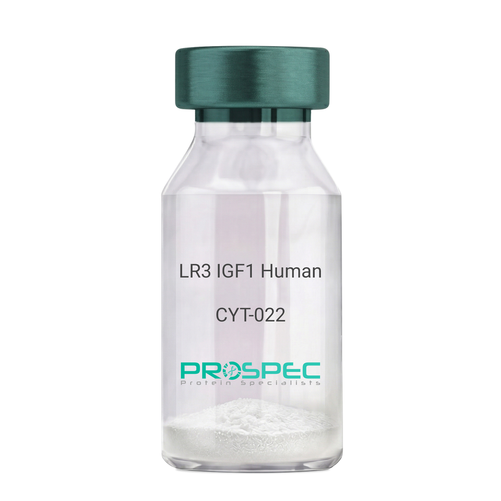
    Name :

    IGF1 LR3 Human

    Description:

    LR3 Insulin Like Growth Factor-1 Human Recombinant

    R3 IGF1, R3 IGF-1, R3IGF1, R3IGF-1, LONG IGF1, LONG IGF-1, LONG R3 IGF1, LONG R3IGF1, LONG R3 IGF-1, LONG R3IGF-1.

    Product # :

    CYT-022

    Price :

    Quantity :

    Shipping Method :

    Room Temp Icon

    Shipped at Room temp

    Add To Cart

    More Info

    • description
    • source
    • formulation
    • purity
    • biological activity
    • More Info
    • sds-page
    • Citations

    Description

    The LR3 is a long-term analog of human IGF-1, specifically designed and manufactured for mammalian cell culture to support large-scale manufacturing of recombinant biopharmaceuticals. Recombinant Human LR3 Insulin Like Growth Factor-1 produced in E.Coli is a single, non-glycosylated, polypeptide chain containing 83 amino acids and having a molecular mass of 9.1kDa.

    Source

    Escherichia Coli.

    Formulation

    Lyophilized from a 0.2µm filtered concentrated solution in 20mM PB, pH 7.2.

    Purity

    Greater than 97.0% as determined by SDS-PAGE.

    Biological Activity

    The ED50 as determined by the stimulation of protein synthesis in L6 myoblasts is less than 10ng/ml, corresponding to a specific activity of 100,000units/mg.

    sds-page

    LR3 IGF1 sds-page - Product image 1

    Citations

    More Info

    • Introduction

      IGF-1 (Insulin-like growth factor-1) is a major hormonal mediator of statural growth. Under regular circumstances, GH (growth hormone) binds to its receptor in the liver, and other tissues, and stimulates the synthesis/secretion of IGF-1. In target tissues, the Type 1 IGF receptor, that is homologous to the insulin receptor, is activated by IGF-1, leading to intracellular signaling which stimulates multiple processes leading to statural growth. IGF-1 metabolic actions are partly directed at stimulating the uptake of glucose, fatty acids, and amino acids so that metabolism supports growing tissues.

    • Synonyms

      R3 IGF1, R3 IGF-1, R3IGF1, R3IGF-1, LONG IGF1, LONG IGF-1, LONG R3 IGF1, LONG R3IGF1, LONG R3 IGF-1, LONG R3IGF-1.

    • Physical Appearance

      Sterile Filtered White lyophilized (freeze-dried) powder.

    • Stability

      Lyophilized LR3 IGF1 although stable at room temperature for 3 weeks, should be stored desiccated below -18°C. Upon reconstitution the LR3 IGF1 should be stored at 4°C between 2-7 days and for future use below -18°C.For long term storage it is recommended to add a carrier protein (0.1% HSA or BSA).Please prevent freeze-thaw cycles.

    • Solubility

      It is recommended to reconstitute the lyophilized LR3 IGF1 in sterile 18M-cm H2O at a concentration of 100µg/ml, which can then be further diluted to other aqueous solutions.

    • Amino Acid Sequence

      MFPAMPLSSLFVNGPRTLCGAELVDALQFVCGDRGFYFNKPTGYGSSSRRAPQTGIV DECCFRSCDLRRLEMYCAPLKPAKSA.

    • Background

      What is the molecular weight/Mw of IGF1 LR3 HUMAN Protein?
      IGF1 LR3 HUMAN Protein has a total Mw of 9.1kDa.

      What is the source or expression system of IGF1 LR3 HUMAN Protein?
      Escherichia Coli.

      What is the Purity of IGF1 LR3 HUMAN Protein?
      IGF1 LR3 HUMAN Protein is >98% pure as determined by SDS-PAGE.

      What is the Biological Activity of IGF1 LR3 HUMAN Protein?
      The ED50 as determined by the stimulation of protein synthesis in L6 myoblasts is less than 10ng/ml, corresponding to a specific activity of 100,000units/mg.

      What is the amino acid sequence of IGF1 LR3 HUMAN Protein?
      MFPAMPLSSLFVNGPRTLCGAELVDALQFVCGDRGFYFNKPTGYGSSSRRAPQTGIV DECCFRSCDLRRLEMYCAPLKPAKSA.

      What applications can IGF1 LR3 HUMAN Protein be used in?
      IGF1 LR3 HUMAN Protein can probably be used in western blot, ELISA and Lateral Flow.

      What is the endotoxin level for IGF1 LR3 HUMAN Protein?
      The endotoxin level is minimal, IGF1 LR3 HUMAN Protein was purified using conventional chromatography techniques.


    ProSpec's products are furnished for LABORATORY RESEARCH USE ONLY. The product may not be used as drugs, agricultural or pesticidal products, food additives or household chemicals.

    Long R3 Igf1 Human
  • View Data Sheet

    Name :

    CCL1 Human, His

    Description:

    I-309 (CCL1) Human Recombinant, His Tag

    Small inducible cytokine A1, CCL1, T lymphocyte-secreted protein I-309, chemokine (C-C motif) ligand 1, P500, SISe, TCA3, I-309, SCYA1.

    Product # :

    CHM-254

    Price :

    Quantity :

    Shipping Method :

    Ice Icon

    Shipped with Ice Packs

    Add To Cart

    More Info

    • description
    • source
    • formulation
    • purity
    • More Info
    • SDS-PAGE
    • Citations

    Description

    I-309 Human Recombinant fused with a 21 amino acid His tag at N-terminus produced in E.Coli is a single, non-glycosylated, polypeptide chain containing 94 amino acids (24-96 a.a.) and having a molecular mass of 10.8kDa. The I-309 is purified by proprietary chromatographic techniques.

    Source

    Escherichia Coli.

    Formulation

    The I-309 solution (0.5 mg/ml) contains 20mM Tris-HCl buffer (pH 7.5), 2mM DTT, 10% glycerol and 50mM NaCl.

    Purity

    Greater than 85.0% as determined by SDS-PAGE.

    SDS-PAGE

    CCL1 Human, His-SDS-PAGE - Product image 1

    Citations

    More Info

    • Introduction

      Chemokine (C-C motif) ligand 1 (CCL1) is a small glycoprotein secreted by activated T cells that belongs to a family inflammatory cytokines known as chemokines. CCL1 attracts monocytes, NK cells, and immature B cells and dendritic cells by interacting with a cell surface chemokine receptor called CCR8. This chemokine resides in a large cluster of CC chemokines on human chromosome 17.

    • Synonyms

      Small inducible cytokine A1, CCL1, T lymphocyte-secreted protein I-309, chemokine (C-C motif) ligand 1, P500, SISe, TCA3, I-309, SCYA1.

    • Physical Appearance

      Sterile Filtered colorless solution.

    • Stability

      Store at 4°C if entire vial will be used within 2-4 weeks. Store, frozen at -20°C for longer periods of time. For long term storage it is recommended to add a carrier protein (0.1% HSA or BSA).Avoid multiple freeze-thaw cycles.

    • Amino Acid Sequence

      MGSSHHHHHH SSGLVPRGSH MKSMQVPFSR CCFSFAEQEI PLRAILCYRN TSSICSNEGL IFKLKRGKEA CALDTVGWVQ RHRKMLRHCP SKRK.

    • Background

      What is the molecular weight/Mw of CCL1 HUMAN, HIS Protein?
      CCL1 HUMAN, HIS Protein has a total Mw of 10.8kDa.

      What is the source or expression system of CCL1 HUMAN, HIS Protein?
      Escherichia Coli.

      What is the Purity of CCL1 HUMAN, HIS Protein?
      CCL1 HUMAN, HIS Protein is >85% pure as determined by SDS-PAGE.

      What is the Biological Activity of CCL1 HUMAN, HIS Protein?
      The biological functionality of CCL1 HUMAN, HIS Protein will be determined in the future.

      What is the amino acid sequence of CCL1 HUMAN, HIS Protein?
      MGSSHHHHHH SSGLVPRGSH MKSMQVPFSR CCFSFAEQEI PLRAILCYRN TSSICSNEGL IFKLKRGKEA CALDTVGWVQ RHRKMLRHCP SKRK.

      What applications can CCL1 HUMAN, HIS Protein be used in?
      CCL1 HUMAN, HIS Protein can probably be used in western blot, ELISA and Lateral Flow.

      What is the endotoxin level for CCL1 HUMAN, HIS Protein?
      The endotoxin level is minimal, CCL1 HUMAN, HIS Protein was purified using conventional chromatography techniques.

      Who supplies recombinant CCL1 HUMAN, HIS that is in stock and available to order?
      ProSpec has the protein in stock and available for immediate delivery.

      Where can I order bulk mg quantities of CCL1 HUMAN, HIS?
      ProSpec supplies bulk mg quantities at affordable prices that meets research and academic budgets.

    ProSpec's products are furnished for LABORATORY RESEARCH USE ONLY. The product may not be used as drugs, agricultural or pesticidal products, food additives or household chemicals.

    I 309 Human His
  • View Data Sheet

    Name :

    EFNA1 Human, HEK

    Description:

    Ephrin A1 Human Recombinant, HEK

    Ephrin-A1, EPLG1, TNFAIP4, LERK1, EFL1, ECKLG, EPH-related receptor tyrosine kinase ligand 1, Immediate early response protein B61, Tumor necrosis factor alpha-induced protein 4, TNF alpha-induced protein 4, ligand of eph-related kinase 1, tumor necrosis factor, alpha-induced protein 4.

    Product # :

    PRO-2477

    Price :

    Quantity :

    Shipping Method :

    Room Temp Icon

    Shipped at Room temp

    Add To Cart

    More Info

    • description
    • source
    • formulation
    • purity
    • More Info
    • Citations

    Description

    EFNA1 Human Recombinant produced in HEK293 cells is a single, glycosylated polypeptide chain (a.a 19-182) containing 170 amino acids including a 6 a.a C-terminal His tag. The total molecular mass is 20.2kDa (calculated).

    Source

    HEK293 cells.

    Formulation

    EFNA1 filtered (0.4 µm) and lyophilized from 0.5mg/ml in phosphate buffered saline, pH 7.5 containing 5 % (w/v) trehalose.

    Purity

    Greater than 95.0% as determined by SDS-PAGE.

    Citations

    More Info

    • Introduction

      EFNA1 belongs to the ephrin (EPH) family. The EPH subfamily is the biggest group of receptor protein kinases and they take part in vital nervous system function and development.

    • Synonyms

      Ephrin-A1, EPLG1, TNFAIP4, LERK1, EFL1, ECKLG, EPH-related receptor tyrosine kinase ligand 1, Immediate early response protein B61, Tumor necrosis factor alpha-induced protein 4, TNF alpha-induced protein 4, ligand of eph-related kinase 1, tumor necrosis factor, alpha-induced protein 4.

    • Physical Appearance

      Filtered White lyophilized (freeze-dried) powder.

    • Stability

      Store lyophilized protein at -20°C. Aliquot the product after reconstitution to avoid repeated freezing/thawing cycles. Reconstituted protein can be stored at 4°C for a limited period of time; it does not show any change after one week at 4°C.

    • Solubility

      It is recommended to add deionized water to prepare a working stock solution of approximately 0.5mg/ml and let the lyophilized pellet dissolve completely. EFNA1 is not sterile! Please filter the product by an appropriate sterile filter before using it in the cell culture.

    • Amino Acid Sequence

      DRHTVFWNSS NPKFRNEDYT IHVQLNDYVD IICPHYEDHS VADAAMEQYI LYLVEHEEYQ LCQPQSKDQV RWQCNRPSAK HGPEKLSEKF QRFTPFTLGK EFKEGHSYYY ISKPIHQHED RCLRLKVTVS GKITHSPQAH DNPQEKRLAA DDPEVRVLHS IGHS HHHHHH.

    ProSpec's products are furnished for LABORATORY RESEARCH USE ONLY. The product may not be used as drugs, agricultural or pesticidal products, food additives or household chemicals.

    Efna1 Protein
  • View Data Sheet

    Name :

    ISOC2 Human

    Description:

    Isochorismatase Domain Containing 2 Human Recombinant

    Isochorismatase domain-containing protein 2, mitochondrial, ISOC2, Isochorismatase Domain Containing 2.

    Product # :

    PRO-1669

    Price :

    Quantity :

    Shipping Method :

    Ice Icon

    Shipped with Ice Packs

    Add To Cart

    More Info

    • description
    • source
    • formulation
    • purity
    • More Info
    • Citations

    Description

    ISOC2 Human Recombinant produced in E.coli is a single, non-glycosylated polypeptide chain containing 170 amino acids (59-205) and having a molecular mass of 18kDa.ISOC2 is fused to a 23 amino acid His-tag at N-terminus & purified by proprietary chromatographic techniques.

    Source

    Escherichia Coli.

    Formulation

    The ISOC2 solution (0.5mg/ml) contains 20mM Tris-HCl buffer (pH 8.0), 0.4M Urea and 10% glycerol.

    Purity

    Greater than 90.0% as determined by SDS-PAGE.

    Citations

    More Info

    • Introduction

      Isochorismatase Domain Containing 2, also known as ISOC2, is a part of the isochorismatase family. SOC2 is localized in mitochondrion, cytoplasm, and nucleus. ISOC2 which interacts with CDKN2A is localizes to the nucleus in the attendance of CDKN2A.

    • Synonyms

      Isochorismatase domain-containing protein 2, mitochondrial, ISOC2, Isochorismatase Domain Containing 2.

    • Physical Appearance

      Sterile Filtered colorless solution.

    • Stability

      Store at 4°C if entire vial will be used within 2-4 weeks. Store, frozen at -20°C for longer periods of time. For long term storage it is recommended to add a carrier protein (0.1% HSA or BSA).Avoid multiple freeze-thaw cycles.

    • Amino Acid Sequence

      MGSSHHHHHH SSGLVPRGSH MGSEQYPQGL GPTVPELGTE GLRPLAKTCF SMVPALQQEL DSRPQLRSVL LCGIEAQACI LNTTLDLLDR GLQVHVVVDA CSSRSQVDRL VALARMRQSG AFLSTSEGLI LQLVGDAVHP QFKEIQKLIK EPAPDSGLLG LFQGQNSLLH.

    ProSpec's products are furnished for LABORATORY RESEARCH USE ONLY. The product may not be used as drugs, agricultural or pesticidal products, food additives or household chemicals.

    Isoc2 Human
  • View Data Sheet

    Name :

    TNF b Human, His

    Description:

    Tumor Necrosis Factor-Beta Human Recombinant, His Tag

    Lymphotoxin-alpha, LT-alpha, TNF-beta, Tumor necrosis factor ligand superfamily member 1, LTA, LT, TNFB, TNFSF1.

    Product # :

    CYT-495

    Price :

    Quantity :

    Shipping Method :

    Ice Icon

    Shipped with Ice Packs

    Add To Cart

    More Info

    • description
    • source
    • formulation
    • purity
    • biological activity
    • More Info
    • Citations

    Description

    TNF-b Human Recombinant produced in E.Coli is a single, non-glycosylated polypeptide chain containing 192 amino acids (35-205 a.a.) and having a molecular mass of 20.9kDa. TNF-b is fused to a 21 amino acid His-tag at N-terminus & purified by proprietary chromatographic techniques.

    Source

    Escherichia Coli.

    Formulation

    TNF-b protein solution (0.25mg/ml) contains 20mM Tris-HCl buffer (pH8.0), 30% glycerol, 0.1M NaCl and 1mM DTT.

    Purity

    Greater than 90% as determined by SDS-PAGE.

    Biological Activity

    Measured in a cytotoxicity assay using L929 mouse fibrosarcoma cells in the presence of the metabolic inhibitor actinomycin D. The ED50 for this effect is <0.3 ng/ml.

    Citations

    More Info

    • Introduction

      TNF-b is a potent multifunctional cytokine produced mainly by activated T and B lymphocytes. The protein has been shown to have a specific potent cytotoxic activity on tumor cells and a variety of other target cells as well as being a mediator of inflammation and immune function.

    • Synonyms

      Lymphotoxin-alpha, LT-alpha, TNF-beta, Tumor necrosis factor ligand superfamily member 1, LTA, LT, TNFB, TNFSF1.

    • Physical Appearance

      Sterile Filtered clear solution.

    • Stability

      Store at 4°C if entire vial will be used within 2-4 weeks. Store, frozen at -20°C for longer periods of time. For long term storage it is recommended to add a carrier protein (0.1% HSA or BSA).Avoid multiple freeze-thaw cycles.

    • Amino Acid Sequence

      MGSSHHHHHH SSGLVPRGSH MLPGVGLTPS AAQTARQHPK MHLAHSTLKP AAHLIGDPSK QNSLLWRANT DRAFLQDGFS LSNNSLLVPT SGIYFVYSQV VFSGKAYSPK ATSSPLYLAH EVQLFSSQYP FHVPLLSSQK MVYPGLQEPW LHSMYHGAAF QLTQGDQLST HTDGIPHLVL SPSTVFFGAF AL.

    ProSpec's products are furnished for LABORATORY RESEARCH USE ONLY. The product may not be used as drugs, agricultural or pesticidal products, food additives or household chemicals.

    Tnf Beta Human His
  • View Data Sheet

    Name :

    TNFA Mouse, Sf9

    Description:

    Tumor Necrosis Factor-alpha Mouse Recombinant, Sf9

    Tnfa, Tnfsf2, Cachectin, TNF-alpha, Tumor necrosis factor ligand superfamily member 2.

    Product # :

    CYT-912

    Price :

    Quantity :

    Shipping Method :

    Ice Icon

    Shipped with Ice Packs

    Add To Cart

    More Info

    • description
    • source
    • formulation
    • purity
    • More Info
    • Citations

    Description

    TNFA Mouse Recombinant produced in Sf9 Baculovirus cells is a single, glycosylated polypeptide chain containing 162 amino acids (80-235 a.a.) and having a molecular mass of 18kDa (Molecular size on SDS-PAGE will appear at approximately 18-28kDa).TNFA is expressed with a 6 amino acid His tag at C-Terminus and purified by proprietary chromatographic techniques.

    Source

    Sf9, Baculovirus cells.

    Formulation

    TNFA protein solution (1mg/ml) contains Phosphate buffered saline (pH7.4).

    Purity

    Greater than 90.0% as determined by SDS-PAGE.

    Citations

    More Info

    • Introduction

      Tumor necrosis factor is a cytokine involved in systemic inflammation and is a member of a group of cytokines that all stimulate the acute phase reaction. TNF is mainly secreted by macrophages.
      TNF causes apoptotic cell death, cellular proliferation, differentiation, inflammation, tumorigenesis and viral replication, TNF is also involved in lipid metabolism, and coagulation. TNF's primary role is in the regulation of immune cells.
      Dysregulation and, in particular, overproduction of TNF have been implicated in a variety of human diseases- autoimmune diseases, insulin resistance, and cancer.

    • Synonyms

      Tnfa, Tnfsf2, Cachectin, TNF-alpha, Tumor necrosis factor ligand superfamily member 2.

    • Physical Appearance

      Sterile filtered colorless solution.

    • Stability

      Store at 4°C if entire vial will be used within 2-4 weeks. Store, frozen at -20°C for longer periods of time. For long term storage it is recommended to add a carrier protein (0.1% HSA or BSA).Avoid multiple freeze-thaw cycles.

    • Amino Acid Sequence

      LRSSSQNSSD KPVAHVVANH QVEEQLEWLS QRANALLANG MDLKDNQLVV PADGLYLVYS QVLFKGQGCP DYVLLTHTVS RFAISYQEKV NLLSAVKSPC PKDTPEGAEL KPWYEPIYLG GVFQLEKGDQ LSAEVNLPKY LDFAESGQVY FGVIALHHHH HH

    ProSpec's products are furnished for LABORATORY RESEARCH USE ONLY. The product may not be used as drugs, agricultural or pesticidal products, food additives or household chemicals.

    Tnfa Mouse Sf9
  • View Data Sheet

    Name :

    TNFR Human, His

    Description:

    Tumor Necrosis Factor Receptor Type Human Recombinant, His Tag

    Tumor necrosis factor receptor superfamily member 1A, Tumor necrosis factor receptor 1, Tumor necrosis factor receptor type I, TNF-R1, TNF-RI, TNFR-I, p60, p55, CD120a, TNFRSF1A, TNFAR, TNFR1, FPF, TBP1, TNF-R, p55-R, TNFR55, TNFR60, TNF-R-I, TNF-R55, MGC19588.

    Product # :

    CYT-673

    Price :

    Quantity :

    Shipping Method :

    Ice Icon

    Shipped with Ice Packs

    Add To Cart

    More Info

    • description
    • source
    • formulation
    • purity
    • More Info
    • Citations

    Description

    TNFR Human Recombinant produced in E.Coli is a single, non-glycosylated, Polypeptide chain containing 161 amino acids fragment (41-201) having a molecular weight of 22.68kDa and fused with a 4.5kDa amino-terminal hexahistidine tag. The TNFR His Tag is purified by proprietary chromatographic techniques.

    Source

    Escherichia Coli.

    Formulation

    TNFR His Tag protein is supplied in 1xPBS, 50% glycerol.

    Purity

    Greater than 95.0% as determined by SDS-PAGE.

    Citations

    More Info

    • Introduction

      TNFR1 belongs to the TNF-receptor superfamily. TNFR1 is a receptor for TNFSF2/TNF-alpha and homotrimeric TNFSF1/lymphotoxin-alpha.
      There are 2 types of soluble TNF receptors: sTNFR-I and sTNFR-II, which act to neutralize the biological activities of TNF alpha and TNF beta. The levels of these soluble receptors seem to increase as a result of shedding of the extracellular domains of the membrane bound receptors. TNF-a, TNFR1 and TNFR2 have roles in cellular differentiation. TNFR1 and TNFR2 function in cell type-specific renal injury.
      TNFR1 is capable of signaling both cell survival and apoptosis. TNFR1-induced apoptosis requires 2 sequential signaling complexes. TNFR1 is capable of activating NF-kappaB, mediate apoptosis, and function as a regulator of inflammation. Oxidative stress promotes TNFR1 and TNFR2 self-interaction, ligand-independent and enhanced ligand-dependent TNF signaling. TNFR1 contributes to the induction of non-cytocidal TNF effects including anti-viral state and activation of the acid sphingomyelinase. Human TNFR1 has a major region which controls cell surface expression. High levels of soluble TNF receptors are found in the amniotic fluid of pregnant women.
      Germline mutations of the extracellular domains of TNFR1 are linked to the autosomal dominant periodic fever syndrome. The impaired receptor clearance is believed to be a mechanism of the disease. Familial hibernian fever (FHF) is caused by defects in TNFRSF1A gene.

    • Synonyms

      Tumor necrosis factor receptor superfamily member 1A, Tumor necrosis factor receptor 1, Tumor necrosis factor receptor type I, TNF-R1, TNF-RI, TNFR-I, p60, p55, CD120a, TNFRSF1A, TNFAR, TNFR1, FPF, TBP1, TNF-R, p55-R, TNFR55, TNFR60, TNF-R-I, TNF-R55, MGC19588.

    • Physical Appearance

      Sterile Filtered clear solution.

    • Stability

      Store at 4°C if entire vial will be used within 2-4 weeks. Store, frozen at -20°C for longer periods of time. Please avoid freeze thaw cycles.

    • Amino Acid Sequence

      DSVCPQGKYIHPQNNSICCTKCHKGTYLYNDCPGPGQ
      DTDCRECESGSFTASENHLRHCLSCSKCRKEMGQVE
      ISSCTVDRDTVCGCRKNQYRHYWSENLFQCFNCSLCL
      NGTVHLSCQEKQNTVCTCHAGFFLRENECVSCSNCKK
      SLECTKLCLPQIEN.

    ProSpec's products are furnished for LABORATORY RESEARCH USE ONLY. The product may not be used as drugs, agricultural or pesticidal products, food additives or household chemicals.

    Tnfr Human His
  • View Data Sheet

    Name :

    TNFRSF4 Mouse

    Description:

    TNF Receptor Superfamily Member 4 Mouse Recombinant

    Tumor necrosis factor receptor superfamily member 4, OX40 antigen, OX40L receptor, Txgp1, Tnfrsf4, tax-transcriptionally activated glycoprotein 1 receptor, TXGP1L, ACT35, Txgp, Ly-70, ACT3, OX4, CD134.

    Product # :

    CYT-1179

    Price :

    Quantity :

    Shipping Method :

    Ice Icon

    Shipped with Ice Packs

    Add To Cart

    More Info

    • description
    • source
    • formulation
    • purity
    • biological activity
    • More Info
    • Citations

    Description

    TNFRSF4 Mouse Recombinant produced in HEK293 cells is a single, glycosylated polypeptide chain (a.a 20-211) containing 435 amino acids and having a molecular mass of 48.6kDa.TNFRSF4 is fused to a 243 amino acid hIgG-His tag at C-terminus and purified by proprietary chromatographic techniques.

    Source

    HEK293 cells.

    Formulation

    TNFRSF4 protein (0.25mg/ml) contains Phosphate-Buffered Saline (pH 7.4) and 10% glycerol.

    Purity

    Greater than 95.0% as determined by SDS-PAGE.

    Biological Activity

    The ED50 range is ≤ 0.4 ug/ml which measured by its binding ability in a functional ELISA with Mouse OX40 Ligand/TNFSF4.

    Citations

    More Info

    • Introduction

      TNFRSF4, also known as TNF Receptor Superfamily Member 4, is a T cell co-stimulatory molecule which belongs to the TNF receptor superfamily. TNFRSF4 coordinates with other co-stimulatory substances such as CD28, CD40, CD30, CD27 and 4-1BB to control the activation of the immune response. TNFRSF4 takes a vital part in antigen-specific T cell expansion as well as survival. TNFRSF4 is up-regulated on CD4+ and CD8+ T cells upon engagement of the TCR by antigen presenting cells along with co-stimulation by CD40-CD40 Ligand and CD28-B7. In addition, TNFRSF4 regulates cytokine production from T cells, antigen presenting cells, natural killer cells and natural killer cells. TNFRSF4 regulates cytokine receptor signaling.

    • Synonyms

      Tumor necrosis factor receptor superfamily member 4, OX40 antigen, OX40L receptor, Txgp1, Tnfrsf4, tax-transcriptionally activated glycoprotein 1 receptor, TXGP1L, ACT35, Txgp, Ly-70, ACT3, OX4, CD134.

    • Physical Appearance

      Sterile Filtered colorless solution.

    • Stability

      Store at 4°C if entire vial will be used within 2-4 weeks. Store, frozen at -20°C for longer periods of time. For long term storage it is recommended to add a carrier protein (0.1% HSA or BSA).Avoid multiple freeze-thaw cycles.

    • Amino Acid Sequence

      DGSMVTARRL NCVKHTYPSG HKCCRECQPG HGMVSRCDHT RDTLCHPCET GFYNEAVNYD TCKQCTQCNH RSGSELKQNC TPTQDTVCRC RPGTQPRQDS GYKLGVDCVP CPPGHFSPGN NQACKPWTNC TLSGKQTRHP ASDSLDAVCE DRSLLATLLW ETQRPTFRPT TVQSTTVWPR TSELPSPPTL VTPEGPLEPK SCDKTHTCPP CPAPELLGGP SVFLFPPKPK DTLMISRTPE VTCVVVDVSH EDPEVKFNWY VDGVEVHNAK TKPREEQYNS TYRVVSVLTV LHQDWLNGKE YKCKVSNKAL PAPIEKTISK AKGQPREPQV YTLPPSRDEL TKNQVSLTCL VKGFYPSDIA VEWESNGQPE NNYKTTPPVL DSDGSFFLYS KLTVDKSRWQ QGNVFSCSVM HEALHNHYTQ KSLSLSPGKH HHHHH

    ProSpec's products are furnished for LABORATORY RESEARCH USE ONLY. The product may not be used as drugs, agricultural or pesticidal products, food additives or household chemicals.

    Tnfrsf4 Mouse
  • View Data Sheet

    Name :

    L Selectin Human

    Description:

    L-selectin Human Recombinant

    L-selectin, Lymph node homing receptor, Leukocyte adhesion molecule 1, LAM-1, Leukocyte surface antigen Leu-8, TQ1, gp90-MEL, Leukocyte-endothelial cell adhesion molecule 1, LECAM1, CD62 antigen-like family member L, CD62L antigen, LAM1, LNHR, LSEL, CD62L, LYAM1, Leu-8, PLNHR, hLHRc, Lyam-1, L-Sel.

    Product # :

    PRO-381

    Price :

    Quantity :

    Shipping Method :

    Ice Icon

    Shipped with Ice Packs

    Add To Cart

    More Info

    • description
    • source
    • formulation
    • purity
    • More Info
    • Citations

    Description

    L-Selectin Human Recombinant is expressed in E. coli containing 294 amino acids 39-332 fused to an amino terminal hexahistidine tag, having a total molecular weight of 37.55kDa.

    Source

    Escherichia Coli.

    Formulation

    L-Sel is supplied in 1x PBS and 50% Glycerol.

    Purity

    Greater than 95% as determined by SDS-PAGE.
    Single band on Western Blot.

    Citations

    More Info

    • Introduction

      L-Selectin belongs to a family of divalent cation-dependent carbohydrate-binding glycoproteins or adhesion molecules. The L-Selectin molecule is composed of various domains: one homologous to lectins, one to epidermal growth factor, and two to the consensus repeat units found in C3/C4 binding proteins.
      L-selectin is expressed constitutively on lymphocytes, monocytes and granulocytes and interacts specifically with carbohydrate groups on activated endothelial cells. L-Selectin may be shed by proteolytic cleavage and circulating levels in biological fluids may be used as an indicator of various pathological conditions. L-Selectin is cleaved by ADAM17.
      L-selectin works as a "homing receptor" for leukocytes to enter secondary lymphoid tissues via the high endothelial venules. Ligands present on endothelial cells will attach to leukocytes expressing L-selectin, which causes the leukocytes to become localized at that juncture. The receptor is also located on the cell surfaces of "naive" T cells, which have not yet encountered their particular antigen. This surface expression is lost following the cells activation.

    • Synonyms

      L-selectin, Lymph node homing receptor, Leukocyte adhesion molecule 1, LAM-1, Leukocyte surface antigen Leu-8, TQ1, gp90-MEL, Leukocyte-endothelial cell adhesion molecule 1, LECAM1, CD62 antigen-like family member L, CD62L antigen, LAM1, LNHR, LSEL, CD62L, LYAM1, Leu-8, PLNHR, hLHRc, Lyam-1, L-Sel.

    • Physical Appearance

      Sterile filtered colorless solution.

    • Stability

      Store at 4°C if entire vial will be used within 1-2 weeks. Store, frozen at -20°C for longer periods of time. Avoid multiple freeze-thaw cycles.

    • Applications

      L-Selectin can be used directly as a positive control in Western blotting, ELISA, immunoprecipitation and other immunological experiments.
      The biological activity of this product has not yet been tested.

    ProSpec's products are furnished for LABORATORY RESEARCH USE ONLY. The product may not be used as drugs, agricultural or pesticidal products, food additives or household chemicals.

    L Selectin Human
  • View Data Sheet

    Name :

    CD79A Human

    Description:

    CD79A Human Recombinant

    CD79a Molecule, Membrane-Bound Immunoglobulin-Associated Protein, CD79A Antigen (Immunoglobulin-Associated Alpha), CD79a Molecule, Immunoglobulin-Associated Alpha, Surface IgM-Associated Protein, MB-1 Membrane Glycoprotein, Ig-Alpha, IGA, B-Cell Antigen Receptor Complex-Associated Protein Alpha Chain, CD79a Antigen, MB-1, MB1.

    Product # :

    PRO-2468

    Price :

    Quantity :

    Shipping Method :

    Room Temp Icon

    Shipped at Room temp

    Add To Cart

    More Info

    • description
    • source
    • formulation
    • purity
    • More Info
    • Citations

    Description

    CD79A Human Recombinant produced in Sf9 Insect cells is a single, glycosylated polypeptide chain containing 119 amino acids (33-143a.a.) and having a molecular mass of 13.6kDa (Molecular size on SDS-PAGE will appear at approximately 18-28kDa).CD79A is expressed with a 8 amino acid His tag at C-Terminus and purified by proprietary chromatographic techniques.

    Source

    Sf9, Insect cells.

    Formulation

    CD79A protein solution (1mg/ml) contains Phosphate Buffered Saline (pH 7.4) and 10% glycerol.

    Purity

    Greater than 95.0% as determined by SDS-PAGE.

    Citations

    More Info

    • Introduction

      CD79A is a signal-transducing part of the B-cell receptor which plays various roles in B cell development and function. CD79A is also needed for BCR surface expression and for efficient differentiation of pro-B and pre-B-cells. CD79A plays an important role in the tumor promoting effects of myeloid cells, and is a novel target for cancer therapy.

    • Synonyms

      CD79a Molecule, Membrane-Bound Immunoglobulin-Associated Protein, CD79A Antigen (Immunoglobulin-Associated Alpha), CD79a Molecule, Immunoglobulin-Associated Alpha, Surface IgM-Associated Protein, MB-1 Membrane Glycoprotein, Ig-Alpha, IGA, B-Cell Antigen Receptor Complex-Associated Protein Alpha Chain, CD79a Antigen, MB-1, MB1.

    • Stability

      Store at 4°C if entire vial will be used within 2-4 weeks. Store, frozen at -20°C for longer periods of time. For long term storage it is recommended to add a carrier protein (0.1% HSA or BSA).Avoid multiple freeze-thaw cycles.

    • Amino Acid Sequence

      LWMHKVPASL MVSLGEDAHF QCPHNSSNNA NVTWWRVLHG NYTWPPEFLG PGEDPNGTLI IQNVNKSHGG IYVCRVQEGN ESYQQSCGTY LRVRQPPPRP FLDMGEGTKN RLEHHHHHH.

    ProSpec's products are furnished for LABORATORY RESEARCH USE ONLY. The product may not be used as drugs, agricultural or pesticidal products, food additives or household chemicals.

    Cd79A Human
  • View Data Sheet

    Name :

    GKN3P Human

    Description:

    Gastrokine 3 Human Recombinant

    Gastrokine 3 Pseudogene, Gastrokine-3, GKN3P.

    Product # :

    PRO-2033

    Price :

    Quantity :

    Shipping Method :

    Ice Icon

    Shipped with Ice Packs

    Add To Cart

    More Info

    • description
    • source
    • formulation
    • purity
    • More Info
    • Citations

    Description

    GKN3P Human Recombinant produced in E.Coli is a single, non-glycosylated, polypeptide chain (Met21-Leu181) containing 171 amino acids including a 10 aa His tag at N-terminus. The total calculated molecular mass is 19.4kDa.

    Source

    Escherichia Coli.

    Formulation

    GKN3P filtered (0.4µm) solution at a concentration of 0.3mg/ml in 0.03M acetate buffer, pH 4.0 and 20% glycerol.

    Purity

    Greater than 95.0% as determined by SDS-PAGE.

    Citations

    More Info

    • Introduction

      Gastrokine 3 (GKN3P) is a Pseudogene, which May inhibit gastric epithelial cell proliferation.

    • Synonyms

      Gastrokine 3 Pseudogene, Gastrokine-3, GKN3P.

    • Physical Appearance

      Filtered colorless solution.

    • Stability

      Store at 4°C if entire vial will be used within 2-4 weeks. Store, frozen at -20°C for longer periods of time. For long term storage it is recommended to add a carrier protein (0.1% HSA or BSA).Avoid multiple freeze-thaw cycles.

    • Amino Acid Sequence

      MKHHHHHHASMMNIRFNHPL YGSFGTQIIH IGAFQGMVSI RDNNIFSEWD GILDYKNALL VAKVFNKMAC VLARMDKAVF PSLDDISKAL DKQAFKYYPS TRGLTYTVLP SWVKNLAQYG KPIKNMCRDD PTYFAQQQKE GTALAIDSNS CFEIQLLSFM GLFICGETPG L.

    ProSpec's products are furnished for LABORATORY RESEARCH USE ONLY. The product may not be used as drugs, agricultural or pesticidal products, food additives or household chemicals.

    Gkn3P Human
  • View Data Sheet

    Name :

    Procalcitonin Rhesus

    Description:

    Procalcitonin Rhesus Recombinant

    Calcitonin.

    Product # :

    HOR-016

    Price :

    Quantity :

    Shipping Method :

    Room Temp Icon

    Shipped at Room temp

    Add To Cart

    More Info

    • description
    • source
    • formulation
    • purity
    • More Info
    • Citations

    Description

    Procalcitonin Rhesus Recombinant produced in E.Coli is a single, non-glycosylated, polypeptide chain (Ala26-Asn140) containing 125 amino acids including a 10 aa His tag at N-terminus. The total calculated molecular mass is 14kDa.

    Source

    Escherichia Coli.

    Formulation

    Procalcitonin was filtered (0.4µm) and lyophilized from 0.5mg/ml solution in 20mM Tris buffer and 50mM NaCl, pH 7.5.

    Purity

    Greater than 95.0% as determined by SDS-PAGE.

    Citations

    More Info

    • Introduction

      Procalcitonin is a peptide hormone mainly produced by the C cells of the thyroid and certain endocrine cells of the lung. Under normal expression conditions, procalcitonin is immediately cleaved into three specific fragments, an N terminal residue, calcitonin and katacalcin. Levels of unprocessed procalcitonin rise significantly after bacterial infection, trauma or shock.

    • Synonyms

      Calcitonin.

    • Physical Appearance

      Filtered White lyophilized (freeze-dried) powder.

    • Stability

      Store lyophilized protein at -20°C. Aliquot the product after reconstitution to avoid repeated freezing/thawing cycles. Reconstituted protein can be stored at 4°C for a limited period of time; it does not show any change after two weeks at 4°C.

    • Solubility

      It is recommended to add deionized water to prepare a working stock solution of approximately 0.5 mg/ml and let the lyophilized pellet dissolve completely. Procalcitonin is not sterile! Please filter the product by an appropriate sterile filter before using it in the cell culture.

    • Amino Acid Sequence

      MKHHHHHHASAPFRSALESS PDPATLSEEE ARLLLAALVQ DYVQMKASEL EQEQETEGSS LDSPRSKRCG NLSTCMLGTY TQDFNKFHTF PQTAIGVGAP GKKRDMSSDL ERNRRRYVSM PQDAN.

    ProSpec's products are furnished for LABORATORY RESEARCH USE ONLY. The product may not be used as drugs, agricultural or pesticidal products, food additives or household chemicals.

    Procalcitonin Rhesus
  • View Data Sheet

    Name :

    Pro-IGF2 Human

    Description:

    Pro-Insulin Like Growth Factor-2 Human Recombinant

    Pro-Insulin Like Growth Factor-2, pro-IGF2.

    Product # :

    CYT-110

    Price :

    Quantity :

    Shipping Method :

    Room Temp Icon

    Shipped at Room temp

    Add To Cart

    More Info

    • description
    • source
    • formulation
    • purity
    • biological activity
    • More Info
    • Citations

    Description

    Pro-IGF2 Human Recombinant produced in HEK cells is a glycosylated monomer, contains 157 a.a. (24-180) having a total molecular weight of 25kDa.The Pro-IGF2 contains a C-terminal propeptide (E peptide) Arg92 to Lys180 and is purified by proprietary chromatographic techniques.

    Source

    HEK.

    Formulation

    The Pro-IGF2 was lyophilized in 50mM Sodium Acetate pH 4.5 and 350mM NaCl.

    Purity

    Greater than 90% as obsereved by SDS-PAGE.

    Biological Activity

    The specific activity was determined by the dose-dependent stimulation of the proliferation of MCF-7 cells (human breast cancer cell line) and is typically 2-8ng/ml.

    Citations

    More Info

    • Introduction

      IGF-2 is a member of the insulin family of polypeptide growth factors that is involved in development and growth. It is an imprinted gene and is expressed only from the paternally inherited allele. It is a candidate gene for eating disorders. There is a read-through, INS-IGF2, which aligns to this gene at the 3' region and to the upstream INS gene at the 5' region. Two alternatively spliced transcript variants encoding the same protein have been found for this gene.

    • Synonyms

      Pro-Insulin Like Growth Factor-2, pro-IGF2.

    • Physical Appearance

      Sterile Filtered White lyophilized (freeze-dried) powder.

    • Stability

      Lyophilized Pro-IGF2 although stable at room temperature for 3 weeks, should be stored desiccated below -18°C. Upon reconstitution Pro-IGF2 should be stored at 4°C between 2-7 days and for future use below -18°C.For long term storage it is recommended to add a carrier protein (0.1% HSA or BSA).Please prevent freeze-thaw cycles.

    • Solubility

      It is recommended to reconstitute the lyophilized Pro-IGF2 in sterile PBS containing 0.1% endotoxin-free recombinant HSA.

    • Background

      What is the molecular weight/Mw of PRO-IGF2 HUMAN Protein?
      PRO-IGF2 HUMAN Protein has a total Mw of 25kDa.

      What is the source or expression system of PRO-IGF2 HUMAN Protein?
      HEK.

      What is the Purity of PRO-IGF2 HUMAN Protein?
      PRO-IGF2 HUMAN Protein is >90% pure as determined by SDS-PAGE.

      What is the Biological Activity of PRO-IGF2 HUMAN Protein?
      The specific activity was determined by the dose-dependent stimulation of the proliferation of MCF-7 cells (human breast cancer cell line) and is typically 2-8ng/ml.

      What applications can PRO-IGF2 HUMAN Protein be used in?
      PRO-IGF2 HUMAN Protein can probably be used in western blot, ELISA and Lateral Flow.

      What is the endotoxin level for PRO-IGF2 HUMAN Protein?
      The endotoxin level is minimal, PRO-IGF2 HUMAN Protein was purified using conventional chromatography techniques.


    ProSpec's products are furnished for LABORATORY RESEARCH USE ONLY. The product may not be used as drugs, agricultural or pesticidal products, food additives or household chemicals.

    Pro Igf2 Human
  • 1
  • …
  • 32
  • 33
  • 34
  • 35
  • 36
  • …
  • 42
Your browser does not support the video tag.

Products

  • Cytokines
  • Growth Factors
  • Chemokines
  • CD Antigens
  • Neurotrophins
  • Hormones
  • Enzymes
  • Viral Antigens
  • Recombinant Proteins
  • Natural Proteins
  • Monoclonal Antibodies
  • Polyclonal Antibodies
  • Thermal Packaging

About

  • Company
  • Founder
  • Contribution
  • Contact Us
  • My Account

Ordering

  • Ordering Method
  • Ordering - Credit Card
  • Ordering PO
  • Shipping Fees
  • FAQ's

Legal

  • Terms & Conditions
  • Privacy
  • Site Map
  • info@prospecbio.com
  • Facebook
  • Instagram
  • Linkedin Profile

© 2026 ProSpec-Tany TechnoGene Ltd. All Rights Reserved.

  • Choosing a selection results in a full page refresh.
  • Opens in a new window.