Search results
1000 results found for “cyclin”
Name
Description
Product #
Price
Quantity
Shipping Method
- View Data Sheet
Name :
IGF1 PAT6F8AT AntibodyDescription:
Insulin-Like Growth Factor-1 Clone PAT6F8AT, Mouse Anti Human
Somatomedin C, IGF-I, IGFI, IGF1, IGF-IA, Mechano growth factor, MGF.
Product # :
ANT-538Price :
Quantity :
Shipping Method :
Shipped with Ice Packs
More Info
- formulation
- More Info
Formulation
1mg/ml containing PBS, pH-7.4, 10% Glycerol and 0.02% Sodium Azide.
More Info
-
Introduction
The somatomedins, or insulin-like growth factors (IGFs), comprise a family of peptides that play important roles in mammalian growth and development. IGF1 mediates many of the growth-promoting effects of growth hormone (GH; MIM 139250). Early studies showed that growth hormone did not directly stimulate the incorporation of sulfate into cartilage, but rather acted through a serum factor, termed 'sulfation factor,' which later became known as 'somatomedin' (Daughaday et al., 1972). Three main somatomedins have been characterized: somatomedin C (IGF1), somatomedin A (IGF2; MIM 147470), and somatomedin B (MIM 193190) (Rotwein, 1986; Rosenfeld, 2003).
-
Synonyms
Somatomedin C, IGF-I, IGFI, IGF1, IGF-IA, Mechano growth factor, MGF.
-
Physical Appearance
Sterile filtered colorless solution.
-
Immunogen
Anti-human IGF1 mAb, clone PAT6F8AT, is derived from hybridization of mouse F0 myeloma cells with spleen cells from BALB/c mice immunized with a recombinant human IGF1 protein 49-118 amino acids purified from E. coli.
-
Ig Subclass
Mouse IgG2b heavy chain and k light chain.
-
Clone
PAT6F8AT.
-
Applications
HSPA5 antibody has been tested by ELISA, Western blot analysis to assure specificity and reactivity. Since application varies, however, each investigation should be titrated by the reagent to obtain optimal results. Recommended starting dilution is 1:1000.
-
Type
Mouse Anti Human Monoclonal.
-
Storage Procedures
For periods up to 1 month store at 4°C, for longer periods of time, store at -20°C. Prevent freeze thaw cycles.
-
Purification Method
IGF1 antibody was purified from mouse ascitic fluids by protein-A affinity chromatography.
ProSpec's products are furnished for LABORATORY RESEARCH USE ONLY. The product may not be used as drugs, agricultural or pesticidal products, food additives or household chemicals.
- View Data Sheet
Name :
KCTD15 PAT2B11AT AntibodyDescription:
Potassium channel tetramerisation domain containing 15, Clone PAT2B11AT, Mouse Anti Human
BTB/POZ domain-containing protein KCTD15, Potassium channel tetramerisation domain containing 15, KCTD15, MGC2628, MGC25497.
Product # :
ANT-696Price :
Quantity :
Shipping Method :
Shipped with Ice Packs
More Info
- formulation
- More Info
Formulation
1mg/ml containing PBS, pH-7.4, 10% Glycerol and 0.02% Sodium Azide.
More Info
-
Introduction
KCTD15 protein is encoded in humans by the KCTD15 gene. KCTD15 is expressed at a high level in the brain and the hypothalamus. The potassium channel KCTD15 was identified as a genetic loci linked to higher than normal BMI in humans along with genes such as GNPDA2, MTCH2, FTO, and TMEM18. SNPs (Single nucleotide polymorphisms) in non-diabetic and diabetic patients showed that FTO was most strongly associated with obesity while MTCH2 and GNPDA2 were still notably associated with higher than normal BMI levels.
-
Synonyms
BTB/POZ domain-containing protein KCTD15, Potassium channel tetramerisation domain containing 15, KCTD15, MGC2628, MGC25497.
-
Immunogen
Anti-human KCTD15 mAb, is derived from hybridization of mouse F0 myeloma cells with spleen cells from BALB/c mice immunized with a recombinant human KCTD15 protein 1-159 amino acids purified from E. coli.
-
Ig Subclass
Mouse IgG2b heavy chain and k light chain.
-
Clone
PAT2B11AT.
-
Applications
KCTD15 antibody has been tested by ELISA, Western blot analysis, Flow cytometry and ICC/IF to assure specificity and reactivity. Since application varies, however, each investigation should be titrated by the reagent to obtain optimal results.
-
Type
Mouse Anti Human Monoclonal.
-
Storage Procedures
For periods up to 1 month store at 4°C, for longer periods of time, store at -20°C. Prevent freeze thaw cycles.
-
Purification Method
KCTD15 antibody was purified from mouse ascitic fluids by protein-G affinity chromatography.
ProSpec's products are furnished for LABORATORY RESEARCH USE ONLY. The product may not be used as drugs, agricultural or pesticidal products, food additives or household chemicals.
- View Data Sheet
Name :
APRIL HumanDescription:
APRIL Human Recombinant
Tumor necrosis factor ligand superfamily member 13, A proliferation-inducing ligand, APRIL, TNF- and APOL-related leukocyte expressed ligand 2, TALL-2, TNF-related death ligand 1, TRDL-1, CD256, TNFSF13, TALL2, ZTNF2, UNQ383/PRO715.
Product # :
CYT-815Price :
Quantity :
Shipping Method :
Shipped with Ice Packs
More Info
- description
- source
- formulation
- purity
- More Info
- sds-page
Description
APRIL Human Recombinant produced in E.coli is a single, non-glycosylated polypeptide chain containing 159 amino acids (105-247) and having a molecular mass of 17.6kDa.APRIL is fused to a 16 amino acid T7-tag at N-terminus and purified by proprietary chromatographic techniques.
Source
Escherichia Coli.
Formulation
APRIL protein solution (1mg/ml) containing 20mM Tris-HCl buffer (pH 8.0), 0.4M UREA and 10% glycerol.
Purity
Greater than 85.0% as determined by SDS-PAGE.
sds-page
More Info
-
Introduction
APRIL which is a part of the TNF ligand superfamily (TNFSF13) is a type II transmembrane protein. Normally, APRIL expression is low in tissues, but is elevated in numerous types of tumors and transformed cell lines.
-
Synonyms
Tumor necrosis factor ligand superfamily member 13, A proliferation-inducing ligand, APRIL, TNF- and APOL-related leukocyte expressed ligand 2, TALL-2, TNF-related death ligand 1, TRDL-1, CD256, TNFSF13, TALL2, ZTNF2, UNQ383/PRO715.
-
Physical Appearance
Sterile filtered colorless solution.
-
Stability
Store at 4°C if entire vial will be used within 2-4 weeks. Store, frozen at -20°C for longer periods of time. For long term storage it is recommended to add a carrier protein (0.1% HSA or BSA).Avoid multiple freeze-thaw cycles.
-
Amino Acid Sequence
MASMTGGQQM GRGSHMAVLT QKQKKQHSVL HLVPINATSK DDSDVTEVMW QPALRRGRGL QAQGYGVRIQ DAGVYLLYSQ VLFQDVTFTM GQVVSREGQG RQETLFRCIR SMPSHPDRAY NSCYSAGVFH LHQGDILSVI IPRARAKLNL SPHGTFLGL.
-
Background
APRIL Human Recombinant: Expanding the Horizons of Immunotherapy
Abstract:
APRIL (A Proliferation-Inducing Ligand) is a promising molecule within the tumor necrosis factor (TNF) superfamily that plays a pivotal role in immune regulation. This research paper provides an overview of APRIL human recombinant, exploring its potential applications in immunotherapy. Understanding the mechanisms and therapeutic implications of APRIL holds promise in the field of immune-related disorders. This article presents a concise analysis of APRIL, highlighting its potential as a therapeutic target.Introduction:
Immunotherapy has revolutionized the treatment of various diseases by harnessing the power of the immune system. APRIL, a member of the TNF superfamily, has emerged as a potential candidate for immunotherapeutic interventions. This paper provides an overview of APRIL, shedding light on its structure, function, and potential applications in immunotherapy.APRIL Structure and Function:
APRIL is a transmembrane protein that can be proteolytically cleaved, leading to the generation of soluble forms. It interacts with its receptors, such as BCMA and TACI, to regulate immune responses. APRIL influences B-cell activation, proliferation, and antibody production, making it a compelling target for immunotherapeutic strategies.Immunotherapeutic Applications of APRIL Human Recombinant:
APRIL human recombinant holds great potential in immunotherapy. By modulating APRIL activity, it may be possible to enhance immune responses against cancer cells or dampen immune dysregulation in autoimmune disorders. Additionally, APRIL-based therapeutics could be developed to target specific immune cell populations or enhance the efficacy of existing immunotherapies.Challenges and Future Directions:
Although APRIL shows promise, there are challenges to overcome. Further research is needed to elucidate the precise mechanisms of APRIL-mediated immune regulation and identify optimal therapeutic approaches. Additionally, safety considerations and potential side effects must be thoroughly evaluated.Conclusion:
APRIL human recombinant represents a valuable tool for advancing immunotherapy. Understanding the structure, function, and therapeutic potential of APRIL opens up new avenues for treating immune-related disorders. Continued research and development in this field have the potential to revolutionize the landscape of immunotherapy, improving patient outcomes and expanding the possibilities for personalized medicine.What is the molecular weight/Mw of APRIL Protein?
APRIL Protein has a total Mw of 17.6kDa.
What is the source or expression system of APRIL Protein?
Escherichia Coli.
What is the Purity of APRIL Protein?
APRIL Protein is >85% pure as determined by SDS-PAGE.
What is the Biological Activity of APRIL Protein?
The biological functionality of APRIL Protein will be determined in the future.
What is the amino acid sequence of APRIL Protein?
MASMTGGQQM GRGSHMAVLT QKQKKQHSVL HLVPINATSK DDSDVTEVMW QPALRRGRGL QAQGYGVRIQ DAGVYLLYSQ VLFQDVTFTM GQVVSREGQG RQETLFRCIR SMPSHPDRAY NSCYSAGVFH LHQGDILSVI IPRARAKLNL SPHGTFLGL.
What applications can APRIL Protein be used in?
APRIL Protein can probably be used in western blot, ELISA and Lateral Flow.
What is the endotoxin level for APRIL Protein?
The endotoxin level is minimal, APRIL Protein was purified using conventional chromatography techniques.
ProSpec's products are furnished for LABORATORY RESEARCH USE ONLY. The product may not be used as drugs, agricultural or pesticidal products, food additives or household chemicals.
- View Data Sheet
Name :
CNOT8 HumanDescription:
CCR4-NOT Transcription Complex, Subunit 8 Human Recombinant
CCR4-NOT transcription complex subunit 8, CAF1, CALIF, hCAF1, POP2, CAF1-like protein, CAF2, CCR4-associated factor 8, Caf1b, CNOT8.
Product # :
PRO-1517Price :
Quantity :
Shipping Method :
Shipped with Ice Packs
More Info
- description
- source
- formulation
- purity
- More Info
Description
CNOT8 Human Recombinant produced in E.coli is a single, non-glycosylated polypeptide chain containing 315 amino acids (1-292) and having a molecular mass of 35.9 kDa.CNOT8 is fused to a 23 amino acid His-tag at N-terminus & purified by proprietary chromatographic techniques.
Source
Escherichia Coli.
Formulation
The CNOT8 solution (0.25mg/ml) contains 20mM Tris-HCl buffer (pH 8.0), 0.15M NaCl, 10% glycerol and 1mM DTT.
Purity
Greater than 85.0% as determined by SDS-PAGE.
More Info
-
Introduction
CCR4-NOT Transcription Complex, Subunit 8 (CNOT8) is a part of the CAF1 family. CNOT8 is a ubiquitous transcription factor necessary for a varied set of processes and functions as part of the CCR-NOT complex. The CCR4-NOT complex which functions as general transcription regulation complex binds BTG2 and holds CHAF1A, CHAF1B, CNOT1, CNOT2, CNOT3, CNOT4, CNOT6 and CNOT8
-
Synonyms
CCR4-NOT transcription complex subunit 8, CAF1, CALIF, hCAF1, POP2, CAF1-like protein, CAF2, CCR4-associated factor 8, Caf1b, CNOT8.
-
Physical Appearance
Sterile Filtered colorless solution.
-
Stability
Store at 4°C if entire vial will be used within 2-4 weeks. Store, frozen at -20°C for longer periods of time. For long term storage it is recommended to add a carrier protein (0.1% HSA or BSA).Avoid multiple freeze-thaw cycles.
-
Amino Acid Sequence
MGSSHHHHHH SSGLVPRGSH MGSMPAALVE NSQVICEVWA SNLEEEMRKI REIVLSYSYI AMDTEFPGVV VRPIGEFRSS IDYQYQLLRC NVDLLKIIQL GLTFTNEKGE YPSGINTWQF NFKFNLTEDM YSQDSIDLLA NSGLQFQKHE EEGIDTLHFA ELLMTSGVVL CDNVKWLSFH SGYDFGYMVK LLTDSRLPEE EHEFFHILNL FFPSIYDVKY LMKSCKNLKG GLQEVADQLD LQRIGRQHQA GSDSLLTGMA FFRMKELFFE DSIDDAKYCG RLYGLGTGVA QKQNEDVDSA QEKMSILAII NNMQQ.
ProSpec's products are furnished for LABORATORY RESEARCH USE ONLY. The product may not be used as drugs, agricultural or pesticidal products, food additives or household chemicals.
- View Data Sheet
Description:
B-Cell Leukemia/Lymphoma 2 Human Recombinant (–BH1)
Apoptosis regulator Bcl-2, BCL2, B-cell CLL/lymphoma 2, Bcl-2.
Product # :
PRO-631Price :
Quantity :
Shipping Method :
Shipped at Room temp
More Info
- description
- source
- formulation
- purity
- More Info
Description
Bcl-2 Des BH1 domain (136-155 residues) Human Recombinant produced in E.Coli is a single, non-glycosylated polypeptide chain containing 198 amino acids 1-135 and 156-218.The Bcl-2 is expressed as His-Tag fusion protein and purified by proprietary chromatographic techniques.
Source
Escherichia Coli.
Formulation
The protein contains 10mM Tris-HCL pH-8, 1mM EDTA and 250mM NaCl.
Purity
Greater than 95.0% as determined by(a) Analysis by RP-HPLC.
(b) Analysis by SDS-PAGE.More Info
-
Introduction
BCL2 gene encodes an integral outer mitochondrial membrane protein that blocks the apoptotic death of some cells such as lymphocytes. Constitutive expression of BCL2, such as in the case of translocation of BCL2 to Ig heavy chain locus, is thought to be the cause of follicular lymphoma. Two transcript variants, produced by alternate splicing, differ in their C-terminal ends.
-
Synonyms
Apoptosis regulator Bcl-2, BCL2, B-cell CLL/lymphoma 2, Bcl-2.
-
Physical Appearance
Sterile Filtered White lyophilized (freeze-dried) powder.
-
Stability
Lyophilized Bcl-2 although stable at room temperature for 3 weeks, should be stored desiccated below -18°C. Upon reconstitution Bcl-2 should be stored at 4°C between 2-7 days and for future use below -18°C.For long term storage it is recommended to add a carrier protein (0.1% HSA or BSA).Please prevent freeze-thaw cycles.
-
Solubility
Suspend Bcl-2 in 100?l of 0.5M Acetic acid, over night at 4°C.
Dilute 10 fold into selected buffer system.
BCL-2 has tendency to form intramolecular disulfide bond, 5mM DTT is recommended in assay buffer. When running SDS-PAGE gel, 10mM DTT is recommended. -
Applications
Input marker or positive control (Western Blotting).
Function study.
ProSpec's products are furnished for LABORATORY RESEARCH USE ONLY. The product may not be used as drugs, agricultural or pesticidal products, food additives or household chemicals.
- View Data Sheet
Name :
hCG ProteinDescription:
Chorionic Gonadotropin Human
Chorionic gonadotropin, hCG, CG.
Product # :
HOR-250Price :
Quantity :
Shipping Method :
Shipped at Room temp
More Info
- description
- source
- formulation
- biological activity
- More Info
- Activity Assay
Description
Human Chorionic Gonadotropin is produced from a sterile preparation of placental glucoprotein urine of pregnant women having a total molecular mass of 36,700 Dalton. The hCG consists of 237 amino acids, a chain-92 amino acids and b chain-145 amino acids. The hCG is purified by proprietary chromatographic techniques.
Source
Urine of pregnant women.
Formulation
The hCG was lyophilized with no additives.
Biological Activity
The activity was found to be 5212IU/mg.
Activity Assay
More Info
-
Introduction
Human chorionic gonadotropin (hCG) is a peptide hormone produced in pregnancy, that is made by the embryosoon after conception and later by the syncytiotrophoblast(part of the placenta). Its role is to prevent the disintegration of the corpus luteumof the ovaryand thereby maintain progesterone production that is critical for a pregnancy in humans. hCG may have additional functions, for instance it is thought that it affects the immune tolerance of the pregnancy. Early pregnancy testing generally is based on the detection or measurement of hCG.
-
Synonyms
Chorionic gonadotropin, hCG, CG.
-
Physical Appearance
Sterile Filtered White lyophilized (freeze-dried) powder.
-
Stability
Lyophilized hCG although stable at room temperature for 3 weeks, should be stored desiccated below -18°C. Upon reconstitution CG-beta should be stored at 4°C between 2-7 days and for future use below -18°C. For long term storage it is recommended to add a carrier protein (0.1% HSA or BSA).Please prevent freeze-thaw cycles.
-
Solubility
It is recommended to reconstitute the lyophilized Human Chorionic Gonadotropin in sterile 18MΩ-cm H2O not less than 100 µg/ml, which can then be further diluted to other aqueous solutions.
-
Contaminants
Free of: HbsAg and antibodies to HIV and HCV.
ProSpec's products are furnished for LABORATORY RESEARCH USE ONLY. The product may not be used as drugs, agricultural or pesticidal products, food additives or household chemicals.
- View Data Sheet
Name :
DHH HumanDescription:
Desert Hedgehog Human Recombinant
HHG-3, Desert Hedgehog homolog, MGC35145, Desert hedgehog protein, DHH.
Product # :
CYT-467Price :
Quantity :
Shipping Method :
Shipped with Ice Packs
More Info
- description
- source
- formulation
- purity
- More Info
Description
DHH Human Recombinant produced in E.Coli is a single, non-glycosylated, polypeptide chain containing 197 amino acids (23-198) and having a molecular mass of 22 kDa. DHH is fused to His-tag (20 a.a.) at N-terminus and is purified by proprietary chromatographic techniques.
Source
Escherichia Coli.
Formulation
DHH solution containing 20mM MES pH-5.5, 0.5mM DTT and 20% glycerol.
Purity
Greater than 95.0% as determined by SDS-PAGE.
More Info
-
Introduction
DHH is part of the Hedgehog family which encodes signaling molecules that are involved in regulating morphogenesis. DHH protein is a precursor that is autocatalytically cleaved, the N-terminal portion is soluble and contains the signalling activity while the C-terminal portion is involved in precursor processing. Additionally, the C-terminal product covalently attaches a cholesterol moiety to the N-terminal product, restricting the N-terminal product to the cell surface and preventing it from freely diffusing throughout the organism. Defects in DHH protein have been associated with partial gonadal dysgenesis (PGD) accompanied by minifascicular polyneuropathy. DHH plays a role both male gonadal differentiation and perineurial development.
DHH plays a role in intercellular signaling which is essential for a variety of patterning events during development. DHH functions as a spermatocyte survival factor in the testes & is essential for testes development. -
Synonyms
HHG-3, Desert Hedgehog homolog, MGC35145, Desert hedgehog protein, DHH.
-
Physical Appearance
Sterile filtered colorless solution.
-
Stability
Store at 4°C if entire vial will be used within 2-4 weeks. Store, frozen at -20°C for longer periods of time. For long term storage it is recommended to add a carrier protein (0.1% HSA or BSA).Please prevent freeze thaw cycles.
-
Amino Acid Sequence
MGSSHHHHHH SSGLVPRGSH MCGPGRGPVG RRRYARKQLV PLLYKQFVPG VPERTLGASG PAEGRVARGS ERFRDLVPNY NPDIIFKDEE NSGADRLMTE RCKERVNALA IAVMNMWPGV RLRVTEGWDE DGHHAQDSLH YEGRALDITT SDRDRNKYGL LARLAVEAGF DWVYYESRNH VHVSVKADNS LAVRAGG.
ProSpec's products are furnished for LABORATORY RESEARCH USE ONLY. The product may not be used as drugs, agricultural or pesticidal products, food additives or household chemicals.
- View Data Sheet
Name :
CD4 biotin AntibodyDescription:
CD4-Biotinylated, Mouse Anti-Human
gp55, HLA-2, L3 / T4, Ly-4, T cell antigen T4/LEU3, T4, sCD4, CD4mut.
Product # :
ANT-167Price :
Quantity :
Shipping Method :
Shipped at Room temp
More Info
- formulation
- More Info
Formulation
1mg/ml in PBS (after reconstitution).
More Info
-
Introduction
CD4 is a cell-surface glycoprotein found on the mature helper T cells and immature thymocytes, as well as on monocytes and macrophages. (Some cytotoxic T cells have CD4 protein as well.) Normally, about 65% of T cells in the blood are CD4+ (have CD4 protein protruding from their membrane). A mature T cell with either have CD4 or CD8, but not both. During one stage of development T cells develop CD4 and CD8 receptors, but they eventually are differentiated in the thymus and become more specialized.
-
Synonyms
gp55, HLA-2, L3 / T4, Ly-4, T cell antigen T4/LEU3, T4, sCD4, CD4mut.
-
Solubility
Reconstitute with of H20. Mix gently, wash the sides of the vial and wait 30-60 seconds before use.
-
Immunogen
Purified human PBL CD4+ T cells.
-
Ig Subclass
Mouse IgG2b.
-
Clone
hCD4.
-
Applications
Blocking and staining antibody. Blocks blinding of HIV to CD4. For staining, use 10 ml/1,000,000 cells. Titer for blocking T cell activation should be determined by the investigator.
-
Available Conjugates
This antibody is also available conjugated to FITC.
-
Type
Mouse Anti Human Monoclonal.
-
Storage Procedures
Lyophilized: store at 4oC. After reconstitution, if not intended for use within a month, aliquot and store at -20oC.
-
Purification Method
Ion exchange column.
ProSpec's products are furnished for LABORATORY RESEARCH USE ONLY. The product may not be used as drugs, agricultural or pesticidal products, food additives or household chemicals.
- View Data Sheet
Name :
PDCD4 AntibodyDescription:
Programmed Cell Death 4, Mouse Anti Human
Programmed cell death protein 4, Nuclear antigen H731-like, Neoplastic transformation inhibitor protein, Protein 197/15a, PDCD4, H731, MGC33046, MGC33047.
Product # :
ANT-338Price :
Quantity :
Shipping Method :
Shipped with Ice Packs
More Info
- formulation
- More Info
Formulation
1mg/ml containing PBS, pH-7.4, & 0.1% Sodium Azide.
More Info
-
Introduction
PDCD4 is restricted to the nucleus in proliferating cells. Expression of PDCD4 is modulated by cytokines in natural killer and T cells. PDCD4 takes part in apoptosis. PDCD4 is a translation inhibitor targeted for degradation during tumor promotion. PDCD4 promotes colonic neoplastic transformation and tumor invasion. PDCD4 is an important target for microrna R-21 in breast cancer cells. Shortage of PDCD4 expression is associated with colorectal cancer. PDCD4 is swiftly phosphorylated by S6K1 kinase protein & afterwords degraded by ubiquitin ligase SCF in response to mitogens. PDCD4 in response to mitogens allows competent protein synthesis & cell growth. PDCD4 is a proapoptotic protein involved in TGF-beta1-induced apoptosis in human HCC cells, and a tumor suppressor in hepatocarcinogenesis. PDCD4 restraines tumor progression in colon carcinoma cells by the novel mechanism of down-regulating MAP4K1 transcription, with consequent inhibition of c-Jun activation and AP-1-dependent transcri
-
Synonyms
Programmed cell death protein 4, Nuclear antigen H731-like, Neoplastic transformation inhibitor protein, Protein 197/15a, PDCD4, H731, MGC33046, MGC33047.
-
Immunogen
Anti-human PDCD4 mAb is derived from hybridization of mouse FO myeloma cells with spleen cells from BALB/c mice immunized with recombinant human PDCD4 amino acids 1-469 purified from E. coli.
-
Ig Subclass
Mouse IgG1 heavy chain and κ light chain.
-
Clone
Pk4C1AT.
-
Applications
PDCD4 antibody has been tested by ELISA and Western blot analysis to assure specificity and reactivity. Since application varies, however, each investigation should be titrated by the reagent to obtain optimal results. Recommended dilution range for Western blot analysis is 1:1,000~ 3,000. Recommended starting dilution is 1:2,000.
-
Type
Mouse Anti Human Monoclonal.
-
Storage Procedures
For periods up to 1 month store at 4°C, for longer periods of time, store at -20°C. Prevent freeze thaw cycles.
-
Purification Method
PDCD4 antibody was purified from mouse ascitic fluids by protein-G affinity chromatography.
ProSpec's products are furnished for LABORATORY RESEARCH USE ONLY. The product may not be used as drugs, agricultural or pesticidal products, food additives or household chemicals.
- View Data Sheet
Name :
TBCB AntibodyDescription:
Tubulin Folding Cofactor B, Mouse Anti Human
Tubulin folding cofactor B, Cytoskeleton-associated protein CKAPI, cytoskeleton associated protein 1, CKAP1, CG22, Tubulin-specific chaperone B.
Product # :
ANT-526Price :
Quantity :
Shipping Method :
Shipped with Ice Packs
More Info
- formulation
- More Info
Formulation
1mg/ml containing PBS, pH-7.4, 10% Glycerol and 0.02% Sodium Azide.
More Info
-
Introduction
TBCB is a member of the Microtubules family. Biosynthesis of functional microtubules include the involvement of a number of chaperones, termed Tubulin folding cofactors A (TBCA), B (TBCB), C (TBCC), D (TBCD) and E (TBCE), which operate on folding intermediates downstream of the cytosolic chaperon, known as TCP. The 244 amino acid cytoplasmic protein TBCB has one CAP-Gly domain and is universally expressed. TBCB takes part in the regulation of tubulin heterodimer dissociation and operates as a negative regulator of axonal growth.
-
Synonyms
Tubulin folding cofactor B, Cytoskeleton-associated protein CKAPI, cytoskeleton associated protein 1, CKAP1, CG22, Tubulin-specific chaperone B.
-
Physical Appearance
Sterile filtered colorless solution.
-
Immunogen
Anti-human TBCB mAb, clone PAT1F6AT, is derived from hybridization of mouse F0 myeloma cells with spleen cells from BALB/c mice immunized with a recombinant human TBCB protein 1-244 amino acids purified from E. coli.
-
Ig Subclass
Mouse IgG2b heavy chain and k light chain.
-
Clone
PAT1F6AT.
-
Applications
The antibody has been tested by ELISA, Western blot analysis to assure specificity and reactivity. Since application varies, however, each investigation should be titrated by the reagent to obtain optimal results. Recommended starting dilution is 1:1500.
-
Type
Mouse Anti Human Monoclonal.
-
Storage Procedures
For periods up to 1 month store at 4°C, for longer periods of time, store at -20°C. Prevent freeze thaw cycles.
-
Purification Method
TBCB antibody was purified from mouse ascitic fluids by protein-A affinity chromatography.
ProSpec's products are furnished for LABORATORY RESEARCH USE ONLY. The product may not be used as drugs, agricultural or pesticidal products, food additives or household chemicals.
- View Data Sheet
Name :
Flagellin FLADescription:
Recombinant Flagellin Listeria Monocytogenes
Product # :
PRO-2772Price :
Quantity :
Shipping Method :
Shipped with Ice Packs
More Info
- description
- source
- formulation
- purity
- More Info
Description
Flagellin Listeria Monocytogenes Recombinant produced in E.Coli is a single, non-glycosylated, polypeptide chain having a molecular mass of approximately 31.2kDa. The Flagellin FLA is fused to a 6xHis tag at N-terminus and purified by proprietary chromatographic techniques.
Source
Escherichia Coli.
Formulation
The Flagellin Listeria Monocytogenes 1mg/ml solution contains 10mM PBS (pH-7.2).
Purity
Greater than 90% as determined by SDS-PAGE.
More Info
-
Introduction
Listeria monocytogenes is a pathogenic bacteria that manufactures flagella.At 37 °C inside humans, it causes MogR to repress the expression of flagellar proteins, thus stopping the manufacturing of flagella. Though, in the low-temperatures externally of the host, the GmaR inactivates MogR, enabling flagellar formation.
-
Physical Appearance
Sterile Filtered colorless solution.
-
Stability
Lyophilized Flagellin FLA although stable at room temperature for 3 weeks, should be stored desiccated below -18°C. Upon reconstitution Flagellin should be stored at 4°C between 2-7 days and for future use below -18°C.Please prevent freeze-thaw cycles.
ProSpec's products are furnished for LABORATORY RESEARCH USE ONLY. The product may not be used as drugs, agricultural or pesticidal products, food additives or household chemicals.
- View Data Sheet
Name :
GHRL ProteinDescription:
Ghrelin Human
Appetite-regulating hormone precursor, Growth hormone secretagogue, Growth hormone-releasing peptide, GHRP, Motilin-related peptide, M46 protein, Ghrelin, Obestatin, MTLRP.
Product # :
HOR-297Price :
Quantity :
Shipping Method :
Shipped at Room temp
More Info
- description
- formulation
- purity
- More Info
Description
Ghrelin Human contains 28 amino acids and a total molecular mass of 3370.9 Dalton and a molecular formula of C149H249N47O42.The GHRL is purified by proprietary chromatographic techniques.
Formulation
GHRL was lyophilized without additives.
Purity
Greater than 97% as determined by:
(a) Analysis by RP-HPLC.
(b) Analysis by SDS-PAGE.More Info
-
Introduction
Obestatin is a hormone that is produced in the cells lining the stomach and small intestine of several mammals including humans; it drastically reduces appetite in mice and is expected to do the same in humans. Obestatin is a peptide hormone - a relatively small protein. It is encoded by the same gene that also encodes ghrelin, a peptide hormone that increases appetite. The protein produced by that gene breaks into two smaller peptides, ghrelin and obestatin. Ghrelin is an endogenous ligand for the growth hormone secretagogue receptor and is involved in regulating growth hormone release. Ghrelin is derived from a preprohormone called preproghrelin, which also generates a second peptide called obestatin. Ghrelin is an endogenous ligand for the orphan G protein-coupled receptor GPR39 and is involved in satiety and decreased food intake.
-
Synonyms
Appetite-regulating hormone precursor, Growth hormone secretagogue, Growth hormone-releasing peptide, GHRP, Motilin-related peptide, M46 protein, Ghrelin, Obestatin, MTLRP.
-
Physical Appearance
Sterile Filtered Yellowish lyophilized (freeze-dried) powder that may appear as a gel form.
-
Stability
Store the lyophilized Ghrelin at -20°C. Aliquot the product after reconstitution to avoid repeated freezing/thawing cycles. Reconstituted GHRL can be stored at 4°C for a limited period of time; it does not show any change after two weeks at 4°C.
-
Solubility
Add deionized water to a working concentration approximately 0.5mg/1ml and let the lyophilized pellet dissolve completely.
-
Amino Acid Sequence
Gly-Ser-Ser(n-octanoyl)-Phe-Leu-Ser-Pro-Glu-His-Gln-Arg-Val-Gln-Gln-Arg-Lys-Glu-Ser-Lys-Lys-Pro-Pro-Ala-Lys-Leu-Gln-Pro-Arg-OH.
ProSpec's products are furnished for LABORATORY RESEARCH USE ONLY. The product may not be used as drugs, agricultural or pesticidal products, food additives or household chemicals.
- View Data Sheet
Name :
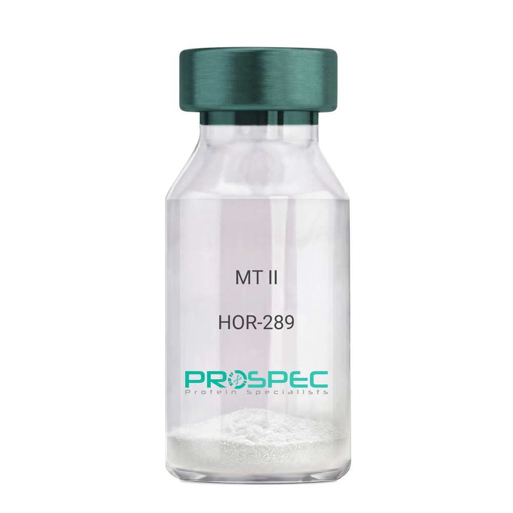
MT IIDescription:
Melanotan-II
Melanotan-II, MT-II, Melanotan.
Product # :
HOR-289Price :
Quantity :
Shipping Method :
Shipped at Room temp
More Info
- description
- formulation
- purity
- More Info
Description
The molecular formula of Melanotan-II is C50H69N15O9. It has the amino acid sequence of Ac-Nle-cyclo [Asp-His-D-Phe-Arg-Trp-Lys]-NH2 and a molecular weight of 1024.2 Dalton. The CAS Number is 121062-08-6.
Formulation
The protein (1mg/ml) was lyophilized with no additives.
Purity
Greater than 99% as determined by by RP-HPLC.
More Info
-
Introduction
Melanotan and Melanotan-II are both analogs of the peptide hormone alpha melanocyte stimulating hormone (a-MSH) that tend to induce skin tanning. Unlike Melanotan, Melanotan-II has the additional effect of increasing libido. Melanotan II has aphrodisiac properties.
-
Synonyms
Melanotan-II, MT-II, Melanotan.
-
Physical Appearance
Sterile Filtered White lyophilized (freeze-dried) powder.
-
Stability
Lyophilized Melanotan-II although stable at room temperature for 3 weeks, should be stored desiccated below -18°C. Upon reconstitution MT-II should be stored at 4°C between 2-7 days and for future use below -18°C.For long term storage it is recommended to add a carrier protein (0.1% HSA or BSA).Please prevent freeze-thaw cycles.
-
Solubility
It is recommended to reconstitute the lyophilized Melanotan-II in sterile 18MΩ-cm H2O not less than 100 µg/ml, which can then be further diluted to other aqueous solutions.
ProSpec's products are furnished for LABORATORY RESEARCH USE ONLY. The product may not be used as drugs, agricultural or pesticidal products, food additives or household chemicals.
- View Data Sheet
Name :
CCL13 HumanDescription:
Monocyte Chemotactic Protein-4 Human Recombinant (CCL13)
Small inducible cytokine A13, CCL13, Monocyte chemotactic protein 4, MCP-4, Monocyte chemoattractant protein 4, CK-beta-10, NCC-1, chemokine (C-C motif) ligand 13, NCC1, CKb10, SCYL1, SCYA13, MGC17134.
Product # :
CHM-319Price :
Quantity :
Shipping Method :
Shipped at Room temp
More Info
- description
- source
- formulation
- purity
- biological activity
- More Info
Description
Monocyte Chemotactic Protein-4 Human Recombinant produced in E.Coli is a non-glycosylated, Polypeptide chain containing 75 amino acids and having a molecular mass of 8.6 kDa. The MCP-4 is purified by proprietary chromatographic techniques.
Source
Escherichia Coli.
Formulation
The protein was lyophilized from a concentrated (1mg/ml) sterile solution in 20mM PB, pH 7.4, 130mM NaCl.
Purity
Greater than 96.0% as determined by:
(a) Analysis by RP-HPLC.
(b) Analysis by SDS-PAGE.Biological Activity
The specific activity as determined by the ability of MCP-4 to chemoattaract human monocytes at 10-100ng/ml, corresponding to a Specific Activity of 10,000-100,000 units/mg.More Info
-
Introduction
Chemokine (C-C motif) ligand 13 (CCL13 / MCP-4) is a small cytokine belonging to the CC chemokine family. The MCP-4 gene is located on human chromosome 17 within a large cluster of other CC chemokines. MCP-4 induces chemotaxis in monocytes, eosinophils, T lymphocytes, and basophils by binding cell surface G-protein linked chemokine receptors such as CCR2, CCR3 and CCR5. Activity of the MCP-4 chemokine has been implicated in allergic reactions such as asthma. MCP-4 can be induced by the inflammatory cytokines interleukin-1 and TNF-a.
-
Synonyms
Small inducible cytokine A13, CCL13, Monocyte chemotactic protein 4, MCP-4, Monocyte chemoattractant protein 4, CK-beta-10, NCC-1, chemokine (C-C motif) ligand 13, NCC1, CKb10, SCYL1, SCYA13, MGC17134.
-
Physical Appearance
Sterile Filtered White lyophilized (freeze-dried) powder.
-
Stability
Lyophilized MCP-4 although stable at room temperature for 3 weeks, should be stored desiccated below -18°C. Upon reconstitution MCP-4 should be stored at 4°C between 2-7 days and for future use below -18°C. For long term storage it is recommended to add a carrier protein (0.1% HSA or BSA).Please prevent freeze-thaw cycles.
-
Solubility
It is recommended to reconstitute the lyophilized MCP-4 in sterile 18M-cm H2O not less than 100µg/ml, which can then be further diluted to other aqueous solutions.
-
Amino Acid Sequence
QPDALNVPSTCCFTFSSKKISLQRLKSYVITTSRCPQKAV
IFRTKLGKEICADPKEKWVQNYMKHLGRKAHTLKT. -
Background
What is the molecular weight/Mw of CCL13 HUMAN Protein?
CCL13 HUMAN Protein has a total Mw of 8.6kDa.
What is the source or expression system of CCL13 HUMAN Protein?
Escherichia Coli.
What is the Purity of CCL13 HUMAN Protein?
CCL13 HUMAN Protein is >96% pure as determined by SDS-PAGE.
What is the Biological Activity of CCL13 HUMAN Protein?
The specific activity as determined by the ability of MCP-4 to chemoattaract human monocytes at 10-100ng/ml, corresponding to a Specific Activity of 10,000-100,000 units/mg.
What is the amino acid sequence of CCL13 HUMAN Protein?
QPDALNVPSTCCFTFSSKKISLQRLKSYVITTSRCPQKAV
IFRTKLGKEICADPKEKWVQNYMKHLGRKAHTLKT.
What applications can CCL13 HUMAN Protein be used in?
CCL13 HUMAN Protein can probably be used in western blot, ELISA and Lateral Flow.
What is the endotoxin level for CCL13 HUMAN Protein?
The endotoxin level is minimal, CCL13 HUMAN Protein was purified using conventional chromatography techniques.
ProSpec's products are furnished for LABORATORY RESEARCH USE ONLY. The product may not be used as drugs, agricultural or pesticidal products, food additives or household chemicals.
- View Data Sheet
Name :
Insulin Human (20-110)Description:
Insulin (20-110 a.a) Human Recombinant
Product # :
CYT-1237Price :
Quantity :
Shipping Method :
Shipped at Room temp
More Info
- description
- source
- formulation
- purity
- More Info
Description
The Insulin Human is created as a recombinant protein with a 4kda N-terminal fusion of His Tag. The Insulin His-Tagged Fusion Protein, produced in E. coli, is a 13kDa protein containing 91 amino acid residues of the Insulin Human, 20-110 amino acids.
Source
Escherichia Coli.
Formulation
Each mg was lyophilized with 1xPBS, 0.4% SDS and 4mM DTT.
Purity
Greater than 90% as determined by SDS-PAGE.
More Info
-
Physical Appearance
Filtered White lyophilized (freeze-dried) powder.
-
Stability
Store lyophilized Insulin at -20°C. Aliquot the product after reconstitution to avoid repeated freezing/thawing cycles. Reconstituted protein can be stored at 4°C for a limited period of time; it does not show any change after two weeks at 4°C.
-
Solubility
It is recommended to add deionized water to prepare a working stock solution of approximately 0.5mg/ml and let the lyophilized pellet dissolve completely. Product is not sterile! Please filter the product by an appropriate sterile filter before using it on cell culture.
-
Amino Acid Sequence
MALWMRLLPL LALLALWGPD PAAAFVNQHL CGSHLVEALY LVCGERGFFY TPKTRREAED LQVGQVELGG GPGAGSLQPL ALEGSLQKRG IVEQCCTSIC SLYQLENYCN
-
Background
Insulin participates in the metabolism of carbohydrates, proteins and fats by regulating glucose homeostasis in the body. Insulin decreases blood glucose concentration. Insulin hormone facilitates the uptake of glucose into cells, mainly in muscle and fat tissues, and stimulates the liver to store glucose as glycogen. Insulin also inhibits the production of gluconeogenesis and promotes the synthesis of proteins and lipids. Insulin increases cell permeability to monosaccharides, fatty acids and amino acids. Insulin accelerates glycolysis, the pentose phosphate cycle and glycogen synthesis in liver.
ProSpec's products are furnished for LABORATORY RESEARCH USE ONLY. The product may not be used as drugs, agricultural or pesticidal products, food additives or household chemicals.
- View Data Sheet
Name :
Thyroglobulin HumanDescription:
Thyroglobulin Human Recombinant
Thyroglobulin, TGN, AITD3, TG.
Product # :
PRO-2803Price :
Quantity :
Shipping Method :
Shipped at Room temp
More Info
- description
- source
- formulation
- purity
- More Info
- sds-page
Description
Thyroglobulin Human produced in a mammalian cell line is a single, non-glycosylated polypeptide chain (1-2768 a.a.) and having a molecular mass of 304640 Dalton. Thyroglobulin Human is fused with GlyAlaProGly4SerHis10-tag at C-terminal and purified by proprietary chromatographic techniques.
Source
Mammalian cell line.
Formulation
Thyroglobulin was lyophilized from PBS, pH 7.4 and 5.4 % sucrose.
Purity
Greater than 95.0% as determined by SDS-PAGE.
sds-page
More Info
-
Synonyms
Thyroglobulin, TGN, AITD3, TG.
-
Physical Appearance
Sterile Filtered White lyophilized (freeze-dried) powder.
-
Stability
Lyophilized Thyroglobulin although stable at room temperature for 3 weeks, should be stored desiccated below -18°C. Upon reconstitution Thyroglobulin should be stored at 4°C between 2-7 days and for future use below -18°C. For long term storage it is recommended to add a carrier protein (0.1% HSA or BSA). Please prevent freeze-thaw cycles.
-
Solubility
It is recommended to reconstitute the lyophilized Thyroglobulin in sterile 18MΩ-cm H2O not less than 100µg/ml, which can then be further diluted to other aqueous solutions.
-
Background
Thyroglobulin, a glycoprotein primarily produced in the thyroid gland, stands at the center of thyroid hormone synthesis. Comprising a series of tyrosine residues, thyroglobulin serves as the scaffold upon which thyroid hormones are assembled. Beyond its pivotal role in thyroid physiology, thyroglobulin has garnered significant attention in the realm of thyroid disease diagnostics, offering valuable insights into thyroid function and disorders. This research delves into the intricacies of thyroglobulin human recombinant protein, exploring its biochemical properties, physiological significance, and its crucial applications in both clinical and research settings.
Structural Complexity of Thyroglobulin:
Thyroglobulin is a large, dimeric protein boasting an intricate structure composed of multiple domains. Within its structure lie tyrosine residues crucial for iodine incorporation, a process fundamental for thyroid hormone synthesis. Its size and complexity reflect the sophistication of thyroid hormone production, as thyroglobulin acts as a reservoir for thyroid hormones within the thyroid follicles.
Physiological Significance in Thyroid Function:
Thyroglobulin plays a central role in the synthesis of triiodothyronine (T3) and thyroxine (T4), the thyroid hormones essential for regulating metabolism and overall body homeostasis. During thyroid hormone synthesis, thyroglobulin is secreted into the follicular lumen, where it undergoes iodination and subsequent proteolysis, releasing T3 and T4. This process highlights the indispensable nature of thyroglobulin in thyroid hormone production, making it a key biomolecule in thyroid physiology.
ProSpec's products are furnished for LABORATORY RESEARCH USE ONLY. The product may not be used as drugs, agricultural or pesticidal products, food additives or household chemicals.
- View Data Sheet
Name :
BD 4 RatDescription:
BD 4 Rat
Beta-defensin 4, BD-4, BD-2, Defensin, beta 4, RBD-2, RBD-4, Defb4, Defb2, Defb3.
Product # :
CYT-066Price :
Quantity :
Shipping Method :
Shipped at Room temp
More Info
- description
- source
- formulation
- purity
- biological activity
- More Info
Description
BD-4 Rat Recombinant produced in E.coli is a single, non-glycosylated polypeptide chain containing 41 amino acids and having a molecular mass of 4.4kDa.The BD-4 is purified by proprietary chromatographic techniques.
Source
Escherichia Coli.
Formulation
BD-4 protein was lyophilized from a 0.2µm filtered concentrated solution in PBS, pH 7.4.
Purity
Greater than 97.0% as determined by SDS-PAGE.
Biological Activity
Measured by its antimicrobial activity against E. coli. The ED50 for this effect is typically 5-50µg/ml.More Info
-
Introduction
Defensins are cationic peptides with a large spectrum of antimicrobial activity that comprise an important arm of the innate immune system. The Alpha defensins are differentiated from the Beta-defensins by the pairing of their 3 disulfide bonds.
4 human Beta-defensins have been identified to date; BD-1, BD-2, BD-3 and BD-4.
Beta-defensins are expressed on some leukocytes and at epithelial surfaces.
In addition to their direct antimicrobial activities, they are chemoattractant towards immature dendritic cells and memory T cells. The beta-defensin proteins are expressed as the C-terminal portion of precursors and are released by proteolytic cleavage of a signal sequence and, in the case of BD-1 (36 a.a.), a propeptide region. Beta-defensins contain a six-cysteine motif that forms three intra-molecular disulfide bonds. Beta-Defensins are 3-5 kDa peptides ranging in size from 33-47 amino acid residues. -
Synonyms
Beta-defensin 4, BD-4, BD-2, Defensin, beta 4, RBD-2, RBD-4, Defb4, Defb2, Defb3.
-
Physical Appearance
Sterile Filtered White lyophilized (freeze-dried) powder.
-
Stability
Lyophilized BD-4 although stable at room temperature for 3 weeks, should be stored desiccated below -18°C. Upon reconstitution BD-4 should be stored at 4°C between 2-7 days and for future use below -18°C. For long term storage it is recommended to add a carrier protein (0.1% HSA or BSA). Please prevent freeze-thaw cycles.
-
Solubility
It is recommended to reconstitute the lyophilized BD-4 in sterile 18M-cm H2O not less than 100µg/ml, which can then be further diluted to other aqueous solutions.
-
Amino Acid Sequence
QSINNPITCL TKGGVCWGPC TGGFRQIGTC GLPRVRCCKK K.
-
Background
What is the molecular weight/Mw of BD4 Protein?
BD4 Protein has a total Mw of 4.4kDa.
What is the source or expression system of BD4 Protein?
Escherichia Coli.
What is the Purity of BD4 Protein?
BD4 Protein is >97% pure as determined by SDS-PAGE.
What is the Biological Activity of BD4 Protein?
Measured by its antimicrobial activity against E. coli. The ED50 for this effect is typically 5-50µg/ml.
What is the amino acid sequence of BD4 Protein?
QSINNPITCL TKGGVCWGPC TGGFRQIGTC GLPRVRCCKK K.
What applications can BD4 Protein be used in?
BD4 Protein can probably be used in western blot, ELISA and Lateral Flow.
What is the endotoxin level for BD4 Protein?
The endotoxin level is minimal, BD4 Protein was purified using conventional chromatography techniques.
ProSpec's products are furnished for LABORATORY RESEARCH USE ONLY. The product may not be used as drugs, agricultural or pesticidal products, food additives or household chemicals.
- View Data Sheet
Name :
BD 3 RatDescription:
Beta Defensin-3 Rat Recombinant
Beta-defensin 3, BD-3, Defensin beta 3, Defb3.
Product # :
CYT-063Price :
Quantity :
Shipping Method :
Shipped at Room temp
More Info
- description
- source
- formulation
- purity
- biological activity
- More Info
Description
BD-3 Rat Recombinant produced in E.coli is a single, non-glycosylated polypeptide chain containing 41 amino acids and having a molecular mass of 4.5kDa.The BD-3 is purified by proprietary chromatographic techniques.
Source
Escherichia Coli.
Formulation
BD-3 protein was lyophilized from a 0.2µm filtered concentrated solution in PBS, pH 7.4.
Purity
Greater than 98.0% as determined by:
(a) Analysis by RP-HPLC.
(b) Analysis by SDS-PAGE.Biological Activity
Measured by its antimicrobial activity against E. coli. The ED50 for this effect is typically 4-20µg/ml, corresponding to a specific activity of 50,000-250,000units/mg.More Info
-
Introduction
Defensins form a family of microbicidal and cytotoxic peptides made by neutrophils. Members of the defensin family are highly similar in protein sequence. This gene encodes defensin, beta 103A, which has broad spectrum antimicrobial activity and may play an important role in innate epithelial defense.
-
Synonyms
Beta-defensin 3, BD-3, Defensin beta 3, Defb3.
-
Physical Appearance
Sterile Filtered White lyophilized (freeze-dried) powder.
-
Stability
Lyophilized BD-3 although stable at room temperature for 3 weeks, should be stored desiccated below -18°C. Upon reconstitution BD-3 should be stored at 4°C between 2-7 days and for future use below -18°C. For long term storage it is recommended to add a carrier protein (0.1% HSA or BSA). Please prevent freeze-thaw cycles.
-
Solubility
It is recommended to reconstitute the lyophilized BD-3 in sterile 18M-cm H2O not less than 100µg/ml, which can then be further diluted to other aqueous solutions.
-
Amino Acid Sequence
KKVYNAVSCM TNGGICWLKC SGTFREIGSC GTRQLKCCKK K.
-
Background
What is the molecular weight/Mw of BD3 Protein?
BD3 Protein has a total Mw of 4.5kDa.
What is the source or expression system of BD3 Protein?
Escherichia Coli.
What is the Purity of BD3 Protein?
BD3 Protein is >98% pure as determined by SDS-PAGE.
What is the Biological Activity of BD3 Protein?
Measured by its antimicrobial activity against E. coli. The ED50 for this effect is typically 4-20µg/ml, corresponding to a specific activity of 50,000-250,000units/mg.
What is the amino acid sequence of BD3 Protein?
KKVYNAVSCM TNGGICWLKC SGTFREIGSC GTRQLKCCKK K.
What applications can BD3 Protein be used in?
BD3 Protein can probably be used in western blot, ELISA and Lateral Flow.
What is the endotoxin level for BD3 Protein?
The endotoxin level is minimal, BD3 Protein was purified using conventional chromatography techniques.
ProSpec's products are furnished for LABORATORY RESEARCH USE ONLY. The product may not be used as drugs, agricultural or pesticidal products, food additives or household chemicals.
- View Data Sheet
Name :
CEA ProteinDescription:
Carcinoembryonic Antigen Human
CEACAM5, Meconium Antigen 100, Carcinoembryonic Antigen, CD66e Antigen, CD66e, Carcinoembryonic Antigen, CEA, oncofetal antigen.
Product # :
PRO-2801Price :
Quantity :
Shipping Method :
Shipped with Ice Packs
More Info
- description
- source
- formulation
- purity
- More Info
Description
CEA produced from patient source colon carcinoma liver metastatic tissue can be used as general marker in screening and monitoring malignant disease states.
Source
Liver tissue.
Formulation
CEA protein solution contains 0.1M PBS, pH 7.4, 0.09 % NaN3 and 2 % methyl-mannoside.
Purity
Greater than 95.0% as determined by SDS-PAGE.
More Info
-
Synonyms
CEACAM5, Meconium Antigen 100, Carcinoembryonic Antigen, CD66e Antigen, CD66e, Carcinoembryonic Antigen, CEA, oncofetal antigen.
-
Physical Appearance
Sterile Filtered colorless solution.
-
Stability
Store at 4°C if entire vial will be used within 2-4 weeks. Store, frozen at -20°C for longer periods of time. For long term storage it is recommended to add a carrier protein (0.1% HSA or BSA). Avoid multiple freeze-thaw cycles.
-
Applications
Blood samples from tissue donors were tested and found to be negative for HBsAg, HIV-1 and HIV-2 antibodies and HCV.
-
Background
Carcinoembryonic Antigen, commonly known as CEA, is a glycoprotein that was initially identified as a tumor marker. Over the years, research into CEA has unveiled its intricate involvement in various physiological processes, not only in cancer but also in the context of normal development and inflammatory conditions. This research aims to delve into the multifaceted roles of CEA, exploring its structural intricacies, regulatory mechanisms, and its implications in health, disease, and beyond.
Structural Complexity of CEA:
CEA, belonging to the immunoglobulin superfamily, is a complex glycoprotein featuring multiple structural domains. Its diverse forms and glycosylation patterns contribute to its functional versatility. CEA is primarily expressed in fetal tissues, but its presence is often detected in adults under pathological conditions, especially in various types of cancer.
CEA in Cancer Biology:
CEA was first recognized as a biomarker for colorectal cancer, but its overexpression is not limited to this context. Elevated CEA levels have been associated with several other malignancies, including breast, lung, and pancreatic cancers. CEA’s involvement in cancer biology ranges from promoting angiogenesis and metastasis to inhibiting immune responses, making it a critical player in tumor progression and evasion.
Beyond Cancer: CEA in Development and Inflammation:
While CEA’s role in cancer is prominent, recent studies have uncovered its participation in normal physiological processes. During embryonic development, CEA is involved in cell adhesion, contributing to tissue organization and morphogenesis. Additionally, CEA expression can be induced in inflammatory conditions, suggesting its involvement in immune responses and tissue repair mechanisms.
CEA as a Diagnostic and Therapeutic Target:
The diverse expression patterns of CEA in various diseases make it a valuable diagnostic tool. CEA assays are widely used for cancer screening, monitoring disease progression, and assessing treatment efficacy. Moreover, CEA’s presence on the surface of cancer cells has made it a target for immunotherapy, enabling the development of targeted therapies aimed at specifically eradicating CEA-positive tumor cells.
ProSpec's products are furnished for LABORATORY RESEARCH USE ONLY. The product may not be used as drugs, agricultural or pesticidal products, food additives or household chemicals.
- View Data Sheet
Name :
SLAMF7 HumanDescription:
SLAMF7 Human Recombinant
SLAMF7, SLAM Family Member 7, CD2-Like Receptor-Activating Cytotoxic Cells, Membrane Protein FOAP-12, CD2 Subset 1, Protein 19A, CRACC, CS1, Novel LY9 (Lymphocyte Antigen 9) Like Protein, CD2-Like Receptor Activating Cytotoxic Cells, 19A24 Protein, CD319 Antigen, Novel Ly9, CD319, 19A.
Product # :
PRO-2261Price :
Quantity :
Shipping Method :
Shipped with Ice Packs
More Info
- description
- source
- formulation
- purity
- More Info
Description
SLAMF7 produced in Sf9 Baculovirus cells is a single, glycosylated polypeptide chain containing 212 amino acids (23-226a.a.) and having a molecular mass of 23.4kDa (Molecular size on SDS-PAGE will appear at approximately 28-40kDa). SLAMF7 is expressed with an 8 amino acid His tag at C-Terminus and purified by proprietary chromatographic techniques.
Source
Sf9, Baculovirus cells.
Formulation
SLAMF7 protein solution (0.5mg/ml) contains Phosphate Buffered Saline (pH 7.4) and 10% glycerol.
Purity
Greater than 95.0% as determined by SDS-PAGE.
More Info
-
Introduction
SLAMF7 belongs to the CS2 family of cell surface receptors. SLAMF7 is a single-pass type 1 membrane protein, SLAMF7 Isoform 1 mediates NK cell activation via aSH2D1A-independent extracellular signal-regulated ERK-mediated pathway. SLAMF7 functions in lymphocyteadhesion. Furthermore, SLAMF7 can exert activating of inhibitory influences on cells of the immune system depending on cellular context as well as the availability of effector proteins.
-
Synonyms
SLAMF7, SLAM Family Member 7, CD2-Like Receptor-Activating Cytotoxic Cells, Membrane Protein FOAP-12, CD2 Subset 1, Protein 19A, CRACC, CS1, Novel LY9 (Lymphocyte Antigen 9) Like Protein, CD2-Like Receptor Activating Cytotoxic Cells, 19A24 Protein, CD319 Antigen, Novel Ly9, CD319, 19A.
-
Physical Appearance
Sterile Filtered clear solution.
-
Stability
Store at 4°C if entire vial will be used within 2-4 weeks. Store, frozen at -20°C for longer periods of time. For long term storage it is recommended to add a carrier protein (0.1% HSA or BSA).Avoid multiple freeze-thaw cycles.
-
Amino Acid Sequence
SGPVKELVGS VGGAVTFPLK SKVKQVDSIV WTFNTTPLVT IQPEGGTIIV TQNRNRERVD FPDGGYSLKL SKLKKNDSGI YYVGIYSSSL QQPSTQEYVL HVYEHLSKPK VTMGLQSNKN GTCVTNLTCC MEHGEEDVIY TWKALGQAAN ESHNGSILPI SWRWGESDMT FICVARNPVS RNFSSPILAR KLCEGAADDP DSSMLEHHHH HH.
ProSpec's products are furnished for LABORATORY RESEARCH USE ONLY. The product may not be used as drugs, agricultural or pesticidal products, food additives or household chemicals.
- View Data Sheet
Name :
IL 3 RatDescription:
Interleukin-3 Rat Recombinant
MCGF (Mast cell growth factor), Multi-CSF, HCGF, P-cell stimulation factor, IL-3, MGC79398, MGC79399.
Product # :
CYT-383Price :
Quantity :
Shipping Method :
Shipped at Room temp
More Info
- description
- source
- formulation
- purity
- biological activity
- More Info
Description
Interleukin-3 Rat Recombinant produced in E.Coli is a single, non-glycosylated polypeptide chain containing 144 amino acids and having a molecular mass of 16.3kDa. The IL-3 is purified by proprietary chromatographic techniques.
Source
Escherichia Coli.
Formulation
Interleukin-3 Rat was lyophilized from a 0.2µm filtered concentrated solution in PBS, pH 7.4.
Purity
Greater than 97.0% as determined by:
(a) Analysis by RP-HPLC.
(b) Analysis by SDS-PAGE.Biological Activity
The ED50 as determined by the dose-dependent stimulation of the proliferation of thymidine uptake by murine MC-9 cells is < 10 ng/ml, corresponding to a specific activity of >1.0 x 10⁵ units/mg.More Info
-
Introduction
Interleukin-3 is a pleiotropic cytokine produced primarily by activated T cells.
IL-3 is thought to function via specific cell surface receptors to stimulate the proliferation, differentiation and survival of haematopoietic cell lines. IL-3 has also been shown to affect the functional activity of a variety of other cell types including mast cells, eosinophils, megakaryocytes and basophils. -
Synonyms
MCGF (Mast cell growth factor), Multi-CSF, HCGF, P-cell stimulation factor, IL-3, MGC79398, MGC79399.
-
Physical Appearance
Sterile Filtered White lyophilized (freeze-dried) powder.
-
Stability
Lyophilized Interleukin-3 although stable at room temperature for 3 weeks, should be stored desiccated below -18°C. Upon reconstitution IL3 should be stored at 4°C between 2-7 days and for future use below -18°C.For long term storage it is recommended to add a carrier protein (0.1% HSA or BSA).Please prevent freeze-thaw cycles.
-
Solubility
It is recommended to reconstitute the lyophilized Interleukin 3 in sterile 18MΩ-cm H2O not less than 100µg/ml, which can then be further diluted to other aqueous solutions.
-
Amino Acid Sequence
MISDRGSDAH HLLRTLDCRT IALEILVKLP YPQVSGLNNS DDKANLRNST LRRVNLDEFL KSQEEFDSQD TTDIKSKLQK LKCCIPAAAS DSVLPGVYNK DLDDFKKKLR FYVIHLKDLQ PVSVSRPPQP TSSSDNFRPM TVEC.
ProSpec's products are furnished for LABORATORY RESEARCH USE ONLY. The product may not be used as drugs, agricultural or pesticidal products, food additives or household chemicals.
- View Data Sheet
Description:
Human Follicle Stimulating Hormone
Follitropin subunit beta, Follicle-stimulating hormone beta subunit, FSH-beta, FSH-B, Follitropin beta chain, FSH.
Product # :
HOR-249Price :
Quantity :
Shipping Method :
Shipped at Room temp
More Info
- description
- source
- formulation
- More Info
Description
FSH Human is a glycoprotein produced from urine of post-menopausal women and having a total molecular mass of 30,000 Dalton.FSH is a heterodimeric hormone consisting of 92 amino acids a chain and 111 amino acids b chain.The FSH is purified by proprietary chromatographic techniques.
Source
Urine of post-menopausal women.
Formulation
The FSH was lyophilized with no additives.
More Info
-
Introduction
Follicle stimulating hormone (FSH) is a hormone synthesised and secreted by gonadotropes in the anterior pituitary gland. FSH and LH act synergistically in reproductionIn women, in the ovary FSH stimulates the growth of immature Graafian follicles to maturation. As the follicle grows it releases inhibin, which shuts off the FSH production.
In men, FSH enhances the production of androgen-binding proteinby the Sertoli cells of the testes and is critical for spermatogenesis.
In both males and females, FSH stimulates the maturation of germ cells. In females, FSH initiates follicular growth, specifically affecting granulosa cells. With the concomitant rise in inhibin B FSH levels then decline in the late follicular phase. This seems to be critical in selecting only the most advanced follicle to proceed to ovulation. At the end of the luteal phase, there is a slight rise in FSH that seems to be of importance to start the next ovulatory cycle.
Like its partner, LH, FSH release at the pituitary gland is controlled by pulses of gonadotropin-releasing hormone(GnRH). Those pulses, in turn, are subject to the estrogen feed-back from the gonads. -
Synonyms
Follitropin subunit beta, Follicle-stimulating hormone beta subunit, FSH-beta, FSH-B, Follitropin beta chain, FSH.
-
Physical Appearance
Sterile Filtered White lyophilized (freeze-dried) powder.
-
Stability
Lyophilized FSH although stable at room temperature for 3 weeks, should be stored desiccated below -18°C. Upon reconstitution FSH-beta should be stored at 4°C between 2-7 days and for future use below -18°C. For long term storage it is recommended to add a carrier protein (0.1% HSA or BSA).Please prevent freeze-thaw cycles.
-
Solubility
It is recommended to reconstitute the lyophilized Follicle Stimulating Hormone in sterile pyrogen free water at 2,000IU/1ml, which can then be further diluted to other aqueous solutions.
-
Background
What is the molecular weight/Mw of FOLLICLE STIMULATING HORMONE HUMAN Protein?
FOLLICLE STIMULATING HORMONE HUMAN Protein has a total Mw of 30kDa.
What is the source or expression system of FOLLICLE STIMULATING HORMONE HUMAN Protein?
Urine of post-menopausal women.
What is the Biological Activity of FOLLICLE STIMULATING HORMONE HUMAN Protein?
The biological functionality of FOLLICLE STIMULATING HORMONE HUMAN Protein will be determined in the future.
What is the amino acid sequence of FOLLICLE STIMULATING HORMONE HUMAN Protein?
FOLLICLE STIMULATING HORMONE HUMAN Protein is composed from 92 amino acids a chain and 111 amino acids b chain.
What applications can FOLLICLE STIMULATING HORMONE HUMAN Protein be used in?
FOLLICLE STIMULATING HORMONE HUMAN Protein can probably be used in western blot, ELISA and Lateral Flow.
What is the endotoxin level for FOLLICLE STIMULATING HORMONE HUMAN Protein?
The endotoxin level is minimal, FOLLICLE STIMULATING HORMONE HUMAN Protein was purified using conventional chromatography techniques.
-
Contaminants
Less than: 0.1% hCG, 0.5%TSH, 0.5% LH, 0.5%GH and 0.5%Prl.Free of HbsAg, antibodies to HIV and HCV.
ProSpec's products are furnished for LABORATORY RESEARCH USE ONLY. The product may not be used as drugs, agricultural or pesticidal products, food additives or household chemicals.
- View Data Sheet
Name :
SSTDescription:
Somatostatin
Growth hormone release-inhibiting factor, SST, SMS, SMST, GHIH.
Product # :
HOR-299Price :
Quantity :
Shipping Method :
Shipped at Room temp
More Info
- description
- formulation
- purity
- More Info
Description
Somatostatin Synthetic is a single, non-glycosylated polypeptide chain containing 14 amino acids, having a molecular mass of 1637.9 Dalton and a Molecular formula of C76H104N18O19S2. The CAS# is 38916-34-6.
Formulation
The protein was lyophilized with no additives.
Purity
Greater than 98.0% as determined by:
(a) Analysis by RP-HPLC.
(b) Analysis by SDS-PAGE.More Info
-
Introduction
Somatostatin (also known as growth hormone inhibiting hormone (GHIH) or somatotropin release-inhibiting hormone (SRIF) is a peptide hormone that regulates the endocrine systemand affects neurotransmission and cell proliferation via interaction with G-protein-coupled somatostatin receptors and inhibition of the release of numerous secondary hormones. Somatostatin has two active forms produced by alternative cleavage of a single preproprotein: one of 14 amino acids, the other of 28 amino acids.
-
Synonyms
Growth hormone release-inhibiting factor, SST, SMS, SMST, GHIH.
-
Physical Appearance
Sterile Filtered White lyophilized (freeze-dried) powder.
-
Stability
Lyophilized SST although stable at room temperature for 3 weeks, should be stored desiccated below -18°C. Upon reconstitution Somatostatin should be stored at 4°C between 2-7 days and for future use below -18°C.For long term storage it is recommended to add a carrier protein (0.1% HSA or BSA).Please prevent freeze-thaw cycles.
-
Solubility
It is recommended to reconstitute the lyophilized Somatostatin in sterile 18MΩ-cm H2O not less than 100 µg/ml, which can then be further diluted to other aqueous solutions.
-
Amino Acid Sequence
Ala-Gly-Cys-Lys-Asn-Phe- Phe-Trp-Lys-Thr-Phe-Thr-Ser-Cys-OH.
ProSpec's products are furnished for LABORATORY RESEARCH USE ONLY. The product may not be used as drugs, agricultural or pesticidal products, food additives or household chemicals.
- View Data Sheet
Name :
MMP 9 HumanDescription:
Matrix Metalloproteinase-9 Human Recombinant
Matrix metalloproteinase-9, MMP-9, 92 kDa gelatinase, Gelatinase B, GELB, MMP9, CLG4B.
Product # :
ENZ-438Price :
Quantity :
Shipping Method :
Shipped with Ice Packs
More Info
- description
- source
- formulation
- purity
- More Info
Description
MMP-9 Human Recombinant produced in E.Coli is single, a non-glycosylated, Polypeptide chain containing 338 amino acids fragment (113-450) corresponding to the catalytic domain of the protein, having a total molecular mass of 42.03kDa and fused with a 4.5kDa amino-terminal hexahistidine tag. The MMP-9 is purified by proprietary chromatographic techniques.
Source
Escherichia Coli.
Formulation
MMP-9 protein is supplied in 20mM Tris-HCl pH 8.0 and 50% glycerol.
Purity
Greater than 95.0% as determined by SDS-PAGE.
More Info
-
Introduction
Matrix metalloproteinases are a family of zinc and calcium-dependent endopeptidases that break down extracellular matrix proteins. The MMP9 is secreted as a 92kDa zymogen. Cleavage of ProMMP-9 results in the active enzyme, having a molecular weight of approximately 82kDa. MMP9 is composed of the following domains: a gelatin-binding domain consisting of three fibronectin type II units, a catalytic domain containing the zinc-binding site, a proline-rich type V collagen-homologous domain and a hemopexin-like domain. MMP9 is produced by the several cell types: monocytes, macrophages, neutrophils, keratinocytes, fibroblasts, osteoclasts and endothelial cells. MMP9 is involved in inflammatory responses, tissue remodeling, wound healing, tumor growth and metastasis. MMP9 may also play an important part in local proteolysis of the extracellular matrix and in leukocyte migration, as well as in bone osteoclastic resorption. MMP9 cleaves type IV and type V collagens into large C-terminal three quarter fragments and shorter N-terminal one quarter fragments. MMP9 can also degrade fibronectin but not laminin or Pz-peptide.
MMP9 defects may be a cause of susceptibility to intervertebral disc disease (IDD), also known as lumbar disk herniation (LDH). -
Synonyms
Matrix metalloproteinase-9, MMP-9, 92 kDa gelatinase, Gelatinase B, GELB, MMP9, CLG4B.
-
Physical Appearance
Sterile Filtered clear solution.
-
Stability
Store at 4°C if entire vial will be used within 2-4 weeks. Store, frozen at -20°C for longer periods of time. Please avoid freeze thaw cycles.
-
Amino Acid Sequence
4.5kDa His Tag-DLKWHHHNITYWIQNYSEDLPRAVIDDAFARAFALWSAVTPLTFTRVYSRDAD
IVIQFGVAEHGDGYPFDGKDGLLAHAFPPGPGIQGDAHFDDDELWSLGKGVVVPTRFGNADGAACHFP
FIFEGRSYSACTTDGRSDGLPWCSTTANYDTDDRFGFCPSERLYTRDGNADGKPCQFPFIFQGQSYSA
CTTDGRSDGYRWCATTANYDRDKLFGFCPTRADSTVMGGNSAGELCVFPFTFLGKEYSTCTSEGRGDG
RLWCATTSNFDSDKKWGFCPDQGYSLFLVAAHEFGHALGLDHSSVPEALMYPMYRFTEGPPLHKDDVN
GIRHLYGP.
ProSpec's products are furnished for LABORATORY RESEARCH USE ONLY. The product may not be used as drugs, agricultural or pesticidal products, food additives or household chemicals.