Search results
1000 results found for “lipocalin”
Name
Description
Product #
Price
Quantity
Shipping Method
- View Data Sheet
Name :
GDF6 HumanDescription:
Bone Morphogenetic protein-13 Human Recombinant
Growth Differentiation Factor 6, Growth/Differentiation Factor 16, Bone Morphogenetic Protein 13, BMP-13, BMP13, GDF-6, Klippel-Feil Malformation, Segmentation Syndrome 1, Klip-Feil Malformation, Klippel-Feil Syndrome, MCOPCB6, SCDO4, CDMP2, LCA17, MCOP4, GDF16, KFS1, KFSL, SGM1, KFM, KFS, GDF6.
Product # :
CYT-938Price :
Quantity :
Shipping Method :
Shipped at Room temp
More Info
- description
- source
- formulation
- purity
- biological activity
- More Info
Description
BMP13 Human Recombinant produced in E.coli is a non-glycosylated disulfide linked homodimer containing 2 chains of 120 amino acids and having a molecular mass of 27.1kDa.The BMP-13 is purified by proprietary chromatographic techniques.
Source
Escherichia Coli.
Formulation
BMP-13 protein was lyophilized from a 0.2µm filtered concentrated solution in 30% Acetonitrile and 0.1% TFA.
Purity
Greater than 95.0% as determined by: (a) Analysis by HPLC. (b) Analysis by SDS-PAGE.
Biological Activity
The ED50 as determined by inducing alkaline phosphatase production of murine ATDC5 cells is less than 2.0µg/ml, corresponding to a specific activity of > 500IU/mg.More Info
-
Introduction
Growth/differentiation factors (GDF1-GDF15) belong to the BMP family of TGF-beta superfamily proteins. These factors are produced as inactive preproproteins which are subsequently cleaved and assembled into active secreted homodimers. BMP13 is a growth factor which controls proliferation and cellular differentiation in the retina and bone formation. BMP13 has a central role in regulating apoptosis during retinal development. GDF proteins are vital during embryonic development, particularly in the skeletal, nervous, and muscular systems. BMP13 gene mutations result in colobomata, which are congenital abnormalities in ocular development, and in Klippel-Feil syndrome (KFS), which is a congenital disorder of spinal segmentation.
-
Synonyms
Growth Differentiation Factor 6, Growth/Differentiation Factor 16, Bone Morphogenetic Protein 13, BMP-13, BMP13, GDF-6, Klippel-Feil Malformation, Segmentation Syndrome 1, Klip-Feil Malformation, Klippel-Feil Syndrome, MCOPCB6, SCDO4, CDMP2, LCA17, MCOP4, GDF16, KFS1, KFSL, SGM1, KFM, KFS, GDF6.
-
Physical Appearance
Sterile Filtered White lyophilized (freeze-dried) powder.
-
Stability
Lyophilized BMP13 although stable at room temperature for 3 weeks, should be stored desiccated below -18°C. Upon reconstitution BMP-13 should be stored at 4°C between 2-7 days and for future use below -18°C. For long term storage it is recommended to add a carrier protein (0.1% HSA or BSA). Please prevent freeze-thaw cycles.
-
Solubility
It is recommended to reconstitute the lyophilized BMP13 in sterile 18M-cm H2O not less than 100µg/ml, which can then be further diluted to other aqueous solutions.
-
Amino Acid Sequence
TAFASRHGKR HGKKSRLRCS KKPLHVNFKE LGWDDWIIAP LEYEAYHCEG VCDFPLRSHL EPTNHAIIQT LMNSMDPGST PPSCCVPTKL TPISILYIDA GNNVVYKQYE DMVVESCGCR.
-
Background
Bone Morphogenetic Protein-13 Human Recombinant: Unraveling its Potential in Tissue Engineering and Regenerative Medicine
Abstract:
Bone Morphogenetic Protein-13 (BMP-13) human recombinant is a pivotal member of the bone morphogenetic protein family, known for its crucial role in tissue development, regeneration, and repair. This research paper aims to provide a comprehensive analysis of BMP-13, including its characteristics, signaling pathways, and potential therapeutic applications. Furthermore, innovative methodologies for the production and optimization of BMP-13 human recombinant are proposed, shedding light on its future implications in the field of tissue engineering and regenerative medicine.
Introduction:
Tissue engineering and regenerative medicine hold great promise in addressing tissue repair and regeneration challenges. BMP-13, a prominent member of the BMP family, plays a vital role in orchestrating cellular responses during tissue development and healing. This paper explores the distinctive features of BMP-13 and presents novel approaches for the production and optimization of BMP-13 human recombinant, aiming to unlock its therapeutic potential in various regenerative contexts.
Characteristics and Signaling Pathways:
BMP-13 is a secreted growth factor belonging to the transforming growth factor-beta (TGF-β) superfamily. It exerts its biological effects by binding to specific cell surface receptors, initiating intricate intracellular signaling cascades. BMP-13 signaling pathways, including Smad-dependent and Smad-independent pathways, regulate critical processes such as cell differentiation, proliferation, and extracellular matrix synthesis, influencing tissue development and repair.
Production of BMP-13 Human Recombinant:
Efficient production methodologies are crucial for harnessing the therapeutic potential of BMP-13 human recombinant. Various recombinant protein expression systems, such as mammalian cells or baculovirus-insect cell systems, have been utilized for the production of functional BMP-13. Optimization strategies, including codon optimization, signal peptide engineering, and protein folding enhancement, have been employed to improve the yield and bioactivity of BMP-13 recombinant protein.
Potential Therapeutic Applications:
BMP-13 human recombinant holds immense promise in the field of tissue engineering and regenerative medicine. Its involvement in cartilage formation, osteogenesis, and tissue repair makes it a potential candidate for the treatment of musculoskeletal disorders, joint injuries, and cartilage defects. Furthermore, the ability of BMP-13 to modulate cell behavior and tissue remodeling indicates its wider therapeutic applications in diverse regenerative processes.
Conclusion:
BMP-13 human recombinant emerges as a crucial regulator in tissue engineering and regenerative medicine, offering significant potential for tissue repair and regeneration. Optimizing production methodologies and further unraveling its signaling mechanisms will undoubtedly enhance its therapeutic applications. Given its involvement in cartilage and bone formation, as well as tissue repair, BMP-13 human recombinant represents a valuable tool for promoting tissue regeneration and addressing the unmet clinical needs in regenerative medicine.
What is the molecular weight/Mw of GDF6 Protein?
GDF6 Protein has a total Mw of 27.1kDa.
What is the source or expression system of GDF6 Protein?
Escherichia Coli.
What is the Purity of GDF6 Protein?
GDF6 Protein is >95% pure as determined by SDS-PAGE.
What is the Biological Activity of GDF6 Protein?
The ED50 as determined by inducing alkaline phosphatase production of murine ATDC5 cells is less than 2.0µg/ml, corresponding to a specific activity of > 500IU/mg.
What is the amino acid sequence of GDF6 Protein?
TAFASRHGKR HGKKSRLRCS KKPLHVNFKE LGWDDWIIAP LEYEAYHCEG VCDFPLRSHL EPTNHAIIQT LMNSMDPGST PPSCCVPTKL TPISILYIDA GNNVVYKQYE DMVVESCGCR.
What applications can GDF6 Protein be used in?
GDF6 Protein can probably be used in western blot, ELISA and Lateral Flow.
What is the endotoxin level for GDF6 Protein?
The endotoxin level is minimal, GDF6 Protein was purified using conventional chromatography techniques.
ProSpec's products are furnished for LABORATORY RESEARCH USE ONLY. The product may not be used as drugs, agricultural or pesticidal products, food additives or household chemicals.
- View Data Sheet
Name :
HAPLN1 Human, HEKDescription:
Hyaluronan And Proteoglycan Link Protein 1 Human Recombinant, HEK
Hyaluronan and proteoglycan link protein 1 precursor, CRTL1, Cartilage-linking protein 1, Cartilage-link protein, Proteoglycan link protein, HAPLN1.
Product # :
PRO-2776Price :
Quantity :
Shipping Method :
Shipped with Ice Packs
More Info
- description
- source
- formulation
- purity
- biological activity
- More Info
Description
HAPLN1 Human Recombinant produced in HEK293 Cells.is a single, glycosylated polypeptide chain containing 345 amino acids (16-354 a.a.) and having a molecular mass of 39.3kDa. HAPLN1 is fused to a 6 amino acid His-tag at C-terminus and is purified by proprietary chromatographic techniques.
Source
HEK293 Cells.
Formulation
HAPLN1 protein solution (0.25mg/ml) containing 20% Glycerol and Phosphate-Buffered Saline (pH 7.4).
Purity
Greater than 95.0% as determined by SDS-PAGE.
Biological Activity
The ED50 range is ≤ 1 ug/ml and is measured by its binding ability in a functional ELISA with Hyaluronic acid.
More Info
-
Synonyms
Hyaluronan and proteoglycan link protein 1 precursor, CRTL1, Cartilage-linking protein 1, Cartilage-link protein, Proteoglycan link protein, HAPLN1.
-
Physical Appearance
Sterile Filtered colorless solution.
-
Stability
Store at 4°C if entire vial will be used within 2-4 weeks. Store, frozen at -20°C for longer periods of time. For long term storage it is recommended to add a carrier protein (0.1% HSA or BSA).Avoid multiple freeze-thaw cycles.
-
Amino Acid Sequence
DHLSDNYTLD HDRAIHIQAE NGPHLLVEAE QAKVFSHRGG NVTLPCKFYR DPTAFGSGIH KIRIKWTKLT SDYLKEVDVF VSMGYHKKTY GGYQGRVFLK GGSDSDASLV ITDLTLEDYG RYKCEVIEGL EDDTVVVALD LQGVVFPYFP RLGRYNLNFH EAQQACLDQD AVIASFDQLY DAWRGGLDWC NAGWLSDGSV QYPITKPREP CGGQNTVPGV RNYGFWDKDK SRYDVFCFTS NFNGRFYYLI HPTKLTYDEA VQACLNDGAQ IAKVGQIFAA WKILGYDRCD AGWLADGSVR YPISRPRRRC SPTEAAVRFV GFPDKKHKLY GVYCFRAYNH HHHHH.
-
Background
Bibliography:
Armingol, E., Officer, A., Harismendy, O., & Lewis, N. E. (2020). Deciphering cell-cell interactions and communication from gene expression. Nature Reviews Genetics, 21(2), 71-88.
Bonnans, C., Chou, J., & Werb, Z. (2014). Remodelling the extracellular matrix in development and disease. Nature Reviews Molecular Cell Biology, 15(12), 786-801.
Bönnemann, C. G. (2011). The collagen VI-related myopathies: muscle meets its matrix. Nature Reviews Neurology, 7(7), 379-390.
Choi, H., Lee, R. H., Bazhanov, N., Oh, J. Y., & Prockop, D. J. (2011). Anti-inflammatory protein TSG-6 secreted by activated MSCs attenuates zymosan-induced mouse peritonitis by decreasing TLR2/NF-κB signaling in resident macrophages. Blood, 118(2), 330-338.
Lesley, J., Hyman, R., & Kincade, P. W. (1993). CD44 and its interaction with extracellularmatrix. Advances in Immunology, 54, 271-335.
Sherman, L. S., Rizvi, T. A., Karyala, S., & Ratner, N. (2000). CD44 enhances neuregulin signaling by Schwann cells. The Journal of Cell Biology, 150(5), 1071-1084.
Toole, B. P. (2004). Hyaluronan: from extracellular glue to pericellular cue. Nature Reviews Cancer, 4(7), 528-539.
Yamada, Y., Itano, N., Narimatsu, H., Kudo, T., Morozumi, K., Hirohashi, S., ... & Kimata, K. (2004). Elevated transcript level of hyaluronan synthase1 gene correlates with poor prognosis of human colon cancer. Clinical & Experimental Metastasis, 21(1), 57-63.
ProSpec's products are furnished for LABORATORY RESEARCH USE ONLY. The product may not be used as drugs, agricultural or pesticidal products, food additives or household chemicals.
- View Data Sheet
Name :
S100B Human, HisDescription:
S100 Calcium Binding Protein B Human Recombinant, His Tag
Protein S100-B, S100 calcium-binding protein B, S-100 protein subunit beta, S-100 protein beta chain, S100B, NEF, S100, S100beta.
Product # :
PRO-306Price :
Quantity :
Shipping Method :
Shipped with Ice Packs
More Info
- description
- source
- formulation
- purity
- More Info
Description
S100B Human Recombinant produced in E.Coli is single, a non-glycosylated, Polypeptide chain containing 112 amino acids fragment (1-92) with a 20 amino acids N-terminal His tag and having a total molecular mass of 12.8kDa. The S100B is purified by proprietary chromatographic techniques.
Source
Escherichia Coli.
Formulation
S100B (1mg/ml) is supplied in 20mM Tris-HCl buffer (pH8.0), 1mM DTT and 10% glycerol.
Purity
Greater than 90.0% as determined by SDS-PAGE.
More Info
-
Introduction
S100b is a member of the S100 family of proteins which are a family of EF-hand calcium binding proteins that exist mostly as dimers of the 20 currently identified individual S100 monomers. The S100B homodimer is expressed in cells of the central nervous system, glial cells and in certain peripheral cells e.g. Schwann cells, melanocytes, adipocytes and chondrocytes. S100 proteins are localized either in the cytoplasm or the nucleus of a wide range of cells. S100 proteins are involved in the regulation of a number of cellular processes such as cell cycle progression and differentiation. There are at least 13 members in the S100 gene family, which are located as a cluster on chromosome 1q21; however, S100b is located at 21q22.3. The determination of S100B in serum levels may be used to monitor the extent of brain injury and malignant melanoma. S100b proteins may have a role in Neurite extension, proliferation of melanoma cells, stimulation of Ca2+ fluxes, inhibition of PKC-mediated phosphorylation, astrocytosis and axonal proliferation, and inhibition of microtubule assembly. Chromosomal rearrangements and altered expression of the S100b gene are implicated in several neurological, neoplastic, and other types of diseases, including Alzheimer's disease, Down's syndrome, epilepsy, amyotrophic lateral sclerosis, melanoma, and type I diabetes.
-
Synonyms
Protein S100-B, S100 calcium-binding protein B, S-100 protein subunit beta, S-100 protein beta chain, S100B, NEF, S100, S100beta.
-
Physical Appearance
Sterile Filtered clear solution.
-
Stability
Store at 4°C if entire vial will be used within 2-4 weeks. Store, frozen at -20°C for longer periods of time. For long term storage it is recommended to add a carrier protein (0.1% HSA or BSA).Avoid multiple freeze-thaw cycles.
-
Amino Acid Sequence
MGSSHHHHHH SSGLVPRGSH MSELEKAMVA LIDVFHQYSG REGDKHKLKK SELKELINNE LSHFLEEIKE QEVVDKVMET LDNDGDGECD FQEFMAFVAM VTTACHEFFE HE.
ProSpec's products are furnished for LABORATORY RESEARCH USE ONLY. The product may not be used as drugs, agricultural or pesticidal products, food additives or household chemicals.
- View Data Sheet
Name :
HOXC11 HumanDescription:
Homeobox C11 Human Recombinant
800x600 Homeobox protein Hox-C11, Homeobox protein Hox-3H, HOXC11, HOX3H.
Product # :
PRO-1658Price :
Quantity :
Shipping Method :
Shipped with Ice Packs
More Info
- description
- source
- formulation
- purity
- More Info
Description
800x600 HOXC11 Human Recombinant produced in E.Coli is a single, non-glycosylated polypeptide chain containing 327 amino acids (1-304 a.a.) and having a molecular mass of 36.1kDa. HOXC11 is fused to a 23 amino acid His-tag at N-terminus & purified by proprietary chromatographic techniques.
Source
Escherichia Coli.
Formulation
HOXC11 protein solution (0.25mg/ml) contains 20mM Tris-HCl buffer (pH 8.0), 0.4M UREA and 10% glycerol.
Purity
Greater than 80.0% as determined by SDS-PAGE.
More Info
-
Introduction
Homeobox C11 (HOXC11) is a member of the Abd-B homeobox family. The homeobox genes encode well conservedfamily of transcription factors that play a vital role in morphogenesis in all multicellular organisms. HOXC11is one of numerous homeobox HOXC genes placed in a cluster on chromosome 12. HOXC11 attaches to a promoterelement of the lactase-phlorizin hydrolase. HOXC11 also takes part in early intestinal development. An alternativelyspliced variant encoding a shorter isoform has been defined however, its full-length nature has not been determined.
-
Synonyms
Homeobox protein Hox-C11, Homeobox protein Hox-3H, HOXC11, HOX3H.
-
Physical Appearance
Sterile Filtered clear solution.
-
Stability
Store at 4°C if entire vial will be used within 2-4 weeks. Store, frozen at -20°C for longer periods of time. For long term storage it is recommended to add a carrier protein (0.1% HSA or BSA).Avoid multiple freeze-thaw cycles.
-
Amino Acid Sequence
MGSSHHHHHH SSGLVPRGSH MGSMFNSVNL GNFCSPSRKE RGADFGERGS CASNLYLPSC TYYMPEFSTV SSFLPQAPSR QISYPYSAQV PPVREVSYGL EPSGKWHHRN SYSSCYAAAD ELMHRECLPP STVTEILMKN EGSYGGHHHP SAPHATPAGF YSSVNKNSVL PQAFDRFFDN AYCGGGDPPA EPPCSGKGEA KGEPEAPPAS GLASRAEAGA EAEAEEENTN PSSSGSAHSV AKEPAKGAAP NAPRTRKKRC PYSKFQIREL EREFFFNVYI NKEKRLQLSR MLNLTDRQVK IWFQNRRMKE KKLSRDRLQY FSGNPLL.
ProSpec's products are furnished for LABORATORY RESEARCH USE ONLY. The product may not be used as drugs, agricultural or pesticidal products, food additives or household chemicals.
- View Data Sheet
Name :
CCL26 HumanDescription:
Eotaxin-3 Human Recombinant (CCL26)
C-C motif chemokine 26, Small-inducible cytokine A26, Eotaxin-3, Macrophage inflammatory protein 4-alpha, MIP-4-alpha, Thymic stroma chemokine-1, TSC-1, CC chemokine IMAC, CCL26, SCYA26, IMAC, MIP-4a, MGC126714, MIP-4alpha.
Product # :
CHM-362Price :
Quantity :
Shipping Method :
Shipped at Room temp
More Info
- description
- source
- formulation
- purity
- biological activity
- More Info
- SDS-PAGE
Description
Eotaxin-3 Human Recombinant produced in E.Coli is a single, non-glycosylated, polypeptide chain containing 71 amino acids and having a molecular mass of 8.4kDa. The Eotaxin-3 is purified by proprietary chromatographic techniques.
Source
Escherichia Coli.
Formulation
Lyophilized from a 0.2μm filtered concentrated solution in PBS, pH 7.4.
Purity
Greater than 97.0% as determined by:(a) Analysis by RP-HPLC.(b) Analysis by SDS-PAGE.
Biological Activity
Determined by its ability to chemoattract BaF3 mouse pro-B cells transfected with mouse CCR3. The ED50 for this effect is typically 0.1-1.0 μg/ml.
SDS-PAGE
More Info
-
Introduction
Eotaxin-3 (CCL26) is a small cytokine that belongs to the CC chemokine family also known as TARC (thymus and activation regulated chemokine). CCL26 is major eotaxin produced and released by alveolar epithelial cells which is involved in autoregulation of CCR3 receptors and other eotaxins. Eotaxin-3 is involved in immunoregulatory and inflammatory processes. Eotaxin-3 specifically binds and stimulates chemotaxis in T cells and elicits its effects by interacting with the chemokine receptor CCR4. CCL26 exhibits chemotactic activity for normal peripheral blood eosinophils and basophils. Eotaxin-3 may play a part in the eosinophil accumulation in atopic diseases. Eotaxin-3 is overexpressed in eosinophilic esophagitis, and the expression level correlates with disease severity. Eotaxin-3 is expressed constitutively in thymus, but only briefly in phytohemagglutinin-stimulated peripheral blood mononuclear cells. CCL26 is one of two Cys-Cys (CC) cytokine genes clustered on the q arm of chromosome 7.
-
Synonyms
C-C motif chemokine 26, Small-inducible cytokine A26, Eotaxin-3, Macrophage inflammatory protein 4-alpha, MIP-4-alpha, Thymic stroma chemokine-1, TSC-1, CC chemokine IMAC, CCL26, SCYA26, IMAC, MIP-4a, MGC126714, MIP-4alpha.
-
Physical Appearance
Sterile Filtered White lyophilized (freeze-dried) powder.
-
Stability
Lyophilized Eotaxin-3 although stable at room temperature for 3 weeks, should be stored desiccated below -18°C. Upon reconstitution CCL26 should be stored at 4°C between 2-7 days and for future use below -18°C.For long term storage it is recommended to add a carrier protein (0.1% HSA or BSA).Please prevent freeze-thaw cycles.
-
Solubility
It is recommended to reconstitute the lyophilized Eotaxin-3 in sterile 18MΩ-cm H2O not less than 100µg/ml, which can then be further diluted to other aqueous solutions.
-
Amino Acid Sequence
TRGSDISKTC CFQYSHKPLP WTWVRSYEFT SNSCSQRAVI FTTKRGKKVC THPRKKWVQK YISLLKTPKQ L.
-
Background
What is the molecular weight/Mw of CCL26 HUMAN Protein?
CCL26 HUMAN Protein has a total Mw of 8.4kDa.
What is the source or expression system of CCL26 HUMAN Protein?
Escherichia Coli.
What is the Purity of CCL26 HUMAN Protein?
CCL26 HUMAN Protein is >97% pure as determined by SDS-PAGE.
What is the Biological Activity of CCL26 HUMAN Protein?
Determined by its ability to chemoattract BaF3 mouse pro-B cells transfected with mouse CCR3. The ED50 for this effect is typically 0.1-1.0 μg/ml.
What is the amino acid sequence of CCL26 HUMAN Protein?
TRGSDISKTC CFQYSHKPLP WTWVRSYEFT SNSCSQRAVI FTTKRGKKVC THPRKKWVQK YISLLKTPKQ L.
What applications can CCL26 HUMAN Protein be used in?
CCL26 HUMAN Protein can probably be used in western blot, ELISA and Lateral Flow.
What is the endotoxin level for CCL26 HUMAN Protein?
The endotoxin level is minimal, CCL26 HUMAN Protein was purified using conventional chromatography techniques.
ProSpec's products are furnished for LABORATORY RESEARCH USE ONLY. The product may not be used as drugs, agricultural or pesticidal products, food additives or household chemicals.
- View Data Sheet
Name :
PEBP1 HumanDescription:
Phosphatidylethanolamine Binding Protein 1 Human Recombinant
Phosphatidylethanolamine-binding protein 1, Prostatic-binding protein, HCNPpp, Neuropolypeptide h3, Raf kinase inhibitor protein, PEBP-1, RKIP, PEBP1, PBP, PEBP, HCNP.
Product # :
PRO-722Price :
Quantity :
Shipping Method :
Shipped with Ice Packs
More Info
- description
- source
- formulation
- purity
- More Info
Description
PEBP1 Human Recombinant produced in E.Coli is a single, non-glycosylated, polypeptide chain containing 187 amino acids (1-187 a.a.) and having a molecular mass of 21kDa.The PEBP1 is purified by proprietary chromatographic techniques.
Source
Escherichia Coli.
Formulation
The PEBP1 protein solution contains 20mM Tris-HCl buffer (pH8.0), 1mM DTT and 10% glycerol.
Purity
Greater than 95.0% as determined by SDS-PAGE.
More Info
-
Introduction
PEBP1 (Phosphatidylethanolamine binding protein 1) belongs to the phosphatidylethanolamine-binding protein family and a serine protease inhibitor that inhibits thrombin, neuropsin. PEBP1 plays a key modulatory part in several protein kinase signaling cascades. PKC phosphorylates PEBP1, resulting in the release of Raf-1 and activation of MEK and ERK. PEBP1 is expressed in many tissues and implicated in the regulation of such physiological processes as membrane biosynthesis, spermatogenesis, neural development, and metastasis suppression.
PEBP1 binds ATP, opioids and phosphatidylethanolamine, however it has lower affinity for phosphatidylinositol and phosphatidylcholine. PEBP1 may also be involved in the function of the presynaptic cholinergic neurons of the CNS. PEBP1 increases the production of choline acetyltransferase although not acetylcholinesterase. Furtheremore, PEBP1 functions in potentially sequestering toxic compounds, including locostatin which may have harmful effects on cells.
Loss of PEBP1 expression may have a significant role as prognostic marker in Gastrointestinal stromal tumors. In addition, PEBP1 is found differentially expressed in the Wernicke's Area from schizophrenia patients. PEBP1 is also, an invasion suppressor protein in nasopharyngeal carcinoma. -
Synonyms
Phosphatidylethanolamine-binding protein 1, Prostatic-binding protein, HCNPpp, Neuropolypeptide h3, Raf kinase inhibitor protein, PEBP-1, RKIP, PEBP1, PBP, PEBP, HCNP.
-
Physical Appearance
Sterile Filtered colorless solution.
-
Stability
Store at 4°C if entire vial will be used within 2-4 weeks. Store, frozen at -20°C for longer periods of time. For long term storage it is recommended to add a carrier protein (0.1% HSA or BSA).Avoid multiple freeze-thaw cycles.
-
Amino Acid Sequence
MPVDLSKWSG PLSLQEVDEQ PQHPLHVTYA GAAVDELGKV LTPTQVKNRP TSISWDGLDS GKLYTLVLTD PDAPSRKDPK YREWHHFLVV NMKGNDISSG TVLSDYVGSG PPKGTGLHRY VWLVYEQDRP LKCDEPILSN RSGDHRGKFK VASFRKKYEL RAPVAGTCYQ AEWDDYVPKL YEQLSGK.
ProSpec's products are furnished for LABORATORY RESEARCH USE ONLY. The product may not be used as drugs, agricultural or pesticidal products, food additives or household chemicals.
- View Data Sheet
Name :
CLTA HumanDescription:
Clathrin, Light Chain A Human Recombinant
Clathrin light chain A, Lca, CLTA.
Product # :
PRO-1144Price :
Quantity :
Shipping Method :
Shipped with Ice Packs
More Info
- description
- source
- formulation
- purity
- More Info
Description
CLTA Human Recombinant produced in E.Coli is a single, non-glycosylated polypeptide chain containing 242 amino acids (1-218 a.a) and having a molecular mass of 26.2kDa (Molecular weight on SDS-PAGE will appear higher).CLTA is fused to a 24 amino acid His-tag at N-terminus & purified by proprietary chromatographic techniques.
Source
Escherichia Coli.
Formulation
CLTA protein solution (1mg/ml) containing 20mM Tris-HCl buffer (pH 8.0), 0.1M NaCl and 10% glycerol.
Purity
Greater than 90.0% as determined by SDS-PAGE.
More Info
-
Introduction
Clathrin, light chain A (CLTA) is a member of the clathrin family. Clathrin, which is a large, soluble protein, is comprised of heavy and light chains. CLTA is one of 2 clathrin light chain proteins which are supposed to function as regulatory elements. CLTA is a key cytosolic coat protein in pits and vesicles originating from the plasma membrane and the trans-Golgi network. In receptor-mediated endocytosis, receptor proteins are encapsulated by Clathrin-coated vesicles.
-
Synonyms
Clathrin light chain A, Lca, CLTA.
-
Physical Appearance
Sterile Filtered colorless solution.
-
Stability
Store at 4°C if entire vial will be used within 2-4 weeks. Store, frozen at -20°C for longer periods of time. For long term storage it is recommended to add a carrier protein (0.1% HSA or BSA).Avoid multiple freeze-thaw cycles.
-
Amino Acid Sequence
MGSSHHHHHH SSGLVPRGSH MGSHMAELDP FGAPAGAPGG PALGNGVAGA GEEDPAAAFL AQQESEIAGI ENDEAFAILD GGAPGPQPHG EPPGGPDAVD GVMNGEYYQE SNGPTDSYAA ISQVDRLQSE PESIRKWREE QMERLEALDA NSRKQEAEWK EKAIKELEEW YARQDEQLQK
TKANNRAAEE AFVNDIDESS PGTEWERVAR LCDFNPKSSK QAKDVSRMRS VLISLKQAPL VH.
ProSpec's products are furnished for LABORATORY RESEARCH USE ONLY. The product may not be used as drugs, agricultural or pesticidal products, food additives or household chemicals.
- View Data Sheet
Name :
XRCC3 HumanDescription:
X-Ray Repair Cross Complementing Protein 3 Human Recombinant
X-ray repair cross complementing protein 3, RAD51-like.
Product # :
PRO-2649Price :
Quantity :
Shipping Method :
Shipped with Ice Packs
More Info
- description
- source
- formulation
- purity
- More Info
Description
XRCC3 Human Recombinant produced in E.Coli is a single, non-glycosylated polypeptide chain containing 366 amino acids (1-346 a.a.) and having a molecular mass of 40 kDa. XRCC3 is fused to a 20 amino acid His tag at N-Terminus and purified by proprietary chromatographic techniques.
Source
Escherichia Coli.
Formulation
The XRCC3 solution (1mg/ml) contains 20mM Tris-HCl buffer (pH 8.0), 10% glycerol and 0.4M urea.
Purity
Greater than 85.0% as determined by SDS-PAGE.
More Info
-
Introduction
Recombinant Human X-Ray Repair Cross Complementing Protein 3, also referred to XRCC3, is a member of RecA family and RAD51 subfamily. The protein takes partin homologous recombination to maintain chromosome stability and repair DNA damage. XRCC3functionally complements Chinese hamster irs1SF, a repair-deficient mutant that shows hypersensitivity to a number of different DNA-damaging agents & chromosomally unstable.
-
Synonyms
X-ray repair cross complementing protein 3, RAD51-like.
-
Physical Appearance
Sterile Filtered colorless solution.
-
Stability
Store at 4°C if entire vial will be used within 2-4 weeks. Store, frozen at -20°C for longer periods of time. For long term storage it is recommended to add a carrier protein (0.1% HSA or BSA).Avoid multiple freeze-thaw cycles.
-
Amino Acid Sequence
MGSSHHHHHH SSGLVPRGSH MDLDLLDLNP RIIAAIKKAK LKSVKEVLHF SGPDLKRLTN LSSPEVWHLL RTASLHLRGS SILTALQLHQ QKERFPTQHQ RLSLGCPVLD ALLRGGLPLD GITELAGRSS AGKTQLALQL CLAVQFPRQH GGLEAGAVYI CTEDAFPHKR LQQLMAQQPR LRTDVPGELL QKLRFGSQIF IEHVADVDTL LECVNKKVPV LLSRGMARLV VIDSVAAPFR
CEFDSQASAP RARHLQSLGA TLRELSSAFQ SPVLCINQVT EAMEEQGAAH GPLGFWDERV SPALGITWAN QLLVRLLADR LREEEAALGC PARTLRVLSA PHLPPSSCSY TISAEGVRGT PGTQSH
ProSpec's products are furnished for LABORATORY RESEARCH USE ONLY. The product may not be used as drugs, agricultural or pesticidal products, food additives or household chemicals.
- View Data Sheet
Name :
ANXA2 HumanDescription:
Annexin A2 Human
ANX2, ANX2L4, CAL1H, LIP2, LPC2, LPC2D, P36, PAP-IV, ANXA2, Annexin A2, Annexin-2, Annexin II, Lipocortin II, Calpactin-1 heavy chain, Calpactin I heavy chain, Chromobindin-8, p36, Protein I, Placental anticoagulant protein IV.
Product # :
PRO-1573Price :
Quantity :
Shipping Method :
Shipped at Room temp
More Info
- description
- source
- formulation
- purity
- More Info
Description
The Human Annexin A2 produced from Human Adipose Tissue has a molecular mass of 38.472kDa (calculated without glycosylation) containing 338 amino acid residues.
Source
Human Adipose Tissue.
Formulation
ANXA2 protein filtered (0.4µm) and lyophilized in 0.5mg/ml in 0.05M phosphate buffer, 0.075M NaCl and pH 6.5.
Purity
Greater than 90.0% as determined by SDS-PAGE.
More Info
-
Introduction
ANXA2 is part of the annexin family and is involved in the regulation of cellular growth and in signal transduction pathways. ANXA2 protein functions as an autocrine factor which increases osteoclast formation and bone resorption. ANXA2 is associated with sickle cell osteonecrosis. Reduced ANXA2 expression is associated with osteosarcoma metastases. ANXA2 is part of the putative cell surface vitamin D binding protein binding site complex and functions to mediate the chemotactic cofactor effect. ANXA2 is involved in dysferlin deficiency and in muscular dystrophies. Human colon adenocarcinoma cell differentiation is related with an up-regulation of ANXA2.
-
Synonyms
ANX2, ANX2L4, CAL1H, LIP2, LPC2, LPC2D, P36, PAP-IV, ANXA2, Annexin A2, Annexin-2, Annexin II, Lipocortin II, Calpactin-1 heavy chain, Calpactin I heavy chain, Chromobindin-8, p36, Protein I, Placental anticoagulant protein IV.
-
Physical Appearance
Filtered White lyophilized (freeze-dried) powder.
-
Stability
Store lyophilized protein at -20°C. Aliquot the product after reconstitution to avoid repeated freezing/thawing cycles. Reconstituted protein can be stored at 4°C for a limited period of time; it does not show any change after two weeks at 4°C.
-
Solubility
It is recommended to add deionized water to prepare a working stock solution of approximately 0.5 mg/ml and let the lyophilized pellet dissolve completely. Product is not sterile! Please filter the product by an appropriate sterile filter before using it in the cell culture.
-
Amino Acid Sequence
STVHEILCKL SLEGDHSTPP SAYGSVKAYT NFDAERDALN IETAIKTKGV DEVTIVNILT NRSNAQRQDI AFAYQRRTKK ELASALKSAL SGHLETVILG LLKTPAQYDA SELKASMKGL GTDEDSLIEI ICSRTNQELQ EINRVYKEMY KTDLEKDIIS DTSGDFRKLM VALAKGRRAE DGSVIDYELI DQDARDLYDA GVKRKGTDVP KWISIMTERS VPHLQKVFDR YKSYSPYDML ESIRKEVKGD LENAFLNLVQ CIQNKPLYFA DRLYDSMKGK GTRDKVLIRI MVSRSEVDML KIRSEFKRKY GKSLYYYIQQ DTKGDYQKAL LYLCGGDD.
ProSpec's products are furnished for LABORATORY RESEARCH USE ONLY. The product may not be used as drugs, agricultural or pesticidal products, food additives or household chemicals.
- View Data Sheet
Name :
OVCA2 HumanDescription:
Ovarian Tumor Suppressor Candidate 2 Human Recombinant
Ovarian tumor suppressor candidate 2, candidate tumor suppressor in ovarian cancer 2, ovarian cancer-associated gene 2 protein.
Product # :
PRO-1123Price :
Quantity :
Shipping Method :
Shipped with Ice Packs
More Info
- description
- source
- formulation
- purity
- More Info
Description
OVCA2 Human Recombinant produced in E. coli is a single polypeptide chain containing 251 amino acids (1-227) and having a molecular mass of 26.9kDa.OVCA2 is fused to a 23 amino acid His-tag at N-terminus & purified by proprietary chromatographic techniques.
Source
E.coli.
Formulation
The OVCA2 solution (0.5mg/ml) contains 20mM Tris-HCl buffer (pH 8.0), 100mM Nacl, 1mM DTT and 10% glycerol.
Purity
Greater than 90% as determined by SDS-PAGE.
More Info
-
Introduction
OVCA2 is a member of the UPF0483 family. Universally expressed, the OVCA2 expression is regulated by retinoids. OVCA2 cooperates with apoptosis-related protein Bag-1, which indicates that OVCA2 takes part in cell proliferation. OVCA2 is thought to perform as a serine-hydrolase due to its catalytic and structural characteristics. Additionally, OVCA2 is believed to be involved in tumor suppression because of its reduced expression in ovarian and other tumor cell lines.
-
Synonyms
Ovarian tumor suppressor candidate 2, candidate tumor suppressor in ovarian cancer 2, ovarian cancer-associated gene 2 protein.
-
Physical Appearance
Sterile Filtered colorless solution.
-
Stability
Store at 4°C if entire vial will be used within 2-4 weeks. Store, frozen at -20°C for longer periods of time. For long term storage it is recommended to add a carrier protein (0.1% HSA or BSA).Avoid multiple freeze-thaw cycles.
-
Amino Acid Sequence
MGSSHHHHHH SSGLVPRGSH MGSHMAAQRP LRVLCLAGFR QSERGFREKT GALRKALRGR AELVCLSGPH PVPDPPGPEG ARSDFGSCPP EEQPRGWWFS EQEADVFSAL EEPAVCRGLE ESLGMVAQAL NRLGPFDGLL GFSQGAALAA LVCALGQAGD PRFPLPRFIL LVSGFCPRGI GFKESILQRP LSLPSLHVFG DTDKVIPSQE SVQLASQFPG AITLTHSGGH FIPAAAPQRQ AYLKFLDQFA E
ProSpec's products are furnished for LABORATORY RESEARCH USE ONLY. The product may not be used as drugs, agricultural or pesticidal products, food additives or household chemicals.
- View Data Sheet
Name :
CCL13 Human, HisDescription:
Monocyte Chemotactic Protein-4 Human Recombinant (CCL13), His-Tag
CKb10, MCP-4, NCC-1, NCC1, SCYA1, SCYL1, CK-beta-10, Small-inducible cytokine A13.
Product # :
CHM-275Price :
Quantity :
Shipping Method :
Shipped with Ice Packs
More Info
- description
- source
- formulation
- purity
- More Info
Description
MCP4 Human Recombinant produced in E.Coli is a non-glycosylated, Polypeptide chain containing 96 amino acids (24-98) and having a molecular mass of 10.8 kDa. The MCP-4 is fused to 21 amino acids His-Tag at N-terminus and purified by proprietary chromatographic techniques.
Source
Escherichia Coli.
Formulation
The His Tag 0.25mg/ml MCP4 protein solution contains PBS pH-7.4, 1mM DTT, 0.1M NaCl and 10% glycerol.
Purity
Greater than 90.0% as determined by SDS-PAGE.
More Info
-
Introduction
CCL13 chemokine is part of the CC chemokine family (TARC). CCL13 protein induces chemotaxis in monocytes, eosinophils, T lymphocytes, and basophils by binding cell surface G-protein linked chemokine receptors such as CCR2, CCR3 and CCR5. CCL13 is induced by the inflammatory cytokines IL1 & TNFA.
-
Synonyms
CKb10, MCP-4, NCC-1, NCC1, SCYA1, SCYL1, CK-beta-10, Small-inducible cytokine A13.
-
Physical Appearance
Sterile Filtered colorless clear solution.
-
Stability
Store at 4°C if entire vial will be used within 2-4 weeks. Store, frozen at -20°C for longer periods of time. Please avoid freeze thaw cycles.
-
Amino Acid Sequence
MGSSHHHHHH SSGLVPRGSH MQPDALNVPS TCCFTFSSKK ISLQRLKSYV ITTSRCPQKA VIFRTKLGKE ICADPKEKWV QNYMKHLGRK
.AHTLKT -
Background
What is the molecular weight/Mw of CCL13 HUMAN, HIS Protein?
CCL13 HUMAN, HIS Protein has a total Mw of 10.8kDa.
What is the source or expression system of CCL13 HUMAN, HIS Protein?
Escherichia Coli.
What is the Purity of CCL13 HUMAN, HIS Protein?
CCL13 HUMAN, HIS Protein is > 90% pure as determined by SDS-PAGE.
What is the Biological Activity of CCL13 HUMAN, HIS Protein?
The biological functionality of CCL13 HUMAN, HIS Protein will be determined in the future.
What is the amino acid sequence of CCL13 HUMAN, HIS Protein?
MGSSHHHHHH SSGLVPRGSH MQPDALNVPS TCCFTFSSKK ISLQRLKSYV ITTSRCPQKA VIFRTKLGKE ICADPKEKWV QNYMKHLGRK
.AHTLKT
What applications can CCL13 HUMAN, HIS Protein be used in?
CCL13 HUMAN, HIS Protein can probably be used in western blot, ELISA and Lateral Flow.
What is the endotoxin level for CCL13 HUMAN, HIS Protein?
The endotoxin level is minimal, CCL13 HUMAN, HIS Protein was purified using conventional chromatography techniques.
ProSpec's products are furnished for LABORATORY RESEARCH USE ONLY. The product may not be used as drugs, agricultural or pesticidal products, food additives or household chemicals.
- View Data Sheet
Name :
APOE4 HumanDescription:
Apolipoprotein E4 Human Recombinant
Apolipoprotein E, APO-E, Alzheimer Disease 2 (APOE*E4-Associated, Late Onset), Apolipoprotein E3, LDLCQ5, LPG, AD2, APOE.
Product # :
CYT-968Price :
Quantity :
Shipping Method :
Shipped at Room temp
More Info
- description
- source
- formulation
- purity
- biological activity
- More Info
Description
Apolipoprotein E4 Human Recombinant produced in E.coli is a single, non-glycosylated polypeptide chain containing a total of 299 amino acids and having a molecular mass of 34.4kDa.
Source
Escherichia Coli.
Formulation
Lyophilized from a sterile (0.2µm) filtered aqueous solution containing 20mM PBS, pH7.8 and 5% trehalose.
Purity
Greater than 95% as determined by:
(a) Analysis by RP-HPLC.
(b) Analysis by SDS-PAGE.
Biological Activity
When Recombinant Human ApoE4 is immobilized at 1µg/mL (100 µl/well), the concentration of recombinant mouse VLDLR that produces 50% of the optimal binding response is found to be approximately 0.075 - 0.375 µg/mL.
More Info
-
Introduction
Apolipoprotein E (APOE) is a chylomicron lipoprotein which is essential for the metabolism of lipoproteins and lipid transport. The APOE gene has 3 alleles, designated APOE2, APOE3, and APOE4. The APOE allelic proteins differ by only one or two amino acids, however have different biological structures and functions. APOE3 is the most common and neutral allele. The APOE4 allele is linked with an increased risk for Alzheimer's Disease (AD) and coronary artery disease (CAD). The APOE4 protein morphology decreases the ability of APOE4 to clear beta-amyloid protein from the brain, resulting in AD progression. The APOE2 allele is linked with type III hyperlipoproteinemia, which is characterized by defects in the clearance of plasma lipoproteins, however APOE2 may have a protective effect against AD.
-
Synonyms
Apolipoprotein E, APO-E, Alzheimer Disease 2 (APOE*E4-Associated, Late Onset), Apolipoprotein E3, LDLCQ5, LPG, AD2, APOE.
-
Physical Appearance
Sterile Filtered White lyophilized (freeze-dried) powder.
-
Stability
Lyophilized APOE4 although stable at room temperature for 3 weeks, should be stored desiccated below -18°C. Upon reconstitution APOE4 should be stored at 4°C between 2-7 days and for future use below -18°C.For long term storage it is recommended to add a carrier protein (0.1% HSA or BSA).Please prevent freeze-thaw cycles.
-
Solubility
It is recommended to reconstitute the lyophilized APOE4 in sterile 18M-cm H2O not less than 100µg/ml, which can then be further diluted to other aqueous solutions.
-
Amino Acid Sequence
KVEQAVETEP EPELRQQTEW QSGQRWELAL GRFWDYLRWV QTLSEQVQEE LLSSQVTQEL RALMDETMKE LKAYKSELEE QLTPVAEETR ARLSKELQAA QARLGADMED VRGRLVQYRG EVQAMLGQST EELRVRLASH LRKLRKRLLR DADDLQKRLA VYQAGAREGA ERGLSAIRER LGPLVEQGRV RAATVGSLAG QPLQERAQAW GERLRARMEE MGSRTRDRLD EVKEQVAEVR AKLEEQAQQI RLQAEAFQAR LKSWFEPLVE DMQRQWAGLV EKVQAAVGTS AAPVPSDNH.
-
Background
What is the molecular weight/Mw of APOE4 Protein?
APOE4 Protein has a total Mw of 34.4kDa.
What is the source or expression system of APOE4 Protein?
Escherichia Coli.
What is the Purity of APOE4 Protein?
APOE4 Protein is >95% pure as determined by SDS-PAGE.
What is the Biological Activity of APOE4 Protein?
When Recombinant Human ApoE4 is immobilized at 1µg/mL (100 µl/well), the concentration of recombinant mouse VLDLR that produces 50% of the optimal binding response is found to be approximately 0.075 - 0.375 µg/mL.
What is the amino acid sequence of APOE4 Protein?
KVEQAVETEP EPELRQQTEW QSGQRWELAL GRFWDYLRWV QTLSEQVQEE LLSSQVTQEL RALMDETMKE LKAYKSELEE QLTPVAEETR ARLSKELQAA QARLGADMED VRGRLVQYRG EVQAMLGQST EELRVRLASH LRKLRKRLLR DADDLQKRLA VYQAGAREGA ERGLSAIRER LGPLVEQGRV RAATVGSLAG QPLQERAQAW GERLRARMEE MGSRTRDRLD EVKEQVAEVR AKLEEQAQQI RLQAEAFQAR LKSWFEPLVE DMQRQWAGLV EKVQAAVGTS AAPVPSDNH.
What applications can APOE4 Protein be used in?
APOE4 Protein can probably be used in western blot, ELISA and Lateral Flow.
What is the endotoxin level for APOE4 Protein?
The endotoxin level is minimal, APOE4 Protein was purified using conventional chromatography techniques.
ProSpec's products are furnished for LABORATORY RESEARCH USE ONLY. The product may not be used as drugs, agricultural or pesticidal products, food additives or household chemicals.
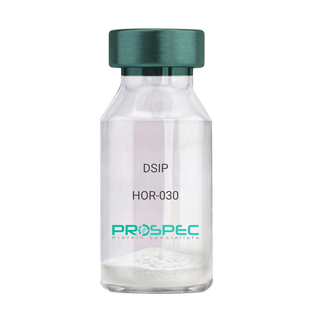
- View Data Sheet
Name :
DSIPDescription:
Delta Sleep Inducing Peptide
Product # :
HOR-030Price :
Quantity :
Shipping Method :
Shipped at Room temp
More Info
- description
- formulation
- purity
- More Info
Description
DSIP Synthetic is a single, non-glycosylated polypeptide chain containing 15 amino acids, having a molecular mass of 1419.55 Dalton and a Molecular formula of C62H98N16O22.
Formulation
The protein was lyophilized with no additives.
Purity
Greater than 97.0% as determined by analysis by RP-HPLC.
More Info
-
Physical Appearance
Sterile Filtered White lyophilized (freeze-dried) powder.
-
Stability
Lyophilized DSIP although stable at room temperature for 3 weeks, should be stored desiccated below -18°C. Upon reconstitution DSIP should be stored at 4°C between 2-7 days and for future use below -18°C. For long term storage it is recommended to add a carrier protein (0.1% HSA or BSA). Please prevent freeze-thaw cycles.
-
Solubility
It is recommended to reconstitute the lyophilized DSIP in sterile 18MΩ-cm H2O not less than 100 µg/ml, which can then be further diluted to other aqueous solutions.
-
Amino Acid Sequence
H-Gly-Glu-Pro-Pro-Pro-Gly-Lys-Pro-Ala-Asp-Asp-Ala-Gly-Leu-Val-OH
-
Background
Delta Sleep-Inducing Peptide (DSIP), also known as Sleep-Promoting Peptide, is a neuropeptide that has been the subject of extensive research due to its potential role in sleep regulation, stress response, and neuroprotection. This nonapeptide, first isolated from the cerebral venous blood of rabbits during sleep, has been shown to induce slow-wave sleep, modulate pain perception, and exhibit potential antioxidant and immunomodulatory properties.
DSIP's primary function is its interaction with the sleep regulatory system. By modulating the release of certain neurotransmitters, DSIP can influence sleep patterns, particularly promoting slow-wave sleep, the most restorative stage of sleep. Studies by Kovalzon et al. (2011) have demonstrated that DSIP can enhance sleep quality in rats, suggesting potential applications in sleep disorders and the promotion of healthy sleep patterns.
In addition to its sleep-inducing effects, DSIP has been shown to possess neuroprotective properties. Research by Zolotarev et al. (2014) found that DSIP could protect neurons from oxidative stress, suggesting potential applications in the treatment of neurodegenerative diseases such as Alzheimer's and Parkinson's disease.
Given its sleep-inducing and neuroprotective effects, DSIP has been proposed as a potential therapeutic agent for a variety of conditions, including sleep disorders, chronic pain, and neurodegenerative diseases. For instance, a study by Spong et al. (2016) found that DSIP could improve sleep quality in patients with chronic insomnia, indicating its potential as a therapeutic agent in the treatment of sleep disorders.
While research on DSIP is promising, it is important to note that most studies have been conducted in animals or in vitro. More research is needed to fully understand the potential effects and applications of DSIP in humans. However, the existing body of research suggests that DSIP could be a promising tool in the treatment of sleep disorders, chronic pain, and neurodegenerative diseases.
ProSpec's products are furnished for LABORATORY RESEARCH USE ONLY. The product may not be used as drugs, agricultural or pesticidal products, food additives or household chemicals.
- View Data Sheet
Name :
Gliadin NativeDescription:
Gliadin Triticum Aestivum Grain Native
Product # :
PRO-2675Price :
Quantity :
Shipping Method :
Shipped with Ice Packs
More Info
- description
- formulation
- purity
- More Info
Description
The native Gliadin Triticum Aestivum Grain is purified from wheat by protein chemical methods.
Formulation
Gliadin is supplied in 20mM HEPES buffer pH-7.4 and 6M Urea.
Purity
Greater than 95.0% as determined by SDS-PAGE.
More Info
-
Introduction
Gliadin is a common substrate of transglutaminase, which generates neo-epitopes by deamidation of glutamine side chains. In the past, serologic tests for gliadin antibodies usually were not very precise and were not enough for accurate diagnosis due to missing deamidated epitopes within the authentic gliadin fraction traditionally used in diagnostic test kits. ProSpec's deamidated Gliadin isoform matches to the deamidated neo-epitopes, which in the natural antigen are formed by transglutaminase-mediated glutamine side chain deamidation.
-
Physical Appearance
Sterile Filtered clear solution.
-
Stability
Store at 4°C if entire vial will be used within 2-4 weeks. Store, frozen at -20°C for longer periods of time. Avoid multiple freeze-thaw cycles.
-
Immunological Functions
1. Binds IgG and IgA-type human auto antibodies in sera of patients diagnosed with celiac disease.2. Immunodot analysis with positive/negative samples.
-
Applications
Western blot with patient sample.
ProSpec's products are furnished for LABORATORY RESEARCH USE ONLY. The product may not be used as drugs, agricultural or pesticidal products, food additives or household chemicals.
- View Data Sheet
Name :
LMNA RatDescription:
Lamin A/C Rat Recombinant
Prelamin-A/C, Lamin-A/C, 70 kDa lamin, LMNA, LMN1, Renal carcinoma antigen NY-REN-32, Progerin.
Product # :
PRO-2667Price :
Quantity :
Shipping Method :
Shipped with Ice Packs
More Info
- description
- source
- formulation
- purity
- More Info
Description
LMNA Rat Recombinant fused with a His tag produced in E.Coli is a single, non-glycosylated, polypeptide chain containing 614 amino acids and having a molecular mass of 68.0kDa. The LMNA is purified by proprietary chromatographic techniques.
Source
Escherichia Coli.
Formulation
The LMNA solution contains 20mM Tris-HCl pH 7.5, 1mM DTT, 0.3M NaCl, 1.5mM EDTA and 10%(v/v) glycerol.
Purity
Greater than 90.0% as determined by SDS-PAGE.
More Info
-
Introduction
Lamin-A is a major component of the nuclear lamina, a dynamic meshwork located just under the nuclear envelope and it is encoded by lamin A/C gene (LMNA).
Lamin-A is synthesized as Prelamin A, a longer precursor that in vivo goes through a serial post-translational modifications that lead to mature Lamin A.
Diverse mutations in the Lamin A/C gene are associated with different diseases that are collectively called laminophaties, including Emery-Dreifuss muscular dystrophy, familiar partial lipodystrophy, limb girdle muscular dystrophy, dilated cardiomyopathy, Charcot-Marie-Tooth disease, and Hutchinson-Gilford progeria syndrome. -
Synonyms
Prelamin-A/C, Lamin-A/C, 70 kDa lamin, LMNA, LMN1, Renal carcinoma antigen NY-REN-32, Progerin.
-
Physical Appearance
Sterile filtered colorless solution.
-
Stability
Store at 4°C if entire vial will be used within 2-4 weeks.Store, frozen at -20°C for longer periods of time.For long term storage it is recommended to add a carrier protein (0.1% HSA or BSA).Avoid multiple freeze-thaw cycles.
-
Amino Acid Sequence
METPSQRRAT RSGAQASSTP LSPTRITRLQ EKEDLQELND RLAVYIDRVR SLETENAGLR LRITESEEVV SREVSGIKAA YEAELGDARK TLDSVAKERA RLQLELSKVR EEFKELKARN TKKEGDLLAA QARLKDLEAL LNSKEAALST ALSEKRTLEG ELHDLRGQVA KLEAALGEAK KQLQDEMLRR VDAENRLQTL KEELDFQKNI YSEELRETKR RHETRLVEID NGKQREFESR LADALQELRA QHEDQVEQYK KELEKTYSAK LDNARQSAER NSNLVGAAHE ELQQSRIRID SLSAQLSQLQ KQLAAKEAKL RDLEDSLARE RDTSRRLLAE KEREMAEMRA RMQQQLDEYQ ELLDIKLALD MEIHAYRKLL EGEEERLRLS PSPTSQRSRG RASSHSSQSQ GGGSVTKKRK LESSESRSSF SQHARTSGRV AVEEVDEEGK FVRLRNKSNE DQSMGNWQIR RQNGDDPLMT YRFPPKFTLK AGQVVTIWAS GAGATHSPPT DLVWKAQNTW GCGSSLRTAL INATGEEVAM RKLVRSLTMV EDDEDEDGDD LLHHHHGSHC SSSGDPAEYN LRSRTVLCGT CGQPADKASA SGSGAQVSSQ NCSIM
ProSpec's products are furnished for LABORATORY RESEARCH USE ONLY. The product may not be used as drugs, agricultural or pesticidal products, food additives or household chemicals.
- View Data Sheet
Name :
METRN MouseDescription:
Meteorin Mouse Recombinant
Meteorin, Hypoxia/reoxygenation regulatory factor, Metrn, 1810034B16Rik, Hyrac.
Product # :
PRO-2241Price :
Quantity :
Shipping Method :
Shipped with Ice Packs
More Info
- description
- source
- formulation
- purity
- More Info
Description
METRN produced in Sf9 Baculovirus cells is a single, glycosylated polypeptide chain containing 276 amino acids (22-291 a.a.) and having a molecular mass of 30.2kDa (Migrates at 28-40kDa on SDS-PAGE under reducing conditions). METRN is expressed with a 6 amino acid His tag at C-Terminus and purified by proprietary chromatographic techniques.
Source
Sf9, Baculovirus cells.
Formulation
METRN protein solution (0.25mg/ml) contains Phosphate Buffered Saline (pH 7.4) and 10% glycerol.
Purity
Greater than 90.0% as determined by SDS-PAGE.
More Info
-
Introduction
Meteorin (METRN) is involved in both glial cell differentiation and axonal network formation during neurogenesis. METRN promotes astrocyte differentiation and transforms cerebellar astrocytes into radial glia. Moreover, the METRN protein stimulates axonal extension in small and intermediate neurons of sensory ganglia by activating nearby satellite glia.
-
Synonyms
Meteorin, Hypoxia/reoxygenation regulatory factor, Metrn, 1810034B16Rik, Hyrac.
-
Physical Appearance
Sterile Filtered colorless solution.
-
Stability
Store at 4°C if entire vial will be used within 2-4 weeks. Store, frozen at -20°C for longer periods of time. For long term storage it is recommended to add a carrier protein (0.1% HSA or BSA). Avoid multiple freeze-thaw cycles.
-
Amino Acid Sequence
GYSEDRCSWR GSGLTQEPGS VGQLTLDCTE GAIEWLYPAG ALRLTLGGPD PGTRPSIVCL RPERPFAGAQ VFAERMTGNL ELLLAEGPDL AGGRCMRWGP RERRALFLQA TPHRDISRRV AAFRFELHED QRAEMSPQAQ GLGVDGACRP CSDAELLLAA CTSDFVIHGT IHGVAHDTEL QESVITVVVA RVIRQTLPLF KEGSSEGQGR ASIRTLLRCG VRPGPGSFLF MGWSRFGEAW LGCAPRFQEF SRVYSAALTT HLNPCEMALD HHHHHH.
ProSpec's products are furnished for LABORATORY RESEARCH USE ONLY. The product may not be used as drugs, agricultural or pesticidal products, food additives or household chemicals.
- View Data Sheet
Name :
SEMA7A HumanDescription:
Semaphorin 7A Human Recombinant
Semaphorin-7A, CDw108, JMH blood group antigen, John-Milton-Hargen human blood group Ag, Semaphorin-K1, Sema K1, Semaphorin-L, Sema L, CD108.
Product # :
PRO-2506Price :
Quantity :
Shipping Method :
Shipped with Ice Packs
More Info
- description
- source
- formulation
- purity
- More Info
Description
SEMA7A produced in Sf9 Insect cells is a single, glycosylated polypeptide chain containing 846 amino acids (45-648 a.a.) and having a molecular mass of 95.7kDa (Molecular size on SDS-PAGE will appear at approximately 70-100kDa).SEMA7A is expressed with an 242 amino acid hIgG-His tag at C-Terminus and purified by proprietary chromatographic techniques.
Source
Sf9, Insect cells.
Formulation
SEMA7A protein solution (0.25mg/ml) contains Phosphate Buffered Saline (pH 7.4) and 10% glycerol.
Purity
Greater than 90.0% as determined by SDS-PAGE.
More Info
-
Introduction
SEMA7A (semaphorin-7A isoform 1), is a membrane-bound semaphorin which is linked with cell surfaces via a glycosylphosphatidylinositol (GPI) linkage. SEMA7A encourages formation of focal adhesion complexes, activation of the protein kinase PTK2/FAK1 and subsequent phosphorylation of MAPK3 & MAPK1. SEMA7A takes a vital part in integrin-mediated signaling and functions in regulating cell migration as well as immune responses. In addition, SEMA7A promotes the production of proinflammatory cytokines by macrophages & monocytes.
-
Synonyms
Semaphorin-7A, CDw108, JMH blood group antigen, John-Milton-Hargen human blood group Ag, Semaphorin-K1, Sema K1, Semaphorin-L, Sema L, CD108.
-
Physical Appearance
Sterile filtered colorless solution.
-
Stability
Store at 4°C if entire vial will be used within 2-4 weeks. Store, frozen at -20°C for longer periods of time. For long term storage it is recommended to add a carrier protein (0.1% HSA or BSA).Avoid multiple freeze-thaw cycles.
-
Amino Acid Sequence
ADPQGHLRSG PRIFAVWKGH VGQDRVDFGQ TEPHTVLFHE PGSSSVWVGG RGKVYLFDFP EGKNASVRTV NIGSTKGSCL DKRDCENYIT LLERRSEGLL ACGTNARHPS CWNLVNGTVV PLGEMRGYAP FSPDENSLVL FEGDEVYSTI RKQEYNGKIP RFRRIRGESE LYTSDTVMQN PQFIKATIVH QDQAYDDKIY YFFREDNPDK NPEAPLNVSR VAQLCRGDQG GESSLSVSKW NTFLKAMLVC SDAATNKNFN RLQDVFLLPD PSGQWRDTRV YGVFSNPWNY SAVCVYSLGD IDKVFRTSSL KGYHSSLPNP RPGKCLPDQQ PIPTETFQVA DRHPEVAQRV EPMGPLKTPL FHSKYHYQKV AVHRMQASHG ETFHVLYLTT DRGTIHKVVE PGEQEHSFAF NIMEIQPFRR AAAIQTMSLD AERRKLYVSS QWEVSQVPLD LCEVYGGGCH GCLMSRDPYC GWDQGRCISI YSSERSVLQS INPAEPHKEC PNPKPDKAPL QKVSLAPNSR YYLSCPMESR HATYSWRHKE NVEQSCEPGH QSPNCILFIE NLTAQQYGHY FCEAQEGSYF REAQHWQLLP EDGIMAEHLL GHACALALEP KSCDKTHTCP PCPAPELLGG PSVFLFPPKP KDTLMISRTP EVTCVVVDVS HEDPEVKFNW YVDGVEVHNA KTKPREEQYN STYRVVSVLT VLHQDWLNGK EYKCKVSNKA LPAPIEKTIS KAKGQPREPQ VYTLPPSRDE LTKNQVSLTC LVKGFYPSDI AVEWESNGQP ENNYKTTPPV LDSDGSFFLY SKLTVDKSRW QQGNVFSCSV MHEALHNHYT QKSLSLSPGK HHHHHH.
ProSpec's products are furnished for LABORATORY RESEARCH USE ONLY. The product may not be used as drugs, agricultural or pesticidal products, food additives or household chemicals.
- View Data Sheet
Name :
PDCL HumanDescription:
Phosducin-Like Human Recombinant
Phosducin-like protein, PHLP, DKFZp564M1863.
Product # :
PRO-1141Price :
Quantity :
Shipping Method :
Shipped with Ice Packs
More Info
- description
- source
- formulation
- purity
- More Info
Description
PDCL Human Recombinant produced in E. coli is a single polypeptide chain containing 325 amino acids (1-301) and having a molecular mass of 36.8 kDa.PDCL is fused to a 24 amino acid His-tag at N-terminus & purified by proprietary chromatographic techniques.
Source
E.coli.
Formulation
The PDCL solution (0.5mg/1ml) contains 20mM Tris-HCl buffer (pH 8.0), 100mM NaCl, 1mM DTT and 20% glycerol.
Purity
Greater than 90% as determined by SDS-PAGE.
More Info
-
Introduction
Phosducin-like protein (PDCL) is a member of the phosducin family. PDCL is a putative modulator of heterotrimeric G proteins. PDCL shares broad amino acid sequence homology with phosducin, a phosphoprotein expressed in the retina and pineal gland. Both PDCL and phosphoducin regulate G-protein signaling by binding to the beta-gamma subunits of G proteins.
-
Synonyms
Phosducin-like protein, PHLP, DKFZp564M1863.
-
Physical Appearance
Sterile Filtered colorless solution.
-
Stability
Store at 4°C if entire vial will be used within 2-4 weeks. Store, frozen at -20°C for longer periods of time. For long term storage it is recommended to add a carrier protein (0.1% HSA or BSA).Avoid multiple freeze-thaw cycles.
-
Amino Acid Sequence
MGSSHHHHHH SSGLVPRGSH MGSHMTTLDD KLLGEKLQYY YSSSEDEDSD HEDKDRGRCA PASSSVPAEA ELAGEGISVN TGPKGVINDW RRFKQLETEQ REEQCREMER LIKKLSMTCR SHLDEEEEQQ KQKDLQEKIS GKMTLKEFAI MNEDQDDEEF LQQYRKQRME EMRQQLHKGP QFKQVFEISS GEGFLDMIDK EQKSIVIMVH IYEDGIPGTE AMNGCMICLA AEYPAVKFCK VKSSVIGASS QFTRNALPAL LIYKGGELIG NFVRVTDQLG DDFFAVDLEA FLQEFGLLPE KEVLVLTSVR NSATCHSEDS DLEID
ProSpec's products are furnished for LABORATORY RESEARCH USE ONLY. The product may not be used as drugs, agricultural or pesticidal products, food additives or household chemicals.
- View Data Sheet
Name :
CCDC69 HumanDescription:
Coiled-Coil Domain Containing 69 Human Recombinant
Coiled-coil domain-containing protein 69, CCDC69.
Product # :
PRO-1880Price :
Quantity :
Shipping Method :
Shipped with Ice Packs
More Info
- description
- source
- formulation
- purity
- More Info
Description
CCDC69 Human Recombinant produced in E.Coli is a single, non-glycosylated polypeptide chain containing 319 amino acids (1-296 a.a) and having a molecular mass of 37.2kDa. CCDC69 is fused to a 23 amino acid His-tag at N-terminus & purified by proprietary chromatographic techniques.
Source
Escherichia Coli.
Formulation
CCDC69 protein solution (0.25mg/ml) containing 20mM Tris-HCl buffer (pH 8.0), 0.15M NaCl, 10% glycerol and 1mM DTT.
Purity
Greater than 90.0% as determined by SDS-PAGE.
More Info
-
Introduction
Coiled-Coil Domain Containing 69, also known as CCDC69 is a protein-coding gene. A significant paralog of CCDC69 is MTUS1.
-
Synonyms
Coiled-coil domain-containing protein 69, CCDC69.
-
Physical Appearance
Sterile Filtered colorless solution.
-
Stability
Store at 4°C if entire vial will be used within 2-4 weeks. Store, frozen at -20°C for longer periods of time. For long term storage it is recommended to add a carrier protein (0.1% HSA or BSA).Avoid multiple freeze-thaw cycles.
-
Amino Acid Sequence
MGSSHHHHHH SSGLVPRGSH MGSMGCRHSR LSSCKPPKKK RQEPEPEQPP RPEPHELGPL NGDTAITVQL CASEEAERHQ KDITRILQQH EEEKKKWAQQ VEKERELELR DRLDEQQRVL EGKNEEALQV LRASYEQEKE ALTHSFREAS STQQETIDRL TSQLEAFQAK MKRVEESILS RNYKKHIQDY GSPSQFWEQE LESLHFVIEM KNERIHELDR RLILMETVKE KNLILEEKIT TLQQENEDLH VRSRNQVVLS RQLSEDLLLT REALEKEVQL RRQLQQEKEE LLYRVLGANA SPAFPLAPVT PTEVSFLAT.
ProSpec's products are furnished for LABORATORY RESEARCH USE ONLY. The product may not be used as drugs, agricultural or pesticidal products, food additives or household chemicals.
- View Data Sheet
Name :
CXCL8 Human (1-72)Description:
Interleukin-8 (1-72 a.a.) Human Recombinant (CXCL8)
IL-8, CXCL8, Monocyte-derived neutrophil chemotactic factor, MDNCF, T-cell chemotactic factor, Neutrophil-activating protein 1, NAP-1, Protein 3-10C, Granulocyte chemotactic protein 1, GCP-1, Monocyte-derived neutrophil-activating peptide, MONAP, Emoctakin, K60, NAF, LECT, LUCT, 3-10C, LYNAP, SCYB8, TSG-1, AMCF-I, b-ENAP.
Product # :
CHM-231Price :
Quantity :
Shipping Method :
Shipped at Room temp
More Info
- description
- source
- formulation
- purity
- biological activity
- More Info
Description
Interleukin-8 Human Recombinant produced in E.Coli is a single, non-glycosylated polypeptide chain containing 72 amino acids and having a molecular mass of 8452 Dalton. The IL-8 is purified by proprietary chromatographic techniques.
Source
Escherichia Coli.
Formulation
The IL-8 was lyophilized from a concentrated (1mg/ml) solution in water containing no additives.
Purity
Greater than 98.0% as determined by:
(a) Analysis by RP-HPLC.
(b) Analysis by SDS-PAGE.Biological Activity
Specific Activity of IL8 in chemotaxis of donor PBL neutrophils, threshold concentration corresponding to 10-100 ng/ml corresponding to a Specific Activity of 10,000-100,000IU/mg.More Info
-
Introduction
Interleukin-8 (IL-8) is a chemokine produced by macrophages and other cell types such as epithelial cells. It is also synthesized by endothelial cells, which store IL-8 in their storage vesicles, the Weibel-Palade bodies. When first encountering an antigen, the primary cells to encounter it are the macrophages who phagocytose the particle. Upon processing, they release chemokines to signal other immune cells to come in to the site of inflammation. IL-8 is one such chemokine. It serves as a chemical signal that attracts neutrophils at the site of inflammation, and therefore is also known as Neutrophil Chemotactic Factor.
-
Synonyms
IL-8, CXCL8, Monocyte-derived neutrophil chemotactic factor, MDNCF, T-cell chemotactic factor, Neutrophil-activating protein 1, NAP-1, Protein 3-10C, Granulocyte chemotactic protein 1, GCP-1, Monocyte-derived neutrophil-activating peptide, MONAP, Emoctakin, K60, NAF, LECT, LUCT, 3-10C, LYNAP, SCYB8, TSG-1, AMCF-I, b-ENAP.
-
Physical Appearance
Sterile Filtered White lyophilized (freeze-dried) powder.
-
Stability
Lyophilized Interleukin-8 although stable at room temperature for 3 weeks, should be stored desiccated below -18°C. Upon reconstitution CXCL8 should be stored at 4°C between 2-7 days and for future use below -18°C. For long term storage it is recommended to add a carrier protein (0.1% HSA or BSA).Please prevent freeze-thaw cycles.
-
Solubility
It is recommended to reconstitute the lyophilized Interleukin-8 in sterile 18MΩ-cm H2O not less than 100µg/ml, which can then be further diluted to other aqueous solutions.
-
Amino Acid Sequence
The sequence of the first five N-terminal amino acids was determined and was found to be Ser-Ala-Lys-Glu-Leu.
-
Background
What is the molecular weight/Mw of CXCL8 HUMAN (1-72) Protein?
CXCL8 HUMAN (1-72) Protein has a total Mw of 8.45kDa.
What is the source or expression system of CXCL8 HUMAN (1-72) Protein?
Escherichia Coli.
What is the Purity of CXCL8 HUMAN (1-72) Protein?
CXCL8 HUMAN (1-72) Protein is >98% pure as determined by SDS-PAGE.
What is the Biological Activity of CXCL8 HUMAN (1-72) Protein?
Specific Activity of IL8 in chemotaxis of donor PBL neutrophils, threshold concentration corresponding to 10-100 ng/ml corresponding to a Specific Activity of 10,000-100,000IU/mg.
What is the amino acid sequence of CXCL8 HUMAN (1-72) Protein?
The sequence of the first five N-terminal amino acids was determined and was found to be Ser-Ala-Lys-Glu-Leu.
What applications can CXCL8 HUMAN (1-72) Protein be used in?
CXCL8 HUMAN (1-72) Protein can probably be used in western blot, ELISA and Lateral Flow.
What is the endotoxin level for CXCL8 HUMAN (1-72) Protein?
The endotoxin level is minimal, CXCL8 HUMAN (1-72) Protein was purified using conventional chromatography techniques.
ProSpec's products are furnished for LABORATORY RESEARCH USE ONLY. The product may not be used as drugs, agricultural or pesticidal products, food additives or household chemicals.
- View Data Sheet
Name :
IL 11 MouseDescription:
Interleukin-11 Mouse Recombinant
AGIF, Adipogenesis inhibitory factor, IL-11, Interleukin-11, Il11.
Product # :
CYT-646Price :
Quantity :
Shipping Method :
Shipped at Room temp
More Info
- description
- source
- formulation
- purity
- biological activity
- More Info
Description
Interleukin-11 Mouse Recombinant produced in E.Coli is a single, non-glycosylated polypeptide chain containing 179 amino acids and having a molecular mass of 19.1kDa. The Mouse IL-11 is purified by proprietary chromatographic techniques.
Source
Escherichia Coli.
Formulation
The protein was lyophilized from a 0.2µm filtered concentrated (1mg/ml) solution in PBS, pH 7.4.
Purity
Greater than 97.0% as determined by:
(a) Analysis by RP-HPLC.
(b) Analysis by SDS-PAGE.Biological Activity
The ED50 as determined by the dose-dependant stimulation of the proliferation of murine T11 was found to be less than 2.0 ng/ml, corresponding to a specific activity of 500,000IU/mg.More Info
-
Introduction
IL11 is a member of the gp130 family of cytokines. These cytokines drive the assembly of multisubunit receptor complexes, all of which contain at least one molecule of the transmembrane signaling receptor IL6ST (gp130). IL-11 is shown to stimulate the T-cell-dependent development of immunoglobulin-producing B cells. It is also found to support the proliferation of hematopoietic stem cells and megakaryocyte progenitor cells.
-
Synonyms
AGIF, Adipogenesis inhibitory factor, IL-11, Interleukin-11, Il11.
-
Physical Appearance
Sterile Filtered White lyophilized (freeze-dried) powder.
-
Stability
Lyophilized Interleukin-11 although stable at room temperature for 3 weeks, should be stored desiccated below -18°C. Upon reconstitution IL11 should be stored at 4°C between 2-7 days and for future use below -18°C.For long term storage it is recommended to add a carrier protein (0.1% HSA or BSA).Please prevent freeze-thaw cycles.
-
Solubility
It is recommended to reconstitute the lyophilized Interleukin 11 in sterile 18MΩ-cm H2O not less than 100µg/ml, which can then be further diluted to other aqueous solutions.
-
Amino Acid Sequence
MPGPPAGSPR VSSDPRADLD SAVLLTRSLL ADTRQLAAQM RDKFPADGDH SLDSLPTLAM SAGTLGSLQL PGVLTRLRVD LMSYLRHVQW LRRAGGPSLK TLEPELGALQ ARLERLLRRL QLLMSRLALP QAAPDQPVIP LGPPASAWGS IRAAHAILGG LHLTLDWAVR GLLLLKTRL.
ProSpec's products are furnished for LABORATORY RESEARCH USE ONLY. The product may not be used as drugs, agricultural or pesticidal products, food additives or household chemicals.
- View Data Sheet
Name :
PECAM1 HumanDescription:
Platelet Endothelial Cell Adhesion Molecule 1 Human Recombinant
Platelet endothelial cell adhesion molecule, PECAM1, CD31, CD31/EndoCAM, endoCAM, GPIIA', PECA1, PECAM-1, Platelet Endothelial Cell Adhesion Molecule 1.
Product # :
CYT-924Price :
Quantity :
Shipping Method :
Shipped with Ice Packs
More Info
- description
- source
- formulation
- purity
- More Info
Description
PECAM1 Human Recombinant produced in Sf9 Baculovirus is a single, non-glycosylated polypeptide chain containing 582 amino acids (28-601a.a.) and having a molecular mass of 65.5kDa (Migrates at 70-100kDa on SDS-PAGE under reducing conditions).PECAM1 is fused to an 8 amino acid His-tag at C-terminus & purified by proprietary chromatographic techniques.
Source
Sf9, Baculovirus cells.
Formulation
PECAM1 protein solution (0.25mg/ml) containing Phosphate Buffered Saline (pH 7.4) and 10% glycerol.
Purity
Greater than 95.0% as determined by SDS-PAGE.
More Info
-
Introduction
Platelet endothelial cell adhesion molecule (PECAM1) induces susceptibility to atherosclerosis. PECAM1 averts phagocyte ingestion of closely apposed viable cells by transmitting detachment signals, and transforms function upon apoptosis, thus promoting tethering of dying cells to phagocytes. The encounter of a viable cell with a phagocyte via the homophilic interaction of PECAM1 on both cell surfaces as a result causing the viable cell's active repulsion from the phagocyte.
-
Synonyms
Platelet endothelial cell adhesion molecule, PECAM1, CD31, CD31/EndoCAM, endoCAM, GPIIA', PECA1, PECAM-1, Platelet Endothelial Cell Adhesion Molecule 1.
-
Physical Appearance
Sterile filtered colorless solution.
-
Stability
Store at 4°C if entire vial will be used within 2-4 weeks. Store, frozen at -20°C for longer periods of time. For long term storage it is recommended to add a carrier protein (0.1% HSA or BSA).Avoid multiple freeze-thaw cycles.
-
Amino Acid Sequence
QENSFTINSV DMKSLPDWTV QNGKNLTLQC FADVSTTSHV KPQHQMLFYK DDVLFYNISS MKSTESYFIP EVRIYDSGTY KCTVIVNNKE KTTAEYQVLV EGVPSPRVTL DKKEAIQGGI VRVNCSVPEE KAPIHFTIEK LELNEKMVKL KREKNSRDQN FVILEFPVEE QDRVLSFRCQ ARIISGIHMQ TSESTKSELV TVTESFSTPK FHISPTGMIM EGAQLHIKCT IQVTHLAQEF PEIIIQKDKA IVAHNRHGNK AVYSVMAMVE HSGNYTCKVE SSRISKVSSI VVNITELFSK PELESSFTHL DQGERLNLSC SIPGAPPANF TIQKEDTIVS QTQDFTKIAS KSDSGTYICT AGIDKVVKKS NTVQIVVCEM LSQPRISYDA QFEVIKGQTI EVRCESISGT LPISYQLLKT SKVLENSTKN SNDPAVFKDN PTEDVEYQCV ADNCHSHAKM LSEVLRVKVI APVDEVQISI LSSKVVESGE DIVLQCAVNE GSGPITYKFY REKEGKPFYQ MTSNATQAFW TKQKASKEQE GEYYCTAFNR ANHASSVPRS KILTVRVILA PWKKVEHHHH HH.
ProSpec's products are furnished for LABORATORY RESEARCH USE ONLY. The product may not be used as drugs, agricultural or pesticidal products, food additives or household chemicals.
- View Data Sheet
Name :
ANGPTL7 HumanDescription:
Angiopoietin-like Protein 7 Human Recombinant
angiopoietin like 7, AngX, CDT6, dJ647M16.1, Angiopoietin-like factor; Angiopoietin-like Protein 7, ANGPTL7, angiopoietinrelated protein 7, Cornea-derived transcript 6 protein
Product # :
CYT-1208Price :
Quantity :
Shipping Method :
Shipped with Ice Packs
More Info
- description
- source
- formulation
- purity
- More Info
Description
ANGPTL7 Human Recombinant produced in HEK293 cells is a single, glycosylated polypeptide chain (27-346a.a) containing 553amino acids and having a molecular mass of 63.2kDa.ANGPTL7 is fused to a 233 amino acid hIgG-His-Tag at C-terminus & purified by proprietary chromatographic techniques.
Source
HEK293 cells.
Formulation
ANGPTL7 protein (0.25mg/ml) contains 10% glycerol and Phosphate-Buffered Saline (pH 7.4).
Purity
Greater than 90.0% as determined by SDS-PAGE.
More Info
-
Introduction
Angiopoietin-related protein 7 (ANGPTL7), belongs to the angiopoietin-like family of molecules. ANGPTL7 is expressed in the corneal stroma, trabecular meshwork, and sclera. ANGPTL7 production is up-regulated in trabecular meshwork cells by glucocorticoids and TGF-Beta and in cartilage by TNF-alpha.
-
Synonyms
angiopoietin like 7, AngX, CDT6, dJ647M16.1, Angiopoietin-like factor; Angiopoietin-like Protein 7, ANGPTL7, angiopoietinrelated protein 7, Cornea-derived transcript 6 protein
-
Physical Appearance
Sterile filtered colorless solution.
-
Stability
Store at 4°C if entire vial will be used within 2-4 weeks.Store, frozen at -20°C for longer periods of time.For long term storage it is recommended to add a carrier protein (0.1% HSA or BSA).Avoid multiple freeze-thaw cycles.
-
Amino Acid Sequence
QKLSKHKTPA QPQLKAANCC EEVKELKAQV ANLSSLLSEL NKKQERDWVS VVMQVMELES NSKRMESRLT DAESKYSEMN NQIDIMQLQA AQTVTQTSAD AIYDCSSLYQ KNYRISGVYK LPPDDFLGSP ELEVFCDMET SGGGWTIIQR RKSGLVSFYR DWKQYKQGFG SIRGDFWLGN EHIHRLSRQP TRLRVEMEDW EGNLRYAEYS HFVLGNELNS YRLFLGNYTG NVGNDALQYH NNTAFSTKDK DNDNCLDKCA QLRKGGYWYN CCTDSNLNGV YYRLGEHNKH LDGITWYGWH GSTYSLKRVE MKIRPEDFKP LEPKSCDKTH TCPPCPAPEL LGGPSVFLFP PKPKDTLMIS RTPEVTCVVV DVSHEDPEVK FNWYVDGVEV HNAKTKPREE QYNSTYRVVS VLTVLHQDWL NGKEYKCKVS NKALPAPIEK TISKAKGQPR EPQVYTLPPS RDELTKNQVS LTCLVKGFYP SDIAVEWESN GQPENNYKTT PPVLDSDGSF FLYSKLTVDK SRWQQGNVFS CSVMHEALHN HYTQKSLSLS PGK
-
Background
Angiopoietin-like Protein 7 Human Recombinant: An Emerging Player in Metabolic Regulation and Therapeutic Potential
Abstract:
Angiopoietin-like protein 7 (ANGPTL7) is a multifunctional protein that has recently gained attention for its potential role in metabolic regulation and as a therapeutic target for metabolic disorders. ANGPTL7 is involved in the modulation of lipid metabolism, adipogenesis, and insulin signaling. The availability of human recombinant ANGPTL7 protein has provided researchers with a valuable tool to unravel its biological functions and explore its therapeutic applications. This review provides an overview of the current knowledge on ANGPTL7 and discusses its potential as a therapeutic intervention in metabolic disorders.
Introduction:
Metabolic disorders, including obesity and type 2 diabetes, pose significant health challenges worldwide. ANGPTL7, a member of the angiopoietin-like protein family, has recently emerged as a potential regulator of metabolic processes. ANGPTL7 affects lipid metabolism, adipose tissue biology, and insulin signaling pathways, making it an intriguing target for therapeutic interventions in metabolic disorders.
Role of ANGPTL7 in Metabolic Regulation:
ANGPTL7 plays a multifaceted role in metabolic regulation. It influences lipid metabolism by regulating lipoprotein lipase (LPL) activity and lipid uptake in adipose tissue and skeletal muscle. ANGPTL7 also affects adipocyte biology and adipogenesis, potentially contributing to the development of obesity and related metabolic complications. Furthermore, ANGPTL7 modulates insulin signaling and glucose metabolism, suggesting its involvement in insulin resistance and diabetes pathogenesis.
Mechanisms of ANGPTL7 Action:
ANGPTL7 exerts its effects through various mechanisms. It interacts with extracellular matrix components, influencing cell adhesion and migration. ANGPTL7 also regulates angiogenesis and vascular remodeling, potentially linking it to metabolic regulation and tissue homeostasis.
Therapeutic Potential of ANGPTL7 Human Recombinant Protein:
The availability of ANGPTL7 human recombinant protein offers new avenues for therapeutic interventions in metabolic disorders. Modulating ANGPTL7 activity through recombinant protein administration or targeted interventions may have significant implications for lipid metabolism, adipose tissue function, and insulin sensitivity. Exploring ANGPTL7 as a therapeutic target holds promise for the development of novel strategies to tackle metabolic disorders.
Conclusion:
ANGPTL7 is an emerging player in metabolic regulation with potential therapeutic implications for metabolic disorders. Its involvement in lipid metabolism, adipose tissue biology, and insulin signaling pathways highlights its importance in maintaining metabolic homeostasis. The availability of ANGPTL7 human recombinant protein opens up new possibilities for further investigations and the development of targeted interventions for metabolic disorders.
What is the molecular weight/Mw of ANGPTL7 Protein?
ANGPTL7 Protein has a total Mw of 63.2kDa.
What is the source or expression system of ANGPTL7 Protein?
HEK293 cells.
What is the Purity of ANGPTL7 Protein?
ANGPTL7 Protein is >90% pure as determined by SDS-PAGE.
What is the Biological Activity of ANGPTL7 Protein?
The biological functionality of ANGPTL7 Protein will be determined in the future.
What is the amino acid sequence of ANGPTL7 Protein?
QKLSKHKTPA QPQLKAANCC EEVKELKAQV ANLSSLLSEL NKKQERDWVS VVMQVMELES NSKRMESRLT DAESKYSEMN NQIDIMQLQA AQTVTQTSAD AIYDCSSLYQ KNYRISGVYK LPPDDFLGSP ELEVFCDMET SGGGWTIIQR RKSGLVSFYR DWKQYKQGFG SIRGDFWLGN EHIHRLSRQP TRLRVEMEDW EGNLRYAEYS HFVLGNELNS YRLFLGNYTG NVGNDALQYH NNTAFSTKDK DNDNCLDKCA QLRKGGYWYN CCTDSNLNGV YYRLGEHNKH LDGITWYGWH GSTYSLKRVE MKIRPEDFKP LEPKSCDKTH TCPPCPAPEL LGGPSVFLFP PKPKDTLMIS RTPEVTCVVV DVSHEDPEVK FNWYVDGVEV HNAKTKPREE QYNSTYRVVS VLTVLHQDWL NGKEYKCKVS NKALPAPIEK TISKAKGQPR EPQVYTLPPS RDELTKNQVS LTCLVKGFYP SDIAVEWESN GQPENNYKTT PPVLDSDGSF FLYSKLTVDK SRWQQGNVFS CSVMHEALHN HYTQKSLSLS PGK
What applications can ANGPTL7 Protein be used in?
ANGPTL7 Protein can probably be used in western blot, ELISA and Lateral Flow.
What is the endotoxin level for ANGPTL7 Protein?
The endotoxin level is minimal, ANGPTL7 Protein was purified using conventional chromatography techniques.
ProSpec's products are furnished for LABORATORY RESEARCH USE ONLY. The product may not be used as drugs, agricultural or pesticidal products, food additives or household chemicals.
- View Data Sheet
Name :
HDL HumanDescription:
High Density Lipoprotein Human
High Density Lipoprotein, HDL.
Product # :
PRO-559Price :
Quantity :
Shipping Method :
Shipped with Ice Packs
More Info
- description
- source
- purity
- More Info
Description
Human High Density Lipoprotein (HDL) produced in Human plasma.
Source
Human plasma.
Purity
Cholesterol level >200mg/l. <1% LDL.
More Info
-
Introduction
HDL is a complex of lipids and proteins in approximately equal amounts that functions as a transporter of cholesterol in the blood. HDL is the smallest of the lipoprotein particles; it’s the densest because it contains the highest proportion of protein. The liver synthesizes these lipoproteins as complexes of apolipoproteins and phospholipid, which bear a resemblance to cholesterol-free flattened spherical lipoprotein particles. They pick up cholesterol; carry it internally, from cells by interaction with the ATP Binding Cassette Transporter A1. Lecithin-cholesterol acyltransferase (plasma enzyme) converts the free cholesterol into cholesteryl ester (a more hydrophobic form of cholesterol) which is then sequestered into the core of the lipoprotein particle ultimately making the newly synthesized HDL spherical. They increase in size as they pass through the bloodstream and integrate more cholesterol and phospholipid molecules from cells and other lipoproteins, for instance by the interaction with the ABCG1 transporter and the phospholipid transport protein. HDL transports their cholesterol generally to the liver or steroidogenic organs such as adrenals, ovary and testes by direct and indirect pathways. The release of HDL cholesterol to adrenals, ovaries and testes is important for the synthesis of steroid hormones. Since the triglycerides are not stable in HDL, they’re degraded by hepatic lipase in order that finally small HDL particles are left which resume the uptake of cholesterol from cells. The cholesterol supplied to the liver is excreted into the bile and consequently intestine either directly or indirectly after conversion into bile acids. High levels of HDL are associated with a decreased risk of atherosclerosis and coronary heart disease.
-
Synonyms
High Density Lipoprotein, HDL.
-
Physical Appearance
Yellow to orange liquid.
-
Stability
Human HDL although stable at 4°C for 1 week, should be stored below -15°C (short term i.e. < 3 months) and below -70°C for long term.
-
Human Virus Test
Starting material donor tested and found negative for HIV I & II antibodies, Hepatitis B surface antigen, Hepatitis C antibodies, HIV1/HCV/HBV NAT and Syphilis.
ProSpec's products are furnished for LABORATORY RESEARCH USE ONLY. The product may not be used as drugs, agricultural or pesticidal products, food additives or household chemicals.