Skip to content

High-quality research proteins.

  • 1-800-805-6531

  • Mon - Sat 8.00-18.00, Sun - Closed

  • info@prospecbio.com

  • PO Box 6591 East Brunswick NJ 08816

  • Products
    • Cytokines
    • Growth factors
    • Chemokines
    • Cd antigens
    • Neurotrophins
    • Hormones
    • Enzymes
    • Viral antigens
    • Recombinant proteins
    • Natural proteins
    • Monoclonal antibodies
    • Polyclonal antibodies
    • Thermal Packaging
  • About
    • Company
    • Founder
    • Contribution
  • Customers
    • Eastern Asia
    • Eastern Europe
    • North America
    • South America
    • South East Asia
    • Southern Asia
    • Western Europe
  • Ordering
    • Ordering Method
    • Ordering - Credit Card
    • Ordering - PO
    • Shipping Fees
    • FAQs
  • Services
    • DNA Cloning
    • Protein Expression
    • Monoclonal Antibody
    • Fermentation
    • Technology License
  • Contact us
Log in

Country/region

  • USD $ | Albania
  • USD $ | Andorra
  • USD $ | Argentina
  • USD $ | Armenia
  • USD $ | Australia
  • USD $ | Austria
  • USD $ | Azerbaijan
  • USD $ | Belarus
  • USD $ | Belgium
  • USD $ | Bolivia
  • USD $ | Brazil
  • USD $ | Bulgaria
  • USD $ | Cambodia
  • USD $ | Canada
  • USD $ | Chile
  • USD $ | China
  • USD $ | Colombia
  • USD $ | Costa Rica
  • USD $ | Croatia
  • USD $ | Cyprus
  • USD $ | Czechia
  • USD $ | Denmark
  • USD $ | Estonia
  • USD $ | Finland
  • USD $ | France
  • USD $ | Georgia
  • USD $ | Germany
  • USD $ | Greece
  • USD $ | Hong Kong SAR
  • USD $ | Hungary
  • USD $ | Iceland
  • USD $ | India
  • USD $ | Ireland
  • USD $ | Israel
  • USD $ | Italy
  • USD $ | Japan
  • USD $ | Kazakhstan
  • USD $ | Latvia
  • USD $ | Liechtenstein
  • USD $ | Lithuania
  • USD $ | Madagascar
  • USD $ | Malta
  • USD $ | Mexico
  • USD $ | Moldova
  • USD $ | Monaco
  • USD $ | Netherlands
  • USD $ | New Zealand
  • USD $ | North Macedonia
  • USD $ | Norway
  • USD $ | Panama
  • USD $ | Paraguay
  • USD $ | Peru
  • USD $ | Philippines
  • USD $ | Poland
  • USD $ | Portugal
  • USD $ | Romania
  • USD $ | Singapore
  • USD $ | Slovakia
  • USD $ | Slovenia
  • USD $ | South Africa
  • USD $ | South Korea
  • USD $ | Spain
  • USD $ | Sri Lanka
  • USD $ | Sweden
  • USD $ | Switzerland
  • USD $ | Taiwan
  • USD $ | Thailand
  • USD $ | Türkiye
  • USD $ | Ukraine
  • USD $ | United Kingdom
  • USD $ | United States
  • USD $ | Uruguay
  • USD $ | Vatican City
  • USD $ | Venezuela
  • USD $ | Vietnam
  • Facebook
  • Instagram
logoProSpec - Recombinant Protein Manufacturer for academic and R&D industry
Become a distributor
Account Log in
Wishlist
Cart Cart(0)
  • Products
    +
    • Cytokines
    • Growth factors
    • Chemokines
    • Cd antigens
    • Neurotrophins
    • Hormones
    • Enzymes
    • Viral antigens
    • Recombinant proteins
    • Natural proteins
    • Monoclonal antibodies
    • Polyclonal antibodies
    • Thermal Packaging
  • About
    +
    • Company
    • Founder
    • Contribution
  • Customers
    +
    • Eastern Asia
    • Eastern Europe
    • North America
    • South America
    • South East Asia
    • Southern Asia
    • Western Europe
  • Ordering
    +
    • Ordering Method
    • Ordering - Credit Card
    • Ordering - PO
    • Shipping Fees
    • FAQs
  • Services
    +
    • DNA Cloning
    • Protein Expression
    • Monoclonal Antibody
    • Fermentation
    • Technology License
  • Contact us

Item added to your cart

View cart
View All
  • Cytokines
  • Growth factors
  • Chemokines
  • Cd antigens
  • Neurotrophins
  • Hormones
  • Enzymes
  • Viral antigens
  • Recombinant proteins
  • Natural proteins
  • Monoclonal antibodies
  • Polyclonal antibodies
  • Thermal Packaging
  • Tumor Necrosis Factor

    Tumor Necrosis Factor

    View All
  • 4-1BB

    4-1BB

    View All
  • Adiponectin

    Adiponectin

    View All
  • AIF1

    AIF1

    View All
  • Other Cytokines

    Other Cytokines

    View All
  • AITRL

    AITRL

    View All
  • Angiopoietin

    Angiopoietin

    View All
  • Apolipoprotein

    Apolipoprotein

    View All
  • B-Cell Activating Factor

    B-Cell Activating Factor

    View All
  • Beta Defensin

    Beta Defensin

    View All
  • Bone Morphogenetic Protein

    Bone Morphogenetic Protein

    View All
  • B type Natriuretic Peptide

    B type Natriuretic Peptide

    View All
  • BST

    BST

    View All
  • Betacellulin

    Betacellulin

    View All
  • Cardiotrophin

    Cardiotrophin

    View All
  • Activin

    Activin

    View All
  • Fibroblast Growth Factor

    Fibroblast Growth Factor

    View All
  • Other Growth Factors

    Other Growth Factors

    View All
  • CSF

    CSF

    View All
  • CTGF

    CTGF

    View All
  • IGFBP

    IGFBP

    View All
  • VEGF Protein

    VEGF Protein

    View All
  • EGF

    EGF

    View All
  • Epigen

    Epigen

    View All
  • Erythropoietin

    Erythropoietin

    View All
  • Insulin-Like Growth Factor

    Insulin-Like Growth Factor

    View All
  • Growth Hormone

    Growth Hormone

    View All
  • HDGF

    HDGF

    View All
  • Hepatocyte Growth Factor

    Hepatocyte Growth Factor

    View All
  • Insulin

    Insulin

    View All
  • BCA-1/ BLC (CXCL13)

    BCA-1/ BLC (CXCL13)

    View All
  • BRAK (CXCL14)

    BRAK (CXCL14)

    View All
  • C-10 (CCL6)

    C-10 (CCL6)

    View All
  • Other Chemokines

    Other Chemokines

    View All
  • MEC (CCL28)

    MEC (CCL28)

    View All
  • LD78-beta (CCL3L1)

    LD78-beta (CCL3L1)

    View All
  • CTACK (CCL27)

    CTACK (CCL27)

    View All
  • CXCL16

    CXCL16

    View All
  • CXCL17

    CXCL17

    View All
  • Platelet Factor-4 (CXCL4)

    Platelet Factor-4 (CXCL4)

    View All
  • ENA-78 (CXCL5)

    ENA-78 (CXCL5)

    View All
  • Eotaxin (CCL11,24,26)

    Eotaxin (CCL11,24,26)

    View All
  • Exodus-2 (CCL21)

    Exodus-2 (CCL21)

    View All
  • Fractalkine (CX3CL1)

    Fractalkine (CX3CL1)

    View All
  • CXCL6

    CXCL6

    View All
  • Other CD Antigens

    Other CD Antigens

    View All
  • CD14

    CD14

    View All
  • CD164

    CD164

    View All
  • CD1

    CD1

    View All
  • CD2

    CD2

    View All
  • CD200

    CD200

    View All
  • CD207

    CD207

    View All
  • CD226

    CD226

    View All
  • CD23

    CD23

    View All
  • CD244

    CD244

    View All
  • CD247

    CD247

    View All
  • CD27

    CD27

    View All
  • CD274

    CD274

    View All
  • CD300

    CD300

    View All
  • CD33

    CD33

    View All
  • Other Neurotrophins

    Other Neurotrophins

    View All
  • Beta-NGF

    Beta-NGF

    View All
  • BDNF

    BDNF

    View All
  • CDNF

    CDNF

    View All
  • CNTF

    CNTF

    View All
  • GDNF

    GDNF

    View All
  • Glia Maturation Factor

    Glia Maturation Factor

    View All
  • MANF

    MANF

    View All
  • Midkine

    Midkine

    View All
  • Neuroglobin

    Neuroglobin

    View All
  • Neurotrophic factors

    Neurotrophic factors

    View All
  • Neuregulin

    Neuregulin

    View All
  • Neuropilin

    Neuropilin

    View All
  • Pigment Epithelium-Derived Factor

    Pigment Epithelium-Derived Factor

    View All
  • Pleiotrophin

    Pleiotrophin

    View All
  • Peptide Hormones

    Peptide Hormones

    View All
  • Other Hormones

    Other Hormones

    View All
  • HCG

    HCG

    View All
  • Endothelin

    Endothelin

    View All
  • Exendin

    Exendin

    View All
  • FSH

    FSH

    View All
  • Glucagon

    Glucagon

    View All
  • GHRP

    GHRP

    View All
  • GLP

    GLP

    View All
  • Thyrostimulin

    Thyrostimulin

    View All
  • Inhibin A

    Inhibin A

    View All
  • LHRH

    LHRH

    View All
  • Melanotan

    Melanotan

    View All
  • Procalcitonin

    Procalcitonin

    View All
  • PTH

    PTH

    View All
  • Peroxiredoxin

    Peroxiredoxin

    View All
  • Peptidase

    Peptidase

    View All
  • Synthetase

    Synthetase

    View All
  • Transferase

    Transferase

    View All
  • Hydrolase

    Hydrolase

    View All
  • Dehydrogenase

    Dehydrogenase

    View All
  • ACE2

    ACE2

    View All
  • Esterase

    Esterase

    View All
  • Protein Kinases

    Protein Kinases

    View All
  • Other Enzymes

    Other Enzymes

    View All
  • Phosphatase

    Phosphatase

    View All
  • Deaminase

    Deaminase

    View All
  • Oxygenase

    Oxygenase

    View All
  • Adenylate Kinase

    Adenylate Kinase

    View All
  • Lyase

    Lyase

    View All
  • Chagas

    Chagas

    View All
  • SARS

    SARS

    View All
  • Influenza

    Influenza

    View All
  • ASFV

    ASFV

    View All
  • Astrovirus

    Astrovirus

    View All
  • Borrelia

    Borrelia

    View All
  • Helicobacter Pylori

    Helicobacter Pylori

    View All
  • Chikungunya

    Chikungunya

    View All
  • Chlamydia

    Chlamydia

    View All
  • Cytomegalo

    Cytomegalo

    View All
  • Dengue

    Dengue

    View All
  • EBV

    EBV

    View All
  • Ebola

    Ebola

    View All
  • Feline Leukemia Virus

    Feline Leukemia Virus

    View All
  • Hepatitis A

    Hepatitis A

    View All
  • Other

    Other

    View All
  • Actin

    Actin

    View All
  • ADAM

    ADAM

    View All
  • Complement Component

    Complement Component

    View All
  • Ag85

    Ag85

    View All
  • Eukaryotic Translation Initiation Factor

    Eukaryotic Translation Initiation Factor

    View All
  • Anterior Gradient Protein

    Anterior Gradient Protein

    View All
  • Heat Shock Protein

    Heat Shock Protein

    View All
  • Alpha-2-HS-Glycoprotein

    Alpha-2-HS-Glycoprotein

    View All
  • Hemoglobin

    Hemoglobin

    View All
  • Allergy

    Allergy

    View All
  • Anaplasma

    Anaplasma

    View All
  • Angiogenin

    Angiogenin

    View All
  • Ankyrin Repeat Domain

    Ankyrin Repeat Domain

    View All
  • Annexin

    Annexin

    View All
  • Other Natural Proteins

    Other Natural Proteins

    View All
  • Aprotinin

    Aprotinin

    View All
  • Transferrin

    Transferrin

    View All
  • Avidin

    Avidin

    View All
  • Anti Coagulation Factors

    Anti Coagulation Factors

    View All
  • Natural Albumin

    Natural Albumin

    View All
  • Natural Coagulation Factors

    Natural Coagulation Factors

    View All
  • Fibronectin

    Fibronectin

    View All
  • Thrombin

    Thrombin

    View All
  • Anti Human Enzyme

    Anti Human Enzyme

    View All
  • Other Monoclonal Antibodies

    Other Monoclonal Antibodies

    View All
  • Anti Human Cytokine

    Anti Human Cytokine

    View All
  • Anti Human Heat Shock Protein

    Anti Human Heat Shock Protein

    View All
  • Anti Mouse Lymphocyte

    Anti Mouse Lymphocyte

    View All
  • Anti Human Chemokine

    Anti Human Chemokine

    View All
  • Anti Human Lymphocyte

    Anti Human Lymphocyte

    View All
  • Anti Viral Monoclonal

    Anti Viral Monoclonal

    View All
  • Anti Mouse Cytokine

    Anti Mouse Cytokine

    View All
  • Anti-GST Monoclonal

    Anti-GST Monoclonal

    View All
  • Other Polyclonal Antibodies

    Other Polyclonal Antibodies

    View All
  • Anti Viral

    Anti Viral

    View All
  • Anti-GST Monoclonal

    Anti-GST Monoclonal

    View All
Back

Search results

1000 results found for “eph receptor”

Name

Description

Product #

Price

Quantity

Shipping Method

  • View Data Sheet

    Name :

    TNFRSF14 Human

    Description:

    HVEM-Fc Human Recombinant

    Tumor Necrosis Factor Receptor Superfamily Member 14, HVEM, TR2, Herpes Virus Entry Mediator A, Tumor Necrosis Factor Receptor-Like 2, Herpesvirus Entry Mediator, HVEA, ATAR, CD270, LIGHTR, CD40-Like Protein, Tumor Necrosis Factor Receptor-Like Gene2.

    Product # :

    CYT-821

    Price :

    Quantity :

    Shipping Method :

    Room Temp Icon

    Shipped at Room temp

    Add To Cart

    More Info

    • description
    • source
    • formulation
    • purity
    • biological activity
    • More Info

    Description

    TNFRSF14 Human Recombinant produced in Pichia Pastoris is a single, glycosylated, polypeptide chain containing 396 amino acids and having a molecular mass of 58.0kDa. However, TNFRSF14 migrates with an apparent molecular mass of 70 kDa in SDS-PAGE under reducing conditions.

    Source

    Pichia Pastoris.

    Formulation

    TNFRSF14 protein was lyophilized from a 0.2 µm filtered concentrated solution in PBS, pH7.4, with 3 % Trehalose.

    Purity

    Greater than 95.0% as determined by: (a) Analysis by RP-HPLC.(b) Analysis by SDS-PAGE.

    Biological Activity

    Fully biologically active when compared to standard. The biologically active as determined by its ability to inhibit TNF-beta -mediated cytotoxicity using murine L929 cells.

    More Info

    • Introduction

      TNFRSF14, a member of the TNF receptor superfamily, is a type I transmembrane protein. TNFRSF14 is expressed in peripheral blood T cells, B cells, monocytes and in various tissues enriched in lymphoid cells. TNFRSF14 operates as a co-stimulatory factor for the activation of lymphoid cells and as a deterrent to infection by herpesvirus. Additionally, TNFRSF14 encourages the proliferation of T cells, and triggers apoptosis of various tumor cells.

    • Synonyms

      Tumor Necrosis Factor Receptor Superfamily Member 14, HVEM, TR2, Herpes Virus Entry Mediator A, Tumor Necrosis Factor Receptor-Like 2, Herpesvirus Entry Mediator, HVEA, ATAR, CD270, LIGHTR, CD40-Like Protein, Tumor Necrosis Factor Receptor-Like Gene2.

    • Physical Appearance

      Sterile Filtered White lyophilized (freeze-dried) powder.

    • Stability

      Lyophilized TNFRSF14 although stable at room temperature for 3 weeks, should be stored desiccated below -18°C. Upon reconstitution TNFRSF14 should be stored at 4°C between 2-7 days and for future use below -18°C. For long term storage it is recommended to add a carrier protein (0.1% HSA or BSA). Please prevent freeze-thaw cycles.

    • Solubility

      It is recommended to reconstitute the lyophilized TNFRSF14 in sterile 18MΩ-cm H2O not less than 100µg/ml, which can then be further diluted to other aqueous solutions.

    • Amino Acid Sequence

      LPSCKEDEYP VGSECCPKCS PGYRVKEACG ELTGTVCEPC PPGTYIAHLN GLSKCLQCQM CDPAMGLRAS RNCSRTENAV CGCSPGHFCI VQDGDHCAAC RAYATSSPGQ RVQKGGTESQ DTLCQNCPPG TFSPNGTLEE CQHQTKCSWL VTKAGAGTSS SHWVEPKSSD KTHTCPPCPA PEFEGAPSVF LFPPKPKDTL MISRTPEVTC VVVDVSHEDP EVKFNWYVDG VEVHNAKTKP REEQYNSTYR VVSVLTVLHQ DWLNGKEYKC KVSNKALPTP IEKTISKAKG QPREPQVYTL PPSRDELTKN QVSLTCLVKG FYPSDIAVEW ESNGQPENNY KTTPPVLDSD GSFFLYSKLT VDKSRWQQGN VFSCSVMHEA LHNHYTQKSL SLSPGK

    ProSpec's products are furnished for LABORATORY RESEARCH USE ONLY. The product may not be used as drugs, agricultural or pesticidal products, food additives or household chemicals.

    Tnfrsf14 Human
  • View Data Sheet

    Name :

    OLR1 Human, Sf9

    Description:

    Oxidized Low Density Lipoprotein Receptor 1 Human Recombinant, Sf9

    Oxidized Low Density Lipoprotein Receptor 1, C-Type Lectin Domain Family 8 Member A, Lectin-Type Oxidized LDL Receptor 1, CLEC8A, HLOX-1, LOX1, Oxidised Low Density Lipoprotein (Lectin-Like) Receptor 1, Oxidized Low Density Lipoprotein (Lectin-Like) Receptor 1, Oxidized Low-Density Lipoprotein Receptor 1, Soluble Form, Oxidized Low-Density Lipoprotein Receptor 1, Scavenger Receptor Class E, Member 1, Lectin-Like Oxidized LDL Receptor 1, Lectin-Like OxLDL Receptor 1, Ox LDL Receptor 1, Ox-LDL Receptor 1, SCARE1, LOXIN, SLOX1, LOX-1, Oxidized low-density lipoprotein receptor 1, Ox-LDL receptor 1, C-type lectin domain family 8 member A, LOX-1, Lectin-like oxLDL receptor 1, hLOX-1, Lectin-type oxidized LDL receptor 1.

    Product # :

    PRO-2373

    Price :

    Quantity :

    Shipping Method :

    Ice Icon

    Shipped with Ice Packs

    Add To Cart

    More Info

    • description
    • source
    • formulation
    • purity
    • More Info

    Description

    OLR1 Human Recombinant produced in Sf9 Baculovirus cells is a single, glycosylated polypeptide chain containing 225 amino acids (58-273a.a) and having a molecular mass of 25.8kDa (Molecular size on SDS-PAGE will appear at approximately 28-40kDa). OLR1 is fused to a 6 amino acid His-tag at C-terminus & purified by proprietary chromatographic techniques.

    Source

    Sf9, Baculovirus cells.

    Formulation

    OLR1 protein solution (1mg/ml) containing Phosphate Buffered Saline (pH 7.4) and 10% glycerol.

    Purity

    Greater than 95% as determined by SDS-PAGE.

    More Info

    • Introduction

      OLR1 is a type II membrane protein which belongs to the C-type lectin family and performs as a cell-surface receptor for Ox-LDL. Ox-LDL has a part in early ather-osclerosis, which includes the transformation of monocyte-derived macro-phages to foam cells in atherosclerotic lesions. In addition, OLR1 protein triggers the activation of the NF?B signal transduction pathway.

    • Synonyms

      Oxidized Low Density Lipoprotein Receptor 1, C-Type Lectin Domain Family 8 Member A, Lectin-Type Oxidized LDL Receptor 1, CLEC8A, HLOX-1, LOX1, Oxidised Low Density Lipoprotein (Lectin-Like) Receptor 1, Oxidized Low Density Lipoprotein (Lectin-Like) Receptor 1, Oxidized Low-Density Lipoprotein Receptor 1, Soluble Form, Oxidized Low-Density Lipoprotein Receptor 1, Scavenger Receptor Class E, Member 1, Lectin-Like Oxidized LDL Receptor 1, Lectin-Like OxLDL Receptor 1, Ox LDL Receptor 1, Ox-LDL Receptor 1, SCARE1, LOXIN, SLOX1, LOX-1, Oxidized low-density lipoprotein receptor 1, Ox-LDL receptor 1, C-type lectin domain family 8 member A, LOX-1, Lectin-like oxLDL receptor 1, hLOX-1, Lectin-type oxidized LDL receptor 1.

    • Physical Appearance

      Sterile Filtered colorless solution.

    • Stability

      Store at 4°C if entire vial will be used within 2-4 weeks. Store, frozen at -20°C for longer periods of time. For long term storage it is recommended to add a carrier protein (0.1% HSA or BSA).Avoid multiple freeze-thaw cycles.

    • Amino Acid Sequence

      ADPMQLSQVS DLLTQEQANL THQKKKLEGQ ISARQQAEEA SQESENELKE MIETLARKLN EKSKEQMELH HQNLNLQETL KRVANCSAPC PQDWIWHGEN CYLFSSGSFN WEKSQEKCLS LDAKLLKINS TADLDFIQQA ISYSSFPFWM GLSRRNPSYP WLWEDGSPLM PHLFRVRGAV SQTYPSGTCA YIQRGAVYAE NCILAAFSIC QKKANLRAQH HHHHH.

    ProSpec's products are furnished for LABORATORY RESEARCH USE ONLY. The product may not be used as drugs, agricultural or pesticidal products, food additives or household chemicals.

    Olr1 Human Sf9
  • View Data Sheet

    Name :

    TNFR2 Human Fc

    Description:

    Tumor Necrosis Factor Receptor 2 Fusion Protein Human Recombinant

    Tumor necrosis factor receptor superfamily member 1B,Tumor necrosis factor receptor 2, TNF-R2, Tumor necrosis factor receptor type II, p75, p80 TNF-alpha receptor, CD120b antigen, Etanercept, TBPII, TNFBR, TNFR80, TNF-R75, p75TNFR, TNF-R-II.

    Product # :

    CYT-422

    Price :

    Quantity :

    Shipping Method :

    Room Temp Icon

    Shipped at Room temp

    Add To Cart

    More Info

    • description
    • source
    • formulation
    • purity
    • biological activity
    • More Info

    Description

    Recombinant Human Tumor Necrosis Factor Receptor 2 Fusion Protein produced in CHO is a dimeric, glycosylated, polypeptide chain consisting of the extracellular ligand-binding portion of the human 75 kilo Dalton (p75) tumor necrosis factor receptor 2 (TNFR2) linked to the Fc portion of human IgG1. The Fc component of TNFR2 contains the CH2 domain, the CH3 domain and hinge region, but not the CH1 domain of IgG1. It consists of 934 amino acids and has an apparent molecular weight of approximately 150 kilo Daltons.The TNFR2 is purified by standard chromatographic techniques.

    Source

    Chinese Hamster Ovarian Cells (CHO).

    Formulation

    Each mg contains 1.6mg mannitol, 0.4 mg sucrose and 48 µg tromethamine.

    Purity

    Greater than 98.0% as determined by:
    (a) Analysis by RP-HPLC.
    (c) Analysis by SDS-PAGE.

    Biological Activity

    Potency is determined by its ability to neutralize TNF-alpha mediated growth inhibition of A375 cells, corresponding to a Specific Activity of 17,000,000 IU/mg.

    More Info

    • Introduction

      TNFR binds specifically to tumor necrosis factor (TNF) and blocks its interaction with cell surface TNF receptors. TNF is a naturally occurring cytokine that is involved in normal inflammatory and immune responses. It plays an important role in the inflammatory processes of rheumatoid arthritis (RA), polyarticular-course juvenile rheumatoid arthritis (JRA), and ankylosing spondylitis and the resulting joint pathology. In addition, TNF plays a role in the inflammatory process of plaque psoriasis. Elevated levels of TNF are found in involved tissues and fluids of patients with RA, psoriatic arthritis, ankylosing spondylitis (AS), and plaque psoriasis. Two distinct receptors for TNF (TNFRs), a 55 kilodalton protein (p55) and a 75 kilodalton protein (p75), exist naturally as monomeric molecules on cell surfaces and in soluble forms. Biological activity of TNF is dependent upon binding to either cell surface TNFR. Recombinant Human TNFR is a dimeric soluble form of the p75 TNF receptor that can bind to two TNF molecules.
      It inhibits the activity of TNF in vitro and has been shown to affect several animal models of inflammation, including murine collagen-induced arthritis. TNFR inhibits binding of both TNF? and TNF? (lymphotoxin alpha [LT?]) to cell surface TNFRs, rendering TNF biologically inactive. Cells expressing transmembrane TNF that bind to TNFR are not lysed in vitro in the presence or absence of complement.
      TNFR can also modulate biological responses that are induced or regulated by TNF, including expression of adhesion molecules responsible for leukocyte migration (i.e., E-selectin and to a lesser extent intercellular adhesion molecule-1 [ICAM-1]), serum levels of cytokines (e.g., IL-6), and serum levels of matrix metalloproteinase-3 (MMP-3 or stromelysin).

    • Synonyms

      Tumor necrosis factor receptor superfamily member 1B,Tumor necrosis factor receptor 2, TNF-R2, Tumor necrosis factor receptor type II, p75, p80 TNF-alpha receptor, CD120b antigen, Etanercept, TBPII, TNFBR, TNFR80, TNF-R75, p75TNFR, TNF-R-II.

    • Physical Appearance

      Sterile Filtered White lyophilized (freeze-dried) powder.

    • Stability

      Lyophilized Tumor Necrosis Factor Receptor although stable at room temperature for 3 weeks, should be stored desiccated below -18°C. Upon reconstitution TNFR2 should be stored at 4°C between 2-7 days and for future use below -18°C.For long term storage it is recommended to add a carrier protein (0.1% HSA or BSA).Please prevent freeze-thaw cycles.

    • Solubility

      It is recommended to reconstitute the lyophilized TNFR2 in sterile 18M-cm H2O not less than 100µg/ml, which can then be further diluted to other aqueous solutions.

    ProSpec's products are furnished for LABORATORY RESEARCH USE ONLY. The product may not be used as drugs, agricultural or pesticidal products, food additives or household chemicals.

    Tnfr2 Human Fc
  • View Data Sheet

    Name :

    Ebola Sudan Protein

    Description:

    Ebola Sudan Recombinant Protein

    Product # :

    EVD-079

    Price :

    Quantity :

    Shipping Method :

    Ice Icon

    Shipped with Ice Packs

    Add To Cart

    More Info

    • description
    • source
    • formulation
    • purity
    • More Info

    Description

    Nucleoprotein (NP) of Ebola virus has strong antigenicity in immune reactions. C-terminal of EBO-S nucleoprotein was expressed and purified from E. coli, it migrated at 15kDa on SDS-PAGE and pI is 4.52. Ebola Sudan was purified by a proprietary chromatographic technique.

    Source

    Escherichia Coli.

    Formulation

    Ebola Sudan protein solution is supplied in Phosphate buffer and 0.02% sodium azide.

    Purity

    >95% pure as determined by 12% SDS-PAGE (Coomassie blue stain).

    More Info

    • Introduction

      Ebolavirus (EVD) belongs to the Filoviridae family of proteins which is comprised of a single-strand, non-infectious RNA genome. EVD genome is about 19,000 base pairs long and covers 7 genes in the order 3'-UTR-NP-VP35-VP40-GP-VP30-VP24-L-5'-UTR. There are 4 different ebolaviruses such as: Zaire (EBO-Z), Sudan (EBO-S), Cote d’Ivoire (EBO-CI) and Reston (EBO-R) that differ in amino acid sequence and location of where the gene overlaps. Similar to filoviruses and ebolavirions, EVD is a filamentous element that could appear in 3 forms: shepherd's crook, U shape or a 6 shape. Ebolaviruses may be coiled, toroid, or branched. Most ebolavirions are 80nm wideand 14,000nm long.

    • Stability

      Store at 4°C if entire vial will be used within 2-4 weeks. Store, frozen at -20°C for longer periods of time. For long term storage it is recommended to add a carrier protein (0.1% HSA or BSA).Avoid multiple freeze-thaw cycles.

    • Applications

      Immunoassay.

    ProSpec's products are furnished for LABORATORY RESEARCH USE ONLY. The product may not be used as drugs, agricultural or pesticidal products, food additives or household chemicals.

    Ebola Sudan
  • View Data Sheet

    Name :

    IL 4 Human, HEK

    Description:

    Interleukin-4 Human Recombinant, HEK

    BCGF, BCDF, B cell stimulating factor, BSF-1, Lymphocyte stimulatory factor 1, IL-4, MGC79402, Binetrakin, Pitrakinra.

    Product # :

    CYT-097

    Price :

    Quantity :

    Shipping Method :

    Room Temp Icon

    Shipped at Room temp

    Add To Cart

    More Info

    • description
    • source
    • formulation
    • purity
    • biological activity
    • More Info

    Description

    IL-4 Human Recombinant produced in HEK cells is a glycosylated monomer, having a molecular weight range of 14-19kDa due to glycosylation.The IL-4 is purified by proprietary chromatographic techniques.

    Source

    HEK.

    Formulation

    The IL-4 was lyophilized from a 0.2µm filtered protein solution (0.68mg/ml) in 1xPBS.

    Purity

    Greater than 95% as obsereved by SDS-PAGE.

    Biological Activity

    The activity was determined by the dose dependent stimulation of the proliferation of human TF-1 cells (human erythroleukemic indicator cell line) and was found to be 0.17ng/ml.

    More Info

    • Introduction

      IL4 is a pleiotropic cytokine produced by activated T cells. IL4 is a ligand for interleukin 4 receptor. The interleukin 4 receptor also binds to IL13, which may contribute to many overlapping functions of this cytokine and IL13. STAT6, a signal transducer and activator of transcription, has been shown to play a central role in mediating the immune regulatory signal of this cytokine. This gene, IL3, IL5, IL13, and CSF2 form a cytokine gene cluster on chromosome 5q, with this gene particularly close to IL13. IL4, IL13 and IL5 are found to be regulated coordinately by several long-range regulatory elements in an over 120 kilobase range on the chromosome. Two alternatively spliced transcript variants of this gene encoding distinct isoforms have been reported.

    • Synonyms

      BCGF, BCDF, B cell stimulating factor, BSF-1, Lymphocyte stimulatory factor 1, IL-4, MGC79402, Binetrakin, Pitrakinra.

    • Physical Appearance

      Sterile Filtered White lyophilized (freeze-dried) powder.

    • Stability

      Lyophilized IL-4 although stable at room temperature for 3 weeks, should be stored desiccated below -18°C. Upon reconstitution IL4 should be stored at 4°C between 2-7 days and for future use below -18°C.For long term storage it is recommended to add a carrier protein (0.1% HSA or BSA).Please prevent freeze-thaw cycles.

    • Solubility

      It is recommended to reconstitute the lyophilized IL-4 in sterile 1xPBS containing 0.1% endotoxin-free recombinant HSA.

    ProSpec's products are furnished for LABORATORY RESEARCH USE ONLY. The product may not be used as drugs, agricultural or pesticidal products, food additives or household chemicals.

    Il 4 Human Hek
  • View Data Sheet

    Name :

    GDNF Human

    Description:

    Glial-Derived Neurotrophic Factor Human Recombinant

    ATF1, ATF2, HFB1-GDNF, GDNF.

    Product # :

    CYT-305

    Price :

    Quantity :

    Shipping Method :

    Room Temp Icon

    Shipped at Room temp

    Add To Cart

    More Info

    • description
    • source
    • formulation
    • purity
    • biological activity
    • More Info
    • sds-page

    Description

    Glial derived Neurotrophic Factor Human Recombinant produced in E.Coli is a non-glycosylated disulfide-linked homodimer containing 2 x 135 amino acids and having a total molecular mass of approximately 30kDa. GDNF is purified by proprietary chromatographic techniques.

    Source

    Escherichia Coli.

    Formulation

    GDNF was lyophilized from a 0.2µm filtered concentrated solution in 1×PBS, pH 7.4 and 5% Trehalose.

    Purity

    Greater than 95.0% as determined by analysis by SDS-PAGE.

    Biological Activity

    The ED50 was determined by the proliferation of rat C6 cells is < 0.1 ng/ml, corresponding to a specific activity of > 1.0x10⁷ units/mg.

    sds-page

    GDNF sds-page - Product image 1

    More Info

    • Introduction

      GDNF promotes the survival and differentiation of minergic neurons in culture, and is able to prevent apoptosis of motor neurons induced by axotomy. The encoded protein is processed to a mature secreted form that exists as a homodimer. The mature form of the protein is a ligand for the product of the RET (rearranged during transfection) protooncogene. In addition to the transcript encoding GDNF, two additional alternative transcripts encoding distinct proteins, referred to as astrocyte-derived trophic factors, have also been described. Mutations in this gene may be associated with Hirschsprung disease.
      GDNF enhances survival and morphological differentiation of minergic neurons and increases their high-affinity uptake.

    • Synonyms

      ATF1, ATF2, HFB1-GDNF, GDNF.

    • Physical Appearance

      Sterile Filtered White lyophilized (freeze-dried) powder.

    • Stability

      Lyophilized Glial-derived Neurotrophic Factor although stable at room temperature for 3 weeks, should be stored desiccated below -18°C. Upon reconstitution GDNF should be stored at 4°C between 2-7 days and for future use below -18°C.For long term storage it is recommended to add a carrier protein (0.1% HSA or BSA).Please prevent freeze-thaw cycles.

    • Solubility

      It is recommended to reconstitute the lyophilized Glial Derived Neurotrophic Factor in sterile 18MΩ-cm H2O not less than 100µg/ml, which can then be further diluted to other aqueous solutions.

    • Amino Acid Sequence

      SPDKQMAVLP RRERNRQAAA ANPENSRGKG RRGQRGKNRG CVLTAIHLNV TDLGLGYETK EELIFRYCSG SCDAAETTYD KILKNLSRNR RLVSDKVGQA CCRPIAFDDD LSFLDDNLVY HILRKHSAKR CGCI.

    • Background

      What is the molecular weight/Mw of GDNF HUMAN Protein?
      GDNF HUMAN Protein has a total Mw of 30kDa.

      What is the source or expression system of GDNF HUMAN Protein?
      Escherichia Coli.

      What is the Purity of GDNF HUMAN Protein?
      GDNF HUMAN Protein is >95% pure as determined by SDS-PAGE.

      What is the Biological Activity of GDNF HUMAN Protein?
      The ED50 was determined by the proliferation of rat C6 cells is < 0.1 ng/ml, corresponding to a specific activity of > 1.0x10⁷ units/mg.

      What is the amino acid sequence of GDNF HUMAN Protein?
      SPDKQMAVLP RRERNRQAAA ANPENSRGKG RRGQRGKNRG CVLTAIHLNV TDLGLGYETK EELIFRYCSG SCDAAETTYD KILKNLSRNR RLVSDKVGQA CCRPIAFDDD LSFLDDNLVY HILRKHSAKR CGCI.
      What applications can GDNF HUMAN Protein be used in?
      GDNF HUMAN Protein can probably be used in western blot, ELISA and Lateral Flow.

      What is the endotoxin level for GDNF HUMAN Protein?
      The endotoxin level is minimal, GDNF HUMAN Protein was purified using conventional chromatography techniques.


    ProSpec's products are furnished for LABORATORY RESEARCH USE ONLY. The product may not be used as drugs, agricultural or pesticidal products, food additives or household chemicals.

    Gdnf Human
  • View Data Sheet

    Name :

    Leptin tA Ovine

    Description:

    Leptin Antagonist Triple Mutant Ovine Recombinant

    Product # :

    CYT-356

    Price :

    Quantity :

    Shipping Method :

    Room Temp Icon

    Shipped at Room temp

    Add To Cart

    More Info

    • description
    • source
    • formulation
    • purity
    • biological activity
    • More Info

    Description

    Leptin Antagonist Triple Mutant Ovine Recombinant is a single non-glycosilated polypeptide chain containing 146 amino and additional Ala at N-terminus acids and having a molecular mass of ~ 16 kDa, Leptin was mutated, resulting in L39A/D40A/F41A mutant. Leptin Antagonist Triple Mutant Ovine Recombinant was purified by proprietary chromatographic techniques.

    Source

    Escherichia coli.

    Formulation

    The protein was lyophilized from a concentrated (0.65mg/ml) solution with 0.003mM NaHCO3.

    Purity

    Greater than 98.0% as determined by:
    (a) Gel filtration analysis.
    (b) Analysis by SDS-PAGE.

    Biological Activity

    ProSpec’s Leptin-Antagonist Triple Mutant Ovine Recombinant is capable of inhibiting leptin-induced proliferation of BAF/3 cells stably transfected with the long form of mouse leptin receptor. It also inhibits various leptin effects in several in vitro bioassays.

    More Info

    • Physical Appearance

      White lyophilized (freeze-dried) powder.

    • Stability

      Lyophilized Leptin Antagonist Triple Mutant Ovine Recombinant although stable at room temperature for several weeks, should be stored desiccated below -18°C. Upon reconstitution at > 0.1 Lep-tA mutant mg/ml and up to 2 mM and filter sterilization Leptin mutant can be stored at 4°C or even room temperature for several weeks making it suitable for long term infusion studies using osmotic pumps. At lower concentration addition of a carrier protein (0.1% HSA or BSA) is suggested. Please prevent freeze-thaw cycles.

    • Solubility

      It is recommended to reconstitute the lyophilized Leptin-Antagonist Triple Mutant Ovine Recombinant in sterile water or sterile 0.4% NaHCO3 adjusted to pH 8, not less than 100µg/ml, which can then be further diluted to other aqueous solutions.

    • Amino Acid Sequence

      The sequence of the first five N-terminal amino acids was determined and was found to be Ala-Val-Pro-Ile-Arg.

    • Protein content

      Protein quantitation was carried out by UV spectroscopy at 280 nm using the absorbency value of 0.21 as the extinction coefficient for a 0.1% (1mg/ml) solution at pH 8.0. This value is calculated by the PC GENE computer analysis program of protein sequences (IntelliGenetics).

    ProSpec's products are furnished for LABORATORY RESEARCH USE ONLY. The product may not be used as drugs, agricultural or pesticidal products, food additives or household chemicals.

    Leptin Ta Ovine
  • View Data Sheet

    Name :

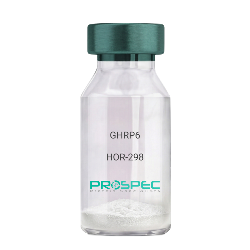
    GHRP6

    Description:

    Growth Hormone Releasing Peptide-6

    GHRP-6, GHRP6

    Product # :

    HOR-298

    Price :

    Quantity :

    Shipping Method :

    Room Temp Icon

    Shipped at Room temp

    Add To Cart

    More Info

    • description
    • formulation
    • purity
    • More Info

    Description

    Growth Hormone Releasing Peptide-6 Synthetic is a single, non-glycosylated polypeptide chain containing 6 amino acids, having a molecular mass of 873 Dalton and a Molecular formula of C46H56N12O6.

    Formulation

    The GHRP-6 peptide was lyophilized with no additives.

    Purity

    Greater than 98.0% as determined by analysis by RP-HPLC.

    More Info

    • Introduction

      GH-releasing peptides (GHRPs) are synthetic peptides that like GHRH act directly on pituitary somatotrophs to stimulate GH release. Growth hormone (GH) release is stimulated by a variety of synthetic secretagogues, of which growth hormone-releasing hexapeptide (GHRP-6) has been most thoroughly studied; it is thought to have actions at both pituitary and hypothalamic site.

    • Synonyms

      GHRP-6, GHRP6

    • Physical Appearance

      Sterile Filtered White lyophilized (freeze-dried) powder.

    • Stability

      Lyophilized Growth Hormone Releasing Peptide-6 although stable at room temperature for 3 weeks, should be stored desiccated below -18°C. Upon reconstitution GHRP-6 should be stored at 4°C between 2-7 days and for future use below -18°C. For long term storage it is recommended to add a carrier protein (0.1% HSA or BSA).Please prevent freeze-thaw cycles.

    • Solubility

      It is recommended to reconstitute the lyophilized GHRP-6 in sterile 18MΩ-cm H2O not less than 100 µg/ml, which can then be further diluted to other aqueous solutions.

    • Amino Acid Sequence

      His-D-Trp-Ala-Trp-D-Phe-Lys-NH2.

    ProSpec's products are furnished for LABORATORY RESEARCH USE ONLY. The product may not be used as drugs, agricultural or pesticidal products, food additives or household chemicals.

    Ghrp 6
  • View Data Sheet

    Name :

    KIR2DL1 Antibody

    Description:

    Killer Cell Immunoglobulin-Like Receptor 2 Domains Long Cytoplasmic Tail 1, Mouse Anti Human

    Killer cell immunoglobulin-like receptor 2DL1, MHC class I NK cell receptor, Natural killer-associated transcript 1, NKAT-1, p58 natural killer cell receptor clones CL-42/47.11, p58 NK receptor, p58.1 MHC class-I-specific NK receptor, CD158 antigen-like family member A, CD158a antigen, KIR2DL1, CD158A, NKAT1, NKAT, p58.1, KIR221, KIR-K64.

    Product # :

    ANT-318

    Price :

    Quantity :

    Shipping Method :

    Ice Icon

    Shipped with Ice Packs

    Add To Cart

    More Info

    • formulation
    • More Info

    Formulation

    1mg/ml containing PBS, pH-7.4, & 0.1% Sodium Azide.

    More Info

    • Introduction

      Killer-cell immunoglobulin-like receptors (KIRs), are a family of cell surface glycoproteins found on Natural Killer (NK) Cells, which are important cells of the immune system. They control the killing function of these cells by interacting with MHC class I molecules, which are expressed on all cell types. This interaction allows them to identify virally infected cells or tumor cells that have a distinctive low level of Class I MHC on their surface. The majority of KIRs are inhibitory, which means that their recognition of MHC suppresses the cytotoxic activity of their NK cell. Only a limited number of KIRs have the capacity to activate cells. The KIR genes are found in a cluster on chromosome 19q13.4 within the 1 Mb leukocyte receptor complex (LRC). KIR molecules are extremely polymorphic, meaning their gene sequences differ significantly between individuals, so that different individuals have different arrays/repertoires of KIR genes. The KIR proteins are categorized by the number of

    • Synonyms

      Killer cell immunoglobulin-like receptor 2DL1, MHC class I NK cell receptor, Natural killer-associated transcript 1, NKAT-1, p58 natural killer cell receptor clones CL-42/47.11, p58 NK receptor, p58.1 MHC class-I-specific NK receptor, CD158 antigen-like family member A, CD158a antigen, KIR2DL1, CD158A, NKAT1, NKAT, p58.1, KIR221, KIR-K64.

    • Physical Appearance

      Sterile Filtered colorless solution.

    • Immunogen

      Anti-human KIR2DL1 mAb is derived from hybridization of mouse SP2/0 myeloma cells with spleen cells from BALB/c mice immunized with recombinant human KIR2DL1 amino acids 23-223 purified from E. coli.

    • Ig Subclass

      Mouse IgG2a heavy chain and κ light chain.

    • Clone

      P2F9AT.

    • Applications

      KIR2DL1 antibody has been tested by ELISA, Western blot and immunoprecipitation analysis to assure specificity and reactivity. Since application varies, however, each investigation should be titrated by the reagent to obtain optimal results. Recommended dilution range for Western blot analysis is 1:500 ~ 2,000. Recommended starting dilution is 1:500.

    • Type

      Mouse Anti Human Monoclonal.

    • Storage Procedures

      For periods up to 1 month store at 4°C, for longer periods of time, store at -20°C. Prevent freeze thaw cycles.

    • Purification Method

      KIR2DL1 antibody was purified from mouse ascitic fluids by protein-G affinity chromatography.

    ProSpec's products are furnished for LABORATORY RESEARCH USE ONLY. The product may not be used as drugs, agricultural or pesticidal products, food additives or household chemicals.

    Kir2Dl1 Antibody
  • View Data Sheet

    Name :

    FADD Antibody

    Description:

    Fas-Associated Death Domain, Mouse Anti Human

    GIG3, MORT1, MGC8528, FADD, Fas (TNFRSF6)-associated via death domain, Protein FADD, FAS-associated death domain protein, FAS-associating death domain-containing protein, Mediator of receptor induced toxicity, Growth-inhibiting gene 3 protein.

    Product # :

    ANT-344

    Price :

    Quantity :

    Shipping Method :

    Ice Icon

    Shipped with Ice Packs

    Add To Cart

    More Info

    • formulation
    • More Info

    Formulation

    1mg/ml containing PBS, pH-7.4, & 0.1% Sodium Azide.

    More Info

    • Introduction

      FADD is an adaptor protein that cooperates with a variety of cell surface receptors and mediates cell apoptotic signals. Using its C-terminal death domain, FADD is recruited by TNFRSF6/Fas-receptor, tumor necrosis factor receptor, TNFRSF25, and TNFSF10/TRAIL-receptor, and consequently it take parts in the death signaling initiated by these receptors. FADD interaction with the receptors reviels the N-terminal effector domain of, which allows it to recruit caspase-8, and thus initiate the cysteine protease cascade. Knockout studies in mice furthermore propose the significance of FADD in premature T cell development. FADD plays a role in survival/proliferation and cell cycle development. FADD also takes part in cellular sublocalization, protein phosphorylation, and inhibitory molecules.

    • Synonyms

      GIG3, MORT1, MGC8528, FADD, Fas (TNFRSF6)-associated via death domain, Protein FADD, FAS-associated death domain protein, FAS-associating death domain-containing protein, Mediator of receptor induced toxicity, Growth-inhibiting gene 3 protein.

    • Immunogen

      Anti-human FADD mAb is derived from hybridization of mouse FO myeloma cells with spleen cells from BALB/c mice immunized with recombinant human FADD amino acids 1-208 purified from E. coli.

    • Ig Subclass

      Mouse IgG2b heavy chain and κ light chain.

    • Clone

      PJ1D2AT.

    • Applications

      FADD antibody has been tested by ELISA, Western blot and immunohistochemistry analysis to assure specificity and reactivity. Since application varies, however, each investigation should be titrated by the reagent to obtain optimal results. Recommended dilution range for Western blot is 1:1,000~1:2,000 and immunohistochemistry analysis is 1:50~100. Recommended starting dilution for Western blot is 1:1,000 and Immunohistochemistry is 1:50.

    • Type

      Mouse Anti Human Monoclonal.

    • Storage Procedures

      For periods up to 1 month store at 4°C, for longer periods of time, store at -20°C. Prevent freeze thaw cycles.

    • Purification Method

      FADD antibody was purified from mouse ascitic fluids by protein-G affinity chromatography.

    ProSpec's products are furnished for LABORATORY RESEARCH USE ONLY. The product may not be used as drugs, agricultural or pesticidal products, food additives or household chemicals.

    Fadd Antibody
  • View Data Sheet

    Name :

    TACI Human

    Description:

    Tumor Necrosis Factor Receptor 13B Human Recombinant

    CD267, CVID, CVID2, TACI, TNFRSF14B, Tumor necrosis factor receptor superfamily, member 13B, Tumor necrosis factor receptor superfamily, member 13B, isoform CRA_a, TNFRSF13B. 

    Product # :

    CYT-819

    Price :

    Quantity :

    Shipping Method :

    Room Temp Icon

    Shipped at Room temp

    Add To Cart

    More Info

    • description
    • source
    • formulation
    • purity
    • biological activity
    • More Info

    Description

    TACI Human Recombinant produced in E.Coli is a single, non-glycosylated, polypeptide chain containing 160 amino acids and having a molecular mass of 18.0kDa.

    Source

    Escherichia Coli.

    Formulation

    TACI protein was lyophilized from a 0.2 µm filtered concentrated solution in PBS, pH 7.4.

    Purity

    Greater than 95.0% as determined by: (a) Analysis by RP-HPLC.(b) Analysis by SDS-PAGE.

    Biological Activity

    Fully biologically active when compared to standard. The biological activity is determined by its ability to block human BAFF induced T2B cell survival using a concentration range of 1.0-3.0 µg/ml corresponding to a specific activity of 334-1000 IU/mg.

    More Info

    • Introduction

      TNFRSF13B (TACI) is a transmembrane receptor protein found predominantly on the surface of B cells (a significant part of the immune system). TACI was at first discovered owing to its ability to interact with calcium-modulator and cyclophilin ligand (CAML). Later on, it was found that TACI plays a key role in humoral immunity by interacting with two members of the TNF family. Also, TACI controls T cell-independent B cell antibody responses, isotype switching, and B cell homeostasis.

    • Synonyms

      CD267, CVID, CVID2, TACI, TNFRSF14B, Tumor necrosis factor receptor superfamily, member 13B, Tumor necrosis factor receptor superfamily, member 13B, isoform CRA_a, TNFRSF13B.

    • Physical Appearance

      Sterile Filtered White lyophilized (freeze-dried) powder.

    • Stability

      Lyophilized TACI although stable at room temperature for 3 weeks, should be stored desiccated below -18°C. Upon reconstitution TACI should be stored at 4°C between 2-7 days and for future use below -18°C. For long term storage it is recommended to add a carrier protein (0.1% HSA or BSA). Please prevent freeze-thaw cycles.

    • Solubility

      It is recommended to reconstitute the lyophilized TACI in sterile 18MΩ-cm H2O not less than 100µg/ml, which can then be further diluted to other aqueous solutions.

    • Amino Acid Sequence

      MSGLGRSRRG GRSRVDQEER FPQGLWTGVA MRSCPEEQYW DPLLGTCMSC KTICNHQSQR TCAAFCRSLS CRKEQGKFYD HLLRDCISCA SICGQHPKQC AYFCENKLRS PVNLPPELRR QRSGEVENNS DNSGRYQGLE HRGSEASPAL PGLKLSADQV

    ProSpec's products are furnished for LABORATORY RESEARCH USE ONLY. The product may not be used as drugs, agricultural or pesticidal products, food additives or household chemicals.

    Human Taci
  • View Data Sheet

    Name :

    VEGF Human

    Description:

    Vascular Endothelial Growth Factor Human Recombinant

    Vascular endothelial growth factor A, VEGF-A, Vascular permeability factor, VPF, VEGF, MGC70609.

    Product # :

    CYT-241

    Price :

    Quantity :

    Shipping Method :

    Room Temp Icon

    Shipped at Room temp

    Add To Cart

    More Info

    • description
    • source
    • formulation
    • purity
    • biological activity
    • More Info

    Description

    Vascular Endothelial Growth Factor Human Recombinant produced in E.Coli is a double, non-glycosylated, polypeptide chain containing 165 amino acids and having a molecular mass of 38.2kDa.The VEGF is purified by proprietary chromatographic techniques.

    Source

    Escherichia Coli.

    Formulation

    The VEGF protein was lyophilized from a concentrated (1mg/ml) solution with no additives.

    Purity

    Greater than 95.0% as determined by:
    (a) Analysis by RP-HPLC.
    (b) Analysis by SDS-PAGE.

    Biological Activity

    Determined by the dose-dependent stimulation of the proliferation of human umbilical vein endothelial cells (HUVEC) using a concentration range of 3.7-5.6 ng/ml, corresponding to a Specific Activity of 178,570-270,270IU/mg.

    More Info

    • Introduction

      Vascular endothelial growth factor (VEGF) is an important signaling protein involved in vessel formation As its name implies, VEGF activity has been mostly studied on cells of the vascular endothelium, although it does have effects on a number of other cell types (e.g. stimulation monocyte/macrophage migration, neurons, cancer cells, kidney epithelial cells ). VEGF mediates increased vascular permeability, induces vasculogenesis and endothelial cell production, promotes cell migration, and inhibits apoptosis. In vitro, VEGF has been shown to stimulate endothelial cell mitogenesis and cell migration. VEGF is also a vasodilator and increases microvascular permeability and was originally referred to as vascular permeability factor. VEGF is located in normal cartilage though only osteoarthritic cartilage expresses the VEGF receptors, NP1, VEGFR1 and VEGFR2. The VEGF level in the culture media from OA chondrocytes was more than 3 folds higher than in media from normal chondrocytes

    • Synonyms

      Vascular endothelial growth factor A, VEGF-A, Vascular permeability factor, VPF, VEGF, MGC70609.

    • Physical Appearance

      Sterile Filtered White lyophilized (freeze-dried) powder.

    • Stability

      Lyophilized VEGF although stable at room temperature for 3 weeks, should be stored desiccated below -18°C. Upon reconstitution VEGF should be stored at 4°C between 2-7 days and for future use below -18°C.For long term storage it is recommended to add a carrier protein (0.1% HSA or BSA).Please prevent freeze-thaw cycles.

    • Solubility

      It is recommended to reconstitute the lyophilized VEGF in sterile 18M-cm H2O not less than 100µg/ml, which can then be further diluted to other aqueous solutions.

    • Amino Acid Sequence

      APMAEGGGQN HHEVVKFMDV YQRSYCHPIE TLVDIFQEYP DEIEYIFKPS CVPLMRCGGC CNDEGLECVP TEESNITMQI MRIKPHQGQH IGEMSFLQHN KCECRPKKDR ARQENPCGPC SERRKHLFVQ DPQTCKCSCK NTDSRCKARQ LELNERTCRC DKPRR

    • Protein content

      VEGF protein quantitation was carried out by two independent methods:1. UV spectroscopy at 280 nm using the absorbency value of 0.2875 as the extinction coefficient for a 0.1% (1mg/ml) solution. This value is calculated by the PC GENE computer analysis program of protein sequences (IntelliGenetics). 2. Analysis by RP-HPLC, using a calibrated solution of VEGF as a Reference Standard.

    ProSpec's products are furnished for LABORATORY RESEARCH USE ONLY. The product may not be used as drugs, agricultural or pesticidal products, food additives or household chemicals.

    Vegf Human
  • View Data Sheet

    Name :

    LECT2 Human

    Description:

    Leukocyte Cell-Derived Chemotaxin 2 Human Recombinant

    Leukocyte Cell-Derived Chemotaxin 2, Leukocyte Cell-Derived Chemotaxin-2, Chondromodulin-II, Chm-II, LECT-2, HLECT2, Chm2, LECT2.

    Product # :

    PRO-2037

    Price :

    Quantity :

    Shipping Method :

    Room Temp Icon

    Shipped at Room temp

    Add To Cart

    More Info

    • description
    • source
    • formulation
    • purity
    • More Info

    Description

    LECT2 Human Recombinant produced in E.Coli is a single, non-glycosylated, polypeptide chain (Gly19-Leu151) containing 143 amino acids including a 10 aa His tag at N-terminus. The total calculated molecular mass is 16kDa.

    Source

    Escherichia Coli.

    Formulation

    LECT2 was filtered (0.4 µm) and lyophilized in 20mM Tris buffer, 50mM NaCl & pH 7.5.

    Purity

    Greater than 95.0% as determined by SDS-PAGE.

    More Info

    • Introduction

      Leukocyte Cell-Derived Chemotaxin 2 (LECT2) functions as a chemotactic factor to neutrophils. LECT2 stimulates the proliferation of chondrocytes and osteoblasts. LECT2 is strongly expressed in the liver and weakly in the testis. LECT2 is a secreted, 16kDa protein which serves as a chemotactic factor to neutrophils and stimulates the growth of chondrocytes and osteoblasts. LECT2 protein has a high sequence similarity to the chondromodulin repeat regions of the chicken myb-induced myeloid 1 protein. A polymorphism in the LECT2 gene is linked with rheumatoid arthritis.

    • Synonyms

      Leukocyte Cell-Derived Chemotaxin 2, Leukocyte Cell-Derived Chemotaxin-2, Chondromodulin-II, Chm-II, LECT-2, HLECT2, Chm2, LECT2.

    • Physical Appearance

      Filtered White lyophilized (freeze-dried) powder.

    • Stability

      Store lyophilized protein at -20°C. Aliquot the product after reconstitution to avoid repeated freezing/thawing cycles. Reconstituted protein can be stored at 4°C for a limited period of time; it does not show any change after two weeks at 4°C.

    • Solubility

      It is recommended to add deionized water to prepare a working stock solution of approximately 0.5mg/ml and let the lyophilized pellet dissolve completely. LECT2 is not sterile! Please filter the product by an appropriate sterile filter before using it in the cell culture.

    • Amino Acid Sequence

      MKHHHHHHASGPWANICAGK SSNEIRTCDR HGCGQYSAQR SQRPHQGVDI LCSAGSTVYA PFTGMIVGQE KPYQNKNAIN NGVRISGRGF CVKMFYIKPI KYKGPIKKGE KLGTLLPLQK VYPGIQSHVH IENCDSSDPT AYL.

    ProSpec's products are furnished for LABORATORY RESEARCH USE ONLY. The product may not be used as drugs, agricultural or pesticidal products, food additives or household chemicals.

    Lect2 Human
  • View Data Sheet

    Name :

    VEGF Mouse

    Description:

    Vascular Endothelial Growth Factor Mouse Recombinant

    Vascular endothelial growth factor A, VEGF-A, Vascular permeability factor, VPF, VEGF, MGC70609.

    Product # :

    CYT-336

    Price :

    Quantity :

    Shipping Method :

    Room Temp Icon

    Shipped at Room temp

    Add To Cart

    More Info

    • description
    • source
    • formulation
    • purity
    • biological activity
    • More Info

    Description

    Vascular Endothelial Growth Factor Mouse Recombinant produced in E.Coli is a disulfide-linked homodimeric, double polypeptide chains containing 165 amino acids and having a molecular mass of 38.8kDa.

    Source

    Escherichia Coli.

    Formulation

    The protein was lyophilized from a 0.2µm filtered solution with PBS, pH 7.4.

    Purity

    Greater than 95.0% as determined by:
    (a) Analysis by RP-HPLC.
    (b) Analysis by SDS-PAGE.

    Biological Activity

    The biological activity is determined by the dose-dependent stimulation of the proliferation of human umbilical vein endothelial cells (HUVEC) using a concentration range of 5.0 ng/ml corresponding to a specific activity of 200,000IU/mg.

    More Info

    • Introduction

      Vascular endothelial growth factor is an important signaling protein involved in both vasculogenesis and angiogenesis. As its name implies, VEGF activity has been mostly studied on cells of the vascular endothelium, although it does have effects on a number of other cell types (e.g. stimulation monocyte/ macrophagemigration, neurons, cancer cells, kidney epithelial cells ). VEGF mediates increased vascular permeability, induces angiogenesis, vasculogenesis and endothelial cell growth, promotes cell migration, and inhibits apoptosis. In vitro, VEGF has been shown to stimulate endothelial cell mitogenesisand cell migration. VEGF is also a vasodilator and increases microvascular permeability and was originally referred to as vascular permeability factor.
      Elevated levels of this protein are linked to POEMS syndrome, also known as Crow-Fukase syndrome. Mutations in this gene have been associated with proliferative and nonproliferative diabetic retinopathy.

    • Synonyms

      Vascular endothelial growth factor A, VEGF-A, Vascular permeability factor, VPF, VEGF, MGC70609.

    • Physical Appearance

      Sterile Filtered White lyophilized (freeze-dried) powder.

    • Stability

      Lyophilized Vascular Endothelial Growth Factor although stable at room temperature for 3 weeks, should be stored desiccated below -18°C. Upon reconstitution VEGF should be stored at 4°C between 2-7 days and for future use below -18°C.For long term storage it is recommended to add a carrier protein (0.1% HSA or BSA).Please prevent freeze-thaw cycles.

    • Solubility

      It is recommended to reconstitute the lyophilized Vascular Endothelial Growth Factor in sterile 18MΩ-cm H2O not less than 100µg/ml, which can then be further diluted to other aqueous solutions.

    • Amino Acid Sequence

      MAPTTEGEQK SHEVIKFMDV YQRSYCRPIE TLVDIFQEYP DEIEYIFKPS CVPLMRCAGC CNDEALECVP TSESNITMQI MRIKPHQSQH IGEMSFLQHS RCECRPKKDR TKPEKHCEPC SERRKHLFVQ DPQTCKCSCK NTDSRCKARQ LELNERTCRC DKPRR.

    ProSpec's products are furnished for LABORATORY RESEARCH USE ONLY. The product may not be used as drugs, agricultural or pesticidal products, food additives or household chemicals.

    Vegf Mouse
  • View Data Sheet

    Name :

    EDN3

    Description:

    Human Endothelin-3

    EDN3, EDN-3, ET-3, ET3, WS4B, HSCR4, MGC15067, MGC61498, Endothelin-3, Preproendothelin-3, PPET3.

    Product # :

    HOR-309

    Price :

    Quantity :

    Shipping Method :

    Room Temp Icon

    Shipped at Room temp

    Add To Cart

    More Info

    • description
    • formulation
    • purity
    • More Info

    Description

    EDN3 contains 21 amino acids having a molecular mass of 2643.08 Dalton, and the molecular formula is: C121H168N26O33S4. Disulfide bonds between cys-1 and cys-15 + cys-3 and cys-11.

    Formulation

    The protein (1mg/ml) was lyophilized with no additives.

    Purity

    Greater than 97.0% as determined by RP-HPLC.

    More Info

    • Introduction

      EDN3 interacts with endothelin receptor B, on the surface of cells. Throughout embryonic development, EDN3 takes part in neural crest cells that migrate from the developing spinal cord to specific regions in the embryo, where they give rise to many different types of cells. EDN3 and EDN3R are necessary for the formation of nerves in the large intestine (enteric nerves) and melanocytes (produce melanin). Mutations in the EDN3 gene is linked with Waardenburg syndrome, type IV that is characterized by changes in skin, hair, and eye coloring. Mutations in the EDN3 gene is linked with Hirschsprung disease that causes severe constipation or intestinal blockage.

    • Synonyms

      EDN3, EDN-3, ET-3, ET3, WS4B, HSCR4, MGC15067, MGC61498, Endothelin-3, Preproendothelin-3, PPET3.

    • Physical Appearance

      Sterile Filtered White lyophilized (freeze-dried) powder.

    • Stability

      Lyophilized EDN3 although stable at room temperature for 3 weeks, should be stored desiccated below -18°C. Upon reconstitution EDN3 should be stored at 4°C between 2-7 days and for future use below -18°C.For long term storage it is recommended to add a carrier protein (0.1% HSA or BSA).Please prevent freeze-thaw cycles.

    • Solubility

      It is recommended to reconstitute the lyophilized EDN3 in sterile Acetic Acid not less than 0.5mg/ml, which can then be further diluted to other aqueous solutions.

    • Amino Acid Sequence

      H-Cys-Thr-Cys-Phe-Thr-Tyr-Lys-Asp-Lys-Glu-Cys-Val-Tyr-Tyr-Cys-His-Leu-Asp-Ile-Ile-Trp-OH.

    • Peptide Content

      88%.

    ProSpec's products are furnished for LABORATORY RESEARCH USE ONLY. The product may not be used as drugs, agricultural or pesticidal products, food additives or household chemicals.

    Edn3 Human
  • View Data Sheet

    Name :

    CoV-2-S1 (319-541)

    Description:

    Coronavirus 2019 Spike Glycoprotein-S1 Receptor Binding Domain (319-541 a.a), Recombinant

    Product # :

    SARS-021

    Price :

    Quantity :

    Shipping Method :

    Ice Icon

    Shipped with Ice Packs

    Add To Cart

    More Info

    • description
    • source
    • formulation
    • purity
    • biological activity
    • More Info

    Description

    The HEK293 derived recombinant protein contains the Coronavirus 2019 Spike Glycoprotein S1 Receptor Binding Domain [ RBD ], Wuhan-Hu-1 strain, amino acids 319-541 fused to His tag at C-terminal.

    Source

    HEK293 Cells.

    Formulation

    CoV-S1 RBD protein is 1mg/ml contains 20mM sodium phosphate (NaPP), 300mM NaCl, pH7.2

    Purity

    Protein is >90% pure as determined SDS-PAGE.

    Biological Activity

    SARS CoV-2 Spike Glycoprotein S1 RBD was tested for its biological functionality by its binding to recombinant ACE2 receptor protein confirmed by structural complementation reporter assay (NanoBiT®).

    More Info

    • Introduction

      A human infecting coronavirus (viral pneumonia) called 2019 novel coronavirus, 2019-nCoV was found in the fish market at the city of Wuhan, Hubei province of China on December 2019.

      The 2019-nCoV shares an 87% identity to the 2 bat-derived severe acute respiratory syndrome 2018 SARS-CoV-2 located in Zhoushan of eastern China. 2019-nCoV has an analogous receptor-BD-structure to that of 2018 SARS-CoV, even though there is a.a. diversity so thus the 2019-nCoV might bind to ACE2 receptor protein (angiotensin-converting enzyme 2) in humans.

      While bats are possibly the host of 2019-nCoV, researchers suspect that animal from the ocean sold at the seafood market was an intermediate host. RSCU analysis proposes that the 2019-nCoV is a recombinant within the viral spike glycoprotein between the bat coronavirus and an unknown coronavirus.

    • Physical Appearance

      Sterile Filtered clear solution.

    • Stability

      CoV-2 S1 RBD Protein is shipped on ice packs. Upon arrival, Store at -20°C.

    • Purification Method

      Purified by Metal-Afinity chromatographic technique.

    ProSpec's products are furnished for LABORATORY RESEARCH USE ONLY. The product may not be used as drugs, agricultural or pesticidal products, food additives or household chemicals.

    product_image.jpg
  • View Data Sheet

    Name :

    ESAT6

    Description:

    Early Secretory Target Mycobacterium Tuberculosis Recombinant

    Early Secretory Target Mycobacterium Tuberculosis, ESAT-6.

    Product # :

    PRO-291

    Price :

    Quantity :

    Shipping Method :

    Ice Icon

    Shipped with Ice Packs

    Add To Cart

    More Info

    • description
    • source
    • formulation
    • purity
    • More Info

    Description

    ESAT-6 Recombinant produced in Baculovirus is a single, glycosylated, polypeptide chain containing 104 amino acids (1-95 a.a) having a total molecular mass of 11kDa.The ESAT-6 fused to a 6 amino acid His-tag & purified by proprietary chromatographic techniques.

    Source

    Baculovirus.

    Formulation

    ESAT-6 protein solution (0.5mg/ml) containing 20mM Tris-HCl buffer (pH 8.0) and 10% glycerol.

    Purity

    Greater than 90.0% as determined by SDS-PAGE.

    More Info

    • Introduction

      Mycobacterium antigen ESAT-6 has been isolated from low molecular weight fractions of the shot-term-culture filtrate (ST-CF) and it can easily be detected in tuberculosis patients.
      The export of ESAT-6 which is a potent T-cell antigen, and related proteins requires a dedicated secretory apparatus that is encoded by a group of genes, several of which also code for proteins that are recognized strongly by T cells. The ESAT-6 systems can consequently be considered as immunogenicity islands and there is mounting evidence that the equivalent genes are subject to selective pressure imposed by the immune system of the host.
      This antigen includes many epitopes detectable in the serum of most patients with tuberculosis (more than 90%). By the attempts to obtain the vaccine on the basis of ESAT-6 it was demonstrated that the optimization of adjuvant is very important when using the combination of dioctadecylammonium bromide and monophosphoryllipide.
      Recently it was shown that ESAT-6 is very potential as diagnostic for differentiation between the mycobacterial infection and BCG vaccination. The main topic in ESAT-6 using is in antibody production and in test-systems for tuberculosis elaboration.

    • Synonyms

      Early Secretory Target Mycobacterium Tuberculosis, ESAT-6.

    • Physical Appearance

      Sterile filtered colorless solution.

    • Stability

      Store at 4°C if entire vial will be used within 2-4 weeks. Store, frozen at -20°C for longer periods of time. For long term storage it is recommended to add a carrier protein (0.1% HSA or BSA).Avoid multiple freeze-thaw cycles.

    • Amino Acid Sequence

      ADPMTEQQWN FAGIEAAASA IQGNVTSIHS LLDEGKQSLT KLAAAWGGSG SEAYQGVQQK WDATATELNN ALQNLARTIS EAGQAMASTE GNVTGMFAHH HHHH.

    ProSpec's products are furnished for LABORATORY RESEARCH USE ONLY. The product may not be used as drugs, agricultural or pesticidal products, food additives or household chemicals.

    Esat6
  • View Data Sheet

    Name :

    FABP7 Human, His

    Description:

    Fatty Acid Binding Protein-7 Human Recombinant, His Tag

    MRG, BLBP, FABPB, B-FABP, DKFZp547J2313, Fatty acid-binding protein brain, Fatty acid-binding protein 7, Brain lipid-binding protein, Mammary-derived growth inhibitor related, FABP7.

    Product # :

    PRO-661

    Price :

    Quantity :

    Shipping Method :

    Ice Icon

    Shipped with Ice Packs

    Add To Cart

    More Info

    • description
    • source
    • formulation
    • purity
    • More Info

    Description

    FABP7 Human Recombinant produced in E.Coli is a single, non-glycosylated polypeptide chain containing 132 amino acids and having a molecular mass of 19.39kDa. FABP7 is fused to His-Tag at N-terminus and purified by standard chromatography techniques.

    Source

    Escherichia Coli.

    Formulation

    The FABP7 protein solution contains 20mM Tris-HCl pH-8 and 50% Glycerol.

    Purity

    Greater than 95.0% as determined by SDS-PAGE.

    More Info

    • Introduction

      FABP7 is a brain fatty acid binding protein. Fatty acid binding proteins (FABPs) are a family of small, highly conserved, cytoplasmic proteins that bind long-chain fatty acids and other hydrophobic ligands. FABPs are are inovlved in fatty acid uptake, transport, and metabolism. FABP7 is expressed in radial glia by the activation of Notch receptors and binds DHA with the highest affinity among all of FABPs. FABP7 plays an important role in transport of hydrophobic ligand with potential morphogenic activity during cns development. FABP7 is required for the establishment of the radial glial fiber system in developing brain, a system that is necessary for the migration of immature neurons to establish cortical layers (by similarity).

    • Synonyms

      MRG, BLBP, FABPB, B-FABP, DKFZp547J2313, Fatty acid-binding protein brain, Fatty acid-binding protein 7, Brain lipid-binding protein, Mammary-derived growth inhibitor related, FABP7.

    • Physical Appearance

      Sterile Filtered colorless solution.

    • Stability

      Store at 4°C if entire vial will be used within 2-4 weeks.Store, frozen at -20°C for longer periods of time. Avoid multiple freeze-thaw cycles.

    ProSpec's products are furnished for LABORATORY RESEARCH USE ONLY. The product may not be used as drugs, agricultural or pesticidal products, food additives or household chemicals.

    Fabp7 Human His
  • View Data Sheet

    Name :

    CDH5 Human

    Description:

    Cadherin 5 Human Recombinant

    Cadherin 5, VE-Cadherin, Cadherin 5 Type 2 VE-Cadherin (Vascular Epithelium), CDH5, Cadherin 5 Type 2 (Vascular Endothelium), Vascular Endothelial Cadherin, 7B4 Antigen, Endothelial-Specific Cadherin, Cd144 Antigen, CD144 Antigen, Cadherin-5, CD144, 7B4.

    Product # :

    PRO-2384

    Price :

    Quantity :

    Shipping Method :

    Ice Icon

    Shipped with Ice Packs

    Add To Cart

    More Info

    • description
    • source
    • formulation
    • purity
    • More Info

    Description

    Cadherin 5 Human Recombinant produced in Sf9 Baculovirus cells is a single, glycosylated polypeptide chain containing 810 amino acids (29-599 a.a.) and having a molecular mass of 91.5kDa (Migrates at 70-100kDa on SDS-PAGE under reducing conditions). CDH5 is expressed with a 239 amino acid hIgG-His-tag at C-Terminus and purified by proprietary chromatographic techniques.

    Source

    Sf9, Insect cells.

    Formulation

    CDH5 solution (0.5mg/ml) contains Phosphate Buffered Saline (pH 7.4) & 10% glycerol.

    Purity

    Greater than 90.0% as determined by SDS-PAGE.

    More Info

    • Introduction

      Cadherin-5 (CDH5) belongs to the atypical/type 2 subgroup of Cadherin homophilic adhesion proteins. Cadherin-5 is a calcium-dependent cell-cell adhesion molecule, which is comprised of 5 extracellular cadherin repeats, a transmembrane region and a highly conserved cytoplasmic tail. Cadherin-5 protein plays a role in the formation, maturation and remodeling of the vascular wall. CDH5 is generally considered to be specific for vascular endothelia in which it is either the sole or the predominant cadherin, often co-existing with N-cadherin. Moreover, the CDH5 protein regulates or is regulated by VEGF R2, type 1 and type 2 TGF-beta receptors, and other endothelial junction proteins such as JAM-C, Claudin-5, and N-Cadherin. CDH5 also connects with alpha-catenin forming a link to the cytoskeleton. CDH5 functions jointly with KRIT1 to create and maintain correct endothelial cell polarity and vascular lumen. Furthermore, Cadherin-5 is essential for activation of PRKCZ and for the localization of phosphorylated PRKCZ, PARD3, TIAM1 and RAP1B to the cell junction. The CDH5 gene is located in a gene cluster in a region on the long arm of chromosome 16 which is involved in loss of heterozygosity events in breast and prostate cancer.

    • Synonyms

      Cadherin 5, VE-Cadherin, Cadherin 5 Type 2 VE-Cadherin (Vascular Epithelium), CDH5, Cadherin 5 Type 2 (Vascular Endothelium), Vascular Endothelial Cadherin, 7B4 Antigen, Endothelial-Specific Cadherin, Cd144 Antigen, CD144 Antigen, Cadherin-5, CD144, 7B4.

    • Physical Appearance

      Sterile filtered colorless solution.

    • Stability

      Store at 4°C if entire vial will be used within 2-4 weeks.Store, frozen at -20°C for longer periods of time. For long term storage it is recommended to add a carrier protein (0.1% HSA or BSA).Avoid multiple freeze-thaw cycles.

    • Amino Acid Sequence

      NPAQRDTHSL LPTHRRQKRD WIWNQMHIDE EKNTSLPHHV GKIKSSVSRK NAKYLLKGEY VGKVFRVDAE TGDVFAIERL DRENISEYHL TAVIVDKDTG ENLETPSSFT IKVHDVNDNW PVFTHRLFNA SVPESSAVGT SVISVTAVDA DDPTVGDHAS VMYQILKGKE YFAIDNSGRI ITITKSLDRE KQARYEIVVE ARDAQGLRGD SGTATVLVTL QDINDNFPFF TQTKYTFVVP EDTRVGTSVG SLFVEDPDEP QNRMTKYSIL RGDYQDAFTI ETNPAHNEGI IKPMKPLDYE YIQQYSFIVE ATDPTIDLRY MSPPAGNRAQ VIINITDVDE PPIFQQPFYH FQLKENQKKP LIGTVLAMDP DAARHSIGYS IRRTSDKGQF FRVTKKGDIY NEKELDREVY PWYNLTVEAK ELDSTGTPTG KESIVQVHIE VLDENDNAPE FAKPYQPKVC ENAVHGQLVL QISAIDKDIT PRNVKFKFIL NTENNFTLTD NHDNTANITV KYGQFDREHT KVHFLPVVIS DNGMPSRTGT STLTVAVCKC NEQGEFTFCE DMAAQVGVSI QLEPKSCDKT HTCPPCPAPE LLGGPSVFLF PPKPKDTLMI SRTPEVTCVV VDVSHEDPEV KFNWYVDGVE VHNAKTKPRE EQYNSTYRVV SVLTVLHQDW LNGKEYKCKV SNKALPAPIE KTISKAKGQP REPQVYTLPP SRDELTKNQV SLTCLVKGFY PSDIAVEWES NGQPENNYKT TPPVLDSDGS FFLYSKLTVD KSRWQQGNVF SCSVMHEALH NHYTQKSLSL SPGKHHHHHH.

    ProSpec's products are furnished for LABORATORY RESEARCH USE ONLY. The product may not be used as drugs, agricultural or pesticidal products, food additives or household chemicals.

    Cdh5 Human
  • View Data Sheet

    Name :

    CEACAM8 Human

    Description:

    Carcinoembryonic Antigen-Related Cell Adhesion Molecule 8 Human Recombinant

    Carcinoembryonic antigen-related cell adhesion molecule 8, CD67 antigen, Carcinoembryonic antigen CGM6, Non-specific cross-reacting antigen NCA-95, CD66b, CGM6, CD67, NCA-95, CEACAM8

    Product # :

    PRO-2716

    Price :

    Quantity :

    Shipping Method :

    Ice Icon

    Shipped with Ice Packs

    Add To Cart

    More Info

    • description
    • source
    • formulation
    • purity
    • More Info

    Description

    CEACAM8 Human produced in Sf9 Baculovirus cells is a single, glycosylated polypeptide chain containing 292 amino acids (35-320 a.a) and having a molecular mass of 32.3kDa.CEACAM8 is fused to a 6 amino acid His-tag at C-terminus & purified by proprietary chromatographic techniques.

    Source

    Sf9, Baculovirus cells.

    Formulation

    The CEACAM8 solution (1mg/1ml) contains Phosphate-Buffered Saline (pH 7.4) and 10% glycerol.

    Purity

    Greater than 95.0% as determined by SDS-PAGE.

    More Info

    • Introduction

      Carcinoembryonic Antigen-Related Cell Adhesion Molecule 8 (CEACAM8) is a cell surface glycoprotein which takes part in cell adhesion in a calciumindependent manner.CEACAM8mediates heterophilic cell adhesion with other carcinoembryonic antigen-related celladhesion molecules (CEACAM6 for example).CEACAM8main role is cell migration,cell adhesion, and pathogen binding.CEACAM-8 isexpressed mainly on the surface of human peripheral blood eosinophils isolated from healthy individuals andused as granulocyte marker.

    • Synonyms

      Carcinoembryonic antigen-related cell adhesion molecule 8, CD67 antigen, Carcinoembryonic antigen CGM6, Non-specific cross-reacting antigen NCA-95, CD66b, CGM6, CD67, NCA-95, CEACAM8

    • Physical Appearance

      Sterile filtered colorless solution.

    • Stability

      Store at 4°C if entire vial will be used within 2-4 weeks.Store, frozen at -20°C for longer periods of time.For long term storage it is recommended to add a carrier protein (0.1% HSA or BSA).Avoid multiple freeze-thaw cycles.

    • Amino Acid Sequence

      QLTIEAVPSN AAEGKEVLLL VHNLPQDPRG YNWYKGETVD ANRRIIGYVI SNQQITPGPA YSNRETIYPN ASLLMRNVTR NDTGSYTLQV IKLNLMSEEV TGQFSVHPET PKPSISSNNS NPVEDKDAVA FTCEPETQNT TYLWWVNGQS LPVSPRLQLS NGNRTLTLLS VTRNDVGPYE CEIQNPASAN FSDPVTLNVL YGPDAPTISP SDTYYHAGVN LNLSCHAASN PPSQYSWSVN GTFQQYTQKL FIPNITTKNS GSYACHTTNS ATGRNRTTVR MITVSDHHHH HH

    ProSpec's products are furnished for LABORATORY RESEARCH USE ONLY. The product may not be used as drugs, agricultural or pesticidal products, food additives or household chemicals.

    Ceacam8 Human
  • View Data Sheet

    Name :

    CETN1 Human

    Description:

    Centrin-1 Human Recombinant

    Centrin EF-hand protein 1, calcium binding protein, Caltractin isoform 2, CETN.

    Product # :

    PRO-1104

    Price :

    Quantity :

    Shipping Method :

    Ice Icon

    Shipped with Ice Packs

    Add To Cart

    More Info

    • description
    • source
    • formulation
    • purity
    • More Info

    Description

    CETN1 Human Recombinant produced E. coli is a single polypeptide chain containing 196 amino acids (1-172) and having a molecular mass of 22.1kDa.CETN1 is fused to a 24 amino acid His-tag at N-terminus & purified by proprietary chromatographic techniques.

    Source

    E.coli.

    Formulation

    The CETN1 solution (0.5mg/1ml) contains 20mM Tris-HCl buffer (pH 8.0), 0.1M NaCl, 1mM DTT and 10% glycerol.

    Purity

    Greater than 90% as determined by SDS-PAGE.

    More Info

    • Introduction

      CETN1 is an EF-hand type Ca2+-binding protein. CETN1 is localized to the centrosome of interphase cells, and reorganizes the region of the spindle poles during mitosis, reflecting the dynamic behavior of the centrosome during the cell cycle. CETN1 has a vital part in the determination of centrosome position and isolation, and in the course of microtubule separating.

    • Synonyms

      Centrin EF-hand protein 1, calcium binding protein, Caltractin isoform 2, CETN.

    • Physical Appearance

      Sterile Filtered colorless solution.

    • Stability

      Store at 4°C if entire vial will be used within 2-4 weeks. Store, frozen at -20°C for longer periods of time. For long term storage it is recommended to add a carrier protein (0.1% HSA or BSA).Avoid multiple freeze-thaw cycles.

    • Amino Acid Sequence

      MGSSHHHHHH SSGLVPRGSH MGSHMASGFK KPSAASTGQK RKVAPKPELT EDQKQEVREA FDLFDVDGSG TIDAKELKVA MRALGFEPRK EEMKKMISEV DREGTGKISF NDFLAVMTQK MSEKDTKEEI LKAFRLFDDD ETGKISFKNL KRVANELGEN LTDEELQEMI DEADRDGDGE VNEEEFLRIM KKTSLY

    ProSpec's products are furnished for LABORATORY RESEARCH USE ONLY. The product may not be used as drugs, agricultural or pesticidal products, food additives or household chemicals.

    Cetn1 Human
  • View Data Sheet

    Name :

    KIR3DL2 Human

    Description:

    Killer Cell Immunoglobulin-Like Receptor, 3 Domains Long Cytoplasmic Tail 2 Human Recombinant, Sf9

    Killer Cell Immunoglobulin Like Receptor, Three Ig Domains And Long Cytoplasmic Tail 2, Killer Cell Immunoglobulin-Like Receptor, Three Domains, Long Cytoplasmic Tail, 2, P70 Natural Killer Cell Receptor Clone CL-5, Natural Killer-Associated Transcript 4, CD158 Antigen-Like Family Member K, MHC Class I NK Cell Receptor, P70 NK Receptor CL-5, CD158K, NKAT-4, NKAT4, Killer Cell Immunoglobulin-Like Receptor 3DL2, Killer-Cell Immunoglobulin-Like Receptor, P70 Killer Cell Inhibitory Receptor, Killer Ig Receptor, KIR Antigen 3DL2, CD158k Antigen, KIR-3DL2, NKAT4B, 3DL2, P140.

    Product # :

    PRO-2466

    Price :

    Quantity :

    Shipping Method :

    Ice Icon

    Shipped with Ice Packs

    Add To Cart

    More Info

    • description
    • source
    • formulation
    • purity
    • More Info

    Description

    KIR3DL2 Human Recombinant produced in Sf9 Baculovirus cells is a single, glycosylated polypeptide chain containing 561 amino acids (22-340a.a.) and having a molecular mass of 62.2kDa (Molecular size on SDS-PAGE will appear at approximately 70-100kDa). KIR3DL2 is expressed with a 239 amino acids hIgG-His tag at C-Terminus and purified by proprietary chromatographic techniques.

    Source

    Sf9, Baculovirus cells.

    Formulation

    KIR3DL2 protein solution (0.5mg/ml) contains Phosphate Buffered Saline (pH 7.4) and 10% glycerol.

    Purity

    Greater than 85.0% as determined by SDS-PAGE.

    More Info

    • Introduction

      Killer Cell Immunoglobulin-Like Receptor, 3 Domains Long Cytoplasmic Tail 2 (KIR3DL2) is a type 1 transmembrane protein of the p70 family of killer cell Ig-like receptors. KIR3DL2 is a receptor on NK cells for HLA-A alleles which Inhibits their activity consequently preventing cell lysis.

    • Synonyms

      Killer Cell Immunoglobulin Like Receptor, Three Ig Domains And Long Cytoplasmic Tail 2, Killer Cell Immunoglobulin-Like Receptor, Three Domains, Long Cytoplasmic Tail, 2, P70 Natural Killer Cell Receptor Clone CL-5, Natural Killer-Associated Transcript 4, CD158 Antigen-Like Family Member K, MHC Class I NK Cell Receptor, P70 NK Receptor CL-5, CD158K, NKAT-4, NKAT4, Killer Cell Immunoglobulin-Like Receptor 3DL2, Killer-Cell Immunoglobulin-Like Receptor, P70 Killer Cell Inhibitory Receptor, Killer Ig Receptor, KIR Antigen 3DL2, CD158k Antigen, KIR-3DL2, NKAT4B, 3DL2, P140.

    • Physical Appearance

      Sterile Filtered colorless solution.

    • Stability

      Store at 4°C if entire vial will be used within 2-4 weeks. Store, frozen at -20°C for longer periods of time. For long term storage it is recommended to add a carrier protein (0.1% HSA or BSA).Avoid multiple freeze-thaw cycles.

    • Amino Acid Sequence

      ADPLMGGQDK PFLSARPSTV VPRGGHVALQ CHYRRGFNNF MLYKEDRSHV PIFHGRIFQE SFIMGPVTPA HAGTYRCRGS RPHSLTGWSA PSNPLVIMVT GNHRKPSLLA HPGPLLKSGE TVILQCWSDV MFEHFFLHRE GISEDPSRLV GQIHDGVSKA NFSIGPLMPV LAGTYRCYGS VPHSPYQLSA PSDPLDIVIT GLYEKPSLSA QPGPTVQAGE NVTLSCSSWS SYDIYHLSRE GEAHERRLRA VPKVNRTFQA DFPLGPATHG GTYRCFGSFR ALPCVWSNSS DPLLVSVTGN PSSSWPSPTE PSSKSGICRH LHVEPKSCDK THTCPPCPAP ELLGGPSVFL FPPKPKDTLM ISRTPEVTCV VVDVSHEDPE VKFNWYVDGV EVHNAKTKPR EEQYNSTYRV VSVLTVLHQD WLNGKEYKCK VSNKALPAPI EKTISKAKGQ PREPQVYTLP PSRDELTKNQ VSLTCLVKGF YPSDIAVEWE SNGQPENNYK TTPPVLDSDG SFFLYSKLTV DKSRWQQGNV FSCSVMHEAL HNHYTQKSLS LSPGKHHHHH H.

    ProSpec's products are furnished for LABORATORY RESEARCH USE ONLY. The product may not be used as drugs, agricultural or pesticidal products, food additives or household chemicals.

    Kir3Dl2 Human
  • View Data Sheet

    Name :

    MMACHC Human

    Description:

    Methylmalonic Aciduria cblC type, with Homocystinuria Human Recombinant

    Methylmalonic aciduria and homocystinuria type C protein, MMACHC, cblC, RP11-291L19.3.

    Product # :

    PRO-1119

    Price :

    Quantity :

    Shipping Method :

    Ice Icon

    Shipped with Ice Packs

    Add To Cart

    More Info

    • description
    • source
    • formulation
    • purity
    • More Info

    Description

    MMACHC Human Recombinant produced in E.Coli is a single, non-glycosylated polypeptide chain containing 306 amino acids (1-282 a.a) and having a molecular mass of 34.3kDa.MMACHC is fused to a 24 amino acid His-tag at N-terminus & purified by proprietary chromatographic techniques.

    Source

    Escherichia Coli.

    Formulation

    MMACHC protein solution (0.5mg/ml) containing 20mM Tris-HCl buffer (pH 8.0), 0.15M NaCl, 10% glycerol and 1mM DTT.

    Purity

    Greater than 90.0% as determined by SDS-PAGE.

    More Info

    • Introduction

      Though the exact role of MMACHC is unknown, its C-terminal region shows similarity to TonB, which is a bacterial protein involved in energy transduction for cobalamin (vitamin B12) uptake. Therefore, it is suggested that MMACHC may have a role in the binding and intracellular trafficking of cobalamin. MMACHC mutations are linked with methylmalonic aciduria and homocystinuria type cblC. MMACHC is widely expressed, it is expressed at higher level in the fetal liver, however it is also expressed in the spleen, lymph node, thymus and bone marrow. MMACHC is weakly or not expressed in the peripheral blood leukocytes.

    • Synonyms

      Methylmalonic aciduria and homocystinuria type C protein, MMACHC, cblC, RP11-291L19.3.

    • Physical Appearance

      Sterile Filtered colorless solution.

    • Stability

      Store at 4°C if entire vial will be used within 2-4 weeks. Store, frozen at -20°C for longer periods of time. For long term storage it is recommended to add a carrier protein (0.1% HSA or BSA).Avoid multiple freeze-thaw cycles.

    • Amino Acid Sequence

      MGSSHHHHHH SSGLVPRGSH MGSHMEPKVA ELKQKIEDTL CPFGFEVYPF QVAWYNELLP PAFHLPLPGP TLAFLVLSTP AMFDRALKPF LQSCHLRMLT DPVDQCVAYH LGRVRESLPE LQIEIIADYE VHPNRRPKIL AQTAAHVAGA AYYYQRQDVE ADPWGNQRIS GVCIHPRFGG
      WFAIRGVVLL PGIEVPDLPP RKPHDCVPTR ADRIALLEGF NFHWRDWTYR DAVTPQERYS EEQKAYFSTP PAQRLALLGL AQPSEKPSSP SPDLPFTTPA PKKPGNPSRA RSWLSPRVSP PASPGP.

    ProSpec's products are furnished for LABORATORY RESEARCH USE ONLY. The product may not be used as drugs, agricultural or pesticidal products, food additives or household chemicals.

    Mmachc Human
  • View Data Sheet

    Name :

    CXCL7 95 a.a Human

    Description:

    Neutrophil Activating Protein-2 (CXCL7) Human Recombinant, 95 a.a.

    Platelet basic protein, PBP, Small inducible cytokine B7, CXCL7, Leukocyte-derived growth factor, LDGF, Macrophage-derived growth factor, MDGF, pro-platelet basic protein (chemokine (C-X-C motif) ligand 7), TC1, TC2, TGB, TGB1, B-TG1, CTAP3, NAP-2, SCYB7, THBGB, LA-PF4, THBGB1, Beta-TG, CTAPIII, CTAP-III.

    Product # :

    CHM-277

    Price :

    Quantity :

    Shipping Method :

    Ice Icon

    Shipped with Ice Packs

    Add To Cart

    More Info

    • description
    • source
    • formulation
    • purity
    • More Info

    Description

    NAP 2 Human Recombinant produced in E.coli is a single, non-glycosylated polypeptide chain containing 95 amino acids (35-128) and having a molecular mass of 10.3 kDa.The NAP 2 is purified by proprietary chromatographic techniques.

    Source

    Escherichia Coli.

    Formulation

    NAP 2 protein 1mg/ml is supplied in 20mM Tris-HCL, pH-7.5, 1mM DTT and 10% Glycerol.

    Purity

    Greater than 90.0% as determined by SDS-PAGE.

    More Info

    • Introduction

      Chemokine (C-X-C motif) ligand (CXCL7) is a small cytokine belonging to the CXC chemokine family. It is a protein that is released in large amounts from platelets following their activation. It stimulates various processes including mitogenesis, synthesis of extracellular matrix, glucose metabolism and synthesis of plasminogen activator.

    • Synonyms

      Platelet basic protein, PBP, Small inducible cytokine B7, CXCL7, Leukocyte-derived growth factor, LDGF, Macrophage-derived growth factor, MDGF, pro-platelet basic protein (chemokine (C-X-C motif) ligand 7), TC1, TC2, TGB, TGB1, B-TG1, CTAP3, NAP-2, SCYB7, THBGB, LA-PF4, THBGB1, Beta-TG, CTAPIII, CTAP-III.

    • Physical Appearance

      NAP 2 is supplied as a sterile filtered clear solution.

    • Stability

      Store at 4°C if entire vial will be used within 2-4 weeks. Store, frozen at -20°C for longer periods of time. For long term storage it is recommended to add a carrier protein (0.1% HSA or BSA).Avoid multiple freeze-thaw cycles.

    • Amino Acid Sequence

      MSSTKGQTKR NLAKGKEESL DSDLYAELRC MCIKTTSGIH PKNIQSLEVI GKGTHCNQVE VIATLKDGRK ICLDPDAPRI KKIVQKKLAG DESAD

    • Background

      What is the molecular weight/Mw of CXCL7 95 A.A HUMAN Protein?
      CXCL7 95 A.A HUMAN Protein has a total Mw of 10.3kDa.

      What is the source or expression system of CXCL7 95 A.A HUMAN Protein?
      Escherichia Coli.

      What is the Purity of CXCL7 95 A.A HUMAN Protein?
      CXCL7 95 A.A HUMAN Protein is > 90% pure as determined by SDS-PAGE.

      What is the Biological Activity of CXCL7 95 A.A HUMAN Protein?
      The biological functionality of CXCL7 95 A.A HUMAN Protein will be determined in the future.

      What is the amino acid sequence of CXCL7 95 A.A HUMAN Protein?
      MSSTKGQTKR NLAKGKEESL DSDLYAELRC MCIKTTSGIH PKNIQSLEVI GKGTHCNQVE VIATLKDGRK ICLDPDAPRI KKIVQKKLAG DESAD

      What applications can CXCL7 95 A.A HUMAN Protein be used in?
      CXCL7 95 A.A HUMAN Protein can probably be used in western blot, ELISA and Lateral Flow.

      What is the endotoxin level for CXCL7 95 A.A HUMAN Protein?
      The endotoxin level is minimal, CXCL7 95 A.A HUMAN Protein was purified using conventional chromatography techniques.


    ProSpec's products are furnished for LABORATORY RESEARCH USE ONLY. The product may not be used as drugs, agricultural or pesticidal products, food additives or household chemicals.

    Nap 2 95 Aa Human
  • 1
  • …
  • 30
  • 31
  • 32
  • 33
  • 34
  • …
  • 42
Your browser does not support the video tag.

Products

  • Cytokines
  • Growth Factors
  • Chemokines
  • CD Antigens
  • Neurotrophins
  • Hormones
  • Enzymes
  • Viral Antigens
  • Recombinant Proteins
  • Natural Proteins
  • Monoclonal Antibodies
  • Polyclonal Antibodies
  • Thermal Packaging

About

  • Company
  • Founder
  • Contribution
  • Contact Us
  • My Account

Ordering

  • Ordering Method
  • Ordering - Credit Card
  • Ordering PO
  • Shipping Fees
  • FAQ's

Legal

  • Terms & Conditions
  • Privacy
  • Site Map
  • info@prospecbio.com
  • Facebook
  • Instagram
  • Linkedin Profile

© 2026 ProSpec-Tany TechnoGene Ltd. All Rights Reserved.

  • Choosing a selection results in a full page refresh.
  • Opens in a new window.