Search results
1000 results found for “Receptor Activity-Modifying Protein”
Name
Description
Product #
Price
Quantity
Shipping Method
- View Data Sheet
Name :
MSRA HumanDescription:
Methionine Sulfoxide Reductase A Human Recombinant
Mitochondrial peptide methionine sulfoxide reductase, Peptide-methionine (S)-S-oxide reductase, Peptide Met(O) reductase, Protein-methionine-S-oxide reductase, PMSR, MSRA.
Product # :
ENZ-621Price :
Quantity :
Shipping Method :
Shipped with Ice Packs
More Info
- description
- source
- formulation
- purity
- More Info
Description
MSRA Human Recombinant produced in E.coli is a single, non-glycosylated polypeptide chain containing 237 amino acids (24-235) and having a molecular mass of 26.2kDa.The MSRA is fused to a 24 amino acid His-Tag at N-terminus and purified by proprietary chromatographic techniques.
Source
Escherichia Coli.
Formulation
MSRA protein solution (0.5mg/ml) is supplied in 20mM Tris-HCl buffer, pH8.0, 10% glycerol, 1mM DTT and 50mM NaCl.
Purity
Greater than 90.0% as determined by SDS-PAGE.
More Info
-
Introduction
Methionine sulfoxide reductase A (MSRA) is a member of the MsrA Met sulfoxide reductase family. The MSRA enzyme has a vital function as a repair enzyme for proteins which have been inactivated by oxidation. MSRA catalyzes the reversible oxidation-reduction of methionine sulfoxide in proteins to methionine. The three substrates of the MSRA enzyme are peptide-L-methionine, thioredoxin disulfide, and H2O, while its 2 products are peptide-L-methionine (R)-S-oxide and thioredoxin. The MSRA protein is ubiquitous and extremely conserved. Human and animal studies have shown the ultimate levels of expression in kidney and nervous tissue.
-
Synonyms
Mitochondrial peptide methionine sulfoxide reductase, Peptide-methionine (S)-S-oxide reductase, Peptide Met(O) reductase, Protein-methionine-S-oxide reductase, PMSR, MSRA.
-
Physical Appearance
Sterile Filtered clear colorless solution.
-
Stability
Store at 4°C if entire vial will be used within 2-4 weeks. Store, frozen at -20°C for longer periods of time. For long term storage it is recommended to add a carrier protein (0.1% HSA or BSA).Avoid multiple freeze-thaw cycles.
-
Amino Acid Sequence
MGSSHHHHHH SSGLVPRGSH MGSHMGNSAS NIVSPQEALP GRKEQTPVAA KHHVNGNRTV EPFPEGTQMA VFGMGCFWGA ERKFWVLKGV YSTQVGFAGG YTSNPTYKEV CSEKTGHAEV VRVVYQPEHM SFEELLKVFW ENHDPTQGMR QGNDHGTQYR SAIYPTSAKQ MEAALSSKEN YQKVLSEHGF GPITTDIREG QTFYYAEDYH QQYLSKNPNG YCGLGGTGVS CPVGIKK.
ProSpec's products are furnished for LABORATORY RESEARCH USE ONLY. The product may not be used as drugs, agricultural or pesticidal products, food additives or household chemicals.
- View Data Sheet
Name :
p38a/SAPK2 HumanDescription:
p38a/SAPK2 Human Recombinant
Mitogen-activated protein kinase 14, EC 2.7.11.24, Mitogen-activated protein kinase p38 alpha, MAP kinase p38 alpha, Cytokine suppressive anti-inflammatory drug-binding protein, CSAID-binding protein, CSBP, MAX-interacting protein 2, MAP kinase MXI2, SAPK2A, RK, p38, EXIP, Mxi2, CSBP1, CSBP2, CSPB1, PRKM14, PRKM15, p38ALPHA.
Product # :
PKA-217Price :
Quantity :
Shipping Method :
Shipped with Ice Packs
More Info
- description
- source
- formulation
- purity
- More Info
Description
p38/SAPK2 is a non-glycosilated polypeptide produced by phosphorylation of the purified p38 alpha with MKK6 having a molecular mass of 42.7 kDa.
Source
Escherichia Coli.
Formulation
p38/SAPK2 is supplied in 25mM Tris-HCl, 150mM NaCl, 1mM DTT, 50% glycerol, pH 8.5.
Purity
Greater than 95% as determined by SDS-PAGE.
More Info
-
Introduction
p38a/SAPK2 is a member of the MAP kinase family. MAP kinases act as an integration point for multiple biochemical signals, and are involved in a wide variety of cellular processes such as proliferation, differentiation, transcription regulation and development. This kinase is activated by various environmental stresses and proinflammatory cytokines. The activation requires its phosphorylation by MAP kinase kinases (MKKs), or its autophosphorylation triggered by the interaction of MAP3K7IP1/TAB1 protein with this kinase. The substrates of this kinase include transcription regulator ATF2, MEF2C, and MAX, cell cycle regulator CDC25B, and tumor suppressor p53, which suggest the roles of this kinase in stress related transcription and cell cycle regulation, as well as in genotoxic stress response. Four alternatively spliced transcript variants of this gene encoding distinct isoforms have been reported.
-
Synonyms
Mitogen-activated protein kinase 14, EC 2.7.11.24, Mitogen-activated protein kinase p38 alpha, MAP kinase p38 alpha, Cytokine suppressive anti-inflammatory drug-binding protein, CSAID-binding protein, CSBP, MAX-interacting protein 2, MAP kinase MXI2, SAPK2A, RK, p38, EXIP, Mxi2, CSBP1, CSBP2, CSPB1, PRKM14, PRKM15, p38ALPHA.
-
Physical Appearance
Sterile Filtered clear solution.
-
Stability
Store at 4°C if entire vial will be used within 2-4 weeks. Store, frozen at -20°C for longer periods of time. Avoid multiple freeze-thaw cycles.
-
Unit Definition
No protease activity detectable, specific activity is 2.357.900 U*/mg (*1 U = 1 pmol/min transferred to myelin basic protein at 30°C).
ProSpec's products are furnished for LABORATORY RESEARCH USE ONLY. The product may not be used as drugs, agricultural or pesticidal products, food additives or household chemicals.
- View Data Sheet
Name :
ESM1 Human, HEKDescription:
Endothelial Cell-Specific Molecule 1 Human Recombinant, HEK
Endothelial cell-specific molecule 1, ESM-1, ESM1, endocan.
Product # :
PRO-2476Price :
Quantity :
Shipping Method :
Shipped at Room temp
More Info
- description
- source
- formulation
- purity
- More Info
Description
ESM1 Human Recombinant produced in HEK293 cells is a single, glycosylated polypeptide chain (a.a 20-184) containing 175 amino acids including a 10 a.a C-terminal His tag. The total molecular mass is 19.5kDa (calculated).
Source
HEK293 cells.
Formulation
ESM1 filtered (0.4 µm) and lyophilized from 0.5mg/ml in phosphate buffered saline.
Purity
Greater than 95.0% as determined by SDS-PAGE.
More Info
-
Introduction
Endothelial cell-specific molecule 1 (ESM1) is a proteoglycan secreted by endothelial cells (mostly in the human lung and kidney tissues) and its mRNA expression is regulated by inflammatory cytokines. ESM1 has potent implications in lung endothelial cell-leukocyte interactions. In addition, ESM1 expression is detected in various epithelia and in adipocytes. ESM1 is involved in angiogenesis and it also promotes angiogenic sprouting. ESM1 expression is upregulated by TNF alpha, IL1 beta, or lipopolysaccharide and downregulated by IFN gamma. Genetically engineered cells overexpressing ESM1 induce tumor formation, implying that ESM1 might be involved in the pathophysiology of tumor growth in vivo.
-
Synonyms
Endothelial cell-specific molecule 1, ESM-1, ESM1, endocan.
-
Physical Appearance
Filtered White lyophilized (freeze-dried) powder.
-
Stability
Store lyophilized protein at -20°C. Aliquot the product after reconstitution to avoid repeated freezing/thawing cycles. Reconstituted protein can be stored at 4°C for a limited period of time; it does not show any change after two weeks at 4°C.
-
Solubility
It is recommended to add deionized water to prepare a working stock solution of approximately 0.5mg/ml and let the lyophilized pellet dissolve completely. ESM1 is not sterile! Please filter the product by an appropriate sterile filter before using it in the cell culture.
-
Amino Acid Sequence
WSNNYAVDCP QHCDSSECKS SPRCKRTVLD DCGCCRVCAA GRGETCYRTV SGMDGMKCGP GLRCQPSNGE DPFGEEFGIC KDCPYGTFGM DCRETCNCQS GICDRGTGKC LKFPFFQYSV TKSSNRFVSL TEHDMASGDG NIVREEVVKE NAAGSPVMRK WLNPR HHHHH HHHHH.
ProSpec's products are furnished for LABORATORY RESEARCH USE ONLY. The product may not be used as drugs, agricultural or pesticidal products, food additives or household chemicals.
- View Data Sheet
Name :
CBX3 HumanDescription:
Chromobox Homolog 3 Human Recombinant
Chromobox homolog 3 (Drosophila HP1 gamma), Heterochromatin protein 1 homolog gamma, Modifier 2 protein, HP1 gamma homolog, HECH, HP1Hs-gamma.
Product # :
PRO-1057Price :
Quantity :
Shipping Method :
Shipped with Ice Packs
More Info
- description
- source
- formulation
- purity
- More Info
Description
CBX3 Human Recombinant produced in E. coli is a single polypeptide chain containing 207 amino acids (1-183) and having a molecular mass of 23.4kDa.CBX3 is fused to a 24 amino acid His-tag at N-terminus & purified by proprietary chromatographic techniques.
Source
E.coli.
Formulation
The CBX3 solution (1mg/ml) contains 20mM Tris-HCl buffer (pH 8.0), 100mM Nacl, 1mM DTT and 20% glycerol.
Purity
Greater than 90% as determined by SDS-PAGE.
More Info
-
Introduction
CBX3 takes part in transcriptional silencing in heterochromatin-like complexes. CBX3 identifies and binds histone H3 tails methylated at 'Lys-9', thereby create an epigenetic repression. Additionally, CBX3 takes part in the formation of functional kinetochore by interacting with MIS12 complex proteins.
-
Synonyms
Chromobox homolog 3 (Drosophila HP1 gamma), Heterochromatin protein 1 homolog gamma, Modifier 2 protein, HP1 gamma homolog, HECH, HP1Hs-gamma.
-
Physical Appearance
Sterile Filtered colorless solution.
-
Stability
Store at 4°C if entire vial will be used within 2-4 weeks. Store, frozen at -20°C for longer periods of time. For long term storage it is recommended to add a carrier protein (0.1% HSA or BSA).Avoid multiple freeze-thaw cycles.
-
Amino Acid Sequence
MGSSHHHHHH SSGLVPRGSH MGSHMASNKT TLQKMGKKQN GKSKKVEEAE PEEFVVEKVL DRRVVNGKVE YFLKWKGFTD ADNTWEPEEN LDCPELIEAF LNSQKAGKEK DGTKRKSLSD SESDDSKSKK KRDAADKPRG FARGLDPERI IGATDSSGEL MFLMKWKDSD EADLVLAKEA NMKCPQIVIA FYEERLTWHS CPEDEAQ.
ProSpec's products are furnished for LABORATORY RESEARCH USE ONLY. The product may not be used as drugs, agricultural or pesticidal products, food additives or household chemicals.
- View Data Sheet
Name :
MEK1 HumanDescription:
Mitogen Activated Kinase Kinase 1 Human Recombinant
Product # :
PKA-256Price :
Quantity :
Shipping Method :
Shipped with Ice Packs
More Info
- description
- source
- formulation
- purity
- biological activity
- More Info
Description
MAP2K1 active Human Recombinant produced in Sf9 cells is a glycosylated, polypeptide chain containing amino acids 2-393 having a molecular mass of 47 kDa. MAP2K1 is fused to a polyhistidine tag and is purified by proprietary chromatographic techniques.
Source
Sf9 Insect Cells.
Formulation
MEK1 is supplied at a concentration of in 40mM Tris, pH-8, 0.15M NaCl, 0.27M sucrose, 1mM DTT, 0.2mM PMSF, 1mM benzamidine, 0.1mM sodium vanadate and 0.03% Brij-35.
Purity
Greater than 90% as determined by SDS-PAGE.
Biological Activity
~125-175 units/mg. One unit of MEK1 activity transfers 1 nmol of phosphate to ERK1/2 peptide per minute at 30°C in a reaction containing 100µM ATP.
Recombinant active MEK1 also phosphorylates ERK1, ERK2, and GSK-3?. Kinase activity may vary depending on the substrate and reaction conditions.
The optimal concentration should be determined for each specific application.More Info
-
Introduction
MAP2K1 is a member of the dual specificity protein kinase family, which plays a role as a mitogen-activated protein (MAP) kinase kinase. MAP kinases, are recognized as extracellular signal-regulated kinases (ERKs), that act as an integration position for multiple biochemical signals. MEK1 is located upstream of MAP kinases and stimulates the enzymatic activity of MAP kinases upon wide variety of extra- and intracellular signals. As a key player of MAP kinase signal transduction pathway, MEK1 is involved in many cellular processes such as proliferation, differentiation, transcription regulation and development. MAP2K1 catalyzes the concomitant phosphorylation of a threonine and a tyrosine residue in a thr-glu-tyr sequence located in map kinases. MEK1 activates erk1 and erk2 map kinases.
-
Physical Appearance
Sterile Filtered clear solution.
-
Stability
Store at 4°C if entire vial will be used within 1-2 weeks. Store frozen at -20°C for longer periods of time. Avoid multiple freeze-thaw cycles.
ProSpec's products are furnished for LABORATORY RESEARCH USE ONLY. The product may not be used as drugs, agricultural or pesticidal products, food additives or household chemicals.
- View Data Sheet
Name :
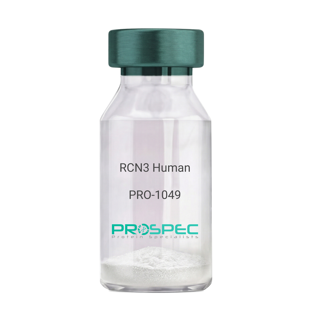
RCN3 HumanDescription:
Reticulocalbin 3 Human Recombinant
Reticulocalbin-3, EF-hand calcium-binding protein RLP49, RCN3, RLP49.
Product # :
PRO-1049Price :
Quantity :
Shipping Method :
Shipped with Ice Packs
More Info
- description
- source
- formulation
- purity
- More Info
Description
RCN3 Human Recombinant produced in E.Coli is a single, non-glycosylated polypeptide chain containing 333 amino acids (21-328 a.a) and having a molecular mass of 37.9kDa.RCN3 is fused to a 25 amino acid His-tag at N-terminus & purified by proprietary chromatographic techniques.
Source
Escherichia Coli.
Formulation
RCN3 protein solution (1mg/ml) containing 20mM Tris-HCl buffer (pH 8.0), 1mM DTT, 10% glycerol and 0.1M NaCl.
Purity
Greater than 95.0% as determined by SDS-PAGE.
More Info
-
Introduction
Reticulocalbin 3 (RCN3) belongs to the CREC (cab45/reticulocalbin/ ERC45/calumenin) family. RCN3 contains 5 Arg-Xaa-Xaa-Arg motifs, which function as target sequences of SPCs (subtilisin-like proprotein convertases), which is a family of serine endoproteases that proteolytically activate proproteins. The synthesis of PACE4 is induced by association and coexpression with RCN3.
-
Synonyms
Reticulocalbin-3, EF-hand calcium-binding protein RLP49, RCN3, RLP49.
-
Physical Appearance
Sterile Filtered colorless solution.
-
Stability
Store at 4°C if entire vial will be used within 2-4 weeks. Store, frozen at -20°C for longer periods of time. For long term storage it is recommended to add a carrier protein (0.1% HSA or BSA).Avoid multiple freeze-thaw cycles.
-
Amino Acid Sequence
MGSSHHHHHH SSGLVPRGSH MGSHMKPSPD AGPHGQGRVH QAAPLSDAPH DDAHGNFQYD HEAFLGREVA KEFDQLTPEE SQARLGRIVD RMDRAGDGDG WVSLAELRAW IAHTQQRHIR DSVSAAWDTY DTDRDGRVGW EELRNATYGH YAPGEEFHDV EDAETYKKML ARDERRFRVA DQDGDSMATR EELTAFLHPE EFPHMRDIVI AETLEDLDRN KDGYVQVEEY IADLYSAEPG EEEPAWVQTE RQQFRDFRDL NKDGHLDGSE VGHWVLPPAQ DQPLVEANHL LHESDTDKDG RLSKAEILGN WNMFVGSQAT NYGEDLTRHH DEL.
ProSpec's products are furnished for LABORATORY RESEARCH USE ONLY. The product may not be used as drugs, agricultural or pesticidal products, food additives or household chemicals.
- View Data Sheet
Name :
FCER1A Human, HEKDescription:
Fc-Epsilon RI-Alpha Human Recombinant, HEK
FCERIA, FCERA, Fc epsilon receptor Ia, Fcepsilon RI-alpha, Fc epsilon RI alpha chain, FcERI, High affinity immunoglobulin epsilon receptor subunit alpha isoform1, IgE Fc receptor subunit alpha, FCER1A, FCE1A.
Product # :
PRO-2779Price :
Quantity :
Shipping Method :
Shipped with Ice Packs
More Info
- description
- source
- formulation
- purity
- biological activity
- More Info
Description
FCER1A Human Recombinant produced in HEK293 Cells is a single, glycosylated polypeptide chain containing 186 amino acids (26-205aa) and having a molecular mass of 21.8kDa. FCER1A is fused to a 6 His-tag at C-terminus & purified by proprietary chromatographic techniques.
Source
HEK293.
Formulation
FCER1A protein solution (1mg/ml) containing 10% Glycerol and Phosphate-Buffered Saline (pH 7.4).
Purity
Greater than 90% as determined by SDS-PAGE.
Biological Activity
The ED50 range ≤ 0.01 ug/ml and is measured by its binding ability in a functional ELISA with Human IgE.
More Info
-
Synonyms
FCERIA, FCERA, Fc epsilon receptor Ia, Fcepsilon RI-alpha, Fc epsilon RI alpha chain, FcERI, High affinity immunoglobulin epsilon receptor subunit alpha isoform1, IgE Fc receptor subunit alpha, FCER1A, FCE1A.
-
Physical Appearance
Sterile Filtered clear solution.
-
Stability
Store at 4°C if entire vial will be used within 2-4 weeks. Store, frozen at -20°C for longer periods of time. For long term storage it is recommended to add a carrier protein (0.1% HSA or BSA). Avoid multiple freeze-thaw cycles.
-
Amino Acid Sequence
VPQKPKVSLN PPWNRIFKGE NVTLTCNGNN FFEVSSTKWF HNGSLSEETN SSLNIVNAKF EDSGEYKCQH QQVNESEPVY LEVFSDWLLL QASAEVVMEG QPLFLRCHGW RNWDVYKVIY YKDGEALKYW YENHNISITN ATVEDSGTYY CTGKVWQLDY ESEPLNITVI KAPREKYWLQ HHHHHH
-
Background
The FCER1A gene encodes the alpha subunit of the high-affinity immunoglobulin E (IgE) receptor, known as FCER1A. This receptor is primarily expressed on mast cells and basophils, and its activation plays a pivotal role in allergic and inflammatory responses. This research aims to explore the significance of FCER1A and its potential implications in allergic disorders and immune-mediated diseases. By investigating the functions and regulation of FCER1A, we can gain insights into its role in immune responses and identify potential therapeutic targets.
The FCER1A receptor is responsible for the binding of IgE antibodies, initiating a cascade of signaling events upon allergen exposure. Crosslinking of IgE-bound FCER1A leads to the release of various inflammatory mediators, such as histamine, cytokines, and leukotrienes, which contribute to the clinical manifestations of allergic reactions. Understanding the molecular mechanisms underlying FCER1A activation and downstream signaling is crucial for comprehending allergic diseases.
In addition to its role in allergies, FCER1A has been implicated in immune-mediated inflammatory diseases, including asthma, atopic dermatitis, and autoimmune conditions. Dysregulation of FCER1A expression and signaling pathways can lead to exaggerated immune responses and chronic inflammation. Investigating the genetic and epigenetic factors influencing FCER1A expression and the interplay between FCER1A and other immune molecules can provide valuable insights into disease pathogenesis.
This research will delve into the molecular mechanisms governing FCER1A expression, activation, and downstream signaling pathways. The paper will explore the regulatory effects of FCER1A on mast cell and basophil activation, the release of inflammatory mediators, and the recruitment of other immune cells. Additionally, it will examine the impact of FCER1A dysregulation in allergic and immune-mediated diseases and discuss the potential of FCER1A as a therapeutic target for intervention.
The study will also investigate the diagnostic and prognostic value of FCER1A in various allergic and immune disorders. Understanding the expression patterns and alterations of FCER1A in different diseases and patient populations may aid in disease stratification, treatment selection, and monitoring of treatment response.
By unraveling the molecular mechanisms underlying FCER1A's functions in allergic and inflammatory responses, this research aims to contribute to our understanding of immune-mediated diseases. Furthermore, it highlights the potential of FCER1A as a target for therapeutic interventions and emphasizes the need for further investigations to develop novel treatments and improve patient outcomes.
ProSpec's products are furnished for LABORATORY RESEARCH USE ONLY. The product may not be used as drugs, agricultural or pesticidal products, food additives or household chemicals.
- View Data Sheet
Name :
IL13RA2 HumanDescription:
Interleukin 13 Receptor, Alpha 2 Human Recombinant
CD213A2, CT19, IL-13R, IL13BP, IL-13 receptor subunit alpha-2, IL-13R subunit alpha-2, CD_antigen=CD213a2, Interleukin-13-binding protein.
Product # :
CYT-757Price :
Quantity :
Shipping Method :
Shipped with Ice Packs
More Info
- description
- source
- formulation
- purity
- More Info
Description
IL13RA2 Human Recombinant produced in E.Coli is a single, non-glycosylated polypeptide chain containing 340 amino acids (27-343 a.a.) and having a molecular mass of 39.5kDa.IL13RA2 is fused to a 23 amino acid His-tag at N-terminus & purified by proprietary chromatographic techniques.
Source
Escherichia Coli.
Formulation
IL13RA2 protein solution (1mg/ml) contains 20mM Tris-HCl buffer, (pH 8.0), 0.4M UREA and 10% glycerol.
Purity
Greater than 85.0% as determined by SDS-PAGE.
More Info
-
Introduction
Interleukin 13 Receptor, Alpha 2 (IL13RA2) is a member of the type I cytokine receptor family, Type 5 subfamily. IL13RA2 is related to l13RA1, a subunit of the interleukin 13 receptor complex. IL13RA2 binds IL13 with high affinity, though lacking the cytoplasmic domain, and does not appear to function as a signal mediator. IL13RA2 plays a role in the internalization of IL13.
-
Synonyms
CD213A2, CT19, IL-13R, IL13BP, IL-13 receptor subunit alpha-2, IL-13R subunit alpha-2, CD_antigen=CD213a2, Interleukin-13-binding protein.
-
Physical Appearance
Sterile Filtered clear solution.
-
Stability
Store at 4°C if entire vial will be used within 2-4 weeks. Store, frozen at -20°C for longer periods of time. For long term storage it is recommended to add a carrier protein (0.1% HSA or BSA).Avoid multiple freeze-thaw cycles.
-
Amino Acid Sequence
MGSSHHHHHH SSGLVPRGSH MGSDTEIKVN PPQDFEIVDP GYLGYLYLQW QPPLSLDHFK ECTVEYELKY RNIGSETWKT IITKNLHYKD GFDLNKGIEA KIHTLLPWQC TNGSEVQSSW AETTYWISPQ GIPETKVQDM DCVYYNWQYL LCSWKPGIGV LLDTNYNLFY WYEGLDHALQ CVDYIKADGQ NIGCRFPYLE ASDYKDFYIC VNGSSENKPI RSSYFTFQLQ NIVKPLPPVY LTFTRESSCE IKLKWSIPLG PIPARCFDYE IEIREDDTTL VTATVENETY TLKTTNETRQ LCFVVRSKVN IYCSDDGIWS EWSDKQCWEG EDLSKKTLLR.
ProSpec's products are furnished for LABORATORY RESEARCH USE ONLY. The product may not be used as drugs, agricultural or pesticidal products, food additives or household chemicals.
- View Data Sheet
Name :
tBID MouseDescription:
Truncated BH3 Interacting Domain Death Agonist Mouse Recombinant
Truncated BH3-interacting domain death agonist, p22 BID, BID, FP497, MGC15319, MGC42355, tBID.
Product # :
PRO-644Price :
Quantity :
Shipping Method :
Shipped with Ice Packs
More Info
- description
- source
- formulation
- purity
- More Info
Description
Truncated BID Mouse Recombinant also called BH3-interacting domain death agonist p15 produced in E.Coli is a single, non-glycosylated polypeptide chain containing 61-195 amino acids (135 a.a.) and having a molecular mass of 15.4 kDa.
Source
Escherichia Coli.
Formulation
The Mouse Truncated BID protein solution contains 10mM Tris-HCl pH-8, 1mM EDTA and 250mM NaCl.
Purity
Greater than 95.0% as determined by:
(a) Analysis by RP-HPLC.
(b) Analysis by SDS-PAGE.More Info
-
Introduction
Truncated BH3 interacting domain death agonist is a truncated form of the pro-apoptotic full-length BID. Truncated BH3 interacting domain death agonist is generated by Caspase-8 cleavage of BID. The truncated form of the protein translocates from the cytosol to mitochondria and transduces apoptotic signals.
BID is a pro-apoptotic Bcl-2 protein having only the BH3 domain. In reaction to apoptotic signaling, BID interacts with another Bcl-2 family of cell death regulators, called Bax, they form a heterodimer resulting to the insertion of Bax into the outer mitochondrial membrane. Bax induces the opening of the mitochondrial voltage-dependent anion channel which lead to the release of cytochrome c and other pro-apoptotic factors from the mitochondria resulting in activation of caspases. BID is a mediator of mitochondrial damage induced by caspase-8 (CASP8). CASP8 cleaves BID, and the COOH-terminal part translocates to mitochondria where it triggers cytochrome c release. The major proteolytic product p15 BID releasea cytochrome c. Isoform 1, Isoform 2 and Isoform 4 induce ice-like proteases and apoptosis while Isoform 3 does not induce apoptosis. -
Synonyms
Truncated BH3-interacting domain death agonist, p22 BID, BID, FP497, MGC15319, MGC42355, tBID.
-
Physical Appearance
Sterile Filtered colorless liquid formulation.
-
Stability
Store at 4°C if entire vial will be used within 2-4 weeks. Store, frozen at -20°C for longer periods of time. For long term storage it is recommended to add a carrier protein (0.1% HSA or BSA).Avoid multiple freeze-thaw cycles.
ProSpec's products are furnished for LABORATORY RESEARCH USE ONLY. The product may not be used as drugs, agricultural or pesticidal products, food additives or household chemicals.
- View Data Sheet
Name :
SNRPA1 HumanDescription:
Small Nuclear Ribonucleoprotein Polypeptide A1 Human Recombinant
U2 small nuclear ribonucleoprotein A', U2 snRNP A', SNRPA1, Lea1.
Product # :
PRO-1015Price :
Quantity :
Shipping Method :
Shipped with Ice Packs
More Info
- description
- source
- formulation
- purity
- More Info
Description
SNRPA1 Human Recombinant produced in E.Coli is a single, non-glycosylated polypeptide chain containing 275 amino acids (1-255 a.a.) and having a molecular mass of 30.5kDa. SNRPA1 is fused to a 20 amino acid His-tag at N-terminus & purified by proprietary chromatographic techniques.
Source
Escherichia Coli.
Formulation
SNRPA1 protein solution (0.5mg/ml) containing 20mM Tris-HCl buffer (pH8.0), 30% glycerol, 0.2M NaCl,2mM DTT and 0.1mM PMSF.
Purity
Greater than 80% as determined by SDS-PAGE.
More Info
-
Introduction
U2 small nuclear ribonucleoprotein A (SNRPA1), a 255 amino acid protein, is a component of the U2 snRNP which forms a complex with U2 snRNP B (U2B). U2 snRNP A and U2 snRNP B form together a complex which binds to the U2 snRNA hairpin IV. The configuration of the U2 snRNP A/U2 snRNP B dimer and the delicate variations of a few key residues regulate the snRNP-RNA-binding specificity.
-
Synonyms
U2 small nuclear ribonucleoprotein A', U2 snRNP A', SNRPA1, Lea1.
-
Physical Appearance
Sterile Filtered clear solution.
-
Stability
Store at 4°C if entire vial will be used within 2-4 weeks. Store, frozen at -20°C for longer periods of time. For long term storage it is recommended to add a carrier protein (0.1% HSA or BSA).Avoid multiple freeze-thaw cycles.
-
Amino Acid Sequence
MGSSHHHHHH SSGLVPRGSH MVKLTAELIE QAAQYTNAVR DRELDLRGYK IPVIENLGAT LDQFDAIDFS DNEIRKLDGF PLLRRLKTLL VNNNRICRIG EGLDQALPCL TELILTNNSL VELGDLDPLA SLKSLTYLSI LRNPVTNKKH YRLYVIYKVP QVRVLDFQKV KLKERQEAEK MFKGKRGAQL AKDIARRSKT FNPGAGLPTD KKKGGPSPGD VEAIKNAIAN ASTLAEVERL KGLLQSGQIP GRERRSGPTD DGEEEMEEDT VTNGS.
ProSpec's products are furnished for LABORATORY RESEARCH USE ONLY. The product may not be used as drugs, agricultural or pesticidal products, food additives or household chemicals.
- View Data Sheet
Name :
RPS27A Human, BiotinDescription:
Ubiquitin Biotinylated Human Recombinant
Ubiquitin, Ribosomal Protein S27a, CEP80, UBA80, UBCEP1, UBCEP80, HUBCEP80, RPS27A.
Product # :
PRO-629Price :
Quantity :
Shipping Method :
Shipped with Ice Packs
More Info
- description
- source
- formulation
- purity
- More Info
Description
Recombinant Human RPS27A protein biotinylated with NHS-biotin produced in E.Coli is a single, non-glycosylated polypeptide chain containing a total of 76 amino acids and having a molecular mass of 8.6 kDa.
Source
Escherichia Coli.
Formulation
The RPS27A is supplied in 1x PBS and 0.05% PBS.
Purity
RPS27A Protein biotinilation is determined by Western Blotting and ELISA analysis using streptavidin–HRP conjugated as a detection reagent. Free biotin is eliminated by dialysis against PBS. Protein concentration is determined by 280nm absorbance.
More Info
-
Introduction
Recombinant Human Ubiquitin having the accession number of P62988 was conjugated to Biotin. RPS27A is a small protein composed of 76 amino acids. RPS27A is found only in eukaryotic organisms among which shows strong sequence conservation. The RPS27A protein is present in all cell types, thus giving rise to its name.
RPS27A is found either in free form or conjugated to proteins through a covalent bond between the glycine at the C-terminal end and the side chains of lysine.
The connection of multiple copies of RPS27A targets the proteins for degradation by the 26S proteosome. RPS27A ligation is an ATP-dependent multi-step process. RPS27A is activated by the E1 enzyme. The attachment of RPS27A to the target protein is catalyzed by the E2 enzyme acting in concert with E3 which is involved in the recognition of the substrate protein. -
Synonyms
Ubiquitin, Ribosomal Protein S27a, CEP80, UBA80, UBCEP1, UBCEP80, HUBCEP80, RPS27A.
-
Physical Appearance
Sterile Filtered colorless liquid formulation.
-
Stability
Store at 4°C if entire vial will be used within 2-4 weeks. Store, frozen at -20°C for longer periods of time. For long term storage it is recommended to add a carrier protein (0.1% HSA or BSA). Please prevent freeze-thaw cycles.
ProSpec's products are furnished for LABORATORY RESEARCH USE ONLY. The product may not be used as drugs, agricultural or pesticidal products, food additives or household chemicals.
- View Data Sheet
Name :
PVR HumanDescription:
Poliovirus Receptor Human Recombinant
Poliovirus receptor, Nectin-like protein 5, NECL-5, CD155, PVR, PVS.
Product # :
PRO-2463Price :
Quantity :
Shipping Method :
Shipped with Ice Packs
More Info
- description
- source
- formulation
- purity
- More Info
Description
PVR Human Recombinant produced in Sf9 Insect cells is a single, glycosylated polypeptide chain containing 331 amino acids (21-343 a.a.) and having a molecular mass of 36.1kDa (Molecular size on SDS-PAGE will appear at approximately 40-57kDa). PVR is expressed with an 8 amino acids His tag at C-Terminus and purified by proprietary chromatographic techniques.
Source
Sf9, Baculovirus cells.
Formulation
PVR protein solution (1mg/ml) contains Phosphate Buffered Saline (pH 7.4) and 10% glycerol.
Purity
Greater than 90.0% as determined by SDS-PAGE.
More Info
-
Introduction
Poliovirus receptor, also known as PVR, is a Type I transmembrane glycoprotein which belongs to the immunoglobulin superfamily. PVR catalyzes a big structural modification in the virus which exposes membrane-binding protein chains. PVR regulates helper T cell differentiation as well as allergic diseases. PVR is expressed in many kinds of human cells and has various functions. PVR is potentially functional as a biomarker for cancer development as well as progression. PVR takes a vital part through both immunological & non-immunological mechanisms in pancreatic cancer.
-
Synonyms
Poliovirus receptor, Nectin-like protein 5, NECL-5, CD155, PVR, PVS.
-
Physical Appearance
Sterile Filtered colorless solution.
-
Stability
Store at 4°C if entire vial will be used within 2-4 weeks. Store, frozen at -20°C for longer periods of time. For long term storage it is recommended to add a carrier protein (0.1% HSA or BSA). Avoid multiple freeze-thaw cycles.
-
Amino Acid Sequence
WPPPGTGDVV VQAPTQVPGF LGDSVTLPCY LQVPNMEVTH VSQLTWARHG ESGSMAVFHQ TQGPSYSESK RLEFVAARLG AELRNASLRM FGLRVEDEGN YTCLFVTFPQ GSRSVDIWLR VLAKPQNTAE VQKVQLTGEP VPMARCVSTG GRPPAQITWH SDLGGMPNTS QVPGFLSGTV TVTSLWILVP SSQVDGKNVT CKVEHESFEK PQLLTVNLTV YYPPEVSISG YDNNWYLGQN EATLTCDARS NPEPTGYNWS TTMGPLPPFA VAQGAQLLIR PVDKPINTTL ICNVTNALGA RQAELTVQVK EGPPSEHSGM SRNLEHHHHH H
ProSpec's products are furnished for LABORATORY RESEARCH USE ONLY. The product may not be used as drugs, agricultural or pesticidal products, food additives or household chemicals.
- View Data Sheet
Name :
CCL6 RatDescription:
C-10 Rat Recombinant (CCL6)
Small inducible cytokine A6, CCL6, C10 protein, c10, MRP-1, Scya6, chemokine (C-C motif) ligand 6.
Product # :
CHM-268Price :
Quantity :
Shipping Method :
Shipped at Room temp
More Info
- description
- source
- formulation
- purity
- biological activity
- More Info
Description
C-10 Rat Recombinant produced in E.Coli is a signle, non-glycosylated, polypeptide chain containing 94 amino acids and having a total molecular mass of 10.4kDa. C-10 is purified by proprietary chromatographic techniques.
Source
Escherichia Coli.
Formulation
Lyophilized from a 0.2µm filtered concentrated (1.0mg/ml) solution in 1×PBS, pH 7.4.
Purity
Greater than 95.0% as determined by
(a) Analysis by RP-HPLC.
(b) Analysis by SDS-PAGE.Biological Activity
Determined by its ability to chemoattract human CCR1 transfected BaF3 mouse proB cells using a concentration range of 0.05-0.25 ug/ml.More Info
-
Introduction
Chemokine (C-C motif) ligand 6 (CCL6) is a small cytokine belonging to the CC chemokine family that has only been identified in rodents.
In mice, CCL6 is expressed in cells from neutrophil and macrophage lineages, and can be greatly induced under conditions suitable for myeloid cell differentiation. It is highly expressed in bone marrow cultures that have been stimulated with the cytokine GM-CSF. Some low levels of gene expression also occur in certain cell lines of myeloid origin (e.g. the immature myeloid cell lines DA3 and 32D cl3, and the macrophage cell line P388D) that can also be greatly induced in culture with GM-CSF. However, in activated T cell lines, expression of CCL6 is greatly reduced. CCL6 can also be induced in the mouse lung by the cytokine interleukin 13. Mouse CCL6 is located on chromosome 11. The cell surface receptor for CCL6 is believed to be the chemokine receptor CCR1. -
Synonyms
Small inducible cytokine A6, CCL6, C10 protein, c10, MRP-1, Scya6, chemokine (C-C motif) ligand 6.
-
Physical Appearance
Sterile Filtered White lyophilized (freeze-dried) powder.
-
Stability
Lyophilized C-10 although stable at room temperature for 3 weeks, should be stored desiccated below -18C. Upon reconstitution C-10 should be stored at 4C between 2-7 days and for future use below -18C.For long term storage it is recommended to add a carrier protein (0.1% HSA or BSA).Please prevent freeze-thaw cycles.
-
Solubility
It is recommended to reconstitute the lyophilized C-10 in sterile 18M-cm H2O not less than 100µg/ml, which can then be further diluted to other aqueous solutions.
-
Amino Acid Sequence
GLIQDTVKED RPFNPTIIHQ GFQDSSDCCF SYASQIPCSR FIYYFPTSGG CTKPGIIFVT RKRKRVCANP SDQRVQTCIS TLKLGPRSGN SAIA.
-
Background
What is the molecular weight/Mw of CCL6 RAT Protein?
CCL6 RAT Protein has a total Mw of 10.4kDa.
What is the source or expression system of CCL6 RAT Protein?
Escherichia Coli.
What is the Purity of CCL6 RAT Protein?
CCL6 RAT Protein is >95% pure as determined by SDS-PAGE.
What is the Biological Activity of CCL6 RAT Protein?
Determined by its ability to chemoattract human CCR1 transfected BaF3 mouse proB cells using a concentration range of 0.05-0.25 ug/ml.
What is the amino acid sequence of CCL6 RAT Protein?
GLIQDTVKED RPFNPTIIHQ GFQDSSDCCF SYASQIPCSR FIYYFPTSGG CTKPGIIFVT RKRKRVCANP SDQRVQTCIS TLKLGPRSGN SAIA.
What applications can CCL6 RAT Protein be used in?
CCL6 RAT Protein can probably be used in western blot, ELISA and Lateral Flow.
What is the endotoxin level for CCL6 RAT Protein?
The endotoxin level is minimal, CCL6 RAT Protein was purified using conventional chromatography techniques.
ProSpec's products are furnished for LABORATORY RESEARCH USE ONLY. The product may not be used as drugs, agricultural or pesticidal products, food additives or household chemicals.
- View Data Sheet
Name :
Adiponectin MouseDescription:
Adiponectin Mouse Recombinant
Acrp30, AdipoQ, GBP-28, APM-1, ACDC.
Product # :
CYT-1168Price :
Quantity :
Shipping Method :
Shipped at Room temp
More Info
- description
- source
- formulation
- purity
- biological activity
- More Info
Description
Acrp30 Mouse Recombinant is a single, glycosylated polypeptide chain containing 230 amino acids (18-247a.a) and having a molecular mass of 24.9kDa (calculated).
Source
HEK293 cells.
Formulation
Acrp30 filtered (0.4 µm) and lyophilized from 0.5mg/ml in 20mM Tris 50mM NaCl and 1mM CaCl2, pH 7.5.
Purity
Greater than 90.0% as determined by SDS-PAGE.
Biological Activity
Measured by its ability to inhibit proliferation of mouse 3T3 fibroblasts stimulated by PDGF. The ED50 for the effect is typically 5-15 μg/mL.
More Info
-
Introduction
Adiponectin, also known as Acrp30, AdipoQ and GBP-28, is a 244 amino acid protein, the product of the apM1 gene, which is physiologically active and specifically and highly expressed in adipose cells (Adipokine). Adiponectin is a part of the soluble defense collagen super family; it has a collagen-like domain structurally homologous with collagen VIII and X and complement factor C1q-like globular domain. APM-1 forms homotrimers, which are the building blocks for higher order complexes found circulating in serum.
-
Synonyms
Acrp30, AdipoQ, GBP-28, APM-1, ACDC.
-
Physical Appearance
Filtered White lyophilized (freeze-dried) powder.
-
Stability
Store lyophilized protein at -20°C. Aliquot the product after reconstitution to avoid repeated freezing/thawing cycles. Reconstituted protein can be stored at 4°C for a limited period of time; it does not show any change after two weeks at 4°C.
-
Solubility
It is recommended to add deionized water to prepare a working stock solution of approximately 0.5mg/ml and let the lyophilized pellet dissolve completely.
-
Amino Acid Sequence
EDDVTTTEEL APALVPPPKG TCAGWMAGIP GHPGHNGTPG RDGRDGTPGE KGEKGDAGLL GPKGETGDVG MTGAEGPRGF PGTPGRKGEP GEAAYVYRSA FSVGLETRVT VPNVPIRFTK IFYNQQNHYD GSTGKFYCNI PGLYYFSYHI TVYMKDVKVS LFKKDKAVLF TYDQYQEKNV DQASGSVLLH LEVGDQVWLQ VYGDGDHNGL YADNVNDSTF TGFLLYHDTN.
-
Background
What is the molecular weight/Mw of ADIPONECTIN Protein?
ADIPONECTIN Protein has a total Mw of 24.9kDa.
What is the source or expression system of ADIPONECTIN Protein?
HEK293 cells.
What is the Purity of ADIPONECTIN Protein?
ADIPONECTIN Protein is >90% pure as determined by SDS-PAGE.
What is the Biological Activity of ADIPONECTIN Protein?
The biological activity Measured by its ability to inhibit proliferation of mouse 3T3 fibroblasts stimulated by PDGF. The ED50 for the effect is typically 5-15 μg/mL.
What is the amino acid sequence of ADIPONECTIN Protein?
EDDVTTTEEL APALVPPPKG TCAGWMAGIP GHPGHNGTPG RDGRDGTPGE KGEKGDAGLL GPKGETGDVG MTGAEGPRGF PGTPGRKGEP GEAAYVYRSA FSVGLETRVT VPNVPIRFTK IFYNQQNHYD GSTGKFYCNI PGLYYFSYHI TVYMKDVKVS LFKKDKAVLF TYDQYQEKNV DQASGSVLLH LEVGDQVWLQ VYGDGDHNGL YADNVNDSTF TGFLLYHDTN.
What applications can ADIPONECTIN Protein be used in?
ADIPONECTIN Protein can probably be used in western blot, ELISA and Lateral Flow.
What is the endotoxin level for ADIPONECTIN Protein?
The endotoxin level is minimal, ADIPONECTIN Protein was purified using conventional chromatography techniques.
ProSpec's products are furnished for LABORATORY RESEARCH USE ONLY. The product may not be used as drugs, agricultural or pesticidal products, food additives or household chemicals.
- View Data Sheet
Name :
Cyclophilin A MouseDescription:
Cyclophilin A Mouse Recombinant
Peptidyl-prolyl cis-trans isomerase A, PPIase A, Cyclophilin A, Cyclosporin A-binding protein, Rotamase A, SP18, Peptidyl-prolyl cis-trans isomerase A, N-terminally processed, Ppia, 2700098C05 Cphn, CyP-18, CypA.
Product # :
ENZ-857Price :
Quantity :
Shipping Method :
Shipped with Ice Packs
More Info
- description
- source
- formulation
- purity
- More Info
Description
Cyclophilin A Mouse Recombinant produced in E.Coli is a single, non-glycosylated polypeptide chain containing 187 amino acids (1-164a.a.) and having a molecular mass of 20.4kDa.Cyclophilin A is fused to a 23 amino acid His-tag at N-terminus & purified by proprietary chromatographic techniques.
Source
E.coli.
Formulation
Cyclophilin A protein solution (0.5mg/ml) containing Phosphate buffered saline (pH7.4), 10% glycerol and 1mM DTT.
Purity
Greater than 95% as determined by SDS-PAGE.
More Info
-
Introduction
PPIases catalyze the cis-trans isomerization of proline imidic peptide bonds in oligopeptides and accelerate the folding of proteins. Cyclophilin-A is a cyclosporin binding-protein and may play a role in cyclosporin A-mediated immunosuppression. Cyclophilin-A can also interact with several HIV proteins, including p55 gag, Vpr, and capsid protein, and has been shown to be necessary for the formation of infectious HIV virions. Multiple pseudogenes that map to different chromosomes have been reported.
-
Synonyms
Peptidyl-prolyl cis-trans isomerase A, PPIase A, Cyclophilin A, Cyclosporin A-binding protein, Rotamase A, SP18, Peptidyl-prolyl cis-trans isomerase A, N-terminally processed, Ppia, 2700098C05 Cphn, CyP-18, CypA.
-
Physical Appearance
Sterile Filtered colorless solution.
-
Stability
Store at 4°C if entire vial will be used within 2-4 weeks. Store, frozen at -20°C for longer periods of time. For long term storage it is recommended to add a carrier protein (0.1% HSA or BSA).Avoid multiple freeze-thaw cycles.
-
Amino Acid Sequence
MGSSHHHHHH SSGLVPRGSH MGSMVNPTVF FDITADDEPL GRVSFELFAD KVPKTAENFR ALSTGEKGFG YKGSSFHRII PGFMCQGGDF TRHNGTGGRS IYGEKFEDEN FILKHTGPGI LSMANAGPNT NGSQFFICTA KTEWLDGKHV VFGKVKEGMN IVEAMERFGS RNGKTSKKIT ISDCGQL.
ProSpec's products are furnished for LABORATORY RESEARCH USE ONLY. The product may not be used as drugs, agricultural or pesticidal products, food additives or household chemicals.
- View Data Sheet
Name :
IL2RG HumanDescription:
Interleukin-2 Receptor Gamma Human Recombinant
Interleukin 2 Receptor Gamma, IL-2 Receptor Subunit Gamma, IL-2R Subunit Gamma, CD132 Antigen, SCIDX1, IL-2RG, GammaC, SCIDX, IMD4, P64, Cytokine Receptor Common Subunit Gamma, Common Cytokine Receptor Gamma Chain, Interleukin-2 Receptor Subunit Gamma, Combined Immunodeficiency X-Linked, Severe Combined Immunodeficiency, CD132, CIDX, IL2RG.
Product # :
CYT-471Price :
Quantity :
Shipping Method :
Shipped with Ice Packs
More Info
- description
- source
- formulation
- purity
- More Info
Description
IL2RG Human Recombinant produced in Sf9 Baculovirus cells is a single, glycosylated polypeptide chain containing 246 amino acids (23-262 a.a.) and having a molecular mass of 29kDa (Migrates at 40-57kDa on SDS-PAGE under reducing conditions).IL2RG is expressed with a 6 amino acid His tag at C-Terminus and purified by proprietary chromatographic techniques.
Source
Sf9, Baculovirus cells.
Formulation
IL2RG protein solution (0.5mg/ml) contains Phosphate Buffered Saline (pH 7.4).
Purity
Greater than 80.0% by SDS-PAGE.
More Info
-
Introduction
Interleukin-2 Receptor Gamma (IL2RG) is a cytokine receptor sub-unit which is common to the receptor complexes for at least six different interleukin receptors. IL2RG is located on the surface of immature blood-forming cells in bone marrow. One end of the IL2RG protein resides outside of the cell where it binds to cytokines and the other end of the IL2RG protein resides in the interior of the cell where it transmits signals to the cell's nucleus. IL2RG teams up with other proteins to direct blood-forming cells to form lymphocytes. In addition, IL2RG directs the growth and maturation of lymphocyte subtypes, which kill viruses, make antibodies, and help regulate the entire immune system.
-
Synonyms
Interleukin 2 Receptor Gamma, IL-2 Receptor Subunit Gamma, IL-2R Subunit Gamma, CD132 Antigen, SCIDX1, IL-2RG, GammaC, SCIDX, IMD4, P64, Cytokine Receptor Common Subunit Gamma, Common Cytokine Receptor Gamma Chain, Interleukin-2 Receptor Subunit Gamma, Combined Immunodeficiency X-Linked, Severe Combined Immunodeficiency, CD132, CIDX, IL2RG.
-
Physical Appearance
Sterile Filtered clear solution.
-
Stability
Store at 4°C if entire vial will be used within 2-4 weeks. Store, frozen at -20°C for longer periods of time. For long term storage it is recommended to add a carrier protein (0.1% HSA or BSA).Please avoid freeze thaw cycles.
-
Amino Acid Sequence
LNTTILTPNG NEDTTADFFL TTMPTDSLSV STLPLPEVQC FVFNVEYMNC TWNSSSEPQP TNLTLHYWYK NSDNDKVQKC SHYLFSEEIT SGCQLQKKEI HLYQTFVVQL QDPREPRRQA TQMLKLQNLV IPWAPENLTL HKLSESQLEL NWNNRFLNHC LEHLVQYRTD WDHSWTEQSV DYRHKFSLPS VDGQKRYTFR VRSRFNPLCG SAQHWSEWSH PIHWGSNTSK ENPFLFALEA HHHHHH.
ProSpec's products are furnished for LABORATORY RESEARCH USE ONLY. The product may not be used as drugs, agricultural or pesticidal products, food additives or household chemicals.
- View Data Sheet
Name :
RELM g MouseDescription:
RELM-Gamma Mouse Recombinant
Resistin-like gamma, RELMgamma,RELM-γ, RELM-g.
Product # :
CYT-1035Price :
Quantity :
Shipping Method :
Shipped at Room temp
More Info
- description
- source
- formulation
- purity
- More Info
Description
RELM g Mouse Recombinant produced in E.Coli is a non glycosylated, homodimeric polypeptide chain containing 2 x 89 amino acids and having a total molecular mass of 18.9kDa. The RELM is purified by proprietary chromatographic techniques.
Source
Escherichia Coli.
Formulation
Lyophilized from a sterile filtered solution containing 0.1 % trifluoroacetic acid (TFA).
Purity
Greater than 95.0% as determined by SDS-PAGE.
More Info
-
Introduction
RELM-gamma is a novel member of the resistin-like molecule/found in inflammatory zone (RELM/FIZZ) family in mice and rats. Microarray and real-time RT-PCR experiments revealed a repression of RELMgamma mRNA in nasal respiratory epithelium of cigarette smoke-exposed versus untreated rats. The analysis of the physiological tissue-specific expression revealed highest expression in hematopoietic tissues, suggesting a cytokine-like role for RELM-gamma. RELM-gamma-mRNA is detectable in bone marrow, spleen, and lung as well as in peripheral blood granulocytes. Promyelocytic HL60 cells transfected with a RELM-gamma expression plasmid have an increased proliferation rate compared to mock-transfected cells and display an altered response to retinoic acid-induced granulocytic differentiation. Taken together, these data provide the first experimental evidence that RELM-gamma is a secreted molecule with a restricted expression pattern that may play a role in promyelocytic differentiation.
-
Synonyms
Resistin-like gamma, RELMgamma,RELM-γ, RELM-g.
-
Physical Appearance
Sterile Filtered White lyophilized (freeze-dried) powder.
-
Stability
Lyophilized RELM g although stable at room temperature for 3 weeks, should be stored desiccated below -18°C. Upon reconstitution RELM g Mouse should be stored at 4°C between 2-7 days and for future use below -18°C. For long term storage it is recommended to add a carrier protein (0.1% HSA or BSA). Please prevent freeze-thaw cycles.
-
Solubility
It is recommended to reconstitute the lyophilized RELM g in sterile 18MΩ-cm H2O at a concentration of 100µg/ml, which can then be further diluted to other aqueous solutions.
-
Amino Acid Sequence
MEGTLESIVE KKVKELLANR DDCPSTVTKT FSCTSITASG RLASCPSGMT VTGCACGYGC GSWDIRDGNT CHCQCSTMDW ATARCCQLA.
ProSpec's products are furnished for LABORATORY RESEARCH USE ONLY. The product may not be used as drugs, agricultural or pesticidal products, food additives or household chemicals.
- View Data Sheet
Name :
IL13RA2 Human, Sf9Description:
Interleukin 13 Receptor Alpha 2, Recombinant Human Sf9
CD213A2, CT19, IL-13R, IL13BP, IL-13 receptor subunit alpha-2, IL-13R subunit alpha-2, CD_antigen=CD213a2, Interleukin-13-binding protein.
Product # :
CYT-1024Price :
Quantity :
Shipping Method :
Shipped with Ice Packs
More Info
- description
- source
- formulation
- purity
- More Info
Description
IL13RA2 Human Recombinant produced in Sf9 Insect cells is a single, glycosylated polypeptide chain containing 559 amino acids (27-343 a.a.) and having a molecular mass of 64.3kDa (Molecular size on SDS-PAGE will appear at approximately 70-100kDa). IL13RA2 is expressed with a 239 amino acid hIgG-His tag at C-Terminus and purified by proprietary chromatographic techniques.
Source
Sf9, Baculovirus cells.
Formulation
IL 13RA2 protein solution (0.5mg/ml) contains Phosphate buffered saline (pH7.4) and 10% glycerol.
Purity
Greater than 85.0% as determined by SDS-PAGE.
More Info
-
Introduction
Interleukin 13 Receptor, Alpha 2 (IL13RA2) is a member of the type I cytokine receptor family, Type 5 subfamily. IL13RA2 is related to l13RA1, a subunit of the interleukin 13 receptor complex. IL13RA2 binds IL13 with high affinity, though lacking the cytoplasmic domain, and does not appear to function as a signal mediator. IL13RA2 plays a role in the internalization of IL13.
-
Synonyms
CD213A2, CT19, IL-13R, IL13BP, IL-13 receptor subunit alpha-2, IL-13R subunit alpha-2, CD_antigen=CD213a2, Interleukin-13-binding protein.
-
Physical Appearance
Sterile filtered colorless solution.
-
Stability
Store at 4°C if entire vial will be used within 2-4 weeks. Store, frozen at -20°C for longer periods of time. For long term storage it is recommended to add a carrier protein (0.1% HSA or BSA).Avoid multiple freeze-thaw cycles.
-
Amino Acid Sequence
ADLDTEIKVN PPQDFEIVDP GYLGYLYLQW QPPLSLDHFK ECTVEYELKY RNIGSETWKT IITKNLHYKD GFDLNKGIEA KIHTLLPWQC TNGSEVQSSW AETTYWISPQ GIPETKVQDM DCVYYNWQYL LCSWKPGIGV LLDTNYNLFY WYEGLDHALQ CVDYIKADGQ NIGCRFPYLE ASDYKDFYIC VNGSSENKPI RSSYFTFQLQ NIVKPLPPVY LTFTRESSCE IKLKWSIPLG PIPARCFDYE IEIREDDTTL VTATVENETY TLKTTNETRQ LCFVVRSKVN IYCSDDGIWS EWSDKQCWEG EDLSKKTLLR LEPKSCDKTH TCPPCPAPEL LGGPSVFLFP PKPKDTLMIS RTPEVTCVVV DVSHEDPEVK FNWYVDGVEV HNAKTKPREE QYNSTYRVVS VLTVLHQDWL NGKEYKCKVS NKALPAPIEK TISKAKGQPR EPQVYTLPPS RDELTKNQVS LTCLVKGFYP SDIAVEWESN GQPENNYKTT PPVLDSDGSF FLYSKLTVDK SRWQQGNVFS CSVMHEALHN
HYTQKSLSLS PGKHHHHHH
ProSpec's products are furnished for LABORATORY RESEARCH USE ONLY. The product may not be used as drugs, agricultural or pesticidal products, food additives or household chemicals.
- View Data Sheet
Name :
GFRA3 Human, Sf9Description:
GDNF Family Receptor Alpha 3 Human Recombinant, Sf9
GDNF Family Receptor Alpha3, GDNFR-alpha-3, GFR-alpha-3, GDNF Receptor Alpha-3, GDNFR3, GDNF Family Receptor Alpha-3, Glial Cell Line-Derived Neurotrophic Factor Receptor Alpha-3, GPI-Linked Receptor, GFRA3.
Product # :
CYT-1013Price :
Quantity :
Shipping Method :
Shipped with Ice Packs
More Info
- description
- source
- formulation
- purity
- More Info
Description
GFRA3 Human Recombinant produced in Sf9 Baculovirus cells is a single, glycosylated polypeptide chain containing 585 amino acids (32-374) and having a molecular mass of 65.5kDa (Molecular size on SDS-PAGE will appear at approximately 70-100kDa). GFRA3 is fused to a 239 amino acid hIgG-His-Tag at C-terminus and purified by proprietary chromatographic techniques.
Source
Sf9, Baculovirus cells.
Formulation
GFRA3 protein solution (0.25mg/ml) containing Phosphate Buffered Saline (pH 7.4) and 10% glycerol.
Purity
Greater than 85.0% as determined by SDS-PAGE.
More Info
-
Introduction
GDNF Family Receptor Alpha 3 (GFRA3) belongs to the GDNF receptor family. GFRA3 creates a signaling receptor complex with RET tyrosine kinase receptor and binds the ligand, artemin (ARTN).
-
Synonyms
GDNF Family Receptor Alpha3, GDNFR-alpha-3, GFR-alpha-3, GDNF Receptor Alpha-3, GDNFR3, GDNF Family Receptor Alpha-3, Glial Cell Line-Derived Neurotrophic Factor Receptor Alpha-3, GPI-Linked Receptor, GFRA3.
-
Physical Appearance
Sterile Filtered clear solution.
-
Stability
Store at 4°C if entire vial will be used within 2-4 weeks. Store, frozen at -20°C for longer periods of time. For long term storage it is recommended to add a carrier protein (0.1% HSA or BSA).Avoid multiple freeze-thaw cycles.
-
Amino Acid Sequence
ADPDPLPTES RLMNSCLQAR RKCQADPTCS AAYHHLDSCT SSISTPLPSE EPSVPADCLE AAQQLRNSSL IGCMCHRRMK NQVACLDIYW TVHRARSLGN YELDVSPYED TVTSKPWKMN LSKLNMLKPD SDLCLKFAML CTLNDKCDRL RKAYGEACSG PHCQRHVCLR QLLTFFEKAA EPHAQGLLLC PCAPNDRGCG ERRRNTIAPN CALPPVAPNC LELRRLCFSD PLCRSRLVDF QTHCHPMDIL GTCATEQSRC LRAYLGLIGT AMTPNFVSNV NTSVALSCTC RGSGNLQEEC EMLEGFFSHN PCLTEAIAAK MRFHSQLFSQ DWPHPTFAVM AHQNENLEPK SCDKTHTCPP CPAPELLGGP SVFLFPPKPK DTLMISRTPE VTCVVVDVSH EDPEVKFNWY VDGVEVHNAK TKPREEQYNS TYRVVSVLTV LHQDWLNGKE YKCKVSNKAL PAPIEKTISK AKGQPREPQV YTLPPSRDEL TKNQVSLTCL VKGFYPSDIA VEWESNGQPE NNYKTTPPVL DSDGSFFLYS KLTVDKSRWQ QGNVFSCSVM HEALHNHYTQ KSLSLSPGKH HHHHH.
-
Background
What is the molecular weight/Mw of GFRA3 HUMAN, SF9 Protein?
GFRA3 HUMAN, SF9 Protein has a total Mw of 65.5kDa.
What is the source or expression system of GFRA3 HUMAN, SF9 Protein?
Sf9, Baculovirus cells.
What is the Purity of GFRA3 HUMAN, SF9 Protein?
GFRA3 HUMAN, SF9 Protein is >85% pure as determined by SDS-PAGE.
What is the Biological Activity of GFRA3 HUMAN, SF9 Protein?
The biological functionality of GFRA3 HUMAN, SF9 Protein will be determined in the future.
What is the amino acid sequence of GFRA3 HUMAN, SF9 Protein?
ADPDPLPTES RLMNSCLQAR RKCQADPTCS AAYHHLDSCT SSISTPLPSE EPSVPADCLE AAQQLRNSSL IGCMCHRRMK NQVACLDIYW TVHRARSLGN YELDVSPYED TVTSKPWKMN LSKLNMLKPD SDLCLKFAML CTLNDKCDRL RKAYGEACSG PHCQRHVCLR QLLTFFEKAA EPHAQGLLLC PCAPNDRGCG ERRRNTIAPN CALPPVAPNC LELRRLCFSD PLCRSRLVDF QTHCHPMDIL GTCATEQSRC LRAYLGLIGT AMTPNFVSNV NTSVALSCTC RGSGNLQEEC EMLEGFFSHN PCLTEAIAAK MRFHSQLFSQ DWPHPTFAVM AHQNENLEPK SCDKTHTCPP CPAPELLGGP SVFLFPPKPK DTLMISRTPE VTCVVVDVSH EDPEVKFNWY VDGVEVHNAK TKPREEQYNS TYRVVSVLTV LHQDWLNGKE YKCKVSNKAL PAPIEKTISK AKGQPREPQV YTLPPSRDEL TKNQVSLTCL VKGFYPSDIA VEWESNGQPE NNYKTTPPVL DSDGSFFLYS KLTVDKSRWQ QGNVFSCSVM HEALHNHYTQ KSLSLSPGKH HHHHH.
What applications can GFRA3 HUMAN, SF9 Protein be used in?
GFRA3 HUMAN, SF9 Protein can probably be used in western blot, ELISA and Lateral Flow.
What is the endotoxin level for GFRA3 HUMAN, SF9 Protein?
The endotoxin level is minimal, GFRA3 HUMAN, SF9 Protein was purified using conventional chromatography techniques.
ProSpec's products are furnished for LABORATORY RESEARCH USE ONLY. The product may not be used as drugs, agricultural or pesticidal products, food additives or household chemicals.
- View Data Sheet
Name :
OmpA S.EnteritidisDescription:
Salmonella Enteritidis Outer Membrane Protein-A Recombinant
Outer Membrane Protein-A, OmpA.
Product # :
PRO-1918Price :
Quantity :
Shipping Method :
Shipped with Ice Packs
More Info
- description
- source
- formulation
- purity
- More Info
Description
The Recombinant Salmonella Enteritidis Outer Membrane Protein A, E.Coli derived, 330 amino acids, contains the ompA immunodominant regions. The protein is fused to a His tag at C-terminal and purified by standard chromatography techniques.
Source
Escherichia Coli.
Formulation
PBS and 25MmM Arginine.
Purity
Protein is >95% pure as determined by 10% PAGE (coomassie staining).
More Info
-
Introduction
The OmpA protein is one of the main outer-membrane proteins of a large array of Gram-negative bacteria such as A.salmonicida, Shigella dysenteriae and E.coli.OmpA’s major physiological functions include maintenance of the structural integrity and morphology of the cells and porin activity, as well as a role in conjugation and bacteriophage binding.Achromogenic atypical Aeromonas salmonicida is the causative agent of goldfish ulcer disease.Virulence of this bacterium is associated with the production of a paracrystalline outer membrane A-layer protein.The species specific structural gene for the monomeric form of A-protein was cloned into a pET-3d plasmid in order to express and produce a recombinant form of the protein in E.coli BL21(DE3). The induced protein was isolated from inclusion bodies by a simple solubilization-renaturation procedure and purified by ion exchange chromatography on Q-Sepharose to over 95% pure monomeric protein.Recombinant A-protein was compared by biochemical, immunological and molecular methods with the A-protein isolated from atypical A.salmonicida bacterial cells by the glycine and the membrane extraction methods.
-
Synonyms
Outer Membrane Protein-A, OmpA.
-
Physical Appearance
Sterile Filtered clear solution.
-
Stability
OmpA S.Enteritidis Recombinant although stable at 4°C for 1 week, should be stored below -18°C. Please prevent freeze thaw cycles.
-
Applications
Immunoassay. Outer membrane protein A (ompA) of S. Enteritidis is a protein directly exposing to outsides of this organism, In hens, the production of antibodies against outer membrane protein A (ompA) during the infection has been demonstrated by inoculating both the complete bacterium and expressed protein produced from ompA DNA vaccine. Vaccination by ompA protein to hens is a poteintail tool to control S. enteritidis contaminated eggs into market, and prevent human foodborne disease from eggs.
ProSpec's products are furnished for LABORATORY RESEARCH USE ONLY. The product may not be used as drugs, agricultural or pesticidal products, food additives or household chemicals.
- View Data Sheet
Name :
MAPK14 HumanDescription:
Mitogen-Activated Protein Kinase 14 Human Recombinant
Mitogen-activated protein kinase 14, Mitogen-activated protein kinase p38 alpha, CSBP1, CSBP2, Mxi2, PRKM14, MAP kinase p38 alpha, MAX-interacting protein 2, EXIP, PRKM15, p38, Cytokine suppressive anti-inflammatory drug-binding protein, MAP kinase MXI2, Stress-activated protein kinase 2a, RK, CSAID binding protein, p38alpha Exip, SAPK2A, EC 2.7.11, EC 2.7.11.24.
Product # :
PKA-014Price :
Quantity :
Shipping Method :
Shipped with Ice Packs
More Info
- description
- source
- formulation
- purity
- More Info
Description
MAPK14 Human Recombinant produced in E.Coli is a single, non-glycosylated polypeptide chain containing 383 amino acids (1-360 a.a.) and having a molecular mass of 43.7kDa.MAPK14 is fused to a 23 amino acid His-tag at N-terminus & purified by proprietary chromatographic techniques.
Source
Escherichia Coli.
Formulation
MAPK14 protein solution (0.5mg/ml) containing 20mM Tris-HCl buffer (pH8.0), 1mM DTT, 100mM NaCl and 10% glycerol.
Purity
Greater than 90.0% as determined by SDS-PAGE analysis.
More Info
-
Introduction
MAPK14 belongs to the MAP kinase family and is most associated with p38 MAP kinases(MAPKs). MAPKs are activated mainly as a reaction to cellular stress and inflammatory cytokines, and inhibitors that target the MAPK14 and MAPK11 have demonstrated ability to cure inflammatory disease. The substrates of this kinase contain transcription regulator ATF2, MEF2C, and MAX, cell cycle regulator CDC25B, and tumor suppressor p53, which demonstrate the parts of MAPK14 in stress-related transcription, cell cycle regulation and genotoxic stress response.
-
Synonyms
Mitogen-activated protein kinase 14, Mitogen-activated protein kinase p38 alpha, CSBP1, CSBP2, Mxi2, PRKM14, MAP kinase p38 alpha, MAX-interacting protein 2, EXIP, PRKM15, p38, Cytokine suppressive anti-inflammatory drug-binding protein, MAP kinase MXI2, Stress-activated protein kinase 2a, RK, CSAID binding protein, p38alpha Exip, SAPK2A, EC 2.7.11, EC 2.7.11.24.
-
Physical Appearance
Sterile Filtered colorless solution.
-
Stability
Store at 4°C if entire vial will be used within 2-4 weeks.Store, frozen at -20°C for longer periods of time. For long term storage it is recommended to add a carrier protein (0.1% HSA or BSA).Avoid multiple freeze-thaw cycles.
-
Amino Acid Sequence
MGSSHHHHHH SSGLVPRGSH MGSMSQERPT FYRQELNKTI WEVPERYQNL SPVGSGAYGS VCAAFDTKTG LRVAVKKLSR PFQSIIHAKR TYRELRLLKH MKHENVIGLL DVFTPARSLE EFNDVYLVTH LMGADLNNIV KCQKLTDDHV QFLIYQILRG LKYIHSADII HRDLKPSNLA VNEDCELKIL DFGLARHTDD EMTGYVATRW YRAPEIMLNW MHYNQTVDIW SVGCIMAELL TGRTLFPGTD HIDQLKLILR LVGTPGAELL KKISSESARN YIQSLTQMPK MNFANVFIGA NPLAVDLLEK MLVLDSDKRI TAAQALAHAY FAQYHDPDDE PVADPYDQSF ESRDLLIDEW KSLTYDEVIS FVPPPLDQEE MES.
ProSpec's products are furnished for LABORATORY RESEARCH USE ONLY. The product may not be used as drugs, agricultural or pesticidal products, food additives or household chemicals.
- View Data Sheet
Name :
MINA HumanDescription:
MYC Induced Nuclear Antigen Human Recombinant
MYC Induced Nuclear Antigen, MINA53, MDIG, 60S Ribosomal Protein L27a Histidine Hydroxylase, Mineral Dust-Induced Gene Protein, Histone Lysine Demethylase MINA Ribosomal Oxygenase MINA, Nucleolar Protein 52, NO52, ROX, Bifunctional Lysine-Specific Demethylase And Histidyl-Hydroxylase MINA, Myc-Induced Nuclear Antigen, 53 KDa, Mineral Dust Induced Gene Protein, MYC-Induced Nuclear Antigen, EC 1.14.11.-, Bifunctional lysine-specific demethylase and histidyl-hydroxylase MINA.
Product # :
PRO-2078Price :
Quantity :
Shipping Method :
Shipped with Ice Packs
More Info
- description
- source
- formulation
- purity
- More Info
Description
MINA Human Recombinant produced in E.Coli is a single, non-glycosylated polypeptide chain containing 485 amino acids (1-465 a.a) and having a molecular mass of 54.9kDa. MINA is fused to a 20 amino acid His-tag at N-terminus & purified by proprietary chromatographic techniques.
Source
Escherichia Coli.
Formulation
MINA protein solution (0.25mg/ml) containing 20mM Tris-HCl buffer (pH 8.0), 0.15M NaCl, 10% glycerol and 1mM DTT.
Purity
Greater than 90.0% as determined by SDS-PAGE.
More Info
-
Introduction
MYC Induced Nuclear Antigen, also known as MINA is an oxygenase which can function both as a histone lysine demethylase and a ribosomal histidine hydroxylase. MINA is involved in the demethylation of trimethylated Lys-9 on histone H3 (H3K9me3), leading to an increase in ribosomal RNA expression. MINA also catalyzes the hydroxylation of 60S ribosomal protein L27a on His-39. In addition, MINA plays a significant role in cell growth and survival. MINA is implicated in ribosome biogenesis, probably in the duration of the assembly process of pre-ribosomal particles.
-
Synonyms
MYC Induced Nuclear Antigen, MINA53, MDIG, 60S Ribosomal Protein L27a Histidine Hydroxylase, Mineral Dust-Induced Gene Protein, Histone Lysine Demethylase MINA Ribosomal Oxygenase MINA, Nucleolar Protein 52, NO52, ROX, Bifunctional Lysine-Specific Demethylase And Histidyl-Hydroxylase MINA, Myc-Induced Nuclear Antigen, 53 KDa, Mineral Dust Induced Gene Protein, MYC-Induced Nuclear Antigen, EC 1.14.11.-, Bifunctional lysine-specific demethylase and histidyl-hydroxylase MINA.
-
Physical Appearance
Sterile Filtered clear solution.
-
Stability
Store at 4°C if entire vial will be used within 2-4 weeks. Store, frozen at -20°C for longer periods of time. For long term storage it is recommended to add a carrier protein (0.1% HSA or BSA).Avoid multiple freeze-thaw cycles.
-
Amino Acid Sequence
MGSSHHHHHH SSGLVPRGSH MPKKAKPTGS GKEEGPAPCK QMKLEAAGGP SALNFDSPSS LFESLISPIK TETFFKEFWE QKPLLIQRDD PALATYYGSL FKLTDLKSLC SRGMYYGRDV NVCRCVNGKK KVLNKDGKAH FLQLRKDFDQ KRATIQFHQP QRFKDELWRI QEKLECYFGS LVGSNVYITP AGSQGLPPHY DDVEVFILQL EGEKHWRLYH PTVPLAREYS VEAEERIGRP VHEFMLKPGD LLYFPRGTIH QADTPAGLAH STHVTISTYQ NNSWGDFLLD TISGLVFDTA KEDVELRTGI PRQLLLQVES TTVATRRLSG FLRTLADRLE GTKELLSSDM KKDFIMHRLP PYSAGDGAEL STPGGKLPRL DSVVRLQFKD HIVLTVLPDQ DQSDETQEKM VYIYHSLKNS RETHMMGNEE ETEFHGLRFP LSHLDALKQI WNSPAISVKD LKLTTDEEKE SLVLSLWTEC LIQVV.
ProSpec's products are furnished for LABORATORY RESEARCH USE ONLY. The product may not be used as drugs, agricultural or pesticidal products, food additives or household chemicals.
- View Data Sheet
Name :
CNTFR HumanDescription:
Ciliary Neurotrophic Factor Receptor Human Recombinant
Ciliary Neurotrophic Factor Receptor, CNTF Receptor Subunit Alpha, CNTFR-Alpha, Ciliary Neurotrophic Factor Receptor Subunit Alpha, Ciliary neurotrophic factor receptor subunit alpha, CNTF receptor subunit alpha, CNTFR-alpha.
Product # :
CYT-883Price :
Quantity :
Shipping Method :
Shipped with Ice Packs
More Info
- description
- source
- formulation
- purity
- More Info
- sds-page
Description
CNTFR Human Recombinant produced in E.Coli is a single, non-glycosylated polypeptide chain containing 341 amino acids (23-342 a.a) and having a molecular mass of 38.1kDa. CNTFR is fused to a 21 amino acid His-tag at N-terminus & purified by proprietary chromatographic techniques.
Source
Escherichia Coli.
Formulation
CNTFR protein solution (1mg/ml) containing 20mM Tris-HCl buffer (pH 8.0) and 10% glycerol.
Purity
Greater than 85.0% as determined by SDS-PAGE.
sds-page
More Info
-
Introduction
Ciliary Neurotrophic Factor Receptor, also known as CNTFR is a member of the type I cytokine receptor family. CNTFR binds to CNTF. The alpha subunit provides the receptor specificity. Sole nucleotide polymorphisms in CNTFR has been associated with variations in muscle strength, in addition to early onset of eating disorders.
-
Synonyms
Ciliary Neurotrophic Factor Receptor, CNTF Receptor Subunit Alpha, CNTFR-Alpha, Ciliary Neurotrophic Factor Receptor Subunit Alpha, Ciliary neurotrophic factor receptor subunit alpha, CNTF receptor subunit alpha, CNTFR-alpha.
-
Physical Appearance
Sterile Filtered colorless clear solution.
-
Stability
Store at 4°C if entire vial will be used within 2-4 weeks. Store, frozen at -20°C for longer periods of time. For long term storage it is recommended to add a carrier protein (0.1% HSA or BSA).Avoid multiple freeze-thaw cycles.
-
Amino Acid Sequence
MGSSHHHHHH SSGLVPRGSH MQRHSPQEAP HVQYERLGSD VTLPCGTANW DAAVTWRVNG TDLAPDLLNG SQLVLHGLEL GHSGLYACFH RDSWHLRHQV LLHVGLPPRE PVLSCRSNTY PKGFYCSWHL PTPTYIPNTF NVTVLHGSKI MVCEKDPALK NRCHIRYMHL FSTIKYKVSI SVSNALGHNA TAITFDEFTI VKPDPPENVV ARPVPSNPRR LEVTWQTPST WPDPESFPLK FFLRYRPLIL DQWQHVELSD GTAHTITDAY AGKEYIIQVA AKDNEIGTWS DWSVAAHATP WTEEPRHLTT EAQAAETTTS TTSSLAPPPT TKICDPGELG S.
-
Background
Unveiling the Potential of Human Recombinant Ciliary Neurotrophic Factor Receptor: Insights and Applications
Abstract:
The Ciliary Neurotrophic Factor Receptor (CNTFR) holds a crucial role in mediating the effects of ciliary neurotrophic factor (CNTF) on neuronal survival and growth. This paper discusses the significance of Human Recombinant CNTFR, its production methods, and its potential applications in neurobiology and therapeutic interventions. The review sheds light on the pivotal role of CNTFR in neuroprotection and neuroregeneration research.
Introduction:
CNTFR, a transmembrane protein, plays a pivotal role in transmitting CNTF-mediated signals to the cell. The availability of Human Recombinant CNTFR allows researchers to investigate its role in neuronal function and develop targeted therapies for neurodegenerative disorders. CNTFR's involvement in modulating neuronal health and promoting regeneration makes it an essential component in neurobiology.
Role in CNTF Signaling:
CNTFR forms a receptor complex with other proteins, including gp130 and LIFRβ, to bind CNTF and initiate downstream signaling pathways. Activation of intracellular signaling cascades, such as JAK/STAT and MAPK, contributes to the neuroprotective and growth-promoting effects of CNTF.
Production Methods:
Human Recombinant CNTFR is produced through gene expression in suitable host cells, often employing bacterial or mammalian systems. Ensuring proper folding and post-translational modifications is essential to maintain its functionality and binding affinity for CNTF.
Therapeutic Applications:
The availability of Human Recombinant CNTFR offers potential therapeutic applications in neurodegenerative diseases like amyotrophic lateral sclerosis (ALS), multiple sclerosis, and retinal degeneration. Manipulating CNTFR-mediated signaling presents opportunities to enhance neuronal survival and regeneration, ultimately improving patient outcomes.
Challenges and Future Directions:
While promising, challenges include optimizing the interaction between CNTFR and CNTF, ensuring efficient delivery to target tissues, and understanding potential off-target effects. Ongoing research is essential to unravel the complete mechanisms of CNTFR-mediated signaling and its implications for therapy.
Conclusion:
Human Recombinant Ciliary Neurotrophic Factor Receptor serves as a crucial tool in advancing our understanding of neuroprotection and regeneration. Its potential to modulate CNTF-mediated effects opens avenues for innovative therapeutic strategies targeting neurodegenerative disorders, exemplifying the intersection of molecular biology and clinical application.
What is the molecular weight/Mw of CNTF Protein?
CNTF Protein has a total Mw of 38.1kDa.
What is the source or expression system of CNTF Protein?
Escherichia Coli.
What is the Purity of CNTF Protein?
CNTF Protein is >85% pure as determined by SDS-PAGE.
What is the Biological Activity of CNTF Protein?
The biological functionality of CNTF Protein will be determined in the future.
What is the amino acid sequence of CNTF Protein?
MGSSHHHHHH SSGLVPRGSH MQRHSPQEAP HVQYERLGSD VTLPCGTANW DAAVTWRVNG TDLAPDLLNG SQLVLHGLEL GHSGLYACFH RDSWHLRHQV LLHVGLPPRE PVLSCRSNTY PKGFYCSWHL PTPTYIPNTF NVTVLHGSKI MVCEKDPALK NRCHIRYMHL FSTIKYKVSI SVSNALGHNA TAITFDEFTI VKPDPPENVV ARPVPSNPRR LEVTWQTPST WPDPESFPLK FFLRYRPLIL DQWQHVELSD GTAHTITDAY AGKEYIIQVA AKDNEIGTWS DWSVAAHATP WTEEPRHLTT EAQAAETTTS TTSSLAPPPT TKICDPGELG S.
What applications can CNTF Protein be used in?
CNTF Protein can probably be used in western blot, ELISA and Lateral Flow.
What is the endotoxin level for CNTF Protein?
The endotoxin level is minimal, CNTF Protein was purified using conventional chromatography tech
ProSpec's products are furnished for LABORATORY RESEARCH USE ONLY. The product may not be used as drugs, agricultural or pesticidal products, food additives or household chemicals.
- View Data Sheet
Name :
Adipsin HumanDescription:
Complement Factor D Human Recombinant
Complement factor D, EC 3.4.21.46, Adipsin, C3 convertase activator, Properdin factor D, CFD, DF, PFD, ADN.
Product # :
PRO-1360Price :
Quantity :
Shipping Method :
Shipped with Ice Packs
More Info
- description
- source
- formulation
- purity
- More Info
Description
Adipsin Human Recombinant produced in E.Coli is a single, non-glycosylated polypeptide chain containing 249 amino acids (26-253 a.a) and having a molecular mass of 26.6kDa.Adipsin is fused to a 21 amino acid His-tag at N-terminus.
Source
Escherichia Coli.
Formulation
Adipsin protein solution (1mg/ml) containing 20mM Tris-HCl buffer (pH8.0), 10% glycerol and 0.4M Urea.
Purity
Greater than 85.0% as determined by SDS-PAGE.
More Info
-
Introduction
Complement Factor D (Adipsin), which belongs to the trypsin family of peptidases, is involved in the alternative complement pathway of the complement system where it cleaves factor B. In the alternative complement pathway, Adipsin is best known for its role in humoral suppression of infectious agents. In addition, Adipsin is a serine protease which is secreted by adipocytes into the bloodstream. Ultimately, Adipsin has a high level of expression in fat, proposing a role for adipose tissue in immune system biology.
-
Synonyms
Complement factor D, EC 3.4.21.46, Adipsin, C3 convertase activator, Properdin factor D, CFD, DF, PFD, ADN.
-
Physical Appearance
Sterile Filtered colorless solution.
-
Stability
Store at 4°C if entire vial will be used within 2-4 weeks. Store, frozen at -20°C for longer periods of time. For long term storage it is recommended to add a carrier protein (0.1% HSA or BSA).Avoid multiple freeze-thaw cycles.
-
Amino Acid Sequence
MGSSHHHHHH SSGLVPRGSH MILGGREAEA HARPYMASVQ LNGAHLCGGV LVAEQWVLSA AHCLEDAADG KVQVLLGAHS LSQPEPSKRL YDVLRAVPHP DSQPDTIDHD LLLLQLSEKA TLGPAVRPLP WQRVDRDVAP GTLCDVAGWG IVNHAGRRPD SLQHVLLPVL DRATCNRRTH HDGAITERLM CAESNRRDSC KGDSGGPLVC GGVLEGVVTS GSRVCGNRKK PGIYTRVASY AAWIDSVLA.
ProSpec's products are furnished for LABORATORY RESEARCH USE ONLY. The product may not be used as drugs, agricultural or pesticidal products, food additives or household chemicals.