Search results
1000 results found for “Papillomavirus”
Name
Description
Product #
Price
Quantity
Shipping Method
- View Data Sheet
Name :
GNLY HumanDescription:
Granulysin Human Recombinant
LAG2, Lymphokine LAG-2, TLA519, NKG5, LAG2, D2S69E, Granulysin, T-cell activation protein 519, GNLY, D2S69E.
Product # :
PRO-852Price :
Quantity :
Shipping Method :
Shipped at Room temp
More Info
- description
- source
- formulation
- purity
- More Info
Description
GNLY Human Recombinant produced in E.Coli is a single, non-glycosylated, polypeptide chain containing 159 amino acids and fused to a double His Tag (N+C terminus) and having a total molecular mass of 18.1 kDa.The GNLY is purified by proprietary chromatographic techniques.
Source
Escherichia Coli.
Formulation
The Granulysin protein was lyophilized from a concentrated (1mg/ml) solution containing no additives.
Purity
Greater than 95.0% as determined by SDS-PAGE.
More Info
-
Introduction
GNLY is part of the SAPLIP family and is located in the cytotoxic granules of T cells, which are discharged upon antigen stimulation. GNLY is localized in cytotoxic granules of cytotoxic T lymphocytes and natural killer cells, and it has antimicrobial activity against M. tuberculosis and other organisms. GNLY is an antimicrobial protein that kills intracellular pathogens. GNLY is active against a wide range of microbes, including Gram-positive and Gram-negative bacteria, fungi, and parasites. Kills Mycobacterium tuberculosis.
-
Synonyms
LAG2, Lymphokine LAG-2, TLA519, NKG5, LAG2, D2S69E, Granulysin, T-cell activation protein 519, GNLY, D2S69E.
-
Physical Appearance
Sterile Filtered White lyophilized (freeze-dried) powder.
-
Stability
Lyophilized Granulysin although stable at room temperature for 3 weeks, should be stored desiccated below -18°C. Upon reconstitution Granulysin should be stored at 4°C between 2-7 days and for future use below -18°C. For long term storage it is recommended to add a carrier protein (0.1% HSA or BSA). Please prevent freeze-thaw cycles.
-
Solubility
It is recommended to reconstitute the lyophilized Granulysin in sterile 18MΩ-cm H2O not less than 100µg/ml, which can then be further diluted to other aqueous solutions.
-
Amino Acid Sequence
MGSSHHHHHHSSGLVPRGSHMMEGLVFSRLSPEYYD
LARAHLRDEEKSCPCLAQEGPQGDLLTKTQELGRDYR
TCLTIVQKLKKMVDKPTQRSVSNAATRVCRTGRSRWR
DVCRNFMRRYQSRVTQGLVAGETAQQICEDLRLCIPS
TGPLGSHHHHHH.
ProSpec's products are furnished for LABORATORY RESEARCH USE ONLY. The product may not be used as drugs, agricultural or pesticidal products, food additives or household chemicals.
- View Data Sheet
Name :
CINP HumanDescription:
Cyclin-Dependent Kinase 2 Interacting Protein Human Recombinant
Cyclin-dependent kinase 2 interacting protein, CDK2-interacting protein, MGC849.
Product # :
PKA-269Price :
Quantity :
Shipping Method :
Shipped with Ice Packs
More Info
- description
- source
- formulation
- purity
- More Info
Description
CINP Human Recombinant produced in E.coli is a single, non-glycosylated polypeptide chain containing 232 amino acids (1-212) and having a molecular mass of 26.4 kDa.The CINP is fused to a 20 amino acid His-Tag at N-terminus and purified by proprietary chromatographic techniques.
Source
Escherichia Coli.
Formulation
CINP protein (1mg/ml) is supplied in 20mM Tris-HCL, pH-8, 0.1M NaCl, 1mM DTT and 20% Glycerol.
Purity
Greater than 95.0% as determined by SDS-PAGE.
More Info
-
Introduction
CINP is a member of the CINP family. CINP cooperates with the components of the replication complex and 2 kinases, CDK2 and CDC7, to provide a working and physical link between CDK2 and CDC7 throughout the firing of the origins of replication.
-
Synonyms
Cyclin-dependent kinase 2 interacting protein, CDK2-interacting protein, MGC849.
-
Physical Appearance
CINP is supplied as a sterile filtered clear solution.
-
Stability
Store at 4°C if entire vial will be used within 2-4 weeks. Store, frozen at -20°C for longer periods of time. For long term storage it is recommended to add a carrier protein (0.1% HSA or BSA).Avoid multiple freeze-thaw cycles.
-
Amino Acid Sequence
MGSSHHHHHH SSGLVPRGSH MEAKTLGTVT PRKPVLSVSA RKIKDNAADW HNLILKWETL NDAGFTTANN IANLKISLLN KDKIELDSSS PASKENEEKV CLEYNEELEK LCEELQATLD GLTKIQVKME KLSSTTKGIC ELENYHYGEE SKRPPLFHTW PTTHFYEVSH KLLEMYRKEL LLKRTVAKEL AHTGDPDLTL SYLSMWLHQP YVESDSRLHL ESMLLETGHR AL
ProSpec's products are furnished for LABORATORY RESEARCH USE ONLY. The product may not be used as drugs, agricultural or pesticidal products, food additives or household chemicals.
- View Data Sheet
Name :
VAMP3 HumanDescription:
Synaptobrevin-3 Human Recombinant
VAMP3, VAMP-3, Cellubrevin, Vesicle-Associated Membrane Protein 3, Synaptobrevin-3, CEB, SYB3.
Product # :
PRO-652Price :
Quantity :
Shipping Method :
Shipped with Ice Packs
More Info
- description
- source
- formulation
- purity
- More Info
Description
VAMP3 Human Recombinant produced in E.Coli is a single, non-glycosylated polypeptide chain containing 77 amino acids and having a molecular mass of 8.7 kDa.
Source
Escherichia Coli.
Formulation
The VAMP3 protein solution contains 20mM Tris pH-7.5 and 10% glycerol.
Purity
Greater than 95.0% as determined by:
(a) Analysis by RP-HPLC.
(b) Analysis by SDS-PAGE.More Info
-
Introduction
VAMP3 is present in recycling endosomes and endosome-derived vesicles. VAMP3 has been implicated in recycling of transferrin receptors to the plasma membrane, secretion of alpha-granules in platelets, recycling of T-cell receptors to the immunological synapses, and membrane trafficking during cell migration. VAMP-3 is present in human platelets and necessary for granule secretion. Synaptobrevins are the main components of a protein complex involved in the docking and/or fusion of synaptic vesicles with the presynaptic membrane. VAMP3 high homology to other VAMPs in its broad tissue distribution and subcellular localization is shown to be the human equivalent of the rodent cellubrevin. In platelets the protein resides on a compartment that is not mobilized to the plasma membrane on calcium or thrombin stimulation.
-
Synonyms
VAMP3, VAMP-3, Cellubrevin, Vesicle-Associated Membrane Protein 3, Synaptobrevin-3, CEB, SYB3.
-
Physical Appearance
Sterile Filtered colorless solution.
-
Stability
Store at 4°C if entire vial will be used within 2-4 weeks. Store, frozen at -20°C for longer periods of time. For long term storage it is recommended to add a carrier protein (0.1% HSA or BSA).Avoid multiple freeze-thaw cycles.
-
Amino Acid Sequence
MSTGPTAATG SNRRLQQTQN QVDEVVDIMR VNVDKVLERD QKLSELDDRA DALQAGASQF ETSAAKLKRK YWWKNCK.
ProSpec's products are furnished for LABORATORY RESEARCH USE ONLY. The product may not be used as drugs, agricultural or pesticidal products, food additives or household chemicals.
- View Data Sheet
Name :
VAPB HumanDescription:
VAMP Associated Protein B and C Human Recombinant
Vesicle-associated membrane protein-associated protein B/C, VAMP-B/VAMP-C, VAMP-associated protein B/C, VAP-B/VAP-C, VAPB, ALS8, VAP-B, VAMP-B.
Product # :
PRO-014Price :
Quantity :
Shipping Method :
Shipped with Ice Packs
More Info
- description
- source
- formulation
- purity
- More Info
Description
VAPB Human Recombinant fused with a 20 amino acid His tag at N-terminus produced in E.Coli is a single, non-glycosylated, polypeptide chain containing 242 amino acids (1-222 a.a.) and having a molecular mass of 27.1kDa (molecular size on SDS-PAGE will appear higher). The VAPB is purified by proprietary chromatographic techniques.
Source
Escherichia Coli.
Formulation
The VAPB solution (1 mg/ml) contains 20mM Tris-HCl buffer (pH8.0) and 10% glycerol.
Purity
Greater than 90.0% as determined by SDS-PAGE.
More Info
-
Introduction
VAPB Vesicle-associated membrane protein (VAMP)-associated protein B (aka VAPB) is a type IV transmembrane protein and belongs to the VAP family of proteins. VAPB may have a role in vesicle trafficking. VAPB is found in plasma and intracellular vesicle membranes as a homodimer and heterodimer with VAPA, and interacts with VAMP1 and VAMP2. VAPB defects are the basis for the amyotrophic lateral sclerosis type 8 and spinal muscular atrophy autosomal dominant Finkel type.
-
Synonyms
Vesicle-associated membrane protein-associated protein B/C, VAMP-B/VAMP-C, VAMP-associated protein B/C, VAP-B/VAP-C, VAPB, ALS8, VAP-B, VAMP-B.
-
Physical Appearance
Sterile Filtered colorless solution.
-
Stability
Store at 4°C if entire vial will be used within 2-4 weeks. Store, frozen at -20°C for longer periods of time. For long term storage it is recommended to add a carrier protein (0.1% HSA or BSA).Avoid multiple freeze-thaw cycles.
-
Amino Acid Sequence
MGSSHHHHHH SSGLVPRGSH MAKVEQVLSL EPQHELKFRG PFTDVVTTNL KLGNPTDRNV CFKVKTTAPR RYCVRPNSGI IDAGASINVS VMLQPFDYDP NEKSKHKFMV QSMFAPTDTS DMEAVWKEAK PEDLMDSKLR CVFELPAEND KPHDVEINKI ISTTASKTET PIVSKSLSSS LDDTEVKKVM EECKRLQGEV QRLREENKQF KEEDGLRMRK TVQSNSPISA LAPTGKEEGL ST.
ProSpec's products are furnished for LABORATORY RESEARCH USE ONLY. The product may not be used as drugs, agricultural or pesticidal products, food additives or household chemicals.
- View Data Sheet
Name :
T7 RNAPDescription:
T7 RNA Polymerase Recombinant
T7 RNAP.
Product # :
ENZ-1180Price :
Quantity :
Shipping Method :
Shipped with Ice Packs
More Info
- description
- source
- formulation
- purity
- More Info
Description
T7 RNA polymerase Recombinant protein is produced by bacteriophage T7 DNA which is expressed in recombinant E. coli bacterial system T7 RNA Polymerase is a DNA-dependent 5'→ 3' RNA polymerase which specifically recognizes T7 promoter sequences.
Source
T7 Bacteriophage RNA Polymerase gene
Formulation
Transcription Buffer 40mM Tris-HCl (25°C, pH-8), 20mM MgCl2, 2.5mM TCEP & 2mM spermidine.
Purity
Greater than 95% as visualized by SDS-PAGE
More Info
-
Introduction
T7 RNA polymerase active enzyme synthesizes RNA at an increasing degree of that of E. coli RNA polymerase and it terminates transcription often. T7 RNA polymerase is very selective for initiation at its own promoter sequences and is resistant to antibiotics that inhibit E. coli RNA polymerase. In-vitro transcription of mRNA is achieved via bacteriophage T7 RNA polymerase using its ability to produce full-length RNA transcripts with high reliability, thoughT7 RNAP can manufacture as well immunostimulatory by products for example dsRNA which affect protein expression.
-
Synonyms
T7 RNAP.
-
Physical Appearance
Sterile Filtered clear solution.
-
Stability
Two years when stored at -20°C, 2 weeks at 4°C. DO NOT STORE AT -70C.
-
Applications
Synthesis of :
- ssRNAs.
- Labeled or unlabeled highly specific RNA probes.
- Capped mRNA using cap analogues.
-
Unit Definition
1U is defined as the amount of enzyme required to incorporate 1nmol of [3H] ATP into acid-insoluble precipitates within 1 hour at 37℃, pH-8.
ProSpec's products are furnished for LABORATORY RESEARCH USE ONLY. The product may not be used as drugs, agricultural or pesticidal products, food additives or household chemicals.
- View Data Sheet
Name :
Intein Bacillus CirculansDescription:
Intein Bacillus Circulans Recombinant
Intein-CBD
Product # :
PRO-958Price :
Quantity :
Shipping Method :
Shipped with Ice Packs
More Info
- description
- source
- formulation
- purity
- More Info
Description
Intein Bacillus Recombinant produced in E.coli is a single, non-glycosylated polypeptide chain containing 533 amino acids (3-518) and having a molecular mass of 59.4 kDa.Intein is fused to a 16 amino acid His-tag at C-terminus & purified by proprietary chromatographic techniques.
Source
E.coli.
Formulation
The Intein solution (1mg/ml) contains 20mM Tris-HCl buffer (pH 8.0), 1mM DTT, 0.1M NaCl and 10% glycerol.
Purity
Greater than 95% as determined by SDS-PAGE.
More Info
-
Introduction
Intein is a section of a protein which can remove itself and return the remaining segment with a peptide bond. In addition, Inteins hold an endonuclease domain which takes part in Intein proliferation. Actually, various genes have unrelated intein-coding segments inserted at altered positions and they were found in all three domains of life (eukaryotes, bacteria, and archaea) and in viruses.
-
Synonyms
Intein-CBD
-
Physical Appearance
Sterile Filtered colorless solution.
-
Stability
Store at 4°C if entire vial will be used within 2-4 weeks. Store, frozen at -20°C for longer periods of time. For long term storage it is recommended to add a carrier protein (0.1% HSA or BSA).Avoid multiple freeze-thaw cycles.
-
Amino Acid Sequence
MKIEEGKLVI GSLEGCFAKG TNVLMADGSI ECIENIEVGN KVMGKDGRPR EVIKLPRGRE TMYSVVQKSQ HRAHKSDSSR EVPELLKFTC NATHELVVRT PRSVRRLSRT IKGVEYFEVI TFEMGQKKAP DGRIVELVKE VSKSYPISEG PERANELVES YRKASNKAYF EWTIEARDLS LLGSHVRKAT YQTYAPILYE NDHFFDYMQK SKFHLTIEGP KVLAYLLGLW IGDGLSDRAT FSVDSRDTSL MERVTEYAEK LNLCAEYKDR KEPQVAKTVN LYSKVVRGAS TNPGVSAWQV NTAYTAGQLV TYNGKTYKCL QPHTSLAGWE PSNVPALWQL QGGHGGIRNN LNTENPLWDA IVGLGFLKDG VKNIPSFLST DNIGTRETFL AGLIDSDGYV TDEHGIKATI KTIHTSVRDG LVSLARSLGL VVSVNAEPAK VDMNVTKHKI SYAIYMSGGD VLLNVLSKCA GSKKFRPAPA AAFARECRGF YFELQELKED DYYGITLSDD SDHQFLLGSQ VVVQNLEHHH HHH
ProSpec's products are furnished for LABORATORY RESEARCH USE ONLY. The product may not be used as drugs, agricultural or pesticidal products, food additives or household chemicals.
- View Data Sheet
Name :
S.Typhi HylEDescription:
Salmonella Typhi Haemolysin E Recombinant
Product # :
STY-004Price :
Quantity :
Shipping Method :
Shipped at Room temp
More Info
- description
- source
- formulation
- purity
- More Info
Description
Recombinant S.Typhi HylE produced in E.coli is a non-glycosylated polypeptide chain having a molecular mass of 34 kDa and fused to a His tag at N-terminus.
Source
Escherichia Coli.
Formulation
Lyophilized from 1mg/ml in 20mM sodium carbonate pH-9.6.
Purity
Greater than 95.0% as determined by SDS-PAGE.
More Info
-
Introduction
Salmonella Typhi is a pathogen causing typhoid fever, affecting over 17 million people with approximately 600,000 deaths annually worldwide. If untreated, typhoid fever cases result in mortality rates ranging from 12-30%.
-
Physical Appearance
Sterile Filtered White lyophilized (freeze-dried) powder.
-
Stability
S.Typhi HylE although stable at room temperature for 4 weeks, should be stored below -18°C. Please prevent freeze thaw cycles.
-
Solubility
It is recommended to reconstitute the lyophilized S.Typhi HylE in sterile 18M-cm H2O not less than 100µg/ml, which can then be further diluted to other aqueous solutions.
ProSpec's products are furnished for LABORATORY RESEARCH USE ONLY. The product may not be used as drugs, agricultural or pesticidal products, food additives or household chemicals.
- View Data Sheet
Name :
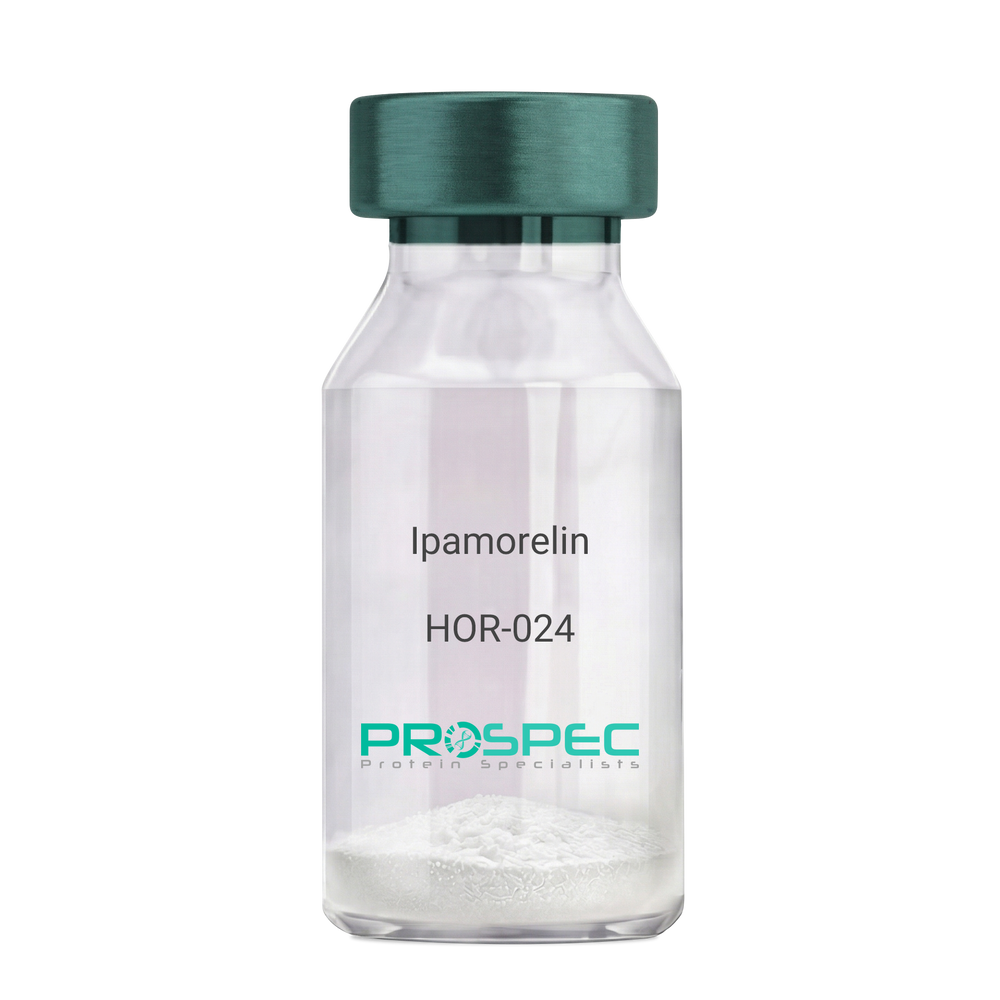
IpamorelinDescription:
Ipamorelin
Ipamorelin
Product # :
HOR-024Price :
Quantity :
Shipping Method :
Shipped at Room temp
More Info
- description
- formulation
- purity
- More Info
Description
Ipamorelin Synthetic is a single, non-glycosylated polypeptide chain containing 4 amino acids, having a molecular mass of 711.85 Dalton and a Molecular formula of C38H49N9O5.
Formulation
The protein was lyophilized with no additives.
Purity
Greater than 97.0% as determined by analysis by RP-HPLC.
More Info
-
Introduction
Ipamorelin is a peptide selective agonist of the ghrelin/growth hormone secretagogue receptor and a growth hormone secretagogue. Ipamorelin is a pentapeptide that was derived from GHRP1. Ipamorelin significantly increases plasma growth hormone levels in both animals and humans. Like pralmorelin and GHRP-6, ipamorelin does not affect prolactin, FSH, LH or TSH levels. However, unlike GHRP2 and GHRP6, but as growth hormone-releasing hormone (GHRH), ipamorelin does not stimulate the secretion of adrenocorticotropic hormone (ACTH) or cortisol, and is highly selective for inducing the secretion only of GH.
-
Synonyms
Ipamorelin
-
Physical Appearance
Sterile Filtered White lyophilized (freeze-dried) powder.
-
Stability
Lyophilized Ipamorelin although stable at room temperature for 3 weeks, should be stored desiccated below -18°C. Upon reconstitution Ipamorelin should be stored at 4°C between 2-7 days and for future use below -18°C. For long term storage it is recommended to add a carrier protein (0.1% HSA or BSA). Please prevent freeze-thaw cycles.
-
Solubility
It is recommended to reconstitute the lyophilized Ipamorelin in sterile 18MΩ-cm H2O not less than 100 µg/ml, which can then be further diluted to other aqueous solutions.
-
Amino Acid Sequence
Aib-His-D-2-Nal-D-Phe-Lys-NH2.
ProSpec's products are furnished for LABORATORY RESEARCH USE ONLY. The product may not be used as drugs, agricultural or pesticidal products, food additives or household chemicals.
- View Data Sheet
Name :
HPRT1 Human, ActiveDescription:
Hypoxanthine-Guanine Phosphoribosyltransferase, Human Recombinant, Active
Hypoxanthine Phosphoribosyltransferase 1, EC 2.4.2.8, HGPRTase, HGPRT, HPRT , Hypoxanthine-Guanine Phosphoribosyltransferase 1, Hypoxanthine-Guanine Phosphoribosyltransferase, Testicular Tissue Protein Li 89, Lesch-Nyhan Syndrome, HPRT1.
Product # :
ENZ-1006Price :
Quantity :
Shipping Method :
Shipped with Ice Packs
More Info
- description
- source
- formulation
- purity
- biological activity
- More Info
Description
HPRT1 Human Recombinant produced in E.Coli is a single, non-glycosylated polypeptide chain containing 238 amino acids (1-218 a.a) and having a molecular mass of 26.7kDa. HPRT1 is fused to a 20 amino acid His-tag at N-terminus & purified by proprietary chromatographic techniques.
Source
Escherichia Coli.
Formulation
HPRT1 protein solution (0.5mg/ml) containing 20mM Tris-HCl buffer (pH 8.0) and 20% glycerol.
Purity
Greater than 95.0% as determined by SDS-PAGE.
Biological Activity
Specific activity is > 15 units/mg and is defined as the amount of enzyme that catalyze the formation of 1 umole of guanosine 5’-monophosphate (GMP) per minute from guanine and phosphoribosyl pyrophosphate at pH 7.5 at 37C.More Info
-
Introduction
HPRT1 has a main part in the generation of purine nucleotides through the purine salvage pathway. HPRT1 primarily functions to salvage purines from degraded DNA to renewed purine synthesis. Therefore, it performs as a catalyst in the reaction between guanine and phosphoribosyl pyrophosphate to form GMP.
-
Synonyms
Hypoxanthine Phosphoribosyltransferase 1, EC 2.4.2.8, HGPRTase, HGPRT, HPRT , Hypoxanthine-Guanine Phosphoribosyltransferase 1, Hypoxanthine-Guanine Phosphoribosyltransferase, Testicular Tissue Protein Li 89, Lesch-Nyhan Syndrome, HPRT1.
-
Physical Appearance
Sterile Filtered clear solution.
-
Stability
Store at 4°C if entire vial will be used within 2-4 weeks. Store, frozen at -20°C for longer periods of time. For long term storage it is recommended to add a carrier protein (0.1% HSA or BSA).Avoid multiple freeze-thaw cycles.
-
Amino Acid Sequence
MGSSHHHHHH SSGLVPRGSH MATRSPGVVI SDDEPGYDLD LFCIPNHYAE DLERVFIPHG LIMDRTERLA RDVMKEMGGH HIVALCVLKG GYKFFADLLD YIKALNRNSD RSIPMTVDFI RLKSYCNDQS TGDIKVIGGD DLSTLTGKNV LIVEDIIDTG KTMQTLLSLV RQYNPKMVKVASLLVKRTPR SVGYKPDFVG FEIPDKFVVG YALDYNEYFR DLNHVCVISE TGKAKYKA
ProSpec's products are furnished for LABORATORY RESEARCH USE ONLY. The product may not be used as drugs, agricultural or pesticidal products, food additives or household chemicals.
- View Data Sheet
Name :
GDF6 HumanDescription:
Bone Morphogenetic protein-13 Human Recombinant
Growth Differentiation Factor 6, Growth/Differentiation Factor 16, Bone Morphogenetic Protein 13, BMP-13, BMP13, GDF-6, Klippel-Feil Malformation, Segmentation Syndrome 1, Klip-Feil Malformation, Klippel-Feil Syndrome, MCOPCB6, SCDO4, CDMP2, LCA17, MCOP4, GDF16, KFS1, KFSL, SGM1, KFM, KFS, GDF6.
Product # :
CYT-938Price :
Quantity :
Shipping Method :
Shipped at Room temp
More Info
- description
- source
- formulation
- purity
- biological activity
- More Info
Description
BMP13 Human Recombinant produced in E.coli is a non-glycosylated disulfide linked homodimer containing 2 chains of 120 amino acids and having a molecular mass of 27.1kDa.The BMP-13 is purified by proprietary chromatographic techniques.
Source
Escherichia Coli.
Formulation
BMP-13 protein was lyophilized from a 0.2µm filtered concentrated solution in 30% Acetonitrile and 0.1% TFA.
Purity
Greater than 95.0% as determined by: (a) Analysis by HPLC. (b) Analysis by SDS-PAGE.
Biological Activity
The ED50 as determined by inducing alkaline phosphatase production of murine ATDC5 cells is less than 2.0µg/ml, corresponding to a specific activity of > 500IU/mg.More Info
-
Introduction
Growth/differentiation factors (GDF1-GDF15) belong to the BMP family of TGF-beta superfamily proteins. These factors are produced as inactive preproproteins which are subsequently cleaved and assembled into active secreted homodimers. BMP13 is a growth factor which controls proliferation and cellular differentiation in the retina and bone formation. BMP13 has a central role in regulating apoptosis during retinal development. GDF proteins are vital during embryonic development, particularly in the skeletal, nervous, and muscular systems. BMP13 gene mutations result in colobomata, which are congenital abnormalities in ocular development, and in Klippel-Feil syndrome (KFS), which is a congenital disorder of spinal segmentation.
-
Synonyms
Growth Differentiation Factor 6, Growth/Differentiation Factor 16, Bone Morphogenetic Protein 13, BMP-13, BMP13, GDF-6, Klippel-Feil Malformation, Segmentation Syndrome 1, Klip-Feil Malformation, Klippel-Feil Syndrome, MCOPCB6, SCDO4, CDMP2, LCA17, MCOP4, GDF16, KFS1, KFSL, SGM1, KFM, KFS, GDF6.
-
Physical Appearance
Sterile Filtered White lyophilized (freeze-dried) powder.
-
Stability
Lyophilized BMP13 although stable at room temperature for 3 weeks, should be stored desiccated below -18°C. Upon reconstitution BMP-13 should be stored at 4°C between 2-7 days and for future use below -18°C. For long term storage it is recommended to add a carrier protein (0.1% HSA or BSA). Please prevent freeze-thaw cycles.
-
Solubility
It is recommended to reconstitute the lyophilized BMP13 in sterile 18M-cm H2O not less than 100µg/ml, which can then be further diluted to other aqueous solutions.
-
Amino Acid Sequence
TAFASRHGKR HGKKSRLRCS KKPLHVNFKE LGWDDWIIAP LEYEAYHCEG VCDFPLRSHL EPTNHAIIQT LMNSMDPGST PPSCCVPTKL TPISILYIDA GNNVVYKQYE DMVVESCGCR.
-
Background
Bone Morphogenetic Protein-13 Human Recombinant: Unraveling its Potential in Tissue Engineering and Regenerative Medicine
Abstract:
Bone Morphogenetic Protein-13 (BMP-13) human recombinant is a pivotal member of the bone morphogenetic protein family, known for its crucial role in tissue development, regeneration, and repair. This research paper aims to provide a comprehensive analysis of BMP-13, including its characteristics, signaling pathways, and potential therapeutic applications. Furthermore, innovative methodologies for the production and optimization of BMP-13 human recombinant are proposed, shedding light on its future implications in the field of tissue engineering and regenerative medicine.
Introduction:
Tissue engineering and regenerative medicine hold great promise in addressing tissue repair and regeneration challenges. BMP-13, a prominent member of the BMP family, plays a vital role in orchestrating cellular responses during tissue development and healing. This paper explores the distinctive features of BMP-13 and presents novel approaches for the production and optimization of BMP-13 human recombinant, aiming to unlock its therapeutic potential in various regenerative contexts.
Characteristics and Signaling Pathways:
BMP-13 is a secreted growth factor belonging to the transforming growth factor-beta (TGF-β) superfamily. It exerts its biological effects by binding to specific cell surface receptors, initiating intricate intracellular signaling cascades. BMP-13 signaling pathways, including Smad-dependent and Smad-independent pathways, regulate critical processes such as cell differentiation, proliferation, and extracellular matrix synthesis, influencing tissue development and repair.
Production of BMP-13 Human Recombinant:
Efficient production methodologies are crucial for harnessing the therapeutic potential of BMP-13 human recombinant. Various recombinant protein expression systems, such as mammalian cells or baculovirus-insect cell systems, have been utilized for the production of functional BMP-13. Optimization strategies, including codon optimization, signal peptide engineering, and protein folding enhancement, have been employed to improve the yield and bioactivity of BMP-13 recombinant protein.
Potential Therapeutic Applications:
BMP-13 human recombinant holds immense promise in the field of tissue engineering and regenerative medicine. Its involvement in cartilage formation, osteogenesis, and tissue repair makes it a potential candidate for the treatment of musculoskeletal disorders, joint injuries, and cartilage defects. Furthermore, the ability of BMP-13 to modulate cell behavior and tissue remodeling indicates its wider therapeutic applications in diverse regenerative processes.
Conclusion:
BMP-13 human recombinant emerges as a crucial regulator in tissue engineering and regenerative medicine, offering significant potential for tissue repair and regeneration. Optimizing production methodologies and further unraveling its signaling mechanisms will undoubtedly enhance its therapeutic applications. Given its involvement in cartilage and bone formation, as well as tissue repair, BMP-13 human recombinant represents a valuable tool for promoting tissue regeneration and addressing the unmet clinical needs in regenerative medicine.
What is the molecular weight/Mw of GDF6 Protein?
GDF6 Protein has a total Mw of 27.1kDa.
What is the source or expression system of GDF6 Protein?
Escherichia Coli.
What is the Purity of GDF6 Protein?
GDF6 Protein is >95% pure as determined by SDS-PAGE.
What is the Biological Activity of GDF6 Protein?
The ED50 as determined by inducing alkaline phosphatase production of murine ATDC5 cells is less than 2.0µg/ml, corresponding to a specific activity of > 500IU/mg.
What is the amino acid sequence of GDF6 Protein?
TAFASRHGKR HGKKSRLRCS KKPLHVNFKE LGWDDWIIAP LEYEAYHCEG VCDFPLRSHL EPTNHAIIQT LMNSMDPGST PPSCCVPTKL TPISILYIDA GNNVVYKQYE DMVVESCGCR.
What applications can GDF6 Protein be used in?
GDF6 Protein can probably be used in western blot, ELISA and Lateral Flow.
What is the endotoxin level for GDF6 Protein?
The endotoxin level is minimal, GDF6 Protein was purified using conventional chromatography techniques.
ProSpec's products are furnished for LABORATORY RESEARCH USE ONLY. The product may not be used as drugs, agricultural or pesticidal products, food additives or household chemicals.
- View Data Sheet
Name :
Influenza-A H1N1 AntibodyDescription:
Influenza-A Hemagglutinin H1N1, Mouse Antibody
Product # :
ANT-214Price :
Quantity :
Shipping Method :
Shipped with Ice Packs
More Info
- description
- formulation
- More Info
Description
Hybridoma clones have been derived from hybridization of Sp2/0 myeloma cells with spleen cells of Balb/c mice immunized with Influenza A/NewCaledonia/20/99 H1N1 derived from allantoic fluid of 10 days old embryonated eggs.
Formulation
In PBS, pH 7.4 and 0.09% NaN3.
More Info
-
Introduction
H1N1 is a subtype specie of Influenza A virus. H1N1 Influenza Virus has mutated into various strains such as the Spanish Flustrain, mild humanflu strains, endemic pigstrains, and various strains found in birds.
The Influenza A Virusis a globular particle about 100nm in diameter, sheathed in a lipid bilayer derived from the plasma membrane of its host. Studded in the lipid bilayer are two integral membrane proteins some 500 molecules of hemagglutinin ("H") and some 100 molecules of neuraminidase ("N"). Within the lipid bilayer are 3000 molecules of matrix protein and 8 pieces of RNA. Each of the 8 RNA molecules is associated with many copies of a nucleoprotein, several molecules of the three subunits of its RNA polymerase some "non-structural" protein molecules of uncertain function. -
Physical Appearance
Sterile Filtered clear solution.
-
Immunogen
Influenza A hemagglutinin H1N1.
-
Ig Subclass
mouse IgG1.
-
Clone
IA-H1N1.
-
Applications
Influenza A haemagglutinin 1 H1N1 immunodetection in direct or indirect ELISA, sandwich immunoassay, Western Blotting. Recommended pairs for Influenza A H1N1 in sandwich immunoassay (coating – conjugate): All pairs detect H1N1 of Influenza A as well as recombinant H1N1 with high specificity and do not interact with haemagglutinin H3N2.
-
Shipping Conditions
Antibody is shipped as in liquid form with ice packs.
-
Type
Mouse Antibody Monoclonal.
-
Storage Procedures
Should be stored at 4C.Do not freeze.
-
Purification Method
Protein-A column.
ProSpec's products are furnished for LABORATORY RESEARCH USE ONLY. The product may not be used as drugs, agricultural or pesticidal products, food additives or household chemicals.
- View Data Sheet
Name :
CDKN1A HumanDescription:
Cyclin-Dependent Kinase Inhibitor 1A Human Recombinant
Cyclin-Dependent Kinase Inhibitor 1A (P21, Cip1), Melanoma Differentiation Associated Protein 6, Wild-Type P53-Activated Fragment 1, CDK-Interaction Protein 1, DNA Synthesis Inhibitor, CDK-Interacting Protein 1, CDKN1, P21Cip1/Waf1, MDA6, P21CIP1, CAP20, WAF1, CIP1, PIC1, P21, SDI1.
Product # :
PKA-043Price :
Quantity :
Shipping Method :
Shipped with Ice Packs
More Info
- description
- source
- formulation
- purity
- More Info
Description
CDKN1A Human Recombinant produced in E. coli is a single polypeptide chain containing 184 amino acids (1-164) and having a molecular mass of 20.2 kDa.CDKN1A is fused to a 20 amino acid His-tag at N-terminus & purified by proprietary chromatographic techniques.
Source
Escherichia Coli.
Formulation
The CDKN1A solution contains 20mM Tris-HCl buffer (pH 8.0), 2M Urea, 0.1M NaCl and 10% glycerol.
Purity
Greater than 85.0% as determined by SDS-PAGE.
More Info
-
Introduction
CDKN1A holds a key part in the cellular response to DNA mutation. CDKN1A overexpression causes cell cycle to stop. When collaborating with CDK2 complexes, CDKN1A aids to prevent kinase activity and inhibits progression through G1/S. Nevertheless, CDKN1A can additionally increase assembly and activity in complexes of CDK4 or CDK6 and cyclin D. CDKN1A is a vital intermediary by which p53/TP53 facilitates its part as an inhibitor of cellular proliferation as a reaction to DNA impairment.
-
Synonyms
Cyclin-Dependent Kinase Inhibitor 1A (P21, Cip1), Melanoma Differentiation Associated Protein 6, Wild-Type P53-Activated Fragment 1, CDK-Interaction Protein 1, DNA Synthesis Inhibitor, CDK-Interacting Protein 1, CDKN1, P21Cip1/Waf1, MDA6, P21CIP1, CAP20, WAF1, CIP1, PIC1, P21, SDI1.
-
Physical Appearance
Sterile Filtered colorless solution.
-
Stability
Store at 4°C if entire vial will be used within 2-4 weeks. Store, frozen at -20°C for longer periods of time. For long term storage it is recommended to add a carrier protein (0.1% HSA or BSA).Avoid multiple freeze-thaw cycles.
-
Amino Acid Sequence
MGSSHHHHHH SSGLVPRGSH MSEPAGDVRQ NPCGSKACRR LFGPVDSEQL SRDCDALMAG CIQEARERWN FDFVTETPLE GDFAWERVRG LGLPKLYLPT GPRRGRDELG GGRRPGTSPA LLQGTAEEDH VDLSLSCTLV PRSGEQAEGS PGGPGDSQGR KRRQTSMTDF YHSKRRLIFS KRKP.
ProSpec's products are furnished for LABORATORY RESEARCH USE ONLY. The product may not be used as drugs, agricultural or pesticidal products, food additives or household chemicals.
- View Data Sheet
Name :
VAMP2 MouseDescription:
Synaptobrevin-2 Recombinant Mouse
Vesicle-associated membrane protein 2, VAMP-2, Synaptobrevin-2, Vamp2, Syb2.
Product # :
PRO-2415Price :
Quantity :
Shipping Method :
Shipped with Ice Packs
More Info
- description
- source
- formulation
- purity
- More Info
Description
VAMP2 Mouse Recombinant produced in E.Coli is a single, non-glycosylated polypeptide chain containing 118 amino acids (1-94 a.a) and having a molecular mass of 12.8kDa. VAMP2 is fused to a 24 amino acid His-tag at N-terminus & purified by proprietary chromatographic techniques.
Source
Escherichia Coli.
Formulation
VAMP2 protein solution (1mg/ml) containing Phosphate Buffered Saline (pH7.4), 1mM EDTA, 0.1mM PMSF and 10% glycerol.
Purity
Greater than 90.0% as determined by SDS-PAGE.
More Info
-
Introduction
Synaptobrevin 2 which is an 18 kDa integral membrane protein localized to the cytoplasmic surface of synaptic vesicle, consists of a proline-rich N-terminal region, a highly conserved hydrophilic domain, followed by a transmembrane anchor and a C-terminal. Synaptobrevin 2 is predominantly expressed in Langerhans islets and glomerular cells. The N-terminal domain of the protein (residues 1-89) forms a specific SNARE complex with the target membrane-associated t- or Q-SNAREs syntaxin 1 and SNAP-25.
-
Synonyms
Vesicle-associated membrane protein 2, VAMP-2, Synaptobrevin-2, Vamp2, Syb2.
-
Physical Appearance
Sterile Filtered clear solution.
-
Stability
Store at 4°C if entire vial will be used within 2-4 weeks. Store, frozen at -20°C for longer periods of time. For long term storage it is recommended to add a carrier protein (0.1% HSA or BSA).Avoid multiple freeze-thaw cycles.
-
Amino Acid Sequence
MGSSHHHHHH SSGLVPRGSH MGSHMSATAA TVPPAAPAGE GGPPAPPPNL TSNRRLQQTQ AQVDEVVDIM RVNVDKVLER DQKLSELDDR ADALQAGASQ FETSAAKLKR KYWWKNLK.
ProSpec's products are furnished for LABORATORY RESEARCH USE ONLY. The product may not be used as drugs, agricultural or pesticidal products, food additives or household chemicals.
- View Data Sheet
Name :
GM-CSF MonkeyDescription:
Granulocyte Macrophage-Colony Stimulating Factor Rhesus Macaque Recombinant
CSF-2, MGI-1GM, GM-CSF, Pluripoietin-alpha, MGC131935, MGC138897.
Product # :
CYT-720Price :
Quantity :
Shipping Method :
Shipped at Room temp
More Info
- description
- source
- formulation
- purity
- biological activity
- More Info
Description
Granulocyte Macrophage Colony Stimulating Factor Monkey Recombinant produced in E.Coli is a single, non-glycosylated, polypeptide chain containing 127 amino acids and having a molecular mass of 14.4 kDa.GM-CSF is purified by proprietary chromatographic techniques.
Source
Escherichia Coli.
Formulation
GM-CSF was lyophilized from a concentrated (1mg/ml) solution containing 1x PBS pH 7.4.
Purity
Greater than 98.0% as determined by SDS-PAGE and RP-HPLC.
Biological Activity
The ED50 as determined by a cell proliferation assay using human TF-1 cells is less than 0.1ng/ml, corresponding to a specific activity of > 10,000,000 IU/mg.More Info
-
Introduction
GMCSF is a cytokine that controls the production, differentiation, and function of granulocytes and macrophages. The active form of the protein is found extracellularly as a homodimer. This gene has been localized to a cluster of related genes at chromosome region 5q31, which is known to be associated with interstitial deletions in the 5q- syndrome and acute myelogenous leukemia. Other genes in the cluster include those encoding interleukins 4, 5, and 13. GM-CSF stimulates the growth and differentiation of hematopoietic precursor cells from various lineages, including granulocytes, macrophages, eosinophils and erythrocytes.
-
Synonyms
CSF-2, MGI-1GM, GM-CSF, Pluripoietin-alpha, MGC131935, MGC138897.
-
Physical Appearance
Sterile Filtered White lyophilized (freeze-dried) powder.
-
Stability
Lyophilized GMCSF although stable at room temperature for 3 weeks, should be stored desiccated below -18°C. Upon reconstitution GMCSF should be stored at 4°C between 2-7 days and for future use below -18°C. For long term storage it is recommended to add a carrier protein (0.1% HSA or BSA). Please prevent freeze-thaw cycles.
-
Solubility
It is recommended to reconstitute the lyophilized GMCSF in sterile 18MΩ-cm H2O not less than 100µg/ml, which can then be further diluted to other aqueous solutions.
-
Amino Acid Sequence
APARSPSPGT QPWEHVNAIQ EARRLLNLSR DTAAEMNKTV EVVSEMFDLQ EPSCLQTRLE LYKQGLQGSL TKLKGPLTMM ASHYKQHCPP TPETSCATQI ITFQSFKENL KDFLLVIPFD CWEPVQE.
ProSpec's products are furnished for LABORATORY RESEARCH USE ONLY. The product may not be used as drugs, agricultural or pesticidal products, food additives or household chemicals.
- View Data Sheet
Name :
NELL2 AntibodyDescription:
NEL-Like 2, Monoclonal Mouse Anti Human Antibody
NEL-like 2, NRP2, Nel-related protein 2, protein kinase C-binding protein NELL2, neural epidermal growth factor-like 2.
Product # :
ANT-030Price :
Quantity :
Shipping Method :
Shipped with Ice Packs
More Info
- formulation
- More Info
Formulation
1mg/ml containing Phosphate-Buffered Saline (pH 7.4) with 0.1% Sodium Azide.
More Info
-
Introduction
NELL2 is expressed in large quantities in neural tissues and has a significant role in the development of neural tissues. In addition, NELL protein has six epidermal growth factor (EGF)-like repeat domains which plays a part in calcium binding.
-
Synonyms
NEL-like 2, NRP2, Nel-related protein 2, protein kinase C-binding protein NELL2, neural epidermal growth factor-like 2.
-
Immunogen
Anti-human NELL2 mAb, is derived from hybridization of mouse F0 myeloma cells with spleen cells from BALB/c mice immunized with a recombinant human NELL2 protein.
-
Ig Subclass
Mouse IgG2b heavy chain and Kappa light chain
-
Clone
AT13E7.
-
Applications
The antibody has been tested by ELISA and Western blot analysis to assure specificity and reactivity. Since application varies, however, each investigation should be titrated by the reagent to obtain optimal results. Recommended dilution range for Western blot analysis is 1:500.
Recommended starting dilution is 1:500 -
Type
Mouse Anti Human Monoclonal.
-
Storage Procedures
For periods up to 1 month store at 4°C, for longer periods of time, store at -20°C. Prevent freeze thaw cycles.
-
Purification Method
Recombinant human NELL2 antibody (30-258aa) is purified from E. coli.
ProSpec's products are furnished for LABORATORY RESEARCH USE ONLY. The product may not be used as drugs, agricultural or pesticidal products, food additives or household chemicals.
- View Data Sheet
Name :
IGF1 PAT6F8AT AntibodyDescription:
Insulin-Like Growth Factor-1 Clone PAT6F8AT, Mouse Anti Human
Somatomedin C, IGF-I, IGFI, IGF1, IGF-IA, Mechano growth factor, MGF.
Product # :
ANT-538Price :
Quantity :
Shipping Method :
Shipped with Ice Packs
More Info
- formulation
- More Info
Formulation
1mg/ml containing PBS, pH-7.4, 10% Glycerol and 0.02% Sodium Azide.
More Info
-
Introduction
The somatomedins, or insulin-like growth factors (IGFs), comprise a family of peptides that play important roles in mammalian growth and development. IGF1 mediates many of the growth-promoting effects of growth hormone (GH; MIM 139250). Early studies showed that growth hormone did not directly stimulate the incorporation of sulfate into cartilage, but rather acted through a serum factor, termed 'sulfation factor,' which later became known as 'somatomedin' (Daughaday et al., 1972). Three main somatomedins have been characterized: somatomedin C (IGF1), somatomedin A (IGF2; MIM 147470), and somatomedin B (MIM 193190) (Rotwein, 1986; Rosenfeld, 2003).
-
Synonyms
Somatomedin C, IGF-I, IGFI, IGF1, IGF-IA, Mechano growth factor, MGF.
-
Physical Appearance
Sterile filtered colorless solution.
-
Immunogen
Anti-human IGF1 mAb, clone PAT6F8AT, is derived from hybridization of mouse F0 myeloma cells with spleen cells from BALB/c mice immunized with a recombinant human IGF1 protein 49-118 amino acids purified from E. coli.
-
Ig Subclass
Mouse IgG2b heavy chain and k light chain.
-
Clone
PAT6F8AT.
-
Applications
HSPA5 antibody has been tested by ELISA, Western blot analysis to assure specificity and reactivity. Since application varies, however, each investigation should be titrated by the reagent to obtain optimal results. Recommended starting dilution is 1:1000.
-
Type
Mouse Anti Human Monoclonal.
-
Storage Procedures
For periods up to 1 month store at 4°C, for longer periods of time, store at -20°C. Prevent freeze thaw cycles.
-
Purification Method
IGF1 antibody was purified from mouse ascitic fluids by protein-A affinity chromatography.
ProSpec's products are furnished for LABORATORY RESEARCH USE ONLY. The product may not be used as drugs, agricultural or pesticidal products, food additives or household chemicals.
- View Data Sheet
Name :
HIV-1 TAT Cys22Description:
HIV-1 TAT Cys22 Recombinant
Product # :
HIV-136Price :
Quantity :
Shipping Method :
Shipped at Room temp
More Info
- description
- source
- formulation
- purity
- More Info
Description
HIV-1 TAT Cys22 Recombinant- produced in E.coli is a single, non-glycosylated, polypeptide chain containing 86 amino acids mutated in the trans activation domain and having chain having a molecular mass of 14.4 kDa.
Source
Escherichia Coli.
Formulation
Lyophilized with 0.1% glycerol.
Purity
Greater than 90.0% as determined by SDS-PAGE.
More Info
-
Introduction
Human immunodeficiency virus type-1 (HIV-1) regulatory Tat protein plays an crucial part in viral replication and infectivity. Throughout acute infection, Tat protein is released extracellularly by infected cells and is taken up by neighboring cells where it transactivates viral replication and inhances virus infectivity.
HIV-1 Tat activates transcription of HIV-1 viral genes by inducing phosphorylation of the C-terminal domain (CTD) of RNA polymerase II (RNAPII). Tat can also disturb cellular metabolism by inhibiting proliferation of antigen-specific T lymphocytes and by inducing cellular apoptosis. Tat-induced apoptosis of T-cells is attributed, in part, to the distortion of microtubules polymerization. LIS1 is a microtubule-associated protein that facilitates microtubule polymerization. -
Physical Appearance
Sterile Filtered White lyophilized (freeze-dried) powder.
-
Stability
Lyophilized HIV-1 TAT Cys22 although stable at room temperature for 1 week, should be stored desiccated below -18°C. Upon reconstitution HIV-1 TAT Cys22 should be stored at 4°C between 2-7 days and for future use below -18°C.For long-term storage it is recommended to add a carrier protein (0.1% HSA or BSA).Please prevent freeze-thaw cycles.
-
Solubility
It is recommended to reconstitute the lyophilized HIV-1 TAT Cys 22 in sterile 18MΩ-cm H2O not less than 100µg/ml, which can then be further diluted to other aqueous solutions.
-
Applications
Recognized by anti-Tat (HIV-1) polyclonal antibody. Reacts with anti-Tat antibodies from human, monkey, rabbit and mouse serum.
-
Specificity
Immunoreactive with all sera of HIV-1 infected individuals.
ProSpec's products are furnished for LABORATORY RESEARCH USE ONLY. The product may not be used as drugs, agricultural or pesticidal products, food additives or household chemicals.
- View Data Sheet
Name :
KLK3 Human, HEKDescription:
Kallikrein-3 Human Recombinant, HEK
Prostate-specific antigen, PSA, Gamma-seminoprotein, Seminin, Kallikrein-3, P-30 antigen, Semenogelase, KLK3, APS, hK3, KLK2A1
Product # :
ENZ-1149Price :
Quantity :
Shipping Method :
Shipped with Ice Packs
More Info
- description
- source
- formulation
- purity
- biological activity
- More Info
Description
KLK3 Human Recombinant produced in HEK cells is a single, glycosylated, polypeptide chain (18-261 a.a) containing a total of 250 amino acids, having a molecular mass of 27.6kDa.KLK3 is fused to a 6 amino acid His-tag at C-terminus,and is purified by proprietary chromatographic techniques.
Source
HEK293 Cells.
Formulation
The KLK3 solution (0.25mg/ml) contains 10% Glycerol and Phosphate-Buffered Saline (pH 7.4).
Purity
Greater than 95.0% as determined by SDS-PAGE.
Biological Activity
Specific activity is > 250pmol/min/ug. Defined by the amount of enzyme that cleaves 1pmole of Succinyl-ArgPro-Tyr-p-Nitroanilide to Succinyl-Arg-Pro-Tyr and p-Nitroanilide per minute at pH 7.5 at 37˚C.
More Info
-
Introduction
Kallikrein-3 or KLK3 is a serine proteases subgroup, enzymes can cleave peptide bonds. KLK3 is found in the prostate and may be hold accountable to the regulation of semen liquefaction via seminogelin hydrolysis. The protein is expressed in low concentrations in men with healthy prostates serum. When elevated quantities are found it may show prostate cancer or different prostate disorders such as prostatitis and benign prostatic hyperplasia.
-
Synonyms
Prostate-specific antigen, PSA, Gamma-seminoprotein, Seminin, Kallikrein-3, P-30 antigen, Semenogelase, KLK3, APS, hK3, KLK2A1
-
Physical Appearance
Sterile Filtered colorless solution.
-
Stability
Store at 4°C if entire vial will be used within 2-4 weeks. Store, frozen at -20°C for longer periods of time. For long term storage it is recommended to add a carrier protein (0.1% HSA or BSA).Avoid multiple freeze-thaw cycles.
-
Amino Acid Sequence
APLILSRIVG GWECEKHSQP WQVLVASRGR AVCGGVLVHP QWVLTAAHCI RNKSVILLGR HSLFHPEDTG QVFQVSHSFP HPLYDMSLLK NRFLRPGDDS SHDLMLLRLS EPAELTDAVK VMDLPTQEPA LGTTCYASGW GSIEPEEFLT PKKLQCVDLH VISNDVCAQV HPQKVTKFML CAGRWTGGKS TCSGDSGGPL VCNGVLQGIT SWGSEPCALP ERPSLYTKVV HYRKWIKDTI VANPHHHHHH
ProSpec's products are furnished for LABORATORY RESEARCH USE ONLY. The product may not be used as drugs, agricultural or pesticidal products, food additives or household chemicals.
- View Data Sheet
Name :
SARS MERS Spike AntibodyDescription:
Mouse Anti MERS-CoV Spike
Middle East respiratory syndrome coronavirus, Human betacoronavirus 2c EMC/2012, MERS-CoV, MERS, MERSCoV SP, Spike glycoprotein, S glycoprotein, S, Spike protein, E2, Peplomer protein.
Product # :
ANT-768Price :
Quantity :
Shipping Method :
Shipped with Ice Packs
More Info
- formulation
- More Info
Formulation
1mg/ml containing Phosphate-Buffered Saline (pH 7.4), 0.02% Sodium Azide and 10% Glycerol.
More Info
-
Introduction
Since April 2012, cases of the Middle East Respiratory Syndrome Coronavirus (MERS-CoV) have been identified in various countries. Coronaviruses are the cause of the common cold, SARS (severe acute respiratory syndrome) and other severe illnesses with high mortality rates, all are classified into coronavirus family. MERS-CoV is a new type of SARS found in the coronavirus family causing severe pneumonia with sudden and serious respiratory illness with high mortality rates as well. Since January 27th 2015, the WHO has reported 956 human cases, including 351 deaths. More cases of the new coronavirus strain are expected. Like in other coronaviruses, large surface spike glycoprotein is a central structural protein of this virus; it is located above the virion surface to bind and enter into the target cell. Spike protein has 2 domains- S1 and S2. The S1 domain is responsible for cellular tropism and interaction with target cell, while the S2 domain is responsible for membrane fusion. The C-terminal of S1 domain contains a receptor binding domain, and is also a potential target for vaccine development and an antigen for diagnosis.
-
Synonyms
Middle East respiratory syndrome coronavirus, Human betacoronavirus 2c EMC/2012, MERS-CoV, MERS, MERSCoV SP, Spike glycoprotein, S glycoprotein, S, Spike protein, E2, Peplomer protein.
-
Physical Appearance
Sterile filtered colorless solution.
-
Immunogen
Recombinant MERS-CoV Spike (18-1296aa) purified from Baculovirus.
-
Ig Subclass
IgG1 kappa.
-
Clone
PAT40G7AT
-
Applications
SARS MERS Spike antibody has been tested by ELISA analysis to assure specificity and reactivity. Since application varies, however, each investigation should be titrated by the reagent to obtain optimal results.
-
Type
Mouse antibody Monoclonal.
-
Storage Procedures
For periods up to 1-month store at 4°C, for longer periods of time, store at -20°C. Prevent freeze thaw cycles.
-
Purification Method
SARS MERS Spike antibody was purified from mouse ascitic fluids by protein-A affinity chromatography.
ProSpec's products are furnished for LABORATORY RESEARCH USE ONLY. The product may not be used as drugs, agricultural or pesticidal products, food additives or household chemicals.
- View Data Sheet
Name :
IFN a 2a AntibodyDescription:
Interferon a 2a Polyclonal Rabbit Anti-Human Antibody
Leukocyte interferon, B cell interferon, Type I interferon, IFNA2, IFN-a 2a.
Product # :
ANT-034Price :
Quantity :
Shipping Method :
Shipped with Ice Packs
More Info
- formulation
- purity
- More Info
Formulation
Lyophilized from 0.2μm sterile filtered solution in phosphate buffered saline, pH 7.4.
Purity
Greater than 98.0% as determined by Analysis by SDS-PAGE.
More Info
-
Introduction
IFN-alpha is produced by macrophages and has antiviral activities. Interferon stimulates the production of two enzymes: protein kinase and an oligoadenylate synthetase.
-
Synonyms
Leukocyte interferon, B cell interferon, Type I interferon, IFNA2, IFN-a 2a.
-
Stability
Store at -20°C. For long term storage freezes in working aliquots at -20°C. Repeated freezing and thawing is not recommended.
-
Solubility
Reconstitute with H20. Mix gently, wash the sides of the vial and wait 30-60 seconds before use.
-
Immunogen
IgG Anti Human Interferon a 2a is developed in rabbit using recombinant Human Interferon a 2a expressed in plants.
-
Applications
ELISA: to detect Human Interferon a 2a by indirect ELISA a dilution of at least 1/500 of this antibody is required. This antibody, in conjunction with compatible secondary reagents (anti rabbit AP conjugated), allows the detection of 0.2-1 ng /well of human Interferon a 2a.
Western Blot: to detect human Interferon a2a by WB analysis this antibody can be used in a dilution of 1/1,000.
-
Antigen Amino Acid Sequence
CDLPQTHSLGSRRTLMLLAQMRKISLFSCLKDRHDFGFPQEEFGNQFQKAETIPVLHEMIQQIFNLFSTKDSSAAWDETLLDKFYTELYQQLNDLEACVIQGVGVTETPLMNEDSILAVRKYFQRITLYLKEKKYSPCAWEVVRAEIMRSFSLSTNLQESLRSKE
-
Type
Polyclonal Rabbit Antibody.
-
Purification Method
Purified IgG prepared by affinity chromatography on protein G.
ProSpec's products are furnished for LABORATORY RESEARCH USE ONLY. The product may not be used as drugs, agricultural or pesticidal products, food additives or household chemicals.
- View Data Sheet
Name :
Influenza-A H3N2 AntibodyDescription:
H3N2/HA1, Mouse Anti-Human
Product # :
ANT-080Price :
Quantity :
Shipping Method :
Shipped with Ice Packs
More Info
- formulation
- More Info
Formulation
1mg/ml containing PBS, pH-7.4, 0.02% Sodium Azide and 10% glycerol.
More Info
-
Introduction
H3N2 is a subtype of the influenza A virus. Its name derives from the forms of the two kinds of proteinson the surface of its coat, hemagglutinin(H) and neuraminidase(N). H3N2 exchanges genes for internal proteins with other influenza subtypes. H3N2 has tended to dominate in prevalence over H1N1, H1N2, and influenza B. H3N2 strain descended from H2N2by antigenic shift, in which genes from multiple subtypes re-assorted to form a new virus. Both the H2N2and H3N2 strains contained genesfrom avian influenzaviruses.
-
Immunogen
Anti-human Influenza-A H3N2 mAb, is derived from hybridization of mouse FO myeloma cells with spleen cells from BALB/c mice immunized with recombinant human H3N2/HA1 amino acids 17-345 purified from Baculovirus.
-
Ig Subclass
Mouse IgG1 heavy chain and k light chain.
-
Clone
PAT1B7AT.
-
Applications
Influenza-A H3N2 antibody has been tested by ELISA, Western blot analysis to assure specificity and reactivity. Since application varies, however, each investigation should be titrated by the reagent to obtain optimal results.
-
Type
Mouse Anti Human Monoclonal.
-
Storage Procedures
For periods up to 1 month store at 4°C, for longer periods of time, store at -20°C. Prevent freeze thaw cycles.
-
Purification Method
H3N2/HA1 antibody was purified from mouse ascitic fluids by protein-G affinity chromatography.
ProSpec's products are furnished for LABORATORY RESEARCH USE ONLY. The product may not be used as drugs, agricultural or pesticidal products, food additives or household chemicals.
- View Data Sheet
Name :
KIR2DL3 AntibodyDescription:
Killer Cell Immunoglobulin-Like Receptor 2 Domains Long Cytoplasmic Tail 3, Mouse Anti Human
Killer cell immunoglobulin-like receptor 2DL3, MHC class I NK cell receptor, Natural killer-associated transcript 2, NKAT-2, NKAT2a, NKAT2b, p58 natural killer cell receptor clone CL-6, p58 NK receptor, p58.2 MHC class-I-specific NK receptor, Killer inhibitory receptor cl 2-3, KIR-023GB, CD158 antigen-like family member B2, CD158b2 antigen, KIR2DL3, CD158B2, KIRCL23, NKAT2, p58, NKAT, GL183, CD158b, KIR-K7b, KIR-K7c, MGC129943.
Product # :
ANT-300Price :
Quantity :
Shipping Method :
Shipped with Ice Packs
More Info
- formulation
- More Info
Formulation
1mg/ml containing PBS, pH-7.4, & 0.1% Sodium Azide.
More Info
-
Introduction
Killer-cell immunoglobulin-like receptors (KIRs), are a family of cell surface glycoproteins found on Natural Killer (NK) Cells, which are important cells of the immune system. They control the killing function of these cells by interacting with MHC class I molecules, which are expressed on all cell types. This interaction allows them to identify virally infected cells or tumor cells that have a distinctive low level of Class I MHC on their surface. The majority of KIRs are inhibitory, which means that their recognition of MHC suppresses the cytotoxic activity of their NK cell. Only a limited number of KIRs have the capacity to activate cells. The KIR genes are found in a cluster on chromosome 19q13.4 within the 1 Mb leukocyte receptor complex (LRC). KIR molecules are extremely polymorphic, meaning their gene sequences differ significantly between individuals, so that different individuals have different arrays/repertoires of KIR genes. The KIR proteins are categorized by the number of extracellular immunoglobulin domains (2D or 3D) and by whether they have a long (L) or short (S) cytoplasmic domain. KIR proteins with the long cytoplasmic domain transduce inhibitory signals upon ligand binding via an immune tyrosine-based inhibitory motif (ITIM). Whereas KIR proteins with the short cytoplasmic domain lack the ITIM motif and instead associate with the TYRO protein tyrosine kinase binding protein to transduce activating signals. KIR2DL3 is an inhibitory Killer Cell Ig-like Receptor (KIR, previously called p58 KIR, cl-6, NKAT2 or KIR-K7), which recognizes class I MHC molecules (HLA-Cw1, -Cw3, -Cw7, and Cw8). KIR2DL3 inhibits the activity of NK cells thus preventing cell lysis.
-
Synonyms
Killer cell immunoglobulin-like receptor 2DL3, MHC class I NK cell receptor, Natural killer-associated transcript 2, NKAT-2, NKAT2a, NKAT2b, p58 natural killer cell receptor clone CL-6, p58 NK receptor, p58.2 MHC class-I-specific NK receptor, Killer inhibitory receptor cl 2-3, KIR-023GB, CD158 antigen-like family member B2, CD158b2 antigen, KIR2DL3, CD158B2, KIRCL23, NKAT2, p58, NKAT, GL183, CD158b, KIR-K7b, KIR-K7c, MGC129943.
-
Physical Appearance
Sterile Filtered colorless solution.
-
Immunogen
Anti-human KIR2DL3 mAb, is derived from hybridization of mouse P3-x63-Ag8.653 myeloma cells with spleen cells from BALB/c mice immunized with recombinant human KIR2DL3 amino acids 19-161 purified from E. coli.
-
Ig Subclass
Mouse IgG2a heavy chains and κ light chain.
-
Clone
P190IIC311AT.
-
Applications
KIR2DL3 antibody has been tested by ELISA and Western blot immunofluorescent staining with flow cytometric analysis to assure specificity. Since application varies, however, each investigation should be titrated by the reagent to obtain optimal results. Applications include flow cytometry (5-10g/1×1000000), ELISA (1:1,000 when tested against the immunized protein) and western blot analysis (1μg/ml).
-
Type
Mouse Anti Human Monoclonal.
-
Storage Procedures
For periods up to 1 month store at 4°C, for longer periods of time, store at -20°C. Prevent freeze thaw cycles.
-
Purification Method
KIR2DL3 antibody was purified from mouse ascitic fluids by protein-G affinity chromatography.
ProSpec's products are furnished for LABORATORY RESEARCH USE ONLY. The product may not be used as drugs, agricultural or pesticidal products, food additives or household chemicals.
- View Data Sheet
Name :
KRT8 AntibodyDescription:
Cytokeratin 8, Mouse Anti Human
Keratin type II cytoskeletal 8, Cytokeratin-8, CK-8, Keratin-8, K8, KRT8, CYK8, KO, CK8, K2C8, CARD2.
Product # :
ANT-470Price :
Quantity :
Shipping Method :
Shipped with Ice Packs
More Info
- formulation
- More Info
Formulation
1mg/ml containing PBS, pH-7.4, 10% Glycerol and 0.02% Sodium Azide.
More Info
-
Introduction
Cytokeratin 8 (KRT8) belongs to the type II keratin family clustered on the long arm of chromosome 12. Type I and type II keratins heteropolymerize to form intermediate-sized filaments in the cytoplasm of epithelial cells. KRT8 typically dimerizes with keratin 18 to form an intermediate filament in simple single-layered epithelial cells. KRT8 has a role in retaining cellular structural integrity and also functions in signal transduction and cellular differentiation. KRT8 gene mutations cause cryptogenic cirrhosis.
-
Synonyms
Keratin type II cytoskeletal 8, Cytokeratin-8, CK-8, Keratin-8, K8, KRT8, CYK8, KO, CK8, K2C8, CARD2.
-
Physical Appearance
Sterile filtered colorless solution.
-
Immunogen
Anti-human KRT8 mAb, clone PAT3D11A, is derived from hybridization of mouse F0 myeloma cells with spleen cells from BALB/c mice immunized with a recombinant human KRT8 protein purified from E. coli.
-
Ig Subclass
Mouse IgG3 heavy chain and Kappa light chain.
-
Clone
PAT3D11A.
-
Applications
The antibody has been tested by ELISA, Western blot analysis to assure specificity and reactivity. Since application varies, however, each investigation should be titrated by the reagent to obtain optimal results. Recommended starting dilution is 1:1000.
-
Type
Mouse Anti Human Monoclonal.
-
Storage Procedures
For periods up to 1 month store at 4°C, for longer periods of time, store at -20°C. Prevent freeze thaw cycles.
ProSpec's products are furnished for LABORATORY RESEARCH USE ONLY. The product may not be used as drugs, agricultural or pesticidal products, food additives or household chemicals.
- View Data Sheet
Name :
Zika Envelope, sf9Description:
Zika Envelope Recombinant, sf9
Product # :
ZKV-005Price :
Quantity :
Shipping Method :
Shipped with Ice Packs
More Info
- description
- source
- formulation
- purity
- More Info
Description
Sf9 derived recombinant Zika Envelope protein, having an Mw of approximately 50kDa.The Zika Envelope protein is fused to a 6xHis tag and purified by proprietary chromatographic technique.
Source
Sf9, Baculovirus cells.
Formulation
Zika Envelope protein contains Phosphate buffered saline, pH 7.4 and 0.09% NaN3.
Purity
Zika Envelope protein is >80% pure as determined by SDS-PAGE.
More Info
-
Introduction
Zika virus (ZIKV) belongs to the family Flaviviridae and the genus Flavivirus, it is transmitted by daytime-active Aedes mosquitoes, such as A. aegypti and A. albopictus. The Zika virus is related to the dengue, yellow fever, Japanese encephalitis, and West Nile viruses. Much like the other flaviviruses, Zika virus is enveloped and icosahedral and has a nonsegmented, single-stranded, positive-sense RNA genome. Zika fever is an infection, which often causes no symptoms or only mild ones, like a mild form of dengue fever, and it is treated by rest. As of February 2016, there has been mounting evidence that Zika fever in pregnant women can cause abnormal brain development in their fetuses by mother-to-child transmission, which may result in miscarriage or microcephaly, however it is not yet known whether Zika virus causes microcephaly. Furthermore, a connection has been established with neurologic conditions in infected adults, including Guillain–Barre syndrome.
-
Physical Appearance
Sterile Filtered solution.
-
Stability
Zika Envelope protein although stable at 4°C for 1 week, should be stored below -18°C. Please prevent freeze thaw cycles.
-
Applications
Each laboratory should determine an optimum working titer for use in its particular application.
ProSpec's products are furnished for LABORATORY RESEARCH USE ONLY. The product may not be used as drugs, agricultural or pesticidal products, food additives or household chemicals.