Search results
1000 results found for “Influenza”
Name
Description
Product #
Price
Quantity
Shipping Method
- View Data Sheet
Name :
IL36B HumanDescription:
Interleukin-36 Beta Human Recombinant
Interleukin 36 beta, interleukin 1 family member 8 (eta), Interleukin-1 homolog 2, IL1F8 (Canonical product IL-1F8a), IL-1F8 (FIL1-eta), Interleukin-1 Superfamily e, IL1H2, MGC126880, MGC126882.
Product # :
CYT-159Price :
Quantity :
Shipping Method :
Shipped at Room temp
More Info
- description
- source
- formulation
- purity
- biological activity
- More Info
Description
IL36B Human Recombinant produced in E.Coli is a single, non-glycosylated, polypeptide chain containing 157 amino acids and having a molecular mass of 17.7kDa.The IL36B is purified by proprietary chromatographic techniques.
Source
Escherichia Coli.
Formulation
Lyophilized from a 0.2µm filtered concentrated solution in 1×PBS, pH 7.4.
Purity
Greater than 95.0% as determined by SDS-PAGE.
Biological Activity
Measured by its binding ability in a functional ELISA to bind recombinant human IL-1 Rrp2 Fc Chimera.More Info
-
Introduction
Human IL-36b belongs to the IL-1 family that includes IL-1b, IL-1a, IL-1ra, IL-18, IL-36ra (IL1F5), IL-36b (IL1F8), IL-36g (IL1F9), IL-37 (IL1F7) and IL-38 (IL-1F10). The IL-1 family members display a 12 b-strand, b-trefoil configuration, and are thought to have ascended from a mutual ancestral gene. IL-36 beta is known to be actively secreted. Cells expressing IL-36 beta include resting and activated monocytes and B cells. The receptor for IL-36 beta is a blend of IL-1 Rrp2 and IL-1 RAcP. Recombinant IL-36 beta stimulates processes involving NF-kB and MAPK in an IL-1 Rrp2-dependent manner.
-
Synonyms
Interleukin 36 beta, interleukin 1 family member 8 (eta), Interleukin-1 homolog 2, IL1F8 (Canonical product IL-1F8a), IL-1F8 (FIL1-eta), Interleukin-1 Superfamily e, IL1H2, MGC126880, MGC126882.
-
Physical Appearance
Sterile Filtered White lyophilized (freeze-dried) powder.
-
Stability
Lyophilized IL36B Human although stable at room temperature for 3 weeks, should be stored desiccated below -18°C. Upon reconstitution IL36B should be stored at 4°C between 2-7 days and for future use below -18°C.Please prevent freeze-thaw cycles.
-
Solubility
It is recommended to reconstitute the lyophilized IL36B in sterile 18M-cm H2O not less than 100µg/ml, which can then be further diluted to other aqueous solutions.
-
Amino Acid Sequence
MNPQREAAPK SYAIRDSRQM VWVLSGNSLI AAPLSRSIKP VTLHLIACRD TEFSDKEKGN MVYLGIKGKD LCLFCAEIQG KPTLQLKEKN IMDLYVEKKA QKPFLFFHNK EGSTSVFQSV SYPGWFIATS TTSGQPIFLT KERGITNNTN FYLDSVE
ProSpec's products are furnished for LABORATORY RESEARCH USE ONLY. The product may not be used as drugs, agricultural or pesticidal products, food additives or household chemicals.
- View Data Sheet
Name :
MIP 1a Mouse, HisDescription:
Macrophage Inflammatory Protein-1 Alpha Mouse Recombinant (CCL3), His Tag
Small inducible cytokine A3, CCL3, Macrophage inflammatory protein 1-alpha, MIP-1-alpha, Tonsillar lymphocyte LD78 alpha protein, G0/G1 switch regulatory protein 19-1, G0S19-1 protein, SIS-beta, PAT 464.1, chemokine (C-C motif) ligand 3, MIP1A, SCYA3, G0S19-1, LD78ALPHA, TY-5, SIS-alpha, Heparin-binding chemotaxis protein, L2G25B.
Product # :
CHM-008Price :
Quantity :
Shipping Method :
Shipped with Ice Packs
More Info
- description
- source
- formulation
- purity
- More Info
Description
MIP-1a Mouse Recombinant produced in E.Coli is a single, non-glycosylated polypeptide chain containing 93 amino acids (24-92 a.a.) and having a molecular mass of 10.4kDa.MIP-1a is fused to a 24 amino acid His-tag at N-terminus & purified by proprietary chromatographic techniques.
Source
Escherichia Coli.
Formulation
MIP-1a protein solution (0.5mg/ml) containing 20mM Sodium citrate buffer (pH 3.5) and 10% glycerol.
Purity
Greater than 90.0% as determined by SDS-PAGE.
More Info
-
Introduction
Macrophage Inflammatory Proteins (MIP) belong to the family of chemotactic cytokines known as chemokines. In humans, there are two major forms, MIP-1a and MIP-1b that are now officially named CCL3 and CCL4 respectively. Both are major factors produced by macrophages after they are stimulated with bacterial endotoxins. They activate human granulocytes (neutrophils, eosinophils and basophils) which can lead to acute neutrophilic inflammation. They also induce the synthesis and release of other pro-inflammatory cytokines such as interleukin 1 (IL-1), IL-6 and TNF-a from fibroblasts and macrophages. The genes for CCL3 and CCL4 are both located on human chromosome 17.
-
Synonyms
Small inducible cytokine A3, CCL3, Macrophage inflammatory protein 1-alpha, MIP-1-alpha, Tonsillar lymphocyte LD78 alpha protein, G0/G1 switch regulatory protein 19-1, G0S19-1 protein, SIS-beta, PAT 464.1, chemokine (C-C motif) ligand 3, MIP1A, SCYA3, G0S19-1, LD78ALPHA, TY-5, SIS-alpha, Heparin-binding chemotaxis protein, L2G25B.
-
Physical Appearance
Sterile Filtered clear solution.
-
Stability
Store at 4°C if entire vial will be used within 2-4 weeks. Store, frozen at -20°C for longer periods of time. For long term storage it is recommended to add a carrier protein (0.1% HSA or BSA).Avoid multiple freeze-thaw cycles.
-
Amino Acid Sequence
MGSSHHHHHH SSGLVPRGSH MGSMAPYGAD TPTACCFSYS RKIPRQFIVD YFETSSLCSQ PGVIFLTKRN RQICADSKET WVQEYITDLE LNA.
-
Background
What is the molecular weight/Mw of MIP 1A MOUSE, HIS Protein?
MIP 1A MOUSE, HIS Protein has a total Mw of 10.4kDa.
What is the source or expression system of MIP 1A MOUSE, HIS Protein?
Escherichia Coli.
What is the Purity of MIP 1A MOUSE, HIS Protein?
MIP 1A MOUSE, HIS Protein is > 90% pure as determined by SDS-PAGE.
What is the Biological Activity of MIP 1A MOUSE, HIS Protein?
The biological functionality of MIP 1A MOUSE, HIS Protein will be determined in the future.
What is the amino acid sequence of MIP 1A MOUSE, HIS Protein?
MGSSHHHHHH SSGLVPRGSH MGSMAPYGAD TPTACCFSYS RKIPRQFIVD YFETSSLCSQ PGVIFLTKRN RQICADSKET WVQEYITDLE LNA..
What applications can MIP 1A MOUSE, HIS Protein be used in?
MIP 1A MOUSE, HIS Protein can probably be used in western blot, ELISA and Lateral Flow.
What is the endotoxin level for MIP 1A MOUSE, HIS Protein?
The endotoxin level is minimal, MIP 1A MOUSE, HIS Protein was purified using conventional chromatography techniques.
ProSpec's products are furnished for LABORATORY RESEARCH USE ONLY. The product may not be used as drugs, agricultural or pesticidal products, food additives or household chemicals.
- View Data Sheet
Name :
IL32 AntibodyDescription:
Interleukin-32, Mouse Anti Human
NK4, TAIF, TAIFa, TAIFb, TAIFc, TAIFd, IL-32beta, IL-32alpha, IL-32delta, IL-32gamma, Interleukin-32, IL-32, Natural killer cells protein 4, Tumor necrosis factor alpha-inducing factor, IL-32a, IL32a, IL32, Interleukin-32 alpha.
Product # :
ANT-007Price :
Quantity :
Shipping Method :
Shipped with Ice Packs
More Info
- formulation
- More Info
Formulation
1mg/ml containing PBS, pH-7.4, & 0.1% Sodium Azide.
More Info
-
Introduction
IL-32 is part of the cytokine family and contains a tyrosine sulfation site, 3 potential N-myristoylation sites, multiple putative phosphorylation sites, and an RGD cell-attachment sequence. IL-32 expression is elevated after the activation of T-cells by mitogens or the activation of NK cells by IL-2. IL-32 induces the production of TNF-a from macrophage cells. IL-32 pro-inflammatory pathway is activated in response to influenza A virus infection. Dysregulation of IL-32 in myelodysplastic syndrome and chronic myelomonocytic leukemia modulates apoptosis and impairs NK function. Induction of TNF, IL-1beta, and IL-6 by IL-32 is intervened by p38-MAPK. IL-32 induced monocyte-to-macrophage differentiation is mediated through nonapoptotic, caspase-3-dependent mechanisms. IL32 plays an important role in the pathogenesis of rheumatoid arthritis. IL-32 is involved in activation-induced cell death in T cells, through its intracellular actions. IL-32 is a cell-associated proinflammatory cytokine,
-
Synonyms
NK4, TAIF, TAIFa, TAIFb, TAIFc, TAIFd, IL-32beta, IL-32alpha, IL-32delta, IL-32gamma, Interleukin-32, IL-32, Natural killer cells protein 4, Tumor necrosis factor alpha-inducing factor, IL-32a, IL32a, IL32, Interleukin-32 alpha.
-
Physical Appearance
Sterile Filtered clear solution.
-
Immunogen
Anti-human IL32 mAb, is derived from hybridization of mouse F0 myeloma cells with spleen cells from BALB/c mice immunized with recombinant human IL32 amino acids 1-331 purified from E. coli.
-
Ig Subclass
Mouse IgG1 heavy chain and κ light chain.
-
Clone
PAT2F9AT.
-
Applications
IL32 antibody has been tested by ELISA, Western blot analysis to assure specificity and reactivity. Since application varies, however, each investigation should be titrated by the reagent to obtain optimal results.
Recommended starting dilution is 1:500. -
Type
Mouse Anti Human Monoclonal.
-
Storage Procedures
For periods up to 1 month store at 4°C, for longer periods of time, store at -20°C. Prevent freeze thaw cycles.
-
Purification Method
IL32 antibody was purified from mouse ascitic fluids by protein-G affinity chromatography.
ProSpec's products are furnished for LABORATORY RESEARCH USE ONLY. The product may not be used as drugs, agricultural or pesticidal products, food additives or household chemicals.
- View Data Sheet
Name :
IgG-Fc antibodyDescription:
IgG-Fc, Mouse Anti-Human
Product # :
ANT-173Price :
Quantity :
Shipping Method :
Shipped at Room temp
More Info
- formulation
- More Info
Formulation
1mg/ml in PBS (after reconstitution).
More Info
-
Solubility
Reconstitute with H20 to get a 1 mg/ml concentration. Mix gently, wash the sides of the vial and wait 30-60 seconds before use.
-
Immunogen
Purified hIgG.
-
Ig Subclass
Mouse IgG2a.
-
Clone
NYRhIgGFc.
-
Note
This antibody does not cross reacts with Fab or F(ab')2 of IgG. Some cross reaction with IgG of other species such as horse and sheep was observed. No cross reaction with human IgA, IgE or IgM.
-
Titer
By direct ELISA, 1:10,000 dilution will yield 0.75 O.D using alkaline phosphatase conjugated rabbit anti-mouse Ig (Jackson Laboratories).
-
Shipping Conditions
Antibody is shipped lyophilized at ambient temperature.
-
Type
Mouse Anti Human Monoclonal.
-
Storage Procedures
In lyophilized form, for long periods, store at 4°C in a dry environment. After reconstitution, if not intended for use within a month, aliquot and store at -20°C.
-
Purification Method
Ion exchange column.
ProSpec's products are furnished for LABORATORY RESEARCH USE ONLY. The product may not be used as drugs, agricultural or pesticidal products, food additives or household chemicals.
- View Data Sheet
Name :
FAS AntibodyDescription:
FAS Blocking/Activating antibody (CD95), Mouse anti Human
FASLG receptor, Apoptosis-mediating surface antigen FAS, Apo-1 antigen, CD95, Tumor necrosis factor receptor superfamily member 6 TNR6, APT1, FAS1, TNFRSF6.
Product # :
ANT-204Price :
Quantity :
Shipping Method :
Shipped at Room temp
More Info
- formulation
- More Info
Formulation
1 mg/ml in PBS (after reconstitution).
More Info
-
Introduction
The Fas receptor (CD95) mediates apoptotic signaling by Fas-ligand expressed on the surface of other cells. The Fas-FasL interaction plays an important role in the immune system and lack of this system leads to autoimmunity, indicating that Fas-mediated apoptosis removes self-reactive lymphocytes. Fas signaling is also involved in immune surveillance to remove transformed cells and virus infected cells. Binding of FAS to oligimerized FasL on another cell activates apoptotic signaling through a cytoplasmic domain termed the death domain that interacts with signaling adaptors including FAF, FADD and DAX to activate the caspase proteolytic cascade. Caspase-8 and caspase-10 are first activated, to then cleave and activate downstream caspases, and a variety of cellular substrates that lead to cell death. Caspases cleave nuclear lamins, causing the nucleus to break down and lose its normal structure and another caspase substrate is DFF, inducing cleavage and degradation of the genome. Other caspase substrates are involved in cytoskeletal structure, cell cycle regulation and signaling pathways. Activation of JNK kinase, activation of Jun, and production of ceramide may also play roles in Fas-mediated apoptosis. Activation of fas-mediated apoptosis is opposed by I-FLICE and FAP. Viruses and tumors may escape immune surveillance in part through suppression of fas-mediated apoptosis using similar mechanisms.
-
Synonyms
FASLG receptor, Apoptosis-mediating surface antigen FAS, Apo-1 antigen, CD95, Tumor necrosis factor receptor superfamily member 6 TNR6, APT1, FAS1, TNFRSF6.
-
Solubility
Reconstitute with H20. Mix gently, wash the sides of the vial and wait 30-60 seconds before use.
-
Immunogen
Recombinant Human FAS.
-
Ig Subclass
mouse IgG1.
-
Clone
NYRhFAS.
-
Note
This antibody is a BLOCKING antibody when in soluble form and will block FAS-mediated apoptosis. It will ACTIVATE FAS-mediated killing when immobilized.
-
Titer
By direct ELISA, 1:10,000 dilution will yield 0.5 O.D using alkaline phosphatase conjugated rabbit anti-mouse Ig (Jackson Laboratories).
-
Shipping Conditions
Antibody is shipped lyophilized at ambient temperature.
-
Type
Mouse Anti Human Monoclonal.
-
Storage Procedures
In lyophilized form, for long periods, store at 4°C in a dry environment. After reconstitution, if not intended for use within a month, aliquot and store at -20°C.
-
Purification Method
Protein-A column.
ProSpec's products are furnished for LABORATORY RESEARCH USE ONLY. The product may not be used as drugs, agricultural or pesticidal products, food additives or household chemicals.
- View Data Sheet
Name :
IL18RAP HumanDescription:
Interleukin 18 Receptor Accessory Protein Human Recombinant
Interleukin-18 receptor accessory protein, IL-18 receptor accessory protein, IL-18RAcP, Accessory protein-like, AcPL, CD218 antigen-like family member B, CDw218b, IL-1R accessory protein-like, IL-1RAcPL, Interleukin-1 receptor 7, IL-1R-7, IL-1R7, Interleukin-18 receptor accessory protein-like, Interleukin-18 receptor beta, IL-18R-beta, IL-18Rbeta, CD218b, IL1R7.
Product # :
CYT-1050Price :
Quantity :
Shipping Method :
Shipped with Ice Packs
More Info
- description
- source
- formulation
- purity
- More Info
Description
IL18RAP produced in Sf9 Insect cells is a single, glycosylated polypeptide chain containing 576 amino acids (20-356 a.a.) and having a molecular mass of 65.4kDa.IL18RAP is expressed with an 239 amino acid His tag at C-Terminus and purified by proprietary chromatographic techniques.
Source
Sf9, Insect cells.
Formulation
IL18RAP protein solution (0.25mg/ml) contains Phosphate Buffered Saline (pH 7.4) AND 10% glycerol.
Purity
Greater than 90.0% as determined by SDS-PAGE.
More Info
-
Introduction
Interleukin-18 receptor accessory protein (IL18RAP), belongs to the IL-1 family of cytokines which holds multiple immunoregulatory functions. It has been found that IL18RAP does not mediate IL18-binding, however, IL18RAP is involved in IL18-dependent signal transduction, which leads to NF-kappa-B and JNK activation. IL18R1 as well as IL18RAP polymorphisms have been linked with the following diseases: schizophrenia, HSV1 seropositivity and atopic asthma.
-
Synonyms
Interleukin-18 receptor accessory protein, IL-18 receptor accessory protein,
IL-18RAcP, Accessory protein-like, AcPL, CD218 antigen-like family member B, CDw218b, IL-1R accessory protein-like, IL-1RAcPL, Interleukin-1 receptor 7, IL-1R-7, IL-1R7, Interleukin-18 receptor accessory protein-like, Interleukin-18 receptor beta, IL-18R-beta, IL-18Rbeta, CD218b, IL1R7. -
Physical Appearance
Sterile filtered colorless solution.
-
Stability
Store at 4°C if entire vial will be used within 2-4 weeks. Store, frozen at -20°C for longer periods of time. For long term storage it is recommended to add a carrier protein (0.1% HSA or BSA).Avoid multiple freeze-thaw cycles.
-
Amino Acid Sequence
FNISGCSTKK LLWTYSTRSE EEFVLFCDLP EPQKSHFCHR NRLSPKQVPE HLPFMGSNDL SDVQWYQQPS NGDPLEDIRK SYPHIIQDKC TLHFLTPGVN NSGSYICRPK MIKSPYDVAC CVKMILEVKP QTNASCEYSA SHKQDLLLGS TGSISCPSLS CQSDAQSPAV TWYKNGKLLS VERSNRIVVD EVYDYHQGTY VCDYTQSDTV SSWTVRAVVQ VRTIVGDTKL KPDILDPVED TLEVELGKPL TISCKARFGF ERVFNPVIKW YIKDSDLEWE VSVPEAKSIK STLKDEIIER NIILEKVTQR DLRRKFVCFV QNSIGNTTQS VQLKEKRLEP KSCDKTHTCP PCPAPELLGG PSVFLFPPKP KDTLMISRTP EVTCVVVDVS HEDPEVKFNW YVDGVEVHNA KTKPREEQYN STYRVVSVLT VLHQDWLNGK EYKCKVSNKA LPAPIEKTIS KAKGQPREPQ VYTLPPSRDE LTKNQVSLTC LVKGFYPSDI AVEWESNGQP ENNYKTTPPV LDSDGSFFLY SKLTVDKSRW QQGNVFSCSV MHEALHNHYT QKSLSLSPGK HHHHHH.
ProSpec's products are furnished for LABORATORY RESEARCH USE ONLY. The product may not be used as drugs, agricultural or pesticidal products, food additives or household chemicals.
- View Data Sheet
Name :
MIP 3a MouseDescription:
Macrophage Inflammatory Protein-3 alpha Mouse Recombinant (CCL20)
Small inducible cytokine A20 precursor, CCL20, Macrophage inflammatory protein 3 alpha, MIP-3-alpha, Liver and activation- regulated chemokine, CC chemokine LARC, Beta chemokine exodus-1, CKb4, LARC, ST38, MIP3A, MIP-3a, SCYA20, C-C motif chemokine 20, CC chemokine ST38, exodus-1.
Product # :
CHM-236Price :
Quantity :
Shipping Method :
Shipped at Room temp
More Info
- description
- source
- formulation
- purity
- biological activity
- More Info
Description
Macrophage Inflammatory Protein-3 alpha Mouse Recombinant produced in E.Coli is a single, non-glycosylated, polypeptide chain containing 70 amino acids and having a molecular mass of 7.9 kDa. The MIP-3a is purified by proprietary chromatographic techniques.
Source
Escherichia Coli.
Formulation
Lyophilized from a concentrated (1mg/ml) solution in water containing no additives.
Purity
Greater than 95.0% as determined by(a) Analysis by RP-HPLC.
(b) Analysis by SDS-PAGE.Biological Activity
The Activity is calculated by its ability to chemoattract primary human T cells at 2-20ng/ml, corresponding to a specific activity of 50,000-500,000 units/mg.
More Info
-
Introduction
CCL20 is a chemotactic factor that draws lymphocytes & neutrophils, rather than monocytes. MIP-3 alpha inhibits proliferation of myeloid progenitors in colony formation assays. MIP3A plays a role in the formation and function of the mucosal lymphoid tissues by attracting lymphocytes and dendritic cells towards epithelial cells. C-terminal processed forms have been shown to be equally chemotactically active for leukocytes. CCL20 holdes antibacterial activity e.coli atcc 25922 and s.aureus atcc 29213. CCL20 gene transcription is activated by H. pylori, which activates NF-kappaB through intracellular signal pathway which involves IkappaB kinase and NF-kappaB-inducing kinase. MIP-3 alpha is invloved in chemokine-mediated lymphocyte trafficking during gastric inflammation in Helicobacter infection. CCL20 expression is involved in the recruitment of CD45R0-positive T cell subsets into the intestinal lamina propria.
MIP-3A is in charge of the advancement of pulpal inflammation through the recruitment of C-C motif Receptor 6-expressing lymphocytes Vaginal epithelial cells respond to factors present in semen by secreting MIP-3 alpha, which increases langerhans cells recruitment during HIV transmission. -
Synonyms
Small inducible cytokine A20 precursor, CCL20, Macrophage inflammatory protein 3 alpha, MIP-3-alpha, Liver and activation- regulated chemokine, CC chemokine LARC, Beta chemokine exodus-1, CKb4, LARC, ST38, MIP3A, MIP-3a, SCYA20, C-C motif chemokine 20, CC chemokine ST38, exodus-1.
-
Physical Appearance
Sterile Filtered White lyophilized (freeze-dried) powder.
-
Stability
Lyophilized MIP-3a although stable at room temperature for 3 weeks, should be stored desiccated below -18°C. Upon reconstitution CCL-20 should be stored at 4°C between 2-7 days and for future use below -18°C.For long term storage it is recommended to add a carrier protein (0.1% HSA or BSA).Please prevent freeze-thaw cycles.
-
Solubility
It is recommended to reconstitute the lyophilized MIP-3a in sterile 18MΩ-cm H2O not less than 100µg/ml, which can then be further diluted to other aqueous solutions.
-
Amino Acid Sequence
ASNYDCCLSY IQTPLPSRAI VGFTRQMADE ACDINAIIFH TKKRKSVCAD PKQNWVKRAV NLLSLRVKKM.
ProSpec's products are furnished for LABORATORY RESEARCH USE ONLY. The product may not be used as drugs, agricultural or pesticidal products, food additives or household chemicals.
- View Data Sheet
Name :
MIP 4 HumanDescription:
Macrophage Inflammatory Protein-4 Human Recombinant (CCL18)
Small inducible cytokine A18, CCL18, Macrophage inflammatory protein 4, MIP-4, Pulmonary and activation-regulated chemokine, CC chemokine PARC, Alternative macrophage activation-associated CC chemokine 1, AMAC-1, Dendritic cell chemokine 1, DC-CK1, chemokine (C-C motif) ligand 18, CKb7, PARC, AMAC1, DCCK1, SCYA18.
Product # :
CHM-322Price :
Quantity :
Shipping Method :
Shipped at Room temp
More Info
- description
- source
- formulation
- purity
- biological activity
- More Info
Description
Macrophage Inflammatory Protein-4 Human Recombinant produced in E.Coli is a single, non-glycosylated, polypeptide chain containing 69 amino acids and having a molecular mass of 7813 Dalton. The MIP-4 is purified by proprietary chromatographic techniques.
Source
Escherichia Coli.
Formulation
MIP-4 protein is lyophilized from a 0.2µm filtered concentrated (1.0mg/ml) solution in 20mM PB, pH 7.4, 100mM NaCl.
Purity
Greater than 97.0% as determined by:
(a) Analysis by RP-HPLC.
(b) Analysis by SDS-PAGE.Biological Activity
The Activity MIP-4 is calculated by the ability to chemoattract Human T lymphocytes at 1-10ng/ml corresponding to a Specific Activity of 100,000-1,000,000IU/mg.More Info
-
Introduction
Chemokine (C-C motif) ligand 18 (CCL18 / MIP-4) is a small cytokine belonging to the CC chemokine family that was previously called PARC (pulmonary and activation-regulated chemokine). MIP-4 is approximately 60% identical in amino acid sequence to CCL3. MIP-4 is expressed at high levels in lung and at lower levels in certain lymphoid tissues, such as the lymph nodes, and is chemotactic for activated T cells and non activated lymphocytes. The gene for human CCL18 contains three exons and is located on chromosome 17.
-
Synonyms
Small inducible cytokine A18, CCL18, Macrophage inflammatory protein 4, MIP-4, Pulmonary and activation-regulated chemokine, CC chemokine PARC, Alternative macrophage activation-associated CC chemokine 1, AMAC-1, Dendritic cell chemokine 1, DC-CK1, chemokine (C-C motif) ligand 18, CKb7, PARC, AMAC1, DCCK1, SCYA18.
-
Physical Appearance
Sterile Filtered White lyophilized (freeze-dried) powder.
-
Stability
Lyophilized MIP-4 although stable at room temperature for 3 weeks, should be stored desiccated below -18°C. Upon reconstitution MIP-4 should be stored at 4°C between 2-7 days and for future use below -18°C. For long term storage it is recommended to add a carrier protein (0.1% HSA or BSA).Please prevent freeze-thaw cycles.
-
Solubility
It is recommended to reconstitute the lyophilized MIP-4 in sterile 18M-cm H2O not less than 100µg/ml, which can then be further diluted to other aqueous solutions.
-
Amino Acid Sequence
The sequence of the first five N-terminal amino acids of MIP-4 was determined and found to be Ala-Gln-Val-Gly-Thr.
ProSpec's products are furnished for LABORATORY RESEARCH USE ONLY. The product may not be used as drugs, agricultural or pesticidal products, food additives or household chemicals.
- View Data Sheet
Name :
Lymphotactin HumanDescription:
Lymphotactin Human Recombinant (XCL1)
XCL1, Cytokine SCM-1, ATAC, Lymphotaxin, SCM-1-alpha, Small inducible cytokine C1, XC chemokine ligand 1, LTN, LPTN, SCM1, SCM-1, SCYC1, SCM-1a.
Product # :
CHM-314Price :
Quantity :
Shipping Method :
Shipped at Room temp
More Info
- description
- source
- formulation
- purity
- biological activity
- More Info
Description
Lymphotactin Human Recombinant produced in E.Coli is a single, non-glycosylated, polypeptide chain containing 92 amino acids and having a molecular mass of 10007 Dalton. The Lymphotactin is purified by proprietary chromatographic techniques.
Source
Escherichia Coli.
Formulation
The XCL1 was lyophilized from a concentrated (1mg/ml) solution in water containing no additives.
Purity
Greater than 99.0% as determined by:
(a) Analysis by RP-HPLC.
(b) Analysis by SDS-PAGE.Biological Activity
The Biological activity is calculated by its ability to chemoattract human T cells at 10-100ng/ml corresponding to a Specific Activity of 10,000-100,000IU/mg.More Info
-
Introduction
Chemokine (C motif) ligand (XCL1) is a small cytokine belonging to the XC chemokine family that is also known as lymphotactin. It is found in high levels in spleen, thymus, intestine and peripheral blood leukocytes, and at lower levels in lung, prostate gland and ovary. Cellular sources for XCL1 include activated thymic and peripheral blood CD8+ T cells. This chemokine attracts T cells. In humans, XCL1 is closely related to another chemokine called XCL2, whose geneis found at the same locus on chromosome 1. XCL1 induces it chemotactic function by binding to a chemokine receptor called XCR1.
-
Synonyms
XCL1, Cytokine SCM-1, ATAC, Lymphotaxin, SCM-1-alpha, Small inducible cytokine C1, XC chemokine ligand 1, LTN, LPTN, SCM1, SCM-1, SCYC1, SCM-1a.
-
Physical Appearance
Sterile Filtered White lyophilized (freeze-dried) powder.
-
Stability
Lyophilized Lymphotactin although stable at room temperature for 3 weeks, should be stored desiccated below -18°C. Upon reconstitution XCL1 should be stored at 4°C between 2-7 days and for future use below -18°C. For long term storage it is recommended to add a carrier protein (0.1% HSA or BSA). Please avoid freeze-thaw cycles.
-
Solubility
It is recommended to reconstitute the lyophilized Lymphotactin in sterile 18MΩ-cm H2O not less than 100µg/ml, which can then be further diluted to other aqueous solutions.
-
Amino Acid Sequence
The sequence of the first five N-terminal amino acids was determined and was found to be Gln-Ser-Glu-Val-Ser.
ProSpec's products are furnished for LABORATORY RESEARCH USE ONLY. The product may not be used as drugs, agricultural or pesticidal products, food additives or household chemicals.
- View Data Sheet
Name :
CRKL HumanDescription:
V-crk Sarcoma Virus CT10 Oncogene Homolog (Avian)-Like Human Recombinant
Crk-like protein, CRKL.
Product # :
PRO-063Price :
Quantity :
Shipping Method :
Shipped with Ice Packs
More Info
- description
- source
- formulation
- purity
- More Info
Description
CRKL Human Recombinant fused with a 20 amino acid His tag at N-terminus produced in E.Coli is a single, non-glycosylated, polypeptide chain containing 323 amino acids (1-303 a.a.) and having a molecular mass of 35.9kDa (Molecular size on SDS-PAGE will appear higher). The CRKL is purified by proprietary chromatographic techniques.
Source
Escherichia Coli.
Formulation
The CRKL solution (1 mg/ml) 20mM Tris-HCl buffer (pH 8.0), 10% glycerol, 2mM DTT and 0.1M NaCl.
Purity
Greater than 90.0% as determined by SDS-PAGE.
More Info
-
Introduction
Crk-like protein (CRKL) is a protein kinase containing SH2 and SH3 (src homology) domains which activates the RAS and JUN kinase signaling pathways and transforms fibroblasts in a RAS-dependent manner. CRKL is a substitute of the BCR-ABL tyrosine kinase, it also has a role in fibroblast transformation by BCR-ABL, and has oncogenic potential.
-
Synonyms
Crk-like protein, CRKL.
-
Physical Appearance
Sterile Filtered colorless solution.
-
Stability
Store at 4°C if entire vial will be used within 2-4 weeks. Store, frozen at -20°C for longer periods of time. For long term storage it is recommended to add a carrier protein (0.1% HSA or BSA).Avoid multiple freeze-thaw cycles.
-
Amino Acid Sequence
MGSSHHHHHH SSGLVPRGSH MSSARFDSSD RSAWYMGPVS RQEAQTRLQG QRHGMFLVRDSSTCPGDYVL SVSENSRVSH YIINSLPNRR FKIGDQEFDH LPALLEFYKI HYLDTTTLIE PAPRYPSPPM GSVSAPNLPT AEDNLEYVRT LYDFPGNDAE DLPFKKGEIL VIIEKPEEQW WSARNKDGRV GMIPVPYVEK LVRSSPHGKH GNRNSNSYGI PEPAHAYAQP QTTTPLPAVS GSPGAAITPL PSTQNGPVFA KAIQKRVPCA YDKTALALEV GDIVKVTRMN INGQWEGEVN GRKGLFPFTH VKIFDPQNPD ENE.
ProSpec's products are furnished for LABORATORY RESEARCH USE ONLY. The product may not be used as drugs, agricultural or pesticidal products, food additives or household chemicals.
- View Data Sheet
Name :
Monkeypox B5RDescription:
Monkeypox B5R Recombinant
Product # :
MPV-002Price :
Quantity :
Shipping Method :
Shipped with Ice Packs
More Info
- description
- source
- formulation
- purity
- More Info
Description
The E.Coli derived recombinant B5R protein contains the Envelope Monkeypox immunodominant regions, having an Mw of 32kDa. The Monkeypox protein is fused to a 6xHis tag at C-terminus and purified by proprietary chromatographic technique
Source
Escherichia Coli.
Formulation
Recombinant Monkeypox protein solution contains PBS, 0.05% sodium nitrate and 25mM K2CO3
Purity
Protein is >95% pure as determined by 12% PAGE (coomassie staining).
More Info
-
Introduction
Monkeypox virus is transmitted to humans from animalssuch as rodents and primates having similar symptoms similar of smallpox although less severe. Monkeypox is an enveloped double-stranded DNA virus approximately 190 kb which is part of the Orthopoxvirus genus of the Poxviridae family. 2monkeypox genetic clades: central African which is more transmissible& west African clade. Monkeypox virus spreadsfrom animal to human &human to humanvia animal bite or by direct body fluid contact. Incubation period lasts12 days. The MPV common ssymptoms include fever, rash, headache, swelling of lymph nodes and muscle pain,
-
Physical Appearance
Sterile Filtered clear solution.
-
Stability
Upon arrival, Store at -20°C. Avoid multiple freeze-thaw cycles.
ProSpec's products are furnished for LABORATORY RESEARCH USE ONLY. The product may not be used as drugs, agricultural or pesticidal products, food additives or household chemicals.
- View Data Sheet
Name :
Dengue-2 CoreDescription:
Recombinant Dengue-2 Core
Product # :
DEN-045Price :
Quantity :
Shipping Method :
Shipped with Ice Packs
More Info
- description
- source
- formulation
- purity
- More Info
Description
Recombinant Dengue-2 Core produced in E. coli having a molecular weight of 12kDa. Dengue-2 Core is fused to a 6 amino acid His-tag at C-terminus & purified by proprietary chromatographic techniques.
Source
E.coli.
Formulation
Dengue-2 Core protein solution contains PBS & 25mM K2CO3.
Purity
Protein is >90% pure as determined by 10% PAGE (coomassie staining).
More Info
-
Physical Appearance
Sterile Filtered clear solution.
-
Stability
Store at 4°C if entire vial will be used within 2-4 weeks. Store, frozen at -20°C for longer periods of time. For long term storage it is recommended to add a carrier protein (0.1% HSA or BSA).Avoid multiple freeze-thaw cycles.
-
Applications
Immunoassay.
-
Background
Dengue fever is a mosquito-borne viral disease caused by the four serotypes of Dengue virus (DENV-1 to DENV-4). With an estimated 390 million infections occurring annually, dengue fever poses a significant global health burden, particularly in tropical and subtropical regions. The DENV core protein is a vital component of the viral particle, playing a crucial role in viral assembly and genome packaging. Understanding the structural and functional characteristics of the DENV-2 core protein is essential for unraveling the viral assembly process and identifying potential targets for antiviral intervention.
This research aims to characterize the DENV-2 core protein in its recombinant form to gain insights into its role in viral replication and explore potential strategies for combating dengue fever.
The primary objective of this study is to express and purify recombinant DENV-2 core protein using various expression systems. Recombinant DNA techniques will be employed to construct expression vectors containing the DENV-2 core gene, followed by expression in bacterial, yeast, or mammalian cell-based systems. The recombinant core protein will be purified using affinity chromatography or other suitable methods, enabling subsequent biochemical and biophysical characterization.
The second objective is to investigate the assembly and structural properties of the DENV-2 core protein. In vitro assays will be conducted to analyze the ability of the purified core protein to self-assemble into oligomeric structures resembling the viral nucleocapsid. Techniques such as electron microscopy and atomic force microscopy will be utilized to determine the morphology and organization of the assembled structures. Furthermore, the interactions between the core protein and other viral and host factors will be explored using protein-protein interaction assays.
The third objective is to identify potential antiviral targets within the DENV-2 core protein. Structural studies, including X-ray crystallography or cryo-electron microscopy, will be employed to elucidate the three-dimensional structure of the recombinant core protein. This structural information will provide valuable insights into the protein's functional domains and potential druggable sites, guiding the development of novel antiviral compounds targeting the DENV-2 core protein.
By characterizing the DENV-2 core protein in its recombinant form, this research aims to deepen our understanding of the assembly process and structural properties of the virus, facilitating the identification of novel antiviral targets. The findings from this study may contribute to the development of effective therapeutic strategies against dengue fever, ultimately reducing the global impact of this mosquito-borne viral disease.
ProSpec's products are furnished for LABORATORY RESEARCH USE ONLY. The product may not be used as drugs, agricultural or pesticidal products, food additives or household chemicals.
- View Data Sheet
Name :
HIV-1 p24 PairedDescription:
Mouse Anti HIV-1 p24 Paired
Product # :
ANT-772Price :
Quantity :
Shipping Method :
Shipped with Ice Packs
More Info
- description
- formulation
- purity
- More Info
Description
HIV-1 p24 Paired antibodies coating and conjugating are used for lateral flow immunoassay and having a Mw of 24kDa. Please note that when ordering for example: 100µg antibody we ship 50µg from each of the antibodies (100µg in total).
Formulation
*HIV-1 p24 Label antibody contains PBS, pH 7.4.
* HIV-1 p24 Capture antibody contains PBS, pH 7.4.
Purity
Greater than 95%.
More Info
-
Introduction
Human immunodeficiency virus (HIV) is a retrovirus that can lead to a condition in which the immune system begins to fail, leading to opportunistic infections. HIV primarily infects vital cells in the human immune system such as helper T cells (specifically CD4+ T cells), macrophage sand dendritic cells. HIV infection leads to low levels of CD4+ T cells through three main mechanisms: firstly, direct viral killing of infected cells; secondly, increased rates of apoptosis in infected cells; and thirdly, killing of infected CD4+ T cells by CD8 cytotoxic lymphocytes that recognize infected cells. When CD4+ T cell numbers decline below a critical level, cell-mediated immunity is lost, and the body becomes progressively more susceptible to opportunistic infections. HIV was classified as a member of the genus Lentivirus, part of the Retroviridae family. Lentiviruses have many common morphologies and biological properties. Many species are infected by lentiviruses, which are characteristically responsible for long-duration illnesses with a long incubation period. Lentiviruses are transmitted as single-stranded, positive-sense, enveloped RNA viruses. Upon entry of the target cell, the viral RNA genome is converted to double-stranded DNA by a virally encoded reverse transcriptase that is present in the virus particle. This viral DNA is then integrated into the cellular DNA by a virally encoded integrase so that the genome can be transcribed. Once the virus has infected the cell, two pathways are possible: either the virus becomes latent and the infected cell continues to function, or the virus becomes active and replicates, and a large number of virus particles are liberated that can then infect other cells. The p24 protein is detected in patient blood as early as 2 weeks after HIV infection, further reducing the window period necessary to accurately detect the HIV status of the patient.
-
Physical Appearance
2 vials of sterile filtered clear colorless solution.
-
Stability
HIV-1 p24 antibody although stable at 4°C for 1 week, should be stored below -18°C. For long term storage it is recommended to add a carrier protein (0.1% HSA or BSA). Please prevent freeze-thaw cycles.
-
Applications
Lateral flow immunoassay.
-
Type
Mouse Anti Human Monoclonal.
-
Purification Method
Purified by protein A affinity column.
ProSpec's products are furnished for LABORATORY RESEARCH USE ONLY. The product may not be used as drugs, agricultural or pesticidal products, food additives or household chemicals.
- View Data Sheet
Name :
IFT20 HumanDescription:
Intraflagellar Transport 20 Homolog Human Recombinant
Intraflagellar Transport 20 Homolog (Chlamydomonas), Intraflagellar Transport Protein IFT20, HIFT20.
Product # :
PRO-1222Price :
Quantity :
Shipping Method :
Shipped with Ice Packs
More Info
- description
- source
- formulation
- purity
- More Info
Description
IFT20 Human Recombinant produced in E. coli is a single polypeptide chain containing 152 amino acids (1-132) and having a molecular mass of 17.0 kDa.IFT20 is fused to a 20 amino acid His-tag at N-terminus & purified by proprietary chromatographic techniques.
Source
E.coli.
Formulation
The IFT20 solution contains 20mM Tris-HCl buffer (pH 8.0), 0.15M NaCl, 1mM DTT and 30% glycerol.
Purity
Greater than 95% as determined by SDS-PAGE.
More Info
-
Introduction
IFT20 is a fragment of intraflagellar transport (IFT) elements which takes part in ciliary process assembly. IFT20 is involved in the transferring of ciliary membrane proteins from the Golgi complex to the cilium and is expressed in virtually all tissues.
-
Synonyms
Intraflagellar Transport 20 Homolog (Chlamydomonas), Intraflagellar Transport Protein IFT20, HIFT20.
-
Physical Appearance
Sterile Filtered colorless solution.
-
Stability
Store at 4°C if entire vial will be used within 2-4 weeks. Store, frozen at -20°C for longer periods of time. For long term storage it is recommended to add a carrier protein (0.1% HSA or BSA).Avoid multiple freeze-thaw cycles.
-
Amino Acid Sequence
MGSSHHHHHH SSGLVPRGSH MAKDILGEAG LHFDELNKLR VLDPEVTQQT IELKEECKDF VDKIGQFQKI VGGLIELVDQ LAKEAENEKM KAIGARNLLK SIAKQREAQQ QQLQALIAEK KMQLERYRVE YEALCKVEAE QNEFIDQFIF QK
ProSpec's products are furnished for LABORATORY RESEARCH USE ONLY. The product may not be used as drugs, agricultural or pesticidal products, food additives or household chemicals.
- View Data Sheet
Name :
BLyS Human, HisDescription:
B cell Activating Factor Human Recombinant, His Tag
BAFF, BLYS, CD257, TALL1, THANK, ZTNF4, TALL-1, TNFSF20, TNFSF13B, B-cell Activating Factor.
Product # :
CYT-545Price :
Quantity :
Shipping Method :
Shipped with Ice Packs
More Info
- description
- source
- formulation
- purity
- More Info
- sds-page
Description
BLyS Human Recombinant fused to His tag at N-terminus produced in E.Coli is a single, non-glycosylated polypeptide chain containing 190 amino acids (134-285 a.a.) and having a molecular mass of 21 kDa. BAFF is fused to a 38 amino acid His-Tag at N-terminus and purified by proprietary chromatographic techniques.
Source
Escherichia Coli.
Formulation
Recombinant His Tag BAFF contains PBS pH-7.4 and 10% glycerol.
Purity
Greater than 95.0% as determined by SDS-PAGE.
sds-page
More Info
-
Introduction
Binds to tnfrsf13b/taci and tnfrsf17/bcma. tnfsf13/april binds to the same 2 receptors, together, they form a 2 ligands -2 receptors pathway involved in the stimulation of b- and t-cell function and the regulation of humoral immunity. a third b-cell specific baff-receptor (baffr/br3) promotes the survival of mature b-cells and the b-cell response.
B Lymphocyte Stimulator functions as a potent B-cell growth factor in costimulation assays.
Administration of BAFF Human recombinant to mice disrupts splenic B-cell and T-cell zones and results in elevated levels of serum immunoglobulin. -
Synonyms
BAFF, BLYS, CD257, TALL1, THANK, ZTNF4, TALL-1, TNFSF20, TNFSF13B, B-cell Activating Factor.
-
Physical Appearance
Sterile Filtered colorless solution.
-
Stability
Store at 4°C if entire vial will be used within 2-4 weeks. Store, frozen at -20°C for longer periods of time. For long term storage it is recommended to add a carrier protein (0.1% HSA or BSA).Avoid multiple freeze-thaw cycles.
-
Amino Acid Sequence
MRGSHHHHHH GMASMTGGQQ MGRDLYDDDD KDRWGSHMAV QGPEETVTQD CLQLIADSET PTIQKGSYTF VPWLLSFKRG SALEEKENKI LVKETGYFFI YGQVLYTDKT YAMGHLIQRK KVHVFGDELS LVTLFRCIQN MPETLPNNSC YSAGIAKLEE GDELQLAIPR ENAQISLDGD VTFFGALKLL
-
Background
What is the molecular weight/Mw of BLYS Protein?
BLYS Protein has a total Mw of 21kDa.
What is the source or expression system of BLYS Protein?
Escherichia Coli.
What is the Purity of BLYS Protein?
BLYS Protein is >95% pure as determined by SDS-PAGE.
What is the Biological Activity of BLYS Protein?
The biological functionality of BLYS Protein will be determined in the future.
What is the amino acid sequence of BLYS Protein?
MRGSHHHHHH GMASMTGGQQ MGRDLYDDDD KDRWGSHMAV QGPEETVTQD CLQLIADSET PTIQKGSYTF VPWLLSFKRG SALEEKENKI LVKETGYFFI YGQVLYTDKT YAMGHLIQRK KVHVFGDELS LVTLFRCIQN MPETLPNNSC YSAGIAKLEE GDELQLAIPR ENAQISLDGD VTFFGALKLL
What applications can BLYS Protein be used in?
BLYS Protein can probably be used in western blot, ELISA and Lateral Flow.
What is the endotoxin level for BLYS Protein?
The endotoxin level is minimal, BLYS Protein was purified using conventional chromatography techniques.
ProSpec's products are furnished for LABORATORY RESEARCH USE ONLY. The product may not be used as drugs, agricultural or pesticidal products, food additives or household chemicals.
- View Data Sheet
Name :
CFL1 HumanDescription:
Cofilin-1 Human Recombinant
CFL-1, CFL1, Cofilin1, Cofilin-1, Cofilin, non-muscle isoform, 18 kDa phosphoprotein, p18.
Product # :
PRO-591Price :
Quantity :
Shipping Method :
Shipped with Ice Packs
More Info
- description
- source
- formulation
- purity
- More Info
Description
Cofilin1 Human Recombinant (aa 1-166) fused to a 20 aa N-terminal His-Tag produced in E.Coli is a single, non-glycosylated polypeptide chain containing a total of 186 amino acids and having a molecular mass of 20kDa.
Source
Escherichia Coli.
Formulation
The protein solution contains 20mM Tris pH-8 and 10% glycerol.
Purity
Greater than 95.0% as determined by Analysis by SDS-PAGE.
More Info
-
Introduction
Cofilin is a family of actin-binding proteins which disassembles actin filaments. It is a widely distributed intracellular actin-modulating protein that binds and depolymerizes filamentous F-actin and inhibits the polymerization of monomeric G-actin in a pH-dependent manner. It is involved in the translocation of actin-cofilin complex from cytoplasm to nucleus.
-
Synonyms
CFL-1, CFL1, Cofilin1, Cofilin-1, Cofilin, non-muscle isoform, 18 kDa phosphoprotein, p18.
-
Physical Appearance
Sterile Filtered colorless solution.
-
Stability
Store at 4°C if entire vial will be used within 2-4 weeks. Store, frozen at -20°C for longer periods of time. For long term storage it is recommended to add a carrier protein (0.1% HSA or BSA).Please prevent freeze-thaw cycles.
-
Amino Acid Sequence
MGSSHHHHHH SSGLVPRGSH MASGVAVSDG VIKVFNDMKV RKSSTPEEVK KRKKAVLFCLSEDKKNIILE EGKEILVGDV GQTVDDPYAT FVKMLPDKDC RYALYDATYE TKESKKEDLVFIFWAPESAP LKSKMIYASS KDAIKKKLTG IKHELQANCY EEVKDRCTLA EKLGGSAVIS LEGKPL.
ProSpec's products are furnished for LABORATORY RESEARCH USE ONLY. The product may not be used as drugs, agricultural or pesticidal products, food additives or household chemicals.
- View Data Sheet
Name :
ICOSLG HumanDescription:
Inducible T-Cell Costimulator Ligand Human Recombinant
Inducible T-Cell Costimulator Ligand, B7-Related Protein 1, B7 Homolog 2, B7-Like Protein Gl50, B7 Homologue 2, B7RP-1, ICOSL, B7-H2, B7RP1, B7H2, Transmembrane Protein B7-H2 ICOS Ligand, Inducible T-Cell Co-Stimulator Ligand, CD275 Antigen, ICOS Ligand, KIAA0653, ICOS-L, CD275, LICOS, GL50, ICOS ligand, B7 homolog 2, B7-H2, B7-like protein Gl50, B7-related protein 1, B7RP-1.
Product # :
PRO-2431Price :
Quantity :
Shipping Method :
Shipped with Ice Packs
More Info
- description
- source
- formulation
- purity
- More Info
Description
ICOSLG produced in Sf9 Baculovirus cells is a single, glycosylated polypeptide chain containing 480 amino acids (19-256a.a.) and having a molecular mass of 53.7kDa (Molecular size on SDS-PAGE will appear at approximately 50-70kDa).ICOSLG is expressed with a 239 amino acid hIgG-His-tag at C-Terminus and purified by proprietary chromatographic techniques.
Source
Sf9, Baculovirus cells.
Formulation
ICOSLG protein solution (1mg/ml) contains Phosphate Buffered Saline (pH 7.4) and 10% glycerol.
Purity
Greater than 90.0% as determined by SDS-PAGE.
More Info
-
Introduction
Inducible T-Cell Costimulator Ligand also known as ICOSLG, is part of the B7 family of co-stimulatory molecules related to B7-1 and B7-2. ICOSLG is a transmembrane glycoprotein with extracellular IgV and IgC domains, in addition it binds to ICOS on activated T cells. The dependent signaling of ICOSLG takes part in a proliferative response.
-
Synonyms
Inducible T-Cell Costimulator Ligand, B7-Related Protein 1, B7 Homolog 2, B7-Like Protein Gl50, B7 Homologue 2, B7RP-1, ICOSL, B7-H2, B7RP1, B7H2, Transmembrane Protein B7-H2 ICOS Ligand, Inducible T-Cell Co-Stimulator Ligand, CD275 Antigen, ICOS Ligand, KIAA0653, ICOS-L, CD275, LICOS, GL50, ICOS ligand, B7 homolog 2, B7-H2, B7-like protein Gl50, B7-related protein 1, B7RP-1.
-
Physical Appearance
Sterile Filtered colorless solution.
-
Stability
Store at 4°C if entire vial will be used within 2-4 weeks. Store, frozen at -20°C for longer periods of time. For long term storage it is recommended to add a carrier protein (0.1% HSA or BSA).Avoid multiple freeze-thaw cycles.
-
Amino Acid Sequence
ADPDTQEKEV RAMVGSDVEL SCACPEGSRF DLNDVYVYWQ TSESKTVVTY HIPQNSSLEN VDSRYRNRAL MSPAGMLRGD FSLRLFNVTP QDEQKFHCLV LSQSLGFQEV LSVEVTLHVA ANFSVPVVSA PHSPSQDELT FTCTSINGYP RPNVYWINKT DNSLLDQALQ NDTVFLNMRG LYDVVSVLRI ARTPSVNIGC CIENVLLQQN LTVGSQTGND IGERDKITEN PVSTGEKNAA TLEPKSCDKT HTCPPCPAPE LLGGPSVFLF PPKPKDTLMI SRTPEVTCVV VDVSHEDPEV KFNWYVDGVE VHNAKTKPRE EQYNSTYRVV SVLTVLHQDW LNGKEYKCKV SNKALPAPIE KTISKAKGQP REPQVYTLPP SRDELTKNQV SLTCLVKGFY PSDIAVEWES NGQPENNYKT TPPVLDSDGS FFLYSKLTVD KSRWQQGNVF SCSVMHEALH NHYTQKSLSL SPGKHHHHHH.
ProSpec's products are furnished for LABORATORY RESEARCH USE ONLY. The product may not be used as drugs, agricultural or pesticidal products, food additives or household chemicals.
- View Data Sheet
Name :
SemaglutideDescription:
Semaglutide
Product # :
HOR-048Price :
Quantity :
Shipping Method :
Shipped at Room temp
More Info
- description
- formulation
- purity
- More Info
- HPLC, MS
Description
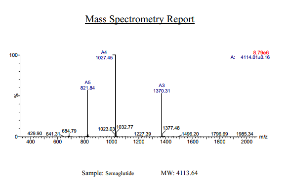
Semaglutide Synthetic is a single, non-glycosylated polypeptide chain containing 31 amino acids, having a molecular mass of 4113 Dalton and a Molecular formula of C187H291N45O59.
Formulation
The protein was lyophilized with no additives.
Purity
Greater than 97.0% as determined by analysis by RP-HPLC.
HPLC, MS
More Info
-
Physical Appearance
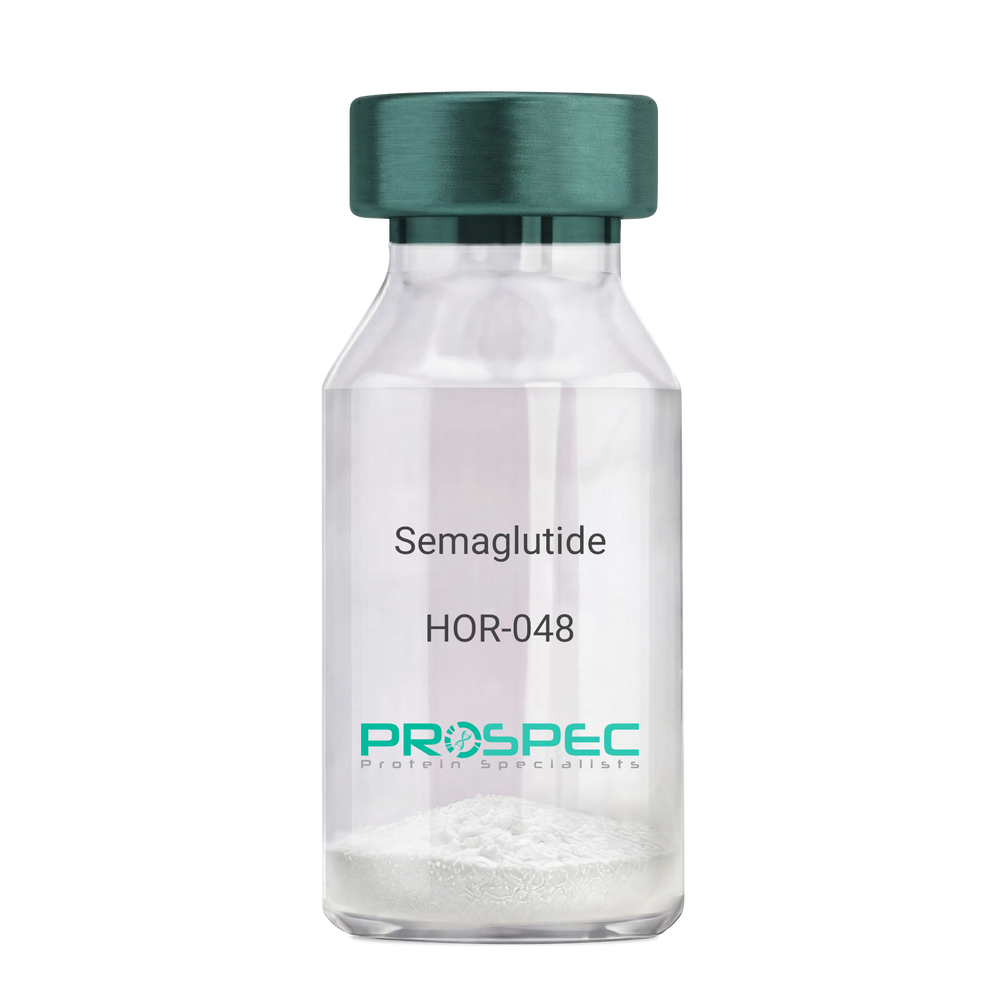
Sterile Filtered White lyophilized (freeze-dried) powder.
-
Stability
Lyophilized Semaglutide although stable at room temperature for 3 weeks, should be stored desiccated below -18°C. Upon reconstitution Semaglutide should be stored at 4°C between 2-7 days and for future use below -18°C. For long term storage it is recommended to add a carrier protein (0.1% HSA or BSA). Please prevent freeze-thaw cycles.
-
Solubility
It is recommended to reconstitute the lyophilized Semaglutide in sterile 18MΩ-cm H2O not less than 100 µg/ml, which can then be further diluted to other aqueous solutions.
-
Amino Acid Sequence
H-His-Aib-Glu-Gly-Thr-Phe-Thr-Ser-Asp-Val-Ser-Ser-Tyr-Leu-Glu-Gly-Gln-Ala-Ala-Lys(AEEAc-AEEAc-γ-Glu-17-carboxyheptadecanoyl)-Glu-Phe-Ile-Ala-Trp-Leu-Val-Arg-Gly-Arg-Gly-OH.
-
Background
Semaglutide, a glucagon-like peptide-1 receptor agonist (GLP-1 RA), has emerged as a breakthrough in the field of diabetes management and metabolic disorders. It is recognized for its potent glucose-lowering effects, weight management properties, and cardiovascular benefits. Beyond diabetes, semaglutide is under investigation for its potential applications in obesity treatment and other related conditions. This research aims to comprehensively explore semaglutide, shedding light on its multifaceted mechanisms of action and its broader therapeutic implications.
The primary objective of this research is to elucidate the mechanisms underlying the glucose-lowering and metabolic effects of semaglutide. In vitro and in vivo experiments will be conducted to investigate how semaglutide interacts with GLP-1 receptors, influences insulin secretion, and modulates glucose homeostasis. Understanding these mechanisms is crucial for harnessing the full therapeutic potential of semaglutide.
The second objective is to assess the clinical relevance of semaglutide in diabetes management. Clinical trials involving individuals with type 2 diabetes will be conducted to evaluate the efficacy, safety, and long-term outcomes of semaglutide treatment. These investigations may provide valuable insights into its use as a monotherapy or adjunct therapy in diabetes care.
The third objective is to explore the potential applications of semaglutide beyond diabetes. Research will investigate its role in obesity treatment, cardiovascular risk reduction, and non-alcoholic fatty liver disease (NAFLD) management. Understanding the multifaceted properties of semaglutide may open new avenues for therapeutic interventions in various metabolic and cardiovascular conditions.
By delving into the diverse functions of semaglutide, this research aims to expand our understanding of its therapeutic potential and clinical applications. The findings may contribute to improved treatment strategies for individuals affected by diabetes, obesity, and related metabolic disorders.
ProSpec's products are furnished for LABORATORY RESEARCH USE ONLY. The product may not be used as drugs, agricultural or pesticidal products, food additives or household chemicals.
- View Data Sheet
Name :
IL17A Human (75-155)Description:
Interleukin-17A (75-155 a.a) Human Recombinant
IL-17A, Interleukin 17A, CTLA8, IL17.
Product # :
CYT-1236Price :
Quantity :
Shipping Method :
Shipped at Room temp
More Info
- description
- source
- formulation
- purity
- More Info
Description
The IL17A Human is created as a recombinant protein with a 4kda N-terminal fusion of His Tag. The IL17A His-Tagged Fusion Protein, produced in E. coli, is a 14kDa protein containing 81 amino acid residues of the IL17A Human, 75-155 amino acids.
Source
Escherichia Coli.
Formulation
Each mg was lyophilized with 1xPBS, 0.4% SDS and 4mM DTT.
Purity
Greater than 90% as determined by SDS-PAGE.
More Info
-
Synonyms
IL-17A, Interleukin 17A, CTLA8, IL17.
-
Physical Appearance
Filtered White lyophilized (freeze-dried) powder.
-
Stability
Store lyophilized IL17A at -20°C. Aliquot the product after reconstitution to avoid repeated freezing/thawing cycles. Reconstituted protein can be stored at 4°C for a limited period of time; it does not show any change after two weeks at 4°C.
-
Solubility
It is recommended to add deionized water to prepare a working stock solution of approximately 0.5mg/ml and let the lyophilized pellet dissolve completely. Product is not sterile! Please filter the product by an appropriate sterile filter before using it on cell culture.
-
Amino Acid Sequence
LHRNE DPERYPSVIW EAKCRHLGCI NADGNVDYHM NSVPIQQEILVLRREPPHCP NSFRLEKILV SVGCTCVTPI VHHVA
-
Background
IL17 is a proinflammatory cytokine produced by activated T cells. IL-17A takes an important part in immune responses, particularly in host defense against infections and in the pathogenesis of various autoimmune diseases. IL-17 regulates the activities of NF-kappaB and mitogen-activated protein kinases. Interleukin-17 stimulates the expression of IL6 and cyclooxygenase-2 and enhances the production of nitric oxide.
ProSpec's products are furnished for LABORATORY RESEARCH USE ONLY. The product may not be used as drugs, agricultural or pesticidal products, food additives or household chemicals.
- View Data Sheet
Name :
Adiponectin Porcine, HEKDescription:
Adiponectin Porcine Recombinant, HEK derived
Acrp30, AdipoQ, GBP-28, APM-1, ACDC.
Product # :
CYT-696Price :
Quantity :
Shipping Method :
Shipped at Room temp
More Info
- description
- source
- formulation
- purity
- More Info
Description
The Acrp30 Porcine Recombinant contains a total of 238 amino acids having a molecular Mass of 26kDa. The Porcine Adiponectin is fused to a 13 amino acid long N-terminal FLAG tag.
Source
HEK293 (Human Embryonic Kidney cell line).
Formulation
Sterile filtered and lyophilized from 0.5mg/ml in 20mM Tris buffer and 50mM NaCl pH-7.5.
Purity
Greater than 90% as determined by SDS PAGE.
More Info
-
Introduction
Adiponectin is a recently discovered 244 amino acid protein, the product of the apM1 gene, which is physiologically active and specifically and highly expressed in adipose cells (Adipokine). The protein belongs to the soluble defense collagen super family; it has a collagen-like domain structurally homologous with collagen VIII and X and complement factor C1q-like globular domain. APM-1 forms homotrimers, which are the building blocks for higher order complexes found circulating in serum.
-
Synonyms
Acrp30, AdipoQ, GBP-28, APM-1, ACDC.
-
Stability
For long term, store lyophilized AdipoQ at -20°C. Aliquot the product after reconstitution to avoid repeated freezing/thawing cycles. Reconstituted protein can be stored at 4°C for a limited period of time; it does not show any change after two weeks at 4°C.The lyophilized protein remains stable for 24 months when stored at -20°C.
-
Solubility
Add deionized water and let the lyophilized pellet dissolve completely.
-
Amino Acid Sequence
HVDYKDDDDK PAGETTEKPG ALLPMPKGAC AGWMAGIPGH PGHNGTPGRD GRDGVPGEKG EKGDTGLTGP KGDTGESGVT GVEGPRGFPG IPGRKGEPGE SAYVYRSAFS VGLETRVTVP NMPIRFTKIF YNQQNHYDVT TGKFHCNIPG LYYFSFHITV LKDVKVSLYK DKAVLFTYDQ QDKNVDQASG VLLYLEKGDQ WLQAYGDEEN GVYADNVNDS FTGFLLYHNIE.
-
Background
What is the molecular weight/Mw of ADIPONECTIN Protein?
ADIPONECTIN Protein has a total Mw of 26kDa.
What is the source or expression system of ADIPONECTIN Protein?
HEK293
What is the Purity of ADIPONECTIN Protein?
ADIPONECTIN Protein is >90% pure as determined by SDS-PAGE.
What is the Biological Activity of ADIPONECTIN Protein?
The biological functionality of ADIPONECTIN Protein will be determined in the future.
What is the amino acid sequence of ADIPONECTIN Protein?
HVDYKDDDDK PAGETTEKPG ALLPMPKGAC AGWMAGIPGH PGHNGTPGRD GRDGVPGEKG EKGDTGLTGP KGDTGESGVT GVEGPRGFPG IPGRKGEPGE SAYVYRSAFS VGLETRVTVP NMPIRFTKIF YNQQNHYDVT TGKFHCNIPG LYYFSFHITV LKDVKVSLYK DKAVLFTYDQ QDKNVDQASG VLLYLEKGDQ WLQAYGDEEN GVYADNVNDS FTGFLLYHNIE.
What applications can ADIPONECTIN Protein be used in?
ADIPONECTIN Protein can probably be used in western blot, ELISA and Lateral Flow.
What is the endotoxin level for ADIPONECTIN Protein?
The endotoxin level is minimal, ADIPONECTIN Protein was purified using conventional chromatography techniques.
ProSpec's products are furnished for LABORATORY RESEARCH USE ONLY. The product may not be used as drugs, agricultural or pesticidal products, food additives or household chemicals.
- View Data Sheet
Name :
AITR HumanDescription:
AITR Human Recombinant
TNFRSF18, AITR, CD357, GITR, GITR-D, Tumor necrosis factor receptor superfamily member 18, Activation-inducible TNFR family receptor, Glucocorticoid-induced TNFR-related protein, CD357, UNQ319/PRO364.
Product # :
CYT-925Price :
Quantity :
Shipping Method :
Shipped with Ice Packs
More Info
- description
- source
- formulation
- purity
- More Info
- sds-page
Description
AITR Human Recombinant produced in Sf9 Baculovirus is a single, glycosylated polypeptide chain containing 145 amino acids (26-162a.a.) and having a molecular mass of 15.6kDa (Migrates at 18-28kDa on SDS-PAGE under reducing conditions).AITR is fused to an 8 amino acid His-tag at C-terminus & purified by proprietary chromatographic techniques.
Source
Escherichia Coli.
Formulation
AITR protein solution (0.5mg/ml) containing Phosphate Buffered Saline (pH 7.4) and 10% glycerol.
Purity
Greater than 95.0% as determined by SDS-PAGE.
sds-page
More Info
-
Synonyms
TNFRSF18, AITR, CD357, GITR, GITR-D, Tumor necrosis factor receptor superfamily member 18, Activation-inducible TNFR family receptor, Glucocorticoid-induced TNFR-related protein, CD357, UNQ319/PRO364.
-
Physical Appearance
Sterile filtered colorless solution.
-
Stability
Store at 4°C if entire vial will be used within 2-4 weeks. Store, frozen at -20°C for longer periods of time. For long term storage it is recommended to add a carrier protein (0.1% HSA or BSA).Avoid multiple freeze-thaw cycles.
-
Amino Acid Sequence
QRPTGGPGCG PGRLLLGTGT DARCCRVHTT RCCRDYPGEE CCSEWDCMCV QPEFHCGDPC CTTCRHHPCP PGQGVQSQGK FSFGFQCIDC ASGTFSGGHE GHCKPWTDCT QFGFLTVFPG NKTHNAVCVP GSPPAEPLEH HHHHH.
-
Background
AITR Human Recombinant: Unveiling its Role in Immune Regulation and Therapeutic Potential
1. Abstract
This research paper aims to provide a comprehensive exploration of the AITR Human Recombinant, a crucial receptor involved in immune regulation. By examining its structure, signaling pathways, biological functions, and implications in disease, we unravel the potential therapeutic applications of AITR in immune-related disorders.
2. Introduction
AITR, also known as TNFRSF18, is a receptor protein that plays a vital role in immune regulation. With its involvement in T-cell responses and immune tolerance, AITR has emerged as an intriguing target for therapeutic interventions in various immune-mediated conditions.
3. Structure and Signaling of AITR
AITR is a transmembrane receptor protein belonging to the tumor necrosis factor receptor superfamily. Its extracellular domain interacts with its ligand, glucocorticoid-induced TNFR-related protein (GITR) ligand, leading to downstream signaling events that modulate immune cell function.
4. Biological Functions of AITR
AITR activation influences T-cell responses by regulating T-cell activation, proliferation, and cytokine production. Additionally, AITR signaling can modulate the balance between effector and regulatory T-cell populations, thereby playing a role in immune tolerance and immune homeostasis.
5. AITR in Disease Pathology
AITR dysregulation has been associated with various immune-related disorders, including autoimmune diseases, cancer, and transplant rejection. Understanding the role of AITR in these pathologies may provide insights into potential therapeutic strategies targeting AITR signaling.
6. Therapeutic Potential of AITR
The unique role of AITR in immune regulation makes it an appealing target for therapeutic interventions. Modulation of AITR signaling holds promise for manipulating immune responses in the context of autoimmune diseases, cancer immunotherapy, and transplantation.
7. Conclusion and Future Perspectives
While our understanding of AITR and its functions has advanced significantly, further research is warranted to unravel its complex signaling pathways and therapeutic potential. Continued investigations into AITR biology will enhance our ability to develop targeted therapies for immune-related disorders.
What is the molecular weight/Mw of AITR Protein?
AITR Protein has a total Mw of 15.6kDa.
What is the source or expression system of AITR Protein?
Escherichia Coli.
What is the Purity of AITR Protein?
AITR Protein is >95% pure as determined by SDS-PAGE.
What is the Biological Activity of AITR Protein?
The biological functionality of AITR Protein will be determined in the future.
What is the amino acid sequence of AITR Protein?
QRPTGGPGCG PGRLLLGTGT DARCCRVHTT RCCRDYPGEE CCSEWDCMCV QPEFHCGDPC CTTCRHHPCP PGQGVQSQGK FSFGFQCIDC ASGTFSGGHE GHCKPWTDCT QFGFLTVFPG NKTHNAVCVP GSPPAEPLEH HHHHH.
What applications can AITR Protein be used in?
AITR Protein can probably be used in western blot, ELISA and Lateral Flow.
What is the endotoxin level for AITR Protein?
The endotoxin level is minimal, AITR Protein was purified using conventional chromatography techniques.
ProSpec's products are furnished for LABORATORY RESEARCH USE ONLY. The product may not be used as drugs, agricultural or pesticidal products, food additives or household chemicals.
- View Data Sheet
Name :
FCGR3A Human, HEKDescription:
CD16a Human Recombinant, HEK
Low affinity immunoglobulin gamma Fc region receptor III-A, CD16a antigen, Fc-gamma, RIII-alpha, Fc-gamma RIII, Fc-gamma RIIIa, FcRIII, FcRIIIa, FcR-10, IgG Fc receptor III-2, CD16a, FCGR3A, FCG3, FCGR3, IGFR3, CD16, FCGRIII.
Product # :
PRO-2770Price :
Quantity :
Shipping Method :
Shipped with Ice Packs
More Info
- description
- source
- formulation
- purity
- biological activity
- More Info
Description
FCGR3A Human Recombinant produced HEK293 Cells is a single, polypeptide chain containing 197 amino acids (18-208 a.a) and having a molecular mass of 22.6 kDa. FCGR3A is fused to a 6 amino acid His-tag at C-terminus & purified by proprietary chromatographic techniques.
Source
HEK293 Cells
Formulation
FCGR3A protein solution (0.5mg/ml) contains Phosphate buffered saline (pH7.4) and 10% glycerol.
Purity
Greater than 95.0% as determined by SDS-PAGE.
Biological Activity
Determined by the binding ability in a functional ELISA with Human IgG1 Fc (cat# pro-2763).
More Info
-
Introduction
Low affinity immunoglobulin gamma Fc region receptor III-A (FCGR3A) is a receptor for the Fc portion of immunoglobulin G, and is involved in the elimination of antigen-antibody complexes from the circulation, as well as other antibody-dependent responses. FCGR3A needs to associate with the gamma subunit of Fc epsilon. The FCGR3A receptor is expressed on natural killer (NK) cells as an integral membrane glycoprotein anchored through a transmembrane peptide, while FCGR3B is expressed on polymorphonuclear neutrophils (PMN) where the receptor is anchored through a phosphatidylinositol (PI) linkage. In addition, FCGR3A is expressed on macrophages, subpopulation of T-cells, immature thymocytes and placental trophoblasts. FCGR3A mediates antibody-dependent cellular cytotoxicity (ADCC) and other antibody-dependent responses, such as phagocytosis. FCGR3A gene mutations are linked with susceptibility to recurrent viral infections, susceptibility to systemic lupus erythematosus, and alloimmune neonatal neutropenia.
-
Synonyms
Low affinity immunoglobulin gamma Fc region receptor III-A, CD16a antigen, Fc-gamma, RIII-alpha, Fc-gamma RIII, Fc-gamma RIIIa, FcRIII, FcRIIIa, FcR-10, IgG Fc receptor III-2, CD16a, FCGR3A, FCG3, FCGR3, IGFR3, CD16, FCGRIII.
-
Physical Appearance
Sterile filtered colorless solution.
-
Stability
Store at 4°C if entire vial will be used within 2-4 weeks. Store, frozen at -20°C for longer periods of time. For long term storage it is recommended to add a carrier protein (0.1% HSA or BSA).Avoid multiple freeze-thaw cycles.
-
Amino Acid Sequence
MRTEDLPKAV VFLEPQWYRV LEKDSVTLKC QGAYSPEDNS TQWFHNESLI SSQASSYFID AATVDDSGEY RCQTNLSTLS DPVQLEVHIG WLLLQAPRWV FKEEDPIHLR CHSWKNTALH KVTYLQNGKG RKYFHHNSDF YIPKATLKDS GSYFCRGLFG SKNVSSETVN ITITQGLAVS TISSFFPPGY Q HHHHHH.
ProSpec's products are furnished for LABORATORY RESEARCH USE ONLY. The product may not be used as drugs, agricultural or pesticidal products, food additives or household chemicals.
- View Data Sheet
Name :
HCV NS3Description:
Hepatitis C Virus NS3 (1450-1643 a.a.) Recombinant
Product # :
HCV-255Price :
Quantity :
Shipping Method :
Shipped with Ice Packs
More Info
- description
- formulation
- purity
- More Info
Description
The E.coli derived 22kDa recombinant protein contains the HCV NS3 genotype 1b immunodominant regions, amino acids 1450-1643. The protein is fused with b-galactosidase (114 kDa) at N-terminus, pI 5.43.
Formulation
20mM Tris-HCl pH-8, 8M urea & 10mM B-ME.
Purity
HCV NS3 protein is >95% pure as determined by 10% PAGE (coomassie staining).
More Info
-
Introduction
HCV is a small 50nm, enveloped, single-stranded, positive sense RNAvirus in the family Flaviviridae.
HCV has a high rate of replication with approximately one trillion particles produced each day in an infected individual. Due to lack of proofreading by the HCV RNA polymerase, the HCV has an exceptionally high mutation rate, a factor that may help it elude the host's immune response. Hepatitis C virus is classified into six genotypes(1-6) with several subtypes within each genotype. The preponderance and distribution of HCV genotypes varies globally. Genotype is clinically important in determining potential response to interferon-based therapy and the required duration of such therapy. Genotypes 1 and 4 are less responsive to interferon-based treatment than are the other genotypes (2, 3, 5 and 6). -
Stability
HCV NS3 although stable at 4°C for 1 week, should be stored below -18°C. Please prevent freeze thaw cycles.
-
Applications
HCV NS3 antigen is suitable for ELISA and Western blots, excellent antigen for detection of HCV with minimal specificity problems.
-
Specificity
Immunoreactive with sera of HCV-infected individuals.
-
Purification Method
HCV NS3 protein was purified by proprietary chromatographic technique.
ProSpec's products are furnished for LABORATORY RESEARCH USE ONLY. The product may not be used as drugs, agricultural or pesticidal products, food additives or household chemicals.
- View Data Sheet
Name :
HTLV-1 gp46 mosaicDescription:
HTLV-I gp46 Mosaic Recombinant
Product # :
HIV-142Price :
Quantity :
Shipping Method :
Shipped with Ice Packs
More Info
- description
- formulation
- purity
- More Info
Description
The E.Coli derived recombinant mosaic protein contains the gp46 immunodominant region, 21-313 amino acids; Mw on SDA-PAGE is 34.2kDa and fuse to 6 histidines at C-terminus.
Formulation
The protein solution contains 20mM Phosphate buffer pH 7.5.
Purity
Protein is >90% pure as determined by SDS-PAGE.
More Info
-
Introduction
Human T-lymphotropic virus (HTLV) is a human, single-stranded RNA retrovirus that causes T-cell leukemia and T-cell lymphoma. The virus activates a subset of T-helper cellscalled Th1cells. The result is a proliferation of Th1 cells and overproduction of Th1 related cytokines (mainly IFN-gamma and TNF-alpha). Feedback mechanisms of these cytokines cause a suppression of the Th2 lymphocytes and a reduction of Th2 cytokine production (mainly IL-4, IL-5, IL-10 and IL-13). The end result is a reduction in the ability of the infected host to mount an adequate immune response to invading organisms that require a predominantly Th2 dependant response (these include parasitic infections and production of mucosal and humoral antibodies).
-
Stability
HTLV-1 gp46 although stable at 4°C for 1 week, should be stored below -18°C. Please prevent freeze thaw cycles.
-
Amino Acid Sequence
PSCCTLTVGV SSYHSKPCNP AQPVCSWTLD LLALSADRAL QPPCPNLVSY SSYHATYSLY LFPHWIKKPN RNGGGYYSAS YSDPCSLKCP YLGCQSWTCP YTGAVSSPYW KFQQDVNFTQ EVSHLNINLH FSKCGFPFSL LVDAPGYDPI WFLNTEPSQL PPTAPPLLSH SNLDHILEPS IPWKSKLLTL VQLTLQSTNY TCIVCIDRAS LSTWHVLYSP NVSVPSPSST PLLYPSLALP APHLTLPFNW THCFDPQIQA IVSSPCHNSL ILPPFSLSPV PTLGSRSRRA.
-
Purification Method
HTLV-1 gp46 was purified by proprietary chromatographic technique.
ProSpec's products are furnished for LABORATORY RESEARCH USE ONLY. The product may not be used as drugs, agricultural or pesticidal products, food additives or household chemicals.