Skip to content

High-quality research proteins.

  • 1-800-805-6531

  • Mon - Sat 8.00-18.00, Sun - Closed

  • info@prospecbio.com

  • PO Box 6591 East Brunswick NJ 08816

  • Products
    • Cytokines
    • Growth factors
    • Chemokines
    • Cd antigens
    • Neurotrophins
    • Hormones
    • Enzymes
    • Viral antigens
    • Recombinant proteins
    • Natural proteins
    • Monoclonal antibodies
    • Polyclonal antibodies
    • Thermal Packaging
  • About
    • Company
    • Founder
    • Contribution
  • Customers
    • Eastern Asia
    • Eastern Europe
    • North America
    • South America
    • South East Asia
    • Southern Asia
    • Western Europe
  • Ordering
    • Ordering Method
    • Ordering - Credit Card
    • Ordering - PO
    • Shipping Fees
    • FAQs
  • Services
    • DNA Cloning
    • Protein Expression
    • Monoclonal Antibody
    • Fermentation
    • Technology License
  • Contact us
Log in

Country/region

  • USD $ | Albania
  • USD $ | Andorra
  • USD $ | Argentina
  • USD $ | Armenia
  • USD $ | Australia
  • USD $ | Austria
  • USD $ | Azerbaijan
  • USD $ | Belarus
  • USD $ | Belgium
  • USD $ | Bolivia
  • USD $ | Brazil
  • USD $ | Bulgaria
  • USD $ | Cambodia
  • USD $ | Canada
  • USD $ | Chile
  • USD $ | China
  • USD $ | Colombia
  • USD $ | Costa Rica
  • USD $ | Croatia
  • USD $ | Cyprus
  • USD $ | Czechia
  • USD $ | Denmark
  • USD $ | Estonia
  • USD $ | Finland
  • USD $ | France
  • USD $ | Georgia
  • USD $ | Germany
  • USD $ | Greece
  • USD $ | Hong Kong SAR
  • USD $ | Hungary
  • USD $ | Iceland
  • USD $ | India
  • USD $ | Ireland
  • USD $ | Israel
  • USD $ | Italy
  • USD $ | Japan
  • USD $ | Kazakhstan
  • USD $ | Latvia
  • USD $ | Liechtenstein
  • USD $ | Lithuania
  • USD $ | Madagascar
  • USD $ | Malta
  • USD $ | Mexico
  • USD $ | Moldova
  • USD $ | Monaco
  • USD $ | Netherlands
  • USD $ | New Zealand
  • USD $ | North Macedonia
  • USD $ | Norway
  • USD $ | Panama
  • USD $ | Paraguay
  • USD $ | Peru
  • USD $ | Philippines
  • USD $ | Poland
  • USD $ | Portugal
  • USD $ | Romania
  • USD $ | Singapore
  • USD $ | Slovakia
  • USD $ | Slovenia
  • USD $ | South Africa
  • USD $ | South Korea
  • USD $ | Spain
  • USD $ | Sri Lanka
  • USD $ | Sweden
  • USD $ | Switzerland
  • USD $ | Taiwan
  • USD $ | Thailand
  • USD $ | Türkiye
  • USD $ | Ukraine
  • USD $ | United Kingdom
  • USD $ | United States
  • USD $ | Uruguay
  • USD $ | Vatican City
  • USD $ | Venezuela
  • USD $ | Vietnam
  • Facebook
  • Instagram
logoProSpec - Recombinant Protein Manufacturer for academic and R&D industry
Become a distributor
Account Log in
Wishlist
Cart Cart(0)
  • Products
    +
    • Cytokines
    • Growth factors
    • Chemokines
    • Cd antigens
    • Neurotrophins
    • Hormones
    • Enzymes
    • Viral antigens
    • Recombinant proteins
    • Natural proteins
    • Monoclonal antibodies
    • Polyclonal antibodies
    • Thermal Packaging
  • About
    +
    • Company
    • Founder
    • Contribution
  • Customers
    +
    • Eastern Asia
    • Eastern Europe
    • North America
    • South America
    • South East Asia
    • Southern Asia
    • Western Europe
  • Ordering
    +
    • Ordering Method
    • Ordering - Credit Card
    • Ordering - PO
    • Shipping Fees
    • FAQs
  • Services
    +
    • DNA Cloning
    • Protein Expression
    • Monoclonal Antibody
    • Fermentation
    • Technology License
  • Contact us

Item added to your cart

View cart
View All
  • Cytokines
  • Growth factors
  • Chemokines
  • Cd antigens
  • Neurotrophins
  • Hormones
  • Enzymes
  • Viral antigens
  • Recombinant proteins
  • Natural proteins
  • Monoclonal antibodies
  • Polyclonal antibodies
  • Thermal Packaging
  • Tumor Necrosis Factor

    Tumor Necrosis Factor

    View All
  • 4-1BB

    4-1BB

    View All
  • Adiponectin

    Adiponectin

    View All
  • AIF1

    AIF1

    View All
  • Other Cytokines

    Other Cytokines

    View All
  • AITRL

    AITRL

    View All
  • Angiopoietin

    Angiopoietin

    View All
  • Apolipoprotein

    Apolipoprotein

    View All
  • B-Cell Activating Factor

    B-Cell Activating Factor

    View All
  • Beta Defensin

    Beta Defensin

    View All
  • Bone Morphogenetic Protein

    Bone Morphogenetic Protein

    View All
  • B type Natriuretic Peptide

    B type Natriuretic Peptide

    View All
  • BST

    BST

    View All
  • Betacellulin

    Betacellulin

    View All
  • Cardiotrophin

    Cardiotrophin

    View All
  • Activin

    Activin

    View All
  • Fibroblast Growth Factor

    Fibroblast Growth Factor

    View All
  • Other Growth Factors

    Other Growth Factors

    View All
  • CSF

    CSF

    View All
  • CTGF

    CTGF

    View All
  • IGFBP

    IGFBP

    View All
  • VEGF Protein

    VEGF Protein

    View All
  • EGF

    EGF

    View All
  • Epigen

    Epigen

    View All
  • Erythropoietin

    Erythropoietin

    View All
  • Insulin-Like Growth Factor

    Insulin-Like Growth Factor

    View All
  • Growth Hormone

    Growth Hormone

    View All
  • HDGF

    HDGF

    View All
  • Hepatocyte Growth Factor

    Hepatocyte Growth Factor

    View All
  • Insulin

    Insulin

    View All
  • BCA-1/ BLC (CXCL13)

    BCA-1/ BLC (CXCL13)

    View All
  • BRAK (CXCL14)

    BRAK (CXCL14)

    View All
  • C-10 (CCL6)

    C-10 (CCL6)

    View All
  • Other Chemokines

    Other Chemokines

    View All
  • MEC (CCL28)

    MEC (CCL28)

    View All
  • LD78-beta (CCL3L1)

    LD78-beta (CCL3L1)

    View All
  • CTACK (CCL27)

    CTACK (CCL27)

    View All
  • CXCL16

    CXCL16

    View All
  • CXCL17

    CXCL17

    View All
  • Platelet Factor-4 (CXCL4)

    Platelet Factor-4 (CXCL4)

    View All
  • ENA-78 (CXCL5)

    ENA-78 (CXCL5)

    View All
  • Eotaxin (CCL11,24,26)

    Eotaxin (CCL11,24,26)

    View All
  • Exodus-2 (CCL21)

    Exodus-2 (CCL21)

    View All
  • Fractalkine (CX3CL1)

    Fractalkine (CX3CL1)

    View All
  • CXCL6

    CXCL6

    View All
  • Other CD Antigens

    Other CD Antigens

    View All
  • CD14

    CD14

    View All
  • CD164

    CD164

    View All
  • CD1

    CD1

    View All
  • CD2

    CD2

    View All
  • CD200

    CD200

    View All
  • CD207

    CD207

    View All
  • CD226

    CD226

    View All
  • CD23

    CD23

    View All
  • CD244

    CD244

    View All
  • CD247

    CD247

    View All
  • CD27

    CD27

    View All
  • CD274

    CD274

    View All
  • CD300

    CD300

    View All
  • CD33

    CD33

    View All
  • Other Neurotrophins

    Other Neurotrophins

    View All
  • Beta-NGF

    Beta-NGF

    View All
  • BDNF

    BDNF

    View All
  • CDNF

    CDNF

    View All
  • CNTF

    CNTF

    View All
  • GDNF

    GDNF

    View All
  • Glia Maturation Factor

    Glia Maturation Factor

    View All
  • MANF

    MANF

    View All
  • Midkine

    Midkine

    View All
  • Neuroglobin

    Neuroglobin

    View All
  • Neurotrophic factors

    Neurotrophic factors

    View All
  • Neuregulin

    Neuregulin

    View All
  • Neuropilin

    Neuropilin

    View All
  • Pigment Epithelium-Derived Factor

    Pigment Epithelium-Derived Factor

    View All
  • Pleiotrophin

    Pleiotrophin

    View All
  • Peptide Hormones

    Peptide Hormones

    View All
  • Other Hormones

    Other Hormones

    View All
  • HCG

    HCG

    View All
  • Endothelin

    Endothelin

    View All
  • Exendin

    Exendin

    View All
  • FSH

    FSH

    View All
  • Glucagon

    Glucagon

    View All
  • GHRP

    GHRP

    View All
  • GLP

    GLP

    View All
  • Thyrostimulin

    Thyrostimulin

    View All
  • Inhibin A

    Inhibin A

    View All
  • LHRH

    LHRH

    View All
  • Melanotan

    Melanotan

    View All
  • Procalcitonin

    Procalcitonin

    View All
  • PTH

    PTH

    View All
  • Peroxiredoxin

    Peroxiredoxin

    View All
  • Peptidase

    Peptidase

    View All
  • Synthetase

    Synthetase

    View All
  • Transferase

    Transferase

    View All
  • Hydrolase

    Hydrolase

    View All
  • Dehydrogenase

    Dehydrogenase

    View All
  • ACE2

    ACE2

    View All
  • Esterase

    Esterase

    View All
  • Protein Kinases

    Protein Kinases

    View All
  • Other Enzymes

    Other Enzymes

    View All
  • Phosphatase

    Phosphatase

    View All
  • Deaminase

    Deaminase

    View All
  • Oxygenase

    Oxygenase

    View All
  • Adenylate Kinase

    Adenylate Kinase

    View All
  • Lyase

    Lyase

    View All
  • Chagas

    Chagas

    View All
  • SARS

    SARS

    View All
  • Influenza

    Influenza

    View All
  • ASFV

    ASFV

    View All
  • Astrovirus

    Astrovirus

    View All
  • Borrelia

    Borrelia

    View All
  • Helicobacter Pylori

    Helicobacter Pylori

    View All
  • Chikungunya

    Chikungunya

    View All
  • Chlamydia

    Chlamydia

    View All
  • Cytomegalo

    Cytomegalo

    View All
  • Dengue

    Dengue

    View All
  • EBV

    EBV

    View All
  • Ebola

    Ebola

    View All
  • Feline Leukemia Virus

    Feline Leukemia Virus

    View All
  • Hepatitis A

    Hepatitis A

    View All
  • Other

    Other

    View All
  • Actin

    Actin

    View All
  • ADAM

    ADAM

    View All
  • Complement Component

    Complement Component

    View All
  • Ag85

    Ag85

    View All
  • Eukaryotic Translation Initiation Factor

    Eukaryotic Translation Initiation Factor

    View All
  • Anterior Gradient Protein

    Anterior Gradient Protein

    View All
  • Heat Shock Protein

    Heat Shock Protein

    View All
  • Alpha-2-HS-Glycoprotein

    Alpha-2-HS-Glycoprotein

    View All
  • Hemoglobin

    Hemoglobin

    View All
  • Allergy

    Allergy

    View All
  • Anaplasma

    Anaplasma

    View All
  • Angiogenin

    Angiogenin

    View All
  • Ankyrin Repeat Domain

    Ankyrin Repeat Domain

    View All
  • Annexin

    Annexin

    View All
  • Other Natural Proteins

    Other Natural Proteins

    View All
  • Aprotinin

    Aprotinin

    View All
  • Transferrin

    Transferrin

    View All
  • Avidin

    Avidin

    View All
  • Anti Coagulation Factors

    Anti Coagulation Factors

    View All
  • Natural Albumin

    Natural Albumin

    View All
  • Natural Coagulation Factors

    Natural Coagulation Factors

    View All
  • Fibronectin

    Fibronectin

    View All
  • Thrombin

    Thrombin

    View All
  • Anti Human Enzyme

    Anti Human Enzyme

    View All
  • Other Monoclonal Antibodies

    Other Monoclonal Antibodies

    View All
  • Anti Human Cytokine

    Anti Human Cytokine

    View All
  • Anti Human Heat Shock Protein

    Anti Human Heat Shock Protein

    View All
  • Anti Mouse Lymphocyte

    Anti Mouse Lymphocyte

    View All
  • Anti Human Chemokine

    Anti Human Chemokine

    View All
  • Anti Human Lymphocyte

    Anti Human Lymphocyte

    View All
  • Anti Viral Monoclonal

    Anti Viral Monoclonal

    View All
  • Anti Mouse Cytokine

    Anti Mouse Cytokine

    View All
  • Anti-GST Monoclonal

    Anti-GST Monoclonal

    View All
  • Other Polyclonal Antibodies

    Other Polyclonal Antibodies

    View All
  • Anti Viral

    Anti Viral

    View All
  • Anti-GST Monoclonal

    Anti-GST Monoclonal

    View All
Back

Search results

1000 results found for “decorin”

Name

Description

Product #

Price

Quantity

Shipping Method

  • View Data Sheet

    Name :

    IL 1 beta Human

    Description:

    Interleukin-1 beta Human Recombinant

    Catabolin, Lymphocyte-activating factor (LAF), Endogenous Pyrogen (EP), Leukocyte Endogenous Mediator (LEM), Mononuclear Cell Factor (MCF), IL1F2, IL-1 beta.

    Product # :

    CYT-208

    Price :

    Quantity :

    Shipping Method :

    Room Temp Icon

    Shipped at Room temp

    Add To Cart

    More Info

    • description
    • source
    • formulation
    • purity
    • biological activity
    • More Info

    Description

    Interleukin-1 beta Human Recombinant produced in E.Coli is a non-glycosylated, Polypeptide chain containing 153 amino acids and having a molecular mass of 17000 Dalton.The IL-1 Beta is purified by proprietary chromatographic techniques.

    Source

    Escherichia Coli.

    Formulation

    IL-1 Beta was lyophilized from a concentrated (1mg/ml) sterile solution containing 50mM Phosphate buffer pH=7.1 and 150mM NaCl.

    Purity

    Greater than 98.0% as determined by:
    (a) Analysis by RP-HPLC.
    (b) Analysis by SDS-PAGE.

    Biological Activity

    The specific activity as determined in the test of augmentation of lymphocyte proliferation assay using mouse thymus was found to be 200,000,000 IU/mg.

    More Info

    • Introduction

      Interleukin-1b is produced by activated macrophages, IL-1 Beta stimulates thymocyte proliferation by inducing il-2 release, b-cell maturation and proliferation, and fibroblast growth factor activity. IL1B proteins are involved in the inflammatory response, being identified as endogenous pyrogens, and are reported to stimulate the release of prostaglandin from synovial cells.

    • Synonyms

      Catabolin, Lymphocyte-activating factor (LAF), Endogenous Pyrogen (EP), Leukocyte Endogenous Mediator (LEM), Mononuclear Cell Factor (MCF), IL1F2, IL-1 beta.

    • Physical Appearance

      Sterile Filtered White lyophilized (freeze-dried) powder.

    • Stability

      Lyophilized Interleukin-1b although stable at room temperature for 3 weeks, should be stored desiccated below -18°C. Upon reconstitution IL-1 Beta should be stored at 4°C between 2-7 days and for future use below -18°C.For long term storage it is recommended to add a carrier protein (0.1% HSA or BSA).Please prevent freeze-thaw cycles.

    • Solubility

      It is recommended to reconstitute the lyophilized Interleukin-1b in sterile 18MΩ-cm H2O not less than 100µg/ml, which can then be further diluted to other aqueous solutions.

    • Amino Acid Sequence

      The sequence of the first five N-terminal amino acids was determined and was found to be Ala-Pro-Val-Arg-Ser.

    • Protein content

      Protein quantitation was carried out by two independent methods:1. UV spectroscopy at 280 nm using the absorbency value of 0.631 as the extinction coefficient for a 0.1% (1mg/ml) solution. This value is calculated by the PC GENE computer analysis program of protein sequences (IntelliGenetics). 2. Analysis by RP-HPLC, using a calibrated solution of IL-1 beta Recombinant as a Reference Standard.

    ProSpec's products are furnished for LABORATORY RESEARCH USE ONLY. The product may not be used as drugs, agricultural or pesticidal products, food additives or household chemicals.

    Il 1 Beta Human
  • View Data Sheet

    Name :

    ARTN Human

    Description:

    Artemin Human Recombinant

    ART, ARTN , EVN, NBN.

    Product # :

    CYT-306

    Price :

    Quantity :

    Shipping Method :

    Room Temp Icon

    Shipped at Room temp

    Add To Cart

    More Info

    • description
    • source
    • formulation
    • purity
    • biological activity
    • More Info

    Description

    Artemin Human Recombinant produced in E.Coli is a disulfide-linked homodimer, non-glycosylated, polypeptide chain containing 2 x 113 amino acids and having a total molecular mass of 24.2 kDa. Artemin is purified by proprietary chromatographic techniques.

    Source

    Escherichia Coli.

    Formulation

    Artemin was lyophilized after extensive dialysis against 10mM sodium citrate pH-4.5 and 25mM sodium chloride.

    Purity

    Greater than 98.0% as determined by:
    (a) Analysis by RP-HPLC.
    (b) Analysis by SDS-PAGE.

    Biological Activity

    The activity is determined by the dose-dependent proliferation of the SH-SY5Y cell line and is typically 4-8 ng/mL. The activity can also be determined by its ability to promote survival and neurite outgrowth.

    More Info

    • Introduction

      The protein encoded by this gene is a member of the glial cell line-derived neurotophic factor (GDNF) family of ligands which are a group of ligands within the TGF-beta superfamily of signaling molecules. GDNFs are unique in having neurotrophic properties and have potential use for gene therapy in neurodegenrative disease. Artemin has been shown in culture to support the survival of a number of periferal neuron populations and at least one population of dopaminergic CNS neurons. Its role in the PNS and CNS is further substantiated by its expression pattern in the proximity of these neurons. This protein is a ligand for the RET receptor and uses GFR-alpha 3 as a coreceptor. Four alternatively spliced transcripts have been described, two of which encode the same protein.

    • Synonyms

      ART, ARTN , EVN, NBN.

    • Physical Appearance

      Sterile Filtered White lyophilized (freeze-dried) powder.

    • Stability

      Lyophilized Artemin although stable at room temperature for 3 weeks, should be stored desiccated below -18°C. Upon reconstitution Artemin Human Recombinant should be stored at 4°C between 2-7 days and for future use below -18°C.For long term storage it is recommended to add a carrier protein (0.1% HSA or BSA).Please prevent freeze-thaw cycles.

    • Solubility

      It is recommended to reconstitute the lyophilized Artemin in sterile 18MΩ-cm H2O not less than 100µg/ml, which can then be further diluted to other aqueous solutions.

    • Amino Acid Sequence

      AGGPGSRARA AGARGCRLRS QLVPVRALGL GHRSDELVRF RFCSGSCRRA RSPHDLSLAS LLGAGALRPP PGSRPVSQPC CRPTRYEAVS FMDVNSTWRT VDRLSATACG CLG.

    • Background

      Artemin Human Recombinant: Unraveling its Role in Neurobiology and Therapeutic Applications

      Abstract:

      Artemin, a member of the glial cell line-derived neurotrophic factor (GDNF) family, holds significant potential in neurobiology and therapeutic interventions. This research paper provides an overview of Artemin human recombinant, elucidating its molecular characteristics, signaling pathways, and therapeutic implications in neurological disorders. Understanding the multifaceted role of Artemin offers new avenues for targeted therapies. This article offers a concise analysis of Artemin, highlighting its impact on neurobiology and its therapeutic applications.

      Introduction:

      Neurological disorders represent a major challenge in healthcare, necessitating innovative therapeutic strategies. Artemin, a member of the GDNF family, has emerged as a promising molecule in neurobiology. This paper provides an overview of Artemin, shedding light on its structure, function, and therapeutic potential.

      Artemin Signaling and Mechanisms:

      Artemin binds to its receptor, Ret tyrosine kinase, and activates downstream signaling pathways, including the PI3K/AKT and MAPK pathways. These signaling cascades play crucial roles in neuronal survival, growth, and differentiation, highlighting the significance of Artemin in neurodevelopment and neuroprotection.

      Artemin in Neurological Disorders:

      Artemin has been implicated in various neurological disorders, including peripheral neuropathies and neurodegenerative diseases. Its neuroprotective properties and ability to enhance neuronal survival and regeneration make it a promising target for therapeutic interventions. Furthermore, Artemin may play a role in pain modulation and sensory neuron function.

      Therapeutic Potential of Artemin Human Recombinant:

      Artemin human recombinant offers promising prospects in the field of neurotherapeutics. Strategies aimed at modulating Artemin signaling or delivering exogenous Artemin hold potential for promoting neuronal survival, regeneration, and functional recovery. Artemin-based therapies could be developed for a range of neurological disorders, including peripheral neuropathies, Parkinson's disease, and spinal cord injuries.

      Challenges and Future Directions:

      While the therapeutic targeting of Artemin shows promise, several challenges lie ahead. Further research is needed to understand the precise mechanisms underlying Artemin's effects and its interactions with other signaling pathways. Additionally, the development of effective delivery methods and the identification of patient subgroups that may benefit from Artemin-based therapies are important considerations for clinical translation.

      Conclusion:

      Artemin human recombinant represents a promising avenue for therapeutic interventions in neurological disorders. Understanding the molecular mechanisms and functional implications of Artemin in neurobiology offers new opportunities for developing innovative treatments. Continued research in this field has the potential to improve the lives of individuals affected by neurological conditions and advance the field of neurotherapeutics.

      What is the molecular weight/Mw of ARTN Protein?
      ARTN Protein has a total Mw of 24.2kDa.

      What is the source or expression system of ARTN Protein?
      Escherichia Coli.

      What is the Purity of ARTN Protein?
      ARTN Protein is >98% pure as determined by SDS-PAGE.

      What is the Biological Activity of ARTN Protein?
      The activity is determined by the dose-dependent proliferation of the SH-SY5Y cell line and is typically 4-8 ng/mL. The activity can also be determined by its ability to promote survival and neurite outgrowth.

      What is the amino acid sequence of ARTN Protein?
      AGGPGSRARA AGARGCRLRS QLVPVRALGL GHRSDELVRF RFCSGSCRRA RSPHDLSLAS LLGAGALRPP PGSRPVSQPC CRPTRYEAVS FMDVNSTWRT VDRLSATACG CLG.

      What applications can ARTN Protein be used in?
      ARTN Protein can probably be used in western blot, ELISA and Lateral Flow.

      What is the endotoxin level for ARTN Protein?
      The endotoxin level is minimal, ARTN Protein was purified using conventional chromatography techniques.

    ProSpec's products are furnished for LABORATORY RESEARCH USE ONLY. The product may not be used as drugs, agricultural or pesticidal products, food additives or household chemicals.

    Artemin Human
  • View Data Sheet

    Name :

    ACADSB Human

    Description:

    Acyl-CoA Dehydrogenase, Short Chain Human Recombinant

    Short/branched chain specific acyl-CoA dehydrogenase mitochondrial, SBCAD, 2-methyl branched chain acyl-CoA dehydrogenase, 2-MEBCAD, 2-methylbutyryl-coenzyme A dehydrogenase, 2-methylbutyryl-CoA dehydrogenase, ACADSB, ACAD7, SBCAD, 2-MEBCAD.

    Product # :

    ENZ-643

    Price :

    Quantity :

    Shipping Method :

    Ice Icon

    Shipped with Ice Packs

    Add To Cart

    More Info

    • description
    • source
    • formulation
    • purity
    • More Info

    Description

    ACADSB Human Recombinant produced in E.coli is a single, non-glycosylated polypeptide chain containing 424 amino acids (34-432) and having a molecular mass of 46.4kDa.ACADSB is fused to a 25 amino acid His-tag at N-terminus & purified by proprietary chromatographic techniques.

    Source

    Escherichia Coli.

    Formulation

    The ACADSB solution (0.5mg/ml) contains 20mM Tris-HCl buffer (pH 8.0), 0.1M NaCl, 10% glycerol and 1mM DTT.

    Purity

    Greater than 90.0% as determined by SDS-PAGE.

    More Info

    • Introduction

      Short/branched chain specific acyl-CoA dehydrogenase (ACADSB) belongs to the acyl-CoA dehydrogenase family of enzymes which catalyze the dehydrogenation of acyl-CoA derivatives in the metabolism of fatty acids or branch chained amino acids. ACADSB catalyzes the degradation of L-isoleucine while having the highest affinity for (s)-2-methylbutyryl-CoA, isobutyryl-CoA and 2-methylhexanoyl-CoA as substrates. ACADSB may use valproyl-CoA as substrate. ACADSB gene defects cause the short/branched-chain acyl-CoA dehydrogenase deficiency (SBCADD), which is an autosomal recessive disorder characterized by an increase of 2-methylbutyrylglycine and 2-methylbutyrylcarnitine in blood and urine.

    • Synonyms

      Short/branched chain specific acyl-CoA dehydrogenase mitochondrial, SBCAD, 2-methyl branched chain acyl-CoA dehydrogenase, 2-MEBCAD, 2-methylbutyryl-coenzyme A dehydrogenase, 2-methylbutyryl-CoA dehydrogenase, ACADSB, ACAD7, SBCAD, 2-MEBCAD.

    • Physical Appearance

      Sterile Filtered clear solution.

    • Stability

      Store at 4°C if entire vial will be used within 2-4 weeks. Store, frozen at -20°C for longer periods of time. For long term storage it is recommended to add a carrier protein (0.1% HSA or BSA).Avoid multiple freeze-thaw cycles.

    • Amino Acid Sequence

      MGSSHHHHHH SSGLVPRGSH MGSHMKSSQS EALLNITNNG IHFAPLQTFT DEEMMIKSSV KKFAQEQIAP LVSTMDENSK MEKSVIQGLF QQGLMGIEVD PEYGGTGASF LSTVLVIEEL AKVDASVAVF CEIQNTLINT LIRKHGTEEQ KATYLPQLTT EKVGSFCLSE AGAGSDSFAL KTRADKEGDY YVLNGSKMWI SSAEHAGLFL VMANVDPTIG YKGITSFLVD RDTPGLHIGK PENKLGLRAS STCPLTFENV KVPEANILGQ IGHGYKYAIG SLNEGRIGIA AQMLGLAQGC FDYTIPYIKE RIQFGKRLFD FQGLQHQVAH VATQLEAARL LTYNAARLLE AGKPFIKEAS MAKYYASEIA GQTTSKCIEW MGGVGYTKDY PVEKYFRDAK IGTIYEGASN IQLNTIAKHI DAEY.

    ProSpec's products are furnished for LABORATORY RESEARCH USE ONLY. The product may not be used as drugs, agricultural or pesticidal products, food additives or household chemicals.

    Acadsb Human
  • View Data Sheet

    Name :

    Adiponectin Mouse, Trimeric

    Description:

    Adiponectin Mouse Recombinant, Trimeric form

    Acrp30, AdipoQ, GBP-28, APM-1, ACDC.

    Product # :

    CYT-247

    Price :

    Quantity :

    Shipping Method :

    Room Temp Icon

    Shipped at Room temp

    Add To Cart

    More Info

    • description
    • source
    • formulation
    • purity
    • More Info

    Description

    Trimeric form of Acrp30 Mouse was expressed in HEK293 cells.The cysteine 39 was replaced with alanine (C39A) 9. mAd-C39A can only form trimer, but not hexamer or HMW form.

    Source

    HEK293 (Human embryonic kidney cell line).

    Formulation

    Mouse Acrp30 filtered (0.4µm) and lyophilized from 0.5 mg/ml in 0.05M phosphate buffer, 0.05M NaCl, pH 7.2.

    Purity

    Acrp30 Mouse purity is greater than 95% as determined by SDS-PAGE.

    More Info

    • Introduction

      Adiponectin is a hormone exclusively expressed from adipose tissue. Many studies demonstrate that adiponectin has direct anti-diabetic, anti-atherogenic and anti-inflammatory functions. APM-1 can increase insulin sensitivity of skeletal muscle, attenuate hepatic lipogenesis and luconeogenesis, regulate NO production in endothelial cells, inhibit proliferation of smooth muscle cells and prevent lipid accumulation of macrophage cells.
      In the circulation, adiponectin is present as three different oligomeric complexes, including the high molecular weight (HMW), the middle molecular weight (MMW, also called hexamer) and low molecular weigh (MMW, also called trimer) forms 8. Different oligomeric complex of adiponectin activates different signaling pathways and exerts distinct functions.

    • Synonyms

      Acrp30, AdipoQ, GBP-28, APM-1, ACDC.

    • Physical Appearance

      Filtered white lyophilized (freeze-dried) powder.

    • Stability

      Store lyophilized Acrp30 Mouse at -20°C. Aliquot the product after reconstitution to avoid repeated freezing/thawing cycles. Reconstituted Acrp30 Mouse can be stored at 4°C for a limited period of time; it does not show any change after two weeks at 4°C.

    • Solubility

      Add deionized water to prepare a working stock solution of approximately 0.5 mg/mL and let the lyophilized pellet dissolve completely. Product is not sterile! Please filter the product by an appropriate sterile filter before using it in the cell culture.

    • Background

      What is the molecular weight/Mw of ADIPONECTIN Protein?
      ADIPONECTIN Protein has a total Mw of 26kDa.

      What is the source or expression system of ADIPONECTIN Protein?
      HEK293

      What is the Purity of ADIPONECTIN Protein?
      ADIPONECTIN Protein is >95% pure as determined by SDS-PAGE.

      What is the Biological Activity of ADIPONECTIN Protein?
      The biological functionality of ADIPONECTIN Protein will be determined in the future.

      What applications can ADIPONECTIN Protein be used in?
      ADIPONECTIN Protein can probably be used in western blot, ELISA and Lateral Flow.

      What is the endotoxin level for ADIPONECTIN Protein?
      The endotoxin level is minimal, ADIPONECTIN Protein was purified using conventional chromatography techniques.

    ProSpec's products are furnished for LABORATORY RESEARCH USE ONLY. The product may not be used as drugs, agricultural or pesticidal products, food additives or household chemicals.

    Adiponectin Mouse Trimeric
  • View Data Sheet

    Name :

    Dengue Envelope-1, Insect

    Description:

    Dengue Virus Subtype 1 Recombinant, Insect Cells

    Product # :

    DEN-033

    Price :

    Quantity :

    Shipping Method :

    Ice Icon

    Shipped with Ice Packs

    Add To Cart

    More Info

    • description
    • source
    • formulation
    • purity
    • More Info

    Description

    Recombinant Dengue Virus Subtype 1 produced in Insect Cells is a polypeptide chain containing amino acids 2-395 and having a molecular weight of 44.8kDa. Dengue Envelope-1 is purified by proprietary chromatographic technique.

    Source

    Insect cells.

    Formulation

    Dengue Envelope-1 protein solution in 1xD-PBS, pH7.3, 0.099% Thimerosal, 1μg/ml of Leupeptin, Aprotinin and Pepstatin A and 2.5% D-trehalose.

    Purity

    Protein is >95% pure as determined by 12.5% SDS-PAGE.

    More Info

    • Introduction

      Caused by one of four closely related virus serotypes of the genus Flavivirus, family Flaviviridae, each serotype is sufficiently different that there is no cross-protection and epidemics caused by multiple serotypes (hyperendemicity) can occur. In cell culture experiments and mice Morpholino antisense oligos have shown specific activity against Dengue virus.

    • Physical Appearance

      Sterile filtered colorless solution.

    • Stability

      Store at 4°C if entire vial will be used within 2-4 weeks. Store, frozen at -20°C for longer periods of time. For long term storage it is recommended to add a carrier protein (0.1% HSA or BSA).Avoid multiple freeze-thaw cycles.

    ProSpec's products are furnished for LABORATORY RESEARCH USE ONLY. The product may not be used as drugs, agricultural or pesticidal products, food additives or household chemicals.

    Dengue Envelope 1 Insect
  • View Data Sheet

    Name :

    Dengue NS1 ST2

    Description:

    Dengue Virus NS1 Subtype 2 Recombinant

    Product # :

    DEN-030

    Price :

    Quantity :

    Shipping Method :

    Ice Icon

    Shipped with Ice Packs

    Add To Cart

    More Info

    • description
    • source
    • formulation
    • purity
    • More Info

    Description

    Recombinant Dengue Virus NS1 Subtype 2 produced in Insect Cells is a polypeptide chain containing amino acids 777-1131 and having a molecular weight of approximately 50kDa.Dengue NS1 ST2 is fused to a His tag and purified by proprietary chromatographic technique.

    Source

    Insect cells.

    Formulation

    Dengue NS1 ST2 protein solution in 1xD-PBS, pH7.4, 0.1% Thimerosal, 5mM EDTA, 1µg/ml of Leupeptin, Aprotinin and Pepstatin A.

    Purity

    Protein is >95% pure as determined by 12.5% SDS-PAGE.

    More Info

    • Introduction

      Caused by one of four closely related virus serotypes of the genus Flavivirus, family Flaviviridae, each serotype is sufficiently different that there is no cross-protection and epidemics caused by multiple serotypes (hyperendemicity) can occur. In cell culture experiments and mice Morpholino antisense oligos have shown specific activity against Dengue virus.

    • Physical Appearance

      Sterile filtered colorless solution.

    • Stability

      Store at 4°C if entire vial will be used within 2-4 weeks. Store, frozen at -20°C for longer periods of time. For long term storage it is recommended to add a carrier protein (0.1% HSA or BSA).Avoid multiple freeze-thaw cycles.

    ProSpec's products are furnished for LABORATORY RESEARCH USE ONLY. The product may not be used as drugs, agricultural or pesticidal products, food additives or household chemicals.

    Dengue Ns1 St2
  • View Data Sheet

    Name :

    IL 13 Rat 109 a.a.

    Description:

    Interleukin-13 109 a.a. Rat Recombinant

    NC300, ALRH, BHR1, P600, IL-13.

    Product # :

    CYT-182

    Price :

    Quantity :

    Shipping Method :

    Room Temp Icon

    Shipped at Room temp

    Add To Cart

    More Info

    • description
    • source
    • formulation
    • purity
    • biological activity
    • More Info

    Description

    Interleukin-13 Rat Recombinant produced in E.Coli is a single, non-glycosylated polypeptide chain containing 109 amino acids and having a molecular mass of 11.9 kDa. The IL-13 is purified by proprietary chromatographic techniques.

    Source

    Escherichia Coli.

    Formulation

    The protein (1mg/ml) was lyophilized in PBS, pH7.4.

    Purity

    Greater than 95% as determined by SDS-PAGE.

    Biological Activity

    ED50 range = 40ng/ml, corresponding to a specific activity of > 25,000IU/mg as determined by the dose dependent proliferation of human TF-1 cells. Optimal concentration for individual application should be determined by a dose response assay.

    More Info

    • Introduction

      IL13 is an immunoregulatory cytokine produced primarily by activated Th2 cells. IL-13 is involved in several stages of B-cell maturation and differentiation. It up-regulates CD23 and MHC class II expression, and promotes IgE isotype switching of B cells. This cytokine down-regulates macrophage activity, thereby inhibits the production of pro-inflammatory cytokines and chemokines. This cytokine is found to be critical to the pathogenesis of allergen-induced asthma but operates through mechanisms independent of IgE and eosinophils. This gene, IL3, IL5, IL4, and CSF2 form a cytokine gene cluster on chromosome 5q, with this gene particularly close to IL4.

    • Synonyms

      NC300, ALRH, BHR1, P600, IL-13.

    • Physical Appearance

      Sterile Filtered White lyophilized (freeze-dried) powder.

    • Stability

      Lyophilized Interleukin-13 although stable at room temperature for 3 weeks, should be stored desiccated below -18°C. Upon reconstitution IL13 should be stored at 4°C between 2-7 days and for future use below -18°C.For long term storage it is recommended to add a carrier protein (0.1% HSA or BSA).Please prevent freeze-thaw cycles.

    • Solubility

      It is recommended to reconstitute the lyophilized Interleukin 13 in sterile 18MΩ-cm H2O not less than 100µg/ml, which can then be further diluted to other aqueous solutions.

    • Amino Acid Sequence

      VRRSTSPPVA LRELIEELSN ITQDQKTSLC NSSIVWSVDI TAGGFCAALE SLTNISSCNA IHRTQRILNG LCNQKASDVA SSPPDTKIEV AQFISKLLNY SKQLFRYGH.

    • Protein content

      Protein quantitation was carried out by two independent methods:1. UV spectroscopy at 280 nm using the absorbency value of 0.69 as the extinction coefficient for a 0.1% (1mg/ml) solution. This value is calculated by the PC GENE computer analysis program of protein sequences (IntelliGenetics). 2. Analysis by RP-HPLC, using a calibrated solution of IL-13 as a Reference Standard.

    ProSpec's products are furnished for LABORATORY RESEARCH USE ONLY. The product may not be used as drugs, agricultural or pesticidal products, food additives or household chemicals.

    Il 13 Rat 109 Aa
  • View Data Sheet

    Name :

    IL 13 Variant Human

    Description:

    Interleukin-13 Variant Human Recombinant

    Interleukin-13, NC30, ALRH, BHR1, P600, IL-13, MGC116786, MGC116788, MGC116789.

    Product # :

    CYT-682

    Price :

    Quantity :

    Shipping Method :

    Room Temp Icon

    Shipped at Room temp

    Add To Cart

    More Info

    • description
    • source
    • formulation
    • purity
    • biological activity
    • More Info

    Description

    Interleukin-13 Variant Human Recombinant produced in E.Coli is a single, non-glycosylated polypeptide chain containing 114 amino acids, with a substitution of Q for R at position 112 compared with the wild type IL-13, having a molecular mass of 12.5 kDa. The IL-13 Variant is purified by proprietary chromatographic techniques.

    Source

    Escherichia Coli.

    Formulation

    Lyophilized from a 0.2µm filtered concentrated solution in 1xPBS, pH 7.2, containing 5% trehalose.

    Purity

    Greater than 95% as determined by(a) Analysis by RP-HPLC.
    (b) Analysis by SDS-PAGE.

    Biological Activity

    The ED50 was determined by the dose dependent prolifiration of TF-1 cells and was found to be < 1ng/ml, corresponding to a specific activity of >1,000,000 units/mg. This analog has also been shown to exhibit increased in vivo activity compared to wild type IL-13.

    More Info

    • Introduction

      IL13 is an immunoregulatory cytokine produced primarily by activated Th2 cells. IL-13 is involved in several stages of B-cell maturation and differentiation. It up-regulates CD23 and MHC class II expression, and promotes IgE isotype switching of B cells. This cytokine down-regulates macrophage activity, thereby inhibits the production of pro-inflammatory cytokines and chemokines. This cytokine is found to be critical to the pathogenesis of allergen-induced asthma but operates through mechanisms independent of IgE and eosinophils. This gene, IL3, IL5, IL4, and CSF2 form a cytokine gene cluster on chromosome 5q, with this gene particularly close to IL4.

    • Synonyms

      Interleukin-13, NC30, ALRH, BHR1, P600, IL-13, MGC116786, MGC116788, MGC116789.

    • Physical Appearance

      Sterile Filtered White lyophilized (freeze-dried) powder.

    • Stability

      Lyophilized Interleukin-13 Variant although stable at room temperature for 3 weeks, should be stored desiccated below -18°C. Upon reconstitution IL13 Variant should be stored at 4°C between 2-7 days and for future use below -18°C.For long term storage it is recommended to add a carrier protein (0.1% HSA or BSA).Please prevent freeze-thaw cycles.

    • Solubility

      It is recommended to reconstitute the lyophilized Interleukin 13 Variant in sterile 18MΩ-cm H2O not less than 100µg/ml, which can then be further diluted to other aqueous solutions.

    • Amino Acid Sequence

      SPGPVPPSTA LRELIEELVN ITQNQKAPLC NGSMVWSINL TAGMYCAALE SLINVSGCSA IEKTQRMLSG FCPHKVSAGQ FSSLHVRDTK IEVAQFVKDL LLHLKKLFRE GQFN.

    ProSpec's products are furnished for LABORATORY RESEARCH USE ONLY. The product may not be used as drugs, agricultural or pesticidal products, food additives or household chemicals.

    Il 13 Variant Human
  • View Data Sheet

    Name :

    OSM Mouse

    Description:

    Oncostatin-M Mouse Recombinant

    Oncostatin-M, OSM, OncoM.

    Product # :

    CYT-168

    Price :

    Quantity :

    Shipping Method :

    Room Temp Icon

    Shipped at Room temp

    Add To Cart

    More Info

    • description
    • source
    • formulation
    • purity
    • biological activity
    • More Info

    Description

    OSM Mouse Recombinant produced in E.coli is a single, non-glycosylated polypeptide chain containing 181 amino acids and having a molecular mass of 20.4kDa.The OSM is purified by proprietary chromatographic techniques.

    Source

    Escherichia Coli.

    Formulation

    OSM protein was lyophilized from a 0.2µm filtered concentrated solution in 1xPBS, pH 7.4.

    Purity

    Greater than 95.0% as determined by:
    (a) Analysis by RP-HPLC.
    (b) Analysis by SDS-PAGE.

    Biological Activity

    The ED50 as determined by the dose-dependent stimulation of the proliferation of NIH-3T3 mouse embryonic fibroblast cells is < 1 ng/ml, corresponding to a specific activity of > 1.0×10⁶ units/mg.

    More Info

    • Introduction

      Oncostatin M is a member of a cytokine family that includes leukemia-inhibitory factor, granulocyte colony-stimulating factor, and interleukin 6. This gene encodes a growth regulator which inhibits the proliferation of a number of tumor cell lines. It regulates cytokine production, including IL-6, G-CSF and GM-CSF from endothelial cells.

    • Synonyms

      Oncostatin-M, OSM, OncoM.

    • Physical Appearance

      Sterile Filtered White lyophilized (freeze-dried) powder.

    • Stability

      Lyophilized Oncostatin M although stable at room temperature for 3 weeks, should be stored desiccated below -18°C. Upon reconstitution Oncostatin should be stored at 4°C between 2-7 days and for future use below -18°C.For long term storage it is recommended to add a carrier protein (0.1% HSA or BSA).Please prevent freeze-thaw cycles.

    • Solubility

      It is recommended to reconstitute the lyophilized Oncostatin M in sterile 18MΩ-cm H2O not less than 100µg/ml, which can then be further diluted to other aqueous solutions.

    • Amino Acid Sequence

      NRGCSNSSSQ LLSQLQNQAN LTGNTESLLE PYIRLQNLNT PDLRAACTQH SVAFPSEDTL RQLSKPHFLS TVYTTLDRVL YQLDALRQKF LKTPAFPKLD SARHNILGIR NNVFCMARLL NHSLEIPEPT QTDSGASRST TTPDVFNTKI GSCGFLWGYH RFMGSVGRVF REWDDGSTRS R.

    ProSpec's products are furnished for LABORATORY RESEARCH USE ONLY. The product may not be used as drugs, agricultural or pesticidal products, food additives or household chemicals.

    Osm Mouse
  • View Data Sheet

    Name :

    IL23R Human

    Description:

    Interleukin-23 Receptor Human Recombinant

    Interleukin 23 Receptor, IL-23 Receptor, IL-23R, IL23R.

    Product # :

    CYT-991

    Price :

    Quantity :

    Shipping Method :

    Ice Icon

    Shipped with Ice Packs

    Add To Cart

    More Info

    • description
    • source
    • formulation
    • purity
    • More Info

    Description

    Interleukin-23 Receptor Human Recombinant produced in Sf9 Baculovirus cells is a single, glycosylated polypeptide chain containing 574 amino acids (24-355a.a.) and having a molecular mass of 65.3kDa (Molecular size on SDS-PAGE will appear at approximately 70-100kDa).IL23R is fused with a 239 amino acids hIgG-His tag at C-Terminus and purified by proprietary chromatographic techniques.

    Source

    Sf9, Baculovirus cells.

    Formulation

    IL23R protein solution (0.25mg/ml) contains Phosphate Buffered Saline (pH7.4) and 10% glycerol.

    Purity

    Greater than 90.0% as determined by SDS-PAGE.

    More Info

    • Introduction

      Interleukin-23 receptor (IL23R) is a member of the type I cytokine receptor family, Type 2 subfamily. IL23R contains two fibronectin type-III domains and is expressed by monocytes, Th1, Th0, NK and dendritic cells. IL23R protein pairs with the receptor molecule IL12RB1/IL12Rbeta1, and both are necessary for IL23A signaling. IL23R protein associates constitutively with JAK2 (Janus kinase 2), and it also binds to transcription activator STAT3 in a ligand-dependent manner. IL23R binds IL23 and mediates T-cells, NK cells and perhaps certain macrophage/myeloid cells stimulation most likely by way of activation of the Jak-Stat signaling cascade. IL23R might be responsible for autoimmune inflammatory diseases and be vital for tumorigenesis.

    • Synonyms

      Interleukin 23 Receptor, IL-23 Receptor, IL-23R, IL23R.

    • Physical Appearance

      Sterile filtered colorless solution.

    • Stability

      Store at 4°C if entire vial will be used within 2-4 weeks. Store, frozen at -20°C for longer periods of time. For long term storage it is recommended to add a carrier protein (0.1% HSA or BSA).Avoid multiple freeze-thaw cycles.

    • Amino Acid Sequence

      ADPGITNINC SGHIWVEPAT IFKMGMNISI YCQAAIKNCQ PRKLHFYKNG IKERFQITRI NKTTARLWYK NFLEPHASMY CTAECPKHFQ ETLICGKDIS SGYPPDIPDE VTCVIYEYSG NMTCTWNAGK LTYIDTKYVV HVKSLETEEE QQYLTSSYIN ISTDSLQGGK KYLVWVQAAN ALGMEESKQL QIHLDDIVIP SAAVISRAET INATVPKTII YWDSQTTIEK VSCEMRYKAT TNQTWNVKEF DTNFTYVQQS EFYLEPNIKY VFQVRCQETG KRYWQPWSSL FFHKTPETVP QVTSKAFQHD TWNSGLTVAS ISTGHLTSDN RGDIGLEPKS CDKTHTCPPC PAPELLGGPS VFLFPPKPKD TLMISRTPEV TCVVVDVSHE DPEVKFNWYV DGVEVHNAKT KPREEQYNST YRVVSVLTVL HQDWLNGKEY KCKVSNKALP APIEKTISKA KGQPREPQVY TLPPSRDELT KNQVSLTCLV KGFYPSDIAV EWESNGQPEN NYKTTPPVLD SDGSFFLYSK LTVDKSRWQQ GNVFSCSVMH EALHNHYTQK SLSLSPGKHH HHHH.

    ProSpec's products are furnished for LABORATORY RESEARCH USE ONLY. The product may not be used as drugs, agricultural or pesticidal products, food additives or household chemicals.

    Il23R Human
  • View Data Sheet

    Name :

    GCDH Human

    Description:

    Glutaryl-Coenzyme A Dehydrogenase Human Recombinant

    ACAD5, GCD, EC 1.3.99.7, GCDH, Glutaryl-Coenzyme A Dehydrogenase, glutaryl-CoA dehydrogenase.

    Product # :

    ENZ-542

    Price :

    Quantity :

    Shipping Method :

    Ice Icon

    Shipped with Ice Packs

    Add To Cart

    More Info

    • description
    • source
    • formulation
    • purity
    • More Info

    Description

    GCDH Recombinant Human produced in E.Coli is a single, non-glycosylated polypeptide chain containing 415 amino acids (45-438 a.a.) and having a molecular mass of 45.8 kDa. The GCDH is fused to 21 amino acid His-Tag at N-terminus and purified by proprietary chromatographic techniques.

    Source

    Escherichia Coli.

    Formulation

    0.5mg/ml solution containing 20mM Tris-HCl, pH-8, 5mM DTT, 0.2M NaCl & 20% glycerol.

    Purity

    Greater than 90.0% as determined by SDS-PAGE.

    More Info

    • Introduction

      GCDH is part of the acyl-CoA dehydrogenase family. GCDH is localized in the mitochondrial matrix as a homotetramer of 45-kD subunits. GCDH catalyzes the oxidative decarboxylation of glutaryl-CoA to crotonyl-CoA and CO(2) in the degradative pathway of L-lysine, L-hydroxylysine, and L-tryptophan metabolism. GCDH uses electron transfer flavoprotein as its electron acceptor.

    • Synonyms

      ACAD5, GCD, EC 1.3.99.7, GCDH, Glutaryl-Coenzyme A Dehydrogenase, glutaryl-CoA dehydrogenase.

    • Physical Appearance

      Sterile filtered colorless solution.

    • Stability

      Store at 4°C if entire vial will be used within 2-4 weeks. Store, frozen at -20°C for longer periods of time. For long term storage it is recommended to add a carrier protein (0.1% HSA or BSA).Avoid multiple freeze-thaw cycles.

    • Amino Acid Sequence

      MGSSHHHHHH SSGLVPRGSH MRPEFDWQDP LVLEEQLTTD EILIRDTFRT YCQERLMPRI LLANRNEVFH REIISEMGEL GVLGPTIKGY GCAGVSSVAY GLLARELERV DSGYRSAMSV QSSLVMHPIY AYGSEEQRQK YLPQLAKGEL LGCFGLTEPN SGSDPSSMET RAHYNSSNKS YTLNGTKTWI TNSPMADLFV VWARCEDGCI RGFLLEKGMR GLSAPRIQGK FSLRASATGM IIMDGVEVPE ENVLPGASSL GGPFGCLNNA RYGIAWGVLG ASEFCLHTAR QYALDRMQFG VPLARNQLIQ KKLADMLTEI TLGLHACLQL GRLKDQDKAA PEMVSLLKRN NCGKALDIAR QARDMLGGNG ISDEYHVIRH AMNLEAVNTY EGTHDIHALI LGRAITGIQA FTASK.

    ProSpec's products are furnished for LABORATORY RESEARCH USE ONLY. The product may not be used as drugs, agricultural or pesticidal products, food additives or household chemicals.

    Gcdh Human
  • View Data Sheet

    Name :

    LECT2 Human

    Description:

    Leukocyte Cell-Derived Chemotaxin 2 Human Recombinant

    Leukocyte Cell-Derived Chemotaxin 2, Leukocyte Cell-Derived Chemotaxin-2, Chondromodulin-II, Chm-II, LECT-2, HLECT2, Chm2, LECT2.

    Product # :

    PRO-2037

    Price :

    Quantity :

    Shipping Method :

    Room Temp Icon

    Shipped at Room temp

    Add To Cart

    More Info

    • description
    • source
    • formulation
    • purity
    • More Info

    Description

    LECT2 Human Recombinant produced in E.Coli is a single, non-glycosylated, polypeptide chain (Gly19-Leu151) containing 143 amino acids including a 10 aa His tag at N-terminus. The total calculated molecular mass is 16kDa.

    Source

    Escherichia Coli.

    Formulation

    LECT2 was filtered (0.4 µm) and lyophilized in 20mM Tris buffer, 50mM NaCl & pH 7.5.

    Purity

    Greater than 95.0% as determined by SDS-PAGE.

    More Info

    • Introduction

      Leukocyte Cell-Derived Chemotaxin 2 (LECT2) functions as a chemotactic factor to neutrophils. LECT2 stimulates the proliferation of chondrocytes and osteoblasts. LECT2 is strongly expressed in the liver and weakly in the testis. LECT2 is a secreted, 16kDa protein which serves as a chemotactic factor to neutrophils and stimulates the growth of chondrocytes and osteoblasts. LECT2 protein has a high sequence similarity to the chondromodulin repeat regions of the chicken myb-induced myeloid 1 protein. A polymorphism in the LECT2 gene is linked with rheumatoid arthritis.

    • Synonyms

      Leukocyte Cell-Derived Chemotaxin 2, Leukocyte Cell-Derived Chemotaxin-2, Chondromodulin-II, Chm-II, LECT-2, HLECT2, Chm2, LECT2.

    • Physical Appearance

      Filtered White lyophilized (freeze-dried) powder.

    • Stability

      Store lyophilized protein at -20°C. Aliquot the product after reconstitution to avoid repeated freezing/thawing cycles. Reconstituted protein can be stored at 4°C for a limited period of time; it does not show any change after two weeks at 4°C.

    • Solubility

      It is recommended to add deionized water to prepare a working stock solution of approximately 0.5mg/ml and let the lyophilized pellet dissolve completely. LECT2 is not sterile! Please filter the product by an appropriate sterile filter before using it in the cell culture.

    • Amino Acid Sequence

      MKHHHHHHASGPWANICAGK SSNEIRTCDR HGCGQYSAQR SQRPHQGVDI LCSAGSTVYA PFTGMIVGQE KPYQNKNAIN NGVRISGRGF CVKMFYIKPI KYKGPIKKGE KLGTLLPLQK VYPGIQSHVH IENCDSSDPT AYL.

    ProSpec's products are furnished for LABORATORY RESEARCH USE ONLY. The product may not be used as drugs, agricultural or pesticidal products, food additives or household chemicals.

    Lect2 Human
  • View Data Sheet

    Name :

    LGALS9 Human

    Description:

    Galectin-9 Human Recombinant

    Lectin galactoside-binding soluble 9, Urate transporter/channel protein, LGALS9A, MGC125973, HUAT, Ecalectin, Galectin-9, MGC117375, MGC125974, HOM-HD-21, LGALS9.

    Product # :

    CYT-708

    Price :

    Quantity :

    Shipping Method :

    Ice Icon

    Shipped with Ice Packs

    Add To Cart

    More Info

    • description
    • source
    • formulation
    • purity
    • More Info
    • SDS-PAGE

    Description

    LGALS9 produced in E.Coli is a single, non-glycosylated polypeptide chain containing 168 amino acids (1-148 a.a.) and having a molecular mass of 18.5 kDa. Galectin-9 is fused to a 20 amino acid His-Tag at N-terminus and purified by proprietary chromatographic techniques.

    Source

    Escherichia Coli.

    Formulation

    The LGALS9 protein solution contains 20mM Tris-HCl, pH-8, 100mM NaCl and 20% glycerol.

    Purity

    Greater than 90.0% as determined by SDS-PAGE.

    SDS-PAGE

    LGALS9 Human-SDS-PAG - Product image 1

    More Info

    • Introduction

      LGLAS9 binds galactosides and has high affinity for the Forssman pentasaccharide. LGLAS9 participates in thymocyte-epithelial interactions relevant to the biology of the thymus and Inhibits cell proliferation. LGLAS9 is a ligand for HAVCR2/TIM3. LGLAS9 Induces T-helper type 1 lymphocyte (Th1) death. LGLAS9 performs as an eosinophil chemoattractant LGLAS9 is an S-type lectin which is over-expressed in Hodgkin's disease tissue and takes part in the interaction between the H&RS cells with their surrounding cells.

    • Synonyms

      Lectin galactoside-binding soluble 9, Urate transporter/channel protein, LGALS9A, MGC125973, HUAT, Ecalectin, Galectin-9, MGC117375, MGC125974, HOM-HD-21, LGALS9.

    • Physical Appearance

      Sterile filtered colorless solution.

    • Stability

      Store at 4°C if entire vial will be used within 2-4 weeks. Store, frozen at -20°C for longer periods of time. For long term storage it is recommended to add a carrier protein (0.1% HSA or BSA). Avoid multiple freeze-thaw cycles.

    • Amino Acid Sequence

      MGSSHHHHHH SSGLVPRGSH MAFSGSQAPY LSPAVPFSGT IQGGLQDGLQ ITVNGTVLSS SGTRFAVNFQ TGFSGNDIAF HFNPRFEDGG YVVCNTRQNG SWGPEERKTH MPFQKGMPFD LCFLVQSSDF KVMVNGILFV QYFHRVPFHR VDTISVNGSV QLSYISFQ.

    • Background

      What is the molecular weight/Mw of LGALS9 HUMAN Protein?
      LGALS9 HUMAN Protein has a total Mw of 18.5kDa.

      What is the source or expression system of LGALS9 HUMAN Protein?
      Escherichia Coli.

      What is the Purity of LGALS9 HUMAN Protein?
      LGALS9 HUMAN Protein is >90% pure as determined by SDS-PAGE.

      What is the Biological Activity of LGALS9 HUMAN Protein?
      The biological functionality of LGALS9 HUMAN Protein will be determined in the future.

      What is the amino acid sequence of LGALS9 HUMAN Protein?
      MGSSHHHHHH SSGLVPRGSH MAFSGSQAPY LSPAVPFSGT IQGGLQDGLQ ITVNGTVLSS SGTRFAVNFQ TGFSGNDIAF HFNPRFEDGG YVVCNTRQNG SWGPEERKTH MPFQKGMPFD LCFLVQSSDF KVMVNGILFV QYFHRVPFHR VDTISVNGSV QLSYISFQ.

      What applications can LGALS9 HUMAN Protein be used in?
      LGALS9 HUMAN Protein can probably be used in western blot, ELISA and Lateral Flow.

      What is the endotoxin level for LGALS9 HUMAN Protein?
      The endotoxin level is minimal, LGALS9 HUMAN Protein was purified using conventional chromatography techniques.


    ProSpec's products are furnished for LABORATORY RESEARCH USE ONLY. The product may not be used as drugs, agricultural or pesticidal products, food additives or household chemicals.

    Lgals9 Human
  • View Data Sheet

    Name :

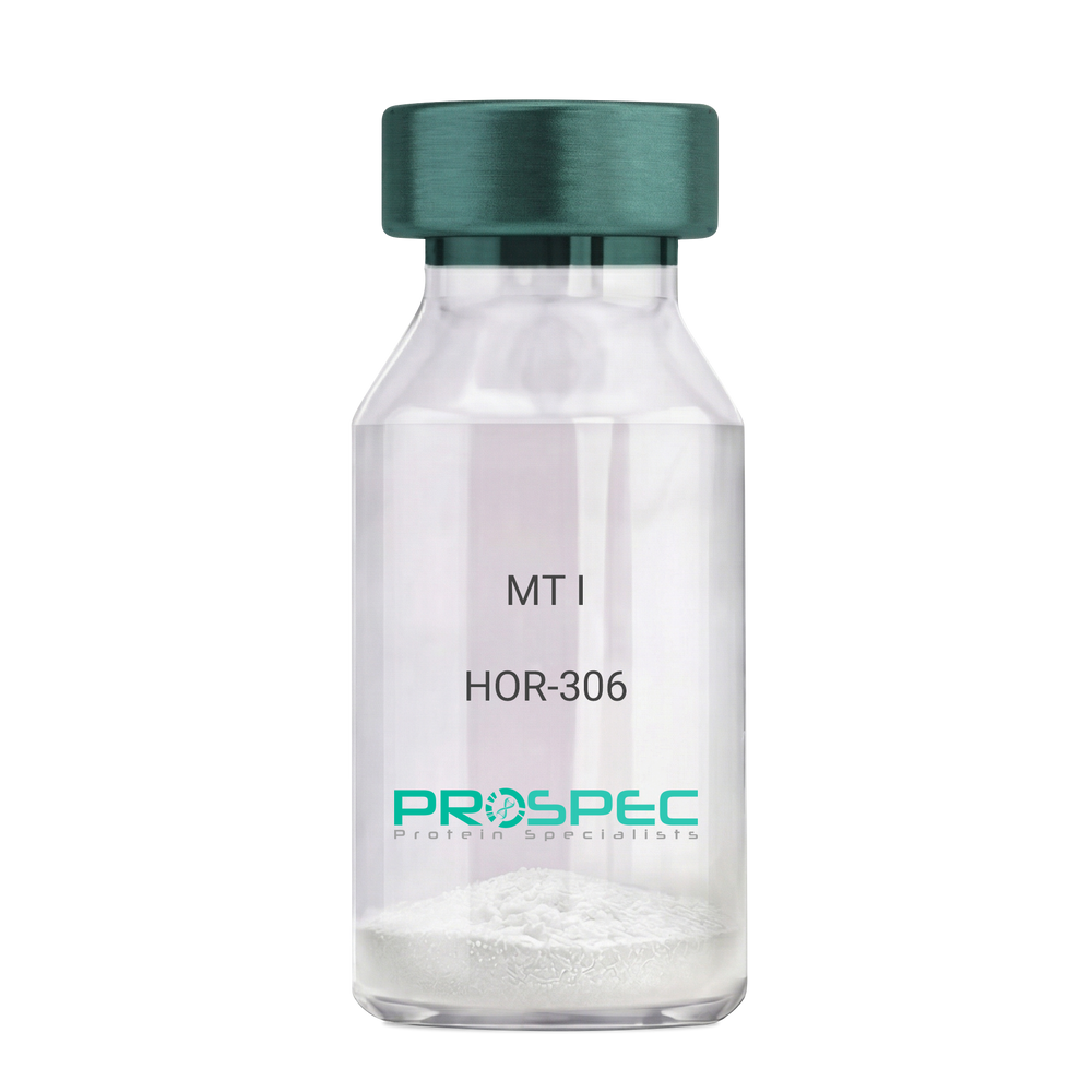
    MT I

    Description:

    Melanotan-I

    Melanotan-I, MT-I, Melanotan-1.

    Product # :

    HOR-306

    Price :

    Quantity :

    Shipping Method :

    Room Temp Icon

    Shipped at Room temp

    Add To Cart

    More Info

    • description
    • formulation
    • purity
    • More Info

    Description

    Melanotan-I has the amino acid sequence of Ser-Tyr-Ser-Nle-Glu-His-D-Phe-Arg-Trp-Gly-Lys-Pro-Val and a molecular weight of 1647.4 Dalton.

    Formulation

    The protein (1mg/ml) was lyophilized with no additives.

    Purity

    Greater than 98.0% as determined by RP-HPLC.

    More Info

    • Synonyms

      Melanotan-I, MT-I, Melanotan-1.

    • Physical Appearance

      Sterile Filtered off-White lyophilized (freeze-dried) powder.

    • Stability

      Lyophilized Melanotan-I although stable at room temperature for 3 weeks, should be stored desiccated below -18°C. Upon reconstitution MT-I should be stored at 4°C between 2-7 days and for future use below -18°C.For long term storage it is recommended to add a carrier protein (0.1% HSA or BSA).Please prevent freeze-thaw cycles.

    • Solubility

      It is recommended to reconstitute the lyophilized Melanotan-I in sterile 1% acetic acid not less than 100 µg/ml, which can then be further diluted to other aqueous solutions.

    ProSpec's products are furnished for LABORATORY RESEARCH USE ONLY. The product may not be used as drugs, agricultural or pesticidal products, food additives or household chemicals.

    Melanotan I
  • View Data Sheet

    Name :

    LGALS4 Human

    Description:

    Galectin-4 Human Recombinant

    Galectin-4, Gal-4, Lactose-binding lectin 4, L-36 lactose-binding protein, L36LBP, Antigen NY-CO-27, LGALS4, lectin galactoside-binding soluble 4, GAL4.

    Product # :

    CYT-686

    Price :

    Quantity :

    Shipping Method :

    Ice Icon

    Shipped with Ice Packs

    Add To Cart

    More Info

    • description
    • source
    • formulation
    • purity
    • More Info
    • sds-page

    Description

    Galectin-4 produced in E.Coli is a single, non-glycosylated polypeptide chain containing 343 amino acids (1-323 a.a.) and having a molecular mass of 38.1kDa.Galectin-4 is fused to 20 amino acid His-Tag at N-terminus and purified by proprietary chromatographic techniques.

    Source

    Escherichia Coli.

    Formulation

    The LGALS4 protein solution contains 20mM Tris-HCl, pH-8, 1mM DTT and 10% glycerol.

    Purity

    Greater than 95.0% as determined by SDS-PAGE.

    sds-page

    LGALS4 Human-SDS-PAGE - Product image 1

    More Info

    • Introduction

      Galectin-4 is a member of the subfamily of galectins composed of two carbohydrate recognition domains having similar peptide chains. The galectins are a family of beta-galactoside-binding proteins having a role in modulating cell-cell and cell-matrix interactions, which inhibits chronic inflammations, GVHD, and allergic responses. LGALS4 expression is limited to small intestine, colon, and rectum, and it is underexpressed in colorectal cancer. LGALS4 binds as an endogenous ligand to glycosphingolipids having 3-O-sulfated Gal residues and bind as well to cholesterol-3-sulfate. LGALS4 takes part in cell adhesion. LGALS4 plays a role in crosslinking the lateral cell membranes of the surface-lining epithelial cells, thus supporting epithelial integrity against mechanical stress exerted by the bowel lume. LGALS4 is in charge of intestinal inflammation via selective regulation of peripheral and mucosal T-cell cell cycle, in addition to cell death by apoptosis of T-cells by a pathway independent of the activation of caspases. LGALS4 blockade decreases TNF-alpha inhibitor induced T-cell death. LGALS4 decreases pro-inflammatory cytokine secretion including IL-6 & IL-17.

    • Synonyms

      Galectin-4, Gal-4, Lactose-binding lectin 4, L-36 lactose-binding protein, L36LBP, Antigen NY-CO-27, LGALS4, lectin galactoside-binding soluble 4, GAL4.

    • Physical Appearance

      Sterile filtered colorless solution.

    • Stability

      Store at 4°C if entire vial will be used within 2-4 weeks. Store, frozen at -20°C for longer periods of time. For long term storage it is recommended to add a carrier protein (0.1% HSA or BSA).Avoid multiple freeze-thaw cycles.

    • Amino Acid Sequence

      MGSSHHHHHH SSGLVPRGSH MAYVPAPGYQ PTYNPTLPYY QPIPGGLNVG MSVYIQGVAS EHMKRFFVNF VVGQDPGSDV AFHFNPRFDG WDKVVFNTLQ GGKWGSEERK RSMPFKKGAA FELVFIVLAE HYKVVVNGNP FYEYGHRLPL QMVTHLQVDG DLQLQSINFI GGQPLRPQGP PMMPPYPGPG HCHQQLNSLP TMEGPPTFNP PVPYFGRLQG GLTARRTIII KGYVPPTGKS FAINFKVGSS GDIALHINPR MGNGTVVRNS LLNGSWGSEE KKITHNPFGP GQFFDLSIRC GLDRFKVYAN GQHLFDFAHR LSAFQRVDTL EIQGDVTLSY VQI.

    • Background

      What is the molecular weight/Mw of LGALS4 HUMAN Protein?
      LGALS4 HUMAN Protein has a total Mw of 38.1kDa.

      What is the source or expression system of LGALS4 HUMAN Protein?
      Escherichia Coli.

      What is the Purity of LGALS4 HUMAN Protein?
      LGALS4 HUMAN Protein is >95% pure as determined by SDS-PAGE.

      What is the Biological Activity of LGALS4 HUMAN Protein?
      The biological functionality of LGALS4 HUMAN Protein will be determined in the future.

      What is the amino acid sequence of LGALS4 HUMAN Protein?
      MGSSHHHHHH SSGLVPRGSH MAYVPAPGYQ PTYNPTLPYY QPIPGGLNVG MSVYIQGVAS EHMKRFFVNF VVGQDPGSDV AFHFNPRFDG WDKVVFNTLQ GGKWGSEERK RSMPFKKGAA FELVFIVLAE HYKVVVNGNP FYEYGHRLPL QMVTHLQVDG DLQLQSINFI GGQPLRPQGP PMMPPYPGPG HCHQQLNSLP TMEGPPTFNP PVPYFGRLQG GLTARRTIII KGYVPPTGKS FAINFKVGSS GDIALHINPR MGNGTVVRNS LLNGSWGSEE KKITHNPFGP GQFFDLSIRC GLDRFKVYAN GQHLFDFAHR LSAFQRVDTL EIQGDVTLSY VQI.

      What applications can LGALS4 HUMAN Protein be used in?
      LGALS4 HUMAN Protein can probably be used in western blot, ELISA and Lateral Flow.

      What is the endotoxin level for LGALS4 HUMAN Protein?
      The endotoxin level is minimal, LGALS4 HUMAN Protein was purified using conventional chromatography techniques.


    ProSpec's products are furnished for LABORATORY RESEARCH USE ONLY. The product may not be used as drugs, agricultural or pesticidal products, food additives or household chemicals.

    Lgals4 Human
  • View Data Sheet

    Name :

    Adiponectin (108-244) Human

    Description:

    Adiponectin (108-244 a.a.) Human Recombinant

    Acrp30, AdipoQ, GBP-28, APM-1, ACDC.

    Product # :

    CYT-073

    Price :

    Quantity :

    Shipping Method :

    Ice Icon

    Shipped with Ice Packs

    Add To Cart

    More Info

    • description
    • source
    • formulation
    • purity
    • More Info
    • sds-page

    Description

    Acrp30 Human Recombinant produced in E.Coli is a single, non-glycosylated polypeptide chain containing 158 amino acids (108-244 a.a.) and having a molecular mass of 18.1kDa.Acrp30 is fused to a 21 amino acid His-tag at N-terminus & purified by proprietary chromatographic techniques.

    Source

    Escherichia Coli.

    Formulation

    Acrp30 protein solution (1mg/ml) contains 20mM Tris-HCl buffer (pH 8.0), 10% glycerol and 0.4M Urea.

    Purity

    Greater than 80.0% as determined by SDS-PAGE.

    sds-page

    Adiponectin-sds-page - Product image 1

    More Info

    • Introduction

      Adiponectin is an adipocyte specific secreted protein that circulates in the plasma. It is induced during adipocyte differentiation and its secretion is stimulated by insulin. Mouse adiponectin shares about 83% amino acid identity with that human. Adiponectin plays a role in various physiological processes such as energy homeostasis and obesity. Adiponectin is reduced in obese humans, and decreased level is associated with insulin resistance and hyperinsulinemia.

    • Synonyms

      Acrp30, AdipoQ, GBP-28, APM-1, ACDC.

    • Stability

      Store at 4°C if entire vial will be used within 2-4 weeks. Store, frozen at -20°C for longer periods of time. For long term storage it is recommended to add a carrier protein (0.1% HSA or BSA).Avoid multiple freeze-thaw cycles.

    • Amino Acid Sequence

      MGSSHHHHHH SSGLVPRGSH MAYVYRSAFS VGLETYVTIP NMPIRFTKIF YNQQNHYDGS TGKFHCNIPG LYYFAYHITV YMKDVKVSLF KKDKAMLFTY DQYQENNVDQ ASGSVLLHLE VGDQVWLQVY GEGERNGLYA DNDNDSTFTG FLLYHDTN.

    • Background

      What is the molecular weight / Mw of ADIPONECTIN Protein?
      ADIPONECTIN Protein has a total Mw of 18.1kDa.

      What is the source or expression system of ADIPONECTIN Protein?
      Escherichia Coli.

      What is the Purity of ADIPONECTIN Protein?
      ADIPONECTIN Protein is >80% pure as determined by SDS-PAGE.

      What is the Biological Activity of ADIPONECTIN Protein?
      The biological functionality of ADIPONECTIN Protein will be determined in the future.

      What is the amino acid sequence of ADIPONECTIN Protein? MGSSHHHHHH SSGLVPRGSH MAYVYRSAFS VGLETYVTIP NMPIRFTKIF YNQQNHYDGS TGKFHCNIPG LYYFAYHITV YMKDVKVSLF KKDKAMLFTY DQYQENNVDQ ASGSVLLHLE VGDQVWLQVY GEGERNGLYA DNDNDSTFTG FLLYHDTN..

      What applications can ADIPONECTIN Protein be used in?
      ADIPONECTIN Protein can probably be used in western blot, ELISA and Lateral Flow.

      What is the endotoxin level for ADIPONECTIN Protein?
      The endotoxin level is minimal, ADIPONECTIN Protein was purified using conventional chromatography techniques

    ProSpec's products are furnished for LABORATORY RESEARCH USE ONLY. The product may not be used as drugs, agricultural or pesticidal products, food additives or household chemicals.

    Acrp30 108 244 Human
  • View Data Sheet

    Name :

    SDF 1a Human, His

    Description:

    Stromal Cell-Derived Factor-1 alpha Human Recombinant, His Tag

    SDF-1, CXCL12, Pre-B cell growth-stimulating factor, PBSF, hIRH, chemokine (C-X-C motif) ligand 12, SDF1, SDF1A, TPAR1, SCYB12, SDF-1a, TLSF-a.

    Product # :

    CHM-241

    Price :

    Quantity :

    Shipping Method :

    Room Temp Icon

    Shipped at Room temp

    Add To Cart

    More Info

    • description
    • source
    • formulation
    • purity
    • More Info

    Description

    Stromal Cell-Derived Factor-1 alpha Human Recombinant produced in E.Coli is a non-glycosylated, Polypeptide chain containing 78 amino acids, having a molecular mass of 9.2 kDa. The SDF-1a is fused to 10 amino acids His-Tag at N-terminus and purified by proprietary chromatographic techniques.

    Source

    Escherichia Coli.

    Formulation

    The protein was filtered (0.4µm) and lyophilized from a concentrated (0.5mg/ml) solution containing 20mM Tris buffer pH-7.5 and 20mM sodium chloride.

    Purity

    Greater than 95.0% as determined SDS-PAGE.

    More Info

    • Introduction

      SDF-1 (stromal cell-derived factor-1) is small cytokine belonging to the chemokine family that is officially designated Chemokine (C-X-C motif) ligand 12 (CXCL12). It is produced in two forms, SDF-1?/CXCL12a and SDF-1?/CXCL12b, by alternate splicing of the same gene. Chemokines are characterized by the presence of four conserved cysteines, which form two disulfide bonds. The CXCL12 proteins belong to the group of CXC chemokines, whose initial pair of cysteines are separated by one intervening amino acid. CXCL12 is strongly chemotactic for lymphocytes and has been implicated as an important cell co-ordinator during development. During embryogenesis it directs the migration of hematopoietic cells from foetal liver to bone marrow. Mice which were knocked-out for CXCL12 gene were lethal before the birth or within just 1 hour of life. As another role, CXCL12a alters also the electrophysiology of neurons. CXCL12 was shown to be expressend in many tissues in mice (including brain, thymus, heart, lung, liver, kidney, spleen and bone marrow).
      The receptor for this chemokine is CXCR4, which was previously called fusin. This CXCL12-CXCR4 interaction used to be considered exclusive (unlike for other chemokines and their receptors), but recently it was suggested that CXCL12 is also bound by CXCR7 receptor.
      The gene for CXCL12 is located on human chromosome 10. In human and mouse both CXCL12 and CXCR4 show high identity of sequence: 99% and 90%, respectively.

    • Synonyms

      SDF-1, CXCL12, Pre-B cell growth-stimulating factor, PBSF, hIRH, chemokine (C-X-C motif) ligand 12, SDF1, SDF1A, TPAR1, SCYB12, SDF-1a, TLSF-a.

    • Physical Appearance

      White lyophilized (freeze-dried) powder.

    • Stability

      Lyophilized SDF1A although stable at room temperature for 3 weeks, should be stored desiccated below -18°C. Upon reconstitution CXCL12 should be stored at 4°C between 2-7 days and for future use below -18°C.For long term storage it is recommended to add a carrier protein (0.1% HSA or BSA).Please prevent freeze-thaw cycles.

    • Solubility

      It is recommended to add deionized water to a working concentration approximately 0.5 mg/ml and let the lyophilized pellet dissolve completely. Product is not sterile! Please filter the product by appropriate sterile filter before using it in the cell culture.

    • Amino Acid Sequence

      MKHHHHHHAS KPVSLSYRCP CRFFESHVAR ANVKHLKILN TPNCALQIVA RLKNNNRQVC IDPKLKWIQE YLEKALNK.

    ProSpec's products are furnished for LABORATORY RESEARCH USE ONLY. The product may not be used as drugs, agricultural or pesticidal products, food additives or household chemicals.

    Sdf 1A Human His
  • View Data Sheet

    Name :

    SDF 1b Human

    Description:

    Stromal Cell Derived Factor-1 Beta Human Recombinant (CXCL12)

    SDF-1, CXCL12, Pre-B cell growth-stimulating factor, PBSF, hIRH, chemokine (C-X-C motif) ligand 12, SDF1, SDF1B, TPAR1, SCYB12, SDF-1b, TLSF-b.

    Product # :

    CHM-325

    Price :

    Quantity :

    Shipping Method :

    Room Temp Icon

    Shipped at Room temp

    Add To Cart

    More Info

    • description
    • source
    • formulation
    • purity
    • biological activity
    • More Info

    Description

    Stromal Cell-Derived Factor-1 beta Human Recombinant produced in E.Coli is a non-glycosylated, Polypeptide chain containing 72 amino acids and having a molecular mass of 8508 Dalton. The SDF-1b is purified by proprietary chromatographic techniques.

    Source

    Escherichia Coli.

    Formulation

    The CXCL12 protein was lyophilized from a concentrated (1mg/ml) sterile solution containing no additives.

    Purity

    Greater than 98.0% as determined by:
    (a) Analysis by RP-HPLC.
    (b) Analysis by SDS-PAGE.

    Biological Activity

    The specific activity as determined by its ability to chemoattract human peripheral T cells activated with PHA and IL-2 using a concentation of 20-80ng/ml corresponding to a Specific Activity of 12,500-50,000IU/mg.

    More Info

    • Introduction

      SDF-1 (stromal cell-derived factor-1) is small cytokine belonging to the chemokine family that is officially designated Chemokine (C-X-C motif) ligand 12 (CXCL12). It is produced in two forms, SDF-1?/CXCL12a and SDF-1?/CXCL12b, by alternate splicing of the same gene. Chemokines are characterized by the presence of four conserved cysteines, which form two disulfide bonds. The CXCL12 proteins belong to the group of CXC chemokines, whose initial pair of cysteines are separated by one intervening amino acid. CXCL12 is strongly chemotactic for lymphocytes and has been implicated as an important cell co-ordinator during development. During embryogenesis it directs the migration of hematopoietic cells from foetal liver to bone marrow. Mice which were knocked-out for CXCL12 gene were lethal before the birth or within just 1 hour of life. As another role, CXCL12a alters also the electrophysiology of neurons. CXCL12 was shown to be expressend in many tissues in mice (including brain, thymus, heart, lung, liver, kidney, spleen and bone marrow).
      The receptor for this chemokine is CXCR4, which was previously called fusin. This CXCL12-CXCR4 interaction used to be considered exclusive (unlike for other chemokines and their receptors), but recently it was suggested that CXCL12 is also bound by CXCR7 receptor.
      The gene for CXCL12 is located on human chromosome 10. In human and mouse both CXCL12 and CXCR4 show high identity of sequence: 99% and 90%, respectively.

    • Synonyms

      SDF-1, CXCL12, Pre-B cell growth-stimulating factor, PBSF, hIRH, chemokine (C-X-C motif) ligand 12, SDF1, SDF1B, TPAR1, SCYB12, SDF-1b, TLSF-b.

    • Physical Appearance

      Sterile Filtered White lyophilized (freeze-dried) powder.

    • Stability

      Lyophilized SDF-1b although stable at room temperature for 3 weeks, should be stored desiccated below -18°C. Upon reconstitution CXCL12 should be stored at 4°C between 2-7 days and for future use below -18°C. For long term storage it is recommended to add a carrier protein (0.1% HSA or BSA).Please prevent freeze-thaw cycles.

    • Solubility

      It is recommended to reconstitute the lyophilized Stromal Cell-Derived Factor-1b in sterile 18MΩ-cm H2O not less than 100µg/ml, which can then be further diluted to other aqueous solutions.

    • Amino Acid Sequence

      The sequence of the first five N-terminal amino acids was determined and was found to be Lys-Pro-Val-Ser-Leu.

    • Protein content

      Protein quantitation was carried out by two independent methods: 1. UV spectroscopy at 280 nm using the absorbency value of 1.06 as the extinction coefficient for a 0.1% (1mg/ml) solution. This value is calculated by the PC GENE computer analysis program of protein sequences (IntelliGenetics). 2. Analysis by RP-HPLC, using a standard solution of SDF-1b as a Reference Standard.

    ProSpec's products are furnished for LABORATORY RESEARCH USE ONLY. The product may not be used as drugs, agricultural or pesticidal products, food additives or household chemicals.

    Sdf 1 B Human
  • View Data Sheet

    Name :

    SDF 1b Mouse

    Description:

    Stromal Cell Derived Factor-1 Beta Mouse Recombinant (CXCL12)

    SDF-1, CXCL12, Pre-B cell growth-stimulating factor, PBSF, hIRH, chemokine (C-X-C motif) ligand 12, SDF1, SDF1B, TPAR1, SCYB12, SDF-1b, TLSF-b, 12-O-tetradecanoylphorbol 13-acetate repressed protein 1, Thymic lymphoma cell-stimulating factor, TLSF.

    Product # :

    CHM-326

    Price :

    Quantity :

    Shipping Method :

    Room Temp Icon

    Shipped at Room temp

    Add To Cart

    More Info

    • description
    • source
    • formulation
    • purity
    • biological activity
    • More Info

    Description

    Stromal Cell-Derived Factor-1 beta Mouse Recombinant produced in E.Coli is a non-glycosylated, Polypeptide chain containing 72 amino acids and having a molecular mass of 8513 Dalton. The SDF-1b is purified by proprietary chromatographic techniques.

    Source

    Escherichia Coli.

    Formulation

    The CXCL12 protein was lyophilized from a concentrated (1mg/ml) sterile solution containing no additives.

    Purity

    Greater than 98.0% as determined by:
    (a) Analysis by RP-HPLC.
    (b) Analysis by SDS-PAGE.

    Biological Activity

    The specific activity as determined by its ability to chemoattract human monocytes at 50-100ng/ml corresponding to a Specific Activity of 10,000-20,000IU/mg.

    More Info

    • Introduction

      SDF-1 (stromal cell-derived factor-1) is small cytokine belonging to the chemokine family that is officially designated Chemokine (C-X-C motif) ligand 12 (CXCL12). It is produced in two forms, SDF-1?/CXCL12a and SDF-1?/CXCL12b, by alternate splicing of the same gene. Chemokines are characterized by the presence of four conserved cysteines, which form two disulfide bonds. The CXCL12 proteins belong to the group of CXC chemokines, whose initial pair of cysteines are separated by one intervening amino acid. CXCL12 is strongly chemotactic for lymphocytes and has been implicated as an important cell co-ordinator during development. During embryogenesis it directs the migration of hematopoietic cells from foetal liver to bone marrow. Mice which were knocked-out for CXCL12 gene were lethal before the birth or within just 1 hour of life. As another role, CXCL12a alters also the electrophysiology of neurons. CXCL12 was shown to be expressend in many tissues in mice (including brain, thymus, heart, lung, liver, kidney, spleen and bone marrow).
      The receptor for this chemokine is CXCR4, which was previously called fusin. This CXCL12-CXCR4 interaction used to be considered exclusive (unlike for other chemokines and their receptors), but recently it was suggested that CXCL12 is also bound by CXCR7 receptor.
      The gene for CXCL12 is located on human chromosome 10. In human and mouse both CXCL12 and CXCR4 show high identity of sequence: 99% and 90%, respectively.

    • Synonyms

      SDF-1, CXCL12, Pre-B cell growth-stimulating factor, PBSF, hIRH, chemokine (C-X-C motif) ligand 12, SDF1, SDF1B, TPAR1, SCYB12, SDF-1b, TLSF-b, 12-O-tetradecanoylphorbol 13-acetate repressed protein 1, Thymic lymphoma cell-stimulating factor, TLSF.

    • Physical Appearance

      Sterile Filtered White lyophilized (freeze-dried) powder.

    • Stability

      Lyophilized SDF-1b although stable at room temperature for 3 weeks, should be stored desiccated below -18°C. Upon reconstitution CXCL12 should be stored at 4°C between 2-7 days and for future use below -18°C.For long term storage it is recommended to add a carrier protein (0.1% HSA or BSA).Please prevent freeze-thaw cycles.

    • Solubility

      It is recommended to reconstitute the lyophilized Stromal Cell-Derived Factor-1b in sterile 18MΩ-cm H2O not less than 100µg/ml, which can then be further diluted to other aqueous solutions.

    • Amino Acid Sequence

      The sequence of the first five N-terminal amino acids was determined and was found to be Lys-Pro-Val-Ser-Leu.

    ProSpec's products are furnished for LABORATORY RESEARCH USE ONLY. The product may not be used as drugs, agricultural or pesticidal products, food additives or household chemicals.

    Sdf 1 B Mouse
  • View Data Sheet

    Name :

    DHH (C23II) Mouse

    Description:

    Desert Hedgehog (C23II) Mouse Recombinant

    Desert hedgehog protein, DHH, HHG-3, C78960.

    Product # :

    CYT-773

    Price :

    Quantity :

    Shipping Method :

    Room Temp Icon

    Shipped at Room temp

    Add To Cart

    More Info

    • description
    • source
    • formulation
    • purity
    • biological activity
    • More Info

    Description

    DHH (C23II) Mouse Recombinant produced in E.Coli is a single, non-glycosylated polypeptide chain containing 177 amino acids and having a molecular mass of 20kDa. The DHH (C23II) is purified by proprietary chromatographic techniques.

    Source

    Escherichia Coli.

    Formulation

    Lyophilized from a 0.2µm filtered concentrated solution in PBS, pH7.4, 1mM DTT and 0.05% Tween-80.

    Purity

    Greater than 97.0% as determined by:
    (a) Analysis by RP-HPLC.
    (b) Analysis by SDS-PAGE.

    Biological Activity

    The ED50 as determined by its ability to induce alkaline phosphatase production by murine MC3T3-E1 cells is 5-20 µg/ml.

    More Info

    • Introduction

      DHH is part of the Hedgehog family which encodes signaling molecules that are involved in regulating morphogenesis. DHH protein is a precursor that is autocatalytically cleaved, the N-terminal portion is soluble and contains the signalling activity while the C-terminal portion is involved in precursor processing. Additionally, the C-terminal product covalently attaches a cholesterol moiety to the N-terminal product, restricting the N-terminal product to the cell surface and preventing it from freely diffusing throughout the organism. Defects in DHH protein have been associated with partial gonadal dysgenesis (PGD) accompanied by minifascicular polyneuropathy. DHH plays a role both male gonadal differentiation and perineurial development.
      DHH plays a role in intercellular signaling which is essential for a variety of patterning events during development. DHH functions as a spermatocyte survival factor in the testes & is essential for testes development.

    • Synonyms

      Desert hedgehog protein, DHH, HHG-3, C78960.

    • Physical Appearance

      Sterile Filtered White lyophilized (freeze-dried) powder.

    • Stability

      Lyophilized DHH (C23II) although stable at room temperature for 3 weeks, should be stored desiccated below -18°C. Upon reconstitution DHH (C23II) should be stored at 4°C between 2-7 days and for future use below -18°C. For long term storage it is recommended to add a carrier protein (0.1% HSA or BSA).Please prevent freeze-thaw cycles.

    • Solubility

      It is recommended to reconstitute the lyophilized DHH (C23II) in sterile 18M-cm H2O not less than 100µg/ml, which can then be further diluted to other aqueous solutions.

    • Amino Acid Sequence

      IIGPGRGPVG RRRYVRKQLV PLLYKQFVPS MPERTLGASG PAEGRVTRGS ERFRDLVPNY NPDIIFKDEE NSGADRLMTE RCKERVNALA IAVMNMWPGV RLRVTEGWDE DGHHAQDSLH YEGRALDITT SDRDRNKYGL LARLAVEAGF DWVYYESRNH IHVSVKADNS LAVRAGG.

    ProSpec's products are furnished for LABORATORY RESEARCH USE ONLY. The product may not be used as drugs, agricultural or pesticidal products, food additives or household chemicals.

    Dhh C23Ii Mouse
  • View Data Sheet

    Name :

    pHGF Porcine

    Description:

    Hepatocyte promoting Growth Factor Porcine

    Scatter Factor (SF), Hepatopoietin (HPTA), HGF, HGFB, F-TCF, pHGF.

    Product # :

    CYT-522

    Price :

    Quantity :

    Shipping Method :

    Room Temp Icon

    Shipped at Room temp

    Add To Cart

    More Info

    • description
    • source
    • formulation
    • purity
    • More Info

    Description

    Hepatocyte Growth Factor porcine is extracted from pig liver.The HGF is purified by proprietary chromatographic techniques.

    Source

    Pig Liver.

    Formulation

    The sterile protein powder is lyophilized with no additives.

    Purity

    Greater than 98.0% as determined by:
    (a) Analysis by RP-HPLC.
    (b) Analysis by SDS-PAGE.

    More Info

    • Introduction

      Hepatocyte Growth Factor (HGF) is a multifunctional growth factor which regulates both cell growth and cell motility. It exerts a strong mitogenic effect on hepatocytes and primary epithelial cells. HGF synergizes with Interleukin-3and GM-CSFto stimulate colony formation of hematopoietic progenitor cells in vitro and may, therefore, also modulate hematopoiesis.

    • Synonyms

      Scatter Factor (SF), Hepatopoietin (HPTA), HGF, HGFB, F-TCF, pHGF.

    • Physical Appearance

      Sterile Filtered White lyophilized (freeze-dried) powder.

    • Stability

      Lyophilized pHGF although stable at room temperature for 3 weeks, should be stored desiccated below -18°C. Upon reconstitution pHGF should be stored at 4°C between 2-7 days and for future use below -18°C. For long term storage it is recommended to add a carrier protein (0.1% HSA or BSA).Please prevent freeze-thaw cycles.

    • Solubility

      It is recommended to reconstitute the lyophilized pHGF in sterile 18MΩ-cm H2O not less than 100µg/ml, which can then be further diluted to other aqueous solutions.

    • Background

      What is the source or expression system of PHGF PORCINE Protein?
      Pig Liver.

      What is the Purity of PHGF PORCINE Protein?
      PHGF PORCINE Protein is >98% pure as determined by SDS-PAGE.

      What is the Biological Activity of PHGF PORCINE Protein?
      The biological functionality of PHGF PORCINE Protein will be determined in the future.

      What applications can PHGF PORCINE Protein be used in?
      PHGF PORCINE Protein can probably be used in western blot, ELISA and Lateral Flow.

      What is the endotoxin level for PHGF PORCINE Protein?
      The endotoxin level is minimal, PHGF PORCINE Protein was purified using conventional chromatography techniques.


    ProSpec's products are furnished for LABORATORY RESEARCH USE ONLY. The product may not be used as drugs, agricultural or pesticidal products, food additives or household chemicals.

    Hgf Porcine
  • View Data Sheet

    Name :

    PPCDC Human

    Description:

    Phosphopantothenoylcysteine Decarboxylase Human Recombinant

    FLJ14585, PPC-DC, MDS018, COAC, CoaC, Phosphopantothenoylcysteine Decarboxylase.

    Product # :

    ENZ-011

    Price :

    Quantity :

    Shipping Method :

    Ice Icon

    Shipped with Ice Packs

    Add To Cart

    More Info

    • description
    • source
    • formulation
    • purity
    • More Info

    Description

    PPCDC produced in E.Coli is a single, non-glycosylated polypeptide chain containing 224 amino acids (1-204a.a.) and having a molecular mass of 24.6kDa.PPCDC is fused to a 20 amino acid His-tag at N-terminus & purified by proprietary chromatographic techniques.

    Source

    Escherichia Coli.

    Formulation

    The PPCDC protein solution (1mg/1ml) is formulated in 20mM Tris-HCl buffer(pH 8.0), 1mM DTT, 0.1M NaCl and 10% glycerol.

    Purity

    Greater than 95% as determined by SDS-PAGE.

    More Info

    • Introduction

      PPCDC is a member of the HFCD (homo oligomeric flavin containing Cys decarboxylase) superfamily which takes a part in the biosynthesis of coenzyme A (CoA) from pantothenate (Vitamin B). The biosynthesis of CoA from pantothenate needs several steps: the phosphorylation of pantothenate, the conversion of 4’-hosphopantothenate to 4''-phosphopantetheine, the adenylation by phosphopantetheine adenylyltransferase to form dephospho-CoA and the phosphorylation by dephospho-CoA kinase to form CoA. PPCDC is a key player in this pathway. PPCDC converts 4''-phosphopantothenate into 4''-phosphopantetheine.

    • Synonyms

      FLJ14585, PPC-DC, MDS018, COAC, CoaC, Phosphopantothenoylcysteine Decarboxylase.

    • Physical Appearance

      Sterile Filtered clear solution.

    • Stability

      Store at 4°C if entire vial will be used within 2-4 weeks. Store, frozen at -20°C for longer periods of time.Please avoid freeze thaw cycles.

    • Amino Acid Sequence

      MGSSHHHHHH SSGLVPRGSH MEPKASCPAA APLMERKFHV LVGVTGSVAA LKLPLLVSKL LDIPGLEVAV VTTERAKHFY SPQDIPVTLY SDADEWEMWK SRSDPVLHID LRRWADLLLV APLDANTLGK VASGICDNLL TCVMRAWDRS KPLLFCPAMN TAMWEHPITA QQVDQLKAFG YVEIPCVAKK LVCGDEGLGA MAEVGTIVDK VKEVLFQHSG FQQS

    ProSpec's products are furnished for LABORATORY RESEARCH USE ONLY. The product may not be used as drugs, agricultural or pesticidal products, food additives or household chemicals.

    Ppcdc Human
  • View Data Sheet

    Name :

    CST3 Mouse

    Description:

    Cystatin-C Mouse Recombinant

    Post G-globulin, CST 3, CST3, Gamma-Trace, Cystatin 3, Amyloid Angiopathy and Cerebral Hemorrhage, Cystatin-C precursor, neuroendocrine basic polypeptide.

    Product # :

    PRO-597

    Price :

    Quantity :

    Shipping Method :

    Room Temp Icon

    Shipped at Room temp

    Add To Cart

    More Info

    • description
    • source
    • formulation
    • purity
    • More Info

    Description

    Cystatin-C Murine Recombinant produced in E.Coli is a single, non-glycosylated, polypeptide chain containing 134 amino acids and having a molecular mass of 15kDa. The Mouse Cystatin-C is fused to His tag at N-Terminus.The Mouse Cystatin-C is purified by proprietary chromatographic techniques.

    Source

    Escherichia Coli.

    Formulation

    The sterile filtered concentrated (0.5mg/ml) protein solution was lyophilized with 20mM Tris & 50mM NaCl pH-7.5.

    Purity

    Greater than 95% as determined by SDS-PAGE.

    More Info

    • Introduction

      Cystatins are a superfamily of cysteine proteinase inhibitors found in both plants and animals. They comprise a group of proteinase inhibitors, widely distributed in tissues and body fluids, and form tight complexes with cysteine proteases such as cathepsin B, H, L and S. Cystatin C, a secreted molecule of this family, is of interest from biochemical, medicine and evolutionary points of view. Cystatin C, with molecular weight of 13260 Da, is composed of 120 amino acids, lacks carbohydrate and has two disulfide bridges located near the carboxyl terminus. Cystatin C is increased in patients with malignant diseases, and is related to the insufficiency of renal function and appears to be a better marker than creatinine. On the other hand, low levels of cystatin C involve cause the breakdown of the elastic laminae and, subsequently, the atherosclerosis and abdominal aortic aneurysm.

    • Synonyms

      Post G-globulin, CST 3, CST3, Gamma-Trace, Cystatin 3, Amyloid Angiopathy and Cerebral Hemorrhage, Cystatin-C precursor, neuroendocrine basic polypeptide.

    • Physical Appearance

      Sterile Filtered White lyophilized (freeze-dried) powder.

    • Stability

      Store lyophilized protein at -20°C. Aliquot the product after reconstitution to avoid repeated freezing/thawing cycles. Reconstituted protein can be stored at 4°C for a limited period of time.

    • Solubility

      Add deionized water to a working concentration of 0.5mg/ml and let the lyophilized pellet dissolve completely.

    • Amino Acid Sequence

      MRGSHHHHHH GMASATPKQG PRMLGAPEEA DANEEGVRRA LDFAVSEYNK GSNDAYHSRA IQVVRARKQL VAGVNYFLDV EMGRTTCTKS QTNLTDCPFH DQPHLMRKAL CSFQIYSVPW KGTHSLTKFSCKNA.

    ProSpec's products are furnished for LABORATORY RESEARCH USE ONLY. The product may not be used as drugs, agricultural or pesticidal products, food additives or household chemicals.

    Cystatin C Mouse
  • View Data Sheet

    Name :

    S100G Human

    Description:

    S100 Calcium Binding Protein G Human Recombinant

    Protein S100-G, Calbindin-D9k, S100 calcium-binding protein G, Vitamin D-dependent calcium-binding protein intestinal, CABP, S100G, CABP9K, CALB3, S100D, CABP1, MGC138379.

    Product # :

    PRO-156

    Price :

    Quantity :

    Shipping Method :

    Room Temp Icon

    Shipped at Room temp

    Add To Cart

    More Info

    • description
    • source
    • formulation
    • More Info

    Description

    The Recombinant Human S100G produced in E.coli has a molecular mass of 10.04kDa containing 87 amino acid residues of the human S100G and fused to a 9 a.a. His tag at N-terminus.

    Source

    Escherichia Coli.

    Formulation

    S100G was filtered (0.4 µm) and lyophilized in 0.5 mg/ml in 20mM Tris and 50mM NaCl, pH 7.5.

    More Info

    • Introduction

      S100G (calbindin D9K) is a vitamin D-dependent calcium-binding protein. S100G, which is a cytosolic protein, is a member of a family of calcium-binding proteins that includes calmodulin, parvalbumin, troponin C, and S100 protein. In the intestine, S100G is vitamin D-dependent and its expression correlates with calcium transport activity. S100G may increase Ca2+ absorption by buffering Ca2+ in the cytoplasm and increase ATP-dependent Ca2+ transport in duodenal basolateral membrane vesicles.

    • Synonyms

      Protein S100-G, Calbindin-D9k, S100 calcium-binding protein G, Vitamin D-dependent calcium-binding protein intestinal, CABP, S100G, CABP9K, CALB3, S100D, CABP1, MGC138379.

    • Stability

      Store lyophilized protein at -20°C. Aliquot the product after reconstitution to avoid repeated freezing/thawing cycles. Reconstituted protein can be stored at 4°C for a limited period of time; it does not show any change after two weeks at 4°C.

    • Solubility

      It is recommended to add deionized water to prepare a working stock solution of approximately 0.5 mg/ml and let the lyophilized pellet dissolve completely. Product is not sterile! Please filter the product by an appropriate sterile filter before using it in the cell culture.

    • Amino Acid Sequence

      MKHHHHHHAS TKKSPEELKRS TKKSPEELKR IFEKYAAKEG DPDQLSKDEL KLLIQAEFPS LLKGPNTLDD LFQELDKNGD GEVSFEEFQV LVKKISQ.

    • Applications

      Western blotting.

    ProSpec's products are furnished for LABORATORY RESEARCH USE ONLY. The product may not be used as drugs, agricultural or pesticidal products, food additives or household chemicals.

    S100G Human
  • 1
  • …
  • 28
  • 29
  • 30
  • 31
  • 32
  • …
  • 42
Your browser does not support the video tag.

Products

  • Cytokines
  • Growth Factors
  • Chemokines
  • CD Antigens
  • Neurotrophins
  • Hormones
  • Enzymes
  • Viral Antigens
  • Recombinant Proteins
  • Natural Proteins
  • Monoclonal Antibodies
  • Polyclonal Antibodies
  • Thermal Packaging

About

  • Company
  • Founder
  • Contribution
  • Contact Us
  • My Account

Ordering

  • Ordering Method
  • Ordering - Credit Card
  • Ordering PO
  • Shipping Fees
  • FAQ's

Legal

  • Terms & Conditions
  • Privacy
  • Site Map
  • info@prospecbio.com
  • Facebook
  • Instagram
  • Linkedin Profile

© 2026 ProSpec-Tany TechnoGene Ltd. All Rights Reserved.

  • Choosing a selection results in a full page refresh.
  • Opens in a new window.