Search results
1000 results found for “decorin”
Name
Description
Product #
Price
Quantity
Shipping Method
- View Data Sheet
Name :
Cyclophilin F RatDescription:
Cyclophilin F Rat Recombinant
Peptidyl-prolyl cis-trans isomerase F, mitochondrial, PPIase F, Cyclophilin D, CyP-D, CypD, Cyclophilin F, Rotamase F, Ppif, Peptidyl-prolyl cis-trans isomerase F, mitochondrial, PPIase.
Product # :
ENZ-903Price :
Quantity :
Shipping Method :
Shipped with Ice Packs
More Info
- description
- source
- formulation
- purity
- More Info
Description
Cyclophilin F Rat Recombinant produced in E.Coli is a single, non-glycosylated polypeptide chain containing 200 amino acids (30-206a.a.) and having a molecular mass of 21.2kDa.Cyclophilin F is fused to a 23 amino acid His-tag at N-terminus & purified by proprietary chromatographic techniques.
Source
Escherichia Coli.
Formulation
Cyclophilin F protein solution (1mg/ml) containing Phosphate buffered saline (pH7.4), 10% glycerol and 1mM DTT.
Purity
Greater than 90.0% as determined by SDS-PAGE.
More Info
-
Introduction
Peptidyl-prolyl cis-trans isomerase F, mitochondrial (Cyclophilin-F) accelerates the folding of proteins. Cyclophilin-F catalyzes the cis-trans isomerization of proline imidic peptide bonds in oligopeptides and involved in regulation of the mitochondrial permeability transition pore (mPTP). Cyclophilin-F, in cooperation with mitochondrial TP53, is involved in activating oxidative stress-induced necrosis. Cyclophilin-F is also involved in modulation of mitochondrial membrane F1F0 ATP synthase activity and regulation of mitochondrial matrix adenine nucleotide levels. Furthermore, Cyclophilin-F has anti-apoptotic activity independently of mPTP and in cooperation with BCL2 inhibits cytochrome c-dependent apoptosis.
-
Synonyms
Peptidyl-prolyl cis-trans isomerase F, mitochondrial, PPIase F, Cyclophilin D, CyP-D, CypD, Cyclophilin F, Rotamase F, Ppif, Peptidyl-prolyl cis-trans isomerase F, mitochondrial, PPIase.
-
Physical Appearance
Sterile Filtered clear solution.
-
Stability
Store at 4°C if entire vial will be used within 2-4 weeks. Store, frozen at -20°C for longer periods of time. For long term storage it is recommended to add a carrier protein (0.1% HSA or BSA).Avoid multiple freeze-thaw cycles.
-
Amino Acid Sequence
MGSSHHHHHH SSGLVPRGSH MGSCSDGGAR GANSSSQNPL VYLDVGADGQ PLGRVVLELK ADVVPKTAEN FRALCTGEKG FGYKGSTFHR VIPAFMCQAG DFTNHNGTGG KSIYGSRFPD ENFTLKHVGP GVLSMANAGP NTNGSQFFIC TIKTDWLDGK HVVFGHVKEG MDVVKKIESF GSKSGKTSKK IVITDCGQLS.
ProSpec's products are furnished for LABORATORY RESEARCH USE ONLY. The product may not be used as drugs, agricultural or pesticidal products, food additives or household chemicals.
- View Data Sheet
Name :
DERADescription:
Deoxyribose-Phosphate Aldolase E.Coli Recombinant
Putative deoxyribose-phosphate aldolase, DERA, 2-deoxy-D-ribose 5-phosphate aldolase, Phosphodeoxyriboaldolase, Deoxyriboaldolase, DERA, CGI-26.
Product # :
ENZ-127Price :
Quantity :
Shipping Method :
Shipped with Ice Packs
More Info
- description
- source
- formulation
- purity
- More Info
Description
DERA produced in E.Coli is a single, non-glycosylated polypeptide chain containing 279 amino acids (1-259 a.a.) and having a molecular mass of 29.9kDa.DERA is fused to a 20 amino acid His-tag at N-terminus & purified by proprietary chromatographic techniques.
Source
Escherichia Coli.
Formulation
The DERA solution (1mg/ml) 20mM Tris-HCl buffer (pH 8.0), 10% glycerol and 2mM DTT.
Purity
Greater than 95.0% as determined by SDS-PAGE.
More Info
-
Introduction
Deoxyribose-phosphate aldolase (DERA) is a member of the deoC/fbaB aldolase protein family involved in the carbohydrate degradation pathway. DERA catalyzes the conversion of 2-deoxy-D-ribose 5-phosphate to D-glyceraldehyde 3-phosphate and an acetyldehyde.
-
Synonyms
Putative deoxyribose-phosphate aldolase, DERA, 2-deoxy-D-ribose 5-phosphate aldolase, Phosphodeoxyriboaldolase, Deoxyriboaldolase, DERA, CGI-26.
-
Physical Appearance
Sterile Filtered colorless solution.
-
Stability
Store at 4°C if entire vial will be used within 2-4 weeks. Store, frozen at -20°C for longer periods of time. For long term storage it is recommended to add a carrier protein (0.1% HSA or BSA).Avoid multiple freeze-thaw cycles.
-
Amino Acid Sequence
MGSSHHHHHH SSGLVPRGSH MTDLKASSLR ALKLMDLTTL NDDDTDEKVI ALCHQAKTPV GNTAAICIYP RFIPIARKTL KEQGTPEIRI ATVTNFPHGN DDIDIALAET RAAIAYGADE VDVVFPYRAL MAGNEQVGFD LVKACKEACA AANVLLKVII ETGELKDEAL IRKASEISIK AGADFIKTST GKVAVNATPE SARIMMEVIR DMGVEKTVGF KPAGGVRTAE DAQKYLAIAD ELFGADWADA RHYRFGASSL LASLLKALGH GDGKSASSY.
ProSpec's products are furnished for LABORATORY RESEARCH USE ONLY. The product may not be used as drugs, agricultural or pesticidal products, food additives or household chemicals.
- View Data Sheet
Name :
NDE1 HumanDescription:
nudE Nuclear Distribution Gene E Homolog 1 Human Recombinant
Nuclear distribution protein nudE homolog 1, NudE, NDE1, NUDE, LIS4, NUDE1, HOM-TES-87.
Product # :
PRO-204Price :
Quantity :
Shipping Method :
Shipped with Ice Packs
More Info
- description
- source
- formulation
- purity
- More Info
Description
NDE1 produced in E.Coli is a single, non-glycosylated polypeptide chain containing 355 amino acids (1-335 a.a.) and having a molecular mass of 39.9kDa (Molecular weight on SDS-PAGE will appear higher).NDE1 is fused to a 20 amino acid His-tag at N-terminus & purified by proprietary chromatographic techniques.
Source
Escherichia Coli.
Formulation
The NDE1 solution (1 mg/ml) contains 20mM Tris-HCl buffer (pH 8.0), 20% glycerol, 5mM DTT and 0.2M NaCl.
Purity
Greater than 85.0% as determined by SDS-PAGE.
More Info
-
Introduction
NDE1 is a cytoplasmic protein which is a member of the nudE protein family. Phosphorylated during mitosis, NDE1 is critical for the formation and function of the mitotic spindle in M phase and functions to regulate the Dynein-mediated transport of kinetochore proteins, as well as centrosome duplication during interphase. NDE1 is believed to interact with NDEL1, LIS1 and Dynein IC1/2, cytosolic in a signaling pathway which regulates the formation of neurons and is fundamental to the development of the cerebral cortex.
-
Synonyms
Nuclear distribution protein nudE homolog 1, NudE, NDE1, NUDE, LIS4, NUDE1, HOM-TES-87.
-
Physical Appearance
Sterile Filtered colorless solution.
-
Stability
NDE1 Human Recombinant although stable at 4°C for 1 week, should be stored below -18°C. Please prevent freeze thaw cycles.
-
Amino Acid Sequence
MGSSHHHHHH SSGLVPRGSH MEDSGKTFSS EEEEANYWKD LAMTYKQRAE NTQEELREFQ EGSREYEAEL ETQLQQIETR NRDLLSENNR LRMELETIKE KFEVQHSEGY RQISALEDDL AQTKAIKDQL QKYIRELEQA NDDLERAKRA TIMSLEDFEQ RLNQAIERNA FLESELDEKE NLLESVQRLK DEARDLRQEL AVQQKQEKPR TPMPSSVEAE RTDTAVQATG SVPSTPIAHR GPSSSLNTPG SFRRGLDDST GGTPLTPAAR ISALNIVGDL LRKVGALESK LASCRNLVYD QSPNRTGGPA SGRSSKNRDG GERRPSSTSV PLGDKGLDTS CRWLSKSTTR SSSSC.
ProSpec's products are furnished for LABORATORY RESEARCH USE ONLY. The product may not be used as drugs, agricultural or pesticidal products, food additives or household chemicals.
- View Data Sheet
Name :
IDH1 HumanDescription:
Isocitrate Dehydrogenase-1 Human Recombinant
Isocitrate dehydrogenase [NADP] cytoplasmic, EC 1.1.1.42, Cytosolic NADP-isocitrate dehydrogenase, Oxalosuccinate decarboxylase, IDH, NADP(+)-specific ICDH, IDP, PICD.
Product # :
ENZ-193Price :
Quantity :
Shipping Method :
Shipped with Ice Packs
More Info
- description
- source
- formulation
- purity
- biological activity
- More Info
Description
IDH1 Human Recombinant produced in E.coli is a single, non-glycosylated polypeptide chain containing 434 amino acids (1-414) and having a molecular mass of 48.8 kDa.IDH1 is fused to a 20 amino acid His-tag at N-terminus & purified by proprietary chromatographic techniques.
Source
E.coli.
Formulation
The IDH1 solution (1mg/ml) contains 20mM Tris-HCl buffer (pH 8.0), 0.1M NaCl,
1mM DTT, 0.1mM PMSF and 20% glycerol.Purity
Greater than 95% as determined by SDS-PAGE.
Biological Activity
The Specific activity is > 0.7 units/ml. One unit will convert 1.0 umole of isocitrate to alpha-ketoglutarate per minute at pH7.5 at 25C.More Info
-
Introduction
Isocitrate Dehydrogenase is an enzyme of the oxidoreductase class that catalyzes the conversion of isocitrate and NAD+ to yield 2-ketoglutarate, carbon dioxide, and NADH. It occurs in cell mitochondria. The enzyme requires Mg2+, Mn2+; it is activated by ADP, citrate, and Ca2+, and inhibited by NADH, NADPH, and ATP. The reaction is the key rate-limiting step of the citric acid (tricarboxylic) cycle.
-
Synonyms
Isocitrate dehydrogenase [NADP] cytoplasmic, EC 1.1.1.42, Cytosolic NADP-isocitrate dehydrogenase, Oxalosuccinate decarboxylase, IDH, NADP(+)-specific ICDH, IDP, PICD.
-
Physical Appearance
Sterile Filtered colorless solution.
-
Stability
Store at 4°C if entire vial will be used within 2-4 weeks. Store, frozen at -20°C for longer periods of time. For long term storage it is recommended to add a carrier protein (0.1% HSA or BSA).Avoid multiple freeze-thaw cycles.
-
Amino Acid Sequence
MGSSHHHHHH SSGLVPRGSH MSKKISGGSV VEMQGDEMTR IIWELIKEKL IFPYVELDLH SYDLGIENRD ATNDQVTKDA AEAIKKHNVG VKCATITPDE KRVEEFKLKQ MWKSPNGTIR NILGGTVFRE AIICKNIPRL VSGWVKPIII GRHAYGDQYR ATDFVVPGPG KVEITYTPSD GTQKVTYLVH NFEEGGGVAM GMYNQDKSIE DFAHSSFQMA LSKGWPLYLS TKNTILKKYD GRFKDIFQEI YDKQYKSQFE AQKIWYEHRL IDDMVAQAMK SEGGFIWACK NYDGDVQSDS VAQGYGSLGM MTSVLVCPDG KTVEAEAAHG TVTRHYRMYQ KGQETSTNPI ASIFAWTRGL AHRAKLDNNK ELAFFANALE EVSIETIEAG FMTKDLAACI KGLPNVQRSD YLNTFEFMDK LGENLKIKLA QAKL
ProSpec's products are furnished for LABORATORY RESEARCH USE ONLY. The product may not be used as drugs, agricultural or pesticidal products, food additives or household chemicals.
- View Data Sheet
Name :
EDA2R HumanDescription:
Ectodysplasin A2 Receptor Human Recombinant
Ectodysplasin A2 Receptor, XEDAR, TNFRSF27, EDA-A2 Receptor, X-Linked Ectodysplasin-A2 Receptor, EDAA2R, EDA-A2R, Tumor Necrosis Factor Receptor Superfamily Member 27, Tumor Necrosis Factor Receptor Superfamily Member XEDAR, EDAR2.
Product # :
PRO-1744Price :
Quantity :
Shipping Method :
Shipped with Ice Packs
More Info
- description
- source
- formulation
- purity
- More Info
Description
EDA2R Human Recombinant produced in E.Coli is a single, non-glycosylated polypeptide chain containing 161 amino acids (1-138 a.a) and having a molecular mass of 17.7kDa.EDA2R is fused to a 23 amino acid His-tag at N-terminus & purified by proprietary chromatographic techniques.
Source
Escherichia Coli.
Formulation
EDA2R protein solution (1mg/ml) containing 20mM Tris-HCl buffer (pH 8.0), 0.4M urea and 10% glycerol.
Purity
Greater than 90.0% as determined by SDS-PAGE.
More Info
-
Introduction
EDA2R (ectodysplasin A2 receptor) mediates the activation of the NF-kappa-B and JNK pathways. Activation seems to be mediated through binding to TRAF3 and TRAF6. In addition, Mutations in EDA give rise to a clinical syndrome characterized by loss of hair, sweat glands, and teeth. EDA2R specifically binds to EDA-A2 isoform. This protein is a type III transmembrane protein of the TNFR (tumor necrosis factor receptor) superfamily, and contains 3 cysteine-rich repeats and a single transmembrane domain however it lacks an N-terminal signal peptide. Alternatively spliced transcript variants have been found for this gene. Among the diseases associated with EDA2R are ectodermal dysplasia 1, hypohidrotic, x-linked, and hypohidrotic ectodermal dysplasia.
-
Synonyms
Ectodysplasin A2 Receptor, XEDAR, TNFRSF27, EDA-A2 Receptor, X-Linked Ectodysplasin-A2 Receptor, EDAA2R, EDA-A2R, Tumor Necrosis Factor Receptor Superfamily Member 27, Tumor Necrosis Factor Receptor Superfamily Member XEDAR, EDAR2.
-
Physical Appearance
Sterile Filtered clear solution.
-
Stability
Store at 4°C if entire vial will be used within 2-4 weeks. Store, frozen at -20°C for longer periods of time.For long term storage it is recommended to add a carrier protein (0.1% HSA or BSA).Please avoid freeze thaw cycles.
-
Amino Acid Sequence
MGSSHHHHHH SSGLVPRGSH MGSMDCQENE YWDQWGRCVT CQRCGPGQEL SKDCGYGEGG DAYCTACPPR RYKSSWGHHR CQSCITCAVI NRVQKVNCTA TSNAVCGDCL PRFYRKTRIG GLQDQECIPC TKQTPTSEVQ CAFQLSLVEA DAPTVPPQEA T
ProSpec's products are furnished for LABORATORY RESEARCH USE ONLY. The product may not be used as drugs, agricultural or pesticidal products, food additives or household chemicals.
- View Data Sheet
Name :
ERLIN2 HumanDescription:
ER Lipid Raft Associated 2 Protein Human Recombinant
Erlin-2, Endoplasmic reticulum lipid raft-associated protein 2, Stomatin-prohibitin-flotillin-HflC/K domain-containing protein 2, SPFH domain-containing protein 2,ERLIN2, C8orf2, SPFH2, UNQ2441, PRO5003, PRO9924, NET32, SPG18.
Product # :
PRO-1266Price :
Quantity :
Shipping Method :
Shipped with Ice Packs
More Info
- description
- source
- formulation
- purity
- More Info
Description
ERLIN2 Human Recombinant produced in E.Coli is a single, non-glycosylated polypeptide chain containing 338 amino acids (25-339 a.a) and having a molecular mass of 37.8 kDa.ERLIN2 is fused to a 23 amino acid His-tag at N-terminus & purified by proprietary chromatographic techniques.
Source
Escherichia Coli.
Formulation
ERLIN2 protein solution (1mg/ml) containing 20mM Tris-HCl buffer (pH 8.0), 2M Urea and 20% glycerol.
Purity
Greater than 95.0% as determined by SDS-PAGE.
More Info
-
Introduction
ER Lipid Raft Associated 2 (ERLIN2) is a domain-containing protein which is a member of the band 7/mec-2 family. ERLIN2 is a ubiquitously expressed 339 amino acid protein.ERLIN2 which is localized to the lipid raft-like domains in the membrane of the endoplasmic reticulum (ER), plays a key role in the ER-associated degradation (ERAD) pathway that eliminates metabolically regulated and abnormal proteins from the ER.
-
Synonyms
Erlin-2, Endoplasmic reticulum lipid raft-associated protein 2, Stomatin-prohibitin-flotillin-HflC/K domain-containing protein 2, SPFH domain-containing protein 2,ERLIN2, C8orf2, SPFH2, UNQ2441, PRO5003, PRO9924, NET32, SPG18.
-
Physical Appearance
Sterile Filtered clear solution.
-
Stability
Store at 4°C if entire vial will be used within 2-4 weeks. Store, frozen at -20°C for longer periods of time. For long term storage it is recommended to add a carrier protein (0.1% HSA or BSA).Avoid multiple freeze-thaw cycles.
-
Amino Acid Sequence
MGSSHHHHHH SSGLVPRGSH MGSKIEEGHI GVYYRGGALL TSTSGPGFHL MLPFITSYKS VQTTLQTDEV KNVPCGTSGG VMIYFDRIEV VNFLVPNAVY DIVKNYTADY DKALIFNKIH HELNQFCSVH TLQEVYIELF DQIDENLKLA LQQDLTSMAP GLVIQAVRVT KPNIPEAIRR NYELMESEKT KLLIAAQKQK VVEKEAETER KKALIEAEKV AQVAEITYGQ KVMEKETEKK ISEIEDAAFL AREKAKADAE CYTAMKIAEA NKLKLTPEYL QLMKYKAIAS NSKIYFGKDI PNMFMDSAGS VSKQFEGLAD KLSFGLEDEP LETATKEN.
ProSpec's products are furnished for LABORATORY RESEARCH USE ONLY. The product may not be used as drugs, agricultural or pesticidal products, food additives or household chemicals.
- View Data Sheet
Name :
PDCD1LG2 HumanDescription:
Programmed Cell Death 1 Ligand 2 Human Recombinant
Programmed cell death 1 ligand 2, PD-1 ligand 2, PD-L2, PDCD1 ligand 2, Programmed death ligand 2, PDCD1LG2, B7-DC, Butyrophilin B7-DC, CD273, B7DC, CD273, PDCD1L2, PDL2, bA574F11.2, Btdc.
Product # :
PRO-2019Price :
Quantity :
Shipping Method :
Shipped with Ice Packs
More Info
- description
- source
- formulation
- purity
- More Info
Description
PDCD1LG2 Human Recombinant produced in E.Coli is a single, non-glycosylated polypeptide chain containing 204 amino acids (20-200 a.a.) and having a molecular mass of 22.8kDa.PDCD1LG2 is fused to a 23 amino acid His-tag at N-terminus & purified by proprietary chromatographic techniques.
Source
E.coli.
Formulation
PDCD1LG2 protein solution (1mg/ml) containing 20mM Tris-HCl buffer (pH8.0), 0.4M Urea and 10% glycerol.
Purity
Greater than 90% as determined by SDS-PAGE.
More Info
-
Introduction
Programmed cell death 1 ligand 2 (PDCD1LG2) which takes part in the costimulatory signal, is necessary for T-cell proliferation. PDCD1LG2 is also involved in IFNG production in a PDCD1-independent way. The interaction with PDCD1 inhibits T-cell proliferation by preventing cytokine production and cell cycle progression.
-
Synonyms
Programmed cell death 1 ligand 2, PD-1 ligand 2, PD-L2, PDCD1 ligand 2, Programmed death ligand 2, PDCD1LG2, B7-DC, Butyrophilin B7-DC, CD273, B7DC, CD273, PDCD1L2, PDL2, bA574F11.2, Btdc.
-
Physical Appearance
Sterile Filtered colorless solution.
-
Stability
Store at 4°C if entire vial will be used within 2-4 weeks. Store, frozen at -20°C for longer periods of time. For long term storage it is recommended to add a carrier protein (0.1% HSA or BSA).Avoid multiple freeze-thaw cycles.
-
Amino Acid Sequence
MGSSHHHHHH SSGLVPRGSH MGSLFTVTVP KELYIIEHGS NVTLECNFDT GSHVNLGAIT ASLQKVENDT SPHRERATLL EEQLPLGKAS FHIPQVQVRD EGQYQCIIIY GVAWDYKYLT LKVKASYRKI NTHILKVPET DEVELTCQAT GYPLAEVSWP NVSVPANTSH SRTPEGLYQV TSVLRLKPPP GRNFSCVFWN THVR.
ProSpec's products are furnished for LABORATORY RESEARCH USE ONLY. The product may not be used as drugs, agricultural or pesticidal products, food additives or household chemicals.
- View Data Sheet
Name :
PDGF BB RatDescription:
Platelet-Derived Growth Factor BB Rat Recombinant
Platelet-derived growth factor subunit B, PDGF subunit B, PDGF-2, Platelet-derived growth factor B chain, Platelet-derived growth factor beta polypeptide, Pdgfb, SIS, c-sis.
Product # :
CYT-740Price :
Quantity :
Shipping Method :
Shipped at Room temp
More Info
- description
- source
- formulation
- purity
- biological activity
- More Info
Description
PDGF-BB Rat Recombinant produced in E.coli is a disulfide-linked homodimeric, non-glycosylated, polypeptide of two B chains containing 2x10⁹ amino acids (218 amino acids in total) and having a molecular mass of 24.4 kDa.The PDGF-BB is purified by proprietary chromatographic techniques.
Source
Escherichia Coli.
Formulation
PDGF-BB was lyophilized from a 0.2µm filtered concentrated solution in 1xPBS, pH 7.0.
Purity
Greater than 98.0% as determined by:
(a) Analysis by RP-HPLC.
(b) Analysis by SDS-PAGE.Biological Activity
The ED50as determined by a cell proliferation assay using murine Balb/c 3T3 cells is less than 2.0 ng/ml, corresponding to a specific activity of > 5.0 × 10⁵ IU/mg.More Info
-
Introduction
PDGF-BB is a member of the platelet-derived growth factor family. The four members of this family are mitogenic factors for cells of mesenchymal origin and are characterized by a motif of eight cysteines. This gene product can exist either as a homodimer (PDGF-BB) or as a heterodimer with the platelet-derived growth factor alpha polypeptide (PDGF-AB), where the dimers are connected by disulfide bonds. Mutations in this gene are associated with meningioma. Reciprocal translocations between chromosomes 22 and 7, at sites where this gene and that for COL1A1 are located, are associated with a particular type of skin tumor called dermatofibrosarcoma protuberans resulting from unregulated expression of growth factor. Two splice variants have been identified for this gene.
-
Synonyms
Platelet-derived growth factor subunit B, PDGF subunit B, PDGF-2, Platelet-derived growth factor B chain, Platelet-derived growth factor beta polypeptide, Pdgfb, SIS, c-sis.
-
Physical Appearance
Sterile Filtered White lyophilized (freeze-dried) powder.
-
Stability
Lyophilized Platelet-derived Growth Factor BB although stable at room temperature for 3 weeks, should be stored desiccated below -18°C. Upon reconstitution PDGF BB should be stored at 4°C between 2-7 days and for future use below -18°C. For long term storage it is recommended to add a carrier protein (0.1% HSA or BSA).Please prevent freeze-thaw cycles.
-
Solubility
It is recommended to reconstitute the lyophilized PDGF-BB in sterile 18M-cm H2O not less than 100µg/ml, which can then be further diluted to other aqueous solutions.
-
Amino Acid Sequence
SLGSLAAAEP AVIAECKTRT EVFQISRNLI DRTNANFLVW PPCVEVQRCS GCCNNRNVQC RASQVQMRPV QVRKIEIVRK KPVFKKATVT LEDHLACKCE TVVTPRPVT.
ProSpec's products are furnished for LABORATORY RESEARCH USE ONLY. The product may not be used as drugs, agricultural or pesticidal products, food additives or household chemicals.
- View Data Sheet
Name :
PGD HumanDescription:
Phosphogluconate Dehydrogenase Human Recombinant
EC 1.1.1.44, 6PGD, PGDH, 6-phosphogluconate dehydrogenase decarboxylating, PGD.
Product # :
ENZ-501Price :
Quantity :
Shipping Method :
Shipped with Ice Packs
More Info
- description
- source
- formulation
- purity
- More Info
Description
PGD Human Recombinant produced in E.Coli is a single, non-glycosylated, polypeptide chain containing 503 amino acids (1-483 a.a.) and having a molecular mass of 55.3 kDa. The PGD is fused to a 20 amino acid His Tag at N-terminus and purified by proprietary chromatographic techniques.
Source
Escherichia Coli.
Formulation
The PGD solution contains 20mM Tris-HCl pH-8, 1mM DTT, 0.1M NaCl and 10% glycerol.
Purity
Greater than 95% as determined by SDS-PAGE.
More Info
-
Introduction
6PGD is the second dehydrogenase in the pentose phosphate shunt. Pentose is neccesary for nucleic acid biosynthesis. The pentose phosphate cycle is a major source of NADPH 6PGD deficiency is usually asymptomatic, and the inheritance of this disorder is autosomal dominant. PGD deficiency raises the erythrocyte pyruvate kinase levels of activity and decreases glutathione synthetase, resulting in hemolysis.
-
Synonyms
EC 1.1.1.44, 6PGD, PGDH, 6-phosphogluconate dehydrogenase decarboxylating, PGD.
-
Physical Appearance
Sterile Filtered clear colorless solution.
-
Stability
Store at 4°C if entire vial will be used within 2-4 weeks. Store, frozen at -20°C for longer periods of time. For long term storage it is recommended to add a carrier protein (0.1% HSA or BSA).Avoid multiple freeze-thaw cycles.
-
Amino Acid Sequence
MGSSHHHHHH SSGLVPRGSH MAQADIALIG LAVMGQNLIL NMNDHGFVVC AFNRTVSKVD DFLANEAKGT KVVGAQSLKE MVSKLKKPRR IILLVKAGQA VDDFIEKLVP LLDTGDIIID GGNSEYRDTT RRCRDLKAKG ILFVGSGVSG GEEGARYGPS LMPGGNKEAW PHIKTIFQGI AAKVGTGEPC CDWVGDEGAG HFVKMVHNGI EYGDMQLICE AYHLMKDVLG MAQDEMAQAF EDWNKTELDS FLIEITANIL KFQDTDGKHL LPKIRDSAGQ KGTGKWTAIS ALEYGVPVTL IGEAVFARCL SSLKDERIQA SKKLKGPQKF QFDGDKKSFL EDIRKALYAS KIISYAQGFM LLRQAATEFG WTLNYGGIAL MWRGGCIIRS VFLGKIKDAF DRNPELQNLL LDDFFKSAVE NCQDSWRRAV STGVQAGIPM PCFTTALSFY DGYRHEMLPA SLIQAQRDYF GAHTYELLAK PGQFIHTNWT GHGGTVSSSS YNA.
ProSpec's products are furnished for LABORATORY RESEARCH USE ONLY. The product may not be used as drugs, agricultural or pesticidal products, food additives or household chemicals.
- View Data Sheet
Name :
LXN MouseDescription:
Latexin Mouse Recombinant
Latexin, Endogenous carboxypeptidase inhibitor, ECI, Tissue carboxypeptidase inhibitor, TCI.
Product # :
PRO-2218Price :
Quantity :
Shipping Method :
Shipped with Ice Packs
More Info
- description
- source
- formulation
- purity
- More Info
Description
LXN Mouse Recombinant produced in E.Coli is a single, non-glycosylated polypeptide chain containing 245 amino acids (1-222 a.a) and having a molecular mass of 27.9kDa. LXN is fused to a 23 amino acid His-tag at N-terminus & purified by proprietary chromatographic techniques.
Source
Escherichia Coli.
Formulation
LXN protein solution (1mg/ml) containing Phosphate Buffer Saline (pH7.4), 30% glycerol and 1mM DTT.
Purity
Greater than 95.0% as determined by SDS-PAGE.
More Info
-
Introduction
Lxn also known as Latexin is a member of the protease inhibitor I47 (latexin) family. Lxn's function is hardly reversible, non-competitive, as well as potent inhibitor of CPA1, CPA2 and CPA4. Furthermore, Lxn plays a role in inflammation. Among the diseases associated with LXN are endocervicitis and listeriosis.
-
Synonyms
Latexin, Endogenous carboxypeptidase inhibitor, ECI, Tissue carboxypeptidase inhibitor, TCI.
-
Physical Appearance
Sterile Filtered colorless solution.
-
Stability
Store at 4°C if entire vial will be used within 2-4 weeks. Store, frozen at -20°C for longer periods of time. For long term storage it is recommended to add a carrier protein (0.1% HSA or BSA).Avoid multiple freeze-thaw cycles.
-
Amino Acid Sequence
MGSSHHHHHH SSGLVPRGSH MGSMEIPPTH YAASRAASVA ENCINYQQGT PHKLFLVQTV QQASKEDIPG RGHKYHLKFS VEEIIQKQVT VNCTAEVLYP QMGQGSAPEV NFTFEGEIGK NPDEEDNTFY QSLMSLKRPL EAQDIPDNFG NVSPQMKPVQ HLAWVACGYV MWQNSTEDTW YKMLKIQTVK QVQRNDDFIE LDYTILLHDI ASQEIIPWQM QVLWHPQYGT KVKHNSRLPK EGQAE.
ProSpec's products are furnished for LABORATORY RESEARCH USE ONLY. The product may not be used as drugs, agricultural or pesticidal products, food additives or household chemicals.
- View Data Sheet
Name :
CLTA HumanDescription:
Clathrin, Light Chain A Human Recombinant
Clathrin light chain A, Lca, CLTA.
Product # :
PRO-1144Price :
Quantity :
Shipping Method :
Shipped with Ice Packs
More Info
- description
- source
- formulation
- purity
- More Info
Description
CLTA Human Recombinant produced in E.Coli is a single, non-glycosylated polypeptide chain containing 242 amino acids (1-218 a.a) and having a molecular mass of 26.2kDa (Molecular weight on SDS-PAGE will appear higher).CLTA is fused to a 24 amino acid His-tag at N-terminus & purified by proprietary chromatographic techniques.
Source
Escherichia Coli.
Formulation
CLTA protein solution (1mg/ml) containing 20mM Tris-HCl buffer (pH 8.0), 0.1M NaCl and 10% glycerol.
Purity
Greater than 90.0% as determined by SDS-PAGE.
More Info
-
Introduction
Clathrin, light chain A (CLTA) is a member of the clathrin family. Clathrin, which is a large, soluble protein, is comprised of heavy and light chains. CLTA is one of 2 clathrin light chain proteins which are supposed to function as regulatory elements. CLTA is a key cytosolic coat protein in pits and vesicles originating from the plasma membrane and the trans-Golgi network. In receptor-mediated endocytosis, receptor proteins are encapsulated by Clathrin-coated vesicles.
-
Synonyms
Clathrin light chain A, Lca, CLTA.
-
Physical Appearance
Sterile Filtered colorless solution.
-
Stability
Store at 4°C if entire vial will be used within 2-4 weeks. Store, frozen at -20°C for longer periods of time. For long term storage it is recommended to add a carrier protein (0.1% HSA or BSA).Avoid multiple freeze-thaw cycles.
-
Amino Acid Sequence
MGSSHHHHHH SSGLVPRGSH MGSHMAELDP FGAPAGAPGG PALGNGVAGA GEEDPAAAFL AQQESEIAGI ENDEAFAILD GGAPGPQPHG EPPGGPDAVD GVMNGEYYQE SNGPTDSYAA ISQVDRLQSE PESIRKWREE QMERLEALDA NSRKQEAEWK EKAIKELEEW YARQDEQLQK
TKANNRAAEE AFVNDIDESS PGTEWERVAR LCDFNPKSSK QAKDVSRMRS VLISLKQAPL VH.
ProSpec's products are furnished for LABORATORY RESEARCH USE ONLY. The product may not be used as drugs, agricultural or pesticidal products, food additives or household chemicals.
- View Data Sheet
Name :
PVALB HumanDescription:
Parvalbumin Human Recombinant
Parvalbumin alpha, PVALB, D22S749, MGC116759.
Product # :
PRO-004Price :
Quantity :
Shipping Method :
Shipped with Ice Packs
More Info
- description
- source
- formulation
- purity
- More Info
Description
PVALB Human Recombinant fused with a 24 amino acid His tag at N-terminus produced in E.Coli is a single, non-glycosylated, polypeptide chain containing 134 amino acids (1-110 a.a.) and having a molecular mass of 14.6kDa. The PVALB is purified by proprietary chromatographic techniques.
Source
Escherichia Coli.
Formulation
The PVALB solution (1 mg/ml) contains 20mM Tris-HCl buffer (pH 8.0), 1mM DTT, 10% glycerol and 50mM NaCl.
Purity
Greater than 95.0% as determined by SDS-PAGE.
More Info
-
Introduction
Parvalbumin alpha (PVALB) is a member of a larger group of EF hand proteins. PVALB is a high affinity calcium ion-binding protein which is structurally and functionally similar to calmodulin and troponin C. Parvalbumin is expressed in a specific population of GABAergic interneurones which are believed to have a role in maintaining the balance between excitation and inhibition in the cortex as well as the hippocampus.
-
Synonyms
Parvalbumin alpha, PVALB, D22S749, MGC116759.
-
Physical Appearance
Sterile Filtered colorless solution.
-
Stability
Store at 4°C if entire vial will be used within 2-4 weeks.Store, frozen at -20°C for longer periods of time. For long term storage it is recommended to add a carrier protein (0.1% HSA or BSA).Avoid multiple freeze-thaw cycles.
-
Amino Acid Sequence
MGSSHHHHHH SSGLVPRGSH MGSHMSMTDL LNAEDIKKAV GAFSATDSFD HKKFFQMVGLKKKSADDVKK VFHMLDKDKS GFIEEDELGF ILKGFSPDAR DLSAKETKML MAAGDKDGDG KIGVDEFSTL VAES.
ProSpec's products are furnished for LABORATORY RESEARCH USE ONLY. The product may not be used as drugs, agricultural or pesticidal products, food additives or household chemicals.
- View Data Sheet
Name :
CCL7 RatDescription:
Monocyte Chemotactic Protein-3 Rat Recombinant (CCL7)
C-C motif chemokine 7, Monocyte chemoattractant protein 3, Monocyte chemotactic protein 3, MCP-3, Small-inducible cytokine A7, Ccl7, Mcp3, Scya7.
Product # :
CHM-280Price :
Quantity :
Shipping Method :
Shipped at Room temp
More Info
- description
- source
- formulation
- purity
- biological activity
- More Info
Description
MCP-3 Rat Recombinant produced in E.coli is a single, non-glycosylated polypeptide chain containing 74 amino acids and having a molecular mass of 8.5kDa.The MCP-3 is purified by proprietary chromatographic techniques.
Source
Escherichia Coli.
Formulation
MCP-3 protein was lyophilized from a 0.2µm filtered concentrated solution in 20mM PB, 300mM NaCl, pH 7.4.
Purity
Greater than 97.0% as determined by:
(a) Analysis by RP-HPLC.
(b) Analysis by SDS-PAGE.Biological Activity
Determined by its ability to chemoattract human peripheral blood monocytes using a concentration range of 50.0-300.0 ng/ml.More Info
-
Introduction
Chemokine (C-C motif) ligand 7 (CCL7) is a small cytokine known as a chemokine that was previously called monocyte-specific chemokine 3 (MCP3). Due to CCL7 possessing two adjacent N-terminal cysteine residues in its mature protein, it is classified among the subfamily of chemokines known as CC chemokines. CCL7 specifically attracts monocytes, and regulates macrophage function. It is produced by certain tumor cell lines and by macrophages. This chemokine is located on chromosome 17 in humans, in a large cluster containing many other CC chemokines and is most closely related to CCL2(previously called MCP1).
-
Synonyms
C-C motif chemokine 7, Monocyte chemoattractant protein 3, Monocyte chemotactic protein 3, MCP-3, Small-inducible cytokine A7, Ccl7, Mcp3, Scya7.
-
Physical Appearance
Sterile Filtered White lyophilized (freeze-dried) powder.
-
Stability
Lyophilized MCP-3 although stable at room temperature for 3 weeks, should be stored desiccated below -18°C. Upon reconstitution MCP3 should be stored at 4°C between 2-7 days and for future use below -18°C. For long term storage it is recommended to add a carrier protein (0.1% HSA or BSA).Please prevent freeze-thaw cycles.
-
Solubility
It is recommended to reconstitute the lyophilized MCP-3 in sterile 18M-cm H2O not less than 100µg/ml, which can then be further diluted to other aqueous solutions.
-
Amino Acid Sequence
QPDGTNSSTC CYVKKQKIPK RNLKSYRKIT SSRCPWEAVI FKTKKGMEVC AEAHQKWVEE AIAYLDMKTS TPKP.
-
Background
What is the molecular weight/Mw of CCL7 RAT Protein?
CCL7 RAT Protein has a total Mw of 8.5kDa.
What is the source or expression system of CCL7 RAT Protein?
Escherichia Coli.
What is the Purity of CCL7 RAT Protein?
CCL7 RAT Protein is >97% pure as determined by SDS-PAGE.
What is the Biological Activity of CCL7 RAT Protein?
Determined by its ability to chemoattract human peripheral blood monocytes using a concentration range of 50.0-300.0 ng/ml.
What is the amino acid sequence of CCL7 RAT Protein?
QPDGTNSSTC CYVKKQKIPK RNLKSYRKIT SSRCPWEAVI FKTKKGMEVC AEAHQKWVEE AIAYLDMKTS TPKP.
What applications can CCL7 RAT Protein be used in?
CCL7 RAT Protein can probably be used in western blot, ELISA and Lateral Flow.
What is the endotoxin level for CCL7 RAT Protein?
The endotoxin level is minimal, CCL7 RAT Protein was purified using conventional chromatography techniques.
ProSpec's products are furnished for LABORATORY RESEARCH USE ONLY. The product may not be used as drugs, agricultural or pesticidal products, food additives or household chemicals.
- View Data Sheet
Name :
GDF3 HumanDescription:
Growth Differentiation Factor-3 Human Recombinant
Growth Differentiation Factor 3, Growth/Differentiation Factor 3 , MCOPCB6, MCOP7, GDF-3, KFS3.
Product # :
CYT-694Price :
Quantity :
Shipping Method :
Shipped at Room temp
More Info
- description
- source
- formulation
- purity
- More Info
Description
GDF3 Human Recombinant produced in E.Coli is a single, non-glycosylated, polypeptide chain containing 124 amino acids and having a total molecular mass of 14.15 kDa.GDF3 is fused to a 10 amino acid His Tag at N-terminus and purified by proprietary chromatographic techniques.
Source
Escherichia Coli.
Formulation
The protein was lyophilized from a concentrated solution (0.5mg/ml) containing 30mM Acetate buffer pH-4.
Purity
Greater than 90.0% as determined by SDS-PAGE.
More Info
-
Introduction
GDF3 is a member of the TGF-beta superfamily though it does not show similarity pattern of conserved cysteine residues. GDF3 is linked to Vg-1 and human BMP-4. GDF3 transcripts are identified mainly in adult bone marrow, spleen, thymus, and adipose tissue. GDF3 expression is upregulated strongly in high-fat-fed C57Bl/6J FABP4/aP2 null mice, which develop obesity but not the related hyperglycemia or hyperinsulinemia characteristic of type II diabetes. GDF3 expression therefore bonds fatty acid metabolism in adipocytes and the expression of a differentiation regulator belonging to the bone morphogenetic proteins.
-
Synonyms
Growth Differentiation Factor 3, Growth/Differentiation Factor 3 , MCOPCB6, MCOP7, GDF-3, KFS3.
-
Physical Appearance
Filtered White lyophilized (freeze-dried) powder.
-
Stability
Lyophilized GDF3 although stable at room temperature for 3 weeks, should be stored desiccated below -18°C. Upon reconstitution GDF3 should be stored at 4°C between 2-7 days and for future use below -18°C.For long term storage it is recommended to add a carrier protein (0.1% HSA or BSA).Please prevent freeze-thaw cycles.
-
Solubility
It is recommended to reconstitute the lyophilized GDF3 in sterile 100mM Acetate buffer pH-4 at a concentration of 0.5mg/ml. For the dilution into higher pH values, it is recommended to dilute the protein to a concentration of 10μg/ml. Please note that in higher concentrations the solubility of GDF3 is limited. The protein is not sterile! Please sterile filter before using it in the cell culture.
-
Amino Acid Sequence
MKHHHHHHAS AAIPVPKLSC KNLCHRHQLF INFRDLGWHK WIIAPKGFMA NYCHGECPFS LTISLNSSNY AFMQALMHAV DPEIPQAVCI PTKLSPISML YQDNNDNVIL RHYEDMVVDECGCG.
-
Background
What is the molecular weight/Mw of GDF3 HUMAN Protein?
GDF3 HUMAN Protein has a total Mw of 14.15XkDa.
What is the source or expression system of GDF3 HUMAN Protein?
Escherichia Coli.
What is the Purity of GDF3 HUMAN Protein?
GDF3 HUMAN Protein is >90% pure as determined by SDS-PAGE.
What is the Biological Activity of GDF3 HUMAN Protein?
The biological functionality of GDF3 HUMAN Protein will be determined in the future.
What is the amino acid sequence of GDF3 HUMAN Protein?
MKHHHHHHAS AAIPVPKLSC KNLCHRHQLF INFRDLGWHK WIIAPKGFMA NYCHGECPFS LTISLNSSNY AFMQALMHAV DPEIPQAVCI PTKLSPISML YQDNNDNVIL RHYEDMVVDECGCG.
What applications can GDF3 HUMAN Protein be used in?
GDF3 HUMAN Protein can probably be used in western blot, ELISA and Lateral Flow.
What is the endotoxin level for GDF3 HUMAN Protein?
The endotoxin level is minimal, GDF3 HUMAN Protein was purified using conventional chromatography techniques.
ProSpec's products are furnished for LABORATORY RESEARCH USE ONLY. The product may not be used as drugs, agricultural or pesticidal products, food additives or household chemicals.
- View Data Sheet
Name :
IL10RA Human, ActiveDescription:
Interleukin 10 Receptor Alpha Human Recombinant, BioActive
Interleukin 10 Receptor, Alpha, IL10R, Interleukin-10 Receptor Subunit 1, IL-10 Receptor Subunit Alpha, IL-10R Subunit Alpha, IL-10R Subunit 1, CDW210A, IL-10R1, IL-10RA, Interleukin-10 Receptor Subunit Alpha, Interleukin-10 Receptor Alpha Chain, CD210 Antigen, HIL-10R, CD210a, CD210, IBD28, IL10RA.
Product # :
CYT-1142Price :
Quantity :
Shipping Method :
Shipped with Ice Packs
More Info
- description
- source
- formulation
- purity
- biological activity
- More Info
Description
IL10RA Human produced in Sf9 Baculovirus cells is a single, glycosylated polypeptide chain containing 220 amino acids (22-235 aa) and having a molecular mass of 25.2kDa.IL10RA is fused to a 6 amino acid His tag at C-terminus and purified by proprietary chromatographic techniques.
Source
Sf9, Baculovirus cells.
Formulation
The IL10RA solution (0.2mg/ml) contains 20% Glycerol and Phosphate-Buffered Saline (pH 7.4).
Purity
Greater than 90.0% as determined by SDS-PAGE.
Biological Activity
Determined by its ability to inhibit proliferation using MC/9 mouse mast cells. ED50 for this effect is ≤ 300 ng/ml with Human IL-10.
More Info
-
Introduction
Interleukin 10 receptor, alpha subunit or CDW210A, is a part of the IL-10 receptor. This protein is encoded by the IL10RA gene in humans. IL10RA is a receptor for IL-10 (interleukin 10), it is part of the interferon receptors family. This protein is responsible for the inhibition of interferon receptors synthesis by taking part in the immunosuppressive signal of interleukin 10. Among its other roles are the insulin receptor substrate-2/PI 3-kinase/AKT pathway and phosphorylation of JAK1 and TYK2 kinases.
-
Synonyms
Interleukin 10 Receptor, Alpha, IL10R, Interleukin-10 Receptor Subunit 1, IL-10 Receptor Subunit Alpha, IL-10R Subunit Alpha, IL-10R Subunit 1, CDW210A, IL-10R1, IL-10RA, Interleukin-10 Receptor Subunit Alpha, Interleukin-10 Receptor Alpha Chain, CD210 Antigen, HIL-10R, CD210a, CD210, IBD28, IL10RA.
-
Physical Appearance
Sterile Filtered colorless solution.
-
Stability
Store at 4°C if entire vial will be used within 2-4 weeks. Store, frozen at -20°C for longer periods of time. For long term storage it is recommended to add a carrier protein (0.1% HSA or BSA).Avoid multiple freeze-thaw cycles.
-
Amino Acid Sequence
HGTELPSPPS VWFEAEFFHH ILHWTPIPNQ SESTCYEVAL LRYGIESWNS ISNCSQTLSY DLTAVTLDLY HSNGYRARVR AVDGSRHSNW TVTNTRFSVD EVTLTVGSVN LEIHNGFILG KIQLPRPKMA PANDTYESIF SHFREYEIAI RKVPGNFTFT HKKVKHENFS CVQVKPSVAS RSNKGMWSKE ECISLTRQYF TVTNHHHHHH
ProSpec's products are furnished for LABORATORY RESEARCH USE ONLY. The product may not be used as drugs, agricultural or pesticidal products, food additives or household chemicals.
- View Data Sheet
Name :
OCT HumanDescription:
Octreotide Human
Product # :
HOR-265Price :
Quantity :
Shipping Method :
Shipped at Room temp
More Info
- description
- formulation
- purity
- More Info
Description
Octreotide Human Synthetic is a single, non-glycosylated, polypeptide chain containing 8 amino acids, having a molecular mass of 1019.26 Dalton and a molecular formula of C49H66N10O10S2.Octreotide is purified by proprietary chromatographic techniques.
Formulation
The Octreotide was lyophilized from a concentrated (1mg/ml) solution with no additives.
Purity
Greater than 98.0% as determined by RP-HPLC.
More Info
-
Introduction
Octreotide acetate is a longer acting synthetic octapeptide analog of naturally occurring somatostatin. It inhibits the secretion of gastro-entero-pancreatic peptide hormones and the release of growth hormone.
-
Physical Appearance
Sterile Filtered White lyophilized (freeze-dried) powder.
-
Stability
Lyophilized Octreotide although stable at room temperature for 3 weeks, should be stored desiccated below -18°C. Upon reconstitution Octreotide should be stored at 4°C between 2-7 days and for future use below -18°C.For long term storage it is recommended to add a carrier protein (0.1% HSA or BSA).Please prevent freeze-thaw cycles.
-
Solubility
It is recommended to reconstitute the lyophilized Octreotide in sterile 18MΩ-cm H2O not less than 100 µg/ml, which can then be further diluted to other aqueous solutions.
-
Amino Acid Sequence
H-D-Phe-Cys-Phe-D-Trp-Lys-Thr-Cys-L-threoninol.
ProSpec's products are furnished for LABORATORY RESEARCH USE ONLY. The product may not be used as drugs, agricultural or pesticidal products, food additives or household chemicals.
- View Data Sheet
Name :
SNX1 HumanDescription:
Sorting Nexin 1 Human Recombinant
HsT17379, SNX1A, Vps5.
Product # :
PRO-665Price :
Quantity :
Shipping Method :
Shipped with Ice Packs
More Info
- description
- source
- formulation
- purity
- More Info
Description
SNX1 Human Recombinant produced in E.Coli is a single, non-glycosylated, polypeptide chain containing 415 amino acids (146-522 a.a.) and having a molecular mass of 48 kDa. SNX1 is fused to 38 a.a. His-Tag at N-terminus and purified by proprietary chromatographic techniques.
Source
Escherichia Coli.
Formulation
The SNX1 0.25mg/ml solution contains 20mM Tris-HCl buffer pH-8, 0.1M NaCl, 1mM DTT and 10% glycerol.
Purity
Greater than 90.0% as determined by SDS-PAGE.
More Info
-
Introduction
SNX1 belongs to the large family of hydrophilic proteins that interact with a range of receptor types that participate in intracellular trafficking. SNX1 and the associated splice variant, SNX1A, bind the EGFR, enable its transport to the lysosome, and thus contribute to the degradation of the receptor. SNX1 is related with cellular membranes, and they, also, associate with EGF, PDGF and the receptor tyrosine kinases.
-
Synonyms
HsT17379, SNX1A, Vps5.
-
Physical Appearance
Sterile filtered colorless solution.
-
Stability
SNX1 although stable at 4°C for 1 week, should be stored below -18°C. For long term storage it is recommended to add a carrier protein (0.1% HSA or BSA). Please prevent freeze thaw cycles.
-
Amino Acid Sequence
MRGSHHHHHH GMASMTGGQQ MGRDLYDDDD KDRWGGSMTV GITDPEKIGD GMNAYVAYKV TTQTSLPLFR SKQFAVKRRF SDFLGLYEKL
SEKHSQNGFI VPPPPEKSLI GMTKVKVGKE DSSSAEFLEK RRAALERYLQ RIVNHPTMLQ DPDVREFLEK EELPRAVGTQ TLSGAGLLKM
FNKATDAVSK MTIKMNESDI WFEEKLQEVE CEEQRLRKLH AVVETLVNHR KELALNTAQF AKSLAMLGSS EDNTALSRAL SQLAEVEEKI
EQLHQEQANN DFFLLAELLS DYIRLLAIVR AAFDQRMKTW QRWQDAQATL QKKREAEARL LWANKPDKLQ QAKDEILEWE SRVTQYERDF
ERISTVVRKE VIRFEKEKSK DFKNHVIKYL ETLLYSQQQL AKYWEAFLPE AKAIS.
ProSpec's products are furnished for LABORATORY RESEARCH USE ONLY. The product may not be used as drugs, agricultural or pesticidal products, food additives or household chemicals.
- View Data Sheet
Name :
EGF Mouse, BiotinDescription:
Epidermal Growth Factor Mouse Recombinant, Biotin
Urogastrone, URG, EGF.
Product # :
CYT-841Price :
Quantity :
Shipping Method :
Shipped with Ice Packs
More Info
- description
- source
- formulation
- purity
- biological activity
- More Info
Description
EGF Mouse Recombinant, Biotin produced in E.Coli is a non-glycosylated polypeptide chain containing 61 amino acids and having a total molecular mass of 7.0kDa. This version of EGF has a N terminal leader sequence hosting a biotin conjugation. There are 0.5 biotins for each EGF protein.
Source
Escherichia Coli.
Formulation
The protein (0.5mg/ml) solution contains sterile PBS.
Purity
Greater than 95.0% as determined by analysis by SDS-PAGE.
Biological Activity
The ED50 as determined by the dose-dependent proliferation of mouse BALB/c 3T3 cells is 0.14-0.2ng/ml, corresponding to a specific activity of 7.1x10⁶units/mg.More Info
-
Introduction
Epidermal growth factor has a profound effect on the differentiation of specific cells in vivo and is a potent mitogenic factor for a variety of cultured cells of both ectodermal and mesodermal origin. The EGF precursor is believed to exist as a membrane-bound molecule which is proteolytically cleaved to generate the 53-amino acid peptide hormone that stimulates cells to divide. EGF stimulates the growth of various epidermal and epithelial tissues in vivo and in vitro and of some fibroblasts in cell culture.
-
Synonyms
Urogastrone, URG, EGF.
-
Physical Appearance
Sterile Filtered clear solution.
-
Stability
Should be stored at 4°C.Please do not freeze.
-
Amino Acid Sequence
MKKIDDDKNS YPGCPSSYDG YCLNGGVCMH IESLDSYTCN CVIGYSGDRC QTRDLEWWEL R.
-
Background
Synergistic Explorations: Epidermal Growth Factor Mouse Recombinant and Biotin Conjugation for Enhanced Therapeutic Potential
Abstract:
This research paper delves into the innovative convergence of Epidermal Growth Factor Mouse Recombinant (EGF-MR) and biotin conjugation, unraveling their intricate interplay, molecular attributes, and therapeutic implications. By employing cutting-edge methodologies involving protein engineering, conjugation chemistry, and cellular assays, this study uncovers the augmented cellular responses driven by EGF-MR-biotin complex. The findings highlight a novel avenue for tailored regenerative medicine and targeted therapy.
Introduction:
Epidermal Growth Factor (EGF) governs pivotal cellular processes. This paper navigates the unexplored realm of Epidermal Growth Factor Mouse Recombinant (EGF-MR) in synergy with biotin conjugation, elucidating their combined molecular attributes and therapeutic potential.
Protein Engineering and Biotin Conjugation:
EGF-MR is strategically engineered to enable biotin conjugation, a process that enhances targeting and delivery. This paper delves into site-specific modification approaches, ensuring precise and controlled conjugation of biotin moieties to EGF-MR.
Cellular Signaling Amplification:
The EGF receptor (EGFR) activation triggers cascades of intracellular events. Structural studies and binding kinetics illuminate how the biotin-conjugated EGF-MR modulates EGFR interactions, amplifying downstream signaling pathways like the MAPK and PI3K/Akt cascades.
Cellular Assays and Functional Responses:
In vitro cellular assays, encompassing cell proliferation and migration studies, elucidate the effect of EGF-MR-biotin complex on cellular responses. Live-cell imaging techniques reveal enhanced cell motility and survival, underpinning the potential therapeutic impact.
Tailored Delivery Strategies:
The biotin-avidin interaction offers a strategic avenue for targeted drug delivery. Employing this interaction, EGF-MR-biotin complex can be directed to specific cell types, revolutionizing precision medicine and enabling tailored therapeutic interventions.
Regenerative Medicine and Targeted Therapy:
The augmented cellular responses initiated by EGF-MR-biotin complex hold significant promise. In regenerative medicine, the complex's potential to accelerate tissue regeneration becomes evident. Furthermore, in targeted therapy, the complex's enhanced cellular uptake offers a novel approach to modulate tumor microenvironments.
Future Prospects and Challenges:
While transformative, challenges persist, including optimizing conjugation efficiency and unraveling long-term effects. Future research should focus on refining delivery strategies and conducting comprehensive long-term studies to harness the full therapeutic potential.
Conclusion:
In a convergence of ingenious methodologies and visionary therapeutic approaches, the synergy between Epidermal Growth Factor Mouse Recombinant and biotin emerges as a captivating frontier. The molecular marriage between EGF-MR and biotin not only amplifies cellular responses but also opens doors for targeted interventions and precision therapies, revolutionizing the landscape of medical advancements.
What is the molecular weight/Mw of MEGF, BIOTIN Protein?
MEGF, BIOTIN Protein has a total Mw of 7kDa.
What is the source or expression system of MEGF, BIOTIN Protein?
Escherichia Coli.
What is the Purity of MEGF, BIOTIN Protein?
MEGF, BIOTIN Protein is >95% pure as determined by SDS-PAGE.
What is the Biological Activity of MEGF, BIOTIN Protein?
The ED50 as determined by the dose-dependent proliferation of mouse BALB/c 3T3 cells is 0.14-0.2ng/ml, corresponding to a specific activity of 7.1x10⁶units/mg.
What is the amino acid sequence of MEGF, BIOTIN Protein?
MKKIDDDKNS YPGCPSSYDG YCLNGGVCMH IESLDSYTCN CVIGYSGDRC QTRDLEWWEL R.
What applications can MEGF, BIOTIN Protein be used in?
MEGF, BIOTIN Protein can probably be used in western blot, ELISA and Lateral Flow.
What is the endotoxin level for MEGF, BIOTIN Protein?
The endotoxin level is minimal, MEGF, BIOTIN Protein was purified using conventional chromatography techniques.
ProSpec's products are furnished for LABORATORY RESEARCH USE ONLY. The product may not be used as drugs, agricultural or pesticidal products, food additives or household chemicals.
- View Data Sheet
Name :
FSTL1 HumanDescription:
Follistatin Like 1 Human Recombinant
Follistatin-related protein 1, Follistatin-like protein 1, FSTL1, FRP, Follistatin Like 1, FSL1.
Product # :
CYT-792Price :
Quantity :
Shipping Method :
Shipped with Ice Packs
More Info
- description
- source
- formulation
- purity
- More Info
- sds-page
Description
FSTL1 Human Recombinant produced in E. coli is a single polypeptide chain containing 309 amino acids (21-308) and having a molecular mass of 34.9 kDa.FSTL1 is fused to a 21 amino acid His-tag at N-terminus & purified by proprietary chromatographic techniques.
Source
E.coli.
Formulation
The FSTL1 solution (0.5mg/1ml) contains 20mM Tris-HCl buffer (pH 8.0), 0.15M NaCl, 20% glycerol and 1mM DTT.
Purity
Greater than 90% as determined by SDS-PAGE.
sds-page
More Info
-
Introduction
FSTL1 protein resembles follistatin, an ACTV-binding protein. FSTL1 is an autoantigen associated with rheumatoid arthritis and it holds an FS section, a follistatin-like sequence having 10 conserved cysteine residues.
-
Synonyms
Follistatin-related protein 1, Follistatin-like protein 1, FSTL1, FRP, Follistatin Like 1, FSL1.
-
Physical Appearance
Sterile Filtered colorless solution.
-
Stability
Store at 4°C if entire vial will be used within 2-4 weeks. Store, frozen at -20°C for longer periods of time. For long term storage it is recommended to add a carrier protein (0.1% HSA or BSA).Avoid multiple freeze-thaw cycles.
-
Amino Acid Sequence
MGSSHHHHHH SSGLVPRGSH MEEELRSKSK ICANVFCGAG RECAVTEKGE PTCLCIEQCK PHKRPVCGSN GKTYLNHCEL HRDACLTGSK IQVDYDGHCK EKKSVSPSAS PVVCYQSNRD ELRRRIIQWL EAEIIPDGWF SKGSNYSEIL DKYFKNFDNG DSRLDSSEFL KFVEQNETAI NITTYPDQEN NKLLRGLCVD ALIELSDENA DWKLSFQEFL KCLNPSFNPP EKKCALEDET YADGAETEVD CNRCVCACGN WVCTAMTCDG KNQKGAQTQT EEEMTRYVQE LQKHQETAEK TKRVSTKEI.
-
Background
What is the molecular weight/Mw of FSTL1 HUMAN Protein?
FSTL1 HUMAN Protein has a total Mw of 34.9kDa.
What is the source or expression system of FSTL1 HUMAN Protein?
Escherichia Coli.
What is the Purity of FSTL1 HUMAN Protein?
FSTL1 HUMAN Protein is >90% pure as determined by SDS-PAGE.
What is the Biological Activity of FSTL1 HUMAN Protein?
The biological functionality of FSTL1 HUMAN Protein will be determined in the future.
What is the amino acid sequence of FSTL1 HUMAN Protein?
MGSSHHHHHH SSGLVPRGSH MEEELRSKSK ICANVFCGAG RECAVTEKGE PTCLCIEQCK PHKRPVCGSN GKTYLNHCEL HRDACLTGSK IQVDYDGHCK EKKSVSPSAS PVVCYQSNRD ELRRRIIQWL EAEIIPDGWF SKGSNYSEIL DKYFKNFDNG DSRLDSSEFL KFVEQNETAI NITTYPDQEN NKLLRGLCVD ALIELSDENA DWKLSFQEFL KCLNPSFNPP EKKCALEDET YADGAETEVD CNRCVCACGN WVCTAMTCDG KNQKGAQTQT EEEMTRYVQE LQKHQETAEK TKRVSTKEI.
What applications can FSTL1 HUMAN Protein be used in?
FSTL1 HUMAN Protein can probably be used in western blot, ELISA and Lateral Flow.
What is the endotoxin level for FSTL1 HUMAN Protein?
The endotoxin level is minimal, FSTL1 HUMAN Protein was purified using conventional chromatography techniques.
ProSpec's products are furnished for LABORATORY RESEARCH USE ONLY. The product may not be used as drugs, agricultural or pesticidal products, food additives or household chemicals.
- View Data Sheet
Name :
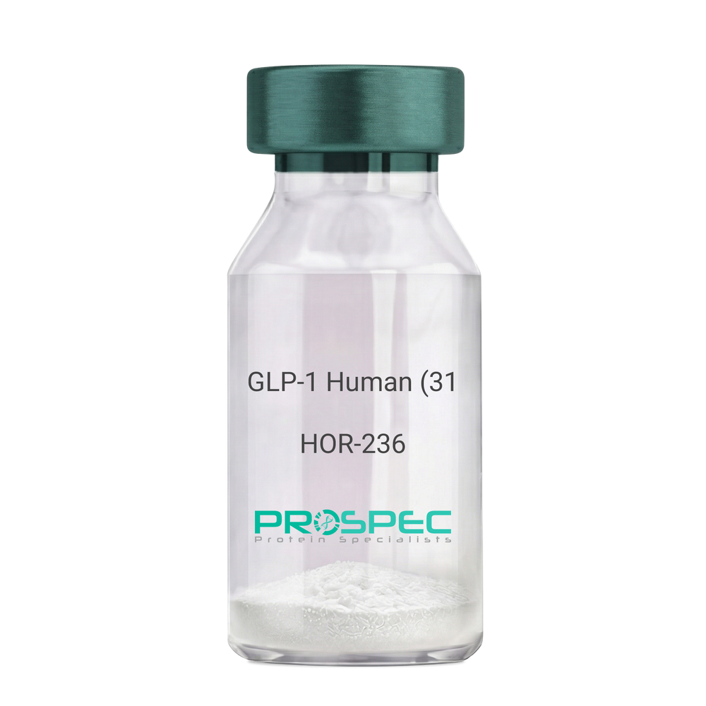
GLP-1 Human (31 a.a.)Description:
Glucagon Like Peptide-1 (31 a.a.) Human Recombinant
GLP1, GLP2, GRPP.
Product # :
HOR-236Price :
Quantity :
Shipping Method :
Shipped at Room temp
More Info
- description
- source
- formulation
- purity
- More Info
Description
Glucagon Like Peptide-1 Human Recombinant produced in E.Coli is a single, non-glycosylated, polypeptide chain containing 31 amino acids and having a molecular mass of 3.3 kDa. The GLP-1 is purified by proprietary chromatographic techniques.
Source
Escherichia Coli.
Formulation
The Recombinant GLP-1 concentrated solution (1mg/ml) was lyophilized after extensive dialyses against 0.1mg sodium phosphate monobasic & 1.6mg sodium phosphate dibasic.
Purity
Greater than 98.0% as determined by:
(a) Analysis by RP-HPLC.
(b) Analysis by SDS-PAGE.More Info
-
Introduction
Glucagon-like peptide-1 (GLP-1) is derived from the transcription product of the proglucagon gene. The major source of GLP-1 in the body is the intestinal L cell that secretes GLP-1 as a guthormone. The biologically active forms of GLP-1 are: GLP-1-(7-37) and GLP-1-(7-36)NH2.
GLP-1 secretion by L cells is dependent on the presence of nutrients in the lumen of the small intestine. The secretagogues (agents that causes or stimulates secretion) of this hormone include major nutrients like carbohydrate, proteinand lipid. Once in the circulation, GLP-1 has a half life of less than 2 minutes, due to rapid degradation by the enzyme dipeptidyl peptidase-4.
GLP-1 possesses several physiological properties that make it a subject of intensive investigation as a potential treatment of diabetes mellitus. The known physiological functions of GLP-1 include: Increases insulin secretion from the pancreas in a glucose-dependent manner, decreases glucagon secretion from the pancreas, increases beta cells mass and insulin gene expression, inhibits acid secretion and gastric emptying in the stomach, decreases food intake by increasing satiety. -
Synonyms
GLP1, GLP2, GRPP.
-
Physical Appearance
Sterile Filtered White lyophilized (freeze-dried) powder.
-
Stability
Lyophilized Glucagon Like Peptide-1 although stable at room temperature for 3 weeks, should be stored desiccated below -18°C. Upon reconstitution GLP-1 should be stored at 4°C between 2-7 days and for future use below -18°C. For long term storage it is recommended to add a carrier protein (0.1% HSA or BSA).Please prevent freeze-thaw cycles.
-
Solubility
It is recommended to reconstitute the lyophilized Glucagon Like Peptide-1 in sterile 18MΩ-cm H2O not less than 100µg/ml, which can then be further diluted to other aqueous solutions.
-
Amino Acid Sequence
HAEGTFTSDV SSYLEGQAAK EFIAWLVKGR G.
ProSpec's products are furnished for LABORATORY RESEARCH USE ONLY. The product may not be used as drugs, agricultural or pesticidal products, food additives or household chemicals.
- View Data Sheet
Name :
TYRP1 HumanDescription:
Tyrosinase Related Protein 1 Human Recombinant
Tyrosinase Related Protein 1, Tyrosinase-Related Protein 1, Melanoma Antigen Gp75, Glycoprotein 75, DHICA Oxidase, Catalase B, CAS2, TRP1, TYRP, TRP, 5,6-Dihydroxyindole-2-Carboxylic Acid Oxidase, EC 1.14.18.1, EC 1.14.18.-, EC 1.14.18, B-PROTEIN, TRP-1, TYRRP, CATB, GP75, OCA3, 5,6-dihydroxyindole-2-carboxylic acid oxidase.
Product # :
PRO-2350Price :
Quantity :
Shipping Method :
Shipped with Ice Packs
More Info
- description
- source
- formulation
- purity
- More Info
Description
TYRP1 Human Recombinant produced in Sf9 Baculovirus cells is a single, non-glycosylated polypeptide chain containing 462 amino acids (25-477a.a) and having a molecular mass of 52.5kDa. (Molecular size on SDS-PAGE will appear at approximately 50-70kDa).TYRP1 is fused to a 6 amino acid His-tag at C-terminus & purified by proprietary chromatographic techniques.
Source
Sf9, Baculovirus cells.
Formulation
TYRP1 protein solution (0.25mg/ml) containing Phosphate Buffered Saline (pH 7.4) and 10% glycerol.
Purity
Greater than 85.0% as determined by SDS-PAGE.
More Info
-
Introduction
TYRP1, also known as 5, 6-dihydroxyindole-2-carboxylic acid oxidase, is a melanosomal enzyme which is a member of the tyrosinase family and takes a significant part in the melanin biosynthetic pathway. TYRP1 is a melanocyte-specific gene which is involved in eumelanin synthesis. Furthermore, TYRP1 is implicated in the oxidation of 5,6-dihydroxyindole-2-carboxylic acid-DHICA into indole-5,6-quinone-2-carboxylic acid. TYRP1 is regulated by the microphthalmia-associatedtranscription factor-MITF.
-
Synonyms
Tyrosinase Related Protein 1, Tyrosinase-Related Protein 1, Melanoma Antigen Gp75, Glycoprotein 75, DHICA Oxidase, Catalase B, CAS2, TRP1, TYRP, TRP, 5,6-Dihydroxyindole-2-Carboxylic Acid Oxidase, EC 1.14.18.1, EC 1.14.18.-, EC 1.14.18, B-PROTEIN, TRP-1, TYRRP, CATB, GP75, OCA3, 5,6-dihydroxyindole-2-carboxylic acid oxidase.
-
Physical Appearance
Sterile Filtered colorless solution.
-
Stability
Store at 4°C if entire vial will be used within 2-4 weeks. Store, frozen at -20°C for longer periods of time. For long term storage it is recommended to add a carrier protein (0.1% HSA or BSA).Avoid multiple freeze-thaw cycles.
-
Amino Acid Sequence
ADLQFPRQCA TVEALRSGMC CPDLSPVSGP GTDRCGSSSG RGRCEAVTAD SRPHSPQYPH DGRDDREVWP LRFFNRTCHC NGNFSGHNCG TCRPGWRGAA CDQRVLIVRR NLLDLSKEEK NHFVRALDMA KRTTHPLFVI ATRRSEEILG PDGNTPQFEN ISIYNYFVWT HYYSVKKTFL GVGQESFGEV DFSHEGPAFL TWHRYHLLRL EKDMQEMLQE PSFSLPYWNF ATGKNVCDIC TDDLMGSRSN FDSTLISPNS VFSQWRVVCD SLEDYDTLGT LCNSTEDGPI RRNPAGNVAR PMVQRLPEPQ DVAQCLEVGL FDTPPFYSNS TNSFRNTVEG YSDPTGKYDP AVRSLHNLAH LFLNGTGGQT HLSPNDPIFV LLHTFTDAVF DEWLRRYNAD ISTFPLENAP IGHNRQYNMV PFWPPVTNTE MFVTAPDNLG YTYEIQWPSR EFSVPEHHHH HH.
ProSpec's products are furnished for LABORATORY RESEARCH USE ONLY. The product may not be used as drugs, agricultural or pesticidal products, food additives or household chemicals.
- View Data Sheet
Name :
GDNF Human, Sf9Description:
Glial-Derived Neurotrophic Factor Human Recombinant, Sf9
Glial cell line-derived neurotrophic factor, hGDNF, Astrocyte-derived trophic factor, ATF, ATF1, ATF2, HFB1-GDNF, HSCR3.
Product # :
CYT-1162Price :
Quantity :
Shipping Method :
Shipped with Ice Packs
More Info
- description
- source
- formulation
- purity
- More Info
Description
GDNF Human Recombinant produced in Baculovirus is a single glycosylated polypeptide chain containing 113amino acids (109-211 aa) and having a molecular mass of 12.8kDa.GDNF is fused to an 10 amino acid His tag at C-terminus and purified by proprietary chromatographic techniques.
Source
Sf9, Baculovirus cells.
Formulation
GDNF protein (0.25mg/ml) contains 10% glycerol and Phosphate-Buffered Saline (pH 7.4).
Purity
Greater than 90.0% as determined by SDS-PAGE.
More Info
-
Introduction
Glial cell-derived neurotrophic factor or GDNF is part of the GDNF group of ligands proteins. GDNF has a crucial part in numerous cell mechanisms such as neurite outgrowth, cell differentiation, cell survival and migration of cells. GDNF enhances neurons survival via GFRa receptors (mainly GFRa1). The mentioned neurons can die as a result from Parkinson's disease or ALS (amyotrophic lateral sclerosis). This protein takes part in the development of the spermatogenesis & kidney, also, it has a role in alcohol metabolism as ameliorating.
-
Synonyms
Glial cell line-derived neurotrophic factor, hGDNF, Astrocyte-derived trophic factor, ATF, ATF1, ATF2, HFB1-GDNF, HSCR3.
-
Physical Appearance
Sterile Filtered colorless solution.
-
Stability
Store at 4°C if entire vial will be used within 2-4 weeks. Store, frozen at -20°C for longer periods of time. For long term storage it is recommended to add a carrier protein (0.1% HSA or BSA).Avoid multiple freeze-thaw cycles.
-
Amino Acid Sequence
ADPMRGQRGK NRGCVLTAIH LNVTDLGLGY ETKEELIFRY CSGSCDAAET TYDKILKNLS RNRRLVSDKV GQACCRPIAF DDDLSFLDDN LVYHILRKHS AKRCGCIHHH HHH
-
Background
What is the molecular weight/Mw of GDNF HUMAN, SF9 Protein?
GDNF HUMAN, SF9 Protein has a total Mw of 12.8kDa.
What is the source or expression system of GDNF HUMAN, SF9 Protein?
Sf9, Baculovirus cells.
What is the Purity of GDNF HUMAN, SF9 Protein?
GDNF HUMAN, SF9 Protein is >90% pure as determined by SDS-PAGE.
What is the Biological Activity of GDNF HUMAN, SF9 Protein?
The biological functionality of GDNF HUMAN, SF9 Protein will be determined in the future.
What is the amino acid sequence of GDNF HUMAN, SF9 Protein?
ADPMRGQRGK NRGCVLTAIH LNVTDLGLGY ETKEELIFRY CSGSCDAAET TYDKILKNLS RNRRLVSDKV GQACCRPIAF DDDLSFLDDN LVYHILRKHS AKRCGCIHHH HHH
What applications can GDNF HUMAN, SF9 Protein be used in?
GDNF HUMAN, SF9 Protein can probably be used in western blot, ELISA and Lateral Flow.
What is the endotoxin level for GDNF HUMAN, SF9 Protein?
The endotoxin level is minimal, GDNF HUMAN, SF9 Protein was purified using conventional chromatography techniques.
ProSpec's products are furnished for LABORATORY RESEARCH USE ONLY. The product may not be used as drugs, agricultural or pesticidal products, food additives or household chemicals.
- View Data Sheet
Name :
HBV core deltaDescription:
Hepatitis B Virus Core delta Recombinant
Product # :
HBV-270Price :
Quantity :
Shipping Method :
Shipped with Ice Packs
More Info
- description
- formulation
- purity
- More Info
Description
The E.coli derived 14kDa recombinant protein contains the HBV core delta ayw immunodominant region amino acids 1-144.
Formulation
7.5mM Phosphate buffer pH-7.2, 75mM NaCl, & 50% glycerol.
Purity
HBV Core Delta protein is >95% pure as determined by SDS-PAGE (coomassie staining).
More Info
-
Introduction
Hepatitis B is one of a few known non-retroviralviruses which employ reverse transcriptionas a part of its replication process. (HIV, a completely unrelated virus, also uses reverse transcription, but it is a retrovirus.) HBV invades the cell by binding to surface receptor and become internalized. The viral core particles then migrate to the hepatocyte nucleus and the partially double-stranded, relaxed circular genomes (RC-DNA) are repaired to form a covalently closed circular DNA (cccDNA), which is the template for viral genomic and sub-genomic RNAs by cellular RNA polymerase II. Of these, the pregenomic RNA (pgRNA is selectively packaged into progeny capsids and is then reverse-transcribed into new RC-DNA. The core can either bud into the endoplasmic reticulum to be enveloped or exported from the cell or recycled back into the genome for conversion to cccDNA.
-
Stability
HBV Core Delta protein although stable at 4°C for 1 week, should be stored below -18°C. Please prevent freeze thaw cycles.
-
Amino Acid Sequence
MDIDPYKEFG ATVELLSFLP SDFFPSVRDL LDNASALYRE ALESPEHCSP
HHTALRQAIL CWGELMTLAT WVGGNLEDPI SRDLVVSYVN TNMGLKFRQL
LWFHISCLTF GRETVIEYLV SFGVWIRTPP AYRPPNAPIL S
-
Applications
HBV Core Delta antigen is suitable for ELISA and Western blots, excellent antigen for detection of HBV with minimal specificity problems.
-
Specificity
Immunoreactive with sera HBV-infected individuals.
-
Purification Method
Purified by proprietary chromatographic technique.
ProSpec's products are furnished for LABORATORY RESEARCH USE ONLY. The product may not be used as drugs, agricultural or pesticidal products, food additives or household chemicals.
- View Data Sheet
Name :
Dengue NS1, ST3Description:
Dengue Virus NS1 Recombinant, Subtype-3
Product # :
DEN-016Price :
Quantity :
Shipping Method :
Shipped with Ice Packs
More Info
- description
- source
- formulation
- purity
- More Info
Description
The E.Coli derived recombinant protein contains the NS1 Dengue Virus full length Type-3 immunodominant regions, migrates as 45kDa on 12% SDS-PAGE gel.The dengue protein is fused to a 6xHis tag at C-terminus.
Source
Escherichia Coli.
Formulation
Phosphate buffered saline and 50mM arginine.
Purity
Protein is >90% pure as determined by 12% PAGE (coomassie staining).
More Info
-
Introduction
Caused by one of four closely related virus serotypes of the genus Flavivirus, family Flaviviridae, each serotype is sufficiently different that there is no cross-protection and epidemics caused by multiple serotypes (hyperendemicity) can occur. In cell culture experiments and mice Morpholino antisense oligos have shown specific activity against Dengue virus.
-
Stability
Dengue NS1, ST3 although stable at 4°C for 1 week, should be stored below -18°C.Please prevent freeze thaw cycles.
-
Applications
Immunoassay.
-
Purification Method
Purified by proprietary chromatographic technique
ProSpec's products are furnished for LABORATORY RESEARCH USE ONLY. The product may not be used as drugs, agricultural or pesticidal products, food additives or household chemicals.