Skip to content

High-quality research proteins.

  • 1-800-805-6531

  • Mon - Sat 8.00-18.00, Sun - Closed

  • info@prospecbio.com

  • PO Box 6591 East Brunswick NJ 08816

  • Products
    • Cytokines
    • Growth factors
    • Chemokines
    • Cd antigens
    • Neurotrophins
    • Hormones
    • Enzymes
    • Viral antigens
    • Recombinant proteins
    • Natural proteins
    • Monoclonal antibodies
    • Polyclonal antibodies
    • Thermal Packaging
  • About
    • Company
    • Founder
    • Contribution
  • Customers
    • Eastern Asia
    • Eastern Europe
    • North America
    • South America
    • South East Asia
    • Southern Asia
    • Western Europe
  • Ordering
    • Ordering Method
    • Ordering - Credit Card
    • Ordering - PO
    • Shipping Fees
    • FAQs
  • Services
    • DNA Cloning
    • Protein Expression
    • Monoclonal Antibody
    • Fermentation
    • Technology License
  • Contact us
Log in

Country/region

  • USD $ | Albania
  • USD $ | Andorra
  • USD $ | Argentina
  • USD $ | Armenia
  • USD $ | Australia
  • USD $ | Austria
  • USD $ | Azerbaijan
  • USD $ | Belarus
  • USD $ | Belgium
  • USD $ | Bolivia
  • USD $ | Brazil
  • USD $ | Bulgaria
  • USD $ | Cambodia
  • USD $ | Canada
  • USD $ | Chile
  • USD $ | China
  • USD $ | Colombia
  • USD $ | Costa Rica
  • USD $ | Croatia
  • USD $ | Cyprus
  • USD $ | Czechia
  • USD $ | Denmark
  • USD $ | Estonia
  • USD $ | Finland
  • USD $ | France
  • USD $ | Georgia
  • USD $ | Germany
  • USD $ | Greece
  • USD $ | Hong Kong SAR
  • USD $ | Hungary
  • USD $ | Iceland
  • USD $ | India
  • USD $ | Ireland
  • USD $ | Israel
  • USD $ | Italy
  • USD $ | Japan
  • USD $ | Kazakhstan
  • USD $ | Latvia
  • USD $ | Liechtenstein
  • USD $ | Lithuania
  • USD $ | Madagascar
  • USD $ | Malta
  • USD $ | Mexico
  • USD $ | Moldova
  • USD $ | Monaco
  • USD $ | Netherlands
  • USD $ | New Zealand
  • USD $ | North Macedonia
  • USD $ | Norway
  • USD $ | Panama
  • USD $ | Paraguay
  • USD $ | Peru
  • USD $ | Philippines
  • USD $ | Poland
  • USD $ | Portugal
  • USD $ | Romania
  • USD $ | Singapore
  • USD $ | Slovakia
  • USD $ | Slovenia
  • USD $ | South Africa
  • USD $ | South Korea
  • USD $ | Spain
  • USD $ | Sri Lanka
  • USD $ | Sweden
  • USD $ | Switzerland
  • USD $ | Taiwan
  • USD $ | Thailand
  • USD $ | Türkiye
  • USD $ | Ukraine
  • USD $ | United Kingdom
  • USD $ | United States
  • USD $ | Uruguay
  • USD $ | Vatican City
  • USD $ | Venezuela
  • USD $ | Vietnam
  • Facebook
  • Instagram
logoProSpec - Recombinant Protein Manufacturer for academic and R&D industry
Become a distributor
Account Log in
Wishlist
Cart Cart(0)
  • Products
    +
    • Cytokines
    • Growth factors
    • Chemokines
    • Cd antigens
    • Neurotrophins
    • Hormones
    • Enzymes
    • Viral antigens
    • Recombinant proteins
    • Natural proteins
    • Monoclonal antibodies
    • Polyclonal antibodies
    • Thermal Packaging
  • About
    +
    • Company
    • Founder
    • Contribution
  • Customers
    +
    • Eastern Asia
    • Eastern Europe
    • North America
    • South America
    • South East Asia
    • Southern Asia
    • Western Europe
  • Ordering
    +
    • Ordering Method
    • Ordering - Credit Card
    • Ordering - PO
    • Shipping Fees
    • FAQs
  • Services
    +
    • DNA Cloning
    • Protein Expression
    • Monoclonal Antibody
    • Fermentation
    • Technology License
  • Contact us

Item added to your cart

View cart
View All
  • Cytokines
  • Growth factors
  • Chemokines
  • Cd antigens
  • Neurotrophins
  • Hormones
  • Enzymes
  • Viral antigens
  • Recombinant proteins
  • Natural proteins
  • Monoclonal antibodies
  • Polyclonal antibodies
  • Thermal Packaging
  • Tumor Necrosis Factor

    Tumor Necrosis Factor

    View All
  • 4-1BB

    4-1BB

    View All
  • Adiponectin

    Adiponectin

    View All
  • AIF1

    AIF1

    View All
  • Other Cytokines

    Other Cytokines

    View All
  • AITRL

    AITRL

    View All
  • Angiopoietin

    Angiopoietin

    View All
  • Apolipoprotein

    Apolipoprotein

    View All
  • B-Cell Activating Factor

    B-Cell Activating Factor

    View All
  • Beta Defensin

    Beta Defensin

    View All
  • Bone Morphogenetic Protein

    Bone Morphogenetic Protein

    View All
  • B type Natriuretic Peptide

    B type Natriuretic Peptide

    View All
  • BST

    BST

    View All
  • Betacellulin

    Betacellulin

    View All
  • Cardiotrophin

    Cardiotrophin

    View All
  • Activin

    Activin

    View All
  • Fibroblast Growth Factor

    Fibroblast Growth Factor

    View All
  • Other Growth Factors

    Other Growth Factors

    View All
  • CSF

    CSF

    View All
  • CTGF

    CTGF

    View All
  • IGFBP

    IGFBP

    View All
  • VEGF Protein

    VEGF Protein

    View All
  • EGF

    EGF

    View All
  • Epigen

    Epigen

    View All
  • Erythropoietin

    Erythropoietin

    View All
  • Insulin-Like Growth Factor

    Insulin-Like Growth Factor

    View All
  • Growth Hormone

    Growth Hormone

    View All
  • HDGF

    HDGF

    View All
  • Hepatocyte Growth Factor

    Hepatocyte Growth Factor

    View All
  • Insulin

    Insulin

    View All
  • BCA-1/ BLC (CXCL13)

    BCA-1/ BLC (CXCL13)

    View All
  • BRAK (CXCL14)

    BRAK (CXCL14)

    View All
  • C-10 (CCL6)

    C-10 (CCL6)

    View All
  • Other Chemokines

    Other Chemokines

    View All
  • MEC (CCL28)

    MEC (CCL28)

    View All
  • LD78-beta (CCL3L1)

    LD78-beta (CCL3L1)

    View All
  • CTACK (CCL27)

    CTACK (CCL27)

    View All
  • CXCL16

    CXCL16

    View All
  • CXCL17

    CXCL17

    View All
  • Platelet Factor-4 (CXCL4)

    Platelet Factor-4 (CXCL4)

    View All
  • ENA-78 (CXCL5)

    ENA-78 (CXCL5)

    View All
  • Eotaxin (CCL11,24,26)

    Eotaxin (CCL11,24,26)

    View All
  • Exodus-2 (CCL21)

    Exodus-2 (CCL21)

    View All
  • Fractalkine (CX3CL1)

    Fractalkine (CX3CL1)

    View All
  • CXCL6

    CXCL6

    View All
  • Other CD Antigens

    Other CD Antigens

    View All
  • CD14

    CD14

    View All
  • CD164

    CD164

    View All
  • CD1

    CD1

    View All
  • CD2

    CD2

    View All
  • CD200

    CD200

    View All
  • CD207

    CD207

    View All
  • CD226

    CD226

    View All
  • CD23

    CD23

    View All
  • CD244

    CD244

    View All
  • CD247

    CD247

    View All
  • CD27

    CD27

    View All
  • CD274

    CD274

    View All
  • CD300

    CD300

    View All
  • CD33

    CD33

    View All
  • Other Neurotrophins

    Other Neurotrophins

    View All
  • Beta-NGF

    Beta-NGF

    View All
  • BDNF

    BDNF

    View All
  • CDNF

    CDNF

    View All
  • CNTF

    CNTF

    View All
  • GDNF

    GDNF

    View All
  • Glia Maturation Factor

    Glia Maturation Factor

    View All
  • MANF

    MANF

    View All
  • Midkine

    Midkine

    View All
  • Neuroglobin

    Neuroglobin

    View All
  • Neurotrophic factors

    Neurotrophic factors

    View All
  • Neuregulin

    Neuregulin

    View All
  • Neuropilin

    Neuropilin

    View All
  • Pigment Epithelium-Derived Factor

    Pigment Epithelium-Derived Factor

    View All
  • Pleiotrophin

    Pleiotrophin

    View All
  • Peptide Hormones

    Peptide Hormones

    View All
  • Other Hormones

    Other Hormones

    View All
  • HCG

    HCG

    View All
  • Endothelin

    Endothelin

    View All
  • Exendin

    Exendin

    View All
  • FSH

    FSH

    View All
  • Glucagon

    Glucagon

    View All
  • GHRP

    GHRP

    View All
  • GLP

    GLP

    View All
  • Thyrostimulin

    Thyrostimulin

    View All
  • Inhibin A

    Inhibin A

    View All
  • LHRH

    LHRH

    View All
  • Melanotan

    Melanotan

    View All
  • Procalcitonin

    Procalcitonin

    View All
  • PTH

    PTH

    View All
  • Peroxiredoxin

    Peroxiredoxin

    View All
  • Peptidase

    Peptidase

    View All
  • Synthetase

    Synthetase

    View All
  • Transferase

    Transferase

    View All
  • Hydrolase

    Hydrolase

    View All
  • Dehydrogenase

    Dehydrogenase

    View All
  • ACE2

    ACE2

    View All
  • Esterase

    Esterase

    View All
  • Protein Kinases

    Protein Kinases

    View All
  • Other Enzymes

    Other Enzymes

    View All
  • Phosphatase

    Phosphatase

    View All
  • Deaminase

    Deaminase

    View All
  • Oxygenase

    Oxygenase

    View All
  • Adenylate Kinase

    Adenylate Kinase

    View All
  • Lyase

    Lyase

    View All
  • Chagas

    Chagas

    View All
  • SARS

    SARS

    View All
  • Influenza

    Influenza

    View All
  • ASFV

    ASFV

    View All
  • Astrovirus

    Astrovirus

    View All
  • Borrelia

    Borrelia

    View All
  • Helicobacter Pylori

    Helicobacter Pylori

    View All
  • Chikungunya

    Chikungunya

    View All
  • Chlamydia

    Chlamydia

    View All
  • Cytomegalo

    Cytomegalo

    View All
  • Dengue

    Dengue

    View All
  • EBV

    EBV

    View All
  • Ebola

    Ebola

    View All
  • Feline Leukemia Virus

    Feline Leukemia Virus

    View All
  • Hepatitis A

    Hepatitis A

    View All
  • Other

    Other

    View All
  • Actin

    Actin

    View All
  • ADAM

    ADAM

    View All
  • Complement Component

    Complement Component

    View All
  • Ag85

    Ag85

    View All
  • Eukaryotic Translation Initiation Factor

    Eukaryotic Translation Initiation Factor

    View All
  • Anterior Gradient Protein

    Anterior Gradient Protein

    View All
  • Heat Shock Protein

    Heat Shock Protein

    View All
  • Alpha-2-HS-Glycoprotein

    Alpha-2-HS-Glycoprotein

    View All
  • Hemoglobin

    Hemoglobin

    View All
  • Allergy

    Allergy

    View All
  • Anaplasma

    Anaplasma

    View All
  • Angiogenin

    Angiogenin

    View All
  • Ankyrin Repeat Domain

    Ankyrin Repeat Domain

    View All
  • Annexin

    Annexin

    View All
  • Other Natural Proteins

    Other Natural Proteins

    View All
  • Aprotinin

    Aprotinin

    View All
  • Transferrin

    Transferrin

    View All
  • Avidin

    Avidin

    View All
  • Anti Coagulation Factors

    Anti Coagulation Factors

    View All
  • Natural Albumin

    Natural Albumin

    View All
  • Natural Coagulation Factors

    Natural Coagulation Factors

    View All
  • Fibronectin

    Fibronectin

    View All
  • Thrombin

    Thrombin

    View All
  • Anti Human Enzyme

    Anti Human Enzyme

    View All
  • Other Monoclonal Antibodies

    Other Monoclonal Antibodies

    View All
  • Anti Human Cytokine

    Anti Human Cytokine

    View All
  • Anti Human Heat Shock Protein

    Anti Human Heat Shock Protein

    View All
  • Anti Mouse Lymphocyte

    Anti Mouse Lymphocyte

    View All
  • Anti Human Chemokine

    Anti Human Chemokine

    View All
  • Anti Human Lymphocyte

    Anti Human Lymphocyte

    View All
  • Anti Viral Monoclonal

    Anti Viral Monoclonal

    View All
  • Anti Mouse Cytokine

    Anti Mouse Cytokine

    View All
  • Anti-GST Monoclonal

    Anti-GST Monoclonal

    View All
  • Other Polyclonal Antibodies

    Other Polyclonal Antibodies

    View All
  • Anti Viral

    Anti Viral

    View All
  • Anti-GST Monoclonal

    Anti-GST Monoclonal

    View All
Back

Search results

1000 results found for “Intercellular Adhesion Molecule”

Name

Description

Product #

Price

Quantity

Shipping Method

  • View Data Sheet

    Name :

    PDIA4 Human, Active

    Description:

    Protein Disulfide Isomerase A4 Human Recombinant, Active

    Endoplasmic reticulum resident protein 72, ERP70, ERP72.

    Product # :

    ENZ-993

    Price :

    Quantity :

    Shipping Method :

    Ice Icon

    Shipped with Ice Packs

    Add To Cart

    More Info

    • description
    • source
    • formulation
    • purity
    • biological activity
    • More Info

    Description

    PDIA4 Human Recombinant produced in E.Coli is a single, non-glycosylated polypeptide chain containing 646 amino acids (21-645 a.a.) and having a molecular weight of 72.9kDa. The PDIA4 is fused to 21 a.a. His-Tag at N-terminus and purified by proprietary chromatographic techniques.

    Source

    Escherichia Coli.

    Formulation

    The PDIA4 1mg/ml protein solution contains 20mM Tris-HCl, pH-8, 1mM DTT, 0.1M NaCl and 10% glycerol.

    Purity

    Greater than 90.0% as determined by SDS-PAGE.

    Biological Activity

    Specific activity > 10 A650/cm/min/mg. Enzymatic activity was confirmed by measuring the aggregation of insulin in the presence of DTT.

    More Info

    • Introduction

      PDIA4 is an endoplasmic reticulum luminal protein that is a stress protein as well as a member of the protein disulfide isomerase family of proteins. PDIA4 participates in the catalysis of protein-S-S-bond rearrangement. PDIA4 and PDIA3 function as proteases, protein disulfide isomerases, phospholipases or an arrangement of these.

    • Synonyms

      Endoplasmic reticulum resident protein 72, ERP70, ERP72.

    • Physical Appearance

      Sterile filtered colorless solution.

    • Stability

      Store at 4°C if entire vial will be used within 2-4 weeks. Store, frozen at -20°C for longer periods of time. For long term storage it is recommended to add a carrier protein (0.1% HSA or BSA). Avoid multiple freeze-thaw cycles.

    • Amino Acid Sequence

      MGSSHHHHHH SSGLVPRGSH MVAGAEGPDE DSSNRENAIE DEEEEEEEDD DEEEDDLEVK EENGVLVLND ANFDNFVADK DTVLLEFYAP WCGHCKQFAP EYEKIANILK DKDPPIPVAK IDATSASVLA SRFDVSGYPT IKILKKGQAV DYEGSRTQEE IVAKVREVSQ PDWTPPPEVT LVLTKENFDE VVNDADIILV EFYAPWCGHC KKLAPEYEKA AKELSKRSPP IPLAKVDATA ETDLAKRFDV SGYPTLKIFR KGRPYDYNGP REKYGIVDYM IEQSGPPSKE ILTLKQVQEF LKDGDDVIII GVFKGESDPA YQQYQDAANN LREDYKFHHT FSTEIAKFLK VSQGQLVVMQ PEKFQSKYEP RSHMMDVQGS TQDSAIKDFV LKYALPLVGH RKVSNDAKRY TRRPLVVVYY SVDFSFDYRA ATQFWRSKVL EVAKDFPEYT FAIADEEDYA GEVKDLGLSE SGEDVNAAIL DESGKKFAME PEEFDSDTLR EFVTAFKKGK LKPVIKSQPV PKNNKGPVKV VVGKTFDSIV MDPKKDVLIE FYAPWCGHCK QLEPVYNSLA KKYKGQKGLV IAKMDATAND VPSDRYKVEG FPTIYFAPSG DKKNPVKFEG GDRDLEHLSK FIEEHATKLS RTKEEL.

    ProSpec's products are furnished for LABORATORY RESEARCH USE ONLY. The product may not be used as drugs, agricultural or pesticidal products, food additives or household chemicals.

    Pdia4 Human Active
  • View Data Sheet

    Name :

    IFNW1 Human

    Description:

    IFN-Omega 1 Human Recombinant

    IFN omega-1, IFN alpha-II-1, IFNW1.

    Product # :

    CYT-040

    Price :

    Quantity :

    Shipping Method :

    Room Temp Icon

    Shipped at Room temp

    Add To Cart

    More Info

    • description
    • source
    • formulation
    • purity
    • biological activity
    • More Info

    Description

    IFN-Omega 1 Human Recombinant produced in E.Coli is a single, non-glycosylated, polypeptide chain containing 172 amino acids and having a molecular mass of 19.9kDa.The IFN-Omega 1 is purified by proprietary chromatographic techniques.

    Source

    Escherichia Coli.

    Formulation

    Lyophilized from a 0.2µm filtered concentrated solution in PBS, pH 7.4.

    Purity

    Greater than 95.0% as determined by:
    (a) Analysis by RP-HPLC.
    (b) Analysis by SDS-PAGE.

    Biological Activity

    The ED50 was determined by a cytotoxicity assay using Human TF-1 cells is < 0.01 ng/ml, corresponding to a specific activity of > 100,000,000 units/mg.

    More Info

    • Introduction

      IFN-Omega 1 is a type I IFN, that can be induced by virus-infected leukocytes. Type I IFN family members, which include IFN-alpha, IFN-beta, and IFN-omega, signal through IFNAR-1/IFNAR-2 receptor complex, and wield antiviral and antiproliferative activities. IFNW1 exhibits about 75% sequence homology with IFN-a, and contains 2 conserved disulfide bonds, which are essential for full biological activity.

    • Synonyms

      IFN omega-1, IFN alpha-II-1, IFNW1.

    • Physical Appearance

      Sterile Filtered White lyophilized (freeze-dried) powder.

    • Stability

      Lyophilized IFNW1 although stable at room temperature for 3 weeks, should be stored desiccated below -18°C. Upon reconstitution IFNW1 should be stored at 4°C between 2-7 days and for future use below -18°C.For long term storage it is recommended to add a carrier protein (0.1% HSA or BSA).Please prevent freeze-thaw cycles.

    • Solubility

      It is recommended to reconstitute the lyophilized IFNW1 in sterile 18M-cm H2O not less than 100µg/ml, which can then be further diluted to other aqueous solutions.

    • Amino Acid Sequence

      MCDLPQNHGL LSRNTLVLLH QMRRISPFLC LKDRRDFRFP QEMVKGSQLQ KAHVMSVLHE MLQQIFSLFH TERSSAAWNM TLLDQLHTGL HQQLQHLETC LLQVVGEGES AGAISSPALT LRRYFQGIRV YLKEKKYSDC AWEVVRMEIM KSLFLSTNMQ ERLRSKDRDL GSS.

    • Background

      What is the molecular weight/Mw of IFNW1 HUMAN Protein?
      IFNW1 HUMAN Protein has a total Mw of 19.9kDa.

      What is the source or expression system of IFNW1 HUMAN Protein?
      Escherichia Coli.

      What is the Purity of IFNW1 HUMAN Protein?
      IFNW1 HUMAN Protein is >95% pure as determined by SDS-PAGE.

      What is the Biological Activity of IFNW1 HUMAN Protein?
      The ED50 was determined by a cytotoxicity assay using Human TF-1 cells is < 0.01 ng/ml, corresponding to a specific activity of > 100,000,000 units/mg.

      What is the amino acid sequence of IFNW1 HUMAN Protein?
      MCDLPQNHGL LSRNTLVLLH QMRRISPFLC LKDRRDFRFP QEMVKGSQLQ KAHVMSVLHE MLQQIFSLFH TERSSAAWNM TLLDQLHTGL HQQLQHLETC LLQVVGEGES AGAISSPALT LRRYFQGIRV YLKEKKYSDC AWEVVRMEIM KSLFLSTNMQ ERLRSKDRDL GSS.

      What applications can IFNW1 HUMAN Protein be used in?
      IFNW1 HUMAN Protein can probably be used in western blot, ELISA and Lateral Flow.

      What is the endotoxin level for IFNW1 HUMAN Protein?
      The endotoxin level is minimal, IFNW1 HUMAN Protein was purified using conventional chromatography techniques.

    ProSpec's products are furnished for LABORATORY RESEARCH USE ONLY. The product may not be used as drugs, agricultural or pesticidal products, food additives or household chemicals.

    Ifnw1 Human
  • View Data Sheet

    Name :

    LMNA Rat

    Description:

    Lamin A/C Rat Recombinant

    Prelamin-A/C, Lamin-A/C, 70 kDa lamin, LMNA, LMN1, Renal carcinoma antigen NY-REN-32, Progerin.

    Product # :

    PRO-2667

    Price :

    Quantity :

    Shipping Method :

    Ice Icon

    Shipped with Ice Packs

    Add To Cart

    More Info

    • description
    • source
    • formulation
    • purity
    • More Info

    Description

    LMNA Rat Recombinant fused with a His tag produced in E.Coli is a single, non-glycosylated, polypeptide chain containing 614 amino acids and having a molecular mass of 68.0kDa. The LMNA is purified by proprietary chromatographic techniques.

    Source

    Escherichia Coli.

    Formulation

    The LMNA solution contains 20mM Tris-HCl pH 7.5, 1mM DTT, 0.3M NaCl, 1.5mM EDTA and 10%(v/v) glycerol.

    Purity

    Greater than 90.0% as determined by SDS-PAGE.

    More Info

    • Introduction

      Lamin-A is a major component of the nuclear lamina, a dynamic meshwork located just under the nuclear envelope and it is encoded by lamin A/C gene (LMNA).
      Lamin-A is synthesized as Prelamin A, a longer precursor that in vivo goes through a serial post-translational modifications that lead to mature Lamin A.
      Diverse mutations in the Lamin A/C gene are associated with different diseases that are collectively called laminophaties, including Emery-Dreifuss muscular dystrophy, familiar partial lipodystrophy, limb girdle muscular dystrophy, dilated cardiomyopathy, Charcot-Marie-Tooth disease, and Hutchinson-Gilford progeria syndrome.

    • Synonyms

      Prelamin-A/C, Lamin-A/C, 70 kDa lamin, LMNA, LMN1, Renal carcinoma antigen NY-REN-32, Progerin.

    • Physical Appearance

      Sterile filtered colorless solution.

    • Stability

      Store at 4°C if entire vial will be used within 2-4 weeks.Store, frozen at -20°C for longer periods of time.For long term storage it is recommended to add a carrier protein (0.1% HSA or BSA).Avoid multiple freeze-thaw cycles.

    • Amino Acid Sequence

      METPSQRRAT RSGAQASSTP LSPTRITRLQ EKEDLQELND RLAVYIDRVR SLETENAGLR LRITESEEVV SREVSGIKAA YEAELGDARK TLDSVAKERA RLQLELSKVR EEFKELKARN TKKEGDLLAA QARLKDLEAL LNSKEAALST ALSEKRTLEG ELHDLRGQVA KLEAALGEAK KQLQDEMLRR VDAENRLQTL KEELDFQKNI YSEELRETKR RHETRLVEID NGKQREFESR LADALQELRA QHEDQVEQYK KELEKTYSAK LDNARQSAER NSNLVGAAHE ELQQSRIRID SLSAQLSQLQ KQLAAKEAKL RDLEDSLARE RDTSRRLLAE KEREMAEMRA RMQQQLDEYQ ELLDIKLALD MEIHAYRKLL EGEEERLRLS PSPTSQRSRG RASSHSSQSQ GGGSVTKKRK LESSESRSSF SQHARTSGRV AVEEVDEEGK FVRLRNKSNE DQSMGNWQIR RQNGDDPLMT YRFPPKFTLK AGQVVTIWAS GAGATHSPPT DLVWKAQNTW GCGSSLRTAL INATGEEVAM RKLVRSLTMV EDDEDEDGDD LLHHHHGSHC SSSGDPAEYN LRSRTVLCGT CGQPADKASA SGSGAQVSSQ NCSIM

    ProSpec's products are furnished for LABORATORY RESEARCH USE ONLY. The product may not be used as drugs, agricultural or pesticidal products, food additives or household chemicals.

    Lmna Rat
  • View Data Sheet

    Name :

    NPM1 Antibody

    Description:

    Nucleophosmin, Mouse Anti Human

    Nucleophosmin, NPM, Nucleolar phosphoprotein B23, Nucleolar protein NO38, Numatrin, NPM1, NPM, B23.

    Product # :

    ANT-571

    Price :

    Quantity :

    Shipping Method :

    Ice Icon

    Shipped with Ice Packs

    Add To Cart

    More Info

    • formulation
    • More Info

    Formulation

    1mg/ml containing PBS, pH-7.4, 10% Glycerol and 0.02% Sodium Azide.

    More Info

    • Introduction

      Nucleophosmin (NPM1) is a phosphoprotein which shuffles between the nucleus and the cytoplasm. NPM1 is assumed to be involved in a number of processes including regulation of the ARF/p53 pathway. Several genes fusion partners have been described, particularly the anaplastic lymphoma kinase gene on chromosome 2. NPM1 gene mutations are linked with acute myeloid leukemia.

    • Synonyms

      Nucleophosmin, NPM, Nucleolar phosphoprotein B23, Nucleolar protein NO38, Numatrin, NPM1, NPM, B23.

    • Physical Appearance

      Sterile filtered colorless solution.

    • Immunogen

      Anti-human NPM1 mAb, is derived from hybridization of mouse F0 myeloma cells with spleen cells from BALB/c mice immunized with a recombinant human NPM1 protein 1-294 amino acids purified from E. coli.

    • Ig Subclass

      Mouse IgG1 heavy chain and k light chain.

    • Clone

      PAT23F1AT.

    • Applications

      NPM1 antibody has been tested by ELISA, Western blot analysis, ICC/IF and Flow cytometry to assure specificity and reactivity. Since application varies, however, each investigation should be titrated by the reagent to obtain optimal results. Recommended dilution range for Western blot analysis is 1:1000. Recommended dilution range for ICC/IF and Flow cytometry is 1:200.

    • Type

      Mouse Anti Human Monoclonal.

    • Storage Procedures

      For periods up to 1 month store at 4°C, for longer periods of time, store at -20°C. Prevent freeze thaw cycles.

    • Purification Method

      NPM1 antibody was purified from mouse ascitic fluids by protein-A affinity chromatography.

    ProSpec's products are furnished for LABORATORY RESEARCH USE ONLY. The product may not be used as drugs, agricultural or pesticidal products, food additives or household chemicals.

    Npm1 Antibody
  • View Data Sheet

    Name :

    TLR2 Antibody

    Description:

    Toll-like receptor 2, Mouse Anti Human

    Toll-like receptor 2, Toll/interleukin-1 receptor-like protein 4, CD282, TLR2, TIL4.

    Product # :

    ANT-408

    Price :

    Quantity :

    Shipping Method :

    Ice Icon

    Shipped with Ice Packs

    Add To Cart

    More Info

    • formulation
    • More Info

    Formulation

    1mg/ml containing PBS, pH-7.4, & 0.1% Sodium Azide.

    More Info

    • Introduction

      TLR2 belongs to the Toll-like receptor (TLR) family that plays a fundamental role in pathogen recognition and activation of innate immunity. TLRs are highly conserved from Drosophila to humans and share structural and functional similarities. They recognize pathogen-associated molecular patterns (PAMPs) which are expressed on infectious agents, and mediate the production of cytokines necessary for the development of effective immunity. The various TLRs exhibit different patterns of expression. TLR2 is expressed most abundantly in peripheral blood leukocytes, and mediates host response to Gram-positive bacteria and yeast via stimulation of NF-kappaB.

    • Synonyms

      Toll-like receptor 2, Toll/interleukin-1 receptor-like protein 4, CD282, TLR2, TIL4.

    • Physical Appearance

      Sterile filtered colorless solution.

    • Immunogen

      Anti-human TLR2 mAb, is derived from hybridization of mouse SP2/O myeloma cells with spleen cells from BALB/c mice immunized with recombinant human TLR2 amino acids 221-384 purified from E. coli.

    • Ig Subclass

      Mouse IgG1 heavy chain and κ light chain.

    • Clone

      P1F10AT.

    • Applications

      TLR2 antibody has been tested by ELISA and Western blot analysis to assure specificity and reactivity. Since application varies, however, each investigation should be titrated by the reagent to obtain optimal results. Recommended dilution range for Western blot analysis is 1:500 ~ 1,000. Recommended starting dilution is 1:500.

    • Type

      Mouse Anti Human Monoclonal.

    • Storage Procedures

      For periods up to 1 month store at 4°C, for longer periods of time, store at -20°C. Prevent freeze thaw cycles.

    • Purification Method

      TLR2 antibody was purified from mouse ascitic fluids by protein-G affinity chromatography.

    ProSpec's products are furnished for LABORATORY RESEARCH USE ONLY. The product may not be used as drugs, agricultural or pesticidal products, food additives or household chemicals.

    Tlr2 Antibody
  • View Data Sheet

    Name :

    TOMM20 Antibody

    Description:

    Mouse Anti Human Translocase Of Outer Mitochondrial Membrane 20

    Mmitochondrial import receptor subunit TOM20 homolog, TOMM20, MAS20, MOM19, KIAA0016, Outer mitochondrial membrane receptor Tom20, Mitochondrial 20 kDa outer membrane protein.

    Product # :

    ANT-712

    Price :

    Quantity :

    Shipping Method :

    Ice Icon

    Shipped with Ice Packs

    Add To Cart

    More Info

    • formulation
    • More Info

    Formulation

    1mg/ml containing PBS, pH-7.4, 10% Glycerol and 0.02% Sodium Azide.

    More Info

    • Introduction

      Mmitochondrial import receptor subunit TOMM20 homolog (TOMM20) is a member of the Tom20 family. The Tom machinery consists of import receptors for the initial binding of cytosolically synthesized preproteins and a general import pore (GIP) for the membrane translocation of various preproteins into the mitochondria. TOMM20 acts as the transit peptide receptor at the surface of the mitochondrion outer membrane and facilitates the movement of preproteins into the TOM40 translocation pore.

    • Synonyms

      Mmitochondrial import receptor subunit TOM20 homolog, TOMM20, MAS20, MOM19, KIAA0016, Outer mitochondrial membrane receptor Tom20, Mitochondrial 20 kDa outer membrane protein.

    • Physical Appearance

      Sterile filtered colorless solution.

    • Immunogen

      Anti-human TOMM20 mAb, is derived from hybridization of mouse F0 myeloma cells with spleen cells from BALB/c mice immunized with a recombinant human TOMM20 protein 25-145 amino acids purified from E. coli.

    • Ig Subclass

      Mouse IgG1 heavy chain and k light chain.

    • Clone

      PAT1B2AT.

    • Applications

      TOMM20 antibody has been tested by ELISA and Western blot analysis to assure specificity and reactivity. Since application varies, however, each investigation should be titrated by the reagent to obtain optimal results.

    • Type

      Mouse Anti Human Monoclonal.

    • Storage Procedures

      For periods up to 1 month store at 4°C, for longer periods of time, store at -20°C. Prevent freeze thaw cycles.

    • Purification Method

      TOMM20 antibody was purified from mouse ascitic fluids by protein-A affinity chromatography.

    ProSpec's products are furnished for LABORATORY RESEARCH USE ONLY. The product may not be used as drugs, agricultural or pesticidal products, food additives or household chemicals.

    Tomm20 Antibody
  • View Data Sheet

    Name :

    CHI3L2 Antibody

    Description:

    Chitinase 3-Like 2, Mouse Anti Human

    Chitinase 3-Like 2, Chondrocyte Protein 39, YKL-39, YKL39, Chitinase-3-Like Protein 2, CHIL2, CHI3L2.

    Product # :

    ANT-703

    Price :

    Quantity :

    Shipping Method :

    Ice Icon

    Shipped with Ice Packs

    Add To Cart

    More Info

    • formulation
    • More Info

    Formulation

    1mg/ml containing PBS, pH-7.4, 10% Glycerol and 0.02% Sodium Azide.

    More Info

    • Introduction

      Chitinase 3-Like 2 (CHI3L2) is similar to bacterial chitinases but lacks chitinase activity. CHI3L2 protein is secreted and is involved in cartilage biogenesis. CHI3L2 is a lectin, which binds chitooligosaccharides and other glycans with high affinity, but not heparin.

    • Synonyms

      Chitinase 3-Like 2, Chondrocyte Protein 39, YKL-39, YKL39, Chitinase-3-Like Protein 2, CHIL2, CHI3L2.

    • Physical Appearance

      Sterile filtered colorless solution.

    • Immunogen

      Anti-human CHI3L2 mAb, is derived from hybridization of mouse F0 myeloma cells with spleen cells from BALB/c mice immunized with recombinant human CHI3L2 amino acids 27-390 purified from E. coli.

    • Ig Subclass

      Mouse IgG2b heavy chain and κ light chain.

    • Clone

      PAT2B5AT.

    • Applications

      CHI3L2 antibody has been tested by ELISA and Western blot analysis to assure specificity and reactivity. Since application varies, however, each investigation should be titrated by the reagent to obtain optimal results.

    • Type

      Mouse Anti Human Monoclonal.

    • Storage Procedures

      For periods up to 1 month store at 4°C, for longer periods of time, store at -20°C. Prevent freeze thaw cycles.

    • Purification Method

      CHI3L2 antibody was purified from mouse ascitic fluids by protein-A affinity chromatography.

    ProSpec's products are furnished for LABORATORY RESEARCH USE ONLY. The product may not be used as drugs, agricultural or pesticidal products, food additives or household chemicals.

    Chi3L2 Antibody
  • View Data Sheet

    Name :

    COX5A Antibody

    Description:

    Cytochrome C Oxidase Subunit Va, Mouse Anti Human

    Cytochrome c oxidase subunit 5A, mitochondrial, Cytochrome c oxidase polypeptide Va, COX5A, VA, COX, COX-VA.

    Product # :

    ANT-605

    Price :

    Quantity :

    Shipping Method :

    Ice Icon

    Shipped with Ice Packs

    Add To Cart

    More Info

    • formulation
    • More Info

    Formulation

    1mg/ml containing PBS, pH-7.4, 10% Glycerol and 0.02% Sodium Azide.

    More Info

    • Introduction

      Cytochrome C Oxidase Subunit Va (COX5A) is a member of the cytochrome c oxidase subunit 5A family. COX (Cytochrome C Oxidase), which is the terminal component of the mitochondrial respiratory chain, catalyzes the electron transfer from reduced Cytochrome C to oxygen. The COX component is a heteromeric complex comprised of three catalytic subunits encoded by mitochondrial genes and multiple structural subunits encoded by nuclear genes. The mitochondrially-encoded subunits serve in electron transfer, and the nuclear-encoded subunits act in the regulation and assembly of the complex. COX5A is the heme A-containing chain of Cytochrome C Oxidase,which is the terminal oxidase in mitochondrial electron transport.

    • Synonyms

      Cytochrome c oxidase subunit 5A, mitochondrial, Cytochrome c oxidase polypeptide Va, COX5A, VA, COX, COX-VA.

    • Physical Appearance

      Sterile filtered colorless solution.

    • Immunogen

      Anti-human COX5A mAb, is derived from hybridization of mouse F0 myeloma cells with spleen cells from BALB/c mice immunized with a recombinant human COX5A protein 42-150 amino acids purified from E. coli.

    • Ig Subclass

      Mouse IgG1 heavy chain and k light chain.

    • Clone

      PAT7A6AT.

    • Applications

      COX5A antibody has been tested by ELISA, Western blot analysis, ICC/IF and Flow cytometry to assure specificity and reactivity. Since application varies, however, each investigation should be titrated by the reagent to obtain optimal results.

    • Type

      Mouse Anti Human Monoclonal.

    • Storage Procedures

      For periods up to 1 month store at 4°C, for longer periods of time, store at -20°C. Prevent freeze thaw cycles.

    • Purification Method

      COX5A antibody was purified from mouse ascitic fluids by protein-A affinity chromatography.

    ProSpec's products are furnished for LABORATORY RESEARCH USE ONLY. The product may not be used as drugs, agricultural or pesticidal products, food additives or household chemicals.

    Cox5A Antibody
  • View Data Sheet

    Name :

    IDE Human, Active

    Description:

    Insulin-Degrading Enzyme Human Recombinant

    Insulin-Degrading Enzyme, Abeta-Degrading Protease, Insulysin, EC 3.4.24.56, Insulinase, Insulin Protease, INSULYSIN, EC 3.4.24, IDE, insulin-degrading enzyme isoform 1. 

    Product # :

    ENZ-1192

    Price :

    Quantity :

    Shipping Method :

    Ice Icon

    Shipped with Ice Packs

    Add To Cart

    More Info

    • description
    • source
    • formulation
    • purity
    • biological activity
    • More Info

    Description

    IDE Human, Active Human Recombinant produced in E.Coli is a single, non-glycosylated, polypeptide chain (42-1019 a.a) containing a total of 984 amino acids, having a molecular mass of 114 kDa. IDE is fused to a 6 amino acid His-tag at C-terminus and is purified by proprietary chromatographic techniques.

    Source

    Escherichia Coli.

    Formulation

    The IDE solution (0.5mg/ml) contains 10% Glycerol, 100mM NaCl, 0.05% Brij35 and 20mM Tris-HCl buffer (pH 7.5).

    Purity

    Greater than 90.0% as determined by SDS-PAGE.

    Biological Activity

    Specific activity is greater than 3,000 pmol/min/ug in which 1 unit will convert 1.0 pmole of Mca-RPPGFSAFK(Dnp)-OH to MCA-Pro-Leu-OH per minute at pH 7.5 at 25°C.

    More Info

    • Synonyms

      Insulin-Degrading Enzyme, Abeta-Degrading Protease, Insulysin, EC 3.4.24.56, Insulinase, Insulin Protease, INSULYSIN, EC 3.4.24, IDE, insulin-degrading enzyme isoform 1.

    • Physical Appearance

      Filtered colorless solution.

    • Stability

      Store at 4°C if entire vial will be used within 2-4 weeks. Store, frozen at -20°C for longer periods of time. For long term storage it is recommended to add a carrier protein (0.1% HSA or BSA). Avoid multiple freeze-thaw cycles.

    • Amino Acid Sequence

      MNNPAIKRIG NHITKSPEDK REYRGLELAN GIKVLLISDP TTDKSSAALD VHIGSLSDPP NIAGLSHFCE HMLFLGTKKY PKENEYSQFL SEHAGSSNAF TSGEHTNYYF DVSHEHLEGA LDRFAQFFLC PLFDESCKDR EVNAVDSEHE KNVMNDAWRL FQLEKATGNP KHPFSKFGTG NKYTLETRPN QEGIDVRQEL LKFHSAYYSS NLMAVCVLGR ESLDDLTNLV VKLFSEVENK NVPLPEFPEH PFQEEHLKQL YKIVPIKDIR NLYVTFPIPD LQKYYKSNPG HYLGHLIGHE GPGSLLSELK SKGWVNTLVG GQKEGARGFM FFIINVDLTE EGLLHVEDII LHMFQYIQKL RAEGPQEWVF QECKDLNAVA FRFKDKERPR GYTSKIAGIL HYYPLEEVLT AEYLLEEFRP DLIEMVLDKL RPENVRVAIV SKSFEGKTDR TEEWYGTQYK QEAIPDEVIK KWQNADLNGK FKLPTKNEFI PTNFEILPLE KEATPYPALI KDTAMSKLWF KQDDKFFLPK ACLNFEFFSP FAYVDPLHCN MAYLYLELLK DSLNEYAYAA ELAGLSYDLQ NTIYGMYLSV KGYNDKQPIL LKKIIEKMAT FEIDEKRFEI IKEAYMRSLN NFRAEQPHQH AMYYLRLLMT EVAWTKDELK EALDDVTLPR LKAFIPQLLS RLHIEALLHG NITKQAALGI MQMVEDTLIE HAHTKPLLPS
      QLVRYREVQL PDRGWFVYQQ RNEVHNNCGI EIYYQTDMQS TSENMFLELF CQIISEPCFN TLRTKEQLGY IVFSGPRRAN GIQGLRFIIQ SEKPPHYLES RVEAFLITME KSIEDMTEEA FQKHIQALAI RRLDKPKKLS AECAKYWGEI ISQQYNFDRD NTEVAYLKTL TKEDIIKFYK EMLAVDAPRR HKVSVHVLAR EMDSCPVVGE FPCQNDINLS QAPALPQPEV IQNMTEFKRG LPLFPLVKPH INFMAAKLHH HHHH.

    • Background

      Insulin-degrading enzyme (IDE) is a crucial protease that plays a significant role in maintaining glucose homeostasis by degrading insulin and other bioactive peptides. Dysregulation of IDE has been implicated in various metabolic disorders, particularly type 2 diabetes mellitus. IDE is also associated with the clearance of amyloid-beta peptides in the brain, making it relevant to Alzheimer's disease pathology. Studying the recombinant form of IDE is fundamental to understanding its functional mechanisms and exploring potential avenues for therapeutic interventions.

      The primary goal of this research is to express and purify recombinant IDE using diverse expression systems. Recombinant DNA techniques will be employed to construct expression vectors containing the IDE gene, followed by expression in bacterial, yeast, or mammalian cell-based systems. The recombinant IDE will be purified using affinity chromatography or other appropriate methods, facilitating subsequent biochemical and biophysical characterization.

      The second objective is to investigate the substrate specificity and catalytic activity of the purified IDE. In vitro enzymatic assays will be conducted to analyse the ability of the recombinant IDE to degrade insulin and other potential substrates. The effects of various factors, such as pH, temperature, and potential modulators, on IDE activity will be evaluated. Additionally, the interactions between IDE and its substrates will be explored using binding assays.

      The third objective is to elucidate the three-dimensional structure of the IDE recombinant using techniques like X-ray crystallography or nuclear magnetic resonance (NMR) spectroscopy. Structural insights into the active site and binding pockets of IDE will provide valuable information for understanding its substrate recognition and catalytic mechanisms. This knowledge could be instrumental in designing targeted therapeutic compounds.

      By characterizing the IDE recombinant, this research aims to contribute to our understanding of its role in insulin metabolism, glucose regulation, and potential therapeutic applications. The findings from this study may have implications for the development of novel treatments for diabetes and other related disorders.

    ProSpec's products are furnished for LABORATORY RESEARCH USE ONLY. The product may not be used as drugs, agricultural or pesticidal products, food additives or household chemicals.

    Ide Human Active
  • View Data Sheet

    Name :

    IL1RL1 Human, Sf9 Active

    Description:

    Interleukin-1 Receptor Like-1 Human Recombinant, Sf9 Active

    Interleukin 1 Receptor Like 1, Homolog Of Mouse Growth Stimulation-Expressed, DER4, ST2, T1, Interleukin 1 Receptor-Related Protein, Interleukin-1 Receptor-Like 1, Growth Stimulation-Expressed, Protein ST2, FIT-1, IL33R, ST2L, ST2V, Interleukin-1 receptor-like 1.

    Product # :

    CYT-1052

    Price :

    Quantity :

    Shipping Method :

    Ice Icon

    Shipped with Ice Packs

    Add To Cart

    More Info

    • description
    • source
    • formulation
    • purity
    • biological activity
    • More Info

    Description

    IL1RL1 produced in Sf9 Baculovirus cells is a single, glycosylated polypeptide chain containing 318 amino acids (19-328a.a.) and having a molecular mass of 36.0kDa. (Molecular size on SDS-PAGE will appear at approximately 40-57kDa).IL1RL1 is expressed with an 8 amino acid His tag at C-Terminus and purified by proprietary chromatographic techniques.

    Source

    Sf9, Baculovirus cells.

    Formulation

    IL1RL1 protein solution (0.5mg/ml) contains 10% glycerol & Phosphate Buffered Saline (pH 7.4).

    Purity

    Greater than 95.0% as determined by SDS-PAGE.

    Biological Activity

    Measured by its ability to inhibit proliferation using D10.G4.1 mouse helper T cells. The ED50 for this effect is less or equal to 10ng/ml with IL-33.

    More Info

    • Introduction

      The IL1RL1 gene is a member of the IL-1 receptor family, encoding a transmembrane protein with a structure similar to IL-1R1. IL1RL1 is a receptor for interleukin-33, its stimulation recruits MYD88, IRAK1, IRAK4, and TRAF6, followed by phosphorylation of MAPK3/ERK1 and/or MAPK1/ERK2, MAPK14, and MAPK8. IL1RL1 may possibly be involved in helper T-cell function. IL1RL1 is highly expressed in kidney, lung, placenta, stomach, skeletal muscle, colon and small intestine.A soluble form of the IL1RL1 is produced from the same gene by alternative splicing and was shown to be expressed in several cell types including fibroblasts and mast cells. Soluble IL1RL1 also acts as a negative regulator of Th2 cytokine production and high levels have been reported in several disease states and conditions including asthma, sepsis and myocardial infarction.Analysis of the similar gene in mouse suggested that the IL1RL1 receptor can be induced by proinflammatory stimuli, and may be involved in the function of helper T cells.

    • Synonyms

      Interleukin 1 Receptor Like 1, Homolog Of Mouse Growth Stimulation-Expressed, DER4, ST2, T1, Interleukin 1 Receptor-Related Protein, Interleukin-1 Receptor-Like 1, Growth Stimulation-Expressed, Protein ST2, FIT-1, IL33R, ST2L, ST2V, Interleukin-1 receptor-like 1.

    • Physical Appearance

      Sterile filtered colorless solution.

    • Stability

      Store at 4°C if entire vial will be used within 2-4 weeks. Store, frozen at -20°C for longer periods of time. For long term storage it is recommended to add a carrier protein (0.1% HSA or BSA).Avoid multiple freeze-thaw cycles.

    • Amino Acid Sequence

      KFSKQSWGLE NEALIVRCPR QGKPSYTVDW YYSQTNKSIP TQERNRVFAS GQLLKFLPAA VADSGIYTCI VRSPTFNRTG YANVTIYKKQ SDCNVPDYLM YSTVSGSEKN SKIYCPTIDL YNWTAPLEWF KNCQALQGSR YRAHKSFLVI DNVMTEDAGD YTCKFIHNEN GANYSVTATR SFTVKDEQGF SLFPVIGAPA QNEIKEVEIG KNANLTCSAC FGKGTQFLAA VLWQLNGTKI TDFGEPRIQQ EEGQNQSFSN GLACLDMVLR IADVKEEDLL LQYDCLALNL HGLRRHTVRL SRKNPIDHHS LEHHHHHH.

    ProSpec's products are furnished for LABORATORY RESEARCH USE ONLY. The product may not be used as drugs, agricultural or pesticidal products, food additives or household chemicals.

    Il1Rl1
  • View Data Sheet

    Name :

    IL 7 Human, HEK

    Description:

    Interleukin-7 Human Recombinant, HEK

    Lymphopoietin 1 (LP-1), pre-B cell factor, IL-7.

    Product # :

    CYT-197

    Price :

    Quantity :

    Shipping Method :

    Room Temp Icon

    Shipped at Room temp

    Add To Cart

    More Info

    • description
    • source
    • formulation
    • purity
    • biological activity
    • More Info

    Description

    IL-7 Human Recombinant produced in HEK cells is a glycosylated monomer, having a molecular weight range of 19-30kDa due to glycosylation.The IL-7 is purified by proprietary chromatographic techniques.

    Source

    HEK.

    Formulation

    The IL7 was lyophilized from 1mg/ml in 1xPBS.

    Purity

    Greater than 95% as obsereved by SDS-PAGE.

    Biological Activity

    The activity was determined by the dose dependent stimulation of the proliferation of murine 2E8 cells and is typically 0.3ng/ml corresponding to a specific activity of 3,333,334units/mg.

    More Info

    • Introduction

      IL-7 is a cytokine important for B and T cell development. This cytokine and the hepatocyte growth factor (HGF) form a heterodimer that functions as a pre-pro-B cell growth-stimulating factor. This cytokine is found to be a cofactor for V(D)J rearrangement of the T cell receptor beta (TCRB) during early T cell development. This cytokine can be produced locally by intestinal epithelial and epithelial goblet cells, and may serve as a regulatory factor for intestinal mucosal lymphocytes. Knockout studies in mice suggested that this cytokine plays an essential role in lymphoid cell survival.

    • Synonyms

      Lymphopoietin 1 (LP-1), pre-B cell factor, IL-7.

    • Physical Appearance

      Sterile Filtered White lyophilized (freeze-dried) powder.

    • Stability

      Lyophilized IL-7 although stable at room temperature for 3 weeks, should be stored desiccated below -18°C. Upon reconstitution IL7 should be stored at 4°C between 2-7 days and for future use below -18°C.For long term storage it is recommended to add a carrier protein (0.1% HSA or BSA).Please prevent freeze-thaw cycles.

    • Solubility

      It is recommended to reconstitute the lyophilized IL-7 in sterile PBS containing 0.1% endotoxin-free recombinant HSA.

    ProSpec's products are furnished for LABORATORY RESEARCH USE ONLY. The product may not be used as drugs, agricultural or pesticidal products, food additives or household chemicals.

    Il 7 Human Hek
  • View Data Sheet

    Name :

    IL1A Human, His Active

    Description:

    Interleukin-1 alpha Human Recombinant, His Tag Active

    Interleukin-1 alpha, IL-1 alpha, Hematopoietin-1, Interleukin-1 alpha: Interleukin-1a, IL-1a, Interleukin-1 alpha, IL1A, interleukin 1 alpha, IL1F1, IL-1A, preinterleukin 1 alpha, pro-interleukin-1-alpha.

    Product # :

    CYT-1134

    Price :

    Quantity :

    Shipping Method :

    Ice Icon

    Shipped with Ice Packs

    Add To Cart

    More Info

    • description
    • source
    • formulation
    • purity
    • biological activity
    • More Info

    Description

    IL1A Human Recombinant produced in E.Coli is a single, non-glycosylated polypeptide chain containing 197 amino acids (113-271 a.a) and having a molecular mass of 22.4kDa. IL1A is fused to a 38 amino acid His-tag at N-terminus & purified by proprietary chromatographic techniques.

    Source

    E.coli.

    Formulation

    IL1A protein solution (1mg/ml) contains 20mM Tris-HCl (pH 7.5) and 10% glycerol.

    Purity

    Greater than 90% as determined by SDS-PAGE.

    Biological Activity

    Measured in a cell proliferation assay using D10.G4.1 mouse helper T cell. The ED50 for this effect is less or equal to 0.04 ng/ml.

    More Info

    • Introduction

      IL-1 alpha is derived fromactivated macrophages. IL1A proteins take part in the inflammatory response, being identified as endogenous pyrogens, and stimulate the release of prostaglandin and collagenase from synovial cells.IL-1 alpha stimulates thymocyte proliferation by inducing il-2 release, b-cell maturation and proliferation, and fibroblast growth factor activity.

    • Synonyms

      Interleukin-1 alpha, IL-1 alpha, Hematopoietin-1, Interleukin-1 alpha: Interleukin-1a, IL-1a, Interleukin-1 alpha, IL1A, interleukin 1 alpha, IL1F1, IL-1A, preinterleukin 1 alpha, pro-interleukin-1-alpha.

    • Physical Appearance

      Sterile Filtered colorless solution.

    • Stability

      Store at 4°C if entire vial will be used within 2-4 weeks. Store, frozen at -20°C for longer periods of time. For long term storage it is recommended to add a carrier protein (0.1% HSA or BSA).Avoid multiple freeze-thaw cycles.

    • Amino Acid Sequence

      MRGSHHHHHH GMASMTGGQQ MGRDLYDDDD KDRWGSHMSA PFSFLSNVKY NFMRIIKYEF ILNDALNQSI IRANDQYLTA AALHNLDEAV KFDMGAYKSS KDDAKITVIL RISKTQLYVT AQDEDQPVLL KEMPEIPKTI TGSETNLLFF WETHGTKNYF TSVAHPNLFI ATKQDYWVCL AGGPPSITDF QILENQA.

    • Background

      What is the molecular weight/Mw of IL1A HUMAN, HIS ACTIVE Protein?
      IL1A HUMAN, HIS ACTIVE Protein has a total Mw of 22.4kDa.

      What is the source or expression system of IL1A HUMAN, HIS ACTIVE Protein?
      Escherichia Coli.

      What is the Purity of IL1A HUMAN, HIS ACTIVE Protein?
      IL1A HUMAN, HIS ACTIVE Protein is >90% pure as determined by SDS-PAGE.

      What is the Biological Activity of IL1A HUMAN, HIS ACTIVE Protein?
      Measured in a cell proliferation assay using D10.G4.1 mouse helper T cell. The ED50 for this effect is less or equal to 0.04 ng/ml.

      What is the amino acid sequence of IL1A HUMAN, HIS ACTIVE Protein?
      MRGSHHHHHH GMASMTGGQQ MGRDLYDDDD KDRWGSHMSA PFSFLSNVKY NFMRIIKYEF ILNDALNQSI IRANDQYLTA AALHNLDEAV KFDMGAYKSS KDDAKITVIL RISKTQLYVT AQDEDQPVLL KEMPEIPKTI TGSETNLLFF WETHGTKNYF TSVAHPNLFI ATKQDYWVCL AGGPPSITDF QILENQA.

      What applications can IL1A HUMAN, HIS ACTIVE Protein be used in?
      IL1A HUMAN, HIS ACTIVE Protein can probably be used in western blot, ELISA and Lateral Flow.

      What is the endotoxin level for IL1A HUMAN, HIS ACTIVE Protein?
      The endotoxin level is minimal, IL1A HUMAN, HIS ACTIVE Protein was purified using conventional chromatography techniques.

    ProSpec's products are furnished for LABORATORY RESEARCH USE ONLY. The product may not be used as drugs, agricultural or pesticidal products, food additives or household chemicals.

    Il1A Protein
  • View Data Sheet

    Name :

    CD19 Human

    Description:

    CD19 Human Recombinant

    CD19 Molecule, B-Lymphocyte Surface Antigen B4, T-Cell Surface Antigen Leu-12, Differentiation Antigen CD19,  CD19 Antigen, B-Lymphocyte Antigen CD19, CVID3, B4.

    Product # :

    PRO-2552

    Price :

    Quantity :

    Shipping Method :

    Ice Icon

    Shipped with Ice Packs

    Add To Cart

    More Info

    • description
    • source
    • formulation
    • purity
    • More Info

    Description

    CD19 produced in Sf9 Baculovirus cells is a single, glycosylated polypeptide chain containing 510 amino acids (21-291a.a.) and having a molecular mass of 57.0kDa. (Molecular size on SDS-PAGE will appear at approximately 50-70kDa).CD19 is expressed with a 239 amino acid hIgG-His tag at C-Terminus and purified by proprietary chromatographic techniques.

    Source

    Sf9, Baculovirus cells.

    Formulation

    CD19 protein solution (0.25mg/ml) contains Phosphate Buffered Saline (pH 7.4) and 10% glycerol.

    Purity

    Greater than 85.0% as determined by SDS-PAGE.

    More Info

    • Introduction

      CD19, also known as B-Lymphocyte Surface Antigen B4, belongs to the Ig superfamily which is expressed on the surface of all B-lymphoid cells with the exception of terminally differentiated plasma cells. CD19 is involved as a signal-transducing receptor in the control of differentiation as well as proliferation. CD19 is as an adaptor protein which drafts cytoplasmic signaling proteins to the membrane and operates within the CD19/CD21 complex to decrease the threshold for B cell receptor signaling pathways. CD19 is present on all B cells, and therefore is a biomarker for lymphoma diagnosis, B lymphocyte development and can be used as a target for leukemia immunotherapies.

    • Synonyms

      CD19 Molecule, B-Lymphocyte Surface Antigen B4, T-Cell Surface Antigen Leu-12, Differentiation Antigen CD19, CD19 Antigen, B-Lymphocyte Antigen CD19, CVID3, B4.

    • Physical Appearance

      Sterile Filtered colorless solution.

    • Stability

      Store at 4°C if entire vial will be used within 2-4 weeks. Store, frozen at -20°C for longer periods of time. For long term storage it is recommended to add a carrier protein (0.1% HSA or BSA). Avoid multiple freeze-thaw cycles.

    • Amino Acid Sequence

      EEPLVVKVEE GDNAVLQCLK GTSDGPTQQL TWSRESPLKP FLKLSLGLPG LGIHMRPLAI WLFIFNVSQQ MGGFYLCQPG PPSEKAWQPG WTVNVEGSGE LFRWNVSDLG GLGCGLKNRS SEGPSSPSGK LMSPKLYVWA KDRPEIWEGE PPCLPPRDSL NQSLSQDLTM APGSTLWLSC GVPPDSVSRG PLSWTHVHPK GPKSLLSLEL KDDRPARDMW VMETGLLLPR ATAQDAGKYY CHRGNLTMSF HLEITARPVL WHWLLRTGGW KLEPKSCDKT HTCPPCPAPE LLGGPSVFLF PPKPKDTLMI SRTPEVTCVV VDVSHEDPEV KFNWYVDGVE VHNAKTKPRE EQYNSTYRVV SVLTVLHQDW LNGKEYKCKV SNKALPAPIE KTISKAKGQP REPQVYTLPP SRDELTKNQV SLTCLVKGFY PSDIAVEWES NGQPENNYKT TPPVLDSDGS FFLYSKLTVD KSRWQQGNVF SCSVMHEALH NHYTQKSLSL SPGKHHHHHH

    ProSpec's products are furnished for LABORATORY RESEARCH USE ONLY. The product may not be used as drugs, agricultural or pesticidal products, food additives or household chemicals.

    Cd19 Human
  • View Data Sheet

    Name :

    SIGLEC5 Human

    Description:

    Sialic Acid Binding Ig Like Lectin 5 Human Recombinant

    Sialic Acid Binding Ig Like Lectin 5, Obesity-Binding Protein 2, OB-Binding Protein 2, CD33 Antigen-Like 2, SIGLEC-5, CD33L2, OB-BP2, OBBP2, Sialic Acid-Binding Immunoglobulin-Like Lectin 5, Sialic Acid Binding Ig-Like Lectin 5, Sialic Acid-Binding Ig-Like Lectin 5, CD170 Antigen, CD170, Sialic acid-binding Ig-like lectin 5, Siglec-5, CD33 antigen-like 2, Obesity-binding protein 2, OB-BP2, OB-binding protein 2, CD170.

    Product # :

    PRO-2432

    Price :

    Quantity :

    Shipping Method :

    Ice Icon

    Shipped with Ice Packs

    Add To Cart

    More Info

    • description
    • source
    • formulation
    • purity
    • More Info

    Description

    SIGLEC5 Human Recombinant produced in Sf9 Baculovirus cells is a single, glycosylated polypeptide chain containing 667 amino acids (17-441a.a.) and having a molecular mass of 74.2kDa (Molecular size on SDS-PAGE will appear at approximately 70-100kDa). SIGLEC5 is expressed with a 239 amino acid hIgG-His Tag at C-Terminus and purified by proprietary chromatographic techniques.

    Source

    Sf9, Baculovirus cells.

    Formulation

    SIGLEC5 protein solution (0.25mg/ml) contains Phosphate Buffered Saline (pH 7.4).

    Purity

    Greater than 90.0% as determined by SDS-PAGE.

    More Info

    • Introduction

      Sialic Acid Binding Ig Like Lectin 5, also known as SIGLEC5 is a member of the immunoglobulin superfamily and SIGLEC family. SIGLEC5 contains of 2 Ig-like C2-type domains and 1 Ig-like V-type domain. In addition, SIGLEC5 is expressed by monocytic/myeloid lineage cells. SIGLEC5 is expressed at high levels in peripheral blood leukocytes, spleen, and at lower levels in lymph node, appendix, pancreas, lung, and thymus.

    • Synonyms

      Sialic Acid Binding Ig Like Lectin 5, Obesity-Binding Protein 2, OB-Binding Protein 2, CD33 Antigen-Like 2, SIGLEC-5, CD33L2, OB-BP2, OBBP2, Sialic Acid-Binding Immunoglobulin-Like Lectin 5, Sialic Acid Binding Ig-Like Lectin 5, Sialic Acid-Binding Ig-Like Lectin 5, CD170 Antigen, CD170, Sialic acid-binding Ig-like lectin 5, Siglec-5, CD33 antigen-like 2, Obesity-binding protein 2, OB-BP2, OB-binding protein 2, CD170.

    • Physical Appearance

      Sterile Filtered colorless solution.

    • Stability

      Store at 4°C if entire vial will be used within 2-4 weeks. Store, frozen at -20°C for longer periods of time. For long term storage it is recommended to add a carrier protein (0.1% HSA or BSA).Avoid multiple freeze-thaw cycles.

    • Amino Acid Sequence

      ADLEKPVYEL QVQKSVTVQE GLCVLVPCSF SYPWRSWYSS PPLYVYWFRD GEIPYYAEVV ATNNPDRRVK PETQGRFRLL GDVQKKNCSL SIGDARMEDT GSYFFRVERG RDVKYSYQQN KLNLEVTALI EKPDIHFLEP LESGRPTRLS CSLPGSCEAG PPLTFSWTGN ALSPLDPETT RSSELTLTPR PEDHGTNLTC QMKRQGAQVT TERTVQLNVS YAPQTITIFR NGIALEILQN TSYLPVLEGQ ALRLLCDAPS NPPAHLSWFQ GSPALNATPI SNTGILELRR VRSAEEGGFT CRAQHPLGFL QIFLNLSVYS LPQLLGPSCS WEAEGLHCRC SFRARPAPSL CWRLEEKPLE GNSSQGSFKV NSSSAGPWAN SSLILHGGLS SDLKVSCKAW NIYGSQSGSV LLLQGRSNLG TGVVPAALLE PKSCDKTHTC PPCPAPELLG GPSVFLFPPK PKDTLMISRT PEVTCVVVDV SHEDPEVKFN WYVDGVEVHN AKTKPREEQY NSTYRVVSVL TVLHQDWLNG KEYKCKVSNK ALPAPIEKTI SKAKGQPREP QVYTLPPSRD ELTKNQVSLT CLVKGFYPSD IAVEWESNGQ PENNYKTTPP VLDSDGSFFL YSKLTVDKSR WQQGNVFSCS VMHEALHNHY TQKSLSLSPG KHHHHHH.

    ProSpec's products are furnished for LABORATORY RESEARCH USE ONLY. The product may not be used as drugs, agricultural or pesticidal products, food additives or household chemicals.

    Siglec5 Human
  • View Data Sheet

    Name :

    CRYAB Antibody

    Description:

    Crystallin Alpha B, Mouse Anti Human

    CRYA2, CTPP2, HSPB5, Crystallin Alpha B, CRYAB.

    Product # :

    ANT-306

    Price :

    Quantity :

    Shipping Method :

    Ice Icon

    Shipped with Ice Packs

    Add To Cart

    More Info

    • formulation
    • More Info

    Formulation

    1mg/ml containing PBS, pH-7.4, & 0.1% Sodium Azide.

    More Info

    • Introduction

      Alpha crystallins are composed of two gene products; alpha-A and alpha-B, for acidic and basic, respectively. Alpha crystallins can be induced by heat shock and are members of the small heat shock protein (sHSP also known as the HSP20). They act as molecular chaperones and hold them in in large soluble aggregates. These heterogeneous aggregates consist of 30-40 subunits; the alpha-A and alpha-B subunits have a 3:1 ratio, respectively. Two additional function of -crystallins are an autokinase activity and participation in the intracellular architecture. Alpha-B is expressed widely in many tissues and organs and occurs in many neurological diseases.

    • Synonyms

      CRYA2, CTPP2, HSPB5, Crystallin Alpha B, CRYAB.

    • Physical Appearance

      Sterile Filtered colorless solution.

    • Immunogen

      Anti-human CRYAB mAb, is derived from hybridization of mouse SP2/O myeloma cells with spleen cells from BALB/c mice immunized with recombinant human CRYAB amino acids 1-175 purified from E. coli.

    • Ig Subclass

      Mouse IgG2b heavy chains and κ light chain.

    • Clone

      P2E8AT.

    • Applications

      CRYAB antibody has been tested by ELISA and Western blot analysis to assure specificity and reactivity. Since application varies, however, each investigation should be titrated by the reagent to obtain optimal results. Recommended dilution range for Western blot analysis is 1:500 ~ 2,000. Recommended starting dilution is 1:1,000.

    • Type

      Mouse Anti Human Monoclonal.

    • Storage Procedures

      For periods up to 1 month store at 4°C, for longer periods of time, store at -20°C. Prevent freeze thaw cycles.

    • Purification Method

      CRYAB antibody was purified from mouse ascitic fluids by protein-G affinity chromatography.

    ProSpec's products are furnished for LABORATORY RESEARCH USE ONLY. The product may not be used as drugs, agricultural or pesticidal products, food additives or household chemicals.

    Cryab Antibody
  • View Data Sheet

    Name :

    NKp46 Antibody

    Description:

    Natural Cytotoxicity Receptor NKp46, Mouse Anti Human

    Natural cytotoxicity triggering receptor 1, Natural killer cell p46-related protein, hNKp46, NK-p46, NKp46, NK cell-activating receptor, Lymphocyte antigen 94 homolog, CD335 antigen, NCR1, LY94, NCRNKp46, CD335.

    Product # :

    ANT-303

    Price :

    Quantity :

    Shipping Method :

    Ice Icon

    Shipped with Ice Packs

    Add To Cart

    More Info

    • formulation
    • More Info

    Formulation

    1mg/ml containing PBS, pH-7.4, & 0.1% Sodium Azide.

    More Info

    • Introduction

      A natural cytotoxicity receptor (NCR) NKp46 has been shown to represent a novel NK cell-specific molecule involved in human NK cell activation. The natural cytotoxicity receptors (NCRs) are a recently characterized family of Ig-like activation receptors that appear to be major triggering receptors in tumor cell recognition. The three known NCRs include NKp46 and NKp30, which are expressed on circulating NKcells, and NKp44, which is expressed only on activating NK cells. NKp46 has been implicated in NK cell-mediated lysis of several autologous tumor cells, pathogen-infected cell lines and mononuclear phagocytes infected with an intracellular bacterium. The Lysis of tumor cells by NK-cells involves recognition by NKp46 of heparan sulfate moieties of membrane heparan sulfate proteoglycans. Furthermore, NKp46 is a surface receptor involved in NK-cell cell death by apoptosis. NKp46 has two extracellular Ig-like domains followed by a ~40 residue stalk region, a type I transmembrane domain, and a short cytoplasmic tail. The extracellular Ig-like domain of NKp46 (22-255aa) is purified by FPLC gel-filtration chromatography, after refolding of the isolated inclusion bodies in a redox buffer. In addition, engagement of the antigen with the monoclonal antibody stimulates intracellular calcium levels and the synthesis of cytokines. CD59 is an NKp46 coreceptor (by physical association) together they activate cytotoxicity of human NK-cells, their engagement results in tyrosine phosphorylation of CD3-zeta chains associated with NKp46. Reduced cell surface expression of NKp46 and other NK-cell receptors is linked to the impaired NK-cell cytolytic function in viremic HIV-1 infection.

    • Synonyms

      Natural cytotoxicity triggering receptor 1, Natural killer cell p46-related protein, hNKp46, NK-p46, NKp46, NK cell-activating receptor, Lymphocyte antigen 94 homolog, CD335 antigen, NCR1, LY94, NCRNKp46, CD335.

    • Physical Appearance

      Sterile Filtered colorless solution.

    • Immunogen

      Anti-human NKp46 mAb, is derived from hybridization of mouse SP2/0 myeloma cells with spleen cells from BALB/c mice immunized with recombinant human NKp46 amino acids 22-255 purified from E. coli.

    • Ig Subclass

      Mouse IgG1 heavy chain and κ light chain.

    • Clone

      Pn1D9AT.

    • Applications

      NKp46 antibody has been tested by immunofluorescent staining with flow cytometric analysis and by Western blot analysis to assure specificity and reactivity. Since application varies, however, each investigation should be titrated by the reagent to obtain optimal results.

    • Type

      Mouse Anti Human Monoclonal.

    • Storage Procedures

      For periods up to 1 month store at 4°C, for longer periods of time, store at -20°C. Prevent freeze thaw cycles.

    • Purification Method

      NKp46 antibody was purified from mouse ascitic fluids by protein-G affinity chromatography.

    ProSpec's products are furnished for LABORATORY RESEARCH USE ONLY. The product may not be used as drugs, agricultural or pesticidal products, food additives or household chemicals.

    Nkp46 Antibody
  • View Data Sheet

    Name :

    GALE Antibody

    Description:

    UDP-Galactose-4-Epimerase, Mouse Anti Human

    UDP-glucose 4-epimerase, EC=5.1.3.2, Galactowaldenase, UDP-galactose 4 epimerase, GALE, SDR1E1, FLJ95174, FLJ97302.

    Product # :

    ANT-627

    Price :

    Quantity :

    Shipping Method :

    Ice Icon

    Shipped with Ice Packs

    Add To Cart

    More Info

    • formulation
    • More Info

    Formulation

    1mg/ml containing PBS, pH-7.4, 10% Glycerol and 0.02% Sodium Azide.

    More Info

    • Introduction

      GALE is an enzyme that participates as the third enzyme in the Leloir pathway of galactose metabolism. GALE is a homodimeric epimerase localized in bacterial, plant, and mammalian cells. GALE inhances the reverse chemical reaction, the conversion of UDP-glucose to UDP-galactose. UDP-galactose builds galactose-containing proteins and fats, which have a crucial part in chemical signaling, building cellular structures, transporting molecules, and producing energy.

    • Synonyms

      UDP-glucose 4-epimerase, EC=5.1.3.2, Galactowaldenase, UDP-galactose 4 epimerase, GALE, SDR1E1, FLJ95174, FLJ97302.

    • Physical Appearance

      Sterile filtered colorless solution.

    • Immunogen

      Anti-human GALE mAb, is derived from hybridization of mouse F0 myeloma cells with spleen cells from BALB/c mice immunized with a recombinant human GALE protein 1-348 amino acids purified from E. coli.

    • Ig Subclass

      Mouse IgG1 heavy chain and k light chain.

    • Clone

      PAT6G10AT.

    • Applications

      The antibody has been tested by ELISA, Western blot analysis, Flow cytometry and ICC/IF to assure specificity and reactivity. Since application varies, however, each investigation should be titrated by the reagent to obtain optimal results.

    • Type

      Mouse Anti Human Monoclonal.

    • Storage Procedures

      For periods up to 1 month store at 4°C, for longer periods of time, store at -20°C. Prevent freeze thaw cycles.

    • Purification Method

      GALE antibody was purified from mouse ascitic fluids by protein-A affinity chromatography.

    ProSpec's products are furnished for LABORATORY RESEARCH USE ONLY. The product may not be used as drugs, agricultural or pesticidal products, food additives or household chemicals.

    Gale Antibody
  • View Data Sheet

    Name :

    GCLM Antibody

    Description:

    Glutamate-Cysteine Ligase, Modifier Subunit, Mouse Anti Human

    Glutamate--cysteine ligase regulatory subunit, GCS light chain, Gamma-ECS regulatory subunit, Gamma-glutamylcysteine synthetase regulatory subunit, Glutamate--cysteine ligase modifier subunit, GCLM, GLCLR.

    Product # :

    ANT-089

    Price :

    Quantity :

    Shipping Method :

    Ice Icon

    Shipped with Ice Packs

    Add To Cart

    More Info

    • formulation
    • More Info

    Formulation

    1mg/ml containing PBS, pH-7.4, & 0.1% Sodium Azide.

    More Info

    • Introduction

      Glutamate-cysteine ligase (GCLM) is the first rate limiting enzyme of glutathione synthesis. The GCLM enzyme is comprised of 2 subunits, a heavy catalytic subunit and a light regulatory subunit. GCLM deficiency is associated with some forms of hemolytic anemia.

    • Synonyms

      Glutamate--cysteine ligase regulatory subunit, GCS light chain, Gamma-ECS regulatory subunit, Gamma-glutamylcysteine synthetase regulatory subunit, Glutamate--cysteine ligase modifier subunit, GCLM, GLCLR.

    • Immunogen

      Anti-human GCLM mAb is derived from hybridization of mouse F0 myeloma cells with spleen cells from BALB/c mice immunized with recombinant human GCLM amino acids 1-274 purified from E. coli.

    • Ig Subclass

      Mouse IgG1 heavy chain and kappa light chain.

    • Clone

      P2D12AT.

    • Type

      Mouse Anti Human Monoclonal.

    • Storage Procedures

      For periods up to 1 month store at 4°C, for longer periods of time, store at -20°C. Prevent freeze thaw cycles.

    • Purification Method

      GCLM antibody was purified from mouse ascitic fluids by protein-G affinity chromatography.

    ProSpec's products are furnished for LABORATORY RESEARCH USE ONLY. The product may not be used as drugs, agricultural or pesticidal products, food additives or household chemicals.

    Gclm Antibody
  • View Data Sheet

    Name :

    AGO2 Human

    Description:

    Argonaute 2 Human Recombinant

    Protein argonaute-2, Argonaute2, hAgo2, Argonaute RISC catalytic component 2, Eukaryotic translation initiation factor 2C 2, eIF-2C 2, eIF2C 2, PAZ Piwi domain protein, PPD, AGO2, EIF2C2, Protein slicer.

    Product # :

    PRO-2577

    Price :

    Quantity :

    Shipping Method :

    Ice Icon

    Shipped with Ice Packs

    Add To Cart

    More Info

    • description
    • source
    • formulation
    • purity
    • More Info

    Description

    AGO2 Human Recombinant produced in E.Coli is a single, non-glycosylated, polypeptide chain (a.a 1-859) containing 869 amino acids including a 10 a.a N-terminal His tag. The total molecular mass is 98.4kDa (calculated).

    Source

    Escherichia Coli.

    Formulation

    AGO2 filltered solution in 50mM acetate buffer, pH 4.0 and 20% (w/v) glycerol.

    Purity

    Greater than 90.0% as determined by SDS-PAGE.

    More Info

    • Introduction

      The Argonaute protein is part of the RISC or RNA-induced silencing complex, as so, the protein has a key part in the slicing processes of RNA. The RNA interference (RNAi) is being held by RISC. Small non-coding RNA fragments bond to the Argonaute proteins, through base pairing, eventually leads to the cleavage of messenger RNA or translation suppression.

    • Synonyms

      Protein argonaute-2, Argonaute2, hAgo2, Argonaute RISC catalytic component 2, Eukaryotic translation initiation factor 2C 2, eIF-2C 2, eIF2C 2, PAZ Piwi domain protein, PPD, AGO2, EIF2C2, Protein slicer.

    • Physical Appearance

      Filtered colorless solution.

    • Stability

      Store at 4°C if entire vial will be used within 2-4 weeks. Store, frozen at -20°C for longer periods of time. For long term storage it is recommended to add a carrier protein (0.1% HSA or BSA). Avoid multiple freeze-thaw cycles.

    • Amino Acid Sequence

      MKHHHHHHAS MYSGAGPALA PPAPPPPIQG YAFKPPPRPD FGTSGRTIKL QANFFEMDIP KIDIYHYELD IKPEKCPRRV NREIVEHMVQ HFKTQIFGDR KPVFDGRKNL YTAMPLPIGR DKVELEVTLP GEGKDRIFKV SIKWVSCVSL QALHDALSGR LPSVPFETIQ ALDVVMRHLP SMRYTPVGRS FFTASEGCSN PLGGGREVWF GFHQSVRPSL WKMMLNIDVS ATAFYKAQPV IEFVCEVLDF KSIEEQQKPL TDSQRVKFTK EIKGLKVEIT HCGQMKRKYR VCNVTRRPAS HQTFPLQQES GQTVECTVAQ YFKDRHKLVL RYPHLPCLQV GQEQKHTYLP LEVCNIVAGQ RCIKKLTDNQ TSTMIRATAR SAPDRQEEIS KLMRSASFNT DPYVREFGIM VKDEMTDVTG RVLQPPSILY GGRNKAIATP VQGVWDMRNK QFHTGIEIKV WAIACFAPQR QCTEVHLKSF TEQLRKISRD AGMPIQGQPC FCKYAQGADS VEPMFRHLKN TYAGLQLVVV ILPGKTPVYA EVKRVGDTVL GMATQCVQMK NVQRTTPQTL SNLCLKINVK LGGVNNILLP QGRPPVFQQP VIFLGADVTH PPAGDGKKPS IAAVVGSMDA HPNRYCATVR VQQHRQEIIQ DLAAMVRELL IQFYKSTRFK PTRIIFYRDG VSEGQFQQVL HHELLAIREA CIKLEKDYQP GITFIVVQKR HHTRLFCTDK NERVGKSGNI PAGTTVDTKI THPTEFDFYL CSHAGIQGTS RPSHYHVLWD DNRFSSDELQ ILTYQLCHTY VRCTRSVSIP APAYYAHLVA FRARYHLVDK EHDSAEGSHT SGQSNGRDHQ ALAKAVQVHQ DTLRTMYFA.

    ProSpec's products are furnished for LABORATORY RESEARCH USE ONLY. The product may not be used as drugs, agricultural or pesticidal products, food additives or household chemicals.

    Ago2 Human
  • View Data Sheet

    Name :

    CD2 FITC Antibody

    Description:

    CD2-FITC (T11, LFA-2), Mouse Anti-Human FITC

    T11, SRBC, LFA-2, LFA-3 receptor, Erythrocyte receptor, Rosette receptor.

    Product # :

    ANT-188

    Price :

    Quantity :

    Shipping Method :

    Room Temp Icon

    Shipped at Room temp

    Add To Cart

    More Info

    • formulation
    • More Info

    Formulation

    1mg/ml in PBS (after reconstitution).

    More Info

    • Introduction

      CD-2 interacts with lymphocyte function-associated antigen (lfa-3) and cd48/bcm1 to mediate adhesion between t-cells and other cell types. CD2 is implicated in the triggering of t- cells, the cytoplasmic domain is implicated in the signaling function.

    • Synonyms

      T11, SRBC, LFA-2, LFA-3 receptor, Erythrocyte receptor, Rosette receptor.

    • Solubility

      Reconstitute with of H20. Mix gently, wash the sides of the vial and wait 30-60 seconds before use.

    • Immunogen

      Purified human PBL T cells.

    • Ig Subclass

      Mouse IgG2a.

    • Clone

      hCD2.

    • Applications

      Staining antibody. For staining, use 10µl/1,000,000 cells. Titer for blocking/activating T cells should be determined by the investigator.

    • Type

      Mouse Anti Human Monoclonal.

    • Storage Procedures

      Lyophilized: store at 4°C. After reconstitution, if not intended for use within a month, aliquot and store at -20°C.

    • Purification Method

      Ion exchange column.

    ProSpec's products are furnished for LABORATORY RESEARCH USE ONLY. The product may not be used as drugs, agricultural or pesticidal products, food additives or household chemicals.

    Cd2 Fitc Antibody
  • View Data Sheet

    Name :

    CD8 Antibody

    Description:

    CD8, Rat Anti-Mouse

    CD8, MAL, p32.

    Product # :

    ANT-280

    Price :

    Quantity :

    Shipping Method :

    Room Temp Icon

    Shipped at Room temp

    Add To Cart

    More Info

    • formulation
    • More Info

    Formulation

    1mg/ml in PBS (after reconstitution).

    More Info

    • Introduction

      CD8 is a cell surface glycoprotein found on most cytotoxic T lymphocytes that mediates efficient cell-cell interactions within the immune system. CD8 acts as a co-receptor, and the T-cell receptor on the T lymphocyte recognize antigen displayed by an antigen presenting cell (APC) in the context of class I MHC molecules. The functional CD8 is either a homodimer composed of two alpha chains, or a heterodimer composed of one alpha and one beta chain. Both alpha and beta chains share significant homology to immunoglobulin variable light chains.
      CD8 identifies cytotoxic/suppressor t-cells that interact with MHC class I bearing targets. CD8 is thought to play a role in the process of t-cell mediated killing. CD8 alpha chains bind to class-I MHC molecules alpha-3 domains.

    • Synonyms

      CD8, MAL, p32.

    • Solubility

      Reconstitute with of H20. Mix gently, wash the sides of the vial and wait 30-60 seconds before use.

    • Immunogen

      Purified Mouse LN T cells.

    • Ig Subclass

      Rat IgM.

    • Clone

      NYRmCD8.

    • Applications

      Cytotoxic and staining antibody. For staining, use 10µl/1,000,000 cells. Titer for cytotoxicity should be determined by the investigator.

    • Available Conjugates

      This antibody is only available conjugated to Biotin and FITC.

    • Type

      Rat Anti Mouse Monoclonal.

    • Storage Procedures

      Lyophilized: store at 4°C. After reconstitution, if not intended for use within a month, aliquot and store at -20°C.

    • Purification Method

      Ion Exchange Column.

    ProSpec's products are furnished for LABORATORY RESEARCH USE ONLY. The product may not be used as drugs, agricultural or pesticidal products, food additives or household chemicals.

    Cd 8 Antibody
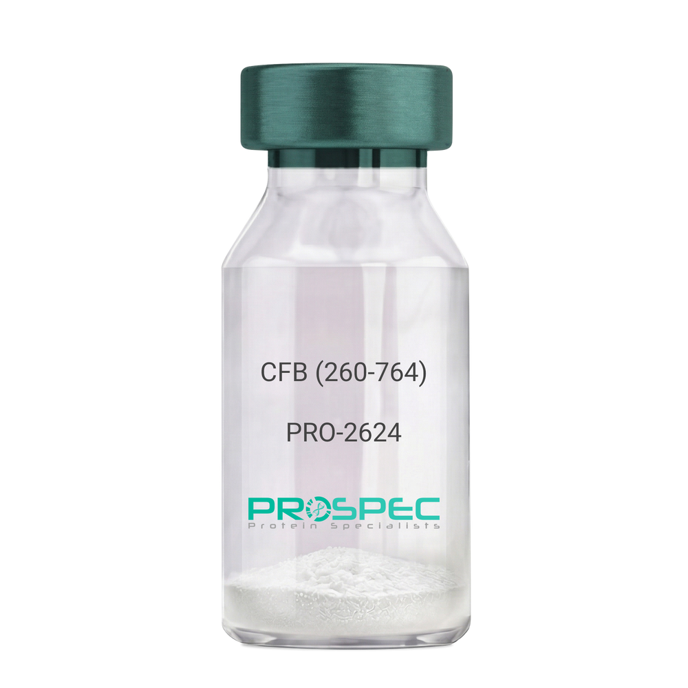
  • View Data Sheet

    Name :

    CFB (260-764) Human, Sf9

    Description:

    Complement Factor B (260-764 a.a.) Human Recombinant, Sf9

    CFB, C3/C5 convertase, Glycine-rich beta glycoprotein, GBG, PBF2, Properdin factor B, BF, BFD, AHUS4, ARMD14, CFAB, CFBD, FB, FBI12, GBG, H2-Bf.

    Product # :

    PRO-2624

    Price :

    Quantity :

    Shipping Method :

    Ice Icon

    Shipped with Ice Packs

    Add To Cart

    More Info

    • description
    • source
    • formulation
    • purity
    • More Info

    Description

    CFB Human Recombinant produced in Sf9 Baculovirus cells is a single, glycosylated polypeptide chain containing 514 amino acids (260-764 a.a) and having a molecular mass of 58.1kDa.CFB is fused to an 9 amino acid His-tag at C-terminus & purified by proprietary chromatographic techniques.

    Source

    Sf9, Baculovirus cells.

    Formulation

    CFB protein solution (0.5mg/ml) containing Phosphate Buffered Saline (pH 7.4) and 20% glycerol.

    Purity

    Greater than 90.0% as determined by SDS-PAGE.

    More Info

    • Introduction

      Factor B is a protein, build from a single chain that circulates in the blood. Complement factor D cleaves and activates factor B, thus creating a catalytic subunit Bb and noncatalytic chain Ba. Subunit Bb is a serine protease that can bind to C3b and forms alternative pathway C3 convertase. Subunit Ba acts as an inhibitor for proliferation of lymphocytes, when Bb is part of the lymphocytes’ proliferation. The gene that codes for this protein is located on chromosome six.

    • Synonyms

      CFB, C3/C5 convertase, Glycine-rich beta glycoprotein, GBG, PBF2, Properdin factor B, BF, BFD, AHUS4, ARMD14, CFAB, CFBD, FB, FBI12, GBG, H2-Bf.

    • Physical Appearance

      Sterile Filtered colorless solution.

    • Stability

      Store at 4°C if entire vial will be used within 2-4 weeks. Store, frozen at -20°C for longer periods of time. For long term storage it is recommended to add a carrier protein (0.1% HSA or BSA).Avoid multiple freeze-thaw cycles.

    • Amino Acid Sequence

      ADPKIVLDPS GSMNIYLVLD GSDSIGASNF TGAKKCLVNL IEKVASYGVK PRYGLVTYAT
      YPKIWVKVSE ADSSNADWVT KQLNEINYED HKLKSGTNTK KALQAVYSMM SWPDDVPPEG
      WNRTRHVIIL MTDGLHNMGG DPITVIDEIR DLLYIGKDRK NPREDYLDVY VFGVGPLVNQ
      VNINALASKK DNEQHVFKVK DMENLEDVFY QMIDESQSLS LCGMVWEHRK GTDYHKQPWQ
      AKISVIRPSK GHESCMGAVV SEYFVLTAAH CFTVDDKEHS IKVSVGGEKR DLEIEVVLFH
      PNYNINGKKE AGIPEFYDYD VALIKLKNKL KYGQTIRPIC LPCTEGTTRA LRLPPTTTCQ
      QQKEELLPAQ DIKALFVSEE EKKLTRKEVY IKNGDKKGSC ERDAQYAPGY DKVKDISEVV
      TPRFLCTGGV SPYADPNTCR GDSGGPLIVH KRSRFIQVGV ISWGVVDVCK NQKRQKQVPA HARDFHINLF QVLPWLKEKL QDEDLGFLHH HHHH

    ProSpec's products are furnished for LABORATORY RESEARCH USE ONLY. The product may not be used as drugs, agricultural or pesticidal products, food additives or household chemicals.

    Complement Factor B
  • View Data Sheet

    Name :

    HEXA Antibody

    Description:

    Hexosaminidase A, Mouse Anti Human

    TSD, hexosaminidase A, Beta-hexosaminidase subunit alpha, Beta-N-acetylhexosaminidase subunit alpha, Hexosaminidase subunit A, N-acetyl-beta-glucosaminidase subunit alpha.

    Product # :

    ANT-480

    Price :

    Quantity :

    Shipping Method :

    Ice Icon

    Shipped with Ice Packs

    Add To Cart

    More Info

    • formulation
    • More Info

    Formulation

    1mg/ml containing PBS, pH-7.4, 10% Glycerol and 0.02% Sodium Azide.

    More Info

    • Introduction

      HEXA is the alpha subunit of the lysosomal enzyme beta-hexosaminidase which, combined with the cofactor GM2 activator protein, catalyzes the degradation of the ganglioside GM2, and other molecules having N-acetyl hexosamines terminus. The two subunits composing Beta-hexosaminidase, alpha and beta, belong to the glycosyl hydrolases family and are encoded by distinct genes. Alpha subunit gene mutations can cause Tay-Sachs disease (GM2-gangliosidosis type I).

    • Synonyms

      TSD, hexosaminidase A, Beta-hexosaminidase subunit alpha, Beta-N-acetylhexosaminidase subunit alpha, Hexosaminidase subunit A, N-acetyl-beta-glucosaminidase subunit alpha.

    • Physical Appearance

      Sterile filtered colorless solution.

    • Immunogen

      Anti-human HEXA mAb, clone PAT20F1A, is derived from hybridization of mouse F0 myeloma cells with spleen cells from BALB/c mice immunized with a recombinant human HEXA protein 89-529 amino acids purified from E. coli.

    • Ig Subclass

      Mouse IgG2a heavy chain and Lambda light chain.

    • Clone

      PAT20F1A.

    • Applications

      The antibody has been tested by ELISA, Western blot analysis to assure specificity and reactivity. Since application varies, however, each investigation should be titrated by the reagent to obtain optimal results. Recommended starting dilution is 1:3000.

    • Type

      Mouse Anti Human Monoclonal.

    • Storage Procedures

      For periods up to 1 month store at 4°C, for longer periods of time, store at -20°C. Prevent freeze thaw cycles.

    • Purification Method

      HEXA antibody was purified by protein-A affinity chromatography.

    ProSpec's products are furnished for LABORATORY RESEARCH USE ONLY. The product may not be used as drugs, agricultural or pesticidal products, food additives or household chemicals.

    Hexa Antibody
  • View Data Sheet

    Name :

    PAIP2 Antibody

    Description:

    Polyadenylate-Binding Protein-Interacting Protein 2, Mouse Anti Human

    Poly(A) Binding Protein Interacting Protein 2, PABP- interacting protein 2, HSPC218, PAIP-2, PAIP2A, Polyadenylate-binding protein-interacting protein 2, PAIP2, MGC72018.

    Product # :

    ANT-094

    Price :

    Quantity :

    Shipping Method :

    Ice Icon

    Shipped with Ice Packs

    Add To Cart

    More Info

    • formulation
    • More Info

    Formulation

    1mg/ml containing PBS, pH-7.4, & 0.1% Sodium Azide.

    More Info

    • Introduction

      PAIP2 has a role as a repressor in the regulation of translation initiation of poly(A)-containing mRNAs. PAIP2 inhibitory activity on translation is mediated through its action on PABPC1. PAIP2 displaces the interaction of PABPC1 with poly(A) RNA and competes with PAIP1 for binding to PABPC1. PAIP2 association with PABPC1 results in disruption of the cytoplasmic poly(A) RNP structure organization.

    • Synonyms

      Poly(A) Binding Protein Interacting Protein 2, PABP- interacting protein 2, HSPC218, PAIP-2, PAIP2A, Polyadenylate-binding protein-interacting protein 2, PAIP2, MGC72018.

    • Physical Appearance

      Sterile Filtered colorless solution.

    • Immunogen

      Anti-human PAIP2 mAb is derived from hybridization of mouse F0 myeloma cells with spleen cells from BALB/c mice immunized with recombinant human CMBL 1-127 amino acids purified from E. coli.

    • Ig Subclass

      Mouse IgG2b heavy chain and ? light chain.

    • Clone

      PAT1C12AT.

    • Applications

      PAIP2 antibody has been tested by ELISA, Western blot analysis to assure specificity and reactivity. Since application varies, however, each investigation should be titrated by the reagent to obtain optimal results. Recommended dilution range for Western blot analysis is 1:500 ~ 1:5000. Recommended starting dilution is 1:1000.

    • Type

      Mouse Anti Human Monoclonal.

    • Storage Procedures

      For periods up to 1 month store at 4°C, for longer periods of time, store at -20°C. Prevent freeze thaw cycles.

    • Purification Method

      PAIP2 antibody was purified from mouse ascitic fluids by protein-G affinity chromatography.

    ProSpec's products are furnished for LABORATORY RESEARCH USE ONLY. The product may not be used as drugs, agricultural or pesticidal products, food additives or household chemicals.

    Paip2 Antibody
  • 1
  • …
  • 28
  • 29
  • 30
  • 31
  • 32
  • …
  • 42
Your browser does not support the video tag.

Products

  • Cytokines
  • Growth Factors
  • Chemokines
  • CD Antigens
  • Neurotrophins
  • Hormones
  • Enzymes
  • Viral Antigens
  • Recombinant Proteins
  • Natural Proteins
  • Monoclonal Antibodies
  • Polyclonal Antibodies
  • Thermal Packaging

About

  • Company
  • Founder
  • Contribution
  • Contact Us
  • My Account

Ordering

  • Ordering Method
  • Ordering - Credit Card
  • Ordering PO
  • Shipping Fees
  • FAQ's

Legal

  • Terms & Conditions
  • Privacy
  • Site Map
  • info@prospecbio.com
  • Facebook
  • Instagram
  • Linkedin Profile

© 2026 ProSpec-Tany TechnoGene Ltd. All Rights Reserved.

  • Choosing a selection results in a full page refresh.
  • Opens in a new window.