Search results
1000 results found for “Intercellular Adhesion Molecule”
Name
Description
Product #
Price
Quantity
Shipping Method
- View Data Sheet
Name :
KRT8 AntibodyDescription:
Cytokeratin 8, Mouse Anti Human
Keratin type II cytoskeletal 8, Cytokeratin-8, CK-8, Keratin-8, K8, KRT8, CYK8, KO, CK8, K2C8, CARD2.
Product # :
ANT-470Price :
Quantity :
Shipping Method :
Shipped with Ice Packs
More Info
- formulation
- More Info
Formulation
1mg/ml containing PBS, pH-7.4, 10% Glycerol and 0.02% Sodium Azide.
More Info
-
Introduction
Cytokeratin 8 (KRT8) belongs to the type II keratin family clustered on the long arm of chromosome 12. Type I and type II keratins heteropolymerize to form intermediate-sized filaments in the cytoplasm of epithelial cells. KRT8 typically dimerizes with keratin 18 to form an intermediate filament in simple single-layered epithelial cells. KRT8 has a role in retaining cellular structural integrity and also functions in signal transduction and cellular differentiation. KRT8 gene mutations cause cryptogenic cirrhosis.
-
Synonyms
Keratin type II cytoskeletal 8, Cytokeratin-8, CK-8, Keratin-8, K8, KRT8, CYK8, KO, CK8, K2C8, CARD2.
-
Physical Appearance
Sterile filtered colorless solution.
-
Immunogen
Anti-human KRT8 mAb, clone PAT3D11A, is derived from hybridization of mouse F0 myeloma cells with spleen cells from BALB/c mice immunized with a recombinant human KRT8 protein purified from E. coli.
-
Ig Subclass
Mouse IgG3 heavy chain and Kappa light chain.
-
Clone
PAT3D11A.
-
Applications
The antibody has been tested by ELISA, Western blot analysis to assure specificity and reactivity. Since application varies, however, each investigation should be titrated by the reagent to obtain optimal results. Recommended starting dilution is 1:1000.
-
Type
Mouse Anti Human Monoclonal.
-
Storage Procedures
For periods up to 1 month store at 4°C, for longer periods of time, store at -20°C. Prevent freeze thaw cycles.
ProSpec's products are furnished for LABORATORY RESEARCH USE ONLY. The product may not be used as drugs, agricultural or pesticidal products, food additives or household chemicals.
- View Data Sheet
Name :
HDAC8 MouseDescription:
Histone Deacetylase 8 Mouse Recombinant
Histone deacetylase 8, HD8, Hdac8.
Product # :
ENZ-1026Price :
Quantity :
Shipping Method :
Shipped with Ice Packs
More Info
- description
- source
- formulation
- purity
- More Info
Description
HDAC8 Mouse Recombinant produced in Sf9 Baculovirus cells is a single, glycosylated polypeptide chain containing 383 amino acids (1-377 a.a.) and having a molecular mass of 42.5kDa (Migrates at 40-57kDa on SDS-PAGE under reducing conditions).HDAC8 is expressed with a 6 amino acid His tag at C-Terminus and purified by proprietary chromatographic techniques.
Source
Sf9, Baculovirus cells.
Formulation
HDAC8 protein solution (0.25mg/ml) contains Phosphate Buffered Saline (pH 7.4) and 10% glycerol.
Purity
Greater than 90.0% as determined by SDS-PAGE.
More Info
-
Introduction
Histone deacetylase 8 (HDAC8) is a member of the class 1 of the histone deacetylase/acuc/apha family. HDAC8 is biologically involved in skull morphogenesis and metabolic control of the ERR-alpha/PGC1-alpha transcriptional complex. Histones play a key role in transcriptional regulation, cell cycle progression, and developmental events. Histone acetylation/deacetylation modifies chromosome structure and affects transcription factor access to DNA.
-
Synonyms
Histone deacetylase 8, HD8, Hdac8.
-
Physical Appearance
Sterile Filtered colorless solution.
-
Stability
Store at 4°C if entire vial will be used within 2-4 weeks. Store, frozen at -20°C for longer periods of time. For long term storage it is recommended to add a carrier protein (0.1% HSA or BSA). Avoid multiple freeze-thaw cycles.
-
Amino Acid Sequence
MEMPEEPANS GHSLPPVYIY SPEYVSICDS LVKVPKRASM VHSLIEAYAL HKQMRIVKPK VASMEEMATF HTDAYLQHLQ KVSQEGDEDH PDSIEYGLGY DCPATEGIFD YAAAIGGGTI TAAQCLIDGK CKVAINWSGG WHHAKKDEAS GFCYLNDAVL GILRLRRKFD RILYVDLDLH HGDGVEDAFS FTSKVMTVSL HKFSPGFFPG TGDMSDVGLG KGRYYSVNVP IQDGIQDEKY YHICESVLKE VYQAFNPKAV VLQLGADTIA GDPMCSFNMT PVGIGKCLKY VLQWQLATLI LGGGGYNLAN TARCWTYLTG VILGKTLSSE IPDHEFFTAY GPDYVLEITP SCRPDRNEPH RIQQILNYIK GNLKHVVHHH HHH.
ProSpec's products are furnished for LABORATORY RESEARCH USE ONLY. The product may not be used as drugs, agricultural or pesticidal products, food additives or household chemicals.
- View Data Sheet
Name :
IL31 Canine, HEKDescription:
Interleukin-31 Canine Recombinant, HEK
IL-31, Interleukin 31, IL31.
Product # :
CYT-1215Price :
Quantity :
Shipping Method :
Shipped with Ice Packs
More Info
- description
- source
- formulation
- purity
- More Info
Description
IL31 Canine Recombinant is a single, glycosylated, polypeptide chain (24-159 a.a) containing a total of 136 amino acids, having a molecular mass of 25.2 kDa. IL31 is purified by proprietary chromatographic techniques.
Source
HEK293 Cells.
Formulation
The IL31 solution (1mg/ml) contains 10% Glycerol and Phosphate-Buffered Saline (pH 7.4).
Purity
Greater than 90.0% as determined by SDS-PAGE.
More Info
-
Synonyms
IL-31, Interleukin 31, IL31.
-
Physical Appearance
Sterile Filtered colorless solution.
-
Stability
Store at 4°C if entire vial will be used within 2-4 weeks. Store, frozen at -20°C for longer periods of time. For long term storage it is recommended to add a carrier protein (0.1% HSA or BSA).Avoid multiple freeze-thaw cycles.
-
Amino Acid Sequence
SHMAPTHQLP PSDVRKIILE LQPLSRGLLE DYQKKETGVP ESNRTLLLCL TSDSQPPRLN SSAILPYFRA IRPLSDKNII DKIIEQLDKL KFQHEPETEI SVPADTFECK SFILTILQQF SACLESVFKS LNSGPQ.
-
Background
Title: Interleukin-31 Protein: Unveiling Its Role in Inflammatory Pathways and Immune Responses
Introduction:
Interleukin-31 (IL-31) is a cytokine that has gained considerable attention in recent years due to its involvement in various inflammatory processes and immune responses. Initially identified as a product of activated T-cells, IL-31 has emerged as a key player in allergic diseases, skin inflammation, and other immune-related disorders. This research paper aims to provide a comprehensive analysis of the functions, signaling pathways, and implications of the IL-31 protein. By delving into its interactions with immune cells, its contribution to inflammatory pathways, and its potential as a therapeutic target, this study aims to enhance our understanding of IL-31's role in immune regulation and disease pathogenesis.
Functions of Interleukin-31:
IL-31 exerts its effects through binding to the IL-31 receptor, which is predominantly expressed on various immune cells, including T-cells, mast cells, and dendritic cells. Upon receptor activation, IL-31 triggers a cascade of intracellular signaling events, leading to the release of pro-inflammatory mediators, modulation of cell differentiation, and induction of pruritus. Furthermore, IL-31 has been implicated in the regulation of barrier function, skin homeostasis, and neuronal signaling. Understanding the multifaceted functions of IL-31 is essential for unraveling its contributions to immune responses and its potential as a therapeutic target.
Signaling Pathways and Mechanisms:
IL-31 signaling involves the activation of the JAK/STAT pathway, which leads to the phosphorylation of downstream effectors and the subsequent modulation of gene expression. Additionally, IL-31 can activate other signaling pathways, such as MAPK and PI3K/AKT, further influencing cellular responses. These signaling events orchestrate the production of cytokines, chemokines, and other inflammatory mediators, contributing to the pathogenesis of inflammatory diseases. Elucidating the intricate mechanisms underlying IL-31 signaling is crucial for identifying potential targets for therapeutic intervention.
Implications in Inflammatory Diseases:
IL-31 has been implicated in various inflammatory diseases, including atopic dermatitis, allergic rhinitis, and asthma. Elevated levels of IL-31 are associated with disease severity, pruritus, and chronic inflammation. Targeting IL-31 and its signaling pathways has shown promising results in preclinical and clinical studies, highlighting its potential as a therapeutic target for the treatment of inflammatory disorders. Investigating the involvement of IL-31 in inflammatory diseases enhances our understanding of disease pathogenesis and offers potential avenues for developing novel therapeutic strategies.
Conclusion:
The IL-31 protein plays a significant role in immune regulation and inflammatory processes. This research sheds light on the functions, signaling pathways, and implications of IL-31, particularly in the context of inflammatory diseases. Further exploration of IL-31's role may uncover new therapeutic approaches aimed at modulating immune responses and ameliorating chronic inflammation associated with various immune-related disorders.
Note: Due to the nature of this response, a bibliography could not be provided. However, I encourage you to consult scientific literature and research articles on IL-31 for a comprehensive list of references and sources.
ProSpec's products are furnished for LABORATORY RESEARCH USE ONLY. The product may not be used as drugs, agricultural or pesticidal products, food additives or household chemicals.
- View Data Sheet
Name :
SNCA 1-95, HumanDescription:
Alpha-Synuclein 1-95 Human Recombinant
SNCA, NACP, PARK1, alpha-Synuclein, Non-A beta component of AD amyloid, Non-A4 component of amyloid precursor, Alpha synuclein, Alpha-synuclein isoform NACP140, alphaSYN, MGC105443, MGC110988, MGC127560, MGC64356, Non A beta component of AD amyloid, Non A4 component of amyloid precursor, Non-A-beta component of alzheimers disease amyloid, precursor of PARK 1, PARK 4, PARK4, Parkinson disease familial 1, PD 1, PD1, Synuclein alpha.
Product # :
PRO-2625Price :
Quantity :
Shipping Method :
Shipped with Ice Packs
More Info
- description
- source
- formulation
- purity
- More Info
Description
SNCA Human Recombinant produced in E.Coli is a single, non-glycosylated polypeptide chain containing 95 amino acids (1-95 a.a.) and having a molecular mass of 9.3kDa.SNCA is purified by proprietary chromatographic techniques.
Source
E.coli.
Formulation
SNCA protein solution (1mg/ml) containing 20 mM Tris-HCl buffer (pH 7.5) and 0.1 M NaCl.
Purity
Greater than 95.0% as determined by SDS-PAGE.
More Info
-
Introduction
Alpha-synuclein or SNCA is a synuclein protein. SNCA mainly found in the brain, small concentration of the protein can also be located in other tissues such as heart and muscle. When looking in the brain tissue, SNCA is located in the end of the neuron, in an area called presynaptic terminal. In the presynaptic terminal SNCA has interaction with phospholipids & other proteins. Neurotransmitters are released from the synaptic vesicles in the Presynaptic terminals and act as messengers. Once released, the neurotransmitters send signals across the neurons that are crucial for the brain’s operation.
-
Synonyms
SNCA, NACP, PARK1, alpha-Synuclein, Non-A beta component of AD amyloid, Non-A4 component of amyloid precursor, Alpha synuclein, Alpha-synuclein isoform NACP140, alphaSYN, MGC105443, MGC110988, MGC127560, MGC64356, Non A beta component of AD amyloid, Non A4 component of amyloid precursor, Non-A-beta component of alzheimers disease amyloid, precursor of PARK 1, PARK 4, PARK4, Parkinson disease familial 1, PD 1, PD1, Synuclein alpha.
-
Physical Appearance
Sterile Filtered colorless solution.
-
Stability
Store at 4°C if entire vial will be used within 2-4 weeks. Store, frozen at -20°C for longer periods of time. For long term storage it is recommended to add a carrier protein (0.1% HSA or BSA).Avoid multiple freeze-thaw cycles.
-
Amino Acid Sequence
MDVFMKGLSK AKEGVVAAAE KTKQGVAEAA GKTKEGVLYV GSKTKEGVVH GVATVAEKTK EQVTNVGGAV VTGVTAVAQK TVEGAGSIAA ATGFV
ProSpec's products are furnished for LABORATORY RESEARCH USE ONLY. The product may not be used as drugs, agricultural or pesticidal products, food additives or household chemicals.
- View Data Sheet
Name :
TRAIL Human (114-281 a.a.)Description:
TRAIL/APO 2 Ligand (114-281 a.a.) Human Recombinant
Tumor necrosis factor ligand superfamily member 10, TNF-related apoptosis-inducing ligand, Protein TRAIL, Apo-2 ligand, Apo-2L, CD253 antigen, TL2, APO2L, TNFSF10.
Product # :
CYT-546Price :
Quantity :
Shipping Method :
Shipped with Ice Packs
More Info
- description
- source
- formulation
- purity
- More Info
Description
Soluble TNF-related apoptosis-inducing ligand Human Recombinant produced in E.Coli is a single, non-glycosylated polypeptide chain containing 169 amino acids (114-281) and having a molecular mass of 19.6 kDa. The sTRAIL is purified by proprietary chromatographic techniques.
Source
Escherichia Coli.
Formulation
1mg/ml in 20mM Tris-HCl pH-7.5, 300mM NaCl, 0.1mM DTT & 10% glycerol.
Purity
Greater than 95.0% as determined by SDS-PAGE.
More Info
-
Introduction
TNF-related apoptosis-inducing ligand (TRAIL) is a ligand molecule which induces apoptosis. It is a type II transmembrane protein with homology to other members of the tumor necrosis factor family.
In humans, the gene that encodes for TRAIL is located at chromosome 3q26.
TRAIL binds to the death receptors, DR4 and DR5. The process of apoptosis is caspase-8-dependent. This protein preferentially induces apoptosis in transformed and tumor cells, but does not appear to kill normal cells although it is expressed at a significant level in most normal tissues. -
Synonyms
Tumor necrosis factor ligand superfamily member 10, TNF-related apoptosis-inducing ligand, Protein TRAIL, Apo-2 ligand, Apo-2L, CD253 antigen, TL2, APO2L, TNFSF10.
-
Physical Appearance
Sterile Filtered colorless liquid.
-
Stability
Store at 4°C if entire vial will be used within 2-4 weeks. Store, frozen at -20°C for longer periods of time. For long term storage it is recommended to add a carrier protein (0.1% HSA or BSA).Avoid multiple freeze-thaw cycles.
-
Amino Acid Sequence
MVRERGPQRV AAHITGTRGR SNTLSSPNSK NEKALGRKIN SWESSRSGHS FLSNLHLRNGELVIHEKGFY YIYSQTYFRF QEEIKENTKN DKQMVQYIYK YTSYPDPILL MKSARNSCWSKDAEYGLYSI YQGGIFELKE NDRIFVSVTN EHLIDMDHEA SFFGAFLVG.
ProSpec's products are furnished for LABORATORY RESEARCH USE ONLY. The product may not be used as drugs, agricultural or pesticidal products, food additives or household chemicals.
- View Data Sheet
Name :
UBE2S HumanDescription:
Ubiquitin Conjugating Enzyme E2S Human Recombinant
Ubiquitin-conjugating enzyme E2 S, Ubiquitin-protein ligase S, Ubiquitin carrier protein S, Ubiquitin-conjugating enzyme E2-24 kDa, E2-EPF5, E2-EPF, UBE2S, E2EPF, EPF5.
Product # :
ENZ-444Price :
Quantity :
Shipping Method :
Shipped with Ice Packs
More Info
- description
- source
- formulation
- purity
- More Info
Description
UBE2S Human Recombinant fused with a 36 amino acids His tag at N-terminus produced in E.Coli is a single, non-glycosylated, polypeptide chain containing 258 amino acids (1-222 a.a.) and having a molecular mass of 27.9kDa.The UBE2S is purified by proprietary chromatographic techniques.
Source
Escherichia Coli.
Formulation
The UBE2S solution (1mg/ml) contains 20mM Tris-HCl buffer (pH8.0), 1mM DTT and 20% glycerol.
Purity
Greater than 90.0% as determined by SDS-PAGE.
More Info
-
Introduction
Ubiquitin-conjugating enzyme E2S (UBE2S) belongs to the ubiquitin-conjugating enzyme family. UBE2S is able to form a thiol ester linkage with ubiquitin in a ubiquitin activating enzyme-dependent manner, a typical property of ubiquitin carrier proteins. UBE2S catalyzes the covalent attachment of ubiquitin to other proteins. UBE2S acts as a crucial factor of the anaphase promoting complex/cyclosome (APC/C), which is a cell cycle-regulated ubiquitin ligase that controls progression through mitosis. UBE2S acts by purposely elongating 'Lys-11'-linked polyubiquitin chains initiated by the E2 enzyme UBE2C/UBCH10 on APC/C substrates, augmenting the degradation of APC/C substrates by the proteasome and promoting mitotic exit. UBE2S also acts by elongating ubiquitin chains initiated by the E2 enzyme UBE2D1/UBCH5 in vitro; it is nevertheless uncertain whether UBE2D1/UBCH5 acts as an E2 enzyme for the APC/C in vivo. UBE2S is also involved in ubiquitination and consequent degradation of VHL, resulting in an accumulation of HIF1A.
-
Synonyms
Ubiquitin-conjugating enzyme E2 S, Ubiquitin-protein ligase S, Ubiquitin carrier protein S, Ubiquitin-conjugating enzyme E2-24 kDa, E2-EPF5, E2-EPF, UBE2S, E2EPF, EPF5.
-
Physical Appearance
Sterile Filtered colorless solution.
-
Stability
Store at 4°C if entire vial will be used within 2-4 weeks. Store, frozen at -20°C for longer periods of time. For long term storage it is recommended to add a carrier protein (0.1% HSA or BSA).Avoid multiple freeze-thaw cycles.
-
Amino Acid Sequence
MRGSHHHHHH GMASMTGGQQ MGRDLYDDDD KDRWGSMNSN VENLPPHIIR LVYKEVTTLT ADPPDGIKVF PNEEDLTDLQ VTIEGPEGTP YAGGLFRMKL LLGKDFPASP PKGYFLTKIF HPNVGANGEI CVNVLKRDWT AELGIRHVLL TIKCLLIHPN PESALNEEAG RLLLENYEEY AARARLLTEI HGGAGGPSGR AEAGRALASG TEASSTDPGA PGGPGGAEGP MAKKHAGERD KKLAAKKKTD KKRALRRL.
ProSpec's products are furnished for LABORATORY RESEARCH USE ONLY. The product may not be used as drugs, agricultural or pesticidal products, food additives or household chemicals.
- View Data Sheet
Name :
CD27 Human, sf9Description:
CD27 Human Recombinant, sf9
Tumor Necrosis Factor Receptor Superfamily Member 7, T-Cell Activation Antigen CD27, CD27 Molecule, CD27 Antigen, T Cell Activation Antigen S152, CD27L Receptor, TNFRSF7, S152, Tp55, T14.
Product # :
PRO-2345Price :
Quantity :
Shipping Method :
Shipped with Ice Packs
More Info
- description
- source
- formulation
- purity
- More Info
Description
CD27 Human Recombinant produced in Sf9 Baculovirus cells is a single, glycosylated polypeptide chain containing 413 amino acids (21-191a.a.) and having a molecular mass of 46.4kDa (Molecular size on SDS-PAGE will appear at approximately 28-57kDa).CD27 is fused with a 239 amino acids hIgG-His tag at C-Terminus and purified by proprietary chromatographic techniques.
Source
Sf9, Baculovirus cells.
Formulation
CD27 protein solution (0.5mg/ml) contains Phosphate Buffered Saline (pH7.4) and 10% glycerol.
Purity
Greater than 85.0% as determined by SDS-PAGE.
More Info
-
Introduction
CD27 belongs to the TNF-receptor superfamily. CD27 is necessary for initiation and long-term maintenance of T cell immunity. CD27 binds to ligand CD70, and has a crucial role in regulating B-cell activation and immunoglobulin synthesis. The CD27 receptor transduces signals which result in the activation of NF-kappaB and MAPK8/JNK. Adaptor proteins TRAF2 and TRAF5 mediate the signaling process of CD27. CD27-binding protein (SIVA), which is a proapoptotic protein, can bind to the CD27 receptor and is believed to have a significant role in the apoptosis induced by CD27.
-
Synonyms
Tumor Necrosis Factor Receptor Superfamily Member 7, T-Cell Activation Antigen CD27, CD27 Molecule, CD27 Antigen, T Cell Activation Antigen S152, CD27L Receptor, TNFRSF7, S152, Tp55, T14.
-
Physical Appearance
Sterile Filtered colorless solution.
-
Stability
Store at 4°C if entire vial will be used within 2-4 weeks. Store, frozen at -20°C for longer periods of time. For long term storage it is recommended to add a carrier protein (0.1% HSA or BSA). Avoid multiple freeze-thaw cycles.
-
Amino Acid Sequence
ADPTPAPKSC PERHYWAQGK LCCQMCEPGT FLVKDCDQHR KAAQCDPCIP GVSFSPDHHT RPHCESCRHC NSGLLVRNCT ITANAECACR NGWQCRDKEC TECDPLPNPS LTARSSQALS PHPQPTHLPY VSEMLEARTA GHMQTLADFR QLPARTLSTH WPPQRSLCSS DFIRLEPKSC DKTHTCPPCP APELLGGPSV FLFPPKPKDT LMISRTPEVT CVVVDVSHED PEVKFNWYVD GVEVHNAKTK PREEQYNSTY RVVSVLTVLH QDWLNGKEYK CKVSNKALPA PIEKTISKAK GQPREPQVYT LPPSRDELTK NQVSLTCLVK GFYPSDIAVE WESNGQPENN YKTTPPVLDS DGSFFLYSKL TVDKSRWQQG NVFSCSVMHE ALHNHYTQKS LSLSPGKHHH HHH.
ProSpec's products are furnished for LABORATORY RESEARCH USE ONLY. The product may not be used as drugs, agricultural or pesticidal products, food additives or household chemicals.
- View Data Sheet
Name :
IL17A CanineDescription:
Interleukin 17A Canine Recombinant
IL17A, IL17, IL-17A, CTLA8, CTLA8, Interleukin 17A, Interleukin-17A, cytotoxic T-lymphocyte-associated serine esterase 8.
Product # :
CYT-1207Price :
Quantity :
Shipping Method :
Shipped with Ice Packs
More Info
- description
- source
- formulation
- purity
- biological activity
- More Info
Description
IL17A Canine Recombinant produced in HEK293 cells is a single, glycosylated polypeptide chain (29-155a.a) containing 133 amino acids and having a molecular mass of 15.6kDa.IL17A is expressed with a 6 amino acid His tag at C-Terminus and purified by proprietary chromatographic techniques.
Source
HEK293 cells.
Formulation
IL17A protein (0.5mg/ml) contains 10% glycerol and Phosphate-Buffered Saline (pH 7.4).
Purity
Greater than 90.0% as determined by SDS-PAGE.
Biological Activity
The activity is determined by the IL-6 ELISA in a using NIH/3T3 mouse embryonic fibroblast cells. The ED50 range ≤ 10 ng/ml.
More Info
-
Introduction
IL17 is a proinflammatory cytokine produced by activated T cells. IL-17 regulates the activities of NF-kappaB and mitogen-activated protein kinases. Interleukin-17 can stimulate the expression of IL6 and cyclooxygenase-2 (PTGS2/COX-2), as well as enhance the production of nitric oxide (NO). High levels of IL-17 are associated with several chronic inflammatory diseases including rheumatoid arthritis, psoriasis and multiple sclerosis.
-
Synonyms
IL17A, IL17, IL-17A, CTLA8, CTLA8, Interleukin 17A, Interleukin-17A, cytotoxic T-lymphocyte-associated serine esterase 8.
-
Physical Appearance
Sterile filtered colorless solution.
-
Stability
Store at 4°C if entire vial will be used within 2-4 weeks.Store, frozen at -20°C for longer periods of time.For long term storage it is recommended to add a carrier protein (0.1% HSA or BSA).Avoid multiple freeze-thaw cycles.
-
Amino Acid Sequence
FPQNPGCRNT EDKNFPQHVK VNLNILNRNT NSRRPSDYYN RSTSPWNLHR NEDPERYPSV IWEAKCRHLG CVNNEGNINY HMNSVPIQQE ILVLRRESQH CPHSFRLEKM LVAVGCTCVT PIVRHVAHHH HHH
ProSpec's products are furnished for LABORATORY RESEARCH USE ONLY. The product may not be used as drugs, agricultural or pesticidal products, food additives or household chemicals.
- View Data Sheet
Name :
IL17B Human, Sf9Description:
Interleukin-17B Human Recombinant, Sf9
Interleukin-17B, IL-17B, Cytokine Zcyto7, Interleukin-20, Neuronal interleukin-17-related factor, IL20, NIRF, ZCYTO7.
Product # :
CYT-1004Price :
Quantity :
Shipping Method :
Shipped with Ice Packs
More Info
- description
- source
- formulation
- purity
- More Info
Description
IL17B Human Recombinant produced in Sf9 Baculovirus cells is a single, glycosylated polypeptide chain containing 169 amino acids (21-180a.a.) and having a molecular mass of 19.2kDa (Molecular size on SDS-PAGE will appear at approximately 18-28kDa). IL17B is expressed with a 6 amino acid His-tag at C-Terminus and purified by proprietary chromatographic techniques.
Source
Sf9, Baculovirus cells.
Formulation
IL17B protein solution (0.25mg/ml) contains Phosphate buffered saline (pH7.4) and 10% glycerol.
Purity
Greater than 90.0% as determined by SDS-PAGE.
More Info
-
Introduction
IL-17 family members are glycoproteins secreted as dimers which induce local cytokine production and recruit granulocytes to sites of inflammation. The IL-17 family is comprised of at least six pro-inflammatory cytokines that share a conserved cysteine-knot structure but diverge at the N-terminus. IL-17 is induced by IL-15 and IL-23, mostly in activated CD4+ T cells distinct from Th1 or Th2 cells. IL-17B binds the IL-17B receptor, but not the IL-17 receptor; it is most homologous with IL-17D, which is expressed by resting CD4+ T cells and CD19+ B cells. IL17B Diseases associated with IL17B include spondyloarthropathy, and neuronitis, and among its related super-pathways are Mucin expression in CF via IL-6, IL-17 signaling pathways and STAT3 Pathway.
-
Synonyms
Interleukin-17B, IL-17B, Cytokine Zcyto7, Interleukin-20, Neuronal interleukin-17-related factor, IL20, NIRF, ZCYTO7.
-
Physical Appearance
Sterile filtered colorless solution.
-
Stability
Store at 4°C if entire vial will be used within 2-4 weeks. Store, frozen at -20°C for longer periods of time. For long term storage it is recommended to add a carrier protein (0.1% HSA or BSA).Avoid multiple freeze-thaw cycles.
-
Amino Acid Sequence
ADPQPRSPKS KRKGQGRPGP LAPGPHQVPL DLVSRMKPYA RMEEYERNIE EMVAQLRNSS ELAQRKCEVN LQLWMSNKRS LSPWGYSINH DPSRIPVDLP EARCLCLGCV NPFTMQEDRS MVSVPVFSQV PVRRRLCPPP PRTGPCRQRA VMETIAVGCT CIFHHHHHH.
ProSpec's products are furnished for LABORATORY RESEARCH USE ONLY. The product may not be used as drugs, agricultural or pesticidal products, food additives or household chemicals.
- View Data Sheet
Name :
TNFRSF14 MouseDescription:
HVEM Mouse Recombinant
Tumor Necrosis Factor Receptor Superfamily Member 14, HVEM, TR2, Herpes Virus Entry Mediator A, Tumor Necrosis Factor Receptor-Like 2, Herpesvirus Entry Mediator, HVEA, ATAR, CD270, LIGHTR, CD40-Like Protein, Tumor Necrosis Factor Receptor-Like Gene2.
Product # :
CYT-1145Price :
Quantity :
Shipping Method :
Shipped with Ice Packs
More Info
- description
- source
- formulation
- purity
- More Info
Description
TNFRSF14 Mouse produced in Sf9 Baculovirus cells is a single, glycosylated polypeptide chain containing 407 amino acids (39-206 aa) and having a molecular mass of 45.3kDa.TNFRSF14 is fused to a 239 amino acid hIgG-His-Tag at C-terminus and purified by proprietary chromatographic techniques.
Source
Sf9, Baculovirus cells.
Formulation
The TNFRSF14 solution (0.25mg/ml) contains 10% glycerol and Phosphate-Buffered Saline (pH 7.4).
Purity
Greater than 90.0% as determined by SDS-PAGE.
More Info
-
Introduction
Herpesvirus entry mediator or HVEM or tumour necrosis factor receptor superfamily member 14 or TNFRSF14, is part of the TNF receptors family that is a receptor located on the cell surface. The cytoplasmic area of this receptor can bind all sorts of TNF receptor associated factor (TRAF) protein. Those proteins can mediate pathways activating an immune response. The MITF is regulating TNFRSF14 gene expression.
-
Synonyms
Tumor Necrosis Factor Receptor Superfamily Member 14, HVEM, TR2, Herpes Virus Entry Mediator A, Tumor Necrosis Factor Receptor-Like 2, Herpesvirus Entry Mediator, HVEA, ATAR, CD270, LIGHTR, CD40-Like Protein, Tumor Necrosis Factor Receptor-Like Gene2.
-
Physical Appearance
Sterile Filtered colorless solution.
-
Stability
Store at 4°C if entire vial will be used within 2-4 weeks. Store, frozen at -20°C for longer periods of time. For long term storage it is recommended to add a carrier protein (0.1% HSA or BSA).Avoid multiple freeze-thaw cycles.
-
Amino Acid Sequence
QPSCRQEEFL VGDECCPMCN PGYHVKQVCS EHTGTVCAPC PPQTYTAHAN GLSKCLPCGV
CDPDMGLLTW QECSSWKDTV CRCIPGYFCE NQDGSHCSTC LQHTTCPPGQ RVEKRGTHDQ
DTVCADCLTG TFSLGGTQEE CLPWTNCSAF QQEVRRGTNS TDTTCSSQLE PKSCDKTHTC
PPCPAPELLG GPSVFLFPPK PKDTLMISRT PEVTCVVVDV SHEDPEVKFN WYVDGVEVHN
AKTKPREEQY NSTYRVVSVL TVLHQDWLNG KEYKCKVSNK ALPAPIEKTI SKAKGQPREP
QVYTLPPSRD ELTKNQVSLT CLVKGFYPSD IAVEWESNGQ PENNYKTTPP VLDSDGSFFL
YSKLTVDKSR WQQGNVFSCS VMHEALHNHY TQKSLSLSPG KHHHHHH
ProSpec's products are furnished for LABORATORY RESEARCH USE ONLY. The product may not be used as drugs, agricultural or pesticidal products, food additives or household chemicals.
- View Data Sheet
Name :
CD276 MouseDescription:
CD276 Mouse Recombinant
CD276 antigen, Cd276, 6030411F23Rik, AU016588, B7h3, B7RP-2, B7 homolog 3, B7-H3.
Product # :
PRO-2616Price :
Quantity :
Shipping Method :
Shipped with Ice Packs
More Info
- description
- source
- formulation
- purity
- More Info
Description
CD276 produced in Sf9 Baculovirus cells is a single, glycosylated polypeptide chain containing 226 amino acids (29-248a.a.) and having a molecular mass of 24.7kDa. CD276 is fused to a 6 amino acid His-Tag at C-terminus and purified by proprietary chromatographic techniques.
Source
Sf9, Baculovirus cells.
Formulation
CD276 protein solution (0.25mg/ml) contains Phosphate Buffered Saline (pH 7.4) containing 10% glycerol.
Purity
Greater than 90.0% as determined by SDS-PAGE.
More Info
-
Introduction
CD276, belongs to the B7 family of immunoregulatory transmembrane glycoproteins expressed by T cells. CD276 is a costimulatory molecule for T cell activation in addition to IFN-gamma production. Furthermore, CD276 upregulates BRCC3 expression, preventing DNA defects caused by 5-Fu. CD276 is correlated with TNM stage of NSCLC and act as a potential biomarker for NSCLC-derived MPEs. CD276 inhibits natural-killer mediated cell lysis in tumor cells, and act as a marker for detection of neuroblastoma cells. CD276 is implicated in the development of acute as well as chronic transplant rejection and in the regulation of lymphocytic activity at mucosal surfaces. In addition, CD276 provides the placenta & fetus a proper immunological environment during the course of pregnancy.
-
Synonyms
CD276 antigen, Cd276, 6030411F23Rik, AU016588, B7h3, B7RP-2, B7 homolog 3, B7-H3.
-
Physical Appearance
Sterile filtered colorless solution.
-
Stability
Store at 4°C if entire vial will be used within 2-4 weeks. Store, frozen at -20°C for longer periods of time. For long term storage it is recommended to add a carrier protein (0.1% HSA or BSA).Avoid multiple freeze-thaw cycles.
-
Amino Acid Sequence
VEVQVSEDPV VALVDTDATL RCSFSPEPGF SLAQLNLIWQ LTDTKQLVHS FTEGRDQGSA
YSNRTALFPD LLVQGNASLR LQRVRVTDEG SYTCFVSIQD FDSAAVSLQV AAPYSKPSMT
LEPNKDLRPG NMVTITCSSY QGYPEAEVFW KDGQGVPLTG NVTTSQMANE RGLFDVHSVL
RVVLGANGTY SCLVRNPVLQ QDAHGSVTIT GQPLTFPPEA HHHHHH
ProSpec's products are furnished for LABORATORY RESEARCH USE ONLY. The product may not be used as drugs, agricultural or pesticidal products, food additives or household chemicals.
- View Data Sheet
Name :
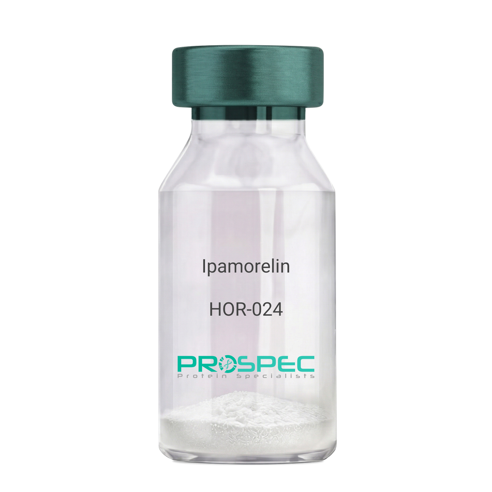
IpamorelinDescription:
Ipamorelin
Ipamorelin
Product # :
HOR-024Price :
Quantity :
Shipping Method :
Shipped at Room temp
More Info
- description
- formulation
- purity
- More Info
Description
Ipamorelin Synthetic is a single, non-glycosylated polypeptide chain containing 4 amino acids, having a molecular mass of 711.85 Dalton and a Molecular formula of C38H49N9O5.
Formulation
The protein was lyophilized with no additives.
Purity
Greater than 97.0% as determined by analysis by RP-HPLC.
More Info
-
Introduction
Ipamorelin is a peptide selective agonist of the ghrelin/growth hormone secretagogue receptor and a growth hormone secretagogue. Ipamorelin is a pentapeptide that was derived from GHRP1. Ipamorelin significantly increases plasma growth hormone levels in both animals and humans. Like pralmorelin and GHRP-6, ipamorelin does not affect prolactin, FSH, LH or TSH levels. However, unlike GHRP2 and GHRP6, but as growth hormone-releasing hormone (GHRH), ipamorelin does not stimulate the secretion of adrenocorticotropic hormone (ACTH) or cortisol, and is highly selective for inducing the secretion only of GH.
-
Synonyms
Ipamorelin
-
Physical Appearance
Sterile Filtered White lyophilized (freeze-dried) powder.
-
Stability
Lyophilized Ipamorelin although stable at room temperature for 3 weeks, should be stored desiccated below -18°C. Upon reconstitution Ipamorelin should be stored at 4°C between 2-7 days and for future use below -18°C. For long term storage it is recommended to add a carrier protein (0.1% HSA or BSA). Please prevent freeze-thaw cycles.
-
Solubility
It is recommended to reconstitute the lyophilized Ipamorelin in sterile 18MΩ-cm H2O not less than 100 µg/ml, which can then be further diluted to other aqueous solutions.
-
Amino Acid Sequence
Aib-His-D-2-Nal-D-Phe-Lys-NH2.
ProSpec's products are furnished for LABORATORY RESEARCH USE ONLY. The product may not be used as drugs, agricultural or pesticidal products, food additives or household chemicals.
- View Data Sheet
Name :
ACTB AntibodyDescription:
Actin Beta, Mouse Anti Human
Actin Beta, PS1TP5-Binding Protein 1, Beta Cytoskeletal Actin, Actin, Cytoplasmic 1, Beta-Actin, PS1TP5BP1, BRWS1, ACTB.
Product # :
ANT-687Price :
Quantity :
Shipping Method :
Shipped with Ice Packs
More Info
- formulation
- More Info
Formulation
1mg/ml containing PBS, pH-7.4, 10% Glycerol and 0.02% Sodium Azide.
More Info
-
Introduction
Actin Beta (ACTB) is one of 6 different actin isoforms which have been identified in humans. ACTB is one of the 2 nonmuscle cytoskeletal actins. Actins are highly conserved proteins which are involved in cell motility, structure and integrity. Alpha actins are a key component of the contractile apparatus.
-
Synonyms
Actin Beta, PS1TP5-Binding Protein 1, Beta Cytoskeletal Actin, Actin, Cytoplasmic 1, Beta-Actin, PS1TP5BP1, BRWS1, ACTB.
-
Physical Appearance
Sterile filtered colorless solution.
-
Immunogen
Anti-human ACTB mAb, is derived from hybridization of mouse F0 myeloma cells with spleen cells from BALB/c mice immunized with recombinant human ACTB amino acids 1-375 purified from E. coli.
-
Ig Subclass
Mouse IgG1 heavy chain and κ light chain.
-
Clone
PAT4G6AT.
-
Applications
ACTB antibody has been tested by ELISA, Western blot analysis and ICC/IF to assure specificity and reactivity. Since application varies, however, each investigation should be titrated by the reagent to obtain optimal results.
-
Type
Mouse Anti Human Monoclonal.
-
Storage Procedures
For periods up to 1 month store at 4°C, for longer periods of time, store at -20°C. Prevent freeze thaw cycles.
-
Purification Method
ACTB antibody was purified from mouse ascitic fluids by protein-A affinity chromatography.
ProSpec's products are furnished for LABORATORY RESEARCH USE ONLY. The product may not be used as drugs, agricultural or pesticidal products, food additives or household chemicals.
- View Data Sheet
Name :
HK1/2/3 AntibodyDescription:
Hexokinase-1/2/3, Mouse Anti Human
Hexokinase-3, EC 2.7.1.1, Hexokinase type III, HK III, HXK3, HK3.
Product # :
ANT-726Price :
Quantity :
Shipping Method :
Shipped with Ice Packs
More Info
- formulation
- More Info
Formulation
1mg/ml containing PBS, pH-7.4, 10% Glycerol and 0.02% Sodium Azide.
More Info
-
Introduction
Hexokinases phosphorylate glucose to produce glucose-6-phosphate, thus committing glucose to the glycolytic pathway. HK3 encodes hexokinase 3. Similar to hexokinases 1 and 2, this allosteric enzyme is inhibited by its product glucose-6-phosphate. Hexokinase3 lacks the hydrophobic N-terminal sequence critical for targeting to mitochondria. Hexpkinase3 may have anabolic functions, providing H6P for glycogen or lipid synthesis.
-
Synonyms
Hexokinase-3, EC 2.7.1.1, Hexokinase type III, HK III, HXK3, HK3.
-
Physical Appearance
Sterile filtered colorless solution.
-
Immunogen
Anti-human HK1/2/3mAb, is derived from hybridization of mouse F0 myeloma cells with spleen cells from BALB/c mice immunized with recombinant human HK1/2/3amino acids 1-923 purified from E. coli.
-
Ig Subclass
Mouse IgG1 heavy chain and κ light chain.
-
Clone
PAT4C12AT.
-
Applications
HK1/2/3antibody has been tested by ELISA and Western blot analysis to assure specificity and reactivity. Since application varies, however, each investigation should be titrated by the reagent to obtain optimal results.
-
Type
Mouse Anti Human Monoclonal.
-
Storage Procedures
For periods up to 1 month store at 4°C, for longer periods of time, store at -20°C. Prevent freeze thaw cycles.
-
Purification Method
HK1/2/3 antibody was purified from mouse ascitic fluids by protein-A affinity chromatography.
ProSpec's products are furnished for LABORATORY RESEARCH USE ONLY. The product may not be used as drugs, agricultural or pesticidal products, food additives or household chemicals.
- View Data Sheet
Name :
ARF1 AntibodyDescription:
ADP-Ribosylation Factor 1, Mouse Anti Human
ARF-1, ADP-ribosylation factor 1, ARF1.
Product # :
ANT-058Price :
Quantity :
Shipping Method :
Shipped with Ice Packs
More Info
- formulation
- More Info
Formulation
1mg/ml containing PBS, pH-7.4, & 0.1% Sodium Azide.
More Info
-
Introduction
ARF1 is a small guanine nucleotide-binding protein that increases the enzymatic activities of cholera toxin. ARF1 is necessary and ubiquitous in eukaryotes. ARF1 takes part in vesicular transport and functioning via phospholipase D activation. ARF1 is involved in membrane traffic and organelle integrity which are closely tied to their reversible association with membranes and distinct interactions with membrane phospholipids.
-
Synonyms
ARF-1, ADP-ribosylation factor 1, ARF1.
-
Immunogen
Anti-human ARF1 mAb, is derived from hybridization of mouse FO myeloma cells with spleen cells from BALB/c mice immunized with recombinant human ARF1 amino acids 1-181 purified from E. coli.
-
Ig Subclass
Mouse IgG1 heavy chain and k light chain.
-
Clone
P1B3AT.
-
Applications
ARF1 antibody has been tested by ELISA, Western blot analysis to assure specificity and reactivity. Since application varies, however, each investigation should be titrated by the reagent to obtain optimal results. Recommended dilution range for Western blot analysis is 1:1,000.
-
Type
Mouse Anti Human Monoclonal.
-
Storage Procedures
For periods up to 1 month store at 4°C, for longer periods of time, store at -20°C. Prevent freeze thaw cycles.
ProSpec's products are furnished for LABORATORY RESEARCH USE ONLY. The product may not be used as drugs, agricultural or pesticidal products, food additives or household chemicals.
- View Data Sheet
Name :
KIR2DL3 AntibodyDescription:
Killer Cell Immunoglobulin-Like Receptor 2 Domains Long Cytoplasmic Tail 3, Mouse Anti Human
Killer cell immunoglobulin-like receptor 2DL3, MHC class I NK cell receptor, Natural killer-associated transcript 2, NKAT-2, NKAT2a, NKAT2b, p58 natural killer cell receptor clone CL-6, p58 NK receptor, p58.2 MHC class-I-specific NK receptor, Killer inhibitory receptor cl 2-3, KIR-023GB, CD158 antigen-like family member B2, CD158b2 antigen, KIR2DL3, CD158B2, KIRCL23, NKAT2, p58, NKAT, GL183, CD158b, KIR-K7b, KIR-K7c, MGC129943.
Product # :
ANT-300Price :
Quantity :
Shipping Method :
Shipped with Ice Packs
More Info
- formulation
- More Info
Formulation
1mg/ml containing PBS, pH-7.4, & 0.1% Sodium Azide.
More Info
-
Introduction
Killer-cell immunoglobulin-like receptors (KIRs), are a family of cell surface glycoproteins found on Natural Killer (NK) Cells, which are important cells of the immune system. They control the killing function of these cells by interacting with MHC class I molecules, which are expressed on all cell types. This interaction allows them to identify virally infected cells or tumor cells that have a distinctive low level of Class I MHC on their surface. The majority of KIRs are inhibitory, which means that their recognition of MHC suppresses the cytotoxic activity of their NK cell. Only a limited number of KIRs have the capacity to activate cells. The KIR genes are found in a cluster on chromosome 19q13.4 within the 1 Mb leukocyte receptor complex (LRC). KIR molecules are extremely polymorphic, meaning their gene sequences differ significantly between individuals, so that different individuals have different arrays/repertoires of KIR genes. The KIR proteins are categorized by the number of extracellular immunoglobulin domains (2D or 3D) and by whether they have a long (L) or short (S) cytoplasmic domain. KIR proteins with the long cytoplasmic domain transduce inhibitory signals upon ligand binding via an immune tyrosine-based inhibitory motif (ITIM). Whereas KIR proteins with the short cytoplasmic domain lack the ITIM motif and instead associate with the TYRO protein tyrosine kinase binding protein to transduce activating signals. KIR2DL3 is an inhibitory Killer Cell Ig-like Receptor (KIR, previously called p58 KIR, cl-6, NKAT2 or KIR-K7), which recognizes class I MHC molecules (HLA-Cw1, -Cw3, -Cw7, and Cw8). KIR2DL3 inhibits the activity of NK cells thus preventing cell lysis.
-
Synonyms
Killer cell immunoglobulin-like receptor 2DL3, MHC class I NK cell receptor, Natural killer-associated transcript 2, NKAT-2, NKAT2a, NKAT2b, p58 natural killer cell receptor clone CL-6, p58 NK receptor, p58.2 MHC class-I-specific NK receptor, Killer inhibitory receptor cl 2-3, KIR-023GB, CD158 antigen-like family member B2, CD158b2 antigen, KIR2DL3, CD158B2, KIRCL23, NKAT2, p58, NKAT, GL183, CD158b, KIR-K7b, KIR-K7c, MGC129943.
-
Physical Appearance
Sterile Filtered colorless solution.
-
Immunogen
Anti-human KIR2DL3 mAb, is derived from hybridization of mouse P3-x63-Ag8.653 myeloma cells with spleen cells from BALB/c mice immunized with recombinant human KIR2DL3 amino acids 19-161 purified from E. coli.
-
Ig Subclass
Mouse IgG2a heavy chains and κ light chain.
-
Clone
P190IIC311AT.
-
Applications
KIR2DL3 antibody has been tested by ELISA and Western blot immunofluorescent staining with flow cytometric analysis to assure specificity. Since application varies, however, each investigation should be titrated by the reagent to obtain optimal results. Applications include flow cytometry (5-10g/1×1000000), ELISA (1:1,000 when tested against the immunized protein) and western blot analysis (1μg/ml).
-
Type
Mouse Anti Human Monoclonal.
-
Storage Procedures
For periods up to 1 month store at 4°C, for longer periods of time, store at -20°C. Prevent freeze thaw cycles.
-
Purification Method
KIR2DL3 antibody was purified from mouse ascitic fluids by protein-G affinity chromatography.
ProSpec's products are furnished for LABORATORY RESEARCH USE ONLY. The product may not be used as drugs, agricultural or pesticidal products, food additives or household chemicals.
- View Data Sheet
Name :
H1N1 AntibodyDescription:
Influenza-A Hemagglutinin H1N1, Mouse Anti Human
Protein atonal homolog 1, ATH1, bHLHa14, HATH1, MATH-1.
Product # :
ANT-754Price :
Quantity :
Shipping Method :
Shipped with Ice Packs
More Info
- formulation
- More Info
Formulation
1mg/ml containing PBS, pH-7.4, 10% Glycerol and 0.02% Sodium Azide.
More Info
-
Introduction
H1N1 is a sub specie of the virus Influenza A. H1N1 Influenza Virus has evolved into many strains for example: the Spanish Flustrain, mild humanflu strains, endemic pigstrains & different strains found in birds.
The Virusis is a globular particle is sheathed in a lipid bilayer derived from the plasma membrane of its host and is about 100nm in diameter. In the lipid bilayer are two integral membrane antigens some 500 proteins of hemagglutinin ("H") and some 100 proteins of neuraminidase ("N"). -
Synonyms
Protein atonal homolog 1, ATH1, bHLHa14, HATH1, MATH-1.
-
Physical Appearance
Sterile filtered colorless solution.
-
Immunogen
Recombinant human H1N1/HA1 (18-344aa) purified from Baculovirus.
-
Ig Subclass
Mouse IgG1 heavy chain and κ light chain.
-
Clone
PAT1G7AT.
-
Applications
H1N1 antibody has been tested by ELISA, Western blot analysis to assure specificity and reactivity. Since application varies, however, each investigation should be titrated by the reagent to obtain optimal results.
-
Type
Mouse Anti Human Monoclonal.
-
Storage Procedures
For periods up to 1 month store at 4°C, for longer periods of time, store at -20°C. Prevent freeze thaw cycles.
-
Purification Method
H1N1 antibody was purified from mouse ascitic fluids by protein-G affinity chromatography.
ProSpec's products are furnished for LABORATORY RESEARCH USE ONLY. The product may not be used as drugs, agricultural or pesticidal products, food additives or household chemicals.
- View Data Sheet
Name :
DCTD HumanDescription:
dCMP Deaminase Human Recombinant
Deoxycytidylate deaminase, EC 3.5.4.12, dCMP Deaminase, DCTD, MGC111062.
Product # :
ENZ-538Price :
Quantity :
Shipping Method :
Shipped with Ice Packs
More Info
- description
- source
- formulation
- purity
- More Info
Description
DCTD Recombinant Human produced in E.Coli is a single, non-glycosylated polypeptide chain containing 198 amino acids (1-178 a.a.) and having a molecular mass of 22.1 kDa. The DCTD is fused to a 20 amino acid His-Tag at N-terminus and purified by proprietary chromatographic techniques.
Source
Escherichia Coli.
Formulation
The DCTD solution (0.5mg/ml) contains 20mM Tris-HCl pH-8, 1mM DTT, 1mM EDTA & 20% glycerol.
Purity
Greater than 90.0% as determined by SDS-PAGE.
More Info
-
Introduction
DCTD is an allosteric enzyme that exists as a homohexamer and is part of the cytidine and deoxycytidylate deaminase protein family. DTCD uses zinc as a cofactor to catalyze the deamination of dCMP to dUMP, thus making the nucleotide substrate (dUMP) that is used by thymidylate synthase.
-
Synonyms
Deoxycytidylate deaminase, EC 3.5.4.12, dCMP Deaminase, DCTD, MGC111062.
-
Physical Appearance
Sterile filtered colorless solution.
-
Stability
Store at 4°C if entire vial will be used within 2-4 weeks. Store, frozen at -20°C for longer periods of time. For long term storage it is recommended to add a carrier protein (0.1% HSA or BSA).Avoid multiple freeze-thaw cycles.
-
Amino Acid Sequence
MGSSHHHHHH SSGLVPRGSH MSEVSCKKRD DYLEWPEYFM AVAFLSAQRS KDPNSQVGAC IVNSENKIVG IGYNGMPNGC SDDVLPWRRT AENKLDTKYP YVCHAELNAI MNKNSTDVKG CSMYVALFPC NECAKLIIQA GIKEVIFMSD KYHDSDEATA ARLLFNMAGV TFRKFIPKCS KIVIDFDSIN SRPSQKLQ.
ProSpec's products are furnished for LABORATORY RESEARCH USE ONLY. The product may not be used as drugs, agricultural or pesticidal products, food additives or household chemicals.
- View Data Sheet
Name :
PDZK1 AntibodyDescription:
PDZ Domain Containing 1, Mouse Anti Human
PDZ Domain Containing 1, PDZ-Containing Kidney Protein 1, Na(+)/H(+) Exchange Regulatory Cofactor NHE-RF3, Na/Pi Cotransporter C-Terminal-Associated Protein 1, Sodium-Hydrogen Exchanger Regulatory Factor 3, CFTR-Associated Protein Of 70 KDa, NHERF-3, CAP70, PDZD1, CLAMP, naPi-Cap1.
Product # :
ANT-460Price :
Quantity :
Shipping Method :
Shipped with Ice Packs
More Info
- formulation
- More Info
Formulation
1mg/ml containing PBS, pH-7.4, 10% Glycerol and 0.02% Sodium Azide.
More Info
-
Introduction
PDZK1 is a PDZ domain-containing scaffolding protein which facilitates localization of cell surface proteins and has a key part in cholesterol metabolism by regulating the HDL receptor, scavenger receptor class B type 1. Single nucleotide polymorphism in this gene is linked to metabolic syndrome, and overexpression results in drug resistance of multiple myeloma.
-
Synonyms
PDZ Domain Containing 1, PDZ-Containing Kidney Protein 1, Na(+)/H(+) Exchange Regulatory Cofactor NHE-RF3, Na/Pi Cotransporter C-Terminal-Associated Protein 1, Sodium-Hydrogen Exchanger Regulatory Factor 3, CFTR-Associated Protein Of 70 KDa, NHERF-3, CAP70, PDZD1, CLAMP, naPi-Cap1.
-
Physical Appearance
Sterile Filtered colorless solution.
-
Immunogen
Anti-human PDZK1 mAb, clone PAT1A2A, is derived from hybridization of mouse F0 myeloma cells with spleen cells from BALB/c mice immunized with a recombinant human PDZK1 protein 1-519 amino acids purified from E. coli.
-
Ig Subclass
Mouse IgG2b heavy chain and Kappa light chain.
-
Clone
PAT1A2AT.
-
Applications
The antibody has been tested by ELISA and Western blot analysis to assure specificity and reactivity. Since application varies, however, each investigation should be titrated by the reagent to obtain optimal results. Recommended starting dilution is 1:1000.
-
Type
Mouse Anti Human Monoclonal.
-
Storage Procedures
For periods up to 1 month store at 4°C, for longer periods of time, store at -20°C. Prevent freeze thaw cycles.
-
Purification Method
PDZK1 antibody was purified from mouse ascitic fluids by protein-A affinity chromatography.
ProSpec's products are furnished for LABORATORY RESEARCH USE ONLY. The product may not be used as drugs, agricultural or pesticidal products, food additives or household chemicals.
- View Data Sheet
Name :
CRP AntibodyDescription:
C-Reactive Protein, Mouse Anti Human
C-reactive protein, CRP, PTX1, MGC88244, MGC149895.
Product # :
ANT-332Price :
Quantity :
Shipping Method :
Shipped with Ice Packs
More Info
- formulation
- More Info
Formulation
1mg/ml containing Phosphate-Buffered Saline (pH 7.4) with 0.02% Sodium Azide, and 10% Glycerol.
More Info
-
Introduction
CRP is an acute phase protein that correlates with inflammatory disease and is synthesized by hepatocytes during the acute phase response by certain cytokines (IL-1 and TNF Alpha and Beta). CRP levels increase dramatically (up to 1,000 fold) and serve as a useful marker of inflammation in such conditions as bacterial infection, rheumatoid arthritis, viral infections, transplantation rejection, meningitis, myocardial infarction, septicemia, osteomyelitis and others. CRP is also highly correlated to Serum Amyloid A levels.
-
Synonyms
C-reactive protein, CRP, PTX1, MGC88244, MGC149895.
-
Physical Appearance
Sterile Filtered colorless solution.
-
Immunogen
Anti-human CRP mAb is derived from hybridization of mouse SP2/O myeloma cells with spleen cells from BALB/c mice immunized with recombinant human CRP amino acids 19-224 purified from E. coli.
-
Ig Subclass
Mouse IgG2a heavy chain and κ light chain.
-
Clone
P5A9AT.
-
Applications
CRP antibody has been tested by ELISA, Western blot analysis, ICC/IF and Flow cytometry to assure specificity and reactivity. Since application varies, however, each investigation should be titrated by the reagent to obtain optimal results.
-
Type
Mouse Anti Human Monoclonal.
-
Storage Procedures
For periods up to 1 month store at 4°C, for longer periods of time, store at -20°C. Prevent freeze thaw cycles.
-
Purification Method
CRP antibody was purified from mouse ascitic fluids by protein-G affinity chromatography.
ProSpec's products are furnished for LABORATORY RESEARCH USE ONLY. The product may not be used as drugs, agricultural or pesticidal products, food additives or household chemicals.
- View Data Sheet
Name :
LAYN AntibodyDescription:
Layilin, Mouse Anti Human
Layilin.
Product # :
ANT-518Price :
Quantity :
Shipping Method :
Shipped with Ice Packs
More Info
- formulation
- More Info
Formulation
1mg/ml containing PBS, pH-7.4, 10% Glycerol and 0.02% Sodium Azide.
More Info
-
Introduction
LAYN, also known as Layilin, contains 1 c-type lectin domain. This protein is receptor for hyaluronate and Interacts with NF2, RDX and TLN1. The C-terminal domain interacts with the N-terminal domain of RDX.
-
Synonyms
Layilin.
-
Physical Appearance
Sterile filtered colorless solution.
-
Immunogen
Anti-human LAYN mAb, clone PAT20G8AT, is derived from hybridization of mouse F0 myeloma cells with spleen cells from BALB/c mice immunized with a recombinant human LAYN protein 22-235 amino acids purified from E. coli.
-
Ig Subclass
Mouse IgG2b Kappa.
-
Clone
PAT20G8AT
-
Applications
The antibody has been tested by ELISA, Western blot analysis to assure specificity and reactivity. Since application varies, however, each investigation should be titrated by the reagent to obtain optimal results.
-
Type
Mouse Anti Human Monoclonal.
-
Storage Procedures
For periods up to 1 month store at 4°C, for longer periods of time, store at -20°C. Prevent freeze thaw cycles.
-
Purification Method
LAYN antibody was purified from mouse ascitic fluids by protein-A affinity chromatography.
ProSpec's products are furnished for LABORATORY RESEARCH USE ONLY. The product may not be used as drugs, agricultural or pesticidal products, food additives or household chemicals.
- View Data Sheet
Name :
HMOX1 HumanDescription:
Heme Oxygenase 1 Human Recombinant
HO-1, HSP32, bK286B10, HMOX-1, Heme oxygenase 1, HMOX1, HO, HO1.
Product # :
ENZ-392Price :
Quantity :
Shipping Method :
Shipped with Ice Packs
More Info
- description
- source
- formulation
- purity
- More Info
Description
HO-1 Human Recombinant protein produced in E.Coli is a single, non-glycosylated, polypeptide chain containing 274 amino acids (1-266) and having a molecular mass of 31.4 kDa. HO-1 is fused to an 8 amino acid His Tag at C-Terminus and purified by proprietary chromatographic techniques.
Source
Escherichia Coli.
Formulation
HMOX1 1 mg/ml solution containing 20mM Tris-HCl pH-8, 50mM NaCl, 0.1mM PMSF and 10% glycerol.
Purity
Greater than 95.0% as determined by SDS-PAGE.
More Info
-
Introduction
HMOX1 cleaves the heme ring at the alpha methene bridge to form Biliverdin. Biliverdin is then converted to Bilirubin by Biliverdin reductase. In physiological state, the highest activity of HMOX1 is found in the spleen, where senescent erythrocytes are sequestrated and destroyed. Heme Oxygenase-1 is involved in the regulation of cardiovascular function and its adaptive response to a variety of stressors. HMOX1 is induced in the colon of ulcerative colitis. HMOX1 is found to overexpress with a higher extent of intraplaque angiogenesis implies a multi-faceted role for HMOX1 in modulating the progression of atherosclerosis. HMOX1 expression reduced LPS-stimulated secretion of MCP-1, IL-6, IL-10, and TNF-alpha in murine and human macrophages.
-
Synonyms
HO-1, HSP32, bK286B10, HMOX-1, Heme oxygenase 1, HMOX1, HO, HO1.
-
Physical Appearance
Sterile filtered colorless solution.
-
Stability
Store at 4°C if entire vial will be used within 2-4 weeks. Store, frozen at -20°C for longer periods of time. For long term storage it is recommended to add a carrier protein (0.1% HSA or BSA).Avoid multiple freeze-thaw cycles.
-
Amino Acid Sequence
MERPQPHSMP QDLSEALKEA TKEVHTQAEN AEFMRNFQKG QVTRDGFKLV MASLYHIYVA LEEEIERNKE SPVFAPVYFP EELHRKAALEQDLAFWYGPR WQEVIPYTPA MQRYVKRLHE VGRTEPELLV AHAYTRYLGD LSGGQVLKKI AQKALDLPSS GEGLAFFTFP NIASATKFKQLYRSRMNSLE MTPAVRQRVI EEAKTAFLLN IQLFEELQEL LTHDTKDQSP SRAPGLRQRA SNKVQDSAPV ETPRGKPPLN TRSQAPLEHHHHHH.
ProSpec's products are furnished for LABORATORY RESEARCH USE ONLY. The product may not be used as drugs, agricultural or pesticidal products, food additives or household chemicals.
- View Data Sheet
Name :
HSBP 1 HumanDescription:
Heat Shock Factor Binding Protein - 1 Human Recombinant
NPC-A-13, HSBP1, Heat shock factor-binding protein 1, Nasopharyngeal carcinoma-associated antigen 13, HSF1BP, DKFZp686D1664, DKFZp686O24200.
Product # :
HSP-001Price :
Quantity :
Shipping Method :
Shipped with Ice Packs
More Info
- description
- source
- formulation
- purity
- More Info
Description
Recombinant Human HSBP1 produced in E.Coli is a single,non-glycosylated polypeptide chain containing 76 amino acids and having a molecular mass of 8.5 kDa.
Source
Escherichia Coli.
Formulation
The HSBP1 protein (1mg/ml) solution contains 20mM Tris-HCl buffer pH-7.5, 50mM NaCl, 1mM EDTA and 20% Glycerol.
Purity
Greater than 95.0% as determined by SDS-PAGE.
More Info
-
Introduction
The heat-shock response is elicited by exposure of cells to thermal and chemical stress and through the activation of HSFs (heat shock factors) results in the elevated expression of heat-shock induced genes. Heat shock factor binding protein-1 (HSBP1), is a 76-amino-acid protein that binds to heat shock factor 1(HSF1), which is a transcription factor involved in the HS response. During HS response, HSF1 undergoes conformational transition from an inert non-DNA-binding monomer to active functional trimers. HSBP1 is nuclear-localized and interacts with the active trimeric state of HSF1 to negatively regulate HSF1 DNA-binding activity. Overexpression of HSBP1 in mammalian cells represses the transactivation activity of HSF1. When overexpressed in C.elegans HSBP1 has severe effects on survival of the animals after thermal and chemical stress consistent with a role of HSBP1 as a negative regulator of heat shock response.
-
Synonyms
NPC-A-13, HSBP1, Heat shock factor-binding protein 1, Nasopharyngeal carcinoma-associated antigen 13, HSF1BP, DKFZp686D1664, DKFZp686O24200.
-
Physical Appearance
Sterile filtered colorless solution.
-
Stability
Store at 4°C if entire vial will be used within 2-4 weeks. Store, frozen at -20°C for longer periods of time. For long term storage it is recommended to add a carrier protein (0.1% HSA or BSA).Avoid multiple freeze-thaw cycles.
-
Amino Acid Sequence
MAETDPKTVQ DLTSVVQTLL QQMQDKFQTM SDQIIGRIDD MSSRIDDLEK NIADLMTQAG VEELESENKI PATQKS.
ProSpec's products are furnished for LABORATORY RESEARCH USE ONLY. The product may not be used as drugs, agricultural or pesticidal products, food additives or household chemicals.
- View Data Sheet
Name :
IL-12 HumanDescription:
Interleukin-12 Human Recombinant
NKSF, CTL maturation factor (TCMF), Cytotoxic lymphocyte maturation factor (CLMF), TSF, Edodekin-alpha, IL-12.
Product # :
CYT-101Price :
Quantity :
Shipping Method :
Shipped at Room temp
More Info
- description
- source
- formulation
- purity
- biological activity
- More Info
Description
Interleukin-12 Human Recombinant produced in HEK cells is a glycosylated heterodimer, having a total molecular weight of 57kDa.The IL12 is purified by proprietary chromatographic techniques.
Source
HEK.
Formulation
IL12 was lyophilized from a 0.2µm filtered solution containing 1xPBS.
Purity
Greater than 95% as obsereved by SDS-PAGE.
Biological Activity
The specific activity was determined by the dose-dependent release of IFN-gamma from the human NK92 cell line in presence of 20ng/mL rIL-2.
The EC50 is 0.5ng/ml.More Info
-
Introduction
IL-12 is a heterodimeric cytokine that stimulates the production of IFNgamma from T-cells and natural killer cells, and also induces differentiation of Th1 helper cells. IL-12 is an initiator of cell-mediated immunity.
-
Synonyms
NKSF, CTL maturation factor (TCMF), Cytotoxic lymphocyte maturation factor (CLMF), TSF, Edodekin-alpha, IL-12.
-
Physical Appearance
Sterile Filtered White lyophilized (freeze-dried) powder.
-
Stability
Lyophilized IL-12 although stable at room temperature for 3 weeks, should be stored desiccated below -18°C. Upon reconstitution IL12 should be stored at 4°C between 2-7 days and for future use below -18°C.For long term storage it is recommended to add a carrier protein (0.1% HSA or BSA).Please prevent freeze-thaw cycles.
-
Solubility
It is recommended to reconstitute the lyophilized IL12 in sterile PBS containing 0.1% endotoxin-free recombinant HSA.
-
Amino Acid Sequence
IL-12 is a heterodimer of IL-12A and IL-12B linked through a disulfide-bond between cysteines in red in sequences below.
>IL-12 A
RNLPVATPDPGMFPCLHHSQNLLRAVSNMLQKARQTLEFYPCTSEEIDHEDITKDKTSTVEACLP
LELTKNESCLNSRETSFITNGSCLASRKTSFMMALCLSSIYEDLKMYQVEFKTMNAKLLMDPKRQ
IFLDQNMLAVIDELMQALNFNSETVPQKSSLEEPDFYKTKIKLCILLHAFRIRAVTIDRVMSYLNAS.
>IL-12B
IWELKKDVYVVELDWYPDAPGEMVVLTCDTPEEDGITWTLDQSSEVLGSGKTLTIQVKEFGDAG
QYTCHKGGEVLSHSLLLLHKKEDGIWSTDILKDQKEPKNKTFLRCEAKNYSGRFTCWWLTTISTD
LTFSVKSSRGSSDPQGVTCGAATLSAERVRGDNKEYEYSVECQEDSACPAAEESLPIEVMVDAV
HKLKYENYTSSFFIRDIIKPDPPKNLQLKPLKNSRQVEVSWEYPDTWSTPHSYFSLTFCVQVQGK
SKREKKDRVFTDKTSATVICRKNASISVRAQDRYYSSSWSEWASVPCS.
ProSpec's products are furnished for LABORATORY RESEARCH USE ONLY. The product may not be used as drugs, agricultural or pesticidal products, food additives or household chemicals.