Skip to content

High-quality research proteins.

  • 1-800-805-6531

  • Mon - Sat 8.00-18.00, Sun - Closed

  • info@prospecbio.com

  • PO Box 6591 East Brunswick NJ 08816

  • Products
    • Cytokines
    • Growth factors
    • Chemokines
    • Cd antigens
    • Neurotrophins
    • Hormones
    • Enzymes
    • Viral antigens
    • Recombinant proteins
    • Natural proteins
    • Monoclonal antibodies
    • Polyclonal antibodies
    • Thermal Packaging
  • About
    • Company
    • Founder
    • Contribution
  • Customers
    • Eastern Asia
    • Eastern Europe
    • North America
    • South America
    • South East Asia
    • Southern Asia
    • Western Europe
  • Ordering
    • Ordering Method
    • Ordering - Credit Card
    • Ordering - PO
    • Shipping Fees
    • FAQs
  • Services
    • DNA Cloning
    • Protein Expression
    • Monoclonal Antibody
    • Fermentation
    • Technology License
  • Contact us
Log in

Country/region

  • USD $ | Albania
  • USD $ | Andorra
  • USD $ | Argentina
  • USD $ | Armenia
  • USD $ | Australia
  • USD $ | Austria
  • USD $ | Azerbaijan
  • USD $ | Belarus
  • USD $ | Belgium
  • USD $ | Bolivia
  • USD $ | Brazil
  • USD $ | Bulgaria
  • USD $ | Cambodia
  • USD $ | Canada
  • USD $ | Chile
  • USD $ | China
  • USD $ | Colombia
  • USD $ | Costa Rica
  • USD $ | Croatia
  • USD $ | Cyprus
  • USD $ | Czechia
  • USD $ | Denmark
  • USD $ | Estonia
  • USD $ | Finland
  • USD $ | France
  • USD $ | Georgia
  • USD $ | Germany
  • USD $ | Greece
  • USD $ | Hong Kong SAR
  • USD $ | Hungary
  • USD $ | Iceland
  • USD $ | India
  • USD $ | Ireland
  • USD $ | Israel
  • USD $ | Italy
  • USD $ | Japan
  • USD $ | Kazakhstan
  • USD $ | Latvia
  • USD $ | Liechtenstein
  • USD $ | Lithuania
  • USD $ | Madagascar
  • USD $ | Malta
  • USD $ | Mexico
  • USD $ | Moldova
  • USD $ | Monaco
  • USD $ | Netherlands
  • USD $ | New Zealand
  • USD $ | North Macedonia
  • USD $ | Norway
  • USD $ | Panama
  • USD $ | Paraguay
  • USD $ | Peru
  • USD $ | Philippines
  • USD $ | Poland
  • USD $ | Portugal
  • USD $ | Romania
  • USD $ | Singapore
  • USD $ | Slovakia
  • USD $ | Slovenia
  • USD $ | South Africa
  • USD $ | South Korea
  • USD $ | Spain
  • USD $ | Sri Lanka
  • USD $ | Sweden
  • USD $ | Switzerland
  • USD $ | Taiwan
  • USD $ | Thailand
  • USD $ | Türkiye
  • USD $ | Ukraine
  • USD $ | United Kingdom
  • USD $ | United States
  • USD $ | Uruguay
  • USD $ | Vatican City
  • USD $ | Venezuela
  • USD $ | Vietnam
  • Facebook
  • Instagram
logoProSpec - Recombinant Protein Manufacturer for academic and R&D industry
Become a distributor
Account Log in
Wishlist
Cart Cart(0)
  • Products
    +
    • Cytokines
    • Growth factors
    • Chemokines
    • Cd antigens
    • Neurotrophins
    • Hormones
    • Enzymes
    • Viral antigens
    • Recombinant proteins
    • Natural proteins
    • Monoclonal antibodies
    • Polyclonal antibodies
    • Thermal Packaging
  • About
    +
    • Company
    • Founder
    • Contribution
  • Customers
    +
    • Eastern Asia
    • Eastern Europe
    • North America
    • South America
    • South East Asia
    • Southern Asia
    • Western Europe
  • Ordering
    +
    • Ordering Method
    • Ordering - Credit Card
    • Ordering - PO
    • Shipping Fees
    • FAQs
  • Services
    +
    • DNA Cloning
    • Protein Expression
    • Monoclonal Antibody
    • Fermentation
    • Technology License
  • Contact us

Item added to your cart

View cart
View All
  • Cytokines
  • Growth factors
  • Chemokines
  • Cd antigens
  • Neurotrophins
  • Hormones
  • Enzymes
  • Viral antigens
  • Recombinant proteins
  • Natural proteins
  • Monoclonal antibodies
  • Polyclonal antibodies
  • Thermal Packaging
  • Tumor Necrosis Factor

    Tumor Necrosis Factor

    View All
  • 4-1BB

    4-1BB

    View All
  • Adiponectin

    Adiponectin

    View All
  • AIF1

    AIF1

    View All
  • Other Cytokines

    Other Cytokines

    View All
  • AITRL

    AITRL

    View All
  • Angiopoietin

    Angiopoietin

    View All
  • Apolipoprotein

    Apolipoprotein

    View All
  • B-Cell Activating Factor

    B-Cell Activating Factor

    View All
  • Beta Defensin

    Beta Defensin

    View All
  • Bone Morphogenetic Protein

    Bone Morphogenetic Protein

    View All
  • B type Natriuretic Peptide

    B type Natriuretic Peptide

    View All
  • BST

    BST

    View All
  • Betacellulin

    Betacellulin

    View All
  • Cardiotrophin

    Cardiotrophin

    View All
  • Activin

    Activin

    View All
  • Fibroblast Growth Factor

    Fibroblast Growth Factor

    View All
  • Other Growth Factors

    Other Growth Factors

    View All
  • CSF

    CSF

    View All
  • CTGF

    CTGF

    View All
  • IGFBP

    IGFBP

    View All
  • VEGF Protein

    VEGF Protein

    View All
  • EGF

    EGF

    View All
  • Epigen

    Epigen

    View All
  • Erythropoietin

    Erythropoietin

    View All
  • Insulin-Like Growth Factor

    Insulin-Like Growth Factor

    View All
  • Growth Hormone

    Growth Hormone

    View All
  • HDGF

    HDGF

    View All
  • Hepatocyte Growth Factor

    Hepatocyte Growth Factor

    View All
  • Insulin

    Insulin

    View All
  • BCA-1/ BLC (CXCL13)

    BCA-1/ BLC (CXCL13)

    View All
  • BRAK (CXCL14)

    BRAK (CXCL14)

    View All
  • C-10 (CCL6)

    C-10 (CCL6)

    View All
  • Other Chemokines

    Other Chemokines

    View All
  • MEC (CCL28)

    MEC (CCL28)

    View All
  • LD78-beta (CCL3L1)

    LD78-beta (CCL3L1)

    View All
  • CTACK (CCL27)

    CTACK (CCL27)

    View All
  • CXCL16

    CXCL16

    View All
  • CXCL17

    CXCL17

    View All
  • Platelet Factor-4 (CXCL4)

    Platelet Factor-4 (CXCL4)

    View All
  • ENA-78 (CXCL5)

    ENA-78 (CXCL5)

    View All
  • Eotaxin (CCL11,24,26)

    Eotaxin (CCL11,24,26)

    View All
  • Exodus-2 (CCL21)

    Exodus-2 (CCL21)

    View All
  • Fractalkine (CX3CL1)

    Fractalkine (CX3CL1)

    View All
  • CXCL6

    CXCL6

    View All
  • Other CD Antigens

    Other CD Antigens

    View All
  • CD14

    CD14

    View All
  • CD164

    CD164

    View All
  • CD1

    CD1

    View All
  • CD2

    CD2

    View All
  • CD200

    CD200

    View All
  • CD207

    CD207

    View All
  • CD226

    CD226

    View All
  • CD23

    CD23

    View All
  • CD244

    CD244

    View All
  • CD247

    CD247

    View All
  • CD27

    CD27

    View All
  • CD274

    CD274

    View All
  • CD300

    CD300

    View All
  • CD33

    CD33

    View All
  • Other Neurotrophins

    Other Neurotrophins

    View All
  • Beta-NGF

    Beta-NGF

    View All
  • BDNF

    BDNF

    View All
  • CDNF

    CDNF

    View All
  • CNTF

    CNTF

    View All
  • GDNF

    GDNF

    View All
  • Glia Maturation Factor

    Glia Maturation Factor

    View All
  • MANF

    MANF

    View All
  • Midkine

    Midkine

    View All
  • Neuroglobin

    Neuroglobin

    View All
  • Neurotrophic factors

    Neurotrophic factors

    View All
  • Neuregulin

    Neuregulin

    View All
  • Neuropilin

    Neuropilin

    View All
  • Pigment Epithelium-Derived Factor

    Pigment Epithelium-Derived Factor

    View All
  • Pleiotrophin

    Pleiotrophin

    View All
  • Peptide Hormones

    Peptide Hormones

    View All
  • Other Hormones

    Other Hormones

    View All
  • HCG

    HCG

    View All
  • Endothelin

    Endothelin

    View All
  • Exendin

    Exendin

    View All
  • FSH

    FSH

    View All
  • Glucagon

    Glucagon

    View All
  • GHRP

    GHRP

    View All
  • GLP

    GLP

    View All
  • Thyrostimulin

    Thyrostimulin

    View All
  • Inhibin A

    Inhibin A

    View All
  • LHRH

    LHRH

    View All
  • Melanotan

    Melanotan

    View All
  • Procalcitonin

    Procalcitonin

    View All
  • PTH

    PTH

    View All
  • Peroxiredoxin

    Peroxiredoxin

    View All
  • Peptidase

    Peptidase

    View All
  • Synthetase

    Synthetase

    View All
  • Transferase

    Transferase

    View All
  • Hydrolase

    Hydrolase

    View All
  • Dehydrogenase

    Dehydrogenase

    View All
  • ACE2

    ACE2

    View All
  • Esterase

    Esterase

    View All
  • Protein Kinases

    Protein Kinases

    View All
  • Other Enzymes

    Other Enzymes

    View All
  • Phosphatase

    Phosphatase

    View All
  • Deaminase

    Deaminase

    View All
  • Oxygenase

    Oxygenase

    View All
  • Adenylate Kinase

    Adenylate Kinase

    View All
  • Lyase

    Lyase

    View All
  • Chagas

    Chagas

    View All
  • SARS

    SARS

    View All
  • Influenza

    Influenza

    View All
  • ASFV

    ASFV

    View All
  • Astrovirus

    Astrovirus

    View All
  • Borrelia

    Borrelia

    View All
  • Helicobacter Pylori

    Helicobacter Pylori

    View All
  • Chikungunya

    Chikungunya

    View All
  • Chlamydia

    Chlamydia

    View All
  • Cytomegalo

    Cytomegalo

    View All
  • Dengue

    Dengue

    View All
  • EBV

    EBV

    View All
  • Ebola

    Ebola

    View All
  • Feline Leukemia Virus

    Feline Leukemia Virus

    View All
  • Hepatitis A

    Hepatitis A

    View All
  • Other

    Other

    View All
  • Actin

    Actin

    View All
  • ADAM

    ADAM

    View All
  • Complement Component

    Complement Component

    View All
  • Ag85

    Ag85

    View All
  • Eukaryotic Translation Initiation Factor

    Eukaryotic Translation Initiation Factor

    View All
  • Anterior Gradient Protein

    Anterior Gradient Protein

    View All
  • Heat Shock Protein

    Heat Shock Protein

    View All
  • Alpha-2-HS-Glycoprotein

    Alpha-2-HS-Glycoprotein

    View All
  • Hemoglobin

    Hemoglobin

    View All
  • Allergy

    Allergy

    View All
  • Anaplasma

    Anaplasma

    View All
  • Angiogenin

    Angiogenin

    View All
  • Ankyrin Repeat Domain

    Ankyrin Repeat Domain

    View All
  • Annexin

    Annexin

    View All
  • Other Natural Proteins

    Other Natural Proteins

    View All
  • Aprotinin

    Aprotinin

    View All
  • Transferrin

    Transferrin

    View All
  • Avidin

    Avidin

    View All
  • Anti Coagulation Factors

    Anti Coagulation Factors

    View All
  • Natural Albumin

    Natural Albumin

    View All
  • Natural Coagulation Factors

    Natural Coagulation Factors

    View All
  • Fibronectin

    Fibronectin

    View All
  • Thrombin

    Thrombin

    View All
  • Anti Human Enzyme

    Anti Human Enzyme

    View All
  • Other Monoclonal Antibodies

    Other Monoclonal Antibodies

    View All
  • Anti Human Cytokine

    Anti Human Cytokine

    View All
  • Anti Human Heat Shock Protein

    Anti Human Heat Shock Protein

    View All
  • Anti Mouse Lymphocyte

    Anti Mouse Lymphocyte

    View All
  • Anti Human Chemokine

    Anti Human Chemokine

    View All
  • Anti Human Lymphocyte

    Anti Human Lymphocyte

    View All
  • Anti Viral Monoclonal

    Anti Viral Monoclonal

    View All
  • Anti Mouse Cytokine

    Anti Mouse Cytokine

    View All
  • Anti-GST Monoclonal

    Anti-GST Monoclonal

    View All
  • Other Polyclonal Antibodies

    Other Polyclonal Antibodies

    View All
  • Anti Viral

    Anti Viral

    View All
  • Anti-GST Monoclonal

    Anti-GST Monoclonal

    View All
Back

Search results

1000 results found for “Leukocyte-Associated Ig-Like Receptor”

Name

Description

Product #

Price

Quantity

Shipping Method

  • View Data Sheet

    Name :

    il 18 Human

    Description:

    Interleukin-18 Human Recombinant

    IGIF, IL-1g, IL-18, IL1F4, MGC12320, IFN-gamma-inducing factor, Interleukin-1 gamma, IL-1 gamma, Iboctadekin.

    Product # :

    CYT-269

    Price :

    Quantity :

    Shipping Method :

    Room Temp Icon

    Shipped at Room temp

    Add To Cart

    More Info

    • description
    • source
    • formulation
    • purity
    • More Info

    Description

    Interleukin-18 Human Recombinant produced in E.Coli is a single, non-glycosylated polypeptide chain containing 157 amino acids and having a molecular mass of 18.2 kDa. The IL-18 is purified by proprietary chromatographic techniques.

    Source

    Escherichia Coli.

    Formulation

    Lyophilized from a 0.2µm filtered concentrated solution in PBS, pH 7.0.

    Purity

    Greater than 95.0% as determined by SDS-PAGE.

    More Info

    • Introduction

      IL-18 is a proinflammatory cytokine. This cytokine can induce the IFN-gamma production of T cells. The combination of this cytokine and IL12 has been shown to inhibit IL4 dependent IgE and IgG1 production, and enhance IgG2a production of B cells. IL-18 binding protein (IL18BP) can specifically interact with this cytokine, and thus negatively regulate its biological activity.

    • Synonyms

      IGIF, IL-1g, IL-18, IL1F4, MGC12320, IFN-gamma-inducing factor, Interleukin-1 gamma, IL-1 gamma, Iboctadekin.

    • Physical Appearance

      Sterile Filtered White lyophilized (freeze-dried) powder.

    • Stability

      Lyophilized Interleukin 18 although stable at room temperature for 3 weeks, should be stored desiccated below -18°C. Upon reconstitution IL18 should be stored at 4°C between 2-7 days and for future use below -18°C.For long term storage it is recommended to add a carrier protein (0.1% HSA or BSA).Please prevent freeze-thaw cycles.

    • Solubility

      It is recommended to reconstitute the lyophilized Interleukin 18 in sterile PBS at 0.1mg/ml, which can then be further diluted to other aqueous solutions.

    • Amino Acid Sequence

      YFGKLESKLS VIRNLNDQVL FIDQGNRPLF EDMTDSDCRD NAPRTIFIIS MYKDSQPRGM AVTISVKCEK ISTLSCENKI ISFKEMNPPD NIKDTKSDII FFQRSVPGHD NKMQFESSSY EGYFLACEKE RDLFKLILKK EDELGDRSIM FTVQNED

    • Background

      Also known as IFN-gamma inducing factor, Interleukin-18 or IL18 is a protein. In humans this protein is encoded by the IL18 gene. The protein is a proinflammatory cytokine.

      Mechanism
      The levels of IL-18 in the human body are increased at sites of inflammation. This includes cases of rheumatoid arthritis as well as other similar conditions. Osteoblastic cells express the protein and it is capable of inhibiting osteoclast formation. It is able to do this through a variety of mechanisms.
      For instance, it is able to stimulate GM-CSF. This is created by T cells and is a response to treatment using IL-18. As well as this, the cytokine does stimulate INF-y production through vivo in bone. Furthermore, the impact on bone resorption and osteoclastogenesis is increased when used in conjunction with IL-12 treatment. Studies have shown that IL-18 provides an indirect stimulus on osteoclastogenesis due to the effect it has on T lymphocytes.
      Furthermore, evidence has shown that IL-18 does increase the production of OPG. This was studied in research on transgenic mice that overexpressed IL-18. In these cases osteoclasts decreased as did bone mass. This suggested that IL-18 also has an impact on bone growth.

      Interactions
      Research has also explored the different interactions of IL-18 on other proteins. This includes the interaction between IL-18 and IL-18R. This has been shown to decrease the power of protective immunity and increase pathogenic responses during an infection involving intracellular bacteria. This interaction suggests that the presence or absence of IL-18R signal does impact the pathogenic compared to protective immunity.
      Another interaction between interleukin 19 and Astrocyte has shown that it can improve neuropathic pain processing following nerve injury. It is proposed this is due to the fact that the nociceptive signals in the spinal cord are augmented due to this reaction.

      Function
      Belonging to the IL-1 superfamily, this cytokine is produced by macrophages as well as various other cells. It operates after binding with the interleukin-18 receptor. Working with IL-12, the protein is then able to induce-cell mediated immunity after an infection from lipopolysaccharide and other microbial products.
      Once stimulated by IL-18 other cells including natural killer and T cells then release IFN-y. This type II IFN plays a crucial part in activating the macrophages of various other cells.
      Together IL12 and IL-18 are able to successfully inhibit IgE and IG1 production that is dependent on IL-4. As well as this, the protein is also able to increase IgG2a production through B cells. IL-18 will interact specifically with this type of cytokine and has a negative impact on regulation of biological activity.

      Structure
      Many researchers have suggested that the structure of IL-18 is a key way to understand it’s receptor activation mechanism. The structure of IL-18 closely resembles of IL-1 and has various similarities. It is folded into a beta-trefoil structure and three sites have been shown to be important for receptor activation. These were revealed through extensive mutagenesis. Two of the sites provide binding sites for the IL-18 receptor and are located in positions similar to IL-1. The third structure seems to be used for IL-18 receptor beta binding.

    ProSpec's products are furnished for LABORATORY RESEARCH USE ONLY. The product may not be used as drugs, agricultural or pesticidal products, food additives or household chemicals.

    Il 18 Human
  • View Data Sheet

    Name :

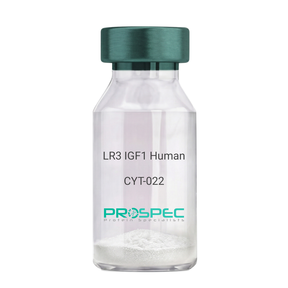
    LR3 IGF1 Human

    Description:

    LR3 Insulin Like Growth Factor-1 Human Recombinant

    R3 IGF1, R3 IGF-1, R3IGF1, R3IGF-1, LONG IGF1, LONG IGF-1, LONG R3 IGF1, LONG R3IGF1, LONG R3 IGF-1, LONG R3IGF-1.

    Product # :

    CYT-022

    Price :

    Quantity :

    Shipping Method :

    Room Temp Icon

    Shipped at Room temp

    Add To Cart

    More Info

    • description
    • source
    • formulation
    • purity
    • biological activity
    • More Info
    • sds-page

    Description

    The LR3 is a long-term analog of human IGF-1, specifically designed and manufactured for mammalian cell culture to support large-scale manufacturing of recombinant biopharmaceuticals. Recombinant Human LR3 Insulin Like Growth Factor-1 produced in E.Coli is a single, non-glycosylated, polypeptide chain containing 83 amino acids and having a molecular mass of 9.1kDa.

    Source

    Escherichia Coli.

    Formulation

    Lyophilized from a 0.2µm filtered concentrated solution in 20mM PB, pH 7.2.

    Purity

    Greater than 98.0% as determined by SDS-PAGE.

    Biological Activity

    The ED50 as determined by the stimulation of protein synthesis in L6 myoblasts is less than 10ng/ml, corresponding to a specific activity of 100,000units/mg.

    sds-page

    LR3 IGF1 sds-page - Product image 1

    More Info

    • Introduction

      IGF-1 (Insulin-like growth factor-1) is a major hormonal mediator of statural growth. Under regular circumstances, GH (growth hormone) binds to its receptor in the liver, and other tissues, and stimulates the synthesis/secretion of IGF-1. In target tissues, the Type 1 IGF receptor, that is homologous to the insulin receptor, is activated by IGF-1, leading to intracellular signaling which stimulates multiple processes leading to statural growth. IGF-1 metabolic actions are partly directed at stimulating the uptake of glucose, fatty acids, and amino acids so that metabolism supports growing tissues.

    • Synonyms

      R3 IGF1, R3 IGF-1, R3IGF1, R3IGF-1, LONG IGF1, LONG IGF-1, LONG R3 IGF1, LONG R3IGF1, LONG R3 IGF-1, LONG R3IGF-1.

    • Physical Appearance

      Sterile Filtered White lyophilized (freeze-dried) powder.

    • Stability

      Lyophilized LR3 IGF1 although stable at room temperature for 3 weeks, should be stored desiccated below -18°C. Upon reconstitution the LR3 IGF1 should be stored at 4°C between 2-7 days and for future use below -18°C.For long term storage it is recommended to add a carrier protein (0.1% HSA or BSA).Please prevent freeze-thaw cycles.

    • Solubility

      It is recommended to reconstitute the lyophilized LR3 IGF1 in sterile 18M-cm H2O at a concentration of 100µg/ml, which can then be further diluted to other aqueous solutions.

    • Amino Acid Sequence

      MFPAMPLSSLFVNGPRTLCGAELVDALQFVCGDRGFYFNKPTGYGSSSRRAPQTGIV DECCFRSCDLRRLEMYCAPLKPAKSA.

    ProSpec's products are furnished for LABORATORY RESEARCH USE ONLY. The product may not be used as drugs, agricultural or pesticidal products, food additives or household chemicals.

    Long R3 Igf1 Human
  • View Data Sheet

    Name :

    LLO PEST free

    Description:

    Listeriolysin-O PEST free Recombinant

    Listeriolysin-O, LLO, hlyA.

    Product # :

    PRO-373

    Price :

    Quantity :

    Shipping Method :

    Ice Icon

    Shipped with Ice Packs

    Add To Cart

    More Info

    • description
    • source
    • formulation
    • purity
    • biological activity
    • More Info

    Description

    Recombinant Listeriolysin O s a single polypeptide protein encoded by the hlyA gene and composed of 529 residues. PEST sequence is 19 amino acids peptide located at the protein NH 2-terminus, that targets the toxin for degradation. This motif is essential for bacterial virulence.

    Source

    Escherichia Coli.

    Formulation

    The protein contains 50mM NaH2PO4, 1mM EDTA, 2.7mM KCl, 1mM DTT, 5% glycerol and 0.5M NaCl.

    Purity

    Greater than 90.0% as determined by SDS-PAGE.

    Biological Activity

    7x10⁴ HU/mg. 2mM DTT could be use to reactivate the toxin.

    More Info

    • Introduction

      Listeriolysin O (aka LLO) is a hemolysin produced by Listeria monocytogenes bacteria, the pathogen responsible for causing listeriosis. The toxin may be regarded as a virulence factor, since it is crucial for the virulence of L. monocytogenes. LLO is a single polypeptide protein encoded by the hlyA gene and composed of 529 residues. LLO is a thiol-activated cholesterol-dependent pore forming toxin protein; therefore, it is activated by reducing agents and inhibited by oxidizing agents. Still, LLO differs from other thiol-activated toxins, as its cytolytic activity is maximized at a pH of 5.5. Inside the acidic phagosomes (average pH ~ 5.9) of cells that have phagocytosed L. monocytogenes, LLO is selectively activated by maximizing activity at a pH of 5.5. Following the phagosome lysis by LLO, the bacterium breaks out into the cytosol, where it is able to grow intracellularly, and the toxin has reduced activity in the more basic cytosol. Thus, LLO permits L. monocytogenes to break out from the phagosomes into the cytosol without harming the plasma membrane of the infected cell, which allows the bacteria to live intracellularly, where they are sheltered from extracellular immune system factors such as the complement system and antibodies. LLO also brings about dephosphorylation of histone H3 and deacetylation of histone H4 in the early phases of infection, before entry of L. monocytogenes into the host cell. The pore-forming activity is not implicated in causing the histone modifications. The modifications of the histones affect the down regulation of genes encoding proteins involved in the inflammatory response. Therefore, LLO may be significant in subverting the host immune response to L. monocytogenes. At its NH2-terminus it possesses a 25 residues long typical signal sequence excited during the secretion process. Moreover, in its NH2-terminus there is also a 19 amino acids PEST- like sequence that may target this toxin for degradation. The PEST-like sequence found in LLO and is considered crucial for virulence, given that mutants lacking the sequence lysed the host cell. Nevertheless, contrary to PEST's supposed role in protein degradation, evidence implies that the PEST-like sequence may control LLO production in the cytosol rather than increase degradation of LLO.

    • Synonyms

      Listeriolysin-O, LLO, hlyA.

    • Physical Appearance

      Sterile Filtered clear solution.

    • Stability

      Store at 4°C if entire vial will be used within 2-4 weeks. Store, frozen at -20°C for longer periods of time. Avoid multiple freeze-thaw cycles.

    ProSpec's products are furnished for LABORATORY RESEARCH USE ONLY. The product may not be used as drugs, agricultural or pesticidal products, food additives or household chemicals.

    Listeriolysin O Pest Free
  • View Data Sheet

    Name :

    TNFR Mouse

    Description:

    Tumor Necrosis Factor Receptor Mouse Recombinant

    Tumor necrosis factor receptor superfamily member 1A, Tumor necrosis factor receptor 1, TNF-R1, Tumor necrosis factor receptor type I, TNF-RI, TNFR-I, p55, p60, CD120a, Tnfrsf1a, Tnfr-1, Tnfr1, FPF, TNF-R, TNFAR, TNFRI, p55-R, TNFR60, Tnfr-2, TNF-R-I, TNF-R55, TNFRp55, TNF-alphaR1, TNFalpha-R1.

    Product # :

    CYT-774

    Price :

    Quantity :

    Shipping Method :

    Room Temp Icon

    Shipped at Room temp

    Add To Cart

    More Info

    • description
    • source
    • formulation
    • purity
    • biological activity
    • More Info

    Description

    TNFR Mouse Recombinant produced in E.coli is a single, non-glycosylated polypeptide chain containing 191 amino acids and having a molecular mass of 21.1kDa.The TNFR is purified by proprietary chromatographic techniques.

    Source

    Escherichia Coli.

    Formulation

    TNFR protein was lyophilized from a 0.2µm filtered concentrated solution in PBS, pH 7.4.

    Purity

    Greater than 97.0% as determined by SDS-PAGE.

    Biological Activity

    The ED50 as determined by its ability to inhibit the TNF-α mediated cytotoxicity in the L-929 cells is less than 1.0 µg/ml, corresponding to a specific activity of > 1000 IU/mg in the presence of 0.1 ng/mL of rMuTNF-a.

    More Info

    • Introduction

      TNFR1 belongs to the TNF-receptor superfamily. TNFR1 is a receptor for TNFSF2/TNF-alpha and homotrimeric TNFSF1/lymphotoxin-alpha.
      There are 2 types of soluble TNF receptors: sTNFR-I and sTNFR-II, which act to neutralize the biological activities of TNF alpha and TNF beta. The levels of these soluble receptors seem to increase as a result of shedding of the extracellular domains of the membrane bound receptors. TNF-a, TNFR1 and TNFR2 have roles in cellular differentiation. TNFR1 and TNFR2 function in cell type-specific renal injury.
      TNFR1 is capable of signaling both cell survival and apoptosis. TNFR1-induced apoptosis requires 2 sequential signaling complexes. TNFR1 is capable of activating NF-kappaB, mediate apoptosis, and function as a regulator of inflammation. Oxidative stress promotes TNFR1 and TNFR2 self-interaction, ligand-independent and enhanced ligand-dependent TNF signaling. TNFR1 contributes to the induction of non-cytocidal TNF effects including anti-viral state and activation of the acid sphingomyelinase. Human TNFR1 has a major region which controls cell surface expression. High levels of soluble TNF receptors are found in the amniotic fluid of pregnant women.
      Germline mutations of the extracellular domains of TNFR1 are linked to the autosomal dominant periodic fever syndrome. The impaired receptor clearance is believed to be a mechanism of the disease. Familial hibernian fever (FHF) is caused by defects in TNFRSF1A gene.

    • Synonyms

      Tumor necrosis factor receptor superfamily member 1A, Tumor necrosis factor receptor 1, TNF-R1, Tumor necrosis factor receptor type I, TNF-RI, TNFR-I, p55, p60, CD120a, Tnfrsf1a, Tnfr-1, Tnfr1, FPF, TNF-R, TNFAR, TNFRI, p55-R, TNFR60, Tnfr-2, TNF-R-I, TNF-R55, TNFRp55, TNF-alphaR1, TNFalpha-R1.

    • Physical Appearance

      Sterile Filtered White lyophilized (freeze-dried) powder.

    • Stability

      Lyophilized TNFR although stable at room temperature for 3 weeks, should be stored desiccated below -18C. Upon reconstitution TNFR should be stored at 4C between 2-7 days and for future use below -18C. For long term storage it is recommended to add a carrier protein (0.1% HSA or BSA). Please prevent freeze-thaw cycles.

    • Solubility

      It is recommended to reconstitute the lyophilized TNFR in sterile 18M-cm H2O not less than 100µg/ml, which can then be further diluted to other aqueous solutions.

    • Amino Acid Sequence

      IHPSGVTGLV PSLGDREKRD SLCPQGKYVH SKNNSICCTK CHKGTYLVSD CPSPGRDTVC RECEKGTFTA SQNYLRQCLS CKTCRKEMSQ VEISPCQADK DTVCGCKENQ FQRYLSETHF QCVDCSPCFN GTVTIPCKET QNTVCNCHAG FFLRESECVP CSHCKKNEEC MKLCLPPPLA NVTNPQDSGT A.

    ProSpec's products are furnished for LABORATORY RESEARCH USE ONLY. The product may not be used as drugs, agricultural or pesticidal products, food additives or household chemicals.

    Tnfr Mouse
  • View Data Sheet

    Name :

    IgG1 Fc Human

    Description:

    Immunoglobulin Heavy Constant Gamma 1 Human Recombinant

    IGHG1, IGHG-1, IGG-1FC, IGG1FC, IGG1-FC

    Product # :

    PRO-2763

    Price :

    Quantity :

    Shipping Method :

    Ice Icon

    Shipped with Ice Packs

    Add To Cart

    More Info

    • description
    • source
    • formulation
    • purity
    • biological activity
    • More Info

    Description

    IgG1 Fc Human Recombinant produced in HEK is a single polypeptide chain containing 231 amino acids (100-330) and having a molecular mass of 25.9kDa. IgG1 Fc is purified by proprietary chromatographic techniques.

    Source

    HEK293 Cells

    Formulation

    The IgG1 Fc solution (1mg/ml) contains 1x PBS and 10% glycerol.

    Purity

    Greater than 90% as determined by SDS-PAGE.

    Biological Activity

    Determined functional ELISA with Human Recombinant CD16a (cat# pro-2360). The ED50 range ≤ 1.5 ug/ml

    More Info

    • Introduction

      IgG1 Fcplays a role in antigen binding activity and immunoglobulin receptor binding activity. IgG1 Fc takes part in the activation of immune & defensive response. IgG1 Fc takes part in the upstream of immunoglobulin mediated immune response, positive regulation of hypersensitivity & regulation of phagocytosis.

    • Synonyms

      IGHG1, IGHG-1, IGG-1FC, IGG1FC, IGG1-FC

    • Physical Appearance

      Sterile Filtered colorless solution.

    • Stability

      Store at 4°C if entire vial will be used within 2-4 weeks. Store, frozen at -20°C for longer periods of time. For long term storage it is recommended to add a carrier protein (0.1% HSA or BSA).Avoid multiple freeze-thaw cycles.

    • Amino Acid Sequence

      PKSCDKTHTC PPCPAPELLG GPSVFLFPPK PKDTLMISRT PEVTCVVVDV SHEDPEVKFN WYVDGVEVHN AKTKPREEQY NSTYRVVSVL TVLHQDWLNG KEYKCKVSNK ALPAPIEKTI SKAKGQPREP QVYTLPPSRD ELTKNQVSLT CLVKGFYPSD IAVEWESNGQ PENNYKTTPP VLDSDGSFFL YSKLTVDKSR WQQGNVFSCS VMHEALHNHY TQKSLSLSPG K

    ProSpec's products are furnished for LABORATORY RESEARCH USE ONLY. The product may not be used as drugs, agricultural or pesticidal products, food additives or household chemicals.

    Igg1 Fc Human
  • View Data Sheet

    Name :

    MIP 3 Human

    Description:

    Macrophage Inflammatory Protein-3 Human Recombinant (CCL23)

    C-C motif chemokine 23, Small-inducible cytokine A23, Macrophage inflammatory protein 3, Myeloid progenitor inhibitory factor 1, CK-beta-8, MIP-3, MPIF-1, CKB-8, CCL23, MIP3, MPIF1, SCYA23, CKb8, Ckb-8-1.

    Product # :

    CHM-358

    Price :

    Quantity :

    Shipping Method :

    Room Temp Icon

    Shipped at Room temp

    Add To Cart

    More Info

    • description
    • source
    • formulation
    • purity
    • biological activity
    • More Info

    Description

    MIP-3 Human Recombinant produced in E.Coli is a single, non-glycosylated, polypeptide chain containing 99 amino acids and having a molecular mass of 11.3kDa. The MIP-3 is purified by proprietary chromatographic techniques.

    Source

    Escherichia Coli.

    Formulation

    Filtered (0.2µm) and lyophilized from a concentrated (1mg/ml) solution in 20mM PB, pH 7.4, 150mM NaCl.

    Purity

    Greater than 97.0% as determined by:
    (a) Analysis by RP-HPLC.
    (b) Analysis by SDS-PAGE.

    Biological Activity

    Determined by its ability to chemoattract human T cell population using a concentration range of 10-50ng/ml corresponding to a Specific Activity of 20,000-100,000IU/mg.

    More Info

    • Introduction

      CCL23 (MIP-3) is a ligand for the CCR1chemokine receptor. CCL23 is one of several cytokine genes clustered on the q-arm of chromosome 17, in a locus containing several other CC chemokines. MIP-3 chemoattracts monocytes, resting T-lymphocytes and neutrophils, but not activated lymphocytes. Furthermore, it was shown that MIP-3 inhibits colony formation of bone marrow myeloid immature progenitors. MIP-3 is mainly expressed in lung and liver tissue, but can be also found in bone marrow and placenta, as well as in some cell lines of myeloid origin.
      Alternative splicing of the CCL23 gene produces 2 mRNAs which encode a short (CK?8) and a long (CK?81) isoform of the MIP-3. CK?8 cDNA encodes a 120 amino acid residue precursor protein with a putative 21 a.a. residue signal peptide which is cleaved to generate a 99 a.a. residue mature CK?8 (a.a. 22-120). Further N-terminal processing of the 99 a.a. residue variant can produce a 75 a.a. residue CK?8 (a.a. 46-120) which is considerably more active than the 99 a.a. residue variant.
      MIP-3 may be involved in the malignant progression of certain human cancer cells which overexpress ErbB2 through the transactivation of ErbB2 tyrosine kinase. MIP-3 may also be involved in angiogenesis via upregulation of matrix metalloproteinase MMP-2 expression.

    • Synonyms

      C-C motif chemokine 23, Small-inducible cytokine A23, Macrophage inflammatory protein 3, Myeloid progenitor inhibitory factor 1, CK-beta-8, MIP-3, MPIF-1, CKB-8, CCL23, MIP3, MPIF1, SCYA23, CKb8, Ckb-8-1.

    • Physical Appearance

      Sterile Filtered White lyophilized (freeze-dried) powder.

    • Stability

      Lyophilized MIP-3 although stable at room temperature for 3 weeks, should be stored desiccated below -18°C. Upon reconstitution CCL23 should be stored at 4°C between 2-7 days and for future use below -18°C.For long term storage it is recommended to add a carrier protein (0.1% HSA or BSA).Please prevent freeze-thaw cycles.

    • Solubility

      It is recommended to reconstitute the lyophilized MIP-3 in sterile 18MΩ-cm H2O not less than 100µg/ml, which can then be further diluted to other aqueous solutions.

    • Amino Acid Sequence

      RVTKDAETEF MMSKLPLENP VLLDRFHATS ADCCISYTPR SIPCSLLESYFETNSECSKP GVIFLTKKGR RFCANPSDKQ VQVCMRMLKL DTRIKTRKN.

    ProSpec's products are furnished for LABORATORY RESEARCH USE ONLY. The product may not be used as drugs, agricultural or pesticidal products, food additives or household chemicals.

    Mip 3 Human
  • View Data Sheet

    Name :

    KIR3DL1 Human

    Description:

    Killer Cell Immunoglobulin-Like Receptor, 3 Domains Long Cytoplasmic Tail 1 Human Recombinant

    Killer cell immunoglobulin-like receptor 3DL1, MHC class I NK cell receptor, Natural killer-associated transcript 3, NKAT-3, p70 natural killer cell receptor clones CL-2/CL-11, HLA-BW4-specific inhibitory NK cell receptor, CD158 antigen-like family member E, CD158e antigen, KIR3DL1, CD158E, NKAT3, NKB1, KIR, NKB1B, CD158E1, MGC119726, MGC119728, MGC126589, MGC126591.

    Product # :

    PRO-427

    Price :

    Quantity :

    Shipping Method :

    Ice Icon

    Shipped with Ice Packs

    Add To Cart

    More Info

    • description
    • source
    • formulation
    • purity
    • More Info

    Description

    Recombinant KIR3DL1 produced in E.Coli is a single, non-glycosylated polypeptide chain containing 132 amino acids and having a molecular mass of 15 kDa.The KIR3DL1 is purified by proprietary chromatographic techniques.

    Source

    Escherichia Coli.

    Formulation

    The protein (1mg/ml) contains 25mM Tris-HCl (pH7.5) and 100mM NaCl.

    Purity

    Greater than 95.0% as determined by(a) Analysis by RP-HPLC.
    (b) Analysis by SDS-PAGE.

    More Info

    • Introduction

      Killer-cell immunoglobulin-like receptors (KIRs), are a family of cell surface glycoproteins found on Natural Killer (NK) Cells, which are important cells of the immune system. They control the killing function of these cells by interacting with MHC class I molecules, which are expressed on all cell types. This interaction allows them to identify virally infected cells or tumor cells that have a distinctive low level of Class I MHC on their surface. The majority of KIRs are inhibitory, which means that their recognition of MHC suppresses the cytotoxic activity of their NK cell. Only a limited number of KIRs have the capacity to activate cells.
      The KIR genes are found in a cluster on chromosome 19q13.4 within the 1 Mb leukocyte receptor complex (LRC). KIR molecules are extremely polymorphic, meaning their gene sequences differ significantly between individuals, so that different individuals have different arrays/repertoires of KIR genes.
      The KIR proteins are categorized by the number of extracellular immunoglobulin domains (2D or 3D) and by whether they have a long (L) or short (S) cytoplasmic domain. KIR proteins with the long cytoplasmic domain transduce inhibitory signals upon ligand binding via an immune tyrosine-based inhibitory motif (ITIM). Whereas KIR proteins with the short cytoplasmic domain lack the ITIM motif and instead associate with the TYRO protein tyrosine kinase binding protein to transduce activating signals.
      The three Ig-domain from of inhibitory killer cell Ig-like receptor 1(KIR3DL1, NKB1, nkat3, p70KIR) is a NK cell receptor for polymorphic HLA-B determinant. KIR3DLl recognizes the Bw4 determinant defined by sequence motifs at positions 77-83 of the HLA-B heavy chain. The cytoplasmic tail of KIR, which contains two immunoreceptor tyrosine-based inhibition motifs (ITIMs), mediates inhibitory signal transduction that prevents killer cell-mediated cytotoxicity. A His-tag fusion protein of KIR3DL1 cytoplasmic tail (361-444aa) was overexpressed as insoluble protein aggregates (inclusion bodies).

    • Synonyms

      Killer cell immunoglobulin-like receptor 3DL1, MHC class I NK cell receptor, Natural killer-associated transcript 3, NKAT-3, p70 natural killer cell receptor clones CL-2/CL-11, HLA-BW4-specific inhibitory NK cell receptor, CD158 antigen-like family member E, CD158e antigen, KIR3DL1, CD158E, NKAT3, NKB1, KIR, NKB1B, CD158E1, MGC119726, MGC119728, MGC126589, MGC126591.

    • Physical Appearance

      Sterile filtered colorless solution.

    • Stability

      Store at 4°C if entire vial will be used within 2-4 weeks. Store, frozen at -20°C for longer periods of time. For long term storage it is recommended to add a carrier protein (0.1% HSA or BSA).Avoid multiple freeze-thaw cycles.

    • Amino Acid Sequence

      MRGSHHHHHH GMASMTGGQQ MGRDLYDDDD KDRWGSTSGT IDKLDIEFHLWCSNKKNAAV MDQEPAGNRT ANSEDSDEQD PEEVTYAQLD HCVFTQRKIT RPSQRPKTPP TDTILYTELP NAKPRSKVVS CP.

    ProSpec's products are furnished for LABORATORY RESEARCH USE ONLY. The product may not be used as drugs, agricultural or pesticidal products, food additives or household chemicals.

    Kir3Dl1 Human
  • View Data Sheet

    Name :

    IL 15 Human, His

    Description:

    Interleukin-15 Human Recombinant, His Tag

    IL-15, MGC9721.

    Product # :

    CYT-490

    Price :

    Quantity :

    Shipping Method :

    Ice Icon

    Shipped with Ice Packs

    Add To Cart

    More Info

    • description
    • source
    • formulation
    • purity
    • biological activity
    • More Info

    Description

    Interleukin-15 His Human Recombinant ?produced in E.Coli is single, a non-glycosylated, Polypeptide chain containing 123 amino acids fragment (49-162) having a total molecular weight of 13.9 kDa with an C-terminal hexahistidine tag. The IL-15 His is purified by proprietary chromatographic techniques.

    Source

    Escherichia Coli.

    Formulation

    IL-15 His is supplied in 20mM Tris-HCl buffer (pH 8.0) 0.2mM PMSF and 10% glycerol.

    Purity

    Greater than 95.0% as determined by SDS-PAGE.

    Biological Activity

    Measured in a cell proliferation assay using CTLL2 mouse cytotoxic T cells. The ED50 for this effect is < 2.5ng/ml, corresponding to a Specific Activity of 400,000units/mg.

    More Info

    • Introduction

      The protein encoded by this gene is a cytokine that regulates T and natural killer cell activation and proliferation. This cytokine and interleukine 2 share many biological activities. They are found to bind common hematopoietin receptor subunits, and may compete for the same receptor, and thus negatively regulate each other's activity. The number of CD8+ memory cells is shown to be controlled by a balance between this cytokine and IL2. This cytokine induces the activation of JAK kinases, as well as the phosphorylation and activation of transcription activators STAT3, STAT5, and STAT6. Studies of the mouse counterpart suggested that this cytokine may increase the expression of apoptosis inhibitor BCL2L1/BCL-x(L), possibly through the transcription activation activity of STAT6, and thus prevent apoptosis. Two alternatively spliced transcript variants of this gene encoding the same protein have been reported.

    • Synonyms

      IL-15, MGC9721.

    • Physical Appearance

      Sterile Filtered clear solution.

    • Stability

      Store at 4°C if entire vial will be used within 2-4 weeks. Store, frozen at -20°C for longer periods of time. Please avoid freeze thaw cycles.

    • Amino Acid Sequence

      MNWVNVISDL KKIEDLIQSM HIDATLYTES DVHPSCKVTA MKCFLLELQV ISLESGDASI HDTVENLIIL ANNSLSSNGN VTESGCKECE ELEEKNIKEF LQSFVHIVQM FINTSLEHHH HHH.

    ProSpec's products are furnished for LABORATORY RESEARCH USE ONLY. The product may not be used as drugs, agricultural or pesticidal products, food additives or household chemicals.

    Il 15 Human His
  • View Data Sheet

    Name :

    IL 9 Human, Sf9

    Description:

    Interleukin-9 Human Recombinant, Sf9

    Interleukin 9, T-Cell Growth Factor P40,Homolog Of Mouse T Cell And Mast Cell Growth Factor 40, P40 T-Cell And Mast Cell Growth Factor, P40 Cytokine, Cytokine P40, IL-9, Interleukin-9, HP40, P40.

    Product # :

    CYT-866

    Price :

    Quantity :

    Shipping Method :

    Ice Icon

    Shipped with Ice Packs

    Add To Cart

    More Info

    • description
    • source
    • formulation
    • purity
    • More Info

    Description

    IL-9 produced in Sf9 Baculovirus cells is a single, glycosylated polypeptide chain (19-144 a.a.) including an 18 aa signal peptide (1-18 aa) and fused to a 6 aa His Tag at C-terminus containing a total of 150 amino acids and having a molecular mass of 14.5kDa.IL9 shows multiple bands between 18-28kDa on SDS-PAGE, reducing conditions and purified by proprietary chromatographic techniques.

    Source

    Sf9, Baculovirus cells.

    Formulation

    IL-9 protein solution (0.5mg/ml) contains Phosphate buffered saline (pH7.4) and 10% glycerol.

    Purity

    Greater than 90.0% as determined by SDS-PAGE.

    More Info

    • Introduction

      IL-9 is a factor which is thought to be a regulator of hematopoiesis. It has been shown to enhance the growth of human mast cells and megakaryoblastic leukemic cells as well as murine helper t-cell clones. IL-9 is a glycoprotein with a molecular weight of 32-39 that is derived from T-cells, and maps to human chromosome 5.

    • Synonyms

      Interleukin 9, T-Cell Growth Factor P40,Homolog Of Mouse T Cell And Mast Cell Growth Factor 40, P40 T-Cell And Mast Cell Growth Factor, P40 Cytokine, Cytokine P40, IL-9, Interleukin-9, HP40, P40.

    • Physical Appearance

      Sterile filtered colorless solution.

    • Stability

      Store at 4°C if entire vial will be used within 2-4 weeks. Store, frozen at -20°C for longer periods of time. For long term storage it is recommended to add a carrier protein (0.1% HSA or BSA).Avoid multiple freeze-thaw cycles.

    • Amino Acid Sequence

      MLLAMVLTSA LLLCSVAGQG CPTLAGILDI NFLINKMQED PASKCHCSAN VTSCLCLGIP SDNCTRPCFS ERLSQMTNTT MQTRYPLIFS RVKKSVEVLK NNKCPYFSCE QPCNQTTAGN ALTFLKSLLE IFQKEKMRGM RGKIHHHHHH.

    ProSpec's products are furnished for LABORATORY RESEARCH USE ONLY. The product may not be used as drugs, agricultural or pesticidal products, food additives or household chemicals.

    Il 9 Human Sf9
  • View Data Sheet

    Name :

    IL36B Human

    Description:

    Interleukin-36 Beta Human Recombinant

    Interleukin 36 beta, interleukin 1 family member 8 (eta), Interleukin-1 homolog 2, IL1F8 (Canonical product IL-1F8a), IL-1F8 (FIL1-eta), Interleukin-1 Superfamily e, IL1H2, MGC126880, MGC126882.

    Product # :

    CYT-159

    Price :

    Quantity :

    Shipping Method :

    Room Temp Icon

    Shipped at Room temp

    Add To Cart

    More Info

    • description
    • source
    • formulation
    • purity
    • biological activity
    • More Info

    Description

    IL36B Human Recombinant produced in E.Coli is a single, non-glycosylated, polypeptide chain containing 157 amino acids and having a molecular mass of 17.7kDa.The IL36B is purified by proprietary chromatographic techniques.

    Source

    Escherichia Coli.

    Formulation

    Lyophilized from a 0.2µm filtered concentrated solution in 1×PBS, pH 7.4.

    Purity

    Greater than 95.0% as determined by SDS-PAGE.

    Biological Activity

    Measured by its binding ability in a functional ELISA to bind recombinant human IL-1 Rrp2 Fc Chimera.

    More Info

    • Introduction

      Human IL-36b belongs to the IL-1 family that includes IL-1b, IL-1a, IL-1ra, IL-18, IL-36ra (IL1F5), IL-36b (IL1F8), IL-36g (IL1F9), IL-37 (IL1F7) and IL-38 (IL-1F10). The IL-1 family members display a 12 b-strand, b-trefoil configuration, and are thought to have ascended from a mutual ancestral gene. IL-36 beta is known to be actively secreted. Cells expressing IL-36 beta include resting and activated monocytes and B cells. The receptor for IL-36 beta is a blend of IL-1 Rrp2 and IL-1 RAcP. Recombinant IL-36 beta stimulates processes involving NF-kB and MAPK in an IL-1 Rrp2-dependent manner.

    • Synonyms

      Interleukin 36 beta, interleukin 1 family member 8 (eta), Interleukin-1 homolog 2, IL1F8 (Canonical product IL-1F8a), IL-1F8 (FIL1-eta), Interleukin-1 Superfamily e, IL1H2, MGC126880, MGC126882.

    • Physical Appearance

      Sterile Filtered White lyophilized (freeze-dried) powder.

    • Stability

      Lyophilized IL36B Human although stable at room temperature for 3 weeks, should be stored desiccated below -18°C. Upon reconstitution IL36B should be stored at 4°C between 2-7 days and for future use below -18°C.Please prevent freeze-thaw cycles.

    • Solubility

      It is recommended to reconstitute the lyophilized IL36B in sterile 18M-cm H2O not less than 100µg/ml, which can then be further diluted to other aqueous solutions.

    • Amino Acid Sequence

      MNPQREAAPK SYAIRDSRQM VWVLSGNSLI AAPLSRSIKP VTLHLIACRD TEFSDKEKGN MVYLGIKGKD LCLFCAEIQG KPTLQLKEKN IMDLYVEKKA QKPFLFFHNK EGSTSVFQSV SYPGWFIATS TTSGQPIFLT KERGITNNTN FYLDSVE

    ProSpec's products are furnished for LABORATORY RESEARCH USE ONLY. The product may not be used as drugs, agricultural or pesticidal products, food additives or household chemicals.

    Il36B Human
  • View Data Sheet

    Name :

    IL36G Human

    Description:

    Interleukin-36 Gamma Human Recombinant

    Interleukin 36 gamma, IL1F9, interleukin 1 family member 9, Interleukin-1 epsilon, IL-1RP2, IL-1H1, IL1E, interleukin 1-related protein 2, Interleukin-1 homolog 1.

    Product # :

    CYT-160

    Price :

    Quantity :

    Shipping Method :

    Room Temp Icon

    Shipped at Room temp

    Add To Cart

    More Info

    • description
    • source
    • formulation
    • purity
    • biological activity
    • More Info

    Description

    IL36G Human Recombinant produced in E.Coli is a single, non-glycosylated, polypeptide chain containing 169 amino acids and having a molecular mass of 18.7kDa.The IL36G is purified by proprietary chromatographic techniques.

    Source

    Escherichia Coli.

    Formulation

    Lyophilized from a 0.2µm filtered concentrated solution in 1×PBS, pH 7.4 and 5% trehalose.

    Purity

    Greater than 95.0% as determined by:
    (a) Analysis by RP-HPLC.
    (b) Analysis by SDS-PAGE.

    Biological Activity

    Measured by its binding ability in a functional ELISA to bind recombinant human IL-1 Rrp2 Fc Chimera.

    More Info

    • Introduction

      IL-36gamma belongs to the IL-1 family which includes IL-1b, IL-1a, IL-1ra, IL-18, IL-36 Ra (IL-1F5), IL-36a (IL-1F6), IL-36b (IL-1F8), IL-37 (IL-1F7) and IL-1F10. ). The IL-1 family members display a 12 b-strand, b-trefoil configuration, and are thought to have ascended from a mutual ancestral gene. IL-36g is an 18-22 kDa, 169aa intracellular and secreted protein which holds no signal sequence, no prosegment and no potential N-linked glycosylation sites. Human IL-36g shares 58%- 69% aa sequence homology with mouse, rat, bovine and equine IL-36g, and 23 - 57% aa sequence homology with other family members. The IL-36g receptor is a mixture of IL-1 Rrp2, mostly located in epithelia and keratinocytes, and the extensively expressed IL-1 RAcP. All IL-36 (a, b and g) activate N F-?B and MAPK pathways in an IL-1 Rrp2 dependent reaction. Additionally, IL-36g induces production of inflammatory cytokines and chemokines like CXCL8/IL-8.

    • Synonyms

      Interleukin 36 gamma, IL1F9, interleukin 1 family member 9, Interleukin-1 epsilon, IL-1RP2, IL-1H1, IL1E, interleukin 1-related protein 2, Interleukin-1 homolog 1.

    • Physical Appearance

      Sterile Filtered White lyophilized (freeze-dried) powder.

    • Stability

      Lyophilized IL36g Human although stable at room temperature for 3 weeks, should be stored desiccated below -18°C. Upon reconstitution IL36g should be stored at 4°C between 2-7 days and for future use below -18°C.Please prevent freeze-thaw cycles.

    • Solubility

      It is recommended to reconstitute the lyophilized IL36g in sterile 18M-cm H2O not less than 100µg/ml, which can then be further diluted to other aqueous solutions.

    • Amino Acid Sequence

      MRGTPGDADG GGRAVYQSMC KPITGTINDL NQQVWTLQGQ NLVAVPRSDS VTPVTVAVIT CKYPEALEQG RGDPIYLGIQ NPEMCLYCEK VGEQPTLQLK EQKIMDLYGQ PEPVKPFLFY RAKTGRTSTL ESVAFPDWFI ASSKRDQPII LTSELGKSYN TAFELNIND

    ProSpec's products are furnished for LABORATORY RESEARCH USE ONLY. The product may not be used as drugs, agricultural or pesticidal products, food additives or household chemicals.

    Il36G Human
  • View Data Sheet

    Name :

    Resistin Rat

    Description:

    Resistin Rat Recombinant

    Cysteine-rich secreted protein FIZZ3, Adipose tissue-specific secretory factor, ADSF, C/EBP-epsilon-regulated myeloid-specific secreted cysteine-rich protein, Cysteine-rich secreted protein A12-alpha-like 2, RSTN, XCP1, RETN1, MGC126603, MGC126609.

    Product # :

    CYT-1129

    Price :

    Quantity :

    Shipping Method :

    Room Temp Icon

    Shipped at Room temp

    Add To Cart

    More Info

    • description
    • source
    • formulation
    • purity
    • More Info

    Description

    Resistin Rat Recombinant produced in E.Coli is disulfide-linked homodimer consisting of 2x95 amino acid polypeptide chains and having a molecular mass of approximately 20.2kDa.Resistin is purified by proprietary chromatographic techniques.

    Source

    Escherichia Coli.

    Formulation

    Lyophilized from a 0.2μm filtered concentrated solution in PBS, pH7.4 and 0.02 % Tween-20.

    Purity

    Greater than 97.0% as determined by:
    (a) Analysis by RP-HPLC.
    (b) Analysis by SDS-PAGE.

    More Info

    • Introduction

      Resistin, a product of the RSTN gene, is a peptide hormone belongs to the class of cysteine-rich secreted proteins (monomeric peptide contains 11 cysteine residues) referred to as the RELM family, and is also described as ADSF (Adipose Tissue-Specific Secretory Factor) or FIZZ3 (Found in Inflammatory Zone 3). Resistin may be an important link between obesity. Mouse resistin, specifically produced and secreted by adipocyte, acts on skeletal muscle myocytes, hepatocytes and adipocytes themselves so that it reduces their sensitivity. Steppan et al. have suggested that resistin suppressed the ability to stimulate glucose uptake. They have also suggested that resistin was present at elevated levels in blood of obese mice, and was down regulated by fasting and by antidiabetic drugs. Way et al., on the other hand, have found that resistin expression is severely suppressed in obesity.
      Other studies have shown that mouse resistin increases during the differentiation of adipocytes, but it also seems to inhibit adipogenesis. In contrast, the human adipogenic differentiation is likely to be associated with a down regulation of resistin gene expression.

    • Synonyms

      Cysteine-rich secreted protein FIZZ3, Adipose tissue-specific secretory factor, ADSF, C/EBP-epsilon-regulated myeloid-specific secreted cysteine-rich protein, Cysteine-rich secreted protein A12-alpha-like 2, RSTN, XCP1, RETN1, MGC126603, MGC126609.

    • Physical Appearance

      Sterile Filtered White lyophilized (freeze-dried) powder.

    • Stability

      Lyophilized Resistin although stable at room temperature for 3 weeks, should be stored desiccated below -18°C. Upon reconstitution Resistin should be stored at 4°C between 2-7 days and for future use below -18°C. Please prevent freeze-thaw cycles.

    • Solubility

      It is recommended to reconstitute the lyophilized Resistin in sterile PBS not less than 100µg/ml, which can then be further diluted to other aqueous solutions.

    • Amino Acid Sequence

      MPSMSLCPMD EAISKKINQD FSSLLPAAMK NTVLHCWSVS SRGRLASCPE GTTVTSCSCG SGCGSWDVRE DTMCHCQCGS IDWTAARCCT LRVGS.

    ProSpec's products are furnished for LABORATORY RESEARCH USE ONLY. The product may not be used as drugs, agricultural or pesticidal products, food additives or household chemicals.

    Rat Resistin Protein
  • View Data Sheet

    Name :

    Ara h 1.0101

    Description:

    Allergen Ara h 1.0101 Recombinant

    Allergen Ara h 1, clone P41B, Allergen Ara h I, Allergen, Ara h 1.

    Product # :

    ALR-006

    Price :

    Quantity :

    Shipping Method :

    Ice Icon

    Shipped with Ice Packs

    Add To Cart

    More Info

    • description
    • source
    • formulation
    • purity
    • More Info

    Description

    Recombinant Allergen Ara h 1.0101 produced in SF9 is a glycosylated, polypeptide chain having a calculated molecular mass of 63,484 Dalton. Ara h 1.0101 is expressed with a 9xHis tag at N-terminus and purified by proprietary chromatographic techniques.

    Source

    Sf9 insect cells.

    Formulation

    Ara h 1.0101 is supplied in 20mM HEPES buffer pH-8.0, 100mM Nacl and 6M Urea.

    Purity

    Greater than 95.0% as determined by SDS-PAGE.

    More Info

    • Introduction

      Ara h 1 is a seed storage protein from Arachis hypogaea which causes an allergic reaction in human. Ara h 1 is found in 95% of peanut allergic patients from North America. Nevertheless, peanut-allergic patients of European populations have fewer sensitizations to Ara h 1.

    • Synonyms

      Allergen Ara h 1, clone P41B, Allergen Ara h I, Allergen, Ara h 1.

    • Physical Appearance

      Sterile Filtered clear solution.

    • Stability

      Store at 4°C if entire vial will be used within 2-4 weeks. Store, frozen at -20°C for longer periods of time. Avoid multiple freeze-thaw cycles.

    • Immunological Functions

      1. Binds IgE type human antibodies. 2. Immunodot test with positive/negative sera panels.

    ProSpec's products are furnished for LABORATORY RESEARCH USE ONLY. The product may not be used as drugs, agricultural or pesticidal products, food additives or household chemicals.

    Ara H 10101
  • View Data Sheet

    Name :

    CD14 Antibody, Biotin

    Description:

    CD14, Mouse Anti-Human Biotin

    Monocyte differentiation antigen CD14, Myeloid cell-specific leucine-rich glycoprotein.

    Product # :

    ANT-252

    Price :

    Quantity :

    Shipping Method :

    Room Temp Icon

    Shipped at Room temp

    Add To Cart

    More Info

    • formulation
    • More Info

    Formulation

    1mg/ml in PBS (after reconstitution).

    More Info

    • Introduction

      CD14 (also known lipopolysaccharide (LPS) receptor) is expressed strongly on monocytes and macrophage and weakly on the surface of neutrophils. CD14 is anchored to cells by linkage to glycosylphosphatidylinositol (GPI) and functions as a high affinity receptor for complexes of LPS and LPS binding protein (LBP). Soluble CD14, also binding to LPS, acts at physiological concentration as an LPS agonist and has, at higher concentrations, an LPS antagonizing effect in cell activation. CD14 has been shown to bind apoptotic cells.

    • Synonyms

      Monocyte differentiation antigen CD14, Myeloid cell-specific leucine-rich glycoprotein.

    • Solubility

      Reconstitute with of H20. Mix gently, wash the sides of the vial and wait 30-60 seconds before use.

    • Immunogen

      Purified human B Cells.

    • Ig Subclass

      Mouse IgG1.

    • Clone

      hCD4.

    • Applications

      For staining, use 10 µl/1,000,000 cells.

    • Available Conjugates

      This antibody is also available as purified antibody and conjugated to FITC.

    • Type

      Mouse Anti Human Monoclonal.

    • Storage Procedures

      Lyophilized: store at 4°C. After reconstitution, if not intended for use within a month, aliquot and store at -20°C.

    • Purification Method

      Ion exchange column.

    ProSpec's products are furnished for LABORATORY RESEARCH USE ONLY. The product may not be used as drugs, agricultural or pesticidal products, food additives or household chemicals.

    Cd14 Antibody Biotin
  • View Data Sheet

    Name :

    G CSF Antibody

    Description:

    Granulocyte Colony Stimulating Factor, Mouse Anti-Human

    CSF-3, MGI-1G, GM-CSF beta, Pluripoietin, Filgrastim, Lenograstim, G-CSF, MGC45931, GCSF.

    Product # :

    ANT-184

    Price :

    Quantity :

    Shipping Method :

    Room Temp Icon

    Shipped at Room temp

    Add To Cart

    More Info

    • formulation
    • More Info

    Formulation

    1mg/ml in PBS (after reconstitution).

    More Info

    • Introduction

      GCSF is a cytokine that controls the production, differentiation, and function of granulocytes. The active protein is found extracellularly. Three transcript variants encoding three different isoforms have been found for this gene.
      Granulocyte/macrophage colony-stimulating factors are cytokines that act in hematopoiesis by controlling the production, differentiation, and function of 2 related white cell populations of the blood, the granulocytes and the monocytes-macrophages. This csf induces granulocytes.

    • Synonyms

      CSF-3, MGI-1G, GM-CSF beta, Pluripoietin, Filgrastim, Lenograstim, G-CSF, MGC45931, GCSF.

    • Solubility

      Reconstitute with sterile H20. Mix gently, wash the sides of the vial and wait 30-60 seconds before use.

    • Immunogen

      r.Human G-CSF.

    • Ig Subclass

      Mouse IgG.

    • Clone

      NYRhGCSF.

    • Applications

      Direct ELISA, Western Blot, Immuneprecipitation.

    • Titer

      By direct ELISA, 1:10,000 dilution will yield 0.4 O.D using alkaline phosphatase conjugated rabbit anti-mouse Ig (Jackson Laboratories).

    • Shipping Conditions

      Antibody is shipped lyophilized at ambient temperature.

    • Type

      Mouse Anti Human Monoclonal.

    • Storage Procedures

      In lyophilized form, for long periods, store at 4oC in a dry environment. After reconstitution, if not intended for use within a month, aliquot and store at -20oC.

    • Purification Method

      Protein A.

    ProSpec's products are furnished for LABORATORY RESEARCH USE ONLY. The product may not be used as drugs, agricultural or pesticidal products, food additives or household chemicals.

    G Csf Antibody
  • View Data Sheet

    Name :

    PLP antibody

    Description:

    Proteolipid Protein of Myelin, Mouse antibody

    PLP, LIPOPHILIN, PMD, MMPL, SPG2, PLP/DM20.

    Product # :

    ANT-200

    Price :

    Quantity :

    Shipping Method :

    Room Temp Icon

    Shipped at Room temp

    Add To Cart

    More Info

    • formulation
    • More Info

    Formulation

    1mg/ml in PBS (after reconstitution).

    More Info

    • Introduction

      Lipophilin is a transmembrane proteolipid protein that is the predominant myelin protein present in the central nervous system. PLP functions in myelination. plays a role in the compaction, stabilization, and maintenance of myelin sheaths, as well as in oligodendrocyte development and axonal survival. Mutations associated with PLP gene cause X-linked Pelizaeus-Merzbacher disease and spastic paraplegia type 2. Two transcript variants encoding distinct isoforms have been identified for PLP gene.
      PLP is the major myelin protein from the central nervous system and plays an important role in the formation or maintenance of the multilamellar structure of myelin.

    • Synonyms

      PLP, LIPOPHILIN, PMD, MMPL, SPG2, PLP/DM20.

    • Solubility

      Reconstitute with H20. Mix gently, wash the sides of the vial and wait 30-60 seconds before use.

    • Immunogen

      synthetic pPLP 139-151.

    • Ig Subclass

      mouse IgG1.

    • Clone

      NYRPLP.

    • Titer

      By direct ELISA, 1:10,000 dilution will yield 0.5 O.D using alkaline phosphatase conjugated rabbit anti-mouse Ig (Jackson Laboratories).

    • Shipping Conditions

      Antibody is shipped lyophilized at ambient temperature.

    • Type

      Mouse Antibody Monoclonal.

    • Storage Procedures

      In lyophilized form, for long periods, store at 4°C in a dry environment. After reconstitution, if not intended for use within a month, aliquot and store at -20°C.

    • Purification Method

      Ion exchange column.

    ProSpec's products are furnished for LABORATORY RESEARCH USE ONLY. The product may not be used as drugs, agricultural or pesticidal products, food additives or household chemicals.

    Plp Antibody
  • View Data Sheet

    Name :

    HA-tag Antibody

    Description:

    HA-tag peptide, Mouse Antibody

    Product # :

    ANT-476

    Price :

    Quantity :

    Shipping Method :

    Room Temp Icon

    Shipped at Room temp

    Add To Cart

    More Info

    • formulation
    • More Info

    Formulation

    1mg/ml in PBS (after reconstitution).

    More Info

    • Solubility

      Reconstitute with H20 to get a 1 mg/ml concentration. Mix gently, wash the sides of the vial and wait 30-60 seconds before use.

    • Immunogen

      HA-tag conjugated proteins.

    • Ig Subclass

      Mouse IgG2b/kappa.

    • Titer

      By direct ELISA, 1 : 20,000 dilution will yield >1 O.D using alkaline phosphatase conjugated rabbit anti-mouse Ig (Jackson Laboratories).

    • Shipping Conditions

      Antibody is shipped lyophilized at ambient temperature.

    • Type

      Mouse Antibody Monoclonal.

    • Storage Procedures

      In lyophilized form, for long periods, store at 4°C in a dry environment. After reconstitution, if not intended for use within a month, aliquot and store at -20°C.

    • Purification Method

      Protein A & Ion exchange column.

    ProSpec's products are furnished for LABORATORY RESEARCH USE ONLY. The product may not be used as drugs, agricultural or pesticidal products, food additives or household chemicals.

    Ha Tag Peptide Antibody
  • View Data Sheet

    Name :

    NKp46 Antibody

    Description:

    Natural Cytotoxicity Receptor NKp46, Mouse Anti Human

    Natural cytotoxicity triggering receptor 1, Natural killer cell p46-related protein, hNKp46, NK-p46, NKp46, NK cell-activating receptor, Lymphocyte antigen 94 homolog, CD335 antigen, NCR1, LY94, NCRNKp46, CD335.

    Product # :

    ANT-303

    Price :

    Quantity :

    Shipping Method :

    Ice Icon

    Shipped with Ice Packs

    Add To Cart

    More Info

    • formulation
    • More Info

    Formulation

    1mg/ml containing PBS, pH-7.4, & 0.1% Sodium Azide.

    More Info

    • Introduction

      A natural cytotoxicity receptor (NCR) NKp46 has been shown to represent a novel NK cell-specific molecule involved in human NK cell activation. The natural cytotoxicity receptors (NCRs) are a recently characterized family of Ig-like activation receptors that appear to be major triggering receptors in tumor cell recognition. The three known NCRs include NKp46 and NKp30, which are expressed on circulating NKcells, and NKp44, which is expressed only on activating NK cells. NKp46 has been implicated in NK cell-mediated lysis of several autologous tumor cells, pathogen-infected cell lines and mononuclear phagocytes infected with an intracellular bacterium. The Lysis of tumor cells by NK-cells involves recognition by NKp46 of heparan sulfate moieties of membrane heparan sulfate proteoglycans. Furthermore, NKp46 is a surface receptor involved in NK-cell cell death by apoptosis. NKp46 has two extracellular Ig-like domains followed by a ~40 residue stalk region, a type I transmembrane domain, and a short cytoplasmic tail. The extracellular Ig-like domain of NKp46 (22-255aa) is purified by FPLC gel-filtration chromatography, after refolding of the isolated inclusion bodies in a redox buffer. In addition, engagement of the antigen with the monoclonal antibody stimulates intracellular calcium levels and the synthesis of cytokines. CD59 is an NKp46 coreceptor (by physical association) together they activate cytotoxicity of human NK-cells, their engagement results in tyrosine phosphorylation of CD3-zeta chains associated with NKp46. Reduced cell surface expression of NKp46 and other NK-cell receptors is linked to the impaired NK-cell cytolytic function in viremic HIV-1 infection.

    • Synonyms

      Natural cytotoxicity triggering receptor 1, Natural killer cell p46-related protein, hNKp46, NK-p46, NKp46, NK cell-activating receptor, Lymphocyte antigen 94 homolog, CD335 antigen, NCR1, LY94, NCRNKp46, CD335.

    • Physical Appearance

      Sterile Filtered colorless solution.

    • Immunogen

      Anti-human NKp46 mAb, is derived from hybridization of mouse SP2/0 myeloma cells with spleen cells from BALB/c mice immunized with recombinant human NKp46 amino acids 22-255 purified from E. coli.

    • Ig Subclass

      Mouse IgG1 heavy chain and κ light chain.

    • Clone

      Pn1D9AT.

    • Applications

      NKp46 antibody has been tested by immunofluorescent staining with flow cytometric analysis and by Western blot analysis to assure specificity and reactivity. Since application varies, however, each investigation should be titrated by the reagent to obtain optimal results.

    • Type

      Mouse Anti Human Monoclonal.

    • Storage Procedures

      For periods up to 1 month store at 4°C, for longer periods of time, store at -20°C. Prevent freeze thaw cycles.

    • Purification Method

      NKp46 antibody was purified from mouse ascitic fluids by protein-G affinity chromatography.

    ProSpec's products are furnished for LABORATORY RESEARCH USE ONLY. The product may not be used as drugs, agricultural or pesticidal products, food additives or household chemicals.

    Nkp46 Antibody
  • View Data Sheet

    Name :

    TSLPR Human, Sf9

    Description:

    Thymic Stromal Lymphopoietin Receptor Human Recombinant, Sf9

    Cytokine receptor-like factor 2, CRLF2, Cytokine receptor-like factor 2, Cytokine receptor-like 2, IL-XR, Thymic stromal lymphopoietin protein receptor, TSLP receptor, CRL2, ILXR, TSLPR, Cytokine receptor-like factor 2 isoform 1, CRLF2Y.

    Product # :

    CYT-995

    Price :

    Quantity :

    Shipping Method :

    Ice Icon

    Shipped with Ice Packs

    Add To Cart

    More Info

    • description
    • source
    • formulation
    • purity
    • More Info

    Description

    TSLPR Human Recombinant produced in Sf9 Baculovirus cells is a single, glycosylated polypeptide chain containing 218 amino acids (23-231) and having a molecular mass of 25.2kDa (Molecular size on SDS-PAGE will appear at approximately 28-40kDa).TSLPR is fused to a 6 amino acid His-Tag at C-terminus and purified by proprietary chromatographic techniques.

    Source

    Sf9, Baculovirus cells.

    Formulation

    TSLPR protein solution (1mg/ml) containing Phosphate Buffered Saline (pH 7.4) and 10% glycerol.

    Purity

    Greater than 95.0% as determined by SDS-PAGE.

    More Info

    • Introduction

      Thymic Stromal Lymphopoietin Receptor (TSLPR) is a member of the type I cytokine receptor family. TSLPR alongside the interleukin 7 receptor (IL7R) activate STAT3, STAT5, and JAK2 pathways, which is regulated processes such as cell proliferation and development of the hematopoietic system. Two transcript variants encoding dissimilar isoforms have been found for this gene. TSLPR is expressed in heart, skeletal muscle, kidney and adult and fetal liver.

    • Synonyms

      Cytokine receptor-like factor 2, CRLF2, Cytokine receptor-like factor 2, Cytokine receptor-like 2, IL-XR, Thymic stromal lymphopoietin protein receptor, TSLP receptor, CRL2, ILXR, TSLPR, Cytokine receptor-like factor 2 isoform 1, CRLF2Y.

    • Physical Appearance

      Sterile Filtered clear solution.

    • Stability

      Store at 4°C if entire vial will be used within 2-4 weeks. Store, frozen at -20°C for longer periods of time. For long term storage it is recommended to add a carrier protein (0.1% HSA or BSA).Avoid multiple freeze-thaw cycles.

    • Amino Acid Sequence

      ADPQGGAAEG VQIQIIYFNL ETVQVTWNAS KYSRTNLTFH YRFNGDEAYD QCTNYLLQEG HTSGCLLDAE QRDDILYFSI RNGTHPVFTA SRWMVYYLKP SSPKHVRFSW HQDAVTVTCS DLSYGDLLYE VQYRSPFDTE WQSKQENTCN VTIEGLDAEK CYSFWVRVKA MEDVYGPDTY PSDWSEVTCW QRGEIRDACA ETPTPPKPKL SKHHHHHH.

    ProSpec's products are furnished for LABORATORY RESEARCH USE ONLY. The product may not be used as drugs, agricultural or pesticidal products, food additives or household chemicals.

    Tslpr Human Sf9
  • View Data Sheet

    Name :

    BAFF Antibody

    Description:

    B-cell Activating Factor, Mouse Anti Human

    BAFF, BLYS, CD257, TALL1, THANK, ZTNF4, TALL-1, TNFSF20, TNFSF13B.

    Product # :

    ANT-367

    Price :

    Quantity :

    Shipping Method :

    Ice Icon

    Shipped with Ice Packs

    Add To Cart

    More Info

    • formulation
    • More Info

    Formulation

    1mg/ml containing PBS, pH-7.4, & 0.1% Sodium Azide.

    More Info

    • Introduction

      Binds to tnfrsf13b/taci and tnfrsf17/bcma. tnfsf13/april binds to the same 2 receptors, together, they form a 2 ligands -2 receptors pathway involved in the stimulation of b- and t-cell function and the regulation of humoral immunity. A third b-cell specific baff-receptor (baffr/br3) promotes the survival of mature b-cells and the b-cell response. B Lymphocyte Stimulator functions as a potent B-cell growth factor in costimulation assays. Administration of BAFF Human recombinant to mice disrupts splenic B-cell and T-cell zones and results in elevated levels of serum immunoglobulin.

    • Synonyms

      BAFF, BLYS, CD257, TALL1, THANK, ZTNF4, TALL-1, TNFSF20, TNFSF13B.

    • Physical Appearance

      Sterile Filtered colorless solution.

    • Immunogen

      Anti-human BAFF mAb, is derived from hybridization of mouse F0 myeloma cells with spleen cells from BALB/c mice immunized with Recombinant human BAFF amino acids 134-285 purified from E. coli.

    • Ig Subclass

      Mouse IgG3 heavy chain and κ light chain.

    • Clone

      PH4C7AT.

    • Applications

      BAFF antibody has been tested by ELISA and Western blot analysis to assure specificity and reactivity. Since application varies, however, each investigation should be titrated by the reagent to obtain optimal results. Recommended dilution range for Western blot analysis is 1:500 ~ 1,000. Recommended starting dilution is 1:500.

    • Type

      Mouse Anti Human Monoclonal.

    • Storage Procedures

      For periods up to 1 month store at 4°C, for longer periods of time, store at -20°C. Prevent freeze thaw cycles.

    • Purification Method

      BAFF antibody was purified from mouse ascitic fluids by protein-G affinity chromatography.

    ProSpec's products are furnished for LABORATORY RESEARCH USE ONLY. The product may not be used as drugs, agricultural or pesticidal products, food additives or household chemicals.

    Baff Antibody
  • View Data Sheet

    Name :

    BMP8B Antibody

    Description:

    Bone Morphogenetic protein-8b, Mouse Anti Human

    Bone morphogenetic protein 8B, BMP-8, BMP-8B, Osteogenic protein 2, OP-2, BMP8B, BMP8, Bone Morphogenetic protein-8b, OP2.

    Product # :

    ANT-558

    Price :

    Quantity :

    Shipping Method :

    Ice Icon

    Shipped with Ice Packs

    Add To Cart

    More Info

    • formulation
    • More Info

    Formulation

    1mg/ml containing PBS, pH-7.4, 10% Glycerol and 0.02% Sodium Azide.

    More Info

    • Introduction

      Bone Morphogenetic protein-8b (BMP8B) belongs to a family of secreted signaling molecules which can induce ectopic bone growth. BMP8B is known for having a possible bone inductive activity as it is related to BMP5 and BMP7. BMP8B is the osteoinductive factor accountable for epithelial osteogenesis.

    • Synonyms

      Bone morphogenetic protein 8B, BMP-8, BMP-8B, Osteogenic protein 2, OP-2, BMP8B, BMP8, Bone Morphogenetic protein-8b, OP2.

    • Physical Appearance

      Sterile filtered colorless solution.

    • Immunogen

      Anti-human BMP8B mAb, is derived from hybridization of mouse F0 myeloma cells with spleen cells from BALB/c mice immunized with a recombinant human BMP8B protein 264-402 amino acids purified from E. coli.

    • Ig Subclass

      Mouse IgG2a heavy chain and k light chain.

    • Clone

      PAT13E6AT.

    • Applications

      BMP8B antibody has been tested by ELISA, Western blot analysis to assure specificity and reactivity. Since application varies, however, each investigation should be titrated by the reagent to obtain optimal results. Recommended starting dilution is 1:1000.

    • Type

      Mouse Anti Human Monoclonal.

    • Storage Procedures

      For periods up to 1 month store at 4°C, for longer periods of time, store at -20°C. Prevent freeze thaw cycles.

    • Purification Method

      BMP8B antibody was purified from mouse ascitic fluids by protein-A affinity chromatography.

    ProSpec's products are furnished for LABORATORY RESEARCH USE ONLY. The product may not be used as drugs, agricultural or pesticidal products, food additives or household chemicals.

    Bmp8B Antibody
  • View Data Sheet

    Name :

    ANXA1 Human

    Description:

    Annexin A1 Human Recombinant

    ANX1, LPC1, ANXA1, Lipocortin I, Calpactin II, Chrombindin-9, p35, Annexin-1, Phospholipase A2 inhibitory protein, Annexin I, Annexin A1.

    Product # :

    PRO-679

    Price :

    Quantity :

    Shipping Method :

    Ice Icon

    Shipped with Ice Packs

    Add To Cart

    More Info

    • description
    • source
    • formulation
    • purity
    • More Info

    Description

    ANXA1 produced in E.Coli is a single, non-glycosylated polypeptide chain containing 346 amino acids (1-346 a.a.) and having a molecular mass of 38.7 kDa.ANXA1 is purified by proprietary chromatographic techniques.

    Source

    Escherichia Coli.

    Formulation

    The ANXA1 protein solution contains 20mM Tris-HCl, pH-8, 100mM NaCl, 1mM DTT and 10% glycerol.

    Purity

    Greater than 90.0% as determined by SDS-PAGE.

    More Info

    • Introduction

      ANXA1 is part of the family of Ca(2+)-dependent phospholipid binding proteins which have a Mw between 35kDa-40kDa and are situated on the cytosolic face of the plasma membrane. ANXA1 protein has a Mw of 40kDa, with phospholipase A2 inhibitory activity to bind from two to four calcium ions with high affinity. Since phospholipase A2 is necessary for the biosynthesis of the potent mediators of inflammation, prostaglandins and leukotrienes, ANXA1 might have potential anti-inflammatory activity. ANXA1 promotes membrane fusion and iplays a role in exocytosis. The recognition of ANXA1 protein by immunocytochemical leads a simple, highly sensitive and specific assay for diagnosis of hairy cell leukemia.

    • Synonyms

      ANX1, LPC1, ANXA1, Lipocortin I, Calpactin II, Chrombindin-9, p35, Annexin-1, Phospholipase A2 inhibitory protein, Annexin I, Annexin A1.

    • Physical Appearance

      Sterile filtered colorless solution.

    • Stability

      Store at 4°C if entire vial will be used within 2-4 weeks. Store, frozen at -20°C for longer periods of time. For long term storage it is recommended to add a carrier protein (0.1% HSA or BSA).Avoid multiple freeze-thaw cycles.

    • Amino Acid Sequence

      MAMVSEFLKQ AWFIENEEQE YVQTVKSSKG GPGSAVSPYP TFNPSSDVAA LHKAIMVKGV DEATIIDILT KRNNAQRQQI KAAYLQETGK
      PLDETLKKAL TGHLEEVVLA LLKTPAQFDA DELRAAMKGL GTDEDTLIEI LASRTNKEIR DINRVYREEL KRDLAKDITS DTSGDFRNAL
      LSLAKGDRSE DFGVNEDLAD SDARALYEAG ERRKGTDVNV FNTILTTRSY PQLRRVFQKY TKYSKHDMNK VLDLELKGDI EKCLTAIVKCATSKPAFFAE KLHQAMKGVG TRHKALIRIM VSRSEIDMND IKAFYQKMYG ISLCQAILDE TKGDYEKILV ALCGGN.

    ProSpec's products are furnished for LABORATORY RESEARCH USE ONLY. The product may not be used as drugs, agricultural or pesticidal products, food additives or household chemicals.

    Anxa1 Human
  • View Data Sheet

    Name :

    TNNI3 Paired Antibody

    Description:

    Mouse Anti Human Troponin I Type 3 Paired

    Troponin I cardiac muscle, Cardiac troponin I, TNNI3, TNNC1, CMH7, RCM1, cTnI, CMD2A, MGC116817.

    Product # :

    ANT-664

    Price :

    Quantity :

    Shipping Method :

    Ice Icon

    Shipped with Ice Packs

    Add To Cart

    More Info

    • description
    • formulation
    • purity
    • More Info

    Description

    TNNI3 Paired antibodies, coating and conjugating are used for lateral flow immunoassay. Please note that when ordering for example: 100µg antibody we ship 50µg from each of the antibodies (100µg in total).

    Formulation

    *Cardiac Troponin-I coating antibody (5mg/1ml) contains 50 mM Na-citrate, pH 6.0, 0.9 % NaCl and 0.095 % NaN3 as a preservative.

    * Cardiac Troponin-I conjugating antibody (1mg/1ml) contains 50 mM Na-citrate, pH 6.0, 0.9 % NaCl and 0.095 % NaN3 as a preservative.

    Purity

    Greater than 95%.

    More Info

    • Introduction

      Troponin I (TnI), troponin T (TnT) and troponin C (TnC) form the troponin complex of the thin filaments of striated muscle. TnI is acts as the inhibitory subunit by blocking actin-myosin interactions and thereby mediating striated muscle relaxation. The TnI subfamily contains 3 genes: TnI-skeletal-fast-twitch, TnI-skeletal-slow-twitch, and TnI-cardiac. The TNNI3 gene encodes the TnI-cardiac protein and is exclusively expressed in cardiac muscle tissues. Mutations in the TNNI3 gene cause familial hypertrophic cardiomyopathy type 7 (CMH7) and familial restrictive cardiomyopathy (RCM).

    • Synonyms

      Troponin I cardiac muscle, Cardiac troponin I, TNNI3, TNNC1, CMH7, RCM1, cTnI, CMD2A, MGC116817.

    • Physical Appearance

      2 vials of sterile filtered clear colorless solution.

    • Stability

      Cardiac Troponin-I although stable at 4°C for 1 week, should be stored below -18°C.For long term storage it is recommended to add a carrier protein (0.1% HSA or BSA).Please prevent freeze-thaw cycles.

    • Applications

      Lateral flow immunoassay.

    • Type

      Mouse Anti Human Monoclonal.

    • Purification Method

      Purified monoclonal IgG by protein A chromatography.

    ProSpec's products are furnished for LABORATORY RESEARCH USE ONLY. The product may not be used as drugs, agricultural or pesticidal products, food additives or household chemicals.

    Tnni3 Paired Antibody
  • View Data Sheet

    Name :

    LCN2 Human

    Description:

    Neutrophil Gelatinase Associated Lipocalin/Lipocalin-2 Human Recombinant

    Neutrophil gelatinase-associated lipocalin, NGAL, p25, 25 kDa alpha-2-microglobulin-related subunit of MMP-9, Lipocalin-2, Oncogene 24p3, LCN2.

    Product # :

    ENZ-783

    Price :

    Quantity :

    Shipping Method :

    Room Temp Icon

    Shipped at Room temp

    Add To Cart

    More Info

    • description
    • source
    • formulation
    • purity
    • biological activity
    • More Info

    Description

    LCN2 Human Recombinant produced in E.Coli is a homodimeric non-glycosylated polypeptide chains consisting of two 178 amino acids and having a molecular mass of 41.0kDa.

    Source

    Escherichia Coli.

    Formulation

    LCN2 protein was lyophilized from a 0.2 µm filtered concentrated solution in PBS, pH 7.4, with 0.05 % Tween-20.

    Purity

    Greater than 95.0% as determined by: (a) Analysis by RP-HPLC.(b) Analysis by SDS-PAGE.

    Biological Activity

    The ED50 as determined by a cell proliferation assay using human TF-1 cells is less than 0.5ng/ml, corresponding to a specific activity of > 2.0 × 1,000,000 IU/mg.

    More Info

    • Introduction

      Recombinant Human Lipocalin-2 also called Neutrophil Gelatinase Associated Lipocalin (NGAL) belongs to a family of lipocans which include 25 proteins (including a1-microglobulin and b-lactoglobulin), which are characterized by their ability to bind small lipophilic substances in their hydrophobic core.
      They thereby serve as transporters of substances like retinal, biliverdins & prostaglandins. There are indications that NGAL is involved in modulation of the inflammatory response and is found in the plasma of patients after stroke.

    • Synonyms

      Neutrophil gelatinase-associated lipocalin, NGAL, p25, 25 kDa alpha-2-microglobulin-related subunit of MMP-9, Lipocalin-2, Oncogene 24p3, LCN2.

    • Physical Appearance

      Sterile Filtered White lyophilized (freeze-dried) powder.

    • Stability

      Lyophilized LCN2 although stable at room temperature for 3 weeks, should be stored desiccated below -18°C. Upon reconstitution LCN2 should be stored at 4°C between 2-7 days and for future use below -18°C. For long term storage it is recommended to add a carrier protein (0.1% HSA or BSA). Please prevent freeze-thaw cycles.

    • Amino Acid Sequence

      QDSTSDLIPA PPLSKVPLQQ NFQDNQFQGK WYVVGLAGNA ILREDKDPQK MYATIYELKE DKSYNVTSVL FRKKKCDYWI RTFVPGCQPG EFTLGNIKSY PGLTSYLVRV VSTNYNQHAM VFFKKVSQNR EYFKITLYGR TKELTSELKE NFIRFSKSLG LPENHIVFPV PIDQCIDG

    ProSpec's products are furnished for LABORATORY RESEARCH USE ONLY. The product may not be used as drugs, agricultural or pesticidal products, food additives or household chemicals.

    Human Lcn2
  • 1
  • …
  • 27
  • 28
  • 29
  • 30
  • 31
  • …
  • 42
Your browser does not support the video tag.

Products

  • Cytokines
  • Growth Factors
  • Chemokines
  • CD Antigens
  • Neurotrophins
  • Hormones
  • Enzymes
  • Viral Antigens
  • Recombinant Proteins
  • Natural Proteins
  • Monoclonal Antibodies
  • Polyclonal Antibodies
  • Thermal Packaging

About

  • Company
  • Founder
  • Contribution
  • Contact Us
  • My Account

Ordering

  • Ordering Method
  • Ordering - Credit Card
  • Ordering PO
  • Shipping Fees
  • FAQ's

Legal

  • Terms & Conditions
  • Privacy
  • Site Map
  • info@prospecbio.com
  • Facebook
  • Instagram
  • Linkedin Profile

© 2026 ProSpec-Tany TechnoGene Ltd. All Rights Reserved.

  • Choosing a selection results in a full page refresh.
  • Opens in a new window.