Skip to content

High-quality research proteins.

  • 1-800-805-6531

  • Mon - Sat 8.00-18.00, Sun - Closed

  • info@prospecbio.com

  • PO Box 6591 East Brunswick NJ 08816

  • Products
    • Cytokines
    • Growth factors
    • Chemokines
    • Cd antigens
    • Neurotrophins
    • Hormones
    • Enzymes
    • Viral antigens
    • Recombinant proteins
    • Natural proteins
    • Monoclonal antibodies
    • Polyclonal antibodies
    • Thermal Packaging
  • About
    • Company
    • Founder
    • Contribution
  • Customers
    • Eastern Asia
    • Eastern Europe
    • North America
    • South America
    • South East Asia
    • Southern Asia
    • Western Europe
  • Ordering
    • Ordering Method
    • Ordering - Credit Card
    • Ordering - PO
    • Shipping Fees
    • FAQs
  • Services
    • DNA Cloning
    • Protein Expression
    • Monoclonal Antibody
    • Fermentation
    • Technology License
  • Contact us
Log in

Country/region

  • USD $ | Albania
  • USD $ | Andorra
  • USD $ | Argentina
  • USD $ | Armenia
  • USD $ | Australia
  • USD $ | Austria
  • USD $ | Azerbaijan
  • USD $ | Belarus
  • USD $ | Belgium
  • USD $ | Bolivia
  • USD $ | Brazil
  • USD $ | Bulgaria
  • USD $ | Cambodia
  • USD $ | Canada
  • USD $ | Chile
  • USD $ | China
  • USD $ | Colombia
  • USD $ | Costa Rica
  • USD $ | Croatia
  • USD $ | Cyprus
  • USD $ | Czechia
  • USD $ | Denmark
  • USD $ | Estonia
  • USD $ | Finland
  • USD $ | France
  • USD $ | Georgia
  • USD $ | Germany
  • USD $ | Greece
  • USD $ | Hong Kong SAR
  • USD $ | Hungary
  • USD $ | Iceland
  • USD $ | India
  • USD $ | Ireland
  • USD $ | Israel
  • USD $ | Italy
  • USD $ | Japan
  • USD $ | Kazakhstan
  • USD $ | Latvia
  • USD $ | Liechtenstein
  • USD $ | Lithuania
  • USD $ | Madagascar
  • USD $ | Malta
  • USD $ | Mexico
  • USD $ | Moldova
  • USD $ | Monaco
  • USD $ | Netherlands
  • USD $ | New Zealand
  • USD $ | North Macedonia
  • USD $ | Norway
  • USD $ | Panama
  • USD $ | Paraguay
  • USD $ | Peru
  • USD $ | Philippines
  • USD $ | Poland
  • USD $ | Portugal
  • USD $ | Romania
  • USD $ | Singapore
  • USD $ | Slovakia
  • USD $ | Slovenia
  • USD $ | South Africa
  • USD $ | South Korea
  • USD $ | Spain
  • USD $ | Sri Lanka
  • USD $ | Sweden
  • USD $ | Switzerland
  • USD $ | Taiwan
  • USD $ | Thailand
  • USD $ | Türkiye
  • USD $ | Ukraine
  • USD $ | United Kingdom
  • USD $ | United States
  • USD $ | Uruguay
  • USD $ | Vatican City
  • USD $ | Venezuela
  • USD $ | Vietnam
  • Facebook
  • Instagram
logoProSpec - Recombinant Protein Manufacturer for academic and R&D industry
Become a distributor
Account Log in
Wishlist
Cart Cart(0)
  • Products
    +
    • Cytokines
    • Growth factors
    • Chemokines
    • Cd antigens
    • Neurotrophins
    • Hormones
    • Enzymes
    • Viral antigens
    • Recombinant proteins
    • Natural proteins
    • Monoclonal antibodies
    • Polyclonal antibodies
    • Thermal Packaging
  • About
    +
    • Company
    • Founder
    • Contribution
  • Customers
    +
    • Eastern Asia
    • Eastern Europe
    • North America
    • South America
    • South East Asia
    • Southern Asia
    • Western Europe
  • Ordering
    +
    • Ordering Method
    • Ordering - Credit Card
    • Ordering - PO
    • Shipping Fees
    • FAQs
  • Services
    +
    • DNA Cloning
    • Protein Expression
    • Monoclonal Antibody
    • Fermentation
    • Technology License
  • Contact us

Item added to your cart

View cart
View All
  • Cytokines
  • Growth factors
  • Chemokines
  • Cd antigens
  • Neurotrophins
  • Hormones
  • Enzymes
  • Viral antigens
  • Recombinant proteins
  • Natural proteins
  • Monoclonal antibodies
  • Polyclonal antibodies
  • Thermal Packaging
  • Tumor Necrosis Factor

    Tumor Necrosis Factor

    View All
  • 4-1BB

    4-1BB

    View All
  • Adiponectin

    Adiponectin

    View All
  • AIF1

    AIF1

    View All
  • Other Cytokines

    Other Cytokines

    View All
  • AITRL

    AITRL

    View All
  • Angiopoietin

    Angiopoietin

    View All
  • Apolipoprotein

    Apolipoprotein

    View All
  • B-Cell Activating Factor

    B-Cell Activating Factor

    View All
  • Beta Defensin

    Beta Defensin

    View All
  • Bone Morphogenetic Protein

    Bone Morphogenetic Protein

    View All
  • B type Natriuretic Peptide

    B type Natriuretic Peptide

    View All
  • BST

    BST

    View All
  • Betacellulin

    Betacellulin

    View All
  • Cardiotrophin

    Cardiotrophin

    View All
  • Activin

    Activin

    View All
  • Fibroblast Growth Factor

    Fibroblast Growth Factor

    View All
  • Other Growth Factors

    Other Growth Factors

    View All
  • CSF

    CSF

    View All
  • CTGF

    CTGF

    View All
  • IGFBP

    IGFBP

    View All
  • VEGF Protein

    VEGF Protein

    View All
  • EGF

    EGF

    View All
  • Epigen

    Epigen

    View All
  • Erythropoietin

    Erythropoietin

    View All
  • Insulin-Like Growth Factor

    Insulin-Like Growth Factor

    View All
  • Growth Hormone

    Growth Hormone

    View All
  • HDGF

    HDGF

    View All
  • Hepatocyte Growth Factor

    Hepatocyte Growth Factor

    View All
  • Insulin

    Insulin

    View All
  • BCA-1/ BLC (CXCL13)

    BCA-1/ BLC (CXCL13)

    View All
  • BRAK (CXCL14)

    BRAK (CXCL14)

    View All
  • C-10 (CCL6)

    C-10 (CCL6)

    View All
  • Other Chemokines

    Other Chemokines

    View All
  • MEC (CCL28)

    MEC (CCL28)

    View All
  • LD78-beta (CCL3L1)

    LD78-beta (CCL3L1)

    View All
  • CTACK (CCL27)

    CTACK (CCL27)

    View All
  • CXCL16

    CXCL16

    View All
  • CXCL17

    CXCL17

    View All
  • Platelet Factor-4 (CXCL4)

    Platelet Factor-4 (CXCL4)

    View All
  • ENA-78 (CXCL5)

    ENA-78 (CXCL5)

    View All
  • Eotaxin (CCL11,24,26)

    Eotaxin (CCL11,24,26)

    View All
  • Exodus-2 (CCL21)

    Exodus-2 (CCL21)

    View All
  • Fractalkine (CX3CL1)

    Fractalkine (CX3CL1)

    View All
  • CXCL6

    CXCL6

    View All
  • Other CD Antigens

    Other CD Antigens

    View All
  • CD14

    CD14

    View All
  • CD164

    CD164

    View All
  • CD1

    CD1

    View All
  • CD2

    CD2

    View All
  • CD200

    CD200

    View All
  • CD207

    CD207

    View All
  • CD226

    CD226

    View All
  • CD23

    CD23

    View All
  • CD244

    CD244

    View All
  • CD247

    CD247

    View All
  • CD27

    CD27

    View All
  • CD274

    CD274

    View All
  • CD300

    CD300

    View All
  • CD33

    CD33

    View All
  • Other Neurotrophins

    Other Neurotrophins

    View All
  • Beta-NGF

    Beta-NGF

    View All
  • BDNF

    BDNF

    View All
  • CDNF

    CDNF

    View All
  • CNTF

    CNTF

    View All
  • GDNF

    GDNF

    View All
  • Glia Maturation Factor

    Glia Maturation Factor

    View All
  • MANF

    MANF

    View All
  • Midkine

    Midkine

    View All
  • Neuroglobin

    Neuroglobin

    View All
  • Neurotrophic factors

    Neurotrophic factors

    View All
  • Neuregulin

    Neuregulin

    View All
  • Neuropilin

    Neuropilin

    View All
  • Pigment Epithelium-Derived Factor

    Pigment Epithelium-Derived Factor

    View All
  • Pleiotrophin

    Pleiotrophin

    View All
  • Peptide Hormones

    Peptide Hormones

    View All
  • Other Hormones

    Other Hormones

    View All
  • HCG

    HCG

    View All
  • Endothelin

    Endothelin

    View All
  • Exendin

    Exendin

    View All
  • FSH

    FSH

    View All
  • Glucagon

    Glucagon

    View All
  • GHRP

    GHRP

    View All
  • GLP

    GLP

    View All
  • Thyrostimulin

    Thyrostimulin

    View All
  • Inhibin A

    Inhibin A

    View All
  • LHRH

    LHRH

    View All
  • Melanotan

    Melanotan

    View All
  • Procalcitonin

    Procalcitonin

    View All
  • PTH

    PTH

    View All
  • Peroxiredoxin

    Peroxiredoxin

    View All
  • Peptidase

    Peptidase

    View All
  • Synthetase

    Synthetase

    View All
  • Transferase

    Transferase

    View All
  • Hydrolase

    Hydrolase

    View All
  • Dehydrogenase

    Dehydrogenase

    View All
  • ACE2

    ACE2

    View All
  • Esterase

    Esterase

    View All
  • Protein Kinases

    Protein Kinases

    View All
  • Other Enzymes

    Other Enzymes

    View All
  • Phosphatase

    Phosphatase

    View All
  • Deaminase

    Deaminase

    View All
  • Oxygenase

    Oxygenase

    View All
  • Adenylate Kinase

    Adenylate Kinase

    View All
  • Lyase

    Lyase

    View All
  • Chagas

    Chagas

    View All
  • SARS

    SARS

    View All
  • Influenza

    Influenza

    View All
  • ASFV

    ASFV

    View All
  • Astrovirus

    Astrovirus

    View All
  • Borrelia

    Borrelia

    View All
  • Helicobacter Pylori

    Helicobacter Pylori

    View All
  • Chikungunya

    Chikungunya

    View All
  • Chlamydia

    Chlamydia

    View All
  • Cytomegalo

    Cytomegalo

    View All
  • Dengue

    Dengue

    View All
  • EBV

    EBV

    View All
  • Ebola

    Ebola

    View All
  • Feline Leukemia Virus

    Feline Leukemia Virus

    View All
  • Hepatitis A

    Hepatitis A

    View All
  • Other

    Other

    View All
  • Actin

    Actin

    View All
  • ADAM

    ADAM

    View All
  • Complement Component

    Complement Component

    View All
  • Ag85

    Ag85

    View All
  • Eukaryotic Translation Initiation Factor

    Eukaryotic Translation Initiation Factor

    View All
  • Anterior Gradient Protein

    Anterior Gradient Protein

    View All
  • Heat Shock Protein

    Heat Shock Protein

    View All
  • Alpha-2-HS-Glycoprotein

    Alpha-2-HS-Glycoprotein

    View All
  • Hemoglobin

    Hemoglobin

    View All
  • Allergy

    Allergy

    View All
  • Anaplasma

    Anaplasma

    View All
  • Angiogenin

    Angiogenin

    View All
  • Ankyrin Repeat Domain

    Ankyrin Repeat Domain

    View All
  • Annexin

    Annexin

    View All
  • Other Natural Proteins

    Other Natural Proteins

    View All
  • Aprotinin

    Aprotinin

    View All
  • Transferrin

    Transferrin

    View All
  • Avidin

    Avidin

    View All
  • Anti Coagulation Factors

    Anti Coagulation Factors

    View All
  • Natural Albumin

    Natural Albumin

    View All
  • Natural Coagulation Factors

    Natural Coagulation Factors

    View All
  • Fibronectin

    Fibronectin

    View All
  • Thrombin

    Thrombin

    View All
  • Anti Human Enzyme

    Anti Human Enzyme

    View All
  • Other Monoclonal Antibodies

    Other Monoclonal Antibodies

    View All
  • Anti Human Cytokine

    Anti Human Cytokine

    View All
  • Anti Human Heat Shock Protein

    Anti Human Heat Shock Protein

    View All
  • Anti Mouse Lymphocyte

    Anti Mouse Lymphocyte

    View All
  • Anti Human Chemokine

    Anti Human Chemokine

    View All
  • Anti Human Lymphocyte

    Anti Human Lymphocyte

    View All
  • Anti Viral Monoclonal

    Anti Viral Monoclonal

    View All
  • Anti Mouse Cytokine

    Anti Mouse Cytokine

    View All
  • Anti-GST Monoclonal

    Anti-GST Monoclonal

    View All
  • Other Polyclonal Antibodies

    Other Polyclonal Antibodies

    View All
  • Anti Viral

    Anti Viral

    View All
  • Anti-GST Monoclonal

    Anti-GST Monoclonal

    View All
Back

Search results

1000 results found for “Leukocyte-Associated Ig-Like Receptor”

Name

Description

Product #

Price

Quantity

Shipping Method

  • View Data Sheet

    Name :

    NAT6 Antibody

    Description:

    N-Acetyltransferase 6, Mouse Anti Human

    Protein fusion-2, FUS2, FUS-2, NAT6, N-acetyltransferase 6, Protein fus-2.

    Product # :

    ANT-395

    Price :

    Quantity :

    Shipping Method :

    Ice Icon

    Shipped with Ice Packs

    Add To Cart

    More Info
    • formulation
    • More Info

    Formulation

    1mg/ml containing PBS, pH-7.4, & 0.1% Sodium Azide.

    More Info

    • Introduction

      NAT6 is an enzyme that catalyzes the transfer of acetyl groups from acetyl-CoA to acrylamines. NAT6 is located mainly in the cytoplasm and its activity has been recognized by its feasibility to acetylate the N-terminus of proteins using a ping-pong-like mechanism and by its substrate specificity. Given that the NAT6 gene maps to the chromosomal region 3p21.3, which includes at least one tumor suppressor gene, the function of NAT6 plays an important role in cancer.

    • Synonyms

      Protein fusion-2, FUS2, FUS-2, NAT6, N-acetyltransferase 6, Protein fus-2.

    • Immunogen

      Anti-human NAT6 mAb is derived from hybridization of mouse F0 myeloma cells with spleen cells from BALB/c mice immunized with recombinant human NAT6 amino acids 1-308 purified from E. coli.

    • Ig Subclass

      Mouse IgG1 heavy chain and κ light chain.

    • Clone

      PAT2F4AT.

    • Applications

      NAT6 antibody has been tested by ELISA and Western blot analysis to assure specificity and reactivity. Since application varies, however, each investigation should be titrated by the reagent to obtain optimal results. Recommended dilution range for Western blot and Immunofluorescence analysis is 1:500 ~ 1000. Recommended starting dilution is 1:500.

    • Type

      Mouse Anti Human Monoclonal.

    • Storage Procedures

      For periods up to 1 month store at 4°C, for longer periods of time, store at -20°C. Prevent freeze thaw cycles.

    • Purification Method

      NAT6 antibody was purified from mouse ascitic fluids by protein-G affinity chromatography.

    ProSpec's products are furnished for LABORATORY RESEARCH USE ONLY. The product may not be used as drugs, agricultural or pesticidal products, food additives or household chemicals.

    Nat6 Antibody
  • View Data Sheet

    Name :

    NRAS Antibody

    Description:

    Neuroblastoma RAS Viral Oncogene Homolog, Mouse Anti Human

    GTPase NRas, HRAS1, ALPS4, N-ras, NRAS1, NS6, Transforming protein N-Ras.

    Product # :

    ANT-047

    Price :

    Quantity :

    Shipping Method :

    Ice Icon

    Shipped with Ice Packs

    Add To Cart

    More Info

    • formulation
    • More Info

    Formulation

    1mg/ml containing PBS, pH-7.4, & 0.1% Sodium Azide.

    More Info

    • Introduction

      NRAS binds GDP/GTP and has intrinsic GTPase activity. NRAS is a membrane protein that shuttles between the Golgi apparatus and the plasma membrane. This transport is regulated through palmitoylation and depalmitoylation by the ZDHHC9-GOLGA7 complex. NRAS is activated to a GTP-bound form by a GTPase activating protein and inactivated to a GDP-bound form by a guanine nucleotide-exchange factor. Defects in this NRAS gene result in juvenile myelomonocytic leukemia.

    • Synonyms

      GTPase NRas, HRAS1, ALPS4, N-ras, NRAS1, NS6, Transforming protein N-Ras.

    • Immunogen

      Anti-human NRAS mAb, is derived from hybridization of mouse FO myeloma cells with spleen cells from BALB/c mice immunized with recombinant human NRAS amino acids 1-186 purified from E. coli.

    • Ig Subclass

      Mouse IgG2a heavy chain and k light chain.

    • Clone

      PAT2G9AT.

    • Applications

      NRAS antibody has been tested by ELISA and Western blot analysis to assure specificity and reactivity. Since application varies, however, each investigation should be titrated by the reagent to obtain optimal results. Recommended dilution range for Western blot analysis is 1:500 ~ 1:5000.
      Recommended starting dilution is 1:1000.

    • Type

      Mouse Anti Human Monoclonal.

    • Storage Procedures

      For periods up to 1 month store at 4°C, for longer periods of time, store at -20°C. Prevent freeze thaw cycles.

    • Purification Method

      NRAS antibody was purified from mouse ascitic fluids by protein-G affinity chromatography.

    ProSpec's products are furnished for LABORATORY RESEARCH USE ONLY. The product may not be used as drugs, agricultural or pesticidal products, food additives or household chemicals.

    Nras Antibody
  • View Data Sheet

    Name :

    ICAM-1 Human

    Description:

    Intercellular Adhesion Molecule-1 Human Recombinant

    Intercellular adhesion molecule 1, ICAM-1, Major group rhinovirus receptor, CD54 antigen, ICAM1, BB2, CD54, P3.58.

    Product # :

    PRO-383

    Price :

    Quantity :

    Shipping Method :

    Ice Icon

    Shipped with Ice Packs

    Add To Cart

    More Info

    • description
    • source
    • formulation
    • purity
    • More Info

    Description

    ICAM-1 Human Recombinant is expressed in E. coli containing 455 amino acids 28-483 fused to an amino terminal hexahistidine tag.The ICAM-1 is purified by proprietary chromatographic techniques.

    Source

    Escherichia Coli.

    Formulation

    ICAM-1 is supplied (0.2mg/ml) in PBS, 50% Glycerol, 2mM Imidazole.

    Purity

    Greater than 95% as determined by SDS-PAGE.

    More Info

    • Introduction

      ICAM-1 also called CD54 is a single chain membrane glycoprotein expressed on the surface of a variety of non-haematopoietic and haematopoietic cell types and has roles in signal transduction, cell signaling and lymphocyte adhesion. ICAM1 binds to integrins such as CD11a / CD18, or CD11b / CD18. ICAM1 is also used by Rhinovirus as a receptor. ICAM-1 is an intercellular adhesion molecule constantly present in low concentrations in the membranes of leukocytes and endothelial cells. When stimulated by cytokine the concentrations significantly increase. ICAM-1 can be stimulated by interleukin-1 (IL-1) and tumor necrosis factor alpha (TNFA) and is expressed by the vascular endothelium, macrophages and lymphocytes. ICAM-1 is a ligand for LFA-1 which is a receptor found on leukocytes. Upon activation, leukocytes bind to endothelial cells via ICAM-1/LFA-1 and then transmigrate into tissues.
      ICAM-1 is implicated in subarachnoid hemorrhage (SAH). Levels of ICAM-1 are shown to be notably elevated in patients with SAH.
      Soluble ICAM-1 is detectable in the plasma and is elevated in patients with various inflammatory conditions.

    • Synonyms

      Intercellular adhesion molecule 1, ICAM-1, Major group rhinovirus receptor, CD54 antigen, ICAM1, BB2, CD54, P3.58.

    • Physical Appearance

      Sterile Filtered clear solution.

    • Stability

      Store at 4°C if entire vial will be used within 1-2 weeks. Store, frozen at -20°C for longer periods of time. Avoid multiple freeze-thaw cycles.

    • Applications

      ICAM-I can be used directly as a positive control in Western blotting, ELISA, immunoprecipitation and other immunological experiments.
      The biological activity of this product has not yet been tested.

    ProSpec's products are furnished for LABORATORY RESEARCH USE ONLY. The product may not be used as drugs, agricultural or pesticidal products, food additives or household chemicals.

    Icam 1 Human
  • View Data Sheet

    Name :

    ACAA1 Antibody

    Description:

    Acetyl-COA Acyltransferase, Mouse Anti Human

    ACAA, PTHIO, THIO.

    Product # :

    ANT-654

    Price :

    Quantity :

    Shipping Method :

    Ice Icon

    Shipped with Ice Packs

    Add To Cart

    More Info

    • formulation
    • More Info

    Formulation

    1mg/ml containing PBS, pH-7.4, 10% Glycerol and 0.02% Sodium Azide.

    More Info

    • Introduction

      ACAA1 is part of the thiolase family of enzymes and is takes part in lipid metabolism. ACAA1 enzyme is localized to the peroxisome and catalyzes the conversion of acyl-CoA and acetyl-CoA to 3-oxoacyl-CoA in the fatty acid oxidation pathway. ACAA1 shows high enzymatic activity in liver, kidney, intestine and white adipose tissue in rats. ACAA1 deficiency causes pseudo-Zellweger syndrome.

    • Synonyms

      ACAA, PTHIO, THIO.

    • Physical Appearance

      Sterile filtered colorless solution.

    • Immunogen

      Anti-human ACAA1 mAb, is derived from hybridization of mouse F0 myeloma cells with spleen cells from BALB/c mice immunized with a recombinant human ACAA1 protein 27-424 amino acids purified from E. coli.

    • Ig Subclass

      Mouse IgG2b heavy chain and k light chain.

    • Clone

      PAT9E5AT.

    • Applications

      The antibody has been tested by ELISA, Western blot analysis, Flow cytometry and ICC/IF to assure specificity and reactivity. Since application varies, however, each investigation should be titrated by the reagent to obtain optimal results.

    • Type

      Mouse Anti Human Monoclonal.

    • Storage Procedures

      For periods up to 1 month store at 4°C, for longer periods of time, store at -20°C. Prevent freeze thaw cycles.

    • Purification Method

      ACAA1 antibody was purified from mouse ascitic fluids by protein-A affinity chromatography.

    ProSpec's products are furnished for LABORATORY RESEARCH USE ONLY. The product may not be used as drugs, agricultural or pesticidal products, food additives or household chemicals.

    Acaa1 Antibody
  • View Data Sheet

    Name :

    ACAT2 Antibody

    Description:

    Acetyl-Coenzyme A acetyltransferase 2, Mouse Anti Human

    Acetyl-CoA acetyltransferase cytosolic, Cytosolic acetoacetyl-CoA thiolase, ACAT2, Acetyl CoA transferase-like protein, ACAT-2.

    Product # :

    ANT-561

    Price :

    Quantity :

    Shipping Method :

    Ice Icon

    Shipped with Ice Packs

    Add To Cart

    More Info

    • formulation
    • More Info

    Formulation

    1mg/ml containing PBS, pH-7.4, 10% Glycerol and 0.02% Sodium Azide.

    More Info

    • Introduction

      ACAT2 enzyme participates in lipid metabolism. ACAT2 takes part in lipoprotein assembly, catalyzing cholesterol esterification in mammalian cells. ACAT2 is an integral membrane protein that localizes to the endoplasmic reticulum of human intestinal cells. ACAT2 deficiency contributes to severe mental retardation and hypotonus.

    • Synonyms

      Acetyl-CoA acetyltransferase cytosolic, Cytosolic acetoacetyl-CoA thiolase, ACAT2, Acetyl CoA transferase-like protein, ACAT-2.

    • Physical Appearance

      Sterile filtered colorless solution.

    • Immunogen

      Anti-human ACAT2 mAb, clone PAT55D5AT, is derived from hybridization of mouse F0 myeloma cells with spleen cells from BALB/c mice immunized with a recombinant human ACAT2 protein 1-397 amino acids purified from E. coli.

    • Ig Subclass

      Mouse IgG2b heavy chain and k light chain.

    • Clone

      PAT55D5AT.

    • Applications

      The antibody has been tested by ELISA, Western blot analysis to assure specificity and reactivity. Since application varies, however, each investigation should be titrated by the reagent to obtain optimal results. Recommended starting dilution is 1:1000.

    • Type

      Mouse Anti Human Monoclonal.

    • Storage Procedures

      For periods up to 1 month store at 4°C, for longer periods of time, store at -20°C. Prevent freeze thaw cycles.

    • Purification Method

      ACAT2 antibody was purified from mouse ascitic fluids by protein-A affinity chromatography.

    ProSpec's products are furnished for LABORATORY RESEARCH USE ONLY. The product may not be used as drugs, agricultural or pesticidal products, food additives or household chemicals.

    Acat2 Antibody
  • View Data Sheet

    Name :

    s100A11 Antibody

    Description:

    S100 Calcium Binding Protein A11, Mouse Anti Human

    Protein S100-A11, S100 calcium-binding protein A11, Calgizzarin, MLN 70, S100A11, MLN70, S100C.

    Product # :

    ANT-666

    Price :

    Quantity :

    Shipping Method :

    Ice Icon

    Shipped with Ice Packs

    Add To Cart

    More Info

    • formulation
    • More Info

    Formulation

    1mg/ml containing PBS, pH-7.4, 10% Glycerol and 0.02% Sodium Azide.

    More Info

    • Introduction

      S100A11 is a member of the S100 family of proteins which contains two EF-hand calcium-binding motifs and is thought to be involved in the regulation of a number of cellular processes including cell cycle progression and differentiation. S100A11 may function in motility, invasion and tubulin polymerisation. S100 proteins are localized either in the cytoplasm or the nucleus of a wide range of cells. There are at least 13 members in the S100 gene family, which are located as a cluster on chromosome 1q21. Chromosomal rearrangements and altered expression of S100A11 have been implicated in tumor metastasis.

    • Synonyms

      Protein S100-A11, S100 calcium-binding protein A11, Calgizzarin, MLN 70, S100A11, MLN70, S100C.

    • Physical Appearance

      Sterile filtered colorless solution.

    • Immunogen

      Anti-human s100A11 mAb, is derived from hybridization of mouse F0 myeloma cells with spleen cells from BALB/c mice immunized with a recombinant human s100A11 protein 1-105 amino acids purified from E. coli.

    • Ig Subclass

      Mouse IgG1 heavy chain and k light chain.

    • Clone

      PAT20D11AT.

    • Applications

      The antibody has been tested by ELISA, Western blot analysis, Flow cytometry and ICC/IF to assure specificity and reactivity. Since application varies, however, each investigation should be titrated by the reagent to obtain optimal results.

    • Type

      Mouse Anti Human Monoclonal.

    • Storage Procedures

      For periods up to 1 month store at 4°C, for longer periods of time, store at -20°C. Prevent freeze thaw cycles.

    • Purification Method

      s100A11 antibody was purified from mouse ascitic fluids by protein-A affinity chromatography.

    ProSpec's products are furnished for LABORATORY RESEARCH USE ONLY. The product may not be used as drugs, agricultural or pesticidal products, food additives or household chemicals.

    S100A11 Antibody
  • View Data Sheet

    Name :

    ACY1 Antibody

    Description:

    AminoAcylase-1, Mouse Anti Human

    ACY1D, ACYLASE, ACY-1, EC 3.5.1.143.

    Product # :

    ANT-555

    Price :

    Quantity :

    Shipping Method :

    Ice Icon

    Shipped with Ice Packs

    Add To Cart

    More Info

    • formulation
    • More Info

    Formulation

    1mg/ml containing PBS, pH-7.4, 10% Glycerol and 0.02% Sodium Azide.

    More Info

    • Introduction

      ACY1 is a cytosolic, homodimeric, zinc-binding enzyme that catalyzes the hydrolysis of acylated L-amino acids to L-amino acids and acyl group, and has been postulated to take part in the catabolism and salvage of acylated amino acids. Defects in ACY1 are the source of aminoacylase-1 deficiency (ACY1D). ACY1D results in a metabolic disorder manifesting with encephalopathy, unspecific psychomotor delay, psychomotor delay with atrophy of the vermis and syringomyelia, marked muscular hypotonia or normal clinical features. Epileptic seizures are a frequent feature.

    • Synonyms

      ACY1D, ACYLASE, ACY-1, EC 3.5.1.143.

    • Physical Appearance

      Sterile filtered colorless solution.

    • Immunogen

      Anti-human ACY1 mAb, is derived from hybridization of mouse F0 myeloma cells with spleen cells from BALB/c mice immunized with recombinant human ACY1 amino acids 1-408 purified from E. coli.

    • Ig Subclass

      Mouse IgG2b heavy chain and ? light chain.

    • Clone

      PAT1E2AT.

    • Applications

      ACY1 antibody has been tested by ELISA, Western blot analysis to assure specificity and reactivity. Since application varies, however, each investigation should be titrated by the reagent to obtain optimal results. Recommended starting dilution is 1:1000.

    • Type

      Mouse Anti Human Monoclonal.

    • Storage Procedures

      For periods up to 1 month store at 4°C, for longer periods of time, store at -20°C. Prevent freeze thaw cycles.

    • Purification Method

      ACY1 antibody was purified from mouse ascitic fluids by protein-A affinity chromatography.

    ProSpec's products are furnished for LABORATORY RESEARCH USE ONLY. The product may not be used as drugs, agricultural or pesticidal products, food additives or household chemicals.

    Acy1 Antibody
  • View Data Sheet

    Name :

    MRRF Antibody

    Description:

    Mitochondrial Ribosome Recycling Factor, Mouse Anti Human

    MRFF, MTRRF, RRF, Ribosome-recycling factor, mitochondrial, Ribosome-releasing factor, mitochondrial.

    Product # :

    ANT-098

    Price :

    Quantity :

    Shipping Method :

    Ice Icon

    Shipped with Ice Packs

    Add To Cart

    More Info

    • formulation
    • More Info

    Formulation

    1mg/ml containing PBS, pH-7.4, 10% Glycerol and 0.01% Sodium Azide.

    More Info

    • Introduction

      Mitochondrial Ribosome Recycling Factor (MRRF) is a member of the RRF family. MRRF attaches to the large ribosomal subunit in the cleft which has a peptidyl transferase center. MRRF controls the release of ribosome from messenger RNA at the termination of protein biosynthesis. Also, it may intensify the efficacy of translation by recycling ribosome from one round of translation to another.

    • Synonyms

      MRFF, MTRRF, RRF, Ribosome-recycling factor, mitochondrial, Ribosome-releasing factor, mitochondrial.

    • Physical Appearance

      Sterile Filtered colorless solution.

    • Immunogen

      Anti-human MRRF mAb, clone PAT7D10A, is derived from hybridization of mouse F0 myeloma cells with spleen cells from BALB/c mice immunized with a recombinant human MRRF protein 56-262 amino acids purified from E. coli.

    • Ig Subclass

      Mouse IgG1 heavy chain and Kappa light chain.

    • Clone

      PAT7D10A.

    • Applications

      The antibody has been tested by ELISA and Western blot analysis to assure specificity and reactivity. Since application varies, however, each investigation should be titrated by the reagent to obtain optimal results. Recommended starting dilution is 1:1000.

    • Type

      Mouse Anti Human Monoclonal.

    • Storage Procedures

      For periods up to 1 month store at 4°C, for longer periods of time, store at -20°C. Prevent freeze thaw cycles.

    • Purification Method

      MRRF antibody was purified from mouse ascitic fluids by protein-G affinity chromatography.

    ProSpec's products are furnished for LABORATORY RESEARCH USE ONLY. The product may not be used as drugs, agricultural or pesticidal products, food additives or household chemicals.

    Mrrf Antibody
  • View Data Sheet

    Name :

    DECR1 Antibody

    Description:

    2,4-Dienoyl CoA Reductase 1, Mouse Anti Human

    2,4-dienoyl-CoA reductase, mitochondrial, 2,4-dienoyl-CoA reductase [NADPH], 4-enoyl-CoA reductase [NADPH], DECR1, DECR, NADPH, SDR18C1.

    Product # :

    ANT-656

    Price :

    Quantity :

    Shipping Method :

    Ice Icon

    Shipped with Ice Packs

    Add To Cart

    More Info

    • formulation
    • More Info

    Formulation

    1mg/ml containing PBS, pH-7.4, 10% Glycerol and 0.02% Sodium Azide.

    More Info

    • Introduction

      DECR1 is a mitochondrial protein which exists as a homotetramer and is a member of a family of short-chain dehydrogenases/reductases. DECR1 acts as an auxiliary enzyme of beta-oxidation andt partakes in the metabolism of unsaturated fatty enoyl-CoA esters. in particular, DECR1 uses NADP+ to catalyze the reduction of 2,4-dienoyl-CoA to yield trans-3-enoyl-CoA that can subsequently be used as an intermediate in the Krebs cycle. Furthermore, DECR1 is believed to work as a tumor suppressor, possibly downregulating the expression of Neu and slowing the rate of tumorigenesis.

    • Synonyms

      2,4-dienoyl-CoA reductase, mitochondrial, 2,4-dienoyl-CoA reductase [NADPH], 4-enoyl-CoA reductase [NADPH], DECR1, DECR, NADPH, SDR18C1.

    • Physical Appearance

      Sterile filtered colorless solution.

    • Immunogen

      Anti-human DECR1 mAb, is derived from hybridization of mouse F0 myeloma cells with spleen cells from BALB/c mice immunized with a recombinant human DECR1 protein 35-335 amino acids purified from E. coli.

    • Ig Subclass

      Mouse IgG2b heavy chain and k light chain.

    • Clone

      PAT3B2AT.

    • Applications

      The antibody has been tested by ELISA, Western blot analysis, Flow cytometry and ICC/IF to assure specificity and reactivity. Since application varies, however, each investigation should be titrated by the reagent to obtain optimal results.

    • Type

      Mouse Anti Human Monoclonal.

    • Storage Procedures

      For periods up to 1 month store at 4°C, for longer periods of time, store at -20°C. Prevent freeze thaw cycles.

    • Purification Method

      DECR1 antibody was purified from mouse ascitic fluids by protein-A affinity chromatography.

    ProSpec's products are furnished for LABORATORY RESEARCH USE ONLY. The product may not be used as drugs, agricultural or pesticidal products, food additives or household chemicals.

    Decr1 Antibody
  • View Data Sheet

    Name :

    MYL4 Antibody

    Description:

    Myosin Light Chain 4, Mouse Anti Human

    Myosin light chain 4 alkali atrial embryonic, ALC1, AMLC, GT1, Myosin light chain 1 embryonic muscle/atrial isoform, Myosin light chain alkali GT-1 isoform, PRO1957, MLC1.

    Product # :

    ANT-500

    Price :

    Quantity :

    Shipping Method :

    Ice Icon

    Shipped with Ice Packs

    Add To Cart

    More Info

    • formulation
    • More Info

    Formulation

    1mg/ml containing PBS, pH-7.4, 10% Glycerol and 0.02% Sodium Azide.

    More Info

    • Introduction

      MYL4 is a hexameric ATPase cellular motor protein which is composed of two nonphosphorylatable alkali light chains, two heavy chains and two phosphorylatable regulatory light chains. MYL4 encodes a myosin alkali light chain which is located in embryonic muscle and adult atria. Two alternatively spliced transcript variants encoding the same protein were found for this gene.

    • Synonyms

      Myosin light chain 4 alkali atrial embryonic, ALC1, AMLC, GT1, Myosin light chain 1 embryonic muscle/atrial isoform, Myosin light chain alkali GT-1 isoform, PRO1957, MLC1.

    • Physical Appearance

      Sterile filtered colorless solution.

    • Immunogen

      Anti-human MYL4 mAb, clone PAT4E8AT, is derived from hybridization of mouse F0 myeloma cells with spleen cells from BALB/c mice immunized with a recombinant human MYL4 protein 1-197 amino acids purified from E. coli.

    • Ig Subclass

      Mouse IgG2a heavy chain and k light chain.

    • Clone

      PAT4E8AT.

    • Applications

      The antibody has been tested by ELISA, Western blot analysis to assure specificity and reactivity. Since application varies, however, each investigation should be titrated by the reagent to obtain optimal results. Recommended starting dilution is 1:1000.

    • Type

      Mouse Anti Human Monoclonal.

    • Storage Procedures

      For periods up to 1 month store at 4°C, for longer periods of time, store at -20°C. Prevent freeze thaw cycles.

    • Purification Method

      MYL4 antibody was purified from mouse ascitic fluids by protein-A affinity chromatography.

    ProSpec's products are furnished for LABORATORY RESEARCH USE ONLY. The product may not be used as drugs, agricultural or pesticidal products, food additives or household chemicals.

    Myl4 Antibody
  • View Data Sheet

    Name :

    DNAL1 Antibody

    Description:

    Dynein Axonemal Light Chain 1, Mouse Anti Human

    Dynein light chain 1, axonemal, C14orf168, CILD16, DNAL1.

    Product # :

    ANT-510

    Price :

    Quantity :

    Shipping Method :

    Ice Icon

    Shipped with Ice Packs

    Add To Cart

    More Info

    • formulation
    • More Info

    Formulation

    1mg/ml containing PBS, pH-7.4, 10% Glycerol and 0.02% Sodium Azide.

    More Info

    • Introduction

      Dynein Axonemal Light Chain 1 (DNAL1) functions as a component of the outer dynein arms complex. DNAL1 acts as the molecular motor which supplies the force to move cilia in an ATP-dependent manner. DNAL1 is expressed in tissues with motile cilia or flagella and takes part in the movement of sperm flagella. Alternate splicing results in numerous transcript variants.

    • Synonyms

      Dynein light chain 1, axonemal, C14orf168, CILD16, DNAL1.

    • Physical Appearance

      Sterile filtered colorless solution.

    • Immunogen

      Anti-human DNAL1 mAb, clone PAT29E4AT, is derived from hybridization of mouse F0 myeloma cells with spleen cells from BALB/c mice immunized with a recombinant human DNAL1 protein 1-190 amino acids purified from E. coli.

    • Ig Subclass

      Mouse IgG2b heavy chain and k light chain.

    • Clone

      PAT29E4AT.

    • Applications

      The antibody has been tested by ELISA, Western blot analysis to assure specificity and reactivity. Since application varies, however, each investigation should be titrated by the reagent to obtain optimal results. Recommended starting dilution is 1:1000.

    • Type

      Mouse Anti Human Monoclonal.

    • Storage Procedures

      For periods up to 1 month store at 4°C, for longer periods of time, store at -20°C. Prevent freeze thaw cycles.

    • Purification Method

      DNAL1 antibody was purified from mouse ascitic fluids by protein-A affinity chromatography.

    ProSpec's products are furnished for LABORATORY RESEARCH USE ONLY. The product may not be used as drugs, agricultural or pesticidal products, food additives or household chemicals.

    Dnal1 Antibody
  • View Data Sheet

    Name :

    IDH1 Antibody

    Description:

    Isocitrate Dehydrogenase-1, Mouse Anti Human

    Isocitrate dehydrogenase [NADP] cytoplasmic, EC 1.1.1.42, Cytosolic NADP-isocitrate dehydrogenase, Oxalosuccinate decarboxylase, IDH, NADP(+)-specific ICDH, IDP, PICD.

    Product # :

    ANT-632

    Price :

    Quantity :

    Shipping Method :

    Ice Icon

    Shipped with Ice Packs

    Add To Cart

    More Info

    • formulation
    • More Info

    Formulation

    1mg/ml containing PBS, pH-7.4, 10% Glycerol and 0.02% Sodium Azide.

    More Info

    • Introduction

      Isocitrate Dehydrogenase is an enzyme of the oxidoreductase class that catalyzes the conversion of isocitrate and NAD+ to yield 2-ketoglutarate, carbon dioxide, and NADH. It occurs in cell mitochondria. The enzyme requires Mg2+, Mn2+; it is activated by ADP, citrate, and Ca2+, and inhibited by NADH, NADPH, and ATP. The reaction is the key rate-limiting step of the citric acid (tricarboxylic) cycle.

    • Synonyms

      Isocitrate dehydrogenase [NADP] cytoplasmic, EC 1.1.1.42, Cytosolic NADP-isocitrate dehydrogenase, Oxalosuccinate decarboxylase, IDH, NADP(+)-specific ICDH, IDP, PICD.

    • Physical Appearance

      Sterile filtered colorless solution.

    • Immunogen

      Anti-human IDH1 mAb, is derived from hybridization of mouse F0 myeloma cells with spleen cells from BALB/c mice immunized with a recombinant human IDH1 protein 1-414 amino acids purified from E. coli.

    • Ig Subclass

      Mouse IgG2a heavy chain and k light chain.

    • Clone

      PAT25H10AT.

    • Applications

      The antibody has been tested by ELISA, Western blot analysis, Flow cytometry and ICC/IF to assure specificity and reactivity. Since application varies, however, each investigation should be titrated by the reagent to obtain optimal results.

    • Type

      Mouse Anti Human Monoclonal.

    • Storage Procedures

      For periods up to 1 month store at 4°C, for longer periods of time, store at -20°C. Prevent freeze thaw cycles.

    • Purification Method

      IDH1 antibody was purified from mouse ascitic fluids by protein-A affinity chromatography.

    ProSpec's products are furnished for LABORATORY RESEARCH USE ONLY. The product may not be used as drugs, agricultural or pesticidal products, food additives or household chemicals.

    Idh1 Antibody
  • View Data Sheet

    Name :

    KCTD15 PAT2B11AT Antibody

    Description:

    Potassium channel tetramerisation domain containing 15, Clone PAT2B11AT, Mouse Anti Human

    BTB/POZ domain-containing protein KCTD15, Potassium channel tetramerisation domain containing 15, KCTD15, MGC2628, MGC25497.

    Product # :

    ANT-696

    Price :

    Quantity :

    Shipping Method :

    Ice Icon

    Shipped with Ice Packs

    Add To Cart

    More Info

    • formulation
    • More Info

    Formulation

    1mg/ml containing PBS, pH-7.4, 10% Glycerol and 0.02% Sodium Azide.

    More Info

    • Introduction

      KCTD15 protein is encoded in humans by the KCTD15 gene. KCTD15 is expressed at a high level in the brain and the hypothalamus. The potassium channel KCTD15 was identified as a genetic loci linked to higher than normal BMI in humans along with genes such as GNPDA2, MTCH2, FTO, and TMEM18. SNPs (Single nucleotide polymorphisms) in non-diabetic and diabetic patients showed that FTO was most strongly associated with obesity while MTCH2 and GNPDA2 were still notably associated with higher than normal BMI levels.

    • Synonyms

      BTB/POZ domain-containing protein KCTD15, Potassium channel tetramerisation domain containing 15, KCTD15, MGC2628, MGC25497.

    • Immunogen

      Anti-human KCTD15 mAb, is derived from hybridization of mouse F0 myeloma cells with spleen cells from BALB/c mice immunized with a recombinant human KCTD15 protein 1-159 amino acids purified from E. coli.

    • Ig Subclass

      Mouse IgG2b heavy chain and k light chain.

    • Clone

      PAT2B11AT.

    • Applications

      KCTD15 antibody has been tested by ELISA, Western blot analysis, Flow cytometry and ICC/IF to assure specificity and reactivity. Since application varies, however, each investigation should be titrated by the reagent to obtain optimal results.

    • Type

      Mouse Anti Human Monoclonal.

    • Storage Procedures

      For periods up to 1 month store at 4°C, for longer periods of time, store at -20°C. Prevent freeze thaw cycles.

    • Purification Method

      KCTD15 antibody was purified from mouse ascitic fluids by protein-G affinity chromatography.

    ProSpec's products are furnished for LABORATORY RESEARCH USE ONLY. The product may not be used as drugs, agricultural or pesticidal products, food additives or household chemicals.

    Kctd15 Pat2B11At Antibody
  • View Data Sheet

    Name :

    CD44 Anti Human

    Description:

    CD44, Mouse Anti Human

    MDU2, MDU3, MIC4, CDW44, CSPG8, HCELL, HUTCH-I, Phagocytic glycoprotein I, PGP-1, Extracellular matrix receptor-III, ECMR-III, Hermes antigen, Hyaluronate receptor, Heparan sulfate proteoglycan, Epican, CDw44.

    Product # :

    ANT-453

    Price :

    Quantity :

    Shipping Method :

    Ice Icon

    Shipped with Ice Packs

    Add To Cart

    More Info

    • formulation
    • More Info

    Formulation

    1mg/ml containing PBS, pH-7.4, & 0.1% Sodium Azide.

    More Info

    • Introduction

      CD44 is a cell-surface glycoprotein that plays a role in cell-cell interactions, cell adhesion and migration. CD44 is a receptor for hyaluronic acid and also interacts with other ligands, such as osteopontin, collagens, and matrix metalloproteinases. CD44 participates in a wide variety of cellular functions such as lymphocyte activation, recirculation and homing, hematopoiesis, and tumor metastasis. CD44 and CD49d are putative activity markers and CD44 a potential novel therapeutic target in multiple sclerosis. Increased CD44 antigen is associated with relapses in non-small cell lung cancers.

    • Synonyms

      MDU2, MDU3, MIC4, CDW44, CSPG8, HCELL, HUTCH-I, Phagocytic glycoprotein I, PGP-1, Extracellular matrix receptor-III, ECMR-III, Hermes antigen, Hyaluronate receptor, Heparan sulfate proteoglycan, Epican, CDw44.

    • Immunogen

      Anti-human CD44 mAb is derived from hybridization of mouse F0 myeloma cells with spleen cells from BALB/c mice immunized with recombinant human CD44 amino acids 21-145 purified from E. coli.

    • Ig Subclass

      Mouse IgG2b heavy chain and κ light chain.

    • Clone

      P5C10AT.

    • Applications

      CD44 antibody has been tested by ELISA and Western blot analysis to assure specificity and reactivity. Since application varies, however, each investigation should be titrated by the reagent to obtain optimal results. Recommended dilution range for Western blot analysis is 1:1,000 ~ 2,000. Recommended starting dilution is 1:1,000.

    • Type

      Mouse Anti Human Monoclonal.

    • Storage Procedures

      For periods up to 1 month store at 4°C, for longer periods of time, store at -20°C. Prevent freeze thaw cycles.

    • Purification Method

      CD44 antibody was purified from mouse ascitic fluids by protein-G affinity chromatography.

    ProSpec's products are furnished for LABORATORY RESEARCH USE ONLY. The product may not be used as drugs, agricultural or pesticidal products, food additives or household chemicals.

    Anti Human Cd44
  • View Data Sheet

    Name :

    TNFRSF8 igG-His Human

    Description:

    CD30 Ligand Receptor, IgG-His Tag Human Recombinant

    Tumor Necrosis Factor Receptor Superfamily Member 8, Tumor Necrosis Factor Receptor Superfamily, Member 8, Lymphocyte Activation Antigen CD30, CD30L Receptor, Ki-1 Antigen, D1S166E, CD30, Cytokine Receptor CD30, CD30 Antigen, Ki-1.

    Product # :

    CYT-983

    Price :

    Quantity :

    Shipping Method :

    Ice Icon

    Shipped with Ice Packs

    Add To Cart

    More Info

    • description
    • source
    • formulation
    • purity
    • More Info

    Description

    TNFRSF8 produced in Sf9 Baculovirus cells is a single, glycosylated polypeptide chain containing 613 amino acids (19-379 a.a.) and having a molecular mass of 66.7kDa (Molecular size on SDS-PAGE will appear at approximately 25-100kDa). TNFRSF8 is expressed with a 252 amino acid hIgG-His-tag at C-Terminus and purified by proprietary chromatographic techniques.

    Source

    Sf9, Baculovirus cells.

    Formulation

    TNFRSF8 protein solution (0.5mg/ml) contains Phosphate Buffered Saline (pH 7.4) and 10% glycerol.

    Purity

    Greater than 85.0% as determined by SDS-PAGE.

    More Info

    • Introduction

      Tumor necrosis factor receptor superfamily member 8 (TNFRSF8) is a receptor for TNFSF8/CD30L. TNFRSF8 has a role in the regulation of cellular growth and transformation of activated lymphoblasts. In addition, the TNFRSF8 protein regulates gene expression via activation of NF-kappa-B. TNFRSF8 being a regulator of apoptosis, induces cell death or proliferation, depending on the cell type.

    • Synonyms

      Tumor Necrosis Factor Receptor Superfamily Member 8, Tumor Necrosis Factor Receptor Superfamily, Member 8, Lymphocyte Activation Antigen CD30, CD30L Receptor, Ki-1 Antigen, D1S166E, CD30, Cytokine Receptor CD30, CD30 Antigen, Ki-1.

    • Physical Appearance

      Sterile Filtered colorless solution.

    • Stability

      Store at 4°C if entire vial will be used within 2-4 weeks. Store, frozen at -20°C for longer periods of time. For long term storage it is recommended to add a carrier protein (0.1% HSA or BSA). Avoid multiple freeze-thaw cycles.

    • Amino Acid Sequence

      ADPFPQDRPF EDTCHGNPSH YYDKAVRRCC YRCPMGLFPT QQCPQRPTDC RKQCEPDYYL DEADRCTACV TCSRDDLVEK TPCAWNSSRV CECRPGMFCS TSAVNSCARC FFHSVCPAGM IVKFPGTAQK NTVCEPASPG VSPACASPEN CKEPSSGTIP QAKPTPVSPA TSSASTMPVR GGTRLAQEAA SKLTRAPDSP SSVGRPSSDP GLSPTQPCPE GSGDCRKQCE PDYYLDEAGR CTACVSCSRD DLVEKTPCAW NSSRTCECRP GMICATSATN SCARCVPYPI CAAETVTKPQ DMAEKDTTFE APPLGTQPDC NPTPENGEAP ASTSPTQSLL VDSQASKTLP IPTSAPVALS STGKAAAFES RACSLEPKSC DKTHTCPPCP APELLGGPSV FLFPPKPKDT LMISRTPEVT CVVVDVSHED PEVKFNWYVD GVEVHNAKTK PREEQYNSTY RVVSVLTVLH QDWLNGKEYK CKVSNKALPA PIEKTISKAK GQPREPQVYT LPPSRDELTK NQVSLTCLVK GFYPSDIAVE WESNGQPENN YKTTPPVLDS DGSFFLYSKL TVDKSRWQQG NVFSCSVMHE ALHNHYTQKS LSLSPGKHHH HHH.

    ProSpec's products are furnished for LABORATORY RESEARCH USE ONLY. The product may not be used as drugs, agricultural or pesticidal products, food additives or household chemicals.

    Tnfrsf8 Igg His Human
  • View Data Sheet

    Name :

    LIFR Human

    Description:

    Leukemia Inhibitory Factor Receptor Alpha Human Recombinant

    Leukemia Inhibitory Factor Receptor Alpha, CD118 Antigen, LIF Receptor, LIF-R, Leukemia Inhibitory Factor Receptor, CD118, SJS2, STWS, SWS, Leukemia inhibitory factor receptor, LIF receptor, LIF-R.

    Product # :

    CYT-949

    Price :

    Quantity :

    Shipping Method :

    Ice Icon

    Shipped with Ice Packs

    Add To Cart

    More Info

    • description
    • source
    • formulation
    • purity
    • More Info

    Description

    LIFR produced in Sf9 Baculovirus cells is a single, glycosylated polypeptide chain containing 798 amino acids (45-833a.a.) and having a molecular mass of 90.5kDa (Molecular size on SDS-PAGE will appear at approximately 100-150kDa). LIFR is expressed with a 6 amino acid His tag at C-Terminus and purified by proprietary chromatographic techniques.

    Source

    Sf9, Baculovirus cells.

    Formulation

    LIFR protein solution (0.5mg/ml) contains Phosphate Buffered Saline (pH 7.4) and 10% glycerol.

    Purity

    Greater than 90.0% as determined by SDS-PAGE.

    More Info

    • Introduction

      Leukemia inhibitory factor receptor (LIFR) is the receptor for leukemia inhibitory factor, a pleiotropic cytokine affecting the differentiation, survival, and proliferation of various cells in the adult and the embryo. LIFR plays an imperative role in a number of aspects of early pregnancy such as blastocyst implantation in the uterus.

    • Synonyms

      Leukemia Inhibitory Factor Receptor Alpha, CD118 Antigen, LIF Receptor, LIF-R, Leukemia Inhibitory Factor Receptor, CD118, SJS2, STWS, SWS, Leukemia inhibitory factor receptor, LIF receptor, LIF-R.

    • Physical Appearance

      Sterile filtered colorless solution.

    • Stability

      Store at 4°C if entire vial will be used within 2-4 weeks. Store, frozen at -20°C for longer periods of time. For long term storage it is recommended to add a carrier protein (0.1% HSA or BSA). Avoid multiple freeze-thaw cycles.

    • Amino Acid Sequence

      ADPQKKGAPH DLKCVTNNLQ VWNCSWKAPS GTGRGTDYEV CIENRSRSCY QLEKTSIKIP ALSHGDYEIT INSLHDFGSS TSKFTLNEQN VSLIPDTPEI LNLSADFSTS TLYLKWNDRG SVFPHRSNVI WEIKVLRKES MELVKLVTHN TTLNGKDTLH HWSWASDMPL ECAIHFVEIR CYIDNLHFSG LEEWSDWSPV KNISWIPDSQ TKVFPQDKVI LVGSDITFCC VSQEKVLSAL IGHTNCPLIH LDGENVAIKI RNISVSASSG TNVVFTTEDN IFGTVIFAGY PPDTPQQLNC ETHDLKEIIC SWNPGRVTAL VGPRATSYTL VESFSGKYVR LKRAEAPTNE SYQLLFQMLP NQEIYNFTLN AHNPLGRSQS TILVNITEKV YPHTPTSFKV KDINSTAVKL SWHLPGNFAK INFLCEIEIK KSNSVQEQRN VTIKGVENSS YLVALDKLNP YTLYTFRIRC STETFWKWSK WSNKKQHLTT EASPSKGPDT WREWSSDGKN LIIYWKPLPI NEANGKILSY NVSCSSDEET QSLSEIPDPQ HKAEIRLDKN DYIISVVAKN SVGSSPPSKI ASMEIPNDDL KIEQVVGMGK GILLTWHYDP NMTCDYVIKW CNSSRSEPCL MDWRKVPSNS TETVIESDEF RPGIRYNFFL YGCRNQGYQL LRSMIGYIEE LAPIVAPNFT VEDTSADSIL VKWEDIPVEE LRGFLRGYLF YFGKGERDTS KMRVLESGRS DIKVKNITDI SQKTLRIADL QGKTSYHLVL RAYTDGGVGP EKSMYVVTKE NSHHHHHH.

    ProSpec's products are furnished for LABORATORY RESEARCH USE ONLY. The product may not be used as drugs, agricultural or pesticidal products, food additives or household chemicals.

    Lifr Human
  • View Data Sheet

    Name :

    IGF1 Antibody

    Description:

    Insulin-Like Growth Factor-1, Mouse Anti-Human

    Somatomedin C, IGF-I, IGFI, IGF1, IGF-IA, Mechano growth factor, MGF.

    Product # :

    ANT-062

    Price :

    Quantity :

    Shipping Method :

    Room Temp Icon

    Shipped at Room temp

    Add To Cart

    More Info

    • formulation
    • More Info

    Formulation

    1mg/ml in PBS (after reconstitution).

    More Info

    • Introduction

      The somatomedins, or insulin-like growth factors (IGFs), comprise a family of peptides that play important roles in mammalian growth and development. IGF1 mediates many of the growth-promoting effects of growth hormone (GH; MIM 139250). Early studies showed that growth hormone did not directly stimulate the incorporation of sulfate into cartilage, but rather acted through a serum factor, termed 'sulfation factor,' which later became known as 'somatomedin' (Daughaday et al., 1972). Three main somatomedins have been characterized: somatomedin C (IGF1), somatomedin A (IGF2; MIM 147470), and somatomedin B (MIM 193190) (Rotwein, 1986; Rosenfeld, 2003).

    • Synonyms

      Somatomedin C, IGF-I, IGFI, IGF1, IGF-IA, Mechano growth factor, MGF.

    • Solubility

      Reconstitute with of H20. Mix gently, wash the sides of the vial and wait 30-60 seconds before use.

    • Immunogen

      r.Human Growth Hormone

    • Ig Subclass

      Mouse IgM

    • Clone

      NYRhIGF1

    • Applications

      Direct ELISA, Western Blot, Immuneprecipitation.

    • Titer

      By direct ELISA, 1:10,000 dilution will yield 0.5 O.D using alkaline phosphatase conjugated rabbit anti-mouse Ig (Jackson Laboratories).

    • Type

      Mouse Anti Human Monoclonal.

    • Storage Procedures

      Lyophilized: store at 4°C. After reconstitution, if not intended for use within a month, aliquot and store at -20°C.

    • Purification Method

      Boric acid precipitation

    ProSpec's products are furnished for LABORATORY RESEARCH USE ONLY. The product may not be used as drugs, agricultural or pesticidal products, food additives or household chemicals.

    Igf1 Antibody
  • View Data Sheet

    Name :

    TNNI1 Paired Antibody

    Description:

    Mouse Anti Human Troponin I Type 1 Paired

    DKFZp451O223, SSTNI, TNN1, Troponin I, slow skeletal muscle ,Troponin I, slow-twitch isoform.

    Product # :

    ANT-724

    Price :

    Quantity :

    Shipping Method :

    Ice Icon

    Shipped with Ice Packs

    Add To Cart

    More Info

    • description
    • formulation
    • purity
    • More Info

    Description

    TNNI1 conjugation antibody and TNNI1 coating antibody are used to develop rapid test for TNNI1 rapid test.

    Please note that when ordering for example: 100µg antibody we ship 50µg from each of the antibodies (100µg in total).

    Formulation

    * TNNI1 conjugation in 50mM Na-citrate, pH 6.0, 0.9% NaCl and 0.095 % NaN3.

    * TNNI1 coating antibody in 50mM Na-citrate, pH 6.0 0.9% NaCl and 0.095 % NaN3.

    Purity

    Greater than 95%.

    More Info

    • Introduction

      Troponin I, (TNNI1) is a member of the troponin I family. Troponin complex has 3 subunits, TNNI1 known as the inhibitory Subunit which prevents the actin-myosin interactions and thus mediating striated muscle relaxation. TNNI1 combines with tropomyosin and regulates calcium sensitivity of striated muscles by structural modifications in actin-myosin complexes.

    • Synonyms

      DKFZp451O223, SSTNI, TNN1, Troponin I, slow skeletal muscle ,Troponin I, slow-twitch isoform.

    • Physical Appearance

      2 vials of sterile Filtered clear colorless solution.

    • Applications

      Lateral flow immunoassay.

    • Type

      Mouse Anti Human Monoclonal.

    • Purification Method

      Purified monoclonal IgG by protein A chromatography.

    ProSpec's products are furnished for LABORATORY RESEARCH USE ONLY. The product may not be used as drugs, agricultural or pesticidal products, food additives or household chemicals.

    Tnni1 Paired Antibody
  • View Data Sheet

    Name :

    sRANKL (158-316) Mouse

    Description:

    Soluble RANK Ligand (158-316 a.a) Mouse Recombinant

    Soluble Receptor Activator of NFkB Ligand, TNFSF11, TRANCE, TNF-related activation-induced cytokine, OPGL, ODF, Osteoclast differentiation factor, Tumor necrosis factor ligand superfamily member 11, Receptor activator of nuclear factor kappa B ligand, RANKL, Osteoprotegerin ligand, CD254 antigen, sRANKL, sOdf.

    Product # :

    CYT-958

    Price :

    Quantity :

    Shipping Method :

    Ice Icon

    Shipped with Ice Packs

    Add To Cart

    More Info

    • description
    • source
    • formulation
    • purity
    • biological activity
    • More Info

    Description

    sRANKL Mouse Recombinant produced in E.Coli is a single, non-glycosylated polypeptide chain containing 160 amino acids (158-316 a.a.) and having a molecular mass of 17.9kDa. sRANKL is purified by proprietary chromatographic techniques.

    Source

    Escherichia Coli.

    Formulation

    sRANKL protein solution (1mg/ml) containing Tris-Hcl buffer pH-8.5 and 0.1M NaCl.

    Purity

    Greater than 90% as determined by SDS-PAGE.

    Biological Activity

    The ED50, as measured by its ability to induce osteoclast differentiation of RAW 264.7 mouse monocyte/macrophage cells, is less than 2ng/ml.

    More Info

    • Introduction

      RANKL binds to tnfrsf11b/opg and to tnfrsf11a/rank. Osteoclast differentiation and activation factor. augments the ability of dendritic cells to stimulate naive t-cell proliferation. May be an important regulator of interactions between t-cells and dendritic cells and may play a role in the regulation of the t-cell-dependent immune response. sRANKL may also play an important role in enhanced bone-resorption in humoral hypercalcemia of malignancy.

    • Synonyms

      Soluble Receptor Activator of NFkB Ligand, TNFSF11, TRANCE, TNF-related activation-induced cytokine, OPGL, ODF, Osteoclast differentiation factor, Tumor necrosis factor ligand superfamily member 11, Receptor activator of nuclear factor kappa B ligand, RANKL, Osteoprotegerin ligand, CD254 antigen, sRANKL, sOdf.

    • Physical Appearance

      Sterile Filtered colorless solution.

    • Stability

      Store at 4°C if entire vial will be used within 2-4 weeks. Store, frozen at -20°C for longer periods of time. For long term storage it is recommended to add a carrier protein (0.1% HSA or BSA). Avoid multiple freeze-thaw cycles.

    • Amino Acid Sequence

      MKPEAQPFAH LTINAASIPS GSHKVTLSSW YHDRGWAKIS NMTLSNGKLR VNQDGFYYLY ANICFRHHET SGSVPTDYLQ LMVYVVKTSI KIPSSHNLMK GGSTKNWSGN SEFHFYSINV GGFFKLRAGE EISIQVSNPS LLDPDQDATY FGAFKVQDID

    ProSpec's products are furnished for LABORATORY RESEARCH USE ONLY. The product may not be used as drugs, agricultural or pesticidal products, food additives or household chemicals.

    Srankl 158 316 Mouse
  • View Data Sheet

    Name :

    LGALS4 Mouse

    Description:

    Galectin-4 Mouse Recombinant

    gal-4 , Galectin-4, Lactose-binding lectin 4, lectin galactoside-binding soluble 4.

    Product # :

    CYT-187

    Price :

    Quantity :

    Shipping Method :

    Ice Icon

    Shipped with Ice Packs

    Add To Cart

    More Info

    • description
    • source
    • formulation
    • purity
    • biological activity
    • More Info
    • SDS-PAGE

    Description

    LGALS4 Mouse Recombinant produced in E.Coli is a single, non-glycosylated polypeptide chain containing 349 amino acids (1-326a.a) and having a molecular mass of 38.8kDa.LGALS4 is fused to a 23 amino acid His-tag at N-terminus & purified by proprietary chromatographic techniques.

    Source

    Escherichia Coli.

    Formulation

    LGALS4 protein solution (0.5mg/ml) containing 20mM Tris-HCl buffer (pH 8.0), 0.1M NaCl, 10% glycerol and 1mM DTT.

    Purity

    Greater than 90.0% as determined by SDS-PAGE.

    Biological Activity

    The ED50 was measured by its ability to agglutinate human red blood cells and was found to be <5 ug/ml.

    SDS-PAGE

    LGALS4 Mouse-SDS-PAGE - Product image 1

    More Info

    • Introduction

      Galectin-4 is a member of the subfamily of galectins composed of two carbohydrate recognition domains having similar peptide chains. The galectins are a family of beta-galactoside-binding proteins having a role in modulating cell-cell and cell-matrix interactions, which inhibits chronic inflammations, GVHD, and allergic responses. LGALS4 expression is limited to small intestine, colon, and rectum, and it is underexpressed in colorectal cancer. LGALS4 binds as an endogenous ligand to glycosphingolipids having 3-O-sulfated Gal residues and bind as well to cholesterol-3-sulfate. LGALS4 takes part in cell adhesion. LGALS4 plays a role in crosslinking the lateral cell membranes of the surface-lining epithelial cells, thus supporting epithelial integrity against mechanical stress exerted by the bowel lume. LGALS4 is in charge of intestinal inflammation via selective regulation of peripheral and mucosal T-cell cell cycle, in addition to cell death by apoptosis of T-cells by a pathway independent of the activation of caspases. LGALS4 blockade decreases TNF-alpha inhibitor induced T-cell death. LGALS4 decreases pro-inflammatory cytokine secretion including IL-6 & IL-17.

    • Synonyms

      gal-4 , Galectin-4, Lactose-binding lectin 4, lectin galactoside-binding soluble 4.

    • Physical Appearance

      Sterile filtered colorless solution.

    • Stability

      Store at 4°C if entire vial will be used within 2-4 weeks. Store, frozen at -20°C for longer periods of time. For long term storage it is recommended to add a carrier protein (0.1% HSA or BSA).Avoid multiple freeze-thaw cycles.

    • Amino Acid Sequence

      MGSSHHHHHH SSGLVPRGSH MGSMAYVPAP GYQPTYNPTL PYKRPIPGGL SVGMSVYIQG MAKENMRRFH VNFAVGQDDG ADVAFHFNPR FDGWDKVVFN TMQSGQWGKE EKKKSMPFQK GKHFELVFMV MPEHYKVVVN GNSFYEYGHR LPVQMVTHLQ VDGDLELQSI NFLGGQPAAA PYPGAMTIPA YPAGSPGYNP PQMNTLPVMT GPPVFNPRVP YVGALQGGLT VRRTIIIKGY VLPTARNFVI NFKVGSSGDI ALHLNPRIGD SVVRNSFMNG SWGAEERKVA YNPFGPGQFF DLSIRCGMDR FKVFANGQHL FDFSHRFQAF QMVDTLEING DITLSYVQI.

    • Background

      What is the molecular weight/Mw of LGALS4 MOUSE Protein?
      LGALS4 MOUSE Protein has a total Mw of 38.8kDa.

      What is the source or expression system of LGALS4 MOUSE Protein?
      Escherichia Coli.

      What is the Purity of LGALS4 MOUSE Protein?
      LGALS4 MOUSE Protein is >90% pure as determined by SDS-PAGE.

      What is the Biological Activity of LGALS4 MOUSE Protein?
      The ED50 was measured by its ability to agglutinate human red blood cells and was found to be <5 ug/ml.

      What is the amino acid sequence of LGALS4 MOUSE Protein?
      MGSSHHHHHH SSGLVPRGSH MGSMAYVPAP GYQPTYNPTL PYKRPIPGGL SVGMSVYIQG MAKENMRRFH VNFAVGQDDG ADVAFHFNPR FDGWDKVVFN TMQSGQWGKE EKKKSMPFQK GKHFELVFMV MPEHYKVVVN GNSFYEYGHR LPVQMVTHLQ VDGDLELQSI NFLGGQPAAA PYPGAMTIPA YPAGSPGYNP PQMNTLPVMT GPPVFNPRVP YVGALQGGLT VRRTIIIKGY VLPTARNFVI NFKVGSSGDI ALHLNPRIGD SVVRNSFMNG SWGAEERKVA YNPFGPGQFF DLSIRCGMDR FKVFANGQHL FDFSHRFQAF QMVDTLEING DITLSYVQI.

      What applications can LGALS4 MOUSE Protein be used in?
      LGALS4 MOUSE Protein can probably be used in western blot, ELISA and Lateral Flow.

      What is the endotoxin level for LGALS4 MOUSE Protein?
      The endotoxin level is minimal, LGALS4 MOUSE Protein was purified using conventional chromatography techniques.


    ProSpec's products are furnished for LABORATORY RESEARCH USE ONLY. The product may not be used as drugs, agricultural or pesticidal products, food additives or household chemicals.

    Lgals4 Mouse
  • View Data Sheet

    Name :

    FCER1A Human 201 a.a

    Description:

    IgE Receptor Subunit A Human Recombinant

    Fc Fragment Of IgE, High Affinity I, Receptor For; Alpha Polypeptide, FCE1A, IgE Fc Receptor Subunit Alpha, FcERI, Fc-Epsilon RI-Alpha, Fc Epsilon RI Alpha-Chain, Fc IgE Receptor, Alpha Polypeptide, High Affinity Immunoglobulin Epsilon Receptor Alpha-Subunit, High Affinity Immunoglobulin Epsilon Receptor Subunit Alpha, Immunoglobulin E Receptor, High-Affinity, Of Mast Cells, Alpha Polypeptide.

    Product # :

    PRO-2357

    Price :

    Quantity :

    Shipping Method :

    Ice Icon

    Shipped with Ice Packs

    Add To Cart

    More Info

    • description
    • source
    • formulation
    • purity
    • More Info

    Description

    FCER1A Human Recombinant produced in E.Coli is a non-glycosylated polypeptide chain containing 201 amino acids.FCER1Ais fused to a 6 amino acid His-tag at C-terminus & purified by proprietary chromatographic techniques.

    Source

    Escherichia Coli.

    Formulation

    1x PBS, 50mM Arginine and 0.05% NaN3.

    Purity

    Greater than 90.0% as determined by SDS-PAGE.

    More Info

    • Introduction

      Fc fragment of IgE, high affinity I, receptor for; alpha polypeptide (FCER1A) binds to the Fc region of immunoglobulins epsilon. FCER1A is a high affinity receptor. In addition FCER1A is responsible for starting the allergic response. Binding of allergen to receptor-bound IgE leads to cell activation and the release of mediators such as histamine which is responsible for the manifestations of allergy. This receptor is contains of an alpha subunit, a beta subunit, and two gamma subunits. FCER1A stands for the alpha subunit. Among the diseases associated with FCER1A are mast-cell leukemia, and allergic asthma.

    • Synonyms

      Fc Fragment Of IgE, High Affinity I, Receptor For; Alpha Polypeptide, FCE1A, IgE Fc Receptor Subunit Alpha, FcERI, Fc-Epsilon RI-Alpha, Fc Epsilon RI Alpha-Chain, Fc IgE Receptor, Alpha Polypeptide, High Affinity Immunoglobulin Epsilon Receptor Alpha-Subunit, High Affinity Immunoglobulin Epsilon Receptor Subunit Alpha, Immunoglobulin E Receptor, High-Affinity, Of Mast Cells, Alpha Polypeptide.

    • Physical Appearance

      Sterile Filtered colorless solution.

    • Stability

      The Recombinant FCER1A protein although stable at 4°C for 1 week, should be stored below -18°C. For long term storage it is recommended to add a carrier protein (0.1% HSA or BSA). Please prevent freeze-thaw cycles.

    • Amino Acid Sequence

      HMAPAMESPTL LCVALLFFAP DGVLAVPQKP KVSLNPPWNR IFKGENVTLT CNGNNFFEVS STKWFHNGSL SEETNSSLNI VNAKFEDSGE YKCQHQQVNE SEPVYLEVFS DWLLLQASAE VVMEGQPLFL RCHGWRNWDV YKVIYYKDGE ALKYWYENHN ISITNATVED SGTYYCTGKV WQLDYESEPL NITVIKAPLEHHHHHH.

    ProSpec's products are furnished for LABORATORY RESEARCH USE ONLY. The product may not be used as drugs, agricultural or pesticidal products, food additives or household chemicals.

    Fcer1A Human 201 Aa
  • View Data Sheet

    Name :

    CD44 Human

    Description:

    CD44 Human Recombinant

    CD44 Molecule (Indian Blood Group), Hematopoietic Cell E- And L-Selectin Ligand, GP90 Lymphocyte Homing/Adhesion Receptor, Chondroitin Sulfate Proteoglycan 8, Extracellular Matrix Receptor III, Heparan Sulfate Proteoglycan, Phagocytic Glycoprotein 1, Hyaluronate Receptor, Hermes Antigen, ECMR-III, HUTCH-I, Epican, CDW44, MDU2, MDU3, MIC4, LHR, CD44 Antigen (Homing Function And Indian Blood Group System),Homing Function And Indian Blood Group System, Cell Surface Glycoprotein CD44, Phagocytic Glycoprotein I, Soluble CD44, CSPG8, HCELL, PGP-1, PGP-I, MC56, Pgp1, IN, CD44 .

    Product # :

    PRO-2540

    Price :

    Quantity :

    Shipping Method :

    Ice Icon

    Shipped with Ice Packs

    Add To Cart

    More Info

    • description
    • source
    • formulation
    • purity
    • More Info

    Description

    CD44 produced in Sf9 Baculovirus cells is a single, glycosylated polypeptide chain (21-220 a.a.) and fused to a 239 aa hIgG-His-tag at C-terminus containing a total of 439 amino acids and having a molecular mass of 49kDa.CD44 shows multiple bands between 57-70kDa on SDS-PAGE, reducing conditions and purified by proprietary chromatographic techniques.

    Source

    Sf9, Baculovirus cells.

    Formulation

    CD44 protein solution (0.25mg/ml) contains 10% glycerol & Phosphate buffered saline (pH7.4).

    Purity

    Greater than 95.0% as determined by SDS-PAGE.

    More Info

    • Introduction

      CD44 is a cell-surface glycoprotein that plays a role in cell-cell interactions, cell adhesion and migration. CD44 is a receptor for hyaluronic acid and also interacts with other ligands, such as osteopontin, collagens, and matrix metalloproteinases. CD44 participates in a wide variety of cellular functions such as lymphocyte activation, recirculation and homing, hematopoiesis, and tumor metastasis.

    • Synonyms

      CD44 Molecule (Indian Blood Group), Hematopoietic Cell E- And L-Selectin Ligand, GP90 Lymphocyte Homing/Adhesion Receptor, Chondroitin Sulfate Proteoglycan 8, Extracellular Matrix Receptor III, Heparan Sulfate Proteoglycan, Phagocytic Glycoprotein 1, Hyaluronate Receptor, Hermes Antigen, ECMR-III, HUTCH-I, Epican, CDW44, MDU2, MDU3, MIC4, LHR, CD44 Antigen (Homing Function And Indian Blood Group System),Homing Function And Indian Blood Group System, Cell Surface Glycoprotein CD44, Phagocytic Glycoprotein I, Soluble CD44, CSPG8, HCELL, PGP-1, PGP-I, MC56, Pgp1, IN, CD44 .

    • Physical Appearance

      Sterile filtered colorless solution.

    • Stability

      Store at 4°C if entire vial will be used within 2-4 weeks. Store, frozen at -20°C for longer periods of time. For long term storage it is recommended to add a carrier protein (0.1% HSA or BSA).Avoid multiple freeze-thaw cycles.

    • Amino Acid Sequence

      QIDLNITCRF AGVFHVEKNG RYSISRTEAA DLCKAFNSTL PTMAQMEKAL SIGFETCRYG FIEGHVVIPR IHPNSICAAN NTGVYILTSN TSQYDTYCFN ASAPPEEDCT SVTDLPNAFD GPITITIVNR DGTRYVQKGE YRTNPEDIYP SNPTDDDVSS GSSSERSSTS GGYIFYTFST VHPIPDEDSP WITDSTDRIP LEPKSCDKTH TCPPCPAPEL LGGPSVFLFP PKPKDTLMIS RTPEVTCVVV DVSHEDPEVK FNWYVDGVEV HNAKTKPREE QYNSTYRVVS VLTVLHQDWL NGKEYKCKVS NKALPAPIEK TISKAKGQPR EPQVYTLPPS RDELTKNQVS LTCLVKGFYP SDIAVEWESN GQPENNYKTT PPVLDSDGSF FLYSKLTVDK SRWQQGNVFS CSVMHEALHN HYTQKSLSLS PGKHHHHHH

    ProSpec's products are furnished for LABORATORY RESEARCH USE ONLY. The product may not be used as drugs, agricultural or pesticidal products, food additives or household chemicals.

    Cd44 Human
  • View Data Sheet

    Name :

    il 18 Human

    Description:

    Interleukin-18 Human Recombinant

    IGIF, IL-1g, IL-18, IL1F4, MGC12320, IFN-gamma-inducing factor, Interleukin-1 gamma, IL-1 gamma, Iboctadekin.

    Product # :

    CYT-269

    Price :

    Quantity :

    Shipping Method :

    Room Temp Icon

    Shipped at Room temp

    Add To Cart

    More Info

    • description
    • source
    • formulation
    • purity
    • More Info

    Description

    Interleukin-18 Human Recombinant produced in E.Coli is a single, non-glycosylated polypeptide chain containing 157 amino acids and having a molecular mass of 18.2 kDa. The IL-18 is purified by proprietary chromatographic techniques.

    Source

    Escherichia Coli.

    Formulation

    Lyophilized from a 0.2µm filtered concentrated solution in PBS, pH 7.0.

    Purity

    Greater than 95.0% as determined by SDS-PAGE.

    More Info

    • Introduction

      IL-18 is a proinflammatory cytokine. This cytokine can induce the IFN-gamma production of T cells. The combination of this cytokine and IL12 has been shown to inhibit IL4 dependent IgE and IgG1 production, and enhance IgG2a production of B cells. IL-18 binding protein (IL18BP) can specifically interact with this cytokine, and thus negatively regulate its biological activity.

    • Synonyms

      IGIF, IL-1g, IL-18, IL1F4, MGC12320, IFN-gamma-inducing factor, Interleukin-1 gamma, IL-1 gamma, Iboctadekin.

    • Physical Appearance

      Sterile Filtered White lyophilized (freeze-dried) powder.

    • Stability

      Lyophilized Interleukin 18 although stable at room temperature for 3 weeks, should be stored desiccated below -18°C. Upon reconstitution IL18 should be stored at 4°C between 2-7 days and for future use below -18°C.For long term storage it is recommended to add a carrier protein (0.1% HSA or BSA).Please prevent freeze-thaw cycles.

    • Solubility

      It is recommended to reconstitute the lyophilized Interleukin 18 in sterile PBS at 0.1mg/ml, which can then be further diluted to other aqueous solutions.

    • Amino Acid Sequence

      YFGKLESKLS VIRNLNDQVL FIDQGNRPLF EDMTDSDCRD NAPRTIFIIS MYKDSQPRGM AVTISVKCEK ISTLSCENKI ISFKEMNPPD NIKDTKSDII FFQRSVPGHD NKMQFESSSY EGYFLACEKE RDLFKLILKK EDELGDRSIM FTVQNED

    • Background

      Also known as IFN-gamma inducing factor, Interleukin-18 or IL18 is a protein. In humans this protein is encoded by the IL18 gene. The protein is a proinflammatory cytokine.

      Mechanism
      The levels of IL-18 in the human body are increased at sites of inflammation. This includes cases of rheumatoid arthritis as well as other similar conditions. Osteoblastic cells express the protein and it is capable of inhibiting osteoclast formation. It is able to do this through a variety of mechanisms.
      For instance, it is able to stimulate GM-CSF. This is created by T cells and is a response to treatment using IL-18. As well as this, the cytokine does stimulate INF-y production through vivo in bone. Furthermore, the impact on bone resorption and osteoclastogenesis is increased when used in conjunction with IL-12 treatment. Studies have shown that IL-18 provides an indirect stimulus on osteoclastogenesis due to the effect it has on T lymphocytes.
      Furthermore, evidence has shown that IL-18 does increase the production of OPG. This was studied in research on transgenic mice that overexpressed IL-18. In these cases osteoclasts decreased as did bone mass. This suggested that IL-18 also has an impact on bone growth.

      Interactions
      Research has also explored the different interactions of IL-18 on other proteins. This includes the interaction between IL-18 and IL-18R. This has been shown to decrease the power of protective immunity and increase pathogenic responses during an infection involving intracellular bacteria. This interaction suggests that the presence or absence of IL-18R signal does impact the pathogenic compared to protective immunity.
      Another interaction between interleukin 19 and Astrocyte has shown that it can improve neuropathic pain processing following nerve injury. It is proposed this is due to the fact that the nociceptive signals in the spinal cord are augmented due to this reaction.

      Function
      Belonging to the IL-1 superfamily, this cytokine is produced by macrophages as well as various other cells. It operates after binding with the interleukin-18 receptor. Working with IL-12, the protein is then able to induce-cell mediated immunity after an infection from lipopolysaccharide and other microbial products.
      Once stimulated by IL-18 other cells including natural killer and T cells then release IFN-y. This type II IFN plays a crucial part in activating the macrophages of various other cells.
      Together IL12 and IL-18 are able to successfully inhibit IgE and IG1 production that is dependent on IL-4. As well as this, the protein is also able to increase IgG2a production through B cells. IL-18 will interact specifically with this type of cytokine and has a negative impact on regulation of biological activity.

      Structure
      Many researchers have suggested that the structure of IL-18 is a key way to understand it’s receptor activation mechanism. The structure of IL-18 closely resembles of IL-1 and has various similarities. It is folded into a beta-trefoil structure and three sites have been shown to be important for receptor activation. These were revealed through extensive mutagenesis. Two of the sites provide binding sites for the IL-18 receptor and are located in positions similar to IL-1. The third structure seems to be used for IL-18 receptor beta binding.

    ProSpec's products are furnished for LABORATORY RESEARCH USE ONLY. The product may not be used as drugs, agricultural or pesticidal products, food additives or household chemicals.

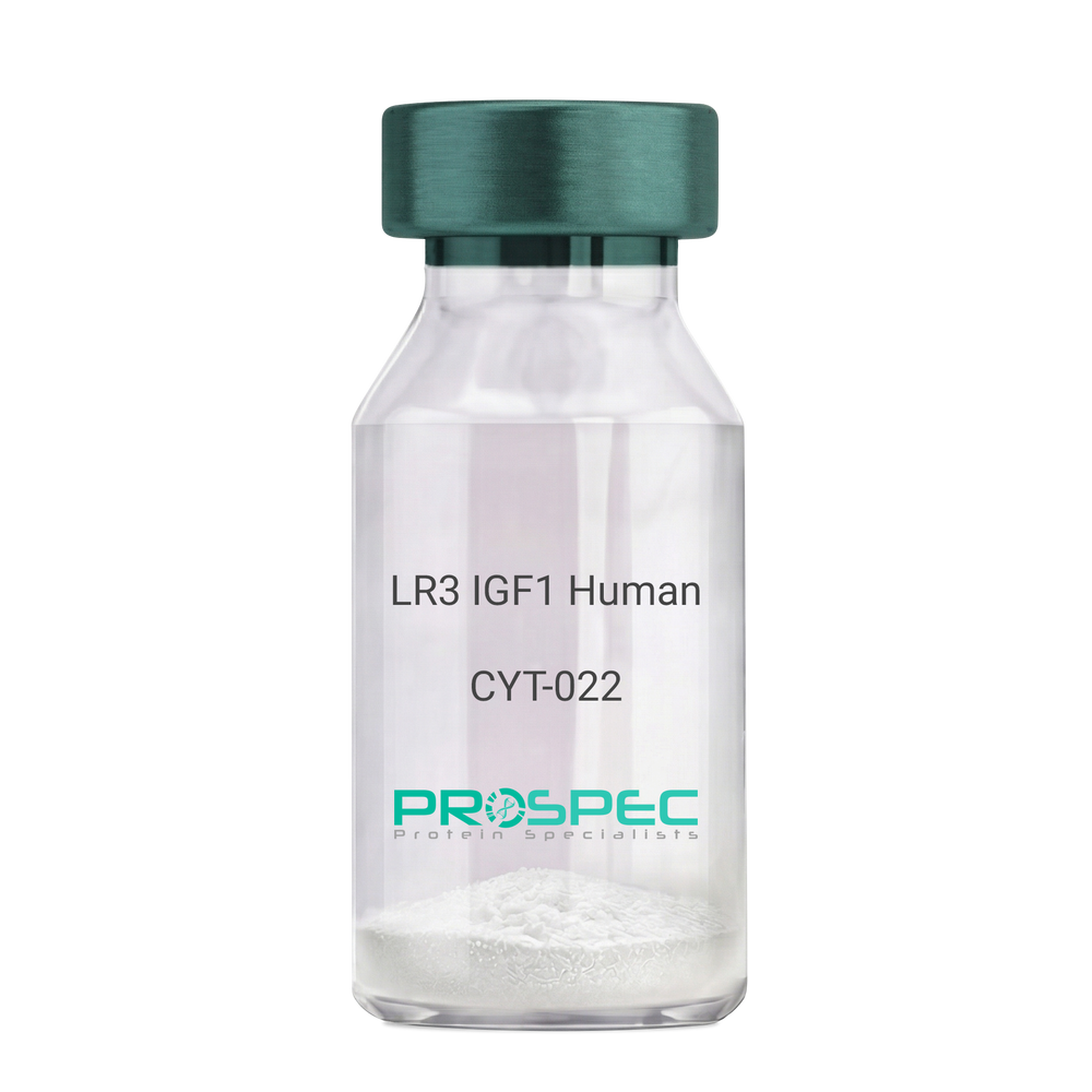
    Il 18 Human
  • View Data Sheet

    Name :

    LR3 IGF1 Human

    Description:

    LR3 Insulin Like Growth Factor-1 Human Recombinant

    R3 IGF1, R3 IGF-1, R3IGF1, R3IGF-1, LONG IGF1, LONG IGF-1, LONG R3 IGF1, LONG R3IGF1, LONG R3 IGF-1, LONG R3IGF-1.

    Product # :

    CYT-022

    Price :

    Quantity :

    Shipping Method :

    Room Temp Icon

    Shipped at Room temp

    Add To Cart

    More Info

    • description
    • source
    • formulation
    • purity
    • biological activity
    • More Info
    • sds-page

    Description

    The LR3 is a long-term analog of human IGF-1, specifically designed and manufactured for mammalian cell culture to support large-scale manufacturing of recombinant biopharmaceuticals. Recombinant Human LR3 Insulin Like Growth Factor-1 produced in E.Coli is a single, non-glycosylated, polypeptide chain containing 83 amino acids and having a molecular mass of 9.1kDa.

    Source

    Escherichia Coli.

    Formulation

    Lyophilized from a 0.2µm filtered concentrated solution in 20mM PB, pH 7.2.

    Purity

    Greater than 98.0% as determined by SDS-PAGE.

    Biological Activity

    The ED50 as determined by the stimulation of protein synthesis in L6 myoblasts is less than 10ng/ml, corresponding to a specific activity of 100,000units/mg.

    sds-page

    LR3 IGF1 sds-page - Product image 1

    More Info

    • Introduction

      IGF-1 (Insulin-like growth factor-1) is a major hormonal mediator of statural growth. Under regular circumstances, GH (growth hormone) binds to its receptor in the liver, and other tissues, and stimulates the synthesis/secretion of IGF-1. In target tissues, the Type 1 IGF receptor, that is homologous to the insulin receptor, is activated by IGF-1, leading to intracellular signaling which stimulates multiple processes leading to statural growth. IGF-1 metabolic actions are partly directed at stimulating the uptake of glucose, fatty acids, and amino acids so that metabolism supports growing tissues.

    • Synonyms

      R3 IGF1, R3 IGF-1, R3IGF1, R3IGF-1, LONG IGF1, LONG IGF-1, LONG R3 IGF1, LONG R3IGF1, LONG R3 IGF-1, LONG R3IGF-1.

    • Physical Appearance

      Sterile Filtered White lyophilized (freeze-dried) powder.

    • Stability

      Lyophilized LR3 IGF1 although stable at room temperature for 3 weeks, should be stored desiccated below -18°C. Upon reconstitution the LR3 IGF1 should be stored at 4°C between 2-7 days and for future use below -18°C.For long term storage it is recommended to add a carrier protein (0.1% HSA or BSA).Please prevent freeze-thaw cycles.

    • Solubility

      It is recommended to reconstitute the lyophilized LR3 IGF1 in sterile 18M-cm H2O at a concentration of 100µg/ml, which can then be further diluted to other aqueous solutions.

    • Amino Acid Sequence

      MFPAMPLSSLFVNGPRTLCGAELVDALQFVCGDRGFYFNKPTGYGSSSRRAPQTGIV DECCFRSCDLRRLEMYCAPLKPAKSA.

    ProSpec's products are furnished for LABORATORY RESEARCH USE ONLY. The product may not be used as drugs, agricultural or pesticidal products, food additives or household chemicals.

    Long R3 Igf1 Human
  • 1
  • …
  • 26
  • 27
  • 28
  • 29
  • 30
  • …
  • 42
Your browser does not support the video tag.

Products

  • Cytokines
  • Growth Factors
  • Chemokines
  • CD Antigens
  • Neurotrophins
  • Hormones
  • Enzymes
  • Viral Antigens
  • Recombinant Proteins
  • Natural Proteins
  • Monoclonal Antibodies
  • Polyclonal Antibodies
  • Thermal Packaging

About

  • Company
  • Founder
  • Contribution
  • Contact Us
  • My Account

Ordering

  • Ordering Method
  • Ordering - Credit Card
  • Ordering PO
  • Shipping Fees
  • FAQ's

Legal

  • Terms & Conditions
  • Privacy
  • Site Map
  • info@prospecbio.com
  • Facebook
  • Instagram
  • Linkedin Profile

© 2026 ProSpec-Tany TechnoGene Ltd. All Rights Reserved.

  • Choosing a selection results in a full page refresh.
  • Opens in a new window.