Search results
681 results found for “glycogen synthase kinase”
Name
Description
Product #
Price
Quantity
Shipping Method
- View Data Sheet
Name :
SNCA A53T HumanDescription:
Alpha Synuclein A53T Human Recombinant
Alpha-synuclein, Non-A beta component of AD amyloid, Non-A4 component of amyloid precursor, NACP, PD1, PARK1, PARK4, MGC110988, a-Synuclein, SNCA.
Product # :
PRO-159Price :
Quantity :
Shipping Method :
Shipped with Ice Packs
More Info
- description
- source
- formulation
- purity
- More Info
Description
A-Synuclein A53T Human Recombinant which is a Parkinson’s disease-related point mutant, produced in E.Coli is a single, non-glycosylated polypeptide chain of 140 amino acids having a molecular mass of 14.4kDa (molecular size on SDS-PAGE will appear higher). The Recombinant Human a-Synuclein A53T is purified by proprietary chromatographic techniques.
Source
Escherichia Coli.
Formulation
The protein (1mg/ml) contains 20mM Tris-HCl buffer (pH 7.5) and 0.1M NaCl.
Purity
Greater than 95.0% as determined by SDS-PAGE.
More Info
-
Introduction
a-Synuclein (amino acids 1-140), an acidic neuronal protein of 140 amino acids, is extremely heat-resistant and is natively unfolded with an extended structure primarily composed of random coils. a-synuclein has been suggested to be implicated in the pathogenesis of Parkinson’s disease and related neurodegenerative disorders, and more recently, to be an important regulatory component of vesicular transport in neuronal cells. Moreover, recent studies have shown that a-synuclein has chaperone activity and that this activity is lost upon removing its C-terminal acidic tail (amino acids 96-140).
-
Synonyms
Alpha-synuclein, Non-A beta component of AD amyloid, Non-A4 component of amyloid precursor, NACP, PD1, PARK1, PARK4, MGC110988, a-Synuclein, SNCA.
-
Physical Appearance
Sterile filtered colorless solution.
-
Stability
Store at 4°C if entire vial will be used within 2-4 weeks. Store, frozen at -20°C for longer periods of time. For long term storage it is recommended to add a carrier protein (0.1% HSA or BSA). Avoid multiple freeze-thaw cycles.
-
Amino Acid Sequence
MDVFMKGLSK AKEGVVAAAE KTKQGVAEAA GKTKEGVLYV GSKTKEGVVH GVTTVAEKTK EQVTNVGGAV VTGVTAVAQK TVEGAGSIAA ATGFVKKDQL GKNEEGAPQE GILEDMPVDP DNEAYEMPSE EGYQDYEPEA.
ProSpec's products are furnished for LABORATORY RESEARCH USE ONLY. The product may not be used as drugs, agricultural or pesticidal products, food additives or household chemicals.
- View Data Sheet
Name :
CoV-2 S1 (1-681)Description:
Coronavirus 2019-nCoV Spike Glycoprotein-S1 (1-681), Recombinant
Product # :
SARS-045Price :
Quantity :
Shipping Method :
Shipped with Ice Packs
More Info
- description
- source
- formulation
- purity
- More Info
Description
The HEK293 derived recombinant protein contains the Coronavirus 2019-nCoV Spike Glycoprotein S1, Wuhan-Hu-1 strain, amino acids 1-681 fused to Fc tag at C-terminal having a Mw of 76 kDa.
Source
HEK293.
Formulation
CoV-2 S1protein solution is supplied PBS pH 7.4.
Purity
Protein is >95% pure as determined SDS-PAGE.
More Info
-
Introduction
A human infecting coronavirus (viral pneumonia) called 2019 novel coronavirus, 2019-nCoV was found in the fish market at the city of Wuhan, Hubei province of China on December 2019.The 2019-nCoV shares an 87% identity to the 2 bat-derived severe acute respiratory syndrome 2018 SARS-CoV-2 located in Zhoushan of eastern China. 2019-nCoV has an analogous receptor-BD-structure to that of 2018 SARS-CoV, even though there is a.a. diversity so thus the 2019-nCoV might bind to ACE2 receptor protein (angiotensin-converting enzyme 2) in humans.While bats are probably the host of 2019-nCoV, researchers suspect that animal from the ocean sold at the seafood market was an intermediate host. RSCU analysis proposes that the 2019-nCoV is a recombinant within the viral spike glycoprotein between the bat coronavirus and an unknown coronavirus.
-
Physical Appearance
Sterile Filtered clear solution.
-
Stability
Protein is shipped on ice packs. Upon arrival, Store at -20°C.
-
Purification Method
Purified by Protein-G chromatography technique.
ProSpec's products are furnished for LABORATORY RESEARCH USE ONLY. The product may not be used as drugs, agricultural or pesticidal products, food additives or household chemicals.
- View Data Sheet
Name :
NTRK3 MouseDescription:
Neurotrophic Receptor Tyrosine Kinase 3 Mouse Recombinant
NT-3 growth factor receptor, GP145-TrkC, Trk-C, Neurotrophic tyrosine kinase receptor type 3, TrkC tyrosine kinase, Ntrk3, TrkC, AW125844, Ntrk3_tv3
Product # :
CYT-1163Price :
Quantity :
Shipping Method :
Shipped with Ice Packs
More Info
- description
- source
- formulation
- purity
- More Info
Description
NTRK3 Mouse Recombinant produced in Baculovirus is a single glycosylated polypeptide chain containing 404 amino acids (32-429 aa) and having a molecular mass of 45.4kDa.NTRK3 is fused to a 6 amino acid His tag at C-terminus and purified by proprietary chromatographic techniques.
Source
Sf9, Baculovirus cells.
Formulation
NTRK3 protein (0.25mg/ml) contains 10% glycerol and Phosphate-Buffered Saline (pH 7.4).
Purity
Greater than 90.0% as determined by SDS-PAGE.
More Info
-
Introduction
Neurotrophic Receptor Tyrosine Kinase 3 (NTRK3)belongs to the receptor tyrosine kinases family and is a high affinity catalytic receptor of neurotrophin NT-3. NTRK3 mediates multiple effects of this neurotrophic factor, which includes neuronal differentiation and survival. NTRK3 inducesvarious pleiotorpic responses in malignant cells, including enhanced tumor cell invasiveness and chemotoxis.Increased NTRK3 expression was observed in neuroblastoma, medulloblastoma, and in neuroectodermal brain tumors.
-
Synonyms
NT-3 growth factor receptor, GP145-TrkC, Trk-C, Neurotrophic tyrosine kinase receptor type 3, TrkC tyrosine kinase, Ntrk3, TrkC, AW125844, Ntrk3_tv3
-
Physical Appearance
Sterile Filtered colorless solution.
-
Stability
Store at 4°C if entire vial will be used within 2-4 weeks. Store, frozen at -20°C for longer periods of time. For long term storage it is recommended to add a carrier protein (0.1% HSA or BSA).Avoid multiple freeze-thaw cycles.
-
Amino Acid Sequence
CPANCVCSKT EINCRRPDDG NLFPLLEGQD SGNSNGNASI NITDISRNIT SIHIENWRGL HTLNAVDMEL YTGLQKLTIK NSGLRNIQPR AFAKNPHLRY INLSSNRLTT LSWQLFQTLS LRELRLEQNF FNCSCDIRWM QLWQEQGEAR LDSQSLYCIS ADGSQLPLFR MNISQCDLPE ISVSHVNLTV REGDNAVITC NGSGSPLPDV DWIVTGLQSI NTHQTNLNWT NVHAINLTLV NVTSEDNGFT LTCIAENVVG MSNASVALTV YYPPRVVSLV EPEVRLEHCI EFVVRGNPTP TLHWLYNGQP LRESKIIHMD YYQEGEVSEG CLLFNKPTHY NNGNYTLIAK NALGTANQTI NGHFLKEPFP ESTDFFDFES DASPTPPITV THKPEEDTHH HHHH
ProSpec's products are furnished for LABORATORY RESEARCH USE ONLY. The product may not be used as drugs, agricultural or pesticidal products, food additives or household chemicals.
- View Data Sheet
Name :
FTCD HumanDescription:
Formiminotransferase Cyclodeaminase Human Recombinant
Formiminotransferase-cyclodeaminase, FTCD, LCHC1, LC-1.
Product # :
ENZ-302Price :
Quantity :
Shipping Method :
Shipped with Ice Packs
More Info
- description
- source
- formulation
- purity
- More Info
Description
Formiminotransferase Cyclodeaminase Human Recombinant (also called liver cytosol type 1) produced in SF9, is a glycosylated, polypeptide chain having a molecular mass of 59,749 Dalton.The FTCD is expressed with a -6xHis tag and purified by proprietary chromatographic techniques.
Source
Sf9 insect cells.
Formulation
FTCD is supplied in 16mM HEPES buffer pH-7.6, 240mM sodium chloride, and 20% glycerol.
Purity
Greater than 95% as determined by SDS-PAGE.
More Info
-
Introduction
Formiminotransferase cyclodeaminase is an enzyme which catalyzes the conversion of formiminoglutamateand tetrahydrofolateinto formiminotetrahydrofolateand glutamate.
Formiminotransferase cyclodeaminase, a bifunctional enzyme of tetrahydrofolate synthesis, is the target antigen of anti-LC1 (liver cytosol antigen type 1) autoantibodies. Presence of LC1 autoantibodies is a marker for type 2 autoimmune hepatitis (for which anti-LKM 1/cytochrome P450 2D6 autoantibodies are a further marker).
Serum LC1 autoantibody concentrations appear to fluctuate in parallel with aminotransferase levels, a particularly intriguing observation that suggests a possible role of LC1 autoreactivity in the pathogenic mechanism leading to hepatocyte injury. -
Synonyms
Formiminotransferase-cyclodeaminase, FTCD, LCHC1, LC-1.
-
Stability
Store at 4°C if entire vial will be used within 2-4 weeks. Store, frozen at -20°Cfor longer periods of time. Avoid multiple freeze-thaw cycles.
-
Immunological Functions
1. Binds IgG-type human auto-antibodies.2. Standard ELISA test (checker-board analysis of positive/negative sera panels, immuno-dot test).
-
coating concentration
0.35-0.7 µg/ml (depending on the type of ELISA plate and coating buffer).Suitable for biotinylation and iodination.
ProSpec's products are furnished for LABORATORY RESEARCH USE ONLY. The product may not be used as drugs, agricultural or pesticidal products, food additives or household chemicals.
- View Data Sheet
Name :
DUSP3 AntibodyDescription:
Dual Specificity Phosphatase 3, Mouse Anti Human
Dual specificity phosphatase 3, VHR, Vaccinia virus phosphatase VH1-related, Dual specificity protein phosphatase VHR, Vaccinia H1-related phosphatase, serine/threonine specific protein phosphatase, DUSP3.
Product # :
ANT-594Price :
Quantity :
Shipping Method :
Shipped with Ice Packs
More Info
- formulation
- More Info
Formulation
1mg/ml containing PBS, pH-7.4, 10% Glycerol and 0.02% Sodium Azide.
More Info
-
Introduction
DUSP3 belongs to the dual specificity protein phosphatase subfamily which inactivates their target kinases by dephosphorylating both the phosphoserine/threonine and phosphotyrosine residues. DUSP3 negatively regulate members of the mitogen-activated protein kinase superfamily that are related to cellular proliferation and differentiation. DUSP3 is expressed in both breast and ovarian tissues and displays activity both for tyrosine-protein phosphate and serine-protein phosphate, but exhibits a strong preference toward phosphotyrosines, specifically dephosphorylates and inactivates ERK1 and ERK2.
-
Synonyms
Dual specificity phosphatase 3, VHR, Vaccinia virus phosphatase VH1-related, Dual specificity protein phosphatase VHR, Vaccinia H1-related phosphatase, serine/threonine specific protein phosphatase, DUSP3.
-
Physical Appearance
Sterile filtered colorless solution.
-
Immunogen
Anti-human DUSP3 mAb, is derived from hybridization of mouse F0 myeloma cells with spleen cells from BALB/c mice immunized with recombinant human DUSP3 amino acids 1-185 purified from E. coli.
-
Ig Subclass
Mouse IgG2b heavy chain and k light chain.
-
Clone
PAT9E6AT.
-
Applications
DUSP3 antibody has been tested by ELISA, Western blot analysis to assure specificity and reactivity. Since application varies, however, each investigation should be titrated by the reagent to obtain optimal results. Recommended starting dilution for Western blot analysis is 1:3000.
-
Type
Mouse Anti Human Monoclonal.
-
Storage Procedures
For periods up to 1 month store at 4°C, for longer periods of time, store at -20°C. Prevent freeze thaw cycles.
-
Purification Method
DUSP3 antibody was purified from mouse ascitic fluids by protein-A affinity chromatography.
ProSpec's products are furnished for LABORATORY RESEARCH USE ONLY. The product may not be used as drugs, agricultural or pesticidal products, food additives or household chemicals.
- View Data Sheet
Name :
PNP HumanDescription:
Purine Nucleoside Phosphorylase Human Recombinant
Purine nucleoside phosphorylase, Inosine phosphorylase, NP, PUNP, PRO1837, FLJ94043, FLJ97288, FLJ97312, MGC117396, MGC125915, MGC125916, PNP.
Product # :
ENZ-025Price :
Quantity :
Shipping Method :
Shipped with Ice Packs
More Info
- description
- source
- formulation
- purity
- More Info
Description
PNP Human Recombinant fused with a 20 amino acid His tag at N-terminus produced in E.Coli is a single, non-glycosylated, polypeptide chain containing 309 amino acids (1-289 a.a.) and having a molecular mass of 34.2kDa. The PNP is purified by proprietary chromatographic techniques.
Source
Escherichia Coli.
Formulation
The PNP solution (1mg/ml) contains 20mM Tris-HCl buffer (pH8.0), 0.1M NaCl, 2mM DTT and 10% glycerol.
Purity
Greater than 90.0% as determined by SDS-PAGE.
More Info
-
Introduction
PNP is a member of the PNP/MTAP phosphorylase family of proteins. PNP catalyzes the reversible phosphorolysis of ribonucleosides and 2’-deoxyribonucleosides with specificity for guanine, hypoxanthine and their analogs. PNP deficiency is a rare autosomal recessive genetic disease linked to a severe defect in T-lymphocyte function and neurologic disorder in children, comprising four percent of combined immunodeficiency cases.
-
Synonyms
Purine nucleoside phosphorylase, Inosine phosphorylase, NP, PUNP, PRO1837, FLJ94043, FLJ97288, FLJ97312, MGC117396, MGC125915, MGC125916, PNP.
-
Physical Appearance
Sterile Filtered colorless solution.
-
Stability
Store at 4°C if entire vial will be used within 2-4 weeks. Store, frozen at -20°C for longer periods of time. For long term storage it is recommended to add a carrier protein (0.1% HSA or BSA).Avoid multiple freeze-thaw cycles.
-
Amino Acid Sequence
MGSSHHHHHH SSGLVPRGSH MENGYTYEDY KNTAEWLLSH TKHRPQVAII CGSGLGGLTD KLTQAQIFDY GEIPNFPRST VPGHAGRLVF GFLNGRACVM MQGRFHMYEG YPLWKVTFPV RVFHLLGVDT LVVTNAAGGL NPKFEVGDIM LIRDHINLPG FSGQNPLRGP NDERFGDRFP AMSDAYDRTM RQRALSTWKQ MGEQRELQEG TYVMVAGPSF ETVAECRVLQ KLGADAVGMS TVPEVIVARH CGLRVFGFSL ITNKVIMDYE SLEKANHEEV LAAGKQAAQK LEQFVSILMA SIPLPDKAS.
ProSpec's products are furnished for LABORATORY RESEARCH USE ONLY. The product may not be used as drugs, agricultural or pesticidal products, food additives or household chemicals.
- View Data Sheet
Name :
MEMO1 AntibodyDescription:
Mediator of Cell Motility 1, Mouse Anti Human
Protein MEMO1, C21orf19-like protein, Hepatitis C virus NS5A-transactivated protein 7, HCV NS5A-transactivated protein 7, Mediator of ErbB2-driven cell motility 1, Mediator of cell motility 1, Memo-1, MEMO1, C2orf4, MEMO, NS5ATP7, CGI-27.
Product # :
ANT-090Price :
Quantity :
Shipping Method :
Shipped with Ice Packs
More Info
- formulation
- More Info
Formulation
1mg/ml containing PBS, pH-7.4, & 0.1% Sodium Azide.
More Info
-
Introduction
Mediator of ErbB2-driven cell motility 1 (MEMO1) is a member of the UPF0103 family. MEMO1 regulates cell migration by transmitting extracellular chemotactic signals to the microtubule cytoskeleton. Furthermore, MEMO1 controls the localization of APC and CLASP2 to the cell membrane, using the regulation of GSK3B activity. MEMO1 is essential for breast carcinoma cell migration, suggesting a key role in tumorigenesis. MEMO1 is also a mediator of ERBB2 signaling.
-
Synonyms
Protein MEMO1, C21orf19-like protein, Hepatitis C virus NS5A-transactivated protein 7, HCV NS5A-transactivated protein 7, Mediator of ErbB2-driven cell motility 1, Mediator of cell motility 1, Memo-1, MEMO1, C2orf4, MEMO, NS5ATP7, CGI-27.
-
Immunogen
Anti-human MEMO1 mAb, is derived from hybridization of mouse FO myeloma cells with spleen cells from BALB/c mice immunized with recombinant human MEMO1 amino acids 1-297 purified from E. coli.
-
Ig Subclass
Mouse IgG1 heavy chain and kappa light chain.
-
Clone
P1E9AT.
-
Type
Mouse Anti Human Monoclonal.
-
Storage Procedures
For periods up to 1 month store at 4°C, for longer periods of time, store at -20°C. Prevent freeze thaw cycles.
-
Purification Method
MEMO1 antibody was purified from mouse ascitic fluids by protein-G affinity chromatography.
ProSpec's products are furnished for LABORATORY RESEARCH USE ONLY. The product may not be used as drugs, agricultural or pesticidal products, food additives or household chemicals.
- View Data Sheet
Name :
HADH HumanDescription:
Hydroxyacyl-Coenzyme A Dehydrogenase Human Recombinant
EC 1.1.1.35, HAD, HADH1, HHF4, MSCHAD, SCHAD, Hydroxyacyl-coenzyme A dehydrogenase, HCDH, Short-chain 3-hydroxyacyl-CoA dehydrogenase, Medium and short-chain L-3-hydroxyacyl-coenzyme A dehydrogenase, HADH, HADHSC, MGC8392.
Product # :
ENZ-499Price :
Quantity :
Shipping Method :
Shipped with Ice Packs
More Info
- description
- source
- formulation
- purity
- More Info
Description
HADH Human Recombinant fused to a 21 amino acids His Tag at N-terminal produced in E.Coli is a single, non-glycosylated, polypeptide chain containing 323 amino acids (13-314 a.a.) and having a molecular mass of 35.1 kDa. The HADH is purified by proprietary chromatographic techniques.
Source
Escherichia Coli.
Formulation
The HADH solution contains 20mM Tris-HCl pH-8, 0.1M NaCl and 20% glycerol.
Purity
Greater than 95% as determined by SDS-PAGE.
More Info
-
Introduction
HADH is part of the 3-hydroxyacyl-CoA dehydrogenase enzyme family. HADH is involved in mitochondrial matrix to catalyze the oxidation of straight-chain 3-hydroxyacyl-CoAs as part of the beta-oxidation pathway. HADH enzymatic activity is at its peak with medium-chain-length fatty acids. Mutations in HADH cause familial hyperinsulinemic hypoglycemia. HADH participates in fatty acid oxidation, where some enzymes work in a step-wise fashion to break metabolize fats and convert them to energy.
-
Synonyms
EC 1.1.1.35, HAD, HADH1, HHF4, MSCHAD, SCHAD, Hydroxyacyl-coenzyme A dehydrogenase, HCDH, Short-chain 3-hydroxyacyl-CoA dehydrogenase, Medium and short-chain L-3-hydroxyacyl-coenzyme A dehydrogenase, HADH, HADHSC, MGC8392.
-
Physical Appearance
Sterile Filtered clear colorless solution.
-
Stability
Store at 4°C if entire vial will be used within 2-4 weeks. Store, frozen at -20°C for longer periods of time. For long term storage it is recommended to add a carrier protein (0.1% HSA or BSA).Avoid multiple freeze-thaw cycles.
-
Amino Acid Sequence
MGSSHHHHHH SSGLVPRGSH MSSSSTASAS AKKIIVKHVT VIGGGLMGAG IAQVAAATGH TVVLVDQTED ILAKSKKGIE ESLRKVAKKK FAENPKAGDE FVEKTLSTIA TSTDAASVVH STDLVVEAIV ENLKVKNELF KRLDKFAAEH TIFASNTSSL QITSIANATT RQDRFAGLHF FNPVPVMKLV EVIKTPMTSQ KTFESLVDFS KALGKHPVSC KDTPGFIVNR LLVPYLMEAI RLYERGDASK EDIDTAMKLG AGYPMGPFEL LDYVGLDTTK FIVDGWHEMD AENPLHQPSP SLNKLVAENK FGKKTGEGFY KYK.
ProSpec's products are furnished for LABORATORY RESEARCH USE ONLY. The product may not be used as drugs, agricultural or pesticidal products, food additives or household chemicals.
- View Data Sheet
Name :
sRANKL (158-316) MouseDescription:
Soluble RANK Ligand (158-316 a.a) Mouse Recombinant
Soluble Receptor Activator of NFkB Ligand, TNFSF11, TRANCE, TNF-related activation-induced cytokine, OPGL, ODF, Osteoclast differentiation factor, Tumor necrosis factor ligand superfamily member 11, Receptor activator of nuclear factor kappa B ligand, RANKL, Osteoprotegerin ligand, CD254 antigen, sRANKL, sOdf.
Product # :
CYT-958Price :
Quantity :
Shipping Method :
Shipped with Ice Packs
More Info
- description
- source
- formulation
- purity
- biological activity
- More Info
Description
sRANKL Mouse Recombinant produced in E.Coli is a single, non-glycosylated polypeptide chain containing 160 amino acids (158-316 a.a.) and having a molecular mass of 17.9kDa. sRANKL is purified by proprietary chromatographic techniques.
Source
Escherichia Coli.
Formulation
sRANKL protein solution (1mg/ml) containing Tris-Hcl buffer pH-8.5 and 0.1M NaCl.
Purity
Greater than 90% as determined by SDS-PAGE.
Biological Activity
The ED50, as measured by its ability to induce osteoclast differentiation of RAW 264.7 mouse monocyte/macrophage cells, is less than 2ng/ml.More Info
-
Introduction
RANKL binds to tnfrsf11b/opg and to tnfrsf11a/rank. Osteoclast differentiation and activation factor. augments the ability of dendritic cells to stimulate naive t-cell proliferation. May be an important regulator of interactions between t-cells and dendritic cells and may play a role in the regulation of the t-cell-dependent immune response. sRANKL may also play an important role in enhanced bone-resorption in humoral hypercalcemia of malignancy.
-
Synonyms
Soluble Receptor Activator of NFkB Ligand, TNFSF11, TRANCE, TNF-related activation-induced cytokine, OPGL, ODF, Osteoclast differentiation factor, Tumor necrosis factor ligand superfamily member 11, Receptor activator of nuclear factor kappa B ligand, RANKL, Osteoprotegerin ligand, CD254 antigen, sRANKL, sOdf.
-
Physical Appearance
Sterile Filtered colorless solution.
-
Stability
Store at 4°C if entire vial will be used within 2-4 weeks. Store, frozen at -20°C for longer periods of time. For long term storage it is recommended to add a carrier protein (0.1% HSA or BSA). Avoid multiple freeze-thaw cycles.
-
Amino Acid Sequence
MKPEAQPFAH LTINAASIPS GSHKVTLSSW YHDRGWAKIS NMTLSNGKLR VNQDGFYYLY ANICFRHHET SGSVPTDYLQ LMVYVVKTSI KIPSSHNLMK GGSTKNWSGN SEFHFYSINV GGFFKLRAGE EISIQVSNPS LLDPDQDATY FGAFKVQDID
ProSpec's products are furnished for LABORATORY RESEARCH USE ONLY. The product may not be used as drugs, agricultural or pesticidal products, food additives or household chemicals.
- View Data Sheet
Name :
ENTPD3 Human, sf9 BioactiveDescription:
Ectonucleoside Triphosphate Diphosphohydrolase 3 Human Recombinant, sf9 Bioactive
Ectonucleoside Triphosphate Diphosphohydrolase 3, Ecto-ATP Diphosphohydrolase 3, CD39 Antigen-Like 3, Ecto-ATPDase 3, Ecto-Apyrase 3, Ecto-ATPase 3, EC 3.6.1.5, NTPDase 3, CD39L3, HB6, NTPDase-3, EC 3.6.1, Ectonucleoside triphosphate diphosphohydrolase 3, NTPDase 3, CD39 antigen-like 3, Ecto-ATP diphosphohydrolase 3, Ecto-ATPDase 3, Ecto-ATPase 3, Ecto-apyrase 3, HB6.
Product # :
ENZ-1020Price :
Quantity :
Shipping Method :
Shipped with Ice Packs
More Info
- description
- source
- formulation
- purity
- biological activity
- More Info
Description
ENTPD3 Human Recombinant produced in Sf9 Baculovirus cells is a single, glycosylated polypeptide chain containing 451 amino acids (44-485a.a.) and having a molecular mass of 50.7kDa (Molecular size on SDS-PAGE will appear at approximately 50-70kDa).ENTPD3 is expressed with a 6 amino acid His tag at C-Terminus and purified by proprietary chromatographic techniques.
Source
Sf9, Baculovirus cells.
Formulation
ENTPD3 protein solution (0.5mg/ml) contains Phosphate Buffered Saline (pH 7.4) and 10% glycerol.
Purity
Greater than 90.0% as determined by SDS-PAGE.
Biological Activity
Specific activity is > 250,000 pmol/min/ug, and is defined as the amount of enzyme that hydrolyze ATP per minute at pH 7.5 at 37C.More Info
-
Introduction
Ectonucleoside Triphosphate Diphosphohydrolase 3, also known as ENTPD3, which owns a threefold preference for the hydrolysis of ATP over ADP is similar to E-type nucleotidases (NTPases). ENTPD3 is a protein coding gene which contains four apyrase-conserved areas which is characteristic of NTPases.
-
Synonyms
Ectonucleoside Triphosphate Diphosphohydrolase 3, Ecto-ATP Diphosphohydrolase 3, CD39 Antigen-Like 3, Ecto-ATPDase 3, Ecto-Apyrase 3, Ecto-ATPase 3, EC 3.6.1.5, NTPDase 3, CD39L3, HB6, NTPDase-3, EC 3.6.1, Ectonucleoside triphosphate diphosphohydrolase 3, NTPDase 3, CD39 antigen-like 3, Ecto-ATP diphosphohydrolase 3, Ecto-ATPDase 3, Ecto-ATPase 3, Ecto-apyrase 3, HB6.
-
Physical Appearance
Sterile Filtered colorless solution.
-
Stability
Store at 4°C if entire vial will be used within 2-4 weeks.Store, frozen at -20°C for longer periods of time. For long term storage it is recommended to add a carrier protein (0.1% HSA or BSA).Avoid multiple freeze-thaw cycles.
-
Amino Acid Sequence
ADLQIHKQEV LPPGLKYGIV LDAGSSRTTV YVYQWPAEKE NNTGVVSQTF KCSVKGSGIS SYGNNPQDVP RAFEECMQKV KGQVPSHLHG STPIHLGATA GMRLLRLQNE TAANEVLESI QSYFKSQPFD FRGAQIISGQ EEGVYGWITA NYLMGNFLEK NLWHMWVHPH GVETTGALDL GGASTQISFV AGEKMDLNTS DIMQVSLYGY VYTLYTHSFQ CYGRNEAEKK FLAMLLQNSP TKNHLTNPCY PRDYSISFTM GHVFDSLCTV DQRPESYNPN DVITFEGTGD PSLCKEKVAS IFDFKACHDQ ETCSFDGVYQ PKIKGPFVAF AGFYYTASAL NLSGSFSLDT FNSSTWNFCS QNWSQLPLLL PKFDEVYARS YCFSANYIYH LFVNGYKFTE ETWPQIHFEK EVGNSSIAWS LGYMLSLTNQ IPAESPLIRL PIEPPHHHHHH.
ProSpec's products are furnished for LABORATORY RESEARCH USE ONLY. The product may not be used as drugs, agricultural or pesticidal products, food additives or household chemicals.
- View Data Sheet
Name :
CoV-2-S1 (319-541)Description:
Coronavirus 2019 Spike Glycoprotein-S1 Receptor Binding Domain (319-541 a.a), Recombinant
Product # :
SARS-021Price :
Quantity :
Shipping Method :
Shipped with Ice Packs
More Info
- description
- source
- formulation
- purity
- biological activity
- More Info
Description
The HEK293 derived recombinant protein contains the Coronavirus 2019 Spike Glycoprotein S1 Receptor Binding Domain [ RBD ], Wuhan-Hu-1 strain, amino acids 319-541 fused to His tag at C-terminal.
Source
HEK293 Cells.
Formulation
CoV-S1 RBD protein is 1mg/ml contains 20mM sodium phosphate (NaPP), 300mM NaCl, pH7.2
Purity
Protein is >90% pure as determined SDS-PAGE.
Biological Activity
SARS CoV-2 Spike Glycoprotein S1 RBD was tested for its biological functionality by its binding to recombinant ACE2 receptor protein confirmed by structural complementation reporter assay (NanoBiT®).
More Info
-
Introduction
A human infecting coronavirus (viral pneumonia) called 2019 novel coronavirus, 2019-nCoV was found in the fish market at the city of Wuhan, Hubei province of China on December 2019.
The 2019-nCoV shares an 87% identity to the 2 bat-derived severe acute respiratory syndrome 2018 SARS-CoV-2 located in Zhoushan of eastern China. 2019-nCoV has an analogous receptor-BD-structure to that of 2018 SARS-CoV, even though there is a.a. diversity so thus the 2019-nCoV might bind to ACE2 receptor protein (angiotensin-converting enzyme 2) in humans.
While bats are possibly the host of 2019-nCoV, researchers suspect that animal from the ocean sold at the seafood market was an intermediate host. RSCU analysis proposes that the 2019-nCoV is a recombinant within the viral spike glycoprotein between the bat coronavirus and an unknown coronavirus.
-
Physical Appearance
Sterile Filtered clear solution.
-
Stability
CoV-2 S1 RBD Protein is shipped on ice packs. Upon arrival, Store at -20°C.
-
Purification Method
Purified by Metal-Afinity chromatographic technique.
ProSpec's products are furnished for LABORATORY RESEARCH USE ONLY. The product may not be used as drugs, agricultural or pesticidal products, food additives or household chemicals.
- View Data Sheet
Name :
Flt3 Ligand Mouse, Sf9Description:
Flt3 Ligand Mouse Recombinant, Sf9
Fms-related tyrosine kinase 3 ligand, Flt3 ligand, Flt3L, SL cytokine, lt3lgF, Flt3l.
Product # :
CYT-910Price :
Quantity :
Shipping Method :
Shipped with Ice Packs
More Info
- description
- source
- formulation
- purity
- More Info
Description
Flt3 Ligand produced in Sf9 Baculovirus cells is a single, glycosylated polypeptide chain (27-189 a.a.) and fused to a 6 aa His Tag at C-terminus containing a total of 169 amino acids and having a molecular mass of 19.3kDa.Flt3 Ligand shows multiple bands between 18-28kDa on SDS-PAGE, reducing conditions and purified by proprietary chromatographic techniques.
Source
Sf9, Baculovirus cells.
Formulation
Flt3 Ligand protein solution (0.5mg/ml) contains Phosphate buffered saline (pH7.4) and 10% glycerol.
Purity
Greater than 85.0% as determined by SDS-PAGE.
More Info
-
Introduction
FLT3 ligand is a receptor for the fl cytokine has a tyrosine-protein kinase activity & a growth factor that regulates proliferation of early hematopoietic cells. Flt3-Ligand synergizes with other CSFs and interleukins to induce growth and differentiation.
-
Synonyms
Fms-related tyrosine kinase 3 ligand, Flt3 ligand, Flt3L, SL cytokine, lt3lgF, Flt3l.
-
Physical Appearance
Sterile filtered colorless solution.
-
Stability
Store at 4°C if entire vial will be used within 2-4 weeks. Store, frozen at -20°C for longer periods of time. For long term storage it is recommended to add a carrier protein (0.1% HSA or BSA).Avoid multiple freeze-thaw cycles.
-
Amino Acid Sequence
GTPDCYFSHS PISSNFKVKF RELTDHLLKD YPVTVAVNLQ DEKHCKALWS LFLAQRWIEQ LKTVAGSKMQ TLLEDVNTEI HFVTSCTFQP LPECLRFVQT NISHLLKDTC TQLLALKPCI GKACQNFSRC LEVQCQPDSS TLLPPRSPIA LEATELPEPR PRQHHHHHH
-
Background
What is the molecular weight/Mw of FLT3 LIGAND MOUSE, SF9 Protein?
FLT3 LIGAND MOUSE, SF9 Protein has a total Mw of 19.3kDa.
What is the source or expression system of FLT3 LIGAND MOUSE, SF9 Protein?
Sf9, Baculovirus cells.
What is the Purity of FLT3 LIGAND MOUSE, SF9 Protein?
FLT3 LIGAND MOUSE, SF9 Protein is >85% pure as determined by SDS-PAGE.
What is the Biological Activity of FLT3 LIGAND MOUSE, SF9 Protein?
The biological functionality of FLT3 LIGAND MOUSE, SF9 Protein will be determined in the future.
What is the amino acid sequence of FLT3 LIGAND MOUSE, SF9 Protein?
GTPDCYFSHS PISSNFKVKF RELTDHLLKD YPVTVAVNLQ DEKHCKALWS LFLAQRWIEQ LKTVAGSKMQ TLLEDVNTEI HFVTSCTFQP LPECLRFVQT NISHLLKDTC TQLLALKPCI GKACQNFSRC LEVQCQPDSS TLLPPRSPIA LEATELPEPR PRQHHHHHH.
What applications can FLT3 LIGAND MOUSE, SF9 Protein be used in?
FLT3 LIGAND MOUSE, SF9 Protein can probably be used in western blot, ELISA and Lateral Flow.
What is the endotoxin level for FLT3 LIGAND MOUSE, SF9 Protein?
The endotoxin level is minimal, FLT3 LIGAND MOUSE, SF9 Protein was purified using conventional chromatography techniques.
ProSpec's products are furnished for LABORATORY RESEARCH USE ONLY. The product may not be used as drugs, agricultural or pesticidal products, food additives or household chemicals.
- View Data Sheet
Name :
RGS16 HumanDescription:
Regulator of G-Protein Signaling 16 Human Recombinant
A28-RGS14, A28-RGS14P, RGS-R, Regulator of G-protein signaling 16, RGS16, Retinally abundant regulator of G-protein signaling, Retinal-specific RGS, hRGS-r, RGSR.
Product # :
PRO-798Price :
Quantity :
Shipping Method :
Shipped with Ice Packs
More Info
- description
- source
- formulation
- purity
- More Info
Description
RGS16 Human Recombinant produced in E.Coli is a single, non-glycosylated, polypeptide chain containing 222 amino acids (1-202 a.a.) and having a molecular mass of 24.9 kDa. The RGS16 is fused to a 20 amino acid His Tag at N-terminal and purified by proprietary chromatographic techniques.
Source
Escherichia Coli.
Formulation
The RGS16 solution (0.5mg/ml) contains Phosphate Buffered Saline (pH7.4), 0.1M NaCl and 20% glycerol.
Purity
Greater than 90% as determined by SDS-PAGE.
More Info
-
Introduction
RGS16 is part of the ''regulator of G protein signaling'' family. RGS16 inhibits signal transduction by increasing the GTPase activity of G protein alpha subunits thereby driving them into their inactive GDP-bound form. RGS16 Binds to G(i)-alpha and G(o)-alpha, but not to G(s)-alpha. RGS16 regulates the kinetics of signaling in the phototransduction cascade.
-
Synonyms
A28-RGS14, A28-RGS14P, RGS-R, Regulator of G-protein signaling 16, RGS16, Retinally abundant regulator of G-protein signaling, Retinal-specific RGS, hRGS-r, RGSR.
-
Physical Appearance
Sterile Filtered clear colorless solution.
-
Stability
Store at 4°C if entire vial will be used within 2-4 weeks. Store, frozen at -20°C for longer periods of time. For long term storage it is recommended to add a carrier protein (0.1% HSA or BSA). Avoid multiple freeze-thaw cycles.
-
Amino Acid Sequence
MGSSHHHHHH SSGLVPRGSH MCRTLAAFPT TCLERAKEFK TRLGIFLHKS ELGCDTGSTG KFEWGSKHSK ENRNFSEDVL GWRESFDLLL SSKNGVAAFH AFLKTEFSEE NLEFWLACEE FKKIRSATKL ASRAHQIFEE FICSEAPKEV NIDHETRELT RMNLQTATAT CFDAAQGKTR TLMEKDSYPR FLKSPAYRDL AAQASAASAT LSSCSLDEPS HT.
ProSpec's products are furnished for LABORATORY RESEARCH USE ONLY. The product may not be used as drugs, agricultural or pesticidal products, food additives or household chemicals.
- View Data Sheet
Name :
ALK/P80 AntibodyDescription:
ALK/P80 Protein, Mouse Anti Human
ALK/P80 Protein, ALK/P80, p80 protein, anaplastic lymphoma kinase, ALK, nucleophosmin, NPM.
Product # :
ANT-331Price :
Quantity :
Shipping Method :
Shipped with Ice Packs
More Info
- formulation
- More Info
Formulation
1mg/ml containing PBS, pH-7.4, & 0.1% Sodium Azide.
More Info
-
Introduction
ALK/p80 is a human p80 protein, identified as a hybrid of the anaplastic lymphoma kinase (ALK) gene and the nucleophosmin (NPM) gene resulting from the t(2;5)(p23;q35) translocation found in a third of large cell lymphomas.
-
Synonyms
ALK/P80 Protein, ALK/P80, p80 protein, anaplastic lymphoma kinase, ALK, nucleophosmin, NPM.
-
Immunogen
Anti-human ALK/P80 mAb is derived from hybridization of mouse SP2/0 myeloma cells with spleen cells from BALB/c mice immunized with recombinant human ALK/P80 amino acids 81-294 purified from E. coli.
-
Ig Subclass
Mouse IgG2b heavy chain and κ light chain.
-
Clone
P5E3AT.
-
Applications
ALK/P80 antibody has been tested by ELISA, Western blot and Immunohistochemical analysis to assure specificity and reactivity. Since application varies, however, each investigation should be titrated by the reagent to obtain optimal results. Recommended dilution range for Western blot analysis is 1:1,000 ~ 2,000. Recommended starting dilution is 1:1,000.
-
Type
Mouse Anti Human Monoclonal.
-
Storage Procedures
For periods up to 1 month store at 4°C, for longer periods of time, store at -20°C. Prevent freeze thaw cycles.
-
Purification Method
ALK/P80 antibody was purified from mouse ascitic fluids by protein-G affinity chromatography.
ProSpec's products are furnished for LABORATORY RESEARCH USE ONLY. The product may not be used as drugs, agricultural or pesticidal products, food additives or household chemicals.
- View Data Sheet
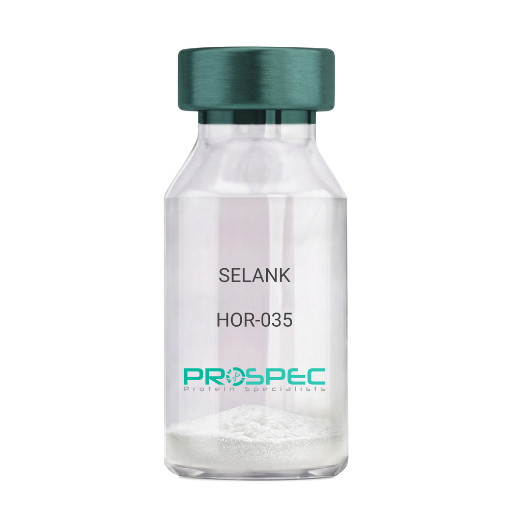
Name :
SELANKDescription:
SELANK
Product # :
HOR-035Price :
Quantity :
Shipping Method :
Shipped at Room temp
More Info
- description
- formulation
- purity
- More Info
Description
SELANK Synthetic is a single, non-glycosylated polypeptide chain containing 7 amino acids, having a molecular mass of 751.89 Dalton and a Molecular formula of C33H57N11O9.
Formulation
The protein was lyophilized with no additives.
Purity
Greater than 97.0% as determined by analysis by RP-HPLC.
More Info
-
Physical Appearance
Sterile Filtered White lyophilized (freeze-dried) powder.
-
Stability
Lyophilized SELANK although stable at room temperature for 3 weeks, should be stored desiccated below -18°C. Upon reconstitution SELANK should be stored at 4°C between 2-7 days and for future use below -18°C. For long term storage it is recommended to add a carrier protein (0.1% HSA or BSA). Please prevent freeze-thaw cycles.
-
Solubility
It is recommended to reconstitute the lyophilized SELANK in sterile 18MΩ-cm H2O not less than 100 µg/ml, which can then be further diluted to other aqueous solutions.
-
Amino Acid Sequence
H-Thr-Lys-Pro-Arg-Pro-Gly-Pro-OH.
-
Background
Selank, also known as TP-7, is a synthetic peptide that has garnered significant attention in the scientific community due to its potential anxiolytic, neuroprotective, and nootropic effects. This heptapeptide, derived from the naturally occurring human tetrapeptide Tuftsin, has been shown to possess a wide range of biological activities, including enhancing memory, reducing anxiety, and potentially mitigating the effects of neurodegenerative diseases.
Selank's primary mechanism of action involves its interaction with the GABAergic system, the body's main inhibitory neurotransmitter system. By modulating the activity of GABA receptors, Selank can exert a calming effect on the brain, reducing anxiety and enhancing cognitive function. Studies by Semenova et al. (2014) have demonstrated that Selank can enhance memory and learning in rats, suggesting potential applications in cognitive enhancement and the treatment of cognitive disorders.
In addition to its anxiolytic and nootropic effects, Selank has been shown to possess neuroprotective properties. Research by Ershov et al. (2016) found that Selank could protect neurons from oxidative stress, suggesting potential applications in the treatment of neurodegenerative diseases such as Alzheimer's and Parkinson's disease.
Given its anxiolytic, nootropic, and neuroprotective effects, Selank has been proposed as a potential therapeutic agent for a variety of conditions, including anxiety disorders, cognitive disorders, and neurodegenerative diseases. For instance, a study by Medvedev et al. (2017) found that Selank could improve symptoms in patients with generalized anxiety disorder, indicating its potential as a therapeutic agent in the treatment of anxiety disorders.
While research on Selank is promising, it is important to note that most studies have been conducted in animals or in vitro. More research is needed to fully understand the potential effects and applications of Selank in humans. However, the existing body of research suggests that Selank could be a promising tool in the treatment of anxiety disorders, cognitive disorders, and neurodegenerative diseases.
ProSpec's products are furnished for LABORATORY RESEARCH USE ONLY. The product may not be used as drugs, agricultural or pesticidal products, food additives or household chemicals.
- View Data Sheet
Name :
WHSC1L1 AntibodyDescription:
Histone-lysine N-methyltransferase NSD3, Mouse Anti Human
Histone-lysine N-methyltransferase NSD3, Nuclear SET domain-containing protein 3, Wolf-Hirschhorn syndrome candidate 1-like protein 1, Protein whistle, WHSC1-like 1 isoform 9 with methyltransferase activity to lysine, WHSC1-like protein 1, WHSC1L1, NSD3, DC28, pp14328, FLJ20353, MGC126766, MGC142029, DKFZp667H044.
Product # :
ANT-412Price :
Quantity :
Shipping Method :
Shipped with Ice Packs
More Info
- formulation
- More Info
Formulation
1mg/ml containing PBS, pH-7.4, & 0.1% Sodium Azide.
More Info
-
Introduction
WHSC1L1 gene is related to the Wolf-Hirschhorn syndrome candidate-1 gene and encodes a protein with PWWP (proline-tryptophan-tryptophan-proline) domains. The function of the WHSC1L1 protein has not been determined. WHSC1L1 is highly expressed in brain, heart and skeletal muscle; however it is expressed at lower level in the liver and the lung.
-
Synonyms
Histone-lysine N-methyltransferase NSD3, Nuclear SET domain-containing protein 3, Wolf-Hirschhorn syndrome candidate 1-like protein 1, Protein whistle, WHSC1-like 1 isoform 9 with methyltransferase activity to lysine, WHSC1-like protein 1, WHSC1L1, NSD3, DC28, pp14328, FLJ20353, MGC126766, MGC142029, DKFZp667H044.
-
Immunogen
Anti-human WHSC1L1 mAb, is derived from hybridization of mouse F0 myeloma cells with spleen cells from BALB/c mice immunized with recombinant human WHSC1L1 amino acids 383-660 purified from E. coli.
-
Ig Subclass
Mouse IgG2b heavy chain and κ light chain.
-
Clone
P2E9AT.
-
Applications
WHSC1L1 antibody has been tested by ELISA and Western blot analysis to assure specificity and reactivity. Since application varies, however, each investigation should be titrated by the reagent to obtain optimal results. Recommended dilution range for Western blot analysis is 1:1,000 ~ 3,000. Recommended starting dilution is 1:2,000.
-
Type
Mouse Anti Human Monoclonal.
-
Storage Procedures
For periods up to 1 month store at 4°C, for longer periods of time, store at -20°C. Prevent freeze thaw cycles.
-
Purification Method
WHSC1L1 antibody was purified from mouse ascitic fluids by protein-G affinity chromatography.
ProSpec's products are furnished for LABORATORY RESEARCH USE ONLY. The product may not be used as drugs, agricultural or pesticidal products, food additives or household chemicals.
- View Data Sheet
Name :
SF20 Mouse, HisDescription:
MYDGF Mouse Recombinant, His Tag
D17Wsu104e, Il25, Ly6elg, MYDGF, Interleukin-25, IL-25, Stromal cell-derived growth factor SF20.
Product # :
CYT-1040Price :
Quantity :
Shipping Method :
Shipped with Ice Packs
More Info
- description
- source
- formulation
- purity
- More Info
- sds-page
Description
MYDGF Mouse Recombinant produced in E.Coli is a single, non-glycosylated polypeptide chain containing 165 amino acids (25-166 a.a) and having a molecular mass of 18.1kDa. MYDGF is fused to a 23 amino acid His-tag at N-terminus & purified by proprietary chromatographic techniques.
Source
Escherichia Coli.
Formulation
MYDGF protein solution (0.5mg/ml) containing 20mM Tris-HCl (pH8.0), 0.1M NaCl and 10% glycerol.
Purity
Greater than 95.0% as determined by SDS-PAGE.
sds-page
More Info
-
Introduction
Myeloid-derived growth factor (Mydgf) is a paracrine-acting protein and a bone marrow-derived monocyte which stimulates cardiac myocyte survival and adaptive angiogenesis for cardiac protection and repair after myocardial infarction. Mydgf induces endothelial cell proliferation through a MAPK1/3-, STAT3- and CCND1-mediated signaling lane. When comparing wild-type mice to mice with a Mydgf-deficiency, the later develop larger infarct scars and more acute contractile dysfunction.
-
Synonyms
D17Wsu104e, Il25, Ly6elg, MYDGF, Interleukin-25, IL-25, Stromal cell-derived growth factor SF20.
-
Physical Appearance
Sterile Filtered clear solution.
-
Stability
Store at 4°C if entire vial will be used within 2-4 weeks. Store, frozen at -20°C for longer periods of time. For long term storage it is recommended to add a carrier protein (0.1% HSA or BSA).Avoid multiple freeze-thaw cycles.
-
Amino Acid Sequence
MGSSHHHHHH SSGLVPRGSH MGSVSEPTTV PFDVRPGGVV HSFSQDVGPG NKFTCTFTYA SQGGTNEQWQ MSLGTSEDSQ HFTCTIWRPQ GKSYLYFTQF KAELRGAEIE YAMAYSKAAF ERESDVPLKS EEFEVTKTAV SHRPGAFKAE LSKLVIVAKA ARSEL.
ProSpec's products are furnished for LABORATORY RESEARCH USE ONLY. The product may not be used as drugs, agricultural or pesticidal products, food additives or household chemicals.
- View Data Sheet
Name :
LGALS3 HumanDescription:
Galectin-3 Human Recombinant
Galectin-3, GAL3, MAC2, CBP35, GALB, GALIG, LGALS2, LGALS3, Galactose-specific lectin 3, Mac-2 antigen, IgE-binding protein, 35 kDa lectin, Carbohydrate-binding protein 35, CBP 35, Laminin-binding protein, Lectin L-29, L-31, Galactoside-binding protein, GALBP.
Product # :
CYT-606Price :
Quantity :
Shipping Method :
Shipped at Room temp
More Info
- description
- source
- formulation
- purity
- More Info
Description
LGALS3 Human Recombinant produced in E.Coli is a single, non-glycosylated, polypeptide chain containing 250 amino acids and having a molecular mass of 26.2kDa. The LGALS3 is purified by proprietary chromatographic techniques.
Source
Escherichia Coli.
Formulation
The Galectin-3 protein was lyophilized from a sterile 0.2 micron filtered aqueous solution containing 10 mM sodium phosphate and 50mM sodium chloride, pH 7.5.
Purity
Greater than 95.0% as determined by analysis by SDS-PAGE.
More Info
-
Introduction
Galectin-3 mediates with the alpha-3, beta-1 integrin the stimulation by cspg4 of endothelial cells migration. Galectin-3 plays an necessary part during the acquisition of vasculogenic mimicry and angiogenic properties associated with melanoma progression. LGALS3 overexpression is highly expressed in early stages of papillary carcinoma, and its expression intensity declines during tumor progression. Serum levels of LGALS3 are high in patients with thyroid malignancy but there is considerable overlap in serum LGALS3 concentrations between those with benign and malignant nodular thyroid disease. LGLAS3 takes part as an immune regulator to inhibit T-cell immune responses and promote tumor growth, as a result providing a new mechanism for tumor immune tolerance.
-
Synonyms
Galectin-3, GAL3, MAC2, CBP35, GALB, GALIG, LGALS2, LGALS3, Galactose-specific lectin 3, Mac-2 antigen, IgE-binding protein, 35 kDa lectin, Carbohydrate-binding protein 35, CBP 35, Laminin-binding protein, Lectin L-29, L-31, Galactoside-binding protein, GALBP.
-
Physical Appearance
Sterile Filtered White lyophilized (freeze-dried) powder.
-
Stability
Lyophilized Galectin-3 Recombinant although stable at room temperature for 3 weeks, should be stored desiccated below -18°C. Upon reconstitution Galectin-3 should be stored at 4°C between 2-7 days and for future use below -18°C.For long term storage it is recommended to add a carrier protein (0.1% HSA or BSA).Please prevent freeze-thaw cycles.
-
Solubility
It is recommended to reconstitute the lyophilized LGALS3 in sterile 18MΩ-cm H2O not less than 100µg/ml, which can then be further diluted to other aqueous solutions.
-
Amino Acid Sequence
MADNFSLHDAL SGSGNPNPQG WPGAWGNQPA GAGGYPGASY PGAYPGQAPP GAYPGQAPPG AYPGAPGAYP GAPAPGVYPG PPSGPGAYPS SGQPSATGAY PATGPYGAPA GPLIVPYNLP LPGGVVPRML ITILGTVKPN ANRIALDFQR GNDVAFHFNP RFNENNRRVI VCNTKLDNNW GREERQSVFP FESGKPFKIQ VLVEPDHFKV AVNDAHLLQY NHRVKKLNEI SKLGISGDID LTSASYTMI.
-
Background
What is the molecular weight/Mw of LGALS3 HUMAN Protein?
LGALS3 HUMAN Protein has a total Mw of 26.2kDa.
What is the source or expression system of LGALS3 HUMAN Protein?
Escherichia Coli.
What is the Purity of LGALS3 HUMAN Protein?
LGALS3 HUMAN Protein is >95% pure as determined by SDS-PAGE.
What is the Biological Activity of LGALS3 HUMAN Protein?
The biological functionality of LGALS3 HUMAN Protein will be determined in the future.
What is the amino acid sequence of LGALS3 HUMAN Protein?
MADNFSLHDAL SGSGNPNPQG WPGAWGNQPA GAGGYPGASY PGAYPGQAPP GAYPGQAPPG AYPGAPGAYP GAPAPGVYPG PPSGPGAYPS SGQPSATGAY PATGPYGAPA GPLIVPYNLP LPGGVVPRML ITILGTVKPN ANRIALDFQR GNDVAFHFNP RFNENNRRVI VCNTKLDNNW GREERQSVFP FESGKPFKIQ VLVEPDHFKV AVNDAHLLQY NHRVKKLNEI SKLGISGDID LTSASYTMI.
What applications can LGALS3 HUMAN Protein be used in?
LGALS3 HUMAN Protein can probably be used in western blot, ELISA and Lateral Flow.
What is the endotoxin level for LGALS3 HUMAN Protein?
The endotoxin level is minimal, LGALS3 HUMAN Protein was purified using conventional chromatography techniques.
ProSpec's products are furnished for LABORATORY RESEARCH USE ONLY. The product may not be used as drugs, agricultural or pesticidal products, food additives or household chemicals.
- View Data Sheet
Name :
SARS Spike (14-1195)Description:
SARS Associated Coronavirus Spike (14-1195 a.a.) Recombinant
Spike glycoprotein, S glycoprotein, Peplomer protein, E2 glycoprotein precursor, Severe acute repiratory Syndrome-related Coronavirus, SARS, SRAS-CoV, SARS-CoV1, E2.
Product # :
SARS-048Price :
Quantity :
Shipping Method :
Shipped with Ice Packs
More Info
- description
- source
- formulation
- purity
- biological activity
- More Info
Description
SARS Spike produced in Sf9 Baculovirus cells is a single, glycosylated polypeptide chain containing 1188 amino acids (14-1195 aa) and having a molecular mass of 131.9kDa.SARS Spike is fused to a 6 amino acid His tag at C-terminus and purified by proprietary chromatographic techniques.
Source
Sf9, Baculovirus cells.
Formulation
The SARS Spike (14-1195) solution (0.25mg/ml) contains Phosphate-Buffered Saline (pH 7.4) and 10% Glycerol.
Purity
Greater than 85.0% as determined by SDS-PAGE.
Biological Activity
Measured by its binding ability in a functional ELISA with Human ACE-2 (CAT# enz-1159).
More Info
-
Introduction
SARS Coronavirus is an enveloped virus containing 3 outer structural proteins, namely the membrane (M), envelope (E), and spike (S) proteins. Spike (S)-glycoprotein of the virus interacts with a cellular receptor and mediates membrane fusion to allow viral entry into susceptible target cells. Accordingly, S-protein takes part in virus infection cycle and is the primary target of neutralizing antibodies.
-
Synonyms
Spike glycoprotein, S glycoprotein, Peplomer protein, E2 glycoprotein precursor, Severe acute repiratory Syndrome-related Coronavirus, SARS, SRAS-CoV, SARS-CoV1, E2.
-
Physical Appearance
Sterile Filtered colorless solution.
-
Stability
Store at 4°C if entire vial will be used within 2-4 weeks. Store, frozen at -20°C for longer periods of time. For long term storage it is recommended to add a carrier protein (0.1% HSA or BSA). Avoid multiple freeze-thaw cycles.
-
Amino Acid Sequence
SDLDRCTTFD DVQAPNYTQH TSSMRGVYYP DEIFRSDTLY LTQDLFLPFY SNVTGFHTIN HTFGNPVIPF KDGIYFAATE KSNVVRGWVF GSTMNNKSQS VIIINNSTNV VIRACNFELC DNPFFAVSKP MGTQTHTMIF DNAFNCTFEY ISDAFSLDVS EKSGNFKHLR EFVFKNKDGF LYVYKGYQPI DVVRDLPSGF NTLKPIFKLP LGINITNFRA ILTAFSPAQD IWGTSAAAYF VGYLKPTTFM LKYDENGTIT DAVDCSQNPL AELKCSVKSF EIDKGIYQTS NFRVVPSGDV VRFPNITNLC PFGEVFNATK FPSVYAWERK KISNCVADYS VLYNSTFFST FKCYGVSATK LNDLCFSNVY ADSFVVKGDD VRQIAPGQTG VIADYNYKLP DDFMGCVLAW NTRNIDATST GNYNYKYRYL RHGKLRPFER DISNVPFSPD GKPCTPPALN CYWPLNDYGF YTTTGIGYQP YRVVVLSFEL LNAPATVCGP KLSTDLIKNQ CVNFNFNGLT GTGVLTPSSK RFQPFQQFGR DVSDFTDSVR DPKTSEILDI SPCAFGGVSV ITPGTNASSE VAVLYQDVNC TDVSTAIHAD QLTPAWRIYS TGNNVFQTQA GCLIGAEHVD TSYECDIPIG AGICASYHTV SLLRSTSQKS IVAYTMSLGA DSSIAYSNNT IAIPTNFSIS ITTEVMPVSM AKTSVDCNMY ICGDSTECAN LLLQYGSFCT QLNRALSGIA AEQDRNTREV FAQVKQMYKT PTLKYFGGFN FSQILPDPLK PTKRSFIEDL LFNKVTLADA GFMKQYGECL GDINARDLIC AQKFNGLTVL PPLLTDDMIA AYTAALVSGT ATAGWTFGAG AALQIPFAMQ MAYRFNGIGV TQNVLYENQK QIANQFNKAI SQIQESLTTT STALGKLQDV VNQNAQALNT LVKQLSSNFG AISSVLNDIL SRLDKVEAEV QIDRLITGRL QSLQTYVTQQ LIRAAEIRAS ANLAATKMSE CVLGQSKRVD FCGKGYHLMS FPQAAPHGVV FLHVTYVPSQ ERNFTTAPAI CHEGKAYFPR EGVFVFNGTS WFITQRNFFS PQIITTDNTF VSGNCDVVIG IINNTVYDPL QPELDSFKEE LDKYFKNHTS PDVDLGDISG INASVVNIQK EIDRLNEVAK NLNESLIDLQ ELGKYEQYIK WPHHHHHH
ProSpec's products are furnished for LABORATORY RESEARCH USE ONLY. The product may not be used as drugs, agricultural or pesticidal products, food additives or household chemicals.
- View Data Sheet
Name :
G6PD AntibodyDescription:
Glucose-6-Phosphate Dehydrogenase, Mouse Anti Human
G6PD, G6PD1, Glucose-6-phosphate 1-dehydrogenase.
Product # :
ANT-391Price :
Quantity :
Shipping Method :
Shipped with Ice Packs
More Info
- formulation
- More Info
Formulation
1mg/ml containing PBS, pH-7.4, & 0.1% Sodium Azide.
More Info
-
Introduction
G6PD is the rate-limiting enzyme of the pentose phosphate pathway, a metabolic pathway that supplies reducing energy to cells by maintaining the level of NADPH. G6PD converts glucose-6-phosphate into 6-phosphoglucono-δ-lactone and at the same time produces NADPH. The NADPH maintains the level of glutathione in these cells that helps protect the red blood cells against oxidative damage. G6PD deficiency causes acute hemolytic anemia, neonatal jaundice or acute hemolysis. G6PD is a cytosolic enzyme encoded by an X-linked gene whose main function is to produce NADPH, a crucial electron donor in the defense against oxidizing agents and in reductive biosynthetic reactions. G6PD produces pentose sugars for nucleic acid synthesis and is a main producer of NADPH reducing power.
-
Synonyms
G6PD, G6PD1, Glucose-6-phosphate 1-dehydrogenase.
-
Immunogen
Anti-human G6PD mAb is derived from hybridization of mouse F0 myeloma cells with spleen cells from BALB/c mice immunized with recombinant human G6PD amino acids 35-506 purified from E. coli.
-
Ig Subclass
Mouse IgG2b heavy chain and κ light chain.
-
Clone
PAT2F6AT.
-
Type
Mouse Anti Human Monoclonal.
-
Storage Procedures
For periods up to 1 month store at 4°C, for longer periods of time, store at -20°C. Prevent freeze thaw cycles.
-
Purification Method
G6PD antibody was purified from mouse ascitic fluids by protein-G affinity chromatography.
ProSpec's products are furnished for LABORATORY RESEARCH USE ONLY. The product may not be used as drugs, agricultural or pesticidal products, food additives or household chemicals.
- View Data Sheet
Name :
HPSE ActiveDescription:
Recombinant Human Heparanase-1 Active
Product # :
ENZ-1032Price :
Quantity :
Shipping Method :
Shipped with Ice Packs
More Info
- description
- formulation
- purity
- biological activity
- More Info
Description
Heparanase Active Enzyme is produced in CHO cells.The protein is purified by several orthogonal chromatography steps.
Formulation
Heparanase Active Enzyme is supplied in20mM Acetate buffer and 750mM NaCl pH 5.4.
Purity
Greater than 95.0% as determined by:
(a) Analysis by RP-HPLC.
(b) Analysis by SDS-PAGE.Biological Activity
The specific activity of Heparanase Active Enzyme in-house standard is about 0.7 Units (1 unit = 1 μmole of reducing ends of heparan sulfate substrate formed per minute per mg Heparanase Active Enzyme at 37°C). The enzymatic activity of each Heparanase Active Enzyme batch is comparable to the standard as determined by activity assay in which immobilized heparan, released due to heparanase activity, is quantified colorimetrically. Recommended reaction buffer: 20 mM Citrate Phosphate buffer, pH 5.4; 50mM NaCl; 1mM CaCl2.
More Info
-
Introduction
Heparanase is an endo β-D-glucuronidase, which degrades heparan sulfate side chains of heparan sulfate proteoglycans (HSPGs) in the extracellular matrix. Heparanase plays an important role in ECM degradation, facilitating the migration and extravasation of tumor cells and inflammatory leukocytes (1,2,3). Upon degradation, heparanase releases growth factors and cytokines that stimulate cell proliferation and chemotaxis (4,5). Heparanase is a heterodimer comprised of a 50 kDa subunit harboring the active site and a 8 kDa subunit. It is produced as a latent 65 kDa precursor and proteolytically processed to its active form (1,6). Heparanase is highly expressed in myeloid leukocytes (i.e. neutrophils) in platelets and in human placenta. Human heparanase was found to be upregulated in various types of primary tumors, correlating in some cases with increased tumor invasiveness and vascularity and with poor prospective survival (7,8).
-
Data Sheet
To view the FULL VERSION data sheet click Heparanase-1 Active Enzyme
-
Specificity
Heparanase Active Enzyme is identified by Western blot analysis with polyclonalrabbit anti-HPA1 antibodies as 2 subunits of 8-kDa and 50-kDa.
-
Shipping Conditions
Heparanase Active Enzyme is shipped frozen on dry ice unless stated otherwise by the customer. Shipping fees to N. America and W. Europe is $400 Shipping fees to Asia, Australia and E. Europe is $500
-
Storage Procedures
Store at –80ºC, avoid repeated freeze-thaw cycles.
ProSpec's products are furnished for LABORATORY RESEARCH USE ONLY. The product may not be used as drugs, agricultural or pesticidal products, food additives or household chemicals.
- View Data Sheet
Name :
TOMM20 AntibodyDescription:
Mouse Anti Human Translocase Of Outer Mitochondrial Membrane 20
Mmitochondrial import receptor subunit TOM20 homolog, TOMM20, MAS20, MOM19, KIAA0016, Outer mitochondrial membrane receptor Tom20, Mitochondrial 20 kDa outer membrane protein.
Product # :
ANT-712Price :
Quantity :
Shipping Method :
Shipped with Ice Packs
More Info
- formulation
- More Info
Formulation
1mg/ml containing PBS, pH-7.4, 10% Glycerol and 0.02% Sodium Azide.
More Info
-
Introduction
Mmitochondrial import receptor subunit TOMM20 homolog (TOMM20) is a member of the Tom20 family. The Tom machinery consists of import receptors for the initial binding of cytosolically synthesized preproteins and a general import pore (GIP) for the membrane translocation of various preproteins into the mitochondria. TOMM20 acts as the transit peptide receptor at the surface of the mitochondrion outer membrane and facilitates the movement of preproteins into the TOM40 translocation pore.
-
Synonyms
Mmitochondrial import receptor subunit TOM20 homolog, TOMM20, MAS20, MOM19, KIAA0016, Outer mitochondrial membrane receptor Tom20, Mitochondrial 20 kDa outer membrane protein.
-
Physical Appearance
Sterile filtered colorless solution.
-
Immunogen
Anti-human TOMM20 mAb, is derived from hybridization of mouse F0 myeloma cells with spleen cells from BALB/c mice immunized with a recombinant human TOMM20 protein 25-145 amino acids purified from E. coli.
-
Ig Subclass
Mouse IgG1 heavy chain and k light chain.
-
Clone
PAT1B2AT.
-
Applications
TOMM20 antibody has been tested by ELISA and Western blot analysis to assure specificity and reactivity. Since application varies, however, each investigation should be titrated by the reagent to obtain optimal results.
-
Type
Mouse Anti Human Monoclonal.
-
Storage Procedures
For periods up to 1 month store at 4°C, for longer periods of time, store at -20°C. Prevent freeze thaw cycles.
-
Purification Method
TOMM20 antibody was purified from mouse ascitic fluids by protein-A affinity chromatography.
ProSpec's products are furnished for LABORATORY RESEARCH USE ONLY. The product may not be used as drugs, agricultural or pesticidal products, food additives or household chemicals.
- View Data Sheet
Name :
BRCC3 AntibodyDescription:
Lys-63-specific deubiquitinase BRCC36, Mouse Anti Human
Lys-63-specific deubiquitinase BRCC36, BRCA1-A complex subunit BRCC36, BRISC complex subunit BRCC36, BRCA1/BRCA2-containing complex subunit 3, BRCA1/BRCA2-containing complex subunit 36, BRCC3, BRCC36, C6.1A, CXorf53, C6.1A, RP11-143H17.2.
Product # :
ANT-448Price :
Quantity :
Shipping Method :
Shipped with Ice Packs
More Info
- formulation
- More Info
Formulation
1mg/ml containing PBS, pH-7.4, & 0.1% Sodium Azide.
More Info
-
Introduction
BRCC3 is a subunit of the BRCA1-BRCA2-containing complex (BRCC), which is an E3 ubiquitin ligase. BRCC3 is also thought to be involved in the cellular response to ionizing radiation and progression through the G2/M checkpoint.
-
Synonyms
Lys-63-specific deubiquitinase BRCC36, BRCA1-A complex subunit BRCC36, BRISC complex subunit BRCC36, BRCA1/BRCA2-containing complex subunit 3, BRCA1/BRCA2-containing complex subunit 36, BRCC3, BRCC36, C6.1A, CXorf53, C6.1A, RP11-143H17.2.
-
Immunogen
Anti-human BRCC3 mAb, is derived from hybridization of mouse F0 myeloma cells with spleen cells from BALB/c mice immunized with recombinant human BRCC3 amino acids 1-316 purified from E. coli.
-
Ig Subclass
Mouse IgG1 heavy chain and κ light chain.
-
Clone
PAT3B1AT.
-
Applications
BRCC3 antibody has been tested by ELISA and Western blot analysis to assure specificity and reactivity. Since application varies, however, each investigation should be titrated by the reagent to obtain optimal results. Recommended dilution range for Western blot analysis is 1:1000 ~ 2000. Recommended starting dilution is 1:1000.
-
Type
Mouse Anti Human Monoclonal.
-
Storage Procedures
For periods up to 1 month store at 4°C, for longer periods of time, store at -20°C. Prevent freeze thaw cycles.
-
Purification Method
BRCC3 antibody was purified from mouse ascitic fluids by protein-G affinity chromatography.
ProSpec's products are furnished for LABORATORY RESEARCH USE ONLY. The product may not be used as drugs, agricultural or pesticidal products, food additives or household chemicals.
- View Data Sheet
Name :
PPP1R14A AntibodyDescription:
Protein Phosphatase-1 Regulatory Subunit-14A, Mouse Anti Human
Protein phosphatase 1 regulatory subunit 14A, 17 kDa PKC-potentiated inhibitory protein of PP1, CPI17, CPI-17, PPP1INL, PPP1R14A.
Product # :
ANT-381Price :
Quantity :
Shipping Method :
Shipped with Ice Packs
More Info
- formulation
- More Info
Formulation
1mg/ml containing PBS, pH-7.4, & 0.1% Sodium Azide.
More Info
-
Introduction
PPP1R14A is a phosphorylation-dependent inhibitor of smooth muscle myosin phosphatase. Inhibition of PPP1R14A results to increased myosin phosphorylation and enhances smooth muscle contraction in the absence of increased intracellular Ca(2+) concentration. Myosin phosphatase can reverse MYL (myosin light chain) phosphorylation to induce a state of relaxation. However, during agonist-induced contraction at constant Ca2+ concurrent inhibition of myosin phosphatase leads to increases in MYL phosphorylation and tension. These calcium-independent increases in myosin phosphorylation and tension are termed calcium sensitization. Human pregnancy is characterized by the increases of PKN1 expression and CPI-17 phosphorylation in the myometrium. PPP1R14A is mapped to chromosome 19q13.13-q13.2. PPP1R14A binds directly to protein kinase C and casein kinase I. PPP1R14A siRNA decreased the level of merlin phosphorylation and consequently Ras and ERK activity in human tumor cell lines. PKC/CPI-17 medi
-
Synonyms
Protein phosphatase 1 regulatory subunit 14A, 17 kDa PKC-potentiated inhibitory protein of PP1, CPI17, CPI-17, PPP1INL, PPP1R14A.
-
Immunogen
Anti-human PPP1R14A mAb is derived from hybridization of mouse FO myeloma cells with spleen cells from BALB/c mice immunized with recombinant human PPP1R14A amino acids 1-147 purified from E. coli.
-
Ig Subclass
Mouse IgG2a heavy chain and κ light chain.
-
Clone
P4H10AT.
-
Applications
PPP1R14A antibody has been tested by ELISA and Western blot analysis to assure specificity and reactivity. Since application varies, however, each investigation should be titrated by the reagent to obtain optimal results. Recommended dilution range for Western blot analysis is 1:500 ~ 1,000. Recommended starting dilution is 1:500.
-
Type
Mouse Anti Human Monoclonal.
-
Storage Procedures
For periods up to 1 month store at 4°C, for longer periods of time, store at -20°C. Prevent freeze thaw cycles.
-
Purification Method
PPP1R14A antibody was purified from mouse ascitic fluids by protein-G affinity chromatography.
ProSpec's products are furnished for LABORATORY RESEARCH USE ONLY. The product may not be used as drugs, agricultural or pesticidal products, food additives or household chemicals.