Skip to content

High-quality research proteins.

  • 1-800-805-6531

  • Mon - Sat 8.00-18.00, Sun - Closed

  • info@prospecbio.com

  • PO Box 6591 East Brunswick NJ 08816

  • Products
    • Cytokines
    • Growth factors
    • Chemokines
    • Cd antigens
    • Neurotrophins
    • Hormones
    • Enzymes
    • Viral antigens
    • Recombinant proteins
    • Natural proteins
    • Monoclonal antibodies
    • Polyclonal antibodies
    • Thermal Packaging
  • About
    • Company
    • Founder
    • Contribution
  • Customers
    • Eastern Asia
    • Eastern Europe
    • North America
    • South America
    • South East Asia
    • Southern Asia
    • Western Europe
  • Ordering
    • Ordering Method
    • Ordering - Credit Card
    • Ordering - PO
    • Shipping Fees
    • FAQs
  • Services
    • DNA Cloning
    • Protein Expression
    • Monoclonal Antibody
    • Fermentation
    • Technology License
  • Contact us
Log in

Country/region

  • USD $ | Albania
  • USD $ | Andorra
  • USD $ | Argentina
  • USD $ | Armenia
  • USD $ | Australia
  • USD $ | Austria
  • USD $ | Azerbaijan
  • USD $ | Belarus
  • USD $ | Belgium
  • USD $ | Bolivia
  • USD $ | Brazil
  • USD $ | Bulgaria
  • USD $ | Cambodia
  • USD $ | Canada
  • USD $ | Chile
  • USD $ | China
  • USD $ | Colombia
  • USD $ | Costa Rica
  • USD $ | Croatia
  • USD $ | Cyprus
  • USD $ | Czechia
  • USD $ | Denmark
  • USD $ | Estonia
  • USD $ | Finland
  • USD $ | France
  • USD $ | Georgia
  • USD $ | Germany
  • USD $ | Greece
  • USD $ | Hong Kong SAR
  • USD $ | Hungary
  • USD $ | Iceland
  • USD $ | India
  • USD $ | Ireland
  • USD $ | Israel
  • USD $ | Italy
  • USD $ | Japan
  • USD $ | Kazakhstan
  • USD $ | Latvia
  • USD $ | Liechtenstein
  • USD $ | Lithuania
  • USD $ | Madagascar
  • USD $ | Malta
  • USD $ | Mexico
  • USD $ | Moldova
  • USD $ | Monaco
  • USD $ | Netherlands
  • USD $ | New Zealand
  • USD $ | North Macedonia
  • USD $ | Norway
  • USD $ | Panama
  • USD $ | Paraguay
  • USD $ | Peru
  • USD $ | Philippines
  • USD $ | Poland
  • USD $ | Portugal
  • USD $ | Romania
  • USD $ | Singapore
  • USD $ | Slovakia
  • USD $ | Slovenia
  • USD $ | South Africa
  • USD $ | South Korea
  • USD $ | Spain
  • USD $ | Sri Lanka
  • USD $ | Sweden
  • USD $ | Switzerland
  • USD $ | Taiwan
  • USD $ | Thailand
  • USD $ | Türkiye
  • USD $ | Ukraine
  • USD $ | United Kingdom
  • USD $ | United States
  • USD $ | Uruguay
  • USD $ | Vatican City
  • USD $ | Venezuela
  • USD $ | Vietnam
  • Facebook
  • Instagram
logoProSpec - Recombinant Protein Manufacturer for academic and R&D industry
Become a distributor
Account Log in
Wishlist
Cart Cart(0)
  • Products
    +
    • Cytokines
    • Growth factors
    • Chemokines
    • Cd antigens
    • Neurotrophins
    • Hormones
    • Enzymes
    • Viral antigens
    • Recombinant proteins
    • Natural proteins
    • Monoclonal antibodies
    • Polyclonal antibodies
    • Thermal Packaging
  • About
    +
    • Company
    • Founder
    • Contribution
  • Customers
    +
    • Eastern Asia
    • Eastern Europe
    • North America
    • South America
    • South East Asia
    • Southern Asia
    • Western Europe
  • Ordering
    +
    • Ordering Method
    • Ordering - Credit Card
    • Ordering - PO
    • Shipping Fees
    • FAQs
  • Services
    +
    • DNA Cloning
    • Protein Expression
    • Monoclonal Antibody
    • Fermentation
    • Technology License
  • Contact us

Item added to your cart

View cart
View All
  • Cytokines
  • Growth factors
  • Chemokines
  • Cd antigens
  • Neurotrophins
  • Hormones
  • Enzymes
  • Viral antigens
  • Recombinant proteins
  • Natural proteins
  • Monoclonal antibodies
  • Polyclonal antibodies
  • Thermal Packaging
  • Tumor Necrosis Factor

    Tumor Necrosis Factor

    View All
  • 4-1BB

    4-1BB

    View All
  • Adiponectin

    Adiponectin

    View All
  • AIF1

    AIF1

    View All
  • Other Cytokines

    Other Cytokines

    View All
  • AITRL

    AITRL

    View All
  • Angiopoietin

    Angiopoietin

    View All
  • Apolipoprotein

    Apolipoprotein

    View All
  • B-Cell Activating Factor

    B-Cell Activating Factor

    View All
  • Beta Defensin

    Beta Defensin

    View All
  • Bone Morphogenetic Protein

    Bone Morphogenetic Protein

    View All
  • B type Natriuretic Peptide

    B type Natriuretic Peptide

    View All
  • BST

    BST

    View All
  • Betacellulin

    Betacellulin

    View All
  • Cardiotrophin

    Cardiotrophin

    View All
  • Activin

    Activin

    View All
  • Fibroblast Growth Factor

    Fibroblast Growth Factor

    View All
  • Other Growth Factors

    Other Growth Factors

    View All
  • CSF

    CSF

    View All
  • CTGF

    CTGF

    View All
  • IGFBP

    IGFBP

    View All
  • VEGF Protein

    VEGF Protein

    View All
  • EGF

    EGF

    View All
  • Epigen

    Epigen

    View All
  • Erythropoietin

    Erythropoietin

    View All
  • Insulin-Like Growth Factor

    Insulin-Like Growth Factor

    View All
  • Growth Hormone

    Growth Hormone

    View All
  • HDGF

    HDGF

    View All
  • Hepatocyte Growth Factor

    Hepatocyte Growth Factor

    View All
  • Insulin

    Insulin

    View All
  • BCA-1/ BLC (CXCL13)

    BCA-1/ BLC (CXCL13)

    View All
  • BRAK (CXCL14)

    BRAK (CXCL14)

    View All
  • C-10 (CCL6)

    C-10 (CCL6)

    View All
  • Other Chemokines

    Other Chemokines

    View All
  • MEC (CCL28)

    MEC (CCL28)

    View All
  • LD78-beta (CCL3L1)

    LD78-beta (CCL3L1)

    View All
  • CTACK (CCL27)

    CTACK (CCL27)

    View All
  • CXCL16

    CXCL16

    View All
  • CXCL17

    CXCL17

    View All
  • Platelet Factor-4 (CXCL4)

    Platelet Factor-4 (CXCL4)

    View All
  • ENA-78 (CXCL5)

    ENA-78 (CXCL5)

    View All
  • Eotaxin (CCL11,24,26)

    Eotaxin (CCL11,24,26)

    View All
  • Exodus-2 (CCL21)

    Exodus-2 (CCL21)

    View All
  • Fractalkine (CX3CL1)

    Fractalkine (CX3CL1)

    View All
  • CXCL6

    CXCL6

    View All
  • Other CD Antigens

    Other CD Antigens

    View All
  • CD14

    CD14

    View All
  • CD164

    CD164

    View All
  • CD1

    CD1

    View All
  • CD2

    CD2

    View All
  • CD200

    CD200

    View All
  • CD207

    CD207

    View All
  • CD226

    CD226

    View All
  • CD23

    CD23

    View All
  • CD244

    CD244

    View All
  • CD247

    CD247

    View All
  • CD27

    CD27

    View All
  • CD274

    CD274

    View All
  • CD300

    CD300

    View All
  • CD33

    CD33

    View All
  • Other Neurotrophins

    Other Neurotrophins

    View All
  • Beta-NGF

    Beta-NGF

    View All
  • BDNF

    BDNF

    View All
  • CDNF

    CDNF

    View All
  • CNTF

    CNTF

    View All
  • GDNF

    GDNF

    View All
  • Glia Maturation Factor

    Glia Maturation Factor

    View All
  • MANF

    MANF

    View All
  • Midkine

    Midkine

    View All
  • Neuroglobin

    Neuroglobin

    View All
  • Neurotrophic factors

    Neurotrophic factors

    View All
  • Neuregulin

    Neuregulin

    View All
  • Neuropilin

    Neuropilin

    View All
  • Pigment Epithelium-Derived Factor

    Pigment Epithelium-Derived Factor

    View All
  • Pleiotrophin

    Pleiotrophin

    View All
  • Peptide Hormones

    Peptide Hormones

    View All
  • Other Hormones

    Other Hormones

    View All
  • HCG

    HCG

    View All
  • Endothelin

    Endothelin

    View All
  • Exendin

    Exendin

    View All
  • FSH

    FSH

    View All
  • Glucagon

    Glucagon

    View All
  • GHRP

    GHRP

    View All
  • GLP

    GLP

    View All
  • Thyrostimulin

    Thyrostimulin

    View All
  • Inhibin A

    Inhibin A

    View All
  • LHRH

    LHRH

    View All
  • Melanotan

    Melanotan

    View All
  • Procalcitonin

    Procalcitonin

    View All
  • PTH

    PTH

    View All
  • Peroxiredoxin

    Peroxiredoxin

    View All
  • Peptidase

    Peptidase

    View All
  • Synthetase

    Synthetase

    View All
  • Transferase

    Transferase

    View All
  • Hydrolase

    Hydrolase

    View All
  • Dehydrogenase

    Dehydrogenase

    View All
  • ACE2

    ACE2

    View All
  • Esterase

    Esterase

    View All
  • Protein Kinases

    Protein Kinases

    View All
  • Other Enzymes

    Other Enzymes

    View All
  • Phosphatase

    Phosphatase

    View All
  • Deaminase

    Deaminase

    View All
  • Oxygenase

    Oxygenase

    View All
  • Adenylate Kinase

    Adenylate Kinase

    View All
  • Lyase

    Lyase

    View All
  • Chagas

    Chagas

    View All
  • SARS

    SARS

    View All
  • Influenza

    Influenza

    View All
  • ASFV

    ASFV

    View All
  • Astrovirus

    Astrovirus

    View All
  • Borrelia

    Borrelia

    View All
  • Helicobacter Pylori

    Helicobacter Pylori

    View All
  • Chikungunya

    Chikungunya

    View All
  • Chlamydia

    Chlamydia

    View All
  • Cytomegalo

    Cytomegalo

    View All
  • Dengue

    Dengue

    View All
  • EBV

    EBV

    View All
  • Ebola

    Ebola

    View All
  • Feline Leukemia Virus

    Feline Leukemia Virus

    View All
  • Hepatitis A

    Hepatitis A

    View All
  • Other

    Other

    View All
  • Actin

    Actin

    View All
  • ADAM

    ADAM

    View All
  • Complement Component

    Complement Component

    View All
  • Ag85

    Ag85

    View All
  • Eukaryotic Translation Initiation Factor

    Eukaryotic Translation Initiation Factor

    View All
  • Anterior Gradient Protein

    Anterior Gradient Protein

    View All
  • Heat Shock Protein

    Heat Shock Protein

    View All
  • Alpha-2-HS-Glycoprotein

    Alpha-2-HS-Glycoprotein

    View All
  • Hemoglobin

    Hemoglobin

    View All
  • Allergy

    Allergy

    View All
  • Anaplasma

    Anaplasma

    View All
  • Angiogenin

    Angiogenin

    View All
  • Ankyrin Repeat Domain

    Ankyrin Repeat Domain

    View All
  • Annexin

    Annexin

    View All
  • Other Natural Proteins

    Other Natural Proteins

    View All
  • Aprotinin

    Aprotinin

    View All
  • Transferrin

    Transferrin

    View All
  • Avidin

    Avidin

    View All
  • Anti Coagulation Factors

    Anti Coagulation Factors

    View All
  • Natural Albumin

    Natural Albumin

    View All
  • Natural Coagulation Factors

    Natural Coagulation Factors

    View All
  • Fibronectin

    Fibronectin

    View All
  • Thrombin

    Thrombin

    View All
  • Anti Human Enzyme

    Anti Human Enzyme

    View All
  • Other Monoclonal Antibodies

    Other Monoclonal Antibodies

    View All
  • Anti Human Cytokine

    Anti Human Cytokine

    View All
  • Anti Human Heat Shock Protein

    Anti Human Heat Shock Protein

    View All
  • Anti Mouse Lymphocyte

    Anti Mouse Lymphocyte

    View All
  • Anti Human Chemokine

    Anti Human Chemokine

    View All
  • Anti Human Lymphocyte

    Anti Human Lymphocyte

    View All
  • Anti Viral Monoclonal

    Anti Viral Monoclonal

    View All
  • Anti Mouse Cytokine

    Anti Mouse Cytokine

    View All
  • Anti-GST Monoclonal

    Anti-GST Monoclonal

    View All
  • Other Polyclonal Antibodies

    Other Polyclonal Antibodies

    View All
  • Anti Viral

    Anti Viral

    View All
  • Anti-GST Monoclonal

    Anti-GST Monoclonal

    View All
Back

Search results

1000 results found for “GDNF”

Name

Description

Product #

Price

Quantity

Shipping Method

  • View Data Sheet

    Name :

    DUT Human

    Description:

    Deoxyuridine Triphosphatase Human Recombinant

    Deoxyuridine 5''-triphosphate nucleotidohydrolase mitochondrial, dUTPase, dUTP pyrophosphatase, Deoxyuridine Triphosphatase, DUT, FLJ20622.

    Product # :

    ENZ-568

    Price :

    Quantity :

    Shipping Method :

    Ice Icon

    Shipped with Ice Packs

    Add To Cart

    More Info

    • description
    • source
    • formulation
    • purity
    • More Info

    Description

    DUT Human Recombinant fused with a 21 amino acid His tag at N-terminus produced in E.Coli is a single, non-glycosylated, polypeptide chain containing 204 amino acids (70-252 a.a.) and having a molecular mass of 21.6kDa. The DUT is purified by proprietary chromatographic techniques.

    Source

    Escherichia Coli.

    Formulation

    The DUT solution (1mg/ml) contains 20mM Tris-HCl buffer(pH 8.0), 10% glycerol, 1mM DTT and 0.1M NaCl.

    Purity

    Greater than 90.0% as determined by SDS-PAGE.

    More Info

    • Introduction

      Deoxyuridine Triphosphatase (DUT) is a ubiquitous enzyme that functions in nucleotide metabolism. Deoxyuridine Triphosphatase, in the presence of magnesium ions, is responsible for hydrolyzing dUTP to dUMP and diphosphate. This reaction is imperative for keeping the intracellular dUTP concentration low so that uracil does not become incorporated into DNA. Extensive integration of uracil into DNA can eventually lead to cell death. This suggests that DUT is crucial for cell viability, further implying that it is a prospective target for anticancer therapy.

    • Synonyms

      Deoxyuridine 5''-triphosphate nucleotidohydrolase mitochondrial, dUTPase, dUTP pyrophosphatase, Deoxyuridine Triphosphatase, DUT, FLJ20622.

    • Physical Appearance

      Sterile Filtered colorless solution.

    • Stability

      Store at 4°C if entire vial will be used within 2-4 weeks. Store, frozen at -20°C for longer periods of time. For long term storage it is recommended to add a carrier protein (0.1% HSA or BSA). Please prevent freeze-thaw cycles.

    • Amino Acid Sequence

      MGSSHHHHHH SSGLVPRGSH MASTVGAAGW KGELPKAGGS PAPGPETPAI SPSKRARPAE VGGMQLRFAR LSEHATAPTR GSARAAGYDL YSAYDYTIPP MEKAVVKTDI QIALPSGCYG RVAPRSGLAA KHFIDVGAGV IDEDYRGNVG VVLFNFGKEK FEVKKGDRIA QLICERIFYP EIEEVQALDD TERGSGGFGS TGKN.

    ProSpec's products are furnished for LABORATORY RESEARCH USE ONLY. The product may not be used as drugs, agricultural or pesticidal products, food additives or household chemicals.

    Dut Human
  • View Data Sheet

    Name :

    RND3 Human

    Description:

    Rho Family GTPase 3 Human Recombinant

    ARHE, memB, Rho8, RhoE, Rho-related GTP-binding protein RhoE, Protein MemB, Rho family GTPase 3, Rho-related GTP-binding protein Rho8, Rnd3.

    Product # :

    PRO-487

    Price :

    Quantity :

    Shipping Method :

    Ice Icon

    Shipped with Ice Packs

    Add To Cart

    More Info

    • description
    • source
    • formulation
    • purity
    • More Info

    Description

    RND3 Recombinant Human produced in E.Coli is a single, non-glycosylated polypeptide chain containing 261 amino acids (1-241 a.a.) and having a molecular mass of 29.2 kDa. The RND3 is fused to a 20 amino acid His-Tag at N-terminus and purified by proprietary chromatographic techniques.

    Source

    Escherichia Coli.

    Formulation

    RND3 Human solution containing 20mM Tris-HCl pH-8, 1mM DTT, 1mM EDTA, 0.1M NaCl & 40% glycerol.

    Purity

    Greater than 90.0% as determined by SDS-PAGE.

    More Info

    • Introduction

      RND3 is a small GTPase which is part of the Ras superfamily that controls the association of the actin cytoskeleton in reaction to extracellular growth factors. RND3 activity causes a decrease in stress fibers and an increase in cell migration, at least in part through regulation of the Rho-dependent kinase ROCK1. RND3 inhibits DNA-damage induced apoptosis and cell cycle arrest. RND3 binds GTP and is resistant to Rho-specific GTPase-activating proteins.

    • Synonyms

      ARHE, memB, Rho8, RhoE, Rho-related GTP-binding protein RhoE, Protein MemB, Rho family GTPase 3, Rho-related GTP-binding protein Rho8, Rnd3.

    • Physical Appearance

      Sterile filtered colorless solution.

    • Stability

      Store at 4°C if entire vial will be used within 2-4 weeks. Store, frozen at -20°C for longer periods of time. For long term storage it is recommended to add a carrier protein (0.1% HSA or BSA).Avoid multiple freeze-thaw cycles.

    • Amino Acid Sequence

      MGSSHHHHHH SSGLVPRGSH MKERRASQKL SSKSIMDPNQ NVKCKIVVVG DSQCGKTALL HVFAKDCFPE NYVPTVFENY TASFEIDTQR IELSLWDTSG SPYYDNVRPL SYPDSDAVLI CFDISRPETL DSVLKKWKGE IQEFCPNTKM LLVGCKSDLR TDVSTLVELS NHRQTPVSYD QGANMAKQIG AATYIECSAL QSENSVRDIF HVATLACVNK TNKNVKRNKS QRATKRISHM PSRPELSAVA TDLRKDKAKS C.

    ProSpec's products are furnished for LABORATORY RESEARCH USE ONLY. The product may not be used as drugs, agricultural or pesticidal products, food additives or household chemicals.

    Rnd3 Human
  • View Data Sheet

    Name :

    EGFL6 Human

    Description:

    EGF Like Domain Multiple 6 Human Recombinant

    EGF Like Domain Multiple 6, MAM and EGF Domains-Containing Gene Protein, MAM and EGF Domain Containing, EGF-Like Protein 6, MAEG, EGF Repeat-Containing Protein 6, W80, EGFL6.

    Product # :

    CYT-974

    Price :

    Quantity :

    Shipping Method :

    Room Temp Icon

    Shipped at Room temp

    Add To Cart

    More Info

    • description
    • source
    • formulation
    • purity
    • biological activity
    • More Info

    Description

    EGF Like Domain Multiple 6 Human Recombinant produced in HEK cells is a polypeptide chain starting at amino acid Asn at position 22 to amino acid Arg at position 363, fused to an FC, 6 x His-tag at C-terminus, containing a total of 348 amino acids and having a predicted molecular mass of 40-55kDa. The EGFL6 is purified by proprietary chromatographic techniques.

    Source

    HEK (Human embryonic kidney cells).

    Formulation

    The EGFL6 protein was lyophilized from a 0.2µm filtered solution in 20mM MES and 500mM NaCl, pH 6.0 with 5% Trehalose.

    Purity

    Greater than 95.0% as determined by SDS-PAGE.

    Biological Activity

    EGFL6 activity is determined by its ability of the immobilized protein to support the adhesion of NIH-3T3 mouse embryonic fibroblast cells. The expected ED50 for this effect is 1-5 μg/ml.

    More Info

    • Introduction

      Epidermal Growth Factor­like Domain Multiple 6 (EGFL6) belongs to the EGF repeat superfamily of proteins, whose members are involved in the regulation of cell cycle, proliferation, and developmental processes. EGFL6 gene product contains a signal peptide, suggesting that EGFL6 is secreted; an EGF repeat region consisting of four complete EGF-like repeats and 1 partial EGF-like repeat, 3 of which have a calcium-binding consensus sequence; an arg-gly-asp integrin association motif; and a MAM domain, which is assumed to have an adhesive function. Within shared regions, human EGFL6 shares 75% and 78% amino acid sequence identity with the mouse and rat orthologs, respectively. EGFL6 is expressed in various fetal tissues during early development such as the lung, heart, liver, spleen, cochlea and the placenta, as well as meningioma tumors.

    • Synonyms

      EGF Like Domain Multiple 6, MAM and EGF Domains-Containing Gene Protein, MAM and EGF Domain Containing, EGF-Like Protein 6, MAEG, EGF Repeat-Containing Protein 6, W80, EGFL6.

    • Physical Appearance

      Sterile Filtered White lyophilized (freeze-dried) powder.

    • Stability

      Lyophilized EGFL6 although stable at room temperature for 3 weeks, should be stored desiccated below -18°C. Upon reconstitution EGFL6 should be stored at 4°C between 2-7 days and for future use below -18°C.For long term storage it is recommended to add a carrier protein (0.1% HSA or BSA).Please prevent freeze-thaw cycles.

    • Solubility

      It is recommended to reconstitute the lyophilized EGFL6 in sterile PBS at 500µg/ml, which can then be further diluted to other aqueous solutions.

    • Background

      Illuminating Epidermal Growth Factor Rat Recombinant: Deciphering Cellular Signaling and Therapeutic Potential

      Abstract:

      This research paper delves into the enigmatic realm of Epidermal Growth Factor Rat Recombinant (EGF-RR), unraveling its intricate molecular attributes, signaling cascades, and therapeutic prospects. By employing cutting-edge methodologies encompassing protein expression, receptor binding assays, and bioinformatics analyses, this study sheds light on the multifaceted interplay between EGF-RR and cellular responses, offering novel avenues for therapeutic interventions.

      Introduction:

      Epidermal Growth Factor (EGF) is pivotal in cellular regulation. This paper navigates the complexities of Epidermal Growth Factor Rat Recombinant (EGF-RR), focusing on its unique molecular properties and potential therapeutic applications.

      Protein Expression and Purification:

      The study embarks on precise gene optimization to enhance EGF-RR expression. Purification techniques like affinity chromatography yield purified EGF-RR, primed for subsequent analyses.

      Receptor Binding Assays and Ligand Interaction:

      Employing advanced receptor binding assays, the paper deciphers EGF-RR's engagement with its cognate receptor. Quantitative assessments uncover binding kinetics, shedding light on the intricacies of EGF-RR's molecular interaction.

      Cellular Signaling Pathways and Responses:

      In vitro cellular assays unveil the signaling cascades ignited by EGF-RR. Through quantitative phosphoproteomic profiling, the study unravels phosphorylation events triggered by EGF-RR, delineating its role in cellular proliferation, migration, and differentiation.

      Bioinformatics Insights and Structural Modeling:

      Bioinformatics tools facilitate molecular dynamics simulations, offering insights into EGF-RR's receptor interactions and downstream signaling pathways. Structural modeling captures EGF-RR's conformational changes during signaling cascades.

      Therapeutic Implications and Future Prospects:

      EGF-RR's intricate signaling dynamics open avenues for therapeutic exploration. Harnessing its potential in wound healing, tissue regeneration, and cancer modulation emerges as a promising avenue for precision medicine.

      Challenges and Future Directions:

      Challenges, including context-specific responses, beckon further investigation. Future research should delve into cross-talk between signaling pathways and EGF-RR's contributions to diverse disease contexts.

      Conclusion:

      A fusion of advanced methodologies and visionary insights unveils Epidermal Growth Factor Rat Recombinant as an intriguing subject. Its molecular intricacies and complex cellular interplay ignite prospects for therapeutic breakthroughs, ushering in a new era of precision medicine.

      What is the molecular weight/Mw of EGFL6 HUMAN Protein?
      EGFL6 HUMAN Protein has a total Mw of 40-55kDa.

      What is the source or expression system of EGFL6 HUMAN Protein?
      HEK (Human embryonic kidney cells).

      What is the Purity of EGFL6 HUMAN Protein?
      EGFL6 HUMAN Protein is >95% pure as determined by SDS-PAGE.

      What is the Biological Activity of EGFL6 HUMAN Protein?
      EGFL6 activity is determined by its ability of the immobilized protein to support the adhesion of NIH-3T3 mouse embryonic fibroblast cells. The expected ED50 for this effect is 1-5 μg/ml.

      What is the amino acid sequence of EGFL6 HUMAN Protein?
      EGFL6 HUMAN Protein is composed from 348 amino acids.

      What applications can EGFL6 HUMAN Protein be used in?
      EGFL6 HUMAN Protein can probably be used in western blot, ELISA and Lateral Flow.

      What is the endotoxin level for EGFL6 HUMAN Protein?
      The endotoxin level is minimal, EGFL6 HUMAN Protein was purified using conventional chromatography techniques.

    ProSpec's products are furnished for LABORATORY RESEARCH USE ONLY. The product may not be used as drugs, agricultural or pesticidal products, food additives or household chemicals.

    Egfl6 Human
  • View Data Sheet

    Name :

    FGF 2 Human, Plant

    Description:

    Fibroblast Growth Factor-Basic Human Recombinant, Plant

    Prostatropin, HBGH-2, HBGF-2, FGF-2, FGF-b, Fibroblast growth factor 2, Basic fibroblast growth factor, Heparin-binding growth factor 2.

    Product # :

    CYT-123

    Price :

    Quantity :

    Shipping Method :

    Room Temp Icon

    Shipped at Room temp

    Add To Cart

    More Info

    • description
    • source
    • formulation
    • purity
    • biological activity
    • More Info

    Description

    FGF-2 Human Recombinant produced in rice is a single, non-glycosylated polypeptide chain containing 146 amino acids and having a molecular mass of ~17kDa.The FGF-b protein is purified by proprietary chromatographic techniques.

    Source

    Rice Grain (Oryza Sativa).

    Formulation

    FGF-b was lyophilized from a concentrated solution without any additives.

    Purity

    Greater than 95.0% as determined by SDS-PAGE.

    Biological Activity

    The ED50, as calculated by the dose-dependent proliferation of Balb/c 3T3 cells expressing FGF receptors is <1 ng/ml, corresponding to a specific activity of >1 x10⁶ Units/mg.

    More Info

    • Introduction

      Basic fibroblast growth factor is a member of the fibroblast growth factor (FGF) family. FGF family members possess broad mitogenic and cell survival activities, and are involved in a variety of biological processes, including embryonic development, cell growth, morphogenesis, tissue repair, tumor growth and invasion. This protein functions as a modifier of endothelial cell migration and proliferation, as well as an angiogenic factor. It acts as a mitogen for a variety of mesoderm- and neuroectoderm-derived cells in vitro, thus is thought to be involved in organogenesis. Three alternatively spliced variants encoding different isoforms have been described. The heparin-binding growth factors are angiogenic agents in vivo and are potent mitogens for a variety of cell types in vitro. There are differences in the tissue distribution and concentration of these 2 growth factors.

    • Synonyms

      Prostatropin, HBGH-2, HBGF-2, FGF-2, FGF-b, Fibroblast growth factor 2, Basic fibroblast growth factor, Heparin-binding growth factor 2.

    • Physical Appearance

      Sterile Filtered White lyophilized (freeze-dried) powder.

    • Stability

      Lyophilized Fibroblast Growth Factor-2 although stable at room temperature for 3 weeks, should be stored desiccated below -18°C. Upon reconstitution FGF-b should be stored at 4°C between 2-7 days and for future use below -18°C.For long term storage it is recommended to add a carrier protein (0.1% HSA or BSA).Please prevent freeze-thaw cycles.

    • Solubility

      It is recommended to reconstitute the lyophilized Fibroblast Growth Factor Basic in sterile 18MΩ-cm H2O at 100µg/ml, which can then be further diluted to other aqueous solutions.

    • Background

      What is the molecular weight/Mw of FGF 2 HUMAN, PLANT Protein?
      FGF 2 HUMAN, PLANT Protein has a total Mw of 17kDa.

      What is the source or expression system of FGF 2 HUMAN, PLANT Protein?
      Rice Grain (Oryza Sativa).

      What is the Purity of FGF 2 HUMAN, PLANT Protein?
      FGF 2 HUMAN, PLANT Protein is >95% pure as determined by SDS-PAGE.

      What is the Biological Activity of FGF 2 HUMAN, PLANT Protein?
      The ED50, as calculated by the dose-dependent proliferation of Balb/c 3T3 cells expressing FGF receptors is <1 ng/ml, corresponding to a specific activity of >1 x10⁶ Units/mg.

      What is the amino acid sequence of FGF 2 HUMAN, PLANT Protein?
      FGF 2 HUMAN, PLANT Protein is composed from 146 amino acids.

      What applications can FGF 2 HUMAN, PLANT Protein be used in?
      FGF 2 HUMAN, PLANT Protein can probably be used in western blot, ELISA and Lateral Flow.

      What is the endotoxin level for FGF 2 HUMAN, PLANT Protein?
      The endotoxin level is minimal, FGF 2 HUMAN, PLANT Protein was purified using conventional chromatography techniques.

    ProSpec's products are furnished for LABORATORY RESEARCH USE ONLY. The product may not be used as drugs, agricultural or pesticidal products, food additives or household chemicals.

    Bfgf Human Plant
  • View Data Sheet

    Name :

    LGALS3 Mouse, Active

    Description:

    Galectin-3 Mouse Recombinant, BioActive

    Lectin galactose binding soluble 3, Lectin, galactose binding, soluble 3, CBP35, GAL3, GALBP, GALIG, LGALS2, MAC2.

    Product # :

    CYT-1151

    Price :

    Quantity :

    Shipping Method :

    Ice Icon

    Shipped with Ice Packs

    Add To Cart

    More Info

    • description
    • source
    • formulation
    • purity
    • biological activity
    • More Info
    • sds-page

    Description

    LGALS3 Mouse Recombinant produced in E.Coli is a single, non-glycosylated polypeptide chain containing 287 amino acids ( 1-264 a.a) and having a molecular mass of 29.8kDa.LGALS3 is fused to a 23 amino acid His-tag at N-terminus & purified by proprietary chromatographic techniques.

    Source

    E.coli.

    Formulation

    LGALS3 protein (0.5mg/ml) contains 20mM Tris-HCl buffer (pH 8.0), 0.15M NaCl, 50% glycerol,1mM DTT and 2mM EDTA.

    Purity

    Greater than 95% as determined by SDS-PAGE.

    Biological Activity

    Measured by its ability to agglutinate human red blood cells. The ED50 for this effect is ≥ 25ug/ml. 

    sds-page

    LGALS3-sds-page - Product image 1

    More Info

    • Introduction

      Galectin 3, or LGALS3, is a protein which belongs to the animal lectins family, that binds betagalactoside residues selectively. LGALS3 is originated and leaves cells through ectocytosis. The protein is capable of inhibition apoptosis and the development of cancer. Galectin 3 is found in epithelial tissues in organisms, it can be located in dendritic cells, Kupffer cells, macrophages etc. LGALS3 levels elevated when inflammation is generating, cell proliferation, trans-activation by viral proteins and cell differentiation.

    • Synonyms

      Lectin galactose binding soluble 3, Lectin, galactose binding, soluble 3, CBP35, GAL3, GALBP, GALIG, LGALS2, MAC2.

    • Physical Appearance

      Sterile Filtered colorless solution.

    • Stability

      Store at 4°C if entire vial will be used within 2-4 weeks. Store, frozen at -20°C for longer periods of time. For long term storage it is recommended to add a carrier protein (0.1% HSA or BSA).Avoid multiple freeze-thaw cycles.

    • Amino Acid Sequence

      MGSSHHHHHH SSGLVPRGSH MGSMADSFSL NDALAGSGNP NPQGYPGAWG NQPGAGGYPG AAYPGAYPGQ APPGAYPGQA PPGAYPGQAP PSAYPGPTAP GAYPGPTAPG AYPGSTAPGA FPGQPGAPGA YPSAPGGYPA AGPYGVPAGP LTVPYDLPLP GGVMPRMLIT IMGTVKPNAN RIVLDFRRGN DVAFHFNPRF NENNRRVIVC NTKQDNNWGK EERQSAFPFE SGKPFKIQVL VEADHFKVAV NDAHLLQYNH RMKNLREISQ LGISGDITLT SANHAMI.

    • Background

      What is the molecular weight/Mw of LGALS3 MOUSE Protein?
      LGALS3 MOUSE Protein has a total Mw of 29.8kDa.

      What is the source or expression system of LGALS3 MOUSE Protein?
      Escherichia Coli.

      What is the Purity of LGALS3 MOUSE Protein?
      LGALS3 MOUSE Protein is >95% pure as determined by SDS-PAGE.

      What is the Biological Activity of LGALS3 MOUSE Protein?
      Measured by its ability to agglutinate human red blood cells. The ED50 for this effect is ≥ 25ug/ml.

      What is the amino acid sequence of LGALS3 MOUSE Protein?
      MGSSHHHHHH SSGLVPRGSH MGSMADSFSL NDALAGSGNP NPQGYPGAWG NQPGAGGYPG AAYPGAYPGQ APPGAYPGQA PPGAYPGQAP PSAYPGPTAP GAYPGPTAPG AYPGSTAPGA FPGQPGAPGA YPSAPGGYPA AGPYGVPAGP LTVPYDLPLP GGVMPRMLIT IMGTVKPNAN RIVLDFRRGN DVAFHFNPRF NENNRRVIVC NTKQDNNWGK EERQSAFPFE SGKPFKIQVL VEADHFKVAV NDAHLLQYNH RMKNLREISQ LGISGDITLT SANHAMI.

      What applications can LGALS3 MOUSE Protein be used in?
      LGALS3 MOUSE Protein can probably be used in western blot, ELISA and Lateral Flow.

    ProSpec's products are furnished for LABORATORY RESEARCH USE ONLY. The product may not be used as drugs, agricultural or pesticidal products, food additives or household chemicals.

    Galectin 3 Mouse
  • View Data Sheet

    Name :

    FLT4 Fc Human

    Description:

    Vascular Endothelial Growth Factor Receptor-3 Fc Chimera Human Recombinant

    Tyrosine-protein kinase receptor FLT4, PCL, FLT41, FMS-LIKE TYROSINE KINASE 4, VEGFR-3.

    Product # :

    PKA-245

    Price :

    Quantity :

    Shipping Method :

    Room Temp Icon

    Shipped at Room temp

    Add To Cart

    More Info

    • description
    • source
    • formulation
    • purity
    • biological activity
    • More Info

    Description

    Soluble FLT4 Human Recombinant fused with the Fc part of human IgG1 produced in baculovirus is a monomeric, glycosylated, polypeptide containing 774 amino acids and having a molecular mass of 260 kDa. The soluble receptor protein contains only the first 7 extracellular domains, which contain all the information necessary for ligand binding.The FLT4 Fc Chimera is purified by proprietary chromatographic techniques.

    Source

    Insect Cells.

    Formulation

    FLT4 Fc Chimera was lyophilized from a concentrated (1 mg/ml) sterile solution containing no additives.

    Purity

    Greater than 90.0% as determined by(a)Analysis by RP-HPLC.
    (b)Analysis by SDS-PAGE.

    Biological Activity

    Measured by its ability to bind recombinant rat VEGF-C in a functional solid phase binding assay. Immobilised recombinant human VEGFR-3/FLT-4 at 5 µg/ml can bind recombinant rat VEGF-C in a linear range of 8-500 ng/ml.

    More Info

    • Introduction

      All three VEGF receptors belong to the class III subfamily of receptor tyrosine kinases (RTKs) characterised by the seven immunoglobulin-like loops in the extracellular domain. The expression of VEGFR-1 to -3 is almost exclusively restricted to hematopoietic precursor cells, vascular and lymphatic endothelial cells and to the monocyte/macrophage lineage. They play key roles in vasculogenesis, hematopoiesis, angiogenesis and lymphangiogenesis. The FLT-4 cDNA encodes a 1298 amino acid (aa) residue precursor protein with a 23 aa residue signal peptide. Mature VEGFR-3/FLT-4 is composed of a 751 aa residue extracellular domain, a 22 aa transmembrane domain and a 482 aa residue cytoplasmic domain. Both VEGF family members VEGF-C and VEGF-D have been shown to bind and activate VEGFR-3/FLT-4. The Flt-4 gene is widely expressed in the early embryo but becomes restricted to the lymphatic endothelial a latter stages of development. It is important for lymphangiogenesis.

    • Synonyms

      Tyrosine-protein kinase receptor FLT4, PCL, FLT41, FMS-LIKE TYROSINE KINASE 4, VEGFR-3.

    • Physical Appearance

      Sterile Filtered White lyophilized (freeze-dried) powder.

    • Stability

      Lyophilized FLT4 although stable at room temperature for 3 weeks, should be stored desiccated below -18°C. Upon reconstitution FLT4 should be stored at 4°C between 2-7 days and for future use below -18°C.For long term storage it is recommended to add a carrier protein (0.1% HSA or BSA).Please prevent freeze-thaw cycles.

    • Solubility

      It is recommended to reconstitute the lyophilized FLT4 Fc Chimera in sterile water not less than 100 µg/ml, which can then be further diluted to other aqueous solutions.

    ProSpec's products are furnished for LABORATORY RESEARCH USE ONLY. The product may not be used as drugs, agricultural or pesticidal products, food additives or household chemicals.

    Flt4 Human Fc
  • View Data Sheet

    Name :

    FLT4 Human

    Description:

    Vascular Endothelial Growth Factor Receptor-3 Human Recombinant

    Tyrosine-protein kinase receptor FLT4, PCL, FLT41, FMS-LIKE TYROSINE KINASE 4, VEGFR-3, VEGFR3.

    Product # :

    PKA-244

    Price :

    Quantity :

    Shipping Method :

    Room Temp Icon

    Shipped at Room temp

    Add To Cart

    More Info

    • description
    • source
    • formulation
    • purity
    • biological activity
    • More Info

    Description

    Soluble FLT4 Human Recombinant fused with a carboxy-terminal 6X histidine-tag produced in baculovirus is a monomeric, glycosylated, polypeptide containing the extracellular part, 25-774 amino acids and having a total molecular mass of 120 kDa. The soluble receptor protein contains only the first 7 extracellular domains, which contain all the information necessary for ligand binding. The FLT4 is purified by proprietary chromatographic techniques.

    Source

    Insect Cells.

    Formulation

    FLT4 was lyophilized from a concentrated (1mg/ml) sterile solution containing 1xPBS.

    Purity

    Greater than 90.0% as determined by SDS-PAGE.

    Biological Activity

    Measured by its ability to bind recombinant rat VEGF-C in a functional solid phase binding assay. Immobilised recombinant human VEGFR-3/FLT-4 at 5 µg/ml can bind recombinant rat VEGF-C in a linear range of 8-500 ng/ml.

    More Info

    • Introduction

      All three VEGF receptors belong to the class III subfamily of receptor tyrosine kinases (RTKs) characterised by the seven immunoglobulin-like loops in the extracellular domain. The expression of VEGFR-1 to -3 is almost exclusively restricted to hematopoietic precursor cells, vascular and lymphatic endothelial cells and to the monocyte/macrophage lineage. They play key roles in vasculogenesis, hematopoiesis, angiogenesis and lymphangiogenesis. The FLT-4 cDNA encodes a 1298 amino acid (aa) residue precursor protein with a 23 aa residue signal peptide. Mature VEGFR-3/FLT-4 is composed of a 751 aa residue extracellular domain, a 22 aa transmembrane domain and a 482 aa residue cytoplasmic domain. Both VEGF family members VEGF-C and VEGF-D have been shown to bind and activate VEGFR-3/FLT-4. The Flt-4 gene is widely expressed in the early embryo but becomes restricted to the lymphatic endothelial a latter stages of development. It is important for lymphangiogenesis.

    • Synonyms

      Tyrosine-protein kinase receptor FLT4, PCL, FLT41, FMS-LIKE TYROSINE KINASE 4, VEGFR-3, VEGFR3.

    • Physical Appearance

      Sterile Filtered White lyophilized (freeze-dried) powder.

    • Stability

      Lyophilized FLT4 although stable at room temperature for 3 weeks, should be stored desiccated below -18°C. Upon reconstitution FLT4 should be stored at 4°C between 2-7 days and for future use below -18°C.For long term storage it is recommended to add a carrier protein (0.1% HSA or BSA).Please prevent freeze-thaw cycles.

    • Solubility

      It is recommended to reconstitute the lyophilized FLT4 in sterile water not less than 100 µg/ml, which can then be further diluted to other aqueous solutions.

    ProSpec's products are furnished for LABORATORY RESEARCH USE ONLY. The product may not be used as drugs, agricultural or pesticidal products, food additives or household chemicals.

    Flt4 Human
  • View Data Sheet

    Name :

    MIF Human

    Description:

    Macrophage Migration Inhibitory Factor Human Recombinant

    Phenylpyruvate tautomerase, Glycosylation-inhibiting factor, GIF, MMIF, MIF.

    Product # :

    CYT-575

    Price :

    Quantity :

    Shipping Method :

    Ice Icon

    Shipped with Ice Packs

    Add To Cart

    More Info

    • description
    • source
    • formulation
    • purity
    • More Info

    Description

    Macrophage Inducing Factor Human Recombinant produced in E. coli is a single, non-glycosylated, polypeptide chain containing 115 amino acids (1-115aa) and having a molecular mass of 12kDa. MIF human Recombinant is purified by proprietary chromatographic techniques.

    Source

    Escherichia Coli.

    Formulation

    1mg/ml solution containing 50mM Tris-HCl pH-8, 0.5mM DTT & 10% glycerol.

    Purity

    Greater than 98.0% as determined by SDS-PAGE.

    More Info

    • Introduction

      The cytokine Macrophage migration inhibitory factor (MIF) has been identified to be secreted by the pituitary gland and the monocyte/macrophage and to play an important role in endotoxic shock. MIF has the unique property of being released from macrophages and T cells in response to physiological concentrations of glucocorticoids. The secretion of MIF is tightly regulated and decreases at high, anti-inflammatory steroid concentration.

    • Synonyms

      Phenylpyruvate tautomerase, Glycosylation-inhibiting factor, GIF, MMIF, MIF.

    • Physical Appearance

      Sterile Filtered clear solution.

    • Stability

      Store at 4°C if entire vial will be used within 2-4 weeks. Store, frozen at -20°C for longer periods of time. For long term storage it is recommended to add a carrier protein (0.1% HSA or BSA).Avoid multiple freeze-thaw cycles.

    • Amino Acid Sequence

      MPMFIVNTNV PRASVPDGFL SELTQQLAQA TGKPPQYIAV HVVPDQLMAF GGSSEPCALC SLHSIGKIGG AQNRSYSKLL CGLLAERLRI SPDRVYINYY DMNAANVGWN NSTFA.

    ProSpec's products are furnished for LABORATORY RESEARCH USE ONLY. The product may not be used as drugs, agricultural or pesticidal products, food additives or household chemicals.

    Mif Human
  • View Data Sheet

    Name :

    Noggin Human, Sf9

    Description:

    Noggin Human Recombinant, Sf9

    SYM1, SYNS1, NOG.

    Product # :

    CYT-1119

    Price :

    Quantity :

    Shipping Method :

    Room Temp Icon

    Shipped at Room temp

    Add To Cart

    More Info

    • description
    • source
    • formulation
    • purity
    • biological activity
    • More Info

    Description

    Noggin produced in Sf9 Baculovirus cells is a glycosylated homodimer containing 205 amino acids and having a molecular mass of 47.9kDa under non-reducing conditions. (Molecular size on SDS-PAGE will appear at approximately 50-80kDa).Noggin is purified by proprietary chromatographic techniques.

    Source

    Sf9, Baculovirus cells.

    Formulation

    Lyophilized from a 0.2μm filtered concentrated solution in PBS, pH7.4 and 0.02 % Tween-20 and 5% trehalose.

    Purity

    Greater than 95.0% as determined by:
    (a) Analysis by RP-HPLC
    (b) Analysis by SDS-PAGE.

    Biological Activity

    Measured by its ability to inhibit BMP-4-induced alkaline phosphatase production by ATDC5 mouse chondrogenic cellsans was fount to be 0.04‑0.2 μg/mL in the presence of 50 ng/mL of Recombinant Human BMP‑4.

    More Info

    • Introduction

      Nogginwhich is encoded by the NOG gene, binds and inactivates members of the transforming growth factor-beta (TGF-beta) superfamily signaling proteins, such as bone morphogenetic protein-4 (BMP4). By diffusing through extracellular matrices more efficiently than members of the TGF-beta superfamily, noggin may play and important role in creating morphogenic gradients. Noggin appears to have pleiotropic effect, both early in development as well as in later stages. Noggin was originally isolated from Xenopus based on its ability to restore normal dorsal-ventral body axis in embryos that had been artificially ventralized by UV treatment. The results of the mouse knockout of noggin suggest that it is involved in numerous developmental processes, such as neural tube fusion and joint formation. There are several dominant human NOG mutations in unrelated families with proximal symphalangism (SYM1) and multiple synostoses syndrome (SYNS1). All NOG mutations altered evolutionarily conserved amino acid residues. The amino acid sequence of human noggin is highly homologous to that of Xenopus, rat and mouse.

    • Synonyms

      SYM1, SYNS1, NOG.

    • Physical Appearance

      Sterile Filtered White lyophilized (freeze-dried) powder.

    • Stability

      Lyophilized Noggin although stable at room temperature for 3 weeks, should be stored desiccated below -18°C. Upon reconstitution Noggin should be stored at 4°C between 2-7 days and for future use below -18°C.Please prevent freeze-thaw cycles.

    • Solubility

      It is recommended to reconstitute the lyophilized Noggin in sterile 18MΩ-cm H2O not less than 100µg/ml, which can then be further diluted to other aqueous solutions.

    • Amino Acid Sequence

      QHYLHIRPAP SDNLPLVDLI EHPDPIFDPK EKDLNETLLR SLLGGHYDPG FMATSPPEDR PGGGGGAAGG AEDLAELDQL LRQRPSGAMP SEIKGLEFSE GLAQGKKQRL SKKLRRKLQM WLWSQTFCPV LYAWNDLGSR FWPRYVKVGS CFSKRSCSVP EGMVCKPSKS VHLTVLRWRC QRRGGQRCGW IPIQYPIISE CKCSC.

    ProSpec's products are furnished for LABORATORY RESEARCH USE ONLY. The product may not be used as drugs, agricultural or pesticidal products, food additives or household chemicals.

    Noggin Protein
  • View Data Sheet

    Name :

    LGALS2 Human

    Description:

    Galectin-2 Human Recombinant

    HL14, Gal-2, Beta-galactoside-binding lectin L-14-II, Lactose-binding lectin 2, S-Lac lectin 2.

    Product # :

    CYT-725

    Price :

    Quantity :

    Shipping Method :

    Ice Icon

    Shipped with Ice Packs

    Add To Cart

    More Info

    • description
    • source
    • formulation
    • purity
    • More Info
    • SDS-PAGE

    Description

    LGALS2 Human Recombinant produced in E.Coli is a single, non-glycosylated, polypeptide chain containing 152 amino acids (1-132 a.a.) and having a molecular mass of 16.8 kDa. The LGALS2 is fused to a 20 amino acid His-Tag at N-terminus and purified by proprietary chromatographic techniques.

    Source

    Escherichia Coli.

    Formulation

    The Galectin-2 His tag 0.5mg/ml protein solution contains 20mM Tris-HCl pH-8, 0.2M NaCl, 10% glycerol & 1mM DTT.

    Purity

    Greater than 95.0% as determined by SDS-PAGE.

    SDS-PAGE

    LGALS2 Human-SDS-PAGE - Product image 1

    More Info

    • Introduction

      LGALS2 is a soluble beta-galactoside binding lectin that controls cell-to-cell adhesion and cell-to-extracellular matrix interactions and takes part in tumor progression, pre-mRNA splicing and apoptosis. LGALS2 induces apoptosis in activated T cells and binds to the cytokine lymphotoxin-alpha (LTA) with threat of myocardial infarction.

    • Synonyms

      HL14, Gal-2, Beta-galactoside-binding lectin L-14-II, Lactose-binding lectin 2, S-Lac lectin 2.

    • Physical Appearance

      Sterile Filtered colorless solution.

    • Stability

      Store at 4°C if entire vial will be used within 2-4 weeks. Store, frozen at -20°C for longer periods of time. For long term storage it is recommended to add a carrier protein (0.1% HSA or BSA).Avoid multiple freeze-thaw cycles.

    • Amino Acid Sequence

      MGSSHHHHHH SSGLVPRGSH MTGELEVKNM DMKPGSTLKI TGSIADGTDG FVINLGQGTD KLNLHFNPRF SESTIVCNSL DGSNWGQEQR
      EDHLCFSPGS EVKFTVTFES DKFKVKLPDG HELTFPNRLG HSHLSYLSVR GGFNMSSFKL KE.

    • Background

      What is the molecular weight/Mw of LGALS2 HUMAN Protein?
      LGALS2 HUMAN Protein has a total Mw of 17kDa.

      What is the source or expression system of LGALS2 HUMAN Protein?
      Escherichia Coli.

      What is the Purity of LGALS2 HUMAN Protein?
      LGALS2 HUMAN Protein is >95% pure as determined by SDS-PAGE.

      What is the Biological Activity of LGALS2 HUMAN Protein?
      The biological functionality of LGALS2 HUMAN Protein will be determined in the future.

      What is the amino acid sequence of LGALS2 HUMAN Protein?
      MGSSHHHHHH SSGLVPRGSH MGSHMACGLV ASNLNLKPGE CLKVRGEVAS DAKSFVLNLG KDSNNLCLHF NPRFNAHGDA NTIVCNTKED GTWGTEHREP AFPFQPGSIT EVCITFDQAD LTIKLPDGHE FKFPNRLNME AINYMAADGD FKIKCVAFE.
      What applications can LGALS2 HUMAN Protein be used in?
      LGALS2 HUMAN Protein can probably be used in western blot, ELISA and Lateral Flow.

      What is the endotoxin level for LGALS2 HUMAN Protein?
      The endotoxin level is minimal, LGALS2 HUMAN Protein was purified using conventional chromatography techniques.


    ProSpec's products are furnished for LABORATORY RESEARCH USE ONLY. The product may not be used as drugs, agricultural or pesticidal products, food additives or household chemicals.

    Lgals2 Human
  • View Data Sheet

    Name :

    GDA Mouse

    Description:

    Guanine Deaminase Mouse Recombinant

    Guanine deaminase, Guanase, Guanine aminase, Guanine aminohydrolase, GAH.

    Product # :

    ENZ-1058

    Price :

    Quantity :

    Shipping Method :

    Ice Icon

    Shipped with Ice Packs

    Add To Cart

    More Info

    • description
    • source
    • formulation
    • purity
    • biological activity
    • More Info

    Description

    GDA Mouse Recombinant produced in E.Coli is a single, non-glycosylated polypeptide chain containing 477 amino acids (1-454 a.a) and having a molecular mass of 53.4kDa.GDA is fused to a 23 amino acid His-tag at N-terminus & purified by proprietary chromatographic techniques.

    Source

    Escherichia Coli.

    Formulation

    GDA protein solution (0.5mg/ml) containing 20mM Tris-HCl(pH8.0), 0.15M NaCl, 1mM DTT and 10% glycerol .

    Purity

    Greater than 95.0% as determined by SDS-PAGE.

    Biological Activity

    Specific activity is > 4,000 pmol/min/ug, and is defined as
    the amount of enzyme that convert guanine to xanthine per minute at pH 8.0 at 37°C.

    More Info

    • Introduction

      GDA is a member of the ATZ/TRZ family and is in charge for the hydrolytic deamination of guanine. GDA takes part in microtubule assembly. Multiple transcript variants encoding different isoforms have been found for GDA.

    • Synonyms

      Guanine deaminase, Guanase, Guanine aminase, Guanine aminohydrolase, GAH.

    • Physical Appearance

      Sterile Filtered colorless solution.

    • Stability

      Store at 4°C if entire vial will be used within 2-4 weeks. Store, frozen at -20°C for longer periods of time. For long term storage it is recommended to add a carrier protein (0.1% HSA or BSA). Avoid multiple freeze-thaw cycles.

    • Amino Acid Sequence

      MGSSHHHHHH SSGLVPRGSH MGSMCAARTP PLALVFRGTF VHSTWTCPME VLRDHLLGVS DSGKIVFLEE SSQQEKLAKE WCFKPCEIRE LSHHEFFMPG LVDTHIHAPQ YAFAGSNVDL PLLEWLNKYT FPTEQRFRST DVAEEVYTRV VRRTLKNGTT TACYFGTIHT DSSLILAEIT DKFGQRAFVG KVCMDLNDTV PEYKETTEES VKETERFVSE MLQKNYPRVK PIVTPRFTLS CTETLMSELG NIAKTHDLYI QSHISENREE IEAVKSLYPS YKNYTDVYDK NNLLTNKTVM AHGCYLSEEE LNIFSERGAS IAHCPNSNLS LSSGLLNVLE VLKHKVKIGL GTDVAGGYSY SMLDAIRRAV MVSNVLLINK VNEKNLTLKE VFRLATLGGS QALGLDSEIG NFEVGKEFDA LLINPRASDS PIDLFYGDFV GDISEAVIQK FLYLGDDRNI EEVYVGGKQV VPFSSSV.

    ProSpec's products are furnished for LABORATORY RESEARCH USE ONLY. The product may not be used as drugs, agricultural or pesticidal products, food additives or household chemicals.

    Gda Mouse
  • View Data Sheet

    Name :

    CNDP1 Human, Active

    Description:

    CNDP Dipeptidase 1 Human Recombinant, Active

    Carnosine Dipeptidase 1 (Metallopeptidase M20 Family), Glutamate Carboxypeptidase-Like Protein 2, CNDP Dipeptidase 1, Serum Carnosinase, Carnosinase 1, CPGL2, CN1, Carnosine Dipeptidase 1, EC 3.4.13.20, HsT2308.

    Product # :

    ENZ-1022

    Price :

    Quantity :

    Shipping Method :

    Ice Icon

    Shipped with Ice Packs

    Add To Cart

    More Info

    • description
    • source
    • formulation
    • purity
    • biological activity
    • More Info

    Description

    CNDP1 Human Recombinant produced in Sf9 Baculovirus cells is a single, glycosylated polypeptide chain containing 489 amino acids (27-507 a.a.) and having a molecular mass of 54.9kDa (Migrates at 50-70kDa on SDS-PAGE under reducing conditions).CNDP1 is expressed with a 8 amino acid His tag at C-Terminus and purified by proprietary chromatographic techniques.

    Source

    Sf9, Insect cells.

    Formulation

    CNDP1 protein solution (0.25mg/ml) contains Phosphate Buffered Saline (pH 7.4) and 10% glycerol.

    Purity

    Greater than 95.0% as determined by SDS-PAGE.

    Biological Activity

    Specific activity is >3,000 pmol/min/ug, and as measured by the hydrolysis of carnosine per minute at pH6.8 at 25°C.

    More Info

    • Introduction

      CNDP Dipeptidase 1, also known as CNDP1 is a member of the peptidase M20A family. CNDP1 Mannheim which is the shortest allelic form has been more common in the absence of nephropathy in addition to being associated with lower serum carnosinase levels. Furthermore, Carnosine inhibited the increased production of fibronectin as well as collagen type VI in podocytes and the increased production of TGF-beta in mesangial cells. Diabetic patients with the CNDP1 Mannheim variant are less at risk for nephropathy. In addition, on renal cells carnosine protects against the adverse effects of high glucose levels.

    • Synonyms

      Carnosine Dipeptidase 1 (Metallopeptidase M20 Family), Glutamate Carboxypeptidase-Like Protein 2, CNDP Dipeptidase 1, Serum Carnosinase, Carnosinase 1, CPGL2, CN1, Carnosine Dipeptidase 1, EC 3.4.13.20, HsT2308.

    • Physical Appearance

      Sterile filtered colorless solution.

    • Stability

      Store at 4°C if entire vial will be used within 2-4 weeks.Store, frozen at -20°C for longer periods of time. For long term storage it is recommended to add a carrier protein (0.1% HSA or BSA).Avoid multiple freeze-thaw cycles.

    • Amino Acid Sequence

      SPSPPPALLE KVFQYIDLHQ DEFVQTLKEW VAIESDSVQP VPRFRQELFR MMAVAADTLQ RLGARVASVD MGPQQLPDGQ SLPIPPVILA ELGSDPTKGT VCFYGHLDVQ PADRGDGWLT DPYVLTEVDG KLYGRGATDN KGPVLAWINA VSAFRALEQD LPVNIKFIIE GMEEAGSVAL EELVEKEKDR FFSGVDYIVI SDNLWISQRK PAITYGTRGN SYFMVEVKCR DQDFHSGTFG GILHEPMADL VALLGSLVDS SGHILVPGIY DEVVPLTEEE INTYKAIHLD LEEYRNSSRV EKFLFDTKEE ILMHLWRYPS LSIHGIEGAF DEPGTKTVIP GRVIGKFSIR LVPHMNVSAV EKQVTRHLED VFSKRNSSNK MVVSMTLGLH PWIANIDDTQ YLAAKRAIRT VFGTEPDMIR DGSTIPIAKM FQEIVHKSVV LIPLGAVDDG EHSQNEKINR WNYIEGTKLF AAFFLEMAQL HLEHHHHHH.

    ProSpec's products are furnished for LABORATORY RESEARCH USE ONLY. The product may not be used as drugs, agricultural or pesticidal products, food additives or household chemicals.

    Cndp1 Human Active
  • View Data Sheet

    Name :

    IFNAR1 Human

    Description:

    Interferon Alpha and Beta Receptor Subunit 1 Human Recombinant

    IFN-alpha/beta R1, IFNAR1, AVP, IFN-alpha-REC, IFNAR, IFNBR, IFRC, Interferon alpha/beta receptor 1, IFN-R-1, IFN-alpha/beta receptor 1, Cytokine receptor class-II member 1, Cytokine receptor family 2 member 1, CRF2-1, Type I interferon receptor 1.

    Product # :

    CYT-1138

    Price :

    Quantity :

    Shipping Method :

    Ice Icon

    Shipped with Ice Packs

    Add To Cart

    More Info

    • description
    • source
    • formulation
    • purity
    • More Info
    • SDS-PAGE

    Description

    IFNAR1 produced in Sf9 Insect cells is a single, glycosylated polypeptide chain containing 415 amino acids (28-436a.a.) and having a molecular mass of 47.9kDa. IFNAR1 is expressed with a 6 amino acid His tag at C-Terminus and purified by proprietary chromatographic techniques.

    Source

    Sf9, Insect cells.

    Formulation

    IFNAR1 protein solution (0.5mg/ml) contains Phosphate Buffered Saline (pH 7.4) and 20% glycerol.

    Purity

    Greater than 95.0% as determined by SDS-PAGE.

    SDS-PAGE

    IFNAR1 Human - Product image 1

    More Info

    • Introduction

      Interferon Alpha And Beta Receptor or IFNAR1, is part of the class II cytokine receptor family. IFNAR1 forms one of the two chains of a receptor for interferons alpha and beta. IFNAR1 binds and activates the receptor that stimulates Janus protein kinases, which then phosphorylate few proteins, including STAT1 & STAT2. Furthermore, IFNAR1 also acts as an antiviral factor.

    • Synonyms

      IFN-alpha/beta R1, IFNAR1, AVP, IFN-alpha-REC, IFNAR, IFNBR, IFRC, Interferon alpha/beta receptor 1, IFN-R-1, IFN-alpha/beta receptor 1, Cytokine receptor class-II member 1, Cytokine receptor family 2 member 1, CRF2-1, Type I interferon receptor 1.

    • Physical Appearance

      Sterile filtered colorless solution.

    • Stability

      Store at 4°C if entire vial will be used within 2-4 weeks. Store, frozen at -20°C for longer periods of time. For long term storage it is recommended to add a carrier protein (0.1% HSA or BSA).Avoid multiple freeze-thaw cycles.

    • Amino Acid Sequence

      KNLKSPQKVE VDIIDDNFIL RWNRSDESVG NVTFSFDYQK TGMDNWIKLS GCQNITSTKC NFSSLKLNVY EEIKLRIRAE KENTSSWYEV DSFTPFRKAQ IGPPEVHLEA EDKAIVIHIS PGTKDSVMWA LDGLSFTYSL VIWKNSSGVE ERIENIYSRH KIYKLSPETT YCLKVKAALL TSWKIGVYSP VHCIKTTVEN ELPPPENIEV SVQNQNYVLK WDYTYANMTF QVQWLHAFLK RNPGNHLYKW KQIPDCENVK TTQCVFPQNV FQKGIYLLRV QASDGNNTSF WSEEIKFDTE IQAFLLPPVF NIRSLSDSFH IYIGAPKQSG NTPVIQDYPL IYEIIFWENT SNAERKIIEK KTDVTVPNLK PLTVYCVKAR AHTMDEKLNK SSVFSDAVCE KTKPGNTSKH HHHHH.

    • Background

      What is the molecular weight/Mw of IFNAR1 HUMAN Protein?
      IFNAR1 HUMAN Protein has a total Mw of 47.9kDa.

      What is the source or expression system of IFNAR1 HUMAN Protein?
      Sf9, Insect cells.

      What is the Purity of IFNAR1 HUMAN Protein?
      IFNAR1 HUMAN Protein is >95% pure as determined by SDS-PAGE.

      What is the Biological Activity of IFNAR1 HUMAN Protein?
      The biological functionality of IFNAR1 HUMAN Protein will be determined in the future.

      What is the amino acid sequence of IFNAR1 HUMAN Protein?
      KNLKSPQKVE VDIIDDNFIL RWNRSDESVG NVTFSFDYQK TGMDNWIKLS GCQNITSTKC NFSSLKLNVY EEIKLRIRAE KENTSSWYEV DSFTPFRKAQ IGPPEVHLEA EDKAIVIHIS PGTKDSVMWA LDGLSFTYSL VIWKNSSGVE ERIENIYSRH KIYKLSPETT YCLKVKAALL TSWKIGVYSP VHCIKTTVEN ELPPPENIEV SVQNQNYVLK WDYTYANMTF QVQWLHAFLK RNPGNHLYKW KQIPDCENVK TTQCVFPQNV FQKGIYLLRV QASDGNNTSF WSEEIKFDTE IQAFLLPPVF NIRSLSDSFH IYIGAPKQSG NTPVIQDYPL IYEIIFWENT SNAERKIIEK KTDVTVPNLK PLTVYCVKAR AHTMDEKLNK SSVFSDAVCE KTKPGNTSKH HHHHH.

      What applications can IFNAR1 HUMAN Protein be used in?
      IFNAR1 HUMAN Protein can probably be used in western blot, ELISA and Lateral Flow.

      What is the endotoxin level for IFNAR1 HUMAN Protein?
      The endotoxin level is minimal, IFNAR1 HUMAN Protein was purified using conventional chromatography techniques.

    ProSpec's products are furnished for LABORATORY RESEARCH USE ONLY. The product may not be used as drugs, agricultural or pesticidal products, food additives or household chemicals.

    Ifnar1 Human
  • View Data Sheet

    Name :

    RELT Human

    Description:

    RELT Human Recombinant

    RELT, TNF Receptor, Receptor Expressed In Lymphoid Tissues, Tumor Necrosis Factor Receptor Superfamily Member 19L, RELT Tumor Necrosis Factor Receptor, TNFRSF19L, Tumor Necrosis Factor Receptor Superfamily, Member 19-Like, TRLT.

    Product # :

    CYT-1101

    Price :

    Quantity :

    Shipping Method :

    Ice Icon

    Shipped with Ice Packs

    Add To Cart

    More Info

    • description
    • source
    • formulation
    • purity
    • More Info

    Description

    RELT produced in Sf9 Baculovirus cells is a single, glycosylated polypeptide chain containing 376 amino acids (26-162a.a.) and having a molecular mass of 41.4kDa. (Molecular size on SDS-PAGE will appear at approximately 40-57kDa).RELT is expressed with a 239 amino acid hIgG-His-tag at C-Terminus and purified by proprietary chromatographic techniques.

    Source

    Sf9, Baculovirus cells.

    Formulation

    RELT protein solution (0.5mg/ml) contains 10% glycerol & Phosphate Buffered Saline (pH 7.4).

    Purity

    Greater than 90% as determined by SDS-PAGE.

    More Info

    • Introduction

      RELTis expressed in hematopoietic tissues and peripheral blood leukocytesand is a part of thetumor necrosis factor receptor superfamily. RELT mediates activation of NF-kappa-B and takes part in T-cell activation.overexpression of RELT in HEK-293 cells induces p38 and JNK signalling and leads to apoptosis. it can also costimulate T-cell proliferation in the presence of CD3 signalling.

    • Synonyms

      RELT, TNF Receptor, Receptor Expressed In Lymphoid Tissues, Tumor Necrosis Factor Receptor Superfamily Member 19L, RELT Tumor Necrosis Factor Receptor, TNFRSF19L, Tumor Necrosis Factor Receptor Superfamily, Member 19-Like, TRLT.

    • Physical Appearance

      Sterile filtered colorless solution.

    • Stability

      Store at 4°C if entire vial will be used within 2-4 weeks. Store, frozen at -20°C for longer periods of time. For long term storage it is recommended to add a carrier protein (0.1% HSA or BSA).Avoid multiple freeze-thaw cycles.

    • Amino Acid Sequence

      STTLWQCPPG EEPDLDPGQG TLCRPCPPGT FSAAWGSSPC QPHARCSLWR RLEAQVGMAT
      RDTLCGDCWP GWFGPWGVPR VPCQPCSWAPLGTHGCDEWG RRARRGVEVA AGASSGGETR QPGNGTRAGG PEETAAQVEP KSCDKTHTCP PCPAPELLGG PSVFLFPPKP KDTLMISRTP EVTCVVVDVS HEDPEVKFNW YVDGVEVHNA KTKPREEQYN STYRVVSVLT VLHQDWLNGK EYKCKVSNKA LPAPIEKTIS KAKGQPREPQ VYTLPPSRDE LTKNQVSLTC LVKGFYPSDI AVEWESNGQP ENNYKTTPPV LDSDGSFFLY SKLTVDKSRW QQGNVFSCSV MHEALHNHYT QKSLSLSPGK HHHHHH

    ProSpec's products are furnished for LABORATORY RESEARCH USE ONLY. The product may not be used as drugs, agricultural or pesticidal products, food additives or household chemicals.

    Relt Human
  • View Data Sheet

    Name :

    FGFR2 Human, (22-289)

    Description:

    Fibroblast Growth Factor Receptor-2 (22-289 a.a.) Human Recombinant

    EC 2.7.10, FGFR-2, BFR-1, CD332, BBDS, CEK3, ECT1, TK14, TK25, CFD1, KSAM, JWS, Fibroblast Growth Factor Receptor 2, Keratinocyte Growth Factor Receptor, Bacteria-Expressed Kinase, EC 2.7.10.1, K-SAM, KGFR, BEK, Protein Tyrosine Kinase, Receptor Like 14, BEK Fibroblast Growth Factor Receptor, Craniofacial Dysostosis 1, Jackson-Weiss Syndrome, Pfeiffer Syndrome, Crouzon Syndrome, CD332 Antigen.

    Product # :

    PKA-118

    Price :

    Quantity :

    Shipping Method :

    Ice Icon

    Shipped with Ice Packs

    Add To Cart

    More Info

    • description
    • source
    • formulation
    • purity
    • More Info

    Description

    FGFR2 produced in Sf9 Baculovirus cells is a single, glycosylated polypeptide chain containing 507 amino acids (22-289a.a.) and having a molecular mass of 56.8kDa. FGFR2 is expressed with a 239 amino acid hIgG-His-Tag at C-Terminus and purified by proprietary chromatographic techniques.

    Source

    Sf9, Baculovirus cells.

    Formulation

    FGFR2 protein solution (0.5mg/ml) contains Phosphate Buffered Saline (pH 7.4) and 10% glycerol.

    Purity

    Greater than 95.0% as determined by SDS-PAGE.

    More Info

    • Introduction

      Fibroblast Growth Factors (FGFs) comprise a family of at least 18 structurally related proteins that take part in a multitude of physiological and pathological cellular processes, includingdifferentiation, cell growth, angiogenesis, wound healing and tumorgenesis. The biological activities of the FGFs are mediated by a family if type I transmembrane tyrosine kinases which undergo dimerization and autophosphorylation after ligand binding. 4 different genes encoding related FGF receptors. Multiple forms of FGFR-1 to -3 are generated by alternative splicing of the mRNAs. A frequent splicing event involving FGFR-1 and -2 results in receptors containing all three Ig domains, referred to as the alpha isoform, or only IgII and IgIII. Only the alpha isoform has been identified for FGFR-3 and FGFR-4. Additional splicing events for FGFR-1 to -3, involving the C-terminal half of the IgIII domain encoded by 2 mutually exclusive alternative exons, generate FGF receptors with alternative IgIII domains (IIIb and IIIc). A IIIa isoform which is a secreted FGF binding protein containing only the N-terminal half of the IgIII domain plus some intron sequences has also been reported for FGFR-1.

    • Synonyms

      EC 2.7.10, FGFR-2, BFR-1, CD332, BBDS, CEK3, ECT1, TK14, TK25, CFD1, KSAM, JWS, Fibroblast Growth Factor Receptor 2, Keratinocyte Growth Factor Receptor, Bacteria-Expressed Kinase, EC 2.7.10.1, K-SAM, KGFR, BEK, Protein Tyrosine Kinase, Receptor Like 14, BEK Fibroblast Growth Factor Receptor, Craniofacial Dysostosis 1, Jackson-Weiss Syndrome, Pfeiffer Syndrome, Crouzon Syndrome, CD332 Antigen.

    • Physical Appearance

      Sterile Filtered colorless solution.

    • Stability

      Store at 4°C if entire vial will be used within 2-4 weeks. Store, frozen at -20°C for longer periods of time. For long term storage it is recommended to add a carrier protein (0.1% HSA or BSA). Avoid multiple freeze-thaw cycles.

    • Amino Acid Sequence

      RPSFSLVEDT TLEPEDAISS GDDEDDTDGA EDFVSENSNN KRAPYWTNTE KMEKRLHAVP
      AANTVKFRCP AGGNPMPTMR WLKNGKEFKQ EHRIGGYKVR NQHWSLIMES VVPSDKGNYT
      CVVENEYGSI NHTYHLDVVE RSPHRPILQA GLPANASTVV GGDVEFVCKV YSDAQPHIQW
      IKHVEKNGSK YGPDGLPYLK VLKHSGINSS NAEVLALFNV TEADAGEYIC KVSNYIGQAN
      QSAWLTVLPK QQAPGREKEI TASPDYLELE PKSCDKTHTC PPCPAPELLG GPSVFLFPPK
      PKDTLMISRT PEVTCVVVDV SHEDPEVKFN WYVDGVEVHN AKTKPREEQY NSTYRVVSVL
      TVLHQDWLNG KEYKCKVSNK ALPAPIEKTI SKAKGQPREP QVYTLPPSRD ELTKNQVSLT
      CLVKGFYPSD IAVEWESNGQ PENNYKTTPP VLDSDGSFFL YSKLTVDKSR WQQGNVFSCSVMHEALHNHY TQKSLSLSPG KHHHHHH.

    ProSpec's products are furnished for LABORATORY RESEARCH USE ONLY. The product may not be used as drugs, agricultural or pesticidal products, food additives or household chemicals.

    Fgfr 2 Human
  • View Data Sheet

    Name :

    FGFR1 Human

    Description:

    Fibroblast Growth Factor Receptor 1 Fc Chimera Human Recombinant

    FGFR-1, bFGF-R, C-FGR, CD331, fms-related tyrosine kinase 2, Pfeiffer syndrome, CEK, FLG, FLT2, KAL2, BFGFR, FGFBR, HBGFR, FGFR1/FGFR1OP2 FUSION GENE, FGFR1/ZNF198 FUSION GENE, FLG FGFR1/BCR FUSION GENE, FLG protein, FMS-LIKE GENE, N-sam tyrosine kinase, basic fibroblast growth factor receptor 1.

    Product # :

    PKA-230

    Price :

    Quantity :

    Shipping Method :

    Room Temp Icon

    Shipped at Room temp

    Add To Cart

    More Info

    • description
    • source
    • formulation
    • purity
    • biological activity
    • More Info

    Description

    Soluble FGFR-1a (IIIc) Fc Chimera Human Recombinant fused with Xa cleavage site with the Fc part of human IgG1 produced in baculovirus is a heterodimeric, glycosylated, Polypeptide chain containing 601 amino acids and having a molecular mass of 170 kDa. The FGFR1 is purified by proprietary chromatographic techniques.

    Source

    Insect Cells.

    Formulation

    CD331 was lyophilized from a concentrated (1 mg/ml) sterile solution containing 1xPBS.

    Purity

    Greater than 90.0% as determined by SDS-PAGE.

    Biological Activity

    Determined by its ability to inhibit human FGF acidic-dependent proliferation on R1 cells. The ED50 for this effect is typically at 15.0-30.0 ng/ml.

    More Info

    • Introduction

      Fibroblast Growth Factors (FGFs) comprise a family of at least eighteen structurally realted proteins that are involved in a multitude of physiological and pathological cellular processes, including cell growth, differentation, angiogenesis, wound healing and tumorgenesis. The biological activities of the FGFs are mediated by a family if type I transmembrane tyrosine kinases which undergo dimerization and autophosphorylation after ligand binding. Four distinct genes encoding closely related FGF receptors, FGFR-1to -4 are known. Multiple forms of FGFR-1 to -3 are generated by alternative splicing of the mRNAs. A frequent splicing event involving FGFR-1 and -2 results in receptors containing all three Ig domains, referred to as the alpha isoform, or only IgII and IgIII, referred to as the ? isoform. Only the alpha isoform has been identified for FGFR-3 and FGFR-4. Additional splicing events for FGFR-1 to -3, involving the C-terminal half of the IgIII domain encoded by two mutually exclusive alternative exons, generate FGF receptors with alternative IgIII domains (IIIb and IIIc). A IIIa isoform which is a secreted FGF binding protein containing only the N-terminal half of the IgIII domain plus some intron sequences has also been reported for FGFR-1. Mutations in FGFR-1 to -3 have been found in patients with birth defects involving craniosynostosis.

    • Synonyms

      FGFR-1, bFGF-R, C-FGR, CD331, fms-related tyrosine kinase 2, Pfeiffer syndrome, CEK, FLG, FLT2, KAL2, BFGFR, FGFBR, HBGFR, FGFR1/FGFR1OP2 FUSION GENE, FGFR1/ZNF198 FUSION GENE, FLG FGFR1/BCR FUSION GENE, FLG protein, FMS-LIKE GENE, N-sam tyrosine kinase, basic fibroblast growth factor receptor 1.

    • Physical Appearance

      Sterile Filtered White lyophilized (freeze-dried) powder.

    • Stability

      Lyophilized FGFR1A although stable at room temperature for 3 weeks, should be stored desiccated below -18°C. Upon reconstitution FGFR1 should be stored at 4°C between 2-7 days and for future use below -18°C.For long term storage it is recommended to add a carrier protein (0.1% HSA or BSA).Please prevent freeze-thaw cycles.

    • Solubility

      It is recommended to reconstitute the lyophilized bFGF-R in sterile PBS not less than 100 µg/ml, which can then be further diluted to other aqueous solutions.

    • Amino Acid Sequence

      RPSPTLPEQAQPWGAPVEVESFLVHPGDLLQLRCRLRDDVQSINWLRDGVQL AESNRTRITGEEVEVQDSVPADSGLYACVTSSPSGSDTTYFSVNVSDALPSS EDDDDDDDSSSEEKETDNTKPNRMPVAPYWTSPEKMEKKLHAVPAAKTVKFK CPSSGTPNPTLRWLKNGKEFKPDHRIGGYKVRYATWSIIMDSVVPSDKGNYT CIVENEYGSINHTYQLDVVERSPHRPILQAGLPANKTVALGSNVEFMCKVYS DPQPHIQWLKHIEVNGSKIGPDNLPYVQILKTAGVNTTDKEMEVLHLRNVSF EDAGEYTCLAGNSIGLSHHSAWLTVLEALEERPAVMTSPLYLEDPRRASIEG RGDPEEPKSCDKTHTCPPCPAPELLGGPSVFLFPPKPKDTLMISRTPEVTCV VVDVSHEDPEVKFNWYVDGVEVHNAKTKPREEQYNSTYRVVSVLTVLHQDWL NGKEYKCKVSNKALPAPIEKTISKAKGQPREPQVYTLPPSRDELTKNQVSLT CLVKGFYPSDIAVEWESNGQPENNYKTTPPVLDSDGSFFLYSKLTVDKSRWQ QGNVFSCSVMHEALHNHYTQKSLSLSPGK

    ProSpec's products are furnished for LABORATORY RESEARCH USE ONLY. The product may not be used as drugs, agricultural or pesticidal products, food additives or household chemicals.

    Fgfr1 Human
  • View Data Sheet

    Name :

    CXCL1 Human

    Description:

    GRO-Alpha Human Recombinant (CXCL1)

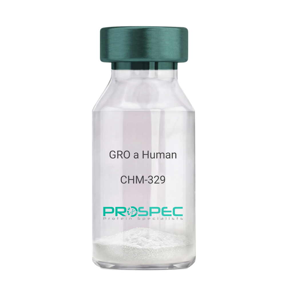
    Growth-regulated protein alpha, CXCL1, Melanoma growth stimulatory activity, MGSA, Neutrophil-activating protein 3, NAP-3, GRO-alpha (1-73), chemokine (C-X-C motif) ligand 1, GRO1, GROa, SCYB1, MGSA-a, MGSA alpha.

    Product # :

    CHM-329

    Price :

    Quantity :

    Shipping Method :

    Room Temp Icon

    Shipped at Room temp

    Add To Cart

    More Info

    • description
    • source
    • formulation
    • purity
    • biological activity
    • More Info

    Description

    GRO Alpha Human Recombinant produced in E.Coli is a single,non-glycosylated, polypeptide chain containing 73 amino acids and having a molecular mass of 7811 Dalton. The GRO-alpha is purified by proprietary chromatographic techniques.

    Source

    Escherichia Coli.

    Formulation

    Lyophilized from a 0.2µm filtered concentrated (1mg/ml) solution in 20mM PB, pH 7.4, 50mM NaCl.

    Purity

    Greater than 97.0% as determined by:
    (a) Analysis by RP-HPLC.
    (b) Analysis by SDS-PAGE.

    Biological Activity

    Determined by its ability to chemoattract human peripheral blood neutrophils using a concentration range of 10.0-100.0 ng/ml.

    More Info

    • Introduction

      Chemokine (C-X-C motif) ligand 1 (CXCL1) is a small cytokine belonging to the CXC chemokine family that was previously called GRO1 oncogene, Neutrophil-activating protein 3 (NAP-3) and melanoma growth stimulating activity, alpha (MSGA-a). It is secreted by human melanoma cells, has mitogenic properties and is implicated in melanoma pathogenesis. CXCL1 is expressed by macrophages, neutrophilsand epithelial cells, and has neutrophil chemoattractant activity. CXCL1 plays a role in spinal cord development by inhibiting the migration of oligodendrocyte precursors and is involved in the processes of angiogenesis, inflammation, wound healing, and tumorigenesis. This chemokine elicits its effects by signaling through the chemokine receptor CXCR2. The gene for CXCL1 is located on human chromosome 4 amongst genes for other CXC chemokines.

    • Synonyms

      Growth-regulated protein alpha, CXCL1, Melanoma growth stimulatory activity, MGSA, Neutrophil-activating protein 3, NAP-3, GRO-alpha (1-73), chemokine (C-X-C motif) ligand 1, GRO1, GROa, SCYB1, MGSA-a, MGSA alpha.

    • Physical Appearance

      Sterile Filtered White lyophilized (freeze-dried) powder.

    • Stability

      Lyophilized GRO-alpha although stable at room temperature for 3 weeks, should be stored desiccated below -18°C. Upon reconstitution CXCL1 should be stored at 4°C between 2-7 days and for future use below -18°C. For long term storage it is recommended to add a carrier protein (0.1% HSA or BSA).Please prevent freeze-thaw cycles.

    • Solubility

      It is recommended to reconstitute the lyophilized CXCL1 in sterile 18MΩ-cm H2O not less than 100µg/ml, which can then be further diluted to other aqueous solutions.

    • Amino Acid Sequence

      The sequence of the first five N-terminal amino acids was determined and was found to be Ala-Ser-Val-Ala-Thr.

    • Background

      What is the molecular weight/Mw of CXCL1 HUMAN Protein?
      CXCL1 HUMAN Protein has a total Mw of 7.81kDa.

      What is the source or expression system of CXCL1 HUMAN Protein?
      Escherichia Coli.

      What is the Purity of CXCL1 HUMAN Protein?
      CXCL1 HUMAN Protein is >97% pure as determined by SDS-PAGE.

      What is the Biological Activity of CXCL1 HUMAN Protein?
      Determined by its ability to chemoattract human peripheral blood neutrophils using a concentration range of 10.0-100.0 ng/ml.

      What is the amino acid sequence of CXCL1 HUMAN Protein?
      The sequence of the first five N-terminal amino acids was determined and was found to be Ala-Ser-Val-Ala-Thr.

      What applications can CXCL1 HUMAN Protein be used in?
      CXCL1 HUMAN Protein can probably be used in western blot, ELISA and Lateral Flow.

      What is the endotoxin level for CXCL1 HUMAN Protein?
      The endotoxin level is minimal, CXCL1 HUMAN Protein was purified using conventional chromatography techniques.


    ProSpec's products are furnished for LABORATORY RESEARCH USE ONLY. The product may not be used as drugs, agricultural or pesticidal products, food additives or household chemicals.

    Gro A Human
  • View Data Sheet

    Name :

    VEGF Antibody

    Description:

    Vascular Endothelial Cell Growth Factor, Mouse Anti-Human

    Vascular endothelial growth factor A, VEGF-A, Vascular permeability factor, VPF, VEGF, MGC70609.

    Product # :

    ANT-125

    Price :

    Quantity :

    Shipping Method :

    Room Temp Icon

    Shipped at Room temp

    Add To Cart

    More Info

    • More Info

    More Info

    • Introduction

      Vascular endothelial growth factoris an important signaling proteininvolved in both vasculogenesisand angiogenesis. As its name implies, VEGF activity has been mostly studied on cells of the vascular endothelium, although it does have effects on a number of other cell types (e.g. stimulation monocyte/macrophagemigration, neurons, cancer cells, kidney epithelial cells ).VEGF mediates increased vascular permeability, induces angiogenesis, vasculogenesis and endothelial cell growth, promotes cell migration, and inhibits apoptosis. In vitro, VEGF has been shown to stimulate endothelial cell mitogenesisand cell migration. VEGF is also a vasodilator and increases microvascular permeability and was originally referred to as vascular permeability factor.
      Elevated levels of this protein is linked to POEMS syndrome, also known as Crow-Fukase syndrome. Mutations in this gene have been associated with proliferative and nonproliferative diabetic retinopathy.

    • Synonyms

      Vascular endothelial growth factor A, VEGF-A, Vascular permeability factor, VPF, VEGF, MGC70609.

    • Solubility

      Reconstitute with sterile H20. Mix gently, wash the sides of the vial and wait 30-60 seconds before use.

    • Immunogen

      r.HumanVEGF.

    • Ig Subclass

      Mouse IgM.

    • Clone

      NYRhVEGF.

    • Applications

      Direct ELISA, Western Blot, Immuneprecipitation.

    • Titer

      By direct ELISA, 1:10,000 dilution will yield 0.15 O.D using alkaline phosphatase conjugated rabbit anti-mouse Ig (Jackson Laboratories).

    • Shipping Conditions

      Antibody is shipped lyophilized at ambient temperature.

    • Type

      Mouse Anti Human Monoclonal.

    • Storage Procedures

      In lyophilized form, for long periods, store at 4oC in a dry environment. After reconstitution, if not intended for use within a month, aliquot and store at -20oC.

    • Purification Method

      Boric acid precipitation Protein concentration1mg/ml in PBS (after reconstitution).

    ProSpec's products are furnished for LABORATORY RESEARCH USE ONLY. The product may not be used as drugs, agricultural or pesticidal products, food additives or household chemicals.

    Vegf Antibody
  • View Data Sheet

    Name :

    MIF Human His C

    Description:

    Macrophage Migration Inhibitory Factor Human Recombinant, His Tag C-Terminus

    Phenylpyruvate tautomerase, Glycosylation-inhibiting factor, GIF, MMIF, MIF.

    Product # :

    CYT-521

    Price :

    Quantity :

    Shipping Method :

    Room Temp Icon

    Shipped at Room temp

    Add To Cart

    More Info

    • description
    • source
    • formulation
    • purity
    • biological activity
    • More Info

    Description

    MIF human Recombinant, fused to His-tag at C-terminus, was cloned into an E. coli expression vector and was purified to apparent homogeneity by using conventional column chromatography techniques.Macrophage Inducing Factor Human Recombinant is a single, non-glycosylated, polypeptide chaincontaining 123 amino acidsand having a molecular mass of 13.5 kDa.

    Source

    Escherichia Coli.

    Formulation

    Human MIF was lyophilized from a 1mg/ml solution containing PBS pH-7.4.

    Purity

    Greater than 95.0% as determined by:
    (a) Analysis by RP-HPLC.
    (b) Analysis by SDS-PAGE.

    Biological Activity

    Measured by its ability to bind rhCD74 in a functional ELISA.

    More Info

    • Introduction

      The cytokine Macrophage migration inhibitory factor (MIF) has been identified to be secreted by the pituitary gland and the monocyte/macrophage and to play an important role in endotoxic shock. MIF has the unique property of being released from macrophages and T cells in response to physiological concentrations of glucocorticoids. The secretion of MIF is tightly regulated and decreases at high, anti-inflammatory steroid concentration.

    • Synonyms

      Phenylpyruvate tautomerase, Glycosylation-inhibiting factor, GIF, MMIF, MIF.

    • Physical Appearance

      Sterile Filtered lyophilized powder.

    • Stability

      Lyophilized MIF although stable at room temperature for 3 weeks, should be stored desiccated below -18°C. Upon reconstitution MIF should be stored at 4°C between 2-7 days and for future use below -18°C.For long term storage it is recommended to add a carrier protein (0.1% HSA or BSA).Please prevent freeze-thaw cycles.

    • Solubility

      It is recommended to reconstitute the lyophilized MIF in sterile 18MΩ-cm H2O not less than 100µg/ml, which can then be further diluted to other aqueous solutions.

    • Amino Acid Sequence

      MPMFIVNTNVPRASVPDGFLSELTQQLAQATGKPPQYIAVHVVPDQLMAFGGSSEPC
      ALCSLHSIGKIGGAQNRSYSKLLCGLLAERLRISPDRVYINYYDMNAANVGWNNSTF
      ALEHHHHHH.

    ProSpec's products are furnished for LABORATORY RESEARCH USE ONLY. The product may not be used as drugs, agricultural or pesticidal products, food additives or household chemicals.

    Mif Human His C
  • View Data Sheet

    Name :

    DPP4 Human, HEK

    Description:

    Dipeptidyl-Peptidase 4 Human Recombinant, HEK

    CD26, ADABP, ADCP2, DPPIV, TP103, DPP4, Dipeptidyl peptidase 4, Dipeptidyl peptidase IV, DPP IV, T-cell activation antigen CD26, Adenosine deaminase complexing protein 2, CD26 antigen.

    Product # :

    ENZ-1187

    Price :

    Quantity :

    Shipping Method :

    Ice Icon

    Shipped with Ice Packs

    Add To Cart

    More Info

    • description
    • source
    • formulation
    • purity
    • biological activity
    • More Info

    Description

    DPP4 Human Recombinant is a single, glycosylated polypeptide chain containing 977 amino acids (29-766a.a) and having a molecular mass of 112.1kDa (calculated). DPP4 is fused to a 239 amino acid hIgG-Tag at C-terminus and is purified by proprietary chromatographic techniques.

    Source

    HEK293 Cells.

    Formulation

    DPP4 protein solution (0.25mg/ml) contains 10% Glycerol and Phosphate-Buffered Saline (pH 7.4).

    Purity

    Greater than 95.0% as determined by Analysis by SDS-PAGE.

    Biological Activity

    Specific activity is > 15,000 pmol/min/ug in which one unit defined as the amount of enzyme that hydrolyze 1pmole of H-Gly-Pro-AMC.HBr to H-Gly-Pro and AMC per minute at pH 8.0 at 37℃.

     The ED50 range ≤250 ng/ml is measured by its binding ability in a functional ELISA with MERS-CoV Spike S1 Subunit (CAT# sars-052)..

    The ED50 range ≤200 ng/ml is measured by its binding ability in a functional ELISA with MERS-CoV Spike RBD (CAT# sars-054).

    The ED50 range ≤120 ng/ml is measured by its binding ability in a functional ELISA with MERS-CoV Spike (CAT# sars-051).

    More Info

    • Synonyms

      CD26, ADABP, ADCP2, DPPIV, TP103, DPP4, Dipeptidyl peptidase 4, Dipeptidyl peptidase IV, DPP IV, T-cell activation antigen CD26, Adenosine deaminase complexing protein 2, CD26 antigen.

    • Physical Appearance

      Sterile Filtered colorless solution.

    • Stability

      Store at 4°C if entire vial will be used within 2-4 weeks. Store, frozen at -20°C for longer periods of time. For long term storage it is recommended to add a carrier protein (0.1% HSA or BSA).Avoid multiple freeze-thaw cycles.

    • Amino Acid Sequence

      DGSMNKGTDD ATADSRKTYT LTDYLKNTYR LKLYSLRWIS DHEYLYKQEN NILVFNAEYG NSSVFLENST FDEFGHSIND YSISPDGQFI LLEYNYVKQW RHSYTASYDI YDLNKRQLIT EERIPNNTQW VTWSPVGHKL AYVWNNDIYV KIEPNLPSYR ITWTGKEDII YNGITDWVYE EEVFSAYSAL WWSPNGTFLA YAQFNDTEVP LIEYSFYSDE SLQYPKTVRV PYPKAGAVNP TVKFFVVNTD SLSSVTNATS IQITAPASML IGDHYLCDVT WATQERISLQ WLRRIQNYSV MDICDYDESS GRWNCLVARQ HIEMSTTGWV GRFRPSEPHF TLDGNSFYKI ISNEEGYRHI CYFQIDKKDC TFITKGTWEV IGIEALTSDY LYYISNEYKG MPGGRNLYKI QLSDYTKVTC LSCELNPERC QYYSVSFSKE AKYYQLRCSG PGLPLYTLHS SVNDKGLRVL EDNSALDKML QNVQMPSKKL DFIILNETKF WYQMILPPHF DKSKKYPLLL DVYAGPCSQK ADTVFRLNWA TYLASTENII VASFDGRGSG YQGDKIMHAI NRRLGTFEVE DQIEAARQFS KMGFVDNKRI AIWGWSYGGY VTSMVLGSGS GVFKCGIAVA PVSRWEYYDS VYTERYMGLP TPEDNLDHYR NSTVMSRAEN FKQVEYLLIH GTADDNVHFQ QSAQISKALV DVGVDFQAMW YTDEDHGIAS STAHQHIYTH MSHFIKQCFS LPKLLEPKSC DKTHTCPPCP APELLGGPSV FLFPPKPKDT LMISRTPEVT CVVVDVSHED PEVKFNWYVD GVEVHNAKTK PREEQYNSTY RVVSVLTVLH QDWLNGKEYK CKVSNKALPA PIEKTISKAK GQPREPQVYT LPPSRDELTK NQVSLTCLVK GFYPSDIAVE WESNGQPENN YKTTPPVLDS DGSFFLYSKL TVDKSRWQQG NVFSCSVMHE ALHNHYTQKS LSLSPGK.

    • Background

      DPP4 protein, also known as Dipeptidyl peptidase-4 or CD26, is a cell surface protease with diverse functions in cell signaling and metabolism. This research aims to investigate the role of DPP4 protein in various physiological processes and its implications in disease pathogenesis. Understanding the biological significance of DPP4 can provide insights into its potential as a therapeutic target for several disorders.

      Function of DPP4 Protein:

      DPP4 protein is involved in the cleavage and regulation of several peptide hormones and chemokines, influencing their bioactivity and half-life. It is widely expressed in various tissues, including immune cells, endothelial cells, and epithelial cells. DPP4 can modulate immune responses, glucose metabolism, and neuropeptide signaling through enzymatic and non-enzymatic activities.

      Role of DPP4 Protein in Immune Regulation:

      DPP4 protein plays a role in immune cell activation and regulation. It is expressed on the surface of T cells, where it functions as a co-stimulatory molecule. DPP4 engagement on T cells promotes T cell activation, cytokine production, and adhesion to endothelial cells. Additionally, DPP4 can cleave and inactivate certain chemokines, thereby influencing chemotaxis and immune cell recruitment.

      Implications of DPP4 Protein in Metabolic Disorders:

      DPP4 protein is involved in glucose metabolism and insulin regulation. It cleaves incretin hormones, such as glucagon-like peptide-1 (GLP-1) and gastric inhibitory polypeptide (GIP), which play crucial roles in glucose homeostasis. Inhibition of DPP4 activity can enhance the action of these incretin hormones, leading to improved glycemic control. Therefore, DPP4 inhibitors have been developed as antidiabetic drugs.

      Association of DPP4 Protein with Cardiovascular Diseases:

      DPP4 protein has been implicated in the pathogenesis of cardiovascular diseases. Elevated DPP4 levels have been observed in patients with heart failure, atherosclerosis, and hypertension. DPP4 can contribute to endothelial dysfunction, inflammation, and vascular remodeling, which are key factors in the development and progression of cardiovascular disorders. Inhibition of DPP4 activity has shown potential as a therapeutic strategy in preclinical studies.

      Given its involvement in various biological processes and disease pathogenesis, DPP4 protein has emerged as a potential therapeutic target. DPP4 inhibitors, which prevent the enzymatic activity of DPP4, have been developed for the treatment of type 2 diabetes. These inhibitors enhance the action of incretin hormones, leading to improved glycemic control. Additionally, ongoing research aims to explore the therapeutic potential of DPP4 inhibitors in other conditions, such as immune-mediated disorders and cardiovascular diseases.

      Conclusion:

      The investigation of DPP4 protein provides insights into its diverse functions in cell signaling, immune regulation, and metabolism. Understanding the role of DPP4 in disease pathogenesis opens avenues for the development of targeted therapies for conditions such as diabetes, cardiovascular diseases, and immune-mediated disorders. Further research on DPP4 protein and its associated pathways may uncover new therapeutic opportunities and improve patient outcomes.

    ProSpec's products are furnished for LABORATORY RESEARCH USE ONLY. The product may not be used as drugs, agricultural or pesticidal products, food additives or household chemicals.

    Dpp4 Human Hek
  • View Data Sheet

    Name :

    GCGR Human

    Description:

    Glucagon Receptor Human Recombinant

    GL-R, GLR, Glucagon Receptor.

    Product # :

    HOR-020

    Price :

    Quantity :

    Shipping Method :

    Room Temp Icon

    Shipped at Room temp

    Add To Cart

    More Info

    • description
    • source
    • formulation
    • purity
    • More Info

    Description

    GCGR Human Recombinant produced in E. coli is a single, non-glycosylated polypeptide chain (a.a 29-142) containing 125 amino acids including an 8 a.a C-terminal His tag. The total molecular mass is 19.9kDa (calculated).

    Source

    E. coli

    Formulation

    GCGR filtered (0.4 µm) and lyophilized from solution in acetonitrile/0,1%TFA and 1% (w/v) trehalose.

    Purity

    Greater than 95.0% as determined by SDS-PAGE.

    More Info

    • Introduction

      Glucagon receptor is part of the glucagon receptor family which also contains GLP-1, GLP-2, GHRH and GIP receptors. GCGR regulates blood glucose levels and is mainly expressed in the pancreas, liver and kidneys. Mutations in GCGR are a cause of non-insulin-dependent diabetes mellitus.

    • Synonyms

      GL-R, GLR, Glucagon Receptor.

    • Physical Appearance

      Filtered White lyophilized (freeze-dried) powder.

    • Stability

      Store lyophilized protein at -20°C. Aliquot the product after reconstitution to avoid repeated freezing/thawing cycles. Reconstituted protein can be stored at 4°C for a limited period of time; it does not show any change after one week at 4°C.

    • Solubility

      It is recommended to add deionized water to prepare a working stock solution of approximately 0.5mg/ml and let the lyophilized pellet dissolve completely. GCGR is not sterile! Please filter the product by an appropriate sterile filter before using it in the cell culture.

    • Amino Acid Sequence

      MQVMDFLFEK WKLYGDQCHH NLSLLPPPTE LVCNRTFDKY SCWPDTPANT TANISCPWYL PWHHKVQHRF VFKRCGPDGQ WVRGPRGQPW RDASQCQMDG EEIEVQKEVA KMYSSFQLEH HHHHH.

    ProSpec's products are furnished for LABORATORY RESEARCH USE ONLY. The product may not be used as drugs, agricultural or pesticidal products, food additives or household chemicals.

    Gcgr Human
  • View Data Sheet

    Name :

    GNAI3 Human

    Description:

    Guanine Nucleotide Binding Protein Alpha Inhibiting Activity 3 Human Recombinant

    Guanine nucleotide binding protein (G protein) alpha inhibiting activity polypeptide 3, G(i) alpha-3, 87U6.

    Product # :

    PRO-1074

    Price :

    Quantity :

    Shipping Method :

    Ice Icon

    Shipped with Ice Packs

    Add To Cart

    More Info

    • description
    • source
    • formulation
    • purity
    • More Info

    Description

    GNAI3 Human Recombinant produced in E. coli is a single polypeptide chain containing 377 amino acids (1-354) and having a molecular mass of 43.0kDa.GNAI3 is fused to a 23 amino acid His-tag at N-terminus & purified by proprietary chromatographic techniques.

    Source

    E.coli.

    Formulation

    The GNAI3 solution (0.5mg/1ml) contains 20mM Tris-HCl buffer (pH 8.0), 0.1M NaCl, 2mM DTT and 20% glycerol.

    Purity

    Greater than 90% as determined by SDS-PAGE.

    More Info

    • Introduction

      GNAI3 is a member of the G-alpha family and G (i/o/t/z) subfamily which act as modulators or transducers in several transmembrane signaling systems. G(k) is the stimulatory G protein of receptor-regulated K+ channels. The active GTP-bound form inhibits the connotation of RGS14 with centrosomes and is essential for the translocation of RGS14 from the cytoplasm to the plasma membrane. GNAI3 takes part in cell division and is known to cooperate with RIC8A, S1PR1, RGS5, RGS10, RGS12, RGS14, RGS16, RGS18, and RGS19.

    • Synonyms

      Guanine nucleotide binding protein (G protein) alpha inhibiting activity polypeptide 3, G(i) alpha-3, 87U6.

    • Physical Appearance

      Sterile Filtered colorless solution.

    • Stability

      Store at 4°C if entire vial will be used within 2-4 weeks. Store, frozen at -20°C for longer periods of time. For long term storage it is recommended to add a carrier protein (0.1% HSA or BSA).Avoid multiple freeze-thaw cycles.

    • Amino Acid Sequence

      MGSSHHHHHH SSGLVPRGSH MGSMGCTLSA EDKAAVERSK MIDRNLREDG EKAAKEVKLL LLGAGESGKS TIVKQMKIIH EDGYSEDECK QYKVVVYSNT IQSIIAIIRA MGRLKIDFGE AARADDARQL FVLAGSAEEG VMTPELAGVI KRLWRDGGVQ ACFSRSREYQ LNDSASYYLN DLDRISQSNY IPTQQDVLRT RVKTTGIVET HFTFKDLYFK MFDVGGQRSE RKKWIHCFEG VTAIIFCVAL SDYDLVLAED EEMNRMHESM KLFDSICNNK WFTETSIILF LNKKDLFEEK IKRSPLTICY PEYTGSNTYE EAAAYIQCQF EDLNRRKDTK EIYTHFTCAT DTKNVQFVFD AVTDVIIKNN LKECGLY

    ProSpec's products are furnished for LABORATORY RESEARCH USE ONLY. The product may not be used as drugs, agricultural or pesticidal products, food additives or household chemicals.

    Gnai3 Human
  • View Data Sheet

    Name :

    CNDP1 Human

    Description:

    CNDP Dipeptidase 1 Human Recombinant

    Carnosine Dipeptidase 1 (Metallopeptidase M20 Family), Glutamate Carboxypeptidase-Like Protein 2, CNDP Dipeptidase 1, Serum Carnosinase, Carnosinase 1, CPGL2, CN1, Carnosine Dipeptidase 1, EC 3.4.13.20, HsT2308.

    Product # :

    ENZ-927

    Price :

    Quantity :

    Shipping Method :

    Ice Icon

    Shipped with Ice Packs

    Add To Cart

    More Info

    • description
    • source
    • formulation
    • purity
    • More Info

    Description

    CNDP1 produced in Sf9 Insect cells is a single, glycosylated polypeptide chain containing 489 amino acids (27-507a.a.) and having a molecular mass of 54.9kDa. (Molecular size on SDS-PAGE will appear at approximately 50-70kDa).CNDP1 is expressed with an 8 amino acid His tag at C-Terminus and purified by proprietary chromatographic techniques.

    Source

    Sf9, Insect cells.

    Formulation

    CNDP1 protein solution (0.25mg/ml) contains Phosphate Buffered Saline (pH 7.4) and 10% glycerol.

    Purity

    Greater than 95.0% as determined by SDS-PAGE.

    More Info

    • Introduction

      CNDP Dipeptidase 1, also known as CNDP1 is a member of the peptidase M20A family. CNDP1 Mannheim which is the shortest allelic form has been more common in the absence of nephropathy in addition to being associated with lower serum carnosinase levels. Furthermore, Carnosine inhibited the increased production of fibronectin as well as collagen type VI in podocytes and the increased production of TGF-beta in mesangial cells. Diabetic patients with the CNDP1 Mannheim variant are less at risk for nephropathy. In addition, on renal cells carnosine protects against the adverse effects of high glucose levels.

    • Synonyms

      Carnosine Dipeptidase 1 (Metallopeptidase M20 Family), Glutamate Carboxypeptidase-Like Protein 2, CNDP Dipeptidase 1, Serum Carnosinase, Carnosinase 1, CPGL2, CN1, Carnosine Dipeptidase 1, EC 3.4.13.20, HsT2308.

    • Physical Appearance

      Sterile filtered colorless solution.

    • Stability

      Store at 4°C if entire vial will be used within 2-4 weeks.Store, frozen at -20°C for longer periods of time. For long term storage it is recommended to add a carrier protein (0.1% HSA or BSA).Avoid multiple freeze-thaw cycles.

    • Amino Acid Sequence

      SPSPPPALLE KVFQYIDLHQ DEFVQTLKEW VAIESDSVQP VPRFRQELFR MMAVAADTLQ RLGARVASVD MGPQQLPDGQ SLPIPPVILA ELGSDPTKGT VCFYGHLDVQ PADRGDGWLT DPYVLTEVDG KLYGRGATDN KGPVLAWINA VSAFRALEQD LPVNIKFIIE GMEEAGSVAL EELVEKEKDR FFSGVDYIVI SDNLWISQRK PAITYGTRGN SYFMVEVKCR DQDFHSGTFG GILHEPMADL VALLGSLVDS SGHILVPGIY DEVVPLTEEE INTYKAIHLD LEEYRNSSRV
      EKFLFDTKEE ILMHLWRYPS LSIHGIEGAF DEPGTKTVIP GRVIGKFSIR LVPHMNVSAV EKQVTRHLED VFSKRNSSNK MVVSMTLGLH PWIANIDDTQ YLAAKRAIRT VFGTEPDMIR DGSTIPIAKM FQEIVHKSVV LIPLGAVDDG EHSQNEKINR WNYIEGTKLF AAFFLEMAQL HLEHHHHHH.

    ProSpec's products are furnished for LABORATORY RESEARCH USE ONLY. The product may not be used as drugs, agricultural or pesticidal products, food additives or household chemicals.

    Cndp1 Human
  • View Data Sheet

    Name :

    RNGTT Human

    Description:

    RNA Guanylyltransferase And 5'-Phosphatase Human Recombinant

    RNA Guanylyltransferase And 5'-Phosphatase, CAP1A, RNA Guanylyltransferase And 5-Phosphatase, HCAP1, HCE1, HCE, MRNA-Capping Enzyme, HCAP 3, mRNA-capping enzyme.

    Product # :

    ENZ-823

    Price :

    Quantity :

    Shipping Method :

    Ice Icon

    Shipped with Ice Packs

    Add To Cart

    More Info

    • description
    • source
    • formulation
    • purity
    • More Info

    Description

    RNGTT Human Recombinant produced in E.Coli is a single, non-glycosylated polypeptide chain containing 620 amino acids (1-597a.a) and having a molecular mass of 70.9kDa. RNGTT is fused to a 23 amino acid His-tag at N-terminus & purified by proprietary chromatographic techniques.

    Source

    Escherichia Coli.

    Formulation

    RNGTT protein solution (0.25mg/ml) containing 20mM Tris-HCl buffer (pH 7.5), 0.2M NaCl, 40% glycerol, 2mM DTT and 0.1mM PMSF.

    Purity

    Greater than 85.0% as determined by SDS-PAGE.

    More Info

    • Introduction

      RNA Guanylyltransferase And 5'-Phosphatase also known as RNGTT is a bifunctional mRNA-capping enzyme which exhibits RNA 5'-triphosphatase activity in the N-terminal section and mRNA guanylyltransferase activity in the C-terminal section. In addition, RNGTT catalyzes the first two steps of cap formation, through removing the gamma-phosphate from the 5'-triphosphate end of nascent mRNA to yield a diphosphate end, and also by transferring the gmp moiety of GTP to the 5'-diphosphate terminus.

    • Synonyms

      RNA Guanylyltransferase And 5'-Phosphatase, CAP1A, RNA Guanylyltransferase And 5-Phosphatase, HCAP1, HCE1, HCE, MRNA-Capping Enzyme, HCAP 3, mRNA-capping enzyme.

    • Physical Appearance

      Sterile filtered colorless solution.

    • Stability

      Store at 4°C if entire vial will be used within 2-4 weeks. Store, frozen at -20°C for longer periods of time. For long term storage it is recommended to add a carrier protein (0.1% HSA or BSA).Avoid multiple freeze-thaw cycles.

    • Amino Acid Sequence

      MGSSHHHHHH SSGLVPRGSH MGSMAHNKIP PRWLNCPRRG QPVAGRFLPL KTMLGPRYDS QVAEENRFHP SMLSNYLKSL KVKMGLLVDL TNTSRFYDRN DIEKEGIKYI KLQCKGHGEC PTTENTETFI RLCERFNERN PPELIGVHCT HGFNRTGFLI CAFLVEKMDW SIEAAVATFA QARPPGIYKG DYLKELFRRY GDIEEAPPPP LLPDWCFEDD EDEDEDEDGK KESEPGSSAS FGKRRKERLK LGAIFLEGVT VKGVTQVTTQ PKLGEVQQKC HQFCGWEGSG FPGAQPVSMD KQNIKLLDLK PYKVSWKADG TRYMMLIDGT NEVFMIDRDN SVFHVSNLEF PFRKDLRMHL SNTLLDGEMI IDRVNGQAVP RYLIYDIIKF NSQPVGDCDF NVRLQCIERE IISPRHEKMK TGLIDKTQEP FSVRNKPFFD ICTSRKLLEG NFAKEVSHEM DGLIFQPTGK YKPGRCDDIL KWKPPSLNSV DFRLKITRMG GEGLLPQNVG LLYVGGYERP FAQIKVTKEL KQYDNKIIEC KFENNSWVFM RQRTDKSFPN AYNTAMAVCN SISNPVTKEM LFEFIDRCTA ASQGQKRKHH LDPDTELMPP PPPKRPRPLT.

    ProSpec's products are furnished for LABORATORY RESEARCH USE ONLY. The product may not be used as drugs, agricultural or pesticidal products, food additives or household chemicals.

    Rngtt Human
  • 1
  • …
  • 26
  • 27
  • 28
  • 29
  • 30
  • …
  • 42
Your browser does not support the video tag.

Products

  • Cytokines
  • Growth Factors
  • Chemokines
  • CD Antigens
  • Neurotrophins
  • Hormones
  • Enzymes
  • Viral Antigens
  • Recombinant Proteins
  • Natural Proteins
  • Monoclonal Antibodies
  • Polyclonal Antibodies
  • Thermal Packaging

About

  • Company
  • Founder
  • Contribution
  • Contact Us
  • My Account

Ordering

  • Ordering Method
  • Ordering - Credit Card
  • Ordering PO
  • Shipping Fees
  • FAQ's

Legal

  • Terms & Conditions
  • Privacy
  • Site Map
  • info@prospecbio.com
  • Facebook
  • Instagram
  • Linkedin Profile

© 2026 ProSpec-Tany TechnoGene Ltd. All Rights Reserved.

  • Choosing a selection results in a full page refresh.
  • Opens in a new window.