Search results
1000 results found for “Decorin”
Name
Description
Product #
Price
Quantity
Shipping Method
- View Data Sheet
Name :
SHC1 HumanDescription:
SHC-Transforming Protein 1 Human Recombinant
SHC (Src homology 2 domain containing) transforming protein 1, SHC, SHCA, SH2 domain protein C1, SHC-transforming protein 3, SHC-transforming protein A, FLJ26504, p66.
Product # :
PRO-883Price :
Quantity :
Shipping Method :
Shipped with Ice Packs
More Info
- description
- source
- formulation
- purity
- More Info
Description
SHC1 Human Recombinant produced in E.coli is a single, non-glycosylated polypeptide chain containing 494 amino acids (1-474) and having a molecular mass of 53.8 kDa.The SHC1 is fused to a 20 amino acid His-Tag at N-terminus and purified by proprietary chromatographic techniques.
Source
Escherichia Coli.
Formulation
SHC1 protein (1mg/ml) is supplied in 20mM Tris-HCl, pH-8, 2mM DTT, 200mM NaCl, 0.1mM PMSF and 20% Glycerol.
Purity
Greater than 90.0% as determined by SDS-PAGE.
More Info
-
Introduction
SHC1 is a member of the SH2 domain family. SHC1 is vital to the regulation of apoptosis and drug resistance in mammalian cells. SHC1 is a signaling adapter which links activated growth factor receptors to signaling pathway. SHC1 is found in neural stem cells but not in mature neurons.
-
Synonyms
SHC (Src homology 2 domain containing) transforming protein 1, SHC, SHCA, SH2 domain protein C1, SHC-transforming protein 3, SHC-transforming protein A, FLJ26504, p66.
-
Physical Appearance
Sterile Filtered colorless solution.
-
Stability
Store at 4°C if entire vial will be used within 2-4 weeks. Store, frozen at -20°C for longer periods of time. For long term storage it is recommended to add a carrier protein (0.1% HSA or BSA).Avoid multiple freeze-thaw cycles.
-
Amino Acid Sequence
MGSSHHHHHH SSGLVPRGSH MNKLSGGGGR RTRVEGGQLG GEEWTRHGSF VNKPTRGWLH PNDKVMGPGV SYLVRYMGCV EVLQSMRALD FNTRTQVTRE AISLVCEAVP GAKGATRRRK PCSRPLSSIL GRSNLKFAGM PITLTVSTSS LNLMAADCKQ IIANHHMQSI SFASGGDPDT AEYVAYVAKD PVNQRACHIL ECPEGLAQDV ISTIGQAFEL RFKQYLRNPP KLVTPHDRMA GFDGSAWDEE EEEPPDHQYY NDFPGKEPPL GGVVDMRLRE GAAPGAARPT APNAQTPSHL GATLPVGQPV GGDPEVRKQM PPPPPCPAGR ELFDDPSYVN VQNLDKARQA VGGAGPPNPA INGSAPRDLF DMKPFEDALR VPPPPQSVSM AEQLRGEPWF HGKLSRREAE ALLQLNGDFL VRESTTTPGQ YVLTGLQSGQ PKHLLLVDPE GVVRTKDHRF ESVSHLISYH MDNHLPIISA GSELCLQQPV ERKL
ProSpec's products are furnished for LABORATORY RESEARCH USE ONLY. The product may not be used as drugs, agricultural or pesticidal products, food additives or household chemicals.
- View Data Sheet
Name :
CCL6 MouseDescription:
C-10 Mouse Recombinant (CCL6)
Small inducible cytokine A6, CCL6, C10 protein, c10, MRP-1, Scya6, chemokine (C-C motif) ligand 6.
Product # :
CHM-307Price :
Quantity :
Shipping Method :
Shipped at Room temp
More Info
- description
- source
- formulation
- purity
- biological activity
- More Info
Description
C-10 Mouse Recombinant produced in E.Coli is a single,non-glycosylated, polypeptide chain containing 95 amino acids and having a molecular mass of 10.7kDa.The CCL6 is purified by proprietary chromatographic techniques.
Source
Escherichia Coli.
Formulation
CCL6 Mouse was lyophilized from a 0.2µm filtered concentrated solution in 1×PBS, pH 7.4.
Purity
Greater than 95.0% as determined by:
(a) Analysis by RP-HPLC.
(b) Analysis by SDS-PAGE.Biological Activity
The Biological activity was determined by its ability to chemoattract human CCR1 transfected BaF3 mouse proB cells using a concentration range of 0.05-0.25µg/ml.
More Info
-
Introduction
Chemokine (C-C motif) ligand 6 (CCL6) is a small cytokine belonging to the CC chemokine family that has only been identified in rodents.
In mice, CCL6 is expressed in cells from neutrophil and macrophage lineages, and can be greatly induced under conditions suitable for myeloid cell differentiation. It is highly expressed in bone marrow cultures that have been stimulated with the cytokine GM-CSF. Some low levels of gene expression also occur in certain cell lines of myeloid origin (e.g. the immature myeloid cell lines DA3 and 32D cl3, and the macrophage cell line P388D) that can also be greatly induced in culture with GM-CSF. However, in activated T cell lines, expression of CCL6 is greatly reduced. CCL6 can also be induced in the mouse lung by the cytokine interleukin 13. Mouse CCL6 is located on chromosome 11. The cell surface receptor for CCL6 is believed to be the chemokine receptor CCR1. -
Synonyms
Small inducible cytokine A6, CCL6, C10 protein, c10, MRP-1, Scya6, chemokine (C-C motif) ligand 6.
-
Physical Appearance
Sterile Filtered White lyophilized (freeze-dried) powder.
-
Stability
Lyophilized C10 protein although stable at room temperature for 3 weeks, should be stored desiccated below -18°C. Upon reconstitution CCL6 should be stored at 4°C between 2-7 days and for future use below -18°C. For long term storage it is recommended to add a carrier protein (0.1% HSA or BSA).Please prevent freeze-thaw cycles.
-
Solubility
It is recommended to reconstitute the lyophilized C10 protein in sterile 18MΩ-cm H2O not less than 100µg/ml, which can then be further diluted to other aqueous solutions.
-
Amino Acid Sequence
GLIQEIEKED RRYNPPIIHQ GFQDTSSDCC FSYATQIPCK RFIYYFPTSG GCIKPGIIFI SRRGTQVCAD PSDRRVQRCL STLKQGPRSG NKVIA.
-
Background
What is the molecular weight/Mw of CCL6 MOUSE Protein?
CCL6 MOUSE Protein has a total Mw of 10.7kDa.
What is the source or expression system of CCL6 MOUSE Protein?
Escherichia Coli.
What is the Purity of CCL6 MOUSE Protein?
CCL6 MOUSE Protein is >95% pure as determined by SDS-PAGE.
What is the Biological Activity of CCL6 MOUSE Protein?
The Biological activity was determined by its ability to chemoattract human CCR1 transfected BaF3 mouse proB cells using a concentration range of 0.05-0.25µg/ml.
What is the amino acid sequence of CCL6 MOUSE Protein?
GLIQEIEKED RRYNPPIIHQ GFQDTSSDCC FSYATQIPCK RFIYYFPTSG GCIKPGIIFI SRRGTQVCAD PSDRRVQRCL STLKQGPRSG NKVIA.
What applications can CCL6 MOUSE Protein be used in?
CCL6 MOUSE Protein can probably be used in western blot, ELISA and Lateral Flow.
What is the endotoxin level for CCL6 MOUSE Protein?
The endotoxin level is minimal, CCL6 MOUSE Protein was purified using conventional chromatography techniques.
ProSpec's products are furnished for LABORATORY RESEARCH USE ONLY. The product may not be used as drugs, agricultural or pesticidal products, food additives or household chemicals.
- View Data Sheet
Name :
PDGF CC HumanDescription:
Platelet Derived Growth Factor-CC Human Recombinant
Platelet Derived Growth Factor C, Spinal Cord-Derived Growth Factor, FALLOTEIN, PDGF-C, VEGF-E, SCDGF, Secretory Growth Factor-Like Protein, Platelet-Derived Growth Factor C, PDGFC.
Product # :
CYT-872Price :
Quantity :
Shipping Method :
Shipped at Room temp
More Info
- description
- source
- formulation
- purity
- biological activity
- More Info
Description
PDGF-CC Human Recombinant (235-345) produced in E.Coli is a disulfide-linked homodimer containing 2x118 amino acids and having a total molecular mass of 26.8kDa.The PDGF-CC is fused to a 7 amino acid His tag [M-HHHHHH] at N-terminal and purified by proprietary chromatographic techniques.
Source
Escherichia Coli.
Formulation
Lyophilized from a 0.2µm filtered solution in Acetonitrile and TFA.
Purity
Greater than 97.0% as determined by SDS-PAGE.
Biological Activity
The ED50, as measured in a proliferation assay using mouse NR6R-3T3 cells, is less than 350ng/ml.More Info
-
Introduction
Platelet Derived Growth Factor-CC (PDGF-CC) belongs to the PGDF family of growth factors. PDGF-CC binds with high-affinity to PDGF R-a and activates PDGF R-ab heterodimers. During development, PDGF-CC is involved in ductal morphogenesis, cardiovascular smooth muscle cell proliferation; also PDGF-CC is an angiogenic factor. Furthermore, PDGF-CC is expressed in numerous tumors and tumor cell lines, and may be linked with tumorigenesis.
-
Synonyms
Platelet Derived Growth Factor C, Spinal Cord-Derived Growth Factor, FALLOTEIN, PDGF-C, VEGF-E, SCDGF, Secretory Growth Factor-Like Protein, Platelet-Derived Growth Factor C, PDGFC.
-
Physical Appearance
Sterile Filtered White lyophilized (freeze-dried) powder.
-
Stability
Lyophilized PDGF-CC although stable at room temperature for 3 weeks, should be stored desiccated below -18°C. Upon reconstitution PDGF-CC should be stored at 4°C between 2-7 days and for future use below -18°C.For long term storage it is recommended to add a carrier protein (0.1% HSA or BSA).Please prevent freeze-thaw cycles.
-
Solubility
It is recommended to reconstitute the lyophilized PDGF-CC in sterile 18M-cm H2O not less than 100µg/ml, which can then be further diluted to other aqueous solutions.
-
Amino Acid Sequence
MHHHHHHVVD LNLLTEEVRL YSCTPRNFSV SIREELKRTD TIFWPGCLLV KRCGGNCACC LHNCNECQCV PSKVTKKYHE VLQLRPKTGV RGLHKSLTDV ALEHHEECDC VCRGSTGG.
ProSpec's products are furnished for LABORATORY RESEARCH USE ONLY. The product may not be used as drugs, agricultural or pesticidal products, food additives or household chemicals.
- View Data Sheet
Name :
PEX26 HumanDescription:
Peroxisomal Biogenesis Factor 26 Human Recombinant
PBD7A, PBD7B, PEX26M1T, Pex26pM1T, Peroxisome assembly protein 26, PEX26.
Product # :
PRO-1544Price :
Quantity :
Shipping Method :
Shipped with Ice Packs
More Info
- description
- source
- formulation
- purity
- More Info
Description
PEX26 Human Recombinant produced in E. coli is a single polypeptide chain containing 269 amino acids (1-246) and having a molecular mass of 29.3kDa. PEX26 is fused to a 23 amino acid His-tag at N-terminus & purified by proprietary chromatographic techniques.
Source
Escherichia Coli.
Formulation
The PEX26 solution (0.5mg/ml) contains 20mM Tris-HCl buffer (pH 8.0), 0.15M NaCl, 20% glycerol and 1mM DTT.
Purity
Greater than 90% as determined by SDS-PAGE.
More Info
-
Introduction
Peroxisomal Biogenesis Factor 26 (PEX26) which is a part of the peroxin-26 gene family is probably required for protein import into peroxisomes. PEX26 attaches PEX1 and PEX6 to peroxisome membranes to form heteromeric AAA ATPase complexes needed for the import of proteins into peroxisomes. Deficiencies in this gene are the cause of peroxisome biogenesis disorder complementation group 8. PBD is a group of peroxisomal disorders evolving from a failure of protein import into the peroxisomal membrane or matrix.
-
Synonyms
PBD7A, PBD7B, PEX26M1T, Pex26pM1T, Peroxisome assembly protein 26, PEX26.
-
Physical Appearance
Sterile Filtered clear solution.
-
Stability
Store at 4°C if entire vial will be used within 2-4 weeks. Store frozen at -20°C for longer periods of time. For long term storage it is recommended to add a carrier protein (0.1% HSA or BSA).Avoid multiple freeze-thaw cycles.
-
Amino Acid Sequence
MGSSHHHHHH SSGLVPRGSH MGSMKSDSST SAAPLRGLGG PLRSSEPVRA VPARAPAVDL LEEAADLLVV HLDFRAALET CERAWQSLAN HAVAEEPAGT SLEVKCSLCV VGIQALAEMD RWQEVLSWVL QYYQVPEKLP PKVLELCILL YSKMQEPGAV LDVVGAWLQD PANQNLPEYG ALAEFHVQRV LLPLGCLSEA EELVVGSAAF GEERRLDVLQ AIHTARQQQK QEHSGSEEAQ KPNLEGSVSH KFLSLPMLVR QLWDSAVSH.
ProSpec's products are furnished for LABORATORY RESEARCH USE ONLY. The product may not be used as drugs, agricultural or pesticidal products, food additives or household chemicals.
- View Data Sheet
Name :
CALR HumanDescription:
Calreticulin Human Recombinant
cC1qR, CRT, FLJ26680, RO, SSA, CRP55, Calreticulin, ERp60, CRTC, CALR.
Product # :
PRO-813Price :
Quantity :
Shipping Method :
Shipped with Ice Packs
More Info
- description
- source
- formulation
- purity
- More Info
Description
CALR Human Recombinant produced in E.Coli is a single, non-glycosylated, polypeptide chain containing 421 amino acids (18-417 a.a.) and having a molecular mass of 48.7 kDa. CALR protein is fused to a 21 amino acid His-Tag at N-terminus and purified by standard chromatography.
Source
Escherichia Coli.
Formulation
CALR Human solution containing 20mM Tris-HCl pH-8, 1mM DTT, 0.1M NaCl and 10% glycerol.
Purity
Greater than 85% as determined by SDS-PAGE.
More Info
-
Introduction
CALR is a multifunctional protein that acts as a main Ca(2+)-binding (storage) protein in the lumen of the endoplasmic reticulum. Calreticulin is localized in the nucleus, and participates in transcription regulation. Calreticulin binds to the synthetic peptide KLGFFKR, which is nearly identical to an amino acid sequence in the DNA-binding domain of the superfamily of nuclear receptors. CALR binds to antibodies in specific sera of systemic lupus and Sjogren patients which have anti-Ro/SSA antibodies, it is well conserved among species, and it is positioned in the endoplasmic and sarcoplasmic reticulum where it binds calcium. The amino terminus of CALR interacts with the DNA-binding domain of the glucocorticoid receptor and prevents the receptor from binding to its specific glucocorticoid response element. CALR reduces the binding of androgen receptor to its hormone-responsive DNA element and inhibits androgen receptor and retinoic acid receptor transcriptional activities in vivo, as well as retinoic acid-induced neuronal differentiation. Therefore, CALR acts as a significant modulator of the regulation of gene transcription by nuclear hormone receptors.
-
Synonyms
cC1qR, CRT, FLJ26680, RO, SSA, CRP55, Calreticulin, ERp60, CRTC, CALR.
-
Physical Appearance
Sterile filtered colorless solution.
-
Stability
Store at 4°C if entire vial will be used within 2-4 weeks. Store, frozen at -20°C for longer periods of time. For long term storage it is recommended to add a carrier protein (0.1% HSA or BSA).Avoid multiple freeze-thaw cycles.
-
Amino Acid Sequence
MGSSHHHHHH SSGLVPRGSH MEPAVYFKEQ FLDGDGWTSR WIESKHKSDF GKFVLSSGKF YGDEEKDKGL QTSQDARFYA LSASFEPFSN KGQTLVVQFT VKHEQNIDCG GGYVKLFPNS LDQTDMHGDS EYNIMFGPDI CGPGTKKVHV IFNYKGKNVL INKDIRCKDD EFTHLYTLIV RPDNTYEVKI DNSQVESGSL EDDWDFLPPK KIKDPDASKP EDWDERAKID DPTDSKPEDW DKPEHIPDPD AKKPEDWDEE MDGEWEPPVI QNPEYKGEWK PRQIDNPDYK GTWIHPEIDN PEYSPDPSIY AYDNFGVLGL DLWQVKSGTI FDNFLITNDE AYAEEFGNET WGVTKAAEKQ MKDKQDEEQR LKEEEEDKKR KEEEEAEDKE DDEDKDEDEE DEEDKEEDEE EDVPGQAKDE L.
ProSpec's products are furnished for LABORATORY RESEARCH USE ONLY. The product may not be used as drugs, agricultural or pesticidal products, food additives or household chemicals.
- View Data Sheet
Name :
IP 10 RatDescription:
IP-10 Rat Recombinant (CXCL10)
C-X-C motif chemokine 10, Gamma-IP10, IP-10, Protein Mob-1, Small-inducible cytokine B10, Cxcl10, Inp10, Mob1, Scyb10.
Product # :
CHM-264Price :
Quantity :
Shipping Method :
Shipped at Room temp
More Info
- description
- source
- formulation
- purity
- biological activity
- More Info
Description
IP-10 Rat Recombinant produced in E.Coli is a single, non-glycosylated, polypeptide chain containing 77 amino acids and having a molecular mass of 8.6kDa.The IP-10 is purified by proprietary chromatographic techniques.
Source
Escherichia Coli.
Formulation
Lyophilized from a 0.2µm filtered concentrated solution in 1×PBS, pH 7.4.
Purity
Greater than 97.0% as determined by:
(a) Analysis by RP-HPLC.
(b) Analysis by SDS-PAGE.Biological Activity
Determined by its ability to chemoattract hCXCR3/HEK293 cells using a concentration range of 10.0-50.0 ng/ml.More Info
-
Introduction
Chemokine (C-X-C motif) ligand 10 (CXCL10) is a small cytokine belonging to the CXC chemokine family. CXCL10 is secreted by several cell types in response to IFN-?. These cell types include monocytes, endothelial cells and fibroblasts. CXCL10 has been attributed to several roles, such as chemoattraction for monocytes and T cells, promotion of T cell adhesion to endothelial cells, antitumor activity, and inhibition of bone marrow colony formation and angiogenesis. The gene for CXCL10 is located on human chromosome 4 in a cluster among several other CXC chemokines. This chemokine elicits its effects by binding to the cell surface chemokine receptor CXCR3. The three-dimensional crystal structure of this chemokine has been determined under 3 different conditions to a resolution of up to 1.92A.
-
Synonyms
C-X-C motif chemokine 10, Gamma-IP10, IP-10, Protein Mob-1, Small-inducible cytokine B10, Cxcl10, Inp10, Mob1, Scyb10.
-
Physical Appearance
Sterile Filtered White lyophilized (freeze-dried) powder.
-
Stability
Lyophilized IP-10 although stable at room temperature for 3 weeks, should be stored desiccated below -18°C. Upon reconstitution CXCL10 should be stored at 4°C between 2-7 days and for future use below -18°C. For long term storage it is recommended to add a carrier protein (0.1% HSA or BSA).Please prevent freeze-thaw cycles.
-
Solubility
It is recommended to reconstitute the lyophilized IP-10 in sterile 18MΩ-cm H2O not less than 100µg/ml, which can then be further diluted to other aqueous solutions.
-
Amino Acid Sequence
IPLARTVRCT CIDFHEQPLR PRAIGKLEII PASLSCPHVE IIATMKKNNE KRCLNPESEA IKSLLKAVSQ RRSKRAP.
ProSpec's products are furnished for LABORATORY RESEARCH USE ONLY. The product may not be used as drugs, agricultural or pesticidal products, food additives or household chemicals.
- View Data Sheet
Name :
PNOC HumanDescription:
Prepronociceptin Human Recombinant
Prepronociceptin, Nocistatin, OFQ, Pre-Pro-N/OFQ, Pronociceptin, Orphanin FQ, Nociceptin, PpN/OFQ, N/OFQ, PPNOC.
Product # :
PRO-1442Price :
Quantity :
Shipping Method :
Shipped with Ice Packs
More Info
- description
- source
- formulation
- purity
- More Info
Description
PNOC Human Recombinant produced in E.coli is a single, non-glycosylated polypeptide chain containing 180 amino acids (20-176) and having a molecular mass of 20.6kDa. PNOC is fused to a 23 amino acid His-tag at N-terminus.
Source
Escherichia Coli.
Formulation
PNOC protein solution (1mg/ml) containing 20mM Tris-HCl buffer (pH 8.0), 0.4M urea and 10% glycerol.
Purity
Greater than 90.0% as determined by SDS-PAGE.
More Info
-
Introduction
Prepronociceptin (PNOC) is the ligand of the opioid receptor-like receptor (OPRL1). PNOC functions as a transmitter in the brain by modulating nociceptive and locomotor behavior. The PNOC protein may also be involved in neuronal differentiation and development.
-
Synonyms
Prepronociceptin, Nocistatin, OFQ, Pre-Pro-N/OFQ, Pronociceptin, Orphanin FQ, Nociceptin, PpN/OFQ, N/OFQ, PPNOC.
-
Physical Appearance
Sterile Filtered colorless solution.
-
Stability
Store at 4°C if entire vial will be used within 2-4 weeks. Store, frozen at -20°C for longer periods of time. For long term storage it is recommended to add a carrier protein (0.1% HSA or BSA).Avoid multiple freeze-thaw cycles.
-
Amino Acid Sequence
MGSSHHHHHH SSGLVPRGSH MGSSCQRDCL TCQEKLHPAL DSFDLEVCIL ECEEKVFPSP LWTPCTKVMA RSSWQLSPAA PEHVAAALYQ PRASEMQHLR RMPRVRSLFQ EQEEPEPGME EAGEMEQKQL QKRFGGFTGA RKSARKLANQ KRFSEFMRQY LVLSMQSSQR RRTLHQNGNV.
ProSpec's products are furnished for LABORATORY RESEARCH USE ONLY. The product may not be used as drugs, agricultural or pesticidal products, food additives or household chemicals.
- View Data Sheet
Name :
KRT16 HumanDescription:
Cytokeratin 16 Human Recombinant
Keratin, type I cytoskeletal 16, Cytokeratin 16, KRT16, Cytokeratin-16, CK-16, Keratin-16, K16, KRT16A, FNEPPK, K1CP, NEPPK.
Product # :
PRO-1899Price :
Quantity :
Shipping Method :
Shipped with Ice Packs
More Info
- description
- source
- formulation
- purity
- More Info
Description
KRT16 Human Recombinant produced in E.coli is a single, non-glycosylated polypeptide chain containing 496 amino acids (1-473) and having a molecular mass of 53.7 kDa.KRT16 is fused to a 23 amino acid His-Tag at N-terminus and purified by proprietary chromatographic techniques.
Source
Escherichia Coli.
Formulation
The KRT16 solution (1mg/ml) contains 20mM Tris-HCl buffer (pH 8.0), 0.4M Urea and 10% glycerol.
Purity
Greater than 90.0% as determined by SDS-PAGE.
More Info
-
Introduction
Cytokeratin 16, also known as KRT16, is a member of the keratin gene family. KRT16 which acts as a regulator of innate immunity in response to skin barrier breach is required for some inflammatory checkpoint for the skin barrier maintenance. KRT16 is coexpressed with keratin 14 in a few epithelial tissues as esophagus, tongue, and hair follicles.
-
Synonyms
Keratin, type I cytoskeletal 16, Cytokeratin 16, KRT16, Cytokeratin-16, CK-16, Keratin-16, K16, KRT16A, FNEPPK, K1CP, NEPPK.
-
Physical Appearance
Sterile Filtered colorless solution.
-
Stability
Store at 4°C if entire vial will be used within 2-4 weeks. Store, frozen at -20°C for longer periods of time. For long term storage it is recommended to add a carrier protein (0.1% HSA or BSA).Avoid multiple freeze-thaw cycles.
-
Amino Acid Sequence
MGSSHHHHHH SSGLVPRGSH MGSMTTCSRQ FTSSSSMKGS CGIGGGIGGG SSRISSVLAG GSCRAPSTYG GGLSVSSRFS SGGACGLGGG YGGGFSSSSS FGSGFGGGYG GGLGAGFGGG LGAGFGGGFA GGDGLLVGSE KVTMQNLNDR LASYLDKVRA LEEANADLEV KIRDWYQRQR PSEIKDYSPY FKTIEDLRNK IIAATIENAQ PILQIDNARL AADDFRTKYE HELALRQTVE ADVNGLRRVL DELTLARTDL EMQIEGLKEE LAYLRKNHEE EMLALRGQTG GDVNVEMDAA PGVDLSRILN EMRDQYEQMA EKNRRDAETW FLSKTEELNK EVASNSELVQ SSRSEVTELR RVLQGLEIEL QSQLSMKASL ENSLEETKGR YCMQLSQIQG LIGSVEEQLA QLRCEMEQQS QEYQILLDVK TRLEQEIATY RRLLEGEDAH LSSQQASGQS YSSREVFTSS SSSSSRQTRP ILKEQSSSSF SQGQSS.
ProSpec's products are furnished for LABORATORY RESEARCH USE ONLY. The product may not be used as drugs, agricultural or pesticidal products, food additives or household chemicals.
- View Data Sheet
Name :
LAIR1 HumanDescription:
Leukocyte-Associated Ig-Like Receptor 1 Human Recombinant
Leukocyte-associated immunoglobulin-like receptor 1, LAIR-1, hLAIR1, CD305 antigen, LAIR1, CD305.
Product # :
PRO-431Price :
Quantity :
Shipping Method :
Shipped with Ice Packs
More Info
- description
- source
- formulation
- purity
- More Info
Description
LAIR1 Recombinant produced in E.Coli is a single, non-glycosylated polypeptide chain containing amino acids 105 and having a molecular mass of 11.5 kDa. The extracellular domain of LAIR-1 (22-125aa) was overexpressed in E.coli, and purified by using conventional chromatography techniques.
Source
Escherichia Coli.
Formulation
The protein (1mg/ml) contains 20mM Tris-HCl (pH7.5) 50mM NaCl and 1mM EDTA.
Purity
Greater than 95.0% as determined by SDS-PAGE.
More Info
-
Introduction
LAIR1 is a glycoprotein (~32kDa) expressed on the surface of the majority of human peripheral blood mononuclear leukocytes including T cells, B cells, NK cells, macrophages and dendritic cells. LAIR-1 functions as an inhibitory receptor in NK cells, T cells and B cells. Inhibitory receptors control the immune response to prevent lysis of cells recognized as self. Lair1 consists of a leader sequence, extracellular domain, transmembrane domain, cytoplasmic region.
LAIR1 is a member of both the immunoglobulin superfamily and the leukocyte-associated inhibitory receptor family. LAIR1 maps to a region of 19q13.4 labeled the leukocyte receptor cluster, which contains at least 29 genes encoding leukocyte-expressed receptors of the immunoglobulin superfamily. -
Synonyms
Leukocyte-associated immunoglobulin-like receptor 1, LAIR-1, hLAIR1, CD305 antigen, LAIR1, CD305.
-
Physical Appearance
Sterile filtered colorless solution.
-
Stability
Store at 4°C if entire vial will be used within 2-4 weeks.Store, frozen at -20°C for longer periods of time. For long term storage it is recommended to add a carrier protein (0.1% HSA or BSA).Avoid multiple freeze-thaw cycles.
-
Amino Acid Sequence
MQEEDLPRPS ISAEPGTVIP LGSHVTFVCR GPVGVQTFRL ERESRSTYND TEDVSQASPSESEARFRIDS VSEGNAGPYR CIYYKPPKWS EQSDYLELLV KETSG.
ProSpec's products are furnished for LABORATORY RESEARCH USE ONLY. The product may not be used as drugs, agricultural or pesticidal products, food additives or household chemicals.
- View Data Sheet
Name :
TXN1 HumanDescription:
Thioredoxin Human Recombinant
Thioredoxin, ATL-derived factor, ADF, Surface-associated sulphydryl protein, SASP, TXN, TRDX, TRX, TRX1, MGC61975, DKFZp686B1993.
Product # :
PRO-569Price :
Quantity :
Shipping Method :
Shipped with Ice Packs
More Info
- description
- source
- formulation
- purity
- biological activity
- More Info
Description
Thioredoxin Human Recombinant produced in E.Coli is a single, non-glycosylated, polypeptide chain containing 105 amino acids and having a molecular mass of 11.7 kDa.
Source
Escherichia Coli.
Formulation
Thioredoxin solution containing 1mg/ml solution containing 1xPBS pH 7.4.
Purity
Greater than 90.0% as determined by SDS-PAGE.
Biological Activity
Specific activity is>150 A650/min/mg, obtained by measuring the increase of insulin precipitation in absorbance at 650 nm resulting from the reduction of insulin.
More Info
-
Introduction
Thioredoxins are small disulphide-containing redox proteins (within the conserved Cys-Gly-Pro-Cys active site) that have been found in all the kingdoms of living organisms. Thioredoxin contains a single disulfide active site and serves as a general protein disulphide oxidoreductase. Thioredoxins are involved in the first unique step in DNA synthesis. It interacts with a broad range of proteins by a redox mechanism based on reversible oxidation of two cysteine thiol groups to a disulphide, accompanied by the transfer of two electrons and two protons. The net result is the covalent interconversion of a disulphide and a dithiol. It has been suggested that thioredoxin may catalyze the formation of correct disulfides during protein folding because of its ability to act as an efficient oxidoreductant. Trx also provides control over a number of transcription factors affecting cell proliferation and death through a mechanism referred to as redox regulation.
-
Synonyms
Thioredoxin, ATL-derived factor, ADF, Surface-associated sulphydryl protein, SASP, TXN, TRDX, TRX, TRX1, MGC61975, DKFZp686B1993.
-
Physical Appearance
Sterile filtered colorless solution.
-
Stability
Store at 4°C if entire vial will be used within 2-4 weeks.Store, frozen at -20°C for longer periods of time.For long term storage it is recommended to add a carrier protein (0.1% HSA or BSA).Avoid multiple freeze-thaw cycles.
-
Amino Acid Sequence
MVKQIESKTA FQEALDAAGD KLVVVDFSAT WCGPCKMIKP FFHSLSEKYS NVIFLEVDVD DCQDVASECE VKCMPTFQFFKKGQKVGEFS GANKEKLEAT INELV.
ProSpec's products are furnished for LABORATORY RESEARCH USE ONLY. The product may not be used as drugs, agricultural or pesticidal products, food additives or household chemicals.
- View Data Sheet
Name :
CHRAC1 HumanDescription:
Chromatin Accessibility Complex 1 Human Recombinant
CHARC1, CHARC15, CHRAC-1, CHRAC-15, CHRAC15, YCL1, Chromatin accessibility complex protein 1, Chromatin accessibility complex 15 kDa protein, DNA polymerase epsilon subunit p15.
Product # :
PRO-1391Price :
Quantity :
Shipping Method :
Shipped with Ice Packs
More Info
- description
- source
- formulation
- purity
- More Info
Description
CHRAC1 Human Recombinant produced in E.Coli is a single, non-glycosylated polypeptide chain containing 154 amino acids (1-131 a.a.) and having a molecular mass of 17.1kDa. CHRAC1 is fused to a 23 amino acid His-tag at N-terminus & purified by proprietary chromatographic techniques.
Source
E.coli.
Formulation
CHRAC1 protein solution (1mg/ml) contains 20mM Tris-HCl buffer (pH 8.0), 0.4M urea and 10% glycerol.
Purity
Greater than 90% as determined by SDS-PAGE.
More Info
-
Introduction
Chromatin Accessibility Complex 1 (CHRAC1) is a histone-fold protein which interacts with further histone-fold proteins to bind DNA in a sequence-independent mode. These histone-fold protein dimers combine within larger enzymatic complexes for DNA replication, transcription, and packaging.
-
Synonyms
CHARC1, CHARC15, CHRAC-1, CHRAC-15, CHRAC15, YCL1, Chromatin accessibility complex protein 1, Chromatin accessibility complex 15 kDa protein, DNA polymerase epsilon subunit p15.
-
Physical Appearance
Sterile Filtered colorless solution.
-
Stability
Store at 4°C if entire vial will be used within 2-4 weeks. Store, frozen at -20°C for longer periods of time. For long term storage it is recommended to add a carrier protein (0.1% HSA or BSA).Avoid multiple freeze-thaw cycles.
-
Amino Acid Sequence
MGSSHHHHHH SSGLVPRGSH MGSMADVVVG KDKGGEQRLI SLPLSRIRVI MKSSPEVSSI NQEALVLTAK ATELFVQCLA TYSYRHGSGK EKKVLTYSDL ANTAQQSETF QFLADILPKK ILASKYLKML KEEKREEDEE NDNDNESDHD EADS.
ProSpec's products are furnished for LABORATORY RESEARCH USE ONLY. The product may not be used as drugs, agricultural or pesticidal products, food additives or household chemicals.
- View Data Sheet
Name :
CLEC1B HumanDescription:
C-type Lectin Domain Family 1, Member B Human Recombinant
C-Type Lectin Domain Family 1, Member B, CLEC2, C-Type Lectin-Like Receptor 2, CLEC2B, CLEC-2, C-Type Lectin Domain Family 1 Member B, C-Type Lectin-Like Receptor-2, 1810061I13Rik, PRO1384, QDED721, C-type lectin domain family 1 member B, C-type lectin-like receptor 2.
Product # :
PRO-2185Price :
Quantity :
Shipping Method :
Shipped with Ice Packs
More Info
- description
- source
- formulation
- purity
- More Info
Description
CLEC1B Human Recombinant produced in E.Coli is a single, non-glycosylated polypeptide chain containing 198 amino acids (55-229 a.a) and having a molecular mass of 23.1kDa. CLEC1B is fused to a 23 amino acid His-tag at N-terminus & purified by proprietary chromatographic techniques.
Source
Escherichia Coli.
Formulation
CLEC1B protein solution (1mg/ml) containing 20mM Tris, PH 8.0 and 10% glycerol.
Purity
Greater than 90.0% as determined by SDS-PAGE.
More Info
-
Introduction
C-type Lectin Domain Family 1 Member B, also known as CLEC1B. NK-Natural killer cells express multiple calcium-dependent (C-type) lectin-like receptors, for instance CD94 (KLRD1; MIM 602894) and NKG2D (KLRC4; MIM 602893), which interact with major histocompatibility complex class I molecules and either inhibit or else activate cytotoxicity and cytokine secretion. CLEC1B is a C-type lectin-like receptor which is expressed in myeloid cells as well as in NK cells.
-
Synonyms
C-Type Lectin Domain Family 1, Member B, CLEC2, C-Type Lectin-Like Receptor 2, CLEC2B, CLEC-2, C-Type Lectin Domain Family 1 Member B, C-Type Lectin-Like Receptor-2, 1810061I13Rik, PRO1384, QDED721, C-type lectin domain family 1 member B, C-type lectin-like receptor 2.
-
Physical Appearance
Sterile Filtered colorless solution.
-
Stability
Store at 4°C if entire vial will be used within 2-4 weeks. Store, frozen at -20°C for longer periods of time. For long term storage it is recommended to add a carrier protein (0.1% HSA or BSA).Avoid multiple freeze-thaw cycles.
-
Amino Acid Sequence
MGSSHHHHHH SSGLVPRGSH MGSSVMQRNY LQGENENRTG TLQQLAKRFC QYVVKQSELK GTFKGHKCSP CDTNWRYYGD SCYGFFRHNL TWEESKQYCT DMNATLLKID NRNIVEYIKA RTHLIRWVGL SRQKSNEVWK WEDGSVISEN MFEFLEDGKG NMNCAYFHNG KMHPTFCENK HYLMCERKAG MTKVDQLP.
ProSpec's products are furnished for LABORATORY RESEARCH USE ONLY. The product may not be used as drugs, agricultural or pesticidal products, food additives or household chemicals.
- View Data Sheet
Name :
CLIC1 HumanDescription:
Chloride Intracellular Channel 1 Human Recombinant
ACBP, ACBD1, CCK-RP, EP, CLIC1, G6, NCC27, Nuclear chloride ion channel 27, Chloride channel ABP, Regulatory nuclear chloride ion channel protein, hRNCC, Chloride intracellular channel protein 1.
Product # :
PRO-833Price :
Quantity :
Shipping Method :
Shipped with Ice Packs
More Info
- description
- source
- formulation
- purity
- More Info
Description
CLIC1 Human Recombinant produced in E.Coli is a single, non-glycosylated, polypeptide chain containing 261amino acids (1-241a.a.) and having a molecular mass of 29 kDa. CLIC1 protein is fused to a 20 amino acid His tag at N-terminus and is purified by standard chromatography.
Source
Escherichia Coli.
Formulation
CLIC1 Human solution containing 20mM Tris-HCl pH-8, 0.1M NaCl & 10% glycerol.
Purity
Greater than 90% as determined by SDS-PAGE.
More Info
-
Introduction
Chloride channels are various group of proteins that control fundamental cellular processes including stabilization of cell membrane potential, transepithelial transport, regulation of intracellular pH, and maintenance of cell volume. CLIC1 is part of the p64 family and is localized to the cell nucleus. CLIC1 displays both nuclear and plasma membrane chloride ion channel activity. CLIC1 inserts into membranes and forms chloride ion channels. CLIC1 channel activity depends on the pH. CLIC1 membrane insertion is redox-regulated and happens under oxydizing conditions.
-
Synonyms
ACBP, ACBD1, CCK-RP, EP, CLIC1, G6, NCC27, Nuclear chloride ion channel 27, Chloride channel ABP, Regulatory nuclear chloride ion channel protein, hRNCC, Chloride intracellular channel protein 1.
-
Physical Appearance
Sterile filtered colorless solution.
-
Stability
Store at 4°C if entire vial will be used within 2-4 weeks. Store, frozen at -20°C for longer periods of time. For long term storage it is recommended to add a carrier protein (0.1% HSA or BSA).Avoid multiple freeze-thaw cycles.
-
Amino Acid Sequence
GSSHHHHHH SSGLVPRGSH MAEEQPQVEL FVKAGSDGAK IGNCPFSQRL FMVLWLKGVT FNVTTVDTKR RTETVQKLCP GGQLPFLLYG TEVHTDTNKI EEFLEAVLCP PRYPKLAALN PESNTAGLDI FAKFSAYIKN SNPALNDNLE KGLLKALKVL DNYLTSPLPE EVDETSAEDE GVSQRKFLDG NELTLADCNL LPKLHIVQVV CKKYRGFTIP EAFRGVHRYL SNAYAREEFA STCPDDEEIE LAYEQVAKAL K.
ProSpec's products are furnished for LABORATORY RESEARCH USE ONLY. The product may not be used as drugs, agricultural or pesticidal products, food additives or household chemicals.
- View Data Sheet
Name :
UQCRH HumanDescription:
Ubiquinol-Cytochrome C Reductase Hinge Protein Human Recombinant
Cytochrome c1 non-heme 11 kDa protein, Ubiquinol-cytochrome c reductase complex 11 kDa protein, UQCRH, Ubiquinol-Cytochrome C Reductase Hinge Protein, Complex III Subunit VIII, Complex III Subunit 6, Mitochondrial Hinge Protein, Ubiquinol-Cytochrome C Reductase, QCR6, UQCR8, Cytochrome B-C1 Complex Subunit 6 Mitochondrial, Ubiquinol-Cytochrome C Reductase Complex III Subunit VIII.
Product # :
PRO-1798Price :
Quantity :
Shipping Method :
Shipped with Ice Packs
More Info
- description
- source
- formulation
- purity
- More Info
Description
UQCRH Human Recombinant produced in E.Coli is a single, non-glycosylated polypeptide chain containing 101 amino acids (14-91 a.a) and having a molecular mass of 11.6kDa.UQCRH is fused to a 23 amino acid His-tag at N-terminus & purified by proprietary chromatographic techniques.
Source
Escherichia Coli.
Formulation
UQCRH protein solution (0.5mg/ml) containing 20mM Tris-HCl buffer (pH 8.0), 0.15M NaCl, 20% glycerol and 1mM DTT.
Purity
Greater than 95.0% as determined by SDS-PAGE.
More Info
-
Introduction
Ubiquinol-Cytochrome C Reductase Hinge Protein (UQCRH) is a member of the UQCRH/QCR6 family. UQCRH is a component of the ubiquinol-cytochrome c reductase complex (complex III or cytochrome b-c1 complex), which is part of the mitochondrial respiratory chain. The UQCRH protein mediates formation of the complex between cytochromes c and c1.
-
Synonyms
Cytochrome c1 non-heme 11 kDa protein, Ubiquinol-cytochrome c reductase complex 11 kDa protein, UQCRH, Ubiquinol-Cytochrome C Reductase Hinge Protein, Complex III Subunit VIII, Complex III Subunit 6, Mitochondrial Hinge Protein, Ubiquinol-Cytochrome C Reductase, QCR6, UQCR8, Cytochrome B-C1 Complex Subunit 6 Mitochondrial, Ubiquinol-Cytochrome C Reductase Complex III Subunit VIII.
-
Physical Appearance
Sterile Filtered colorless solution.
-
Stability
Store at 4°C if entire vial will be used within 2-4 weeks. Store, frozen at -20°C for longer periods of time. For long term storage it is recommended to add a carrier protein (0.1% HSA or BSA).Avoid multiple freeze-thaw cycles.
-
Amino Acid Sequence
MGSSHHHHHH SSGLVPRGSH MGSGDPEEEE EEEEELVDPL TTVREQCEQL EKCVKARERL ELCDERVSSR SHTEEDCTEE LFDFLHARDH CVAHKLFNNL K.
ProSpec's products are furnished for LABORATORY RESEARCH USE ONLY. The product may not be used as drugs, agricultural or pesticidal products, food additives or household chemicals.
- View Data Sheet
Name :
Vaspin HumanDescription:
Vaspin Recombinant Human
Serpin A12 precursor, Visceral adipose-specific serpin, Visceral adipose tissue- derived serine protease inhibitor, Vaspin, OL-64, SERPINA12, Serine (or cysteine) proteinase inhibitor, clade A, antitrypsin, alpha-1 antiproteinase.
Product # :
CYT-1132Price :
Quantity :
Shipping Method :
Shipped at Room temp
More Info
- description
- source
- formulation
- purity
- More Info
Description
Vaspin Human Recombinant produced in E.Coli is a single, non- glycosylated polypeptide chain containing 394 amino acids and having a molecular mass of 45.1kDa. Vaspin is purified by proprietary chromatographic techniques.
Source
Escherichia Coli.
Formulation
Lyophilized from a 0.2μm filtered concentrated solution in 20mM Tris-HCl, pH 8.0, 150mM NaCl and 0.02 % Tween-20.
Purity
Greater than 98.0% as determined by:
(a) Analysis by RP-HPLC.
(b) Analysis by SDS-PAGE.More Info
-
Introduction
Vaspin (visceral adipose-specific SERPIN) is a newly identified adipokine, which is a member of serine protease inhibitor family. Vaspin is also a unique insulin sensitizing adipocytokine in obesity. A recent publication indicates that induction of human vaspin mRNA expression in adipose tissue is regulated in a fat depot-specific manner and could be associated with parameters of obesity, insulin resistance, and glucose metabolism.
-
Synonyms
Serpin A12 precursor, Visceral adipose-specific serpin, Visceral adipose tissue- derived serine protease inhibitor, Vaspin, OL-64, SERPINA12, Serine (or cysteine) proteinase inhibitor, clade A, antitrypsin, alpha-1 antiproteinase.
-
Physical Appearance
Sterile Filtered White lyophilized (freeze-dried) powder.
-
Stability
Lyophilized Vaspin although stable at room temperature for 3 weeks, should be stored desiccated below -18°C. Upon reconstitution Vaspin should be stored at 4°C between 2-7 days and for future use below -18°C.Please prevent freeze-thaw cycles.
-
Solubility
It is recommended to reconstitute the lyophilized Vaspin in sterile 18MΩ-cm H2O not less than 100µg/ml, which can then be further diluted to other aqueous solutions.
-
Amino Acid Sequence
LKPSFSPRNY KALSEVQGWK QRMAAKELAR QNMDLGFKLL KKLAFYNPGR NIFLSPLSIS TAFSMLCLGA QDSTLDEIKQ GFNFRKMPEK DLHEGFHYII HELTQKTQDL KLSIGNTLFI DQRLQPQRKF LEDAKNFYSA ETILTNFQNL EMAQKQINDF ISQKTHGKIN NLIENIDPGT VMLLANYIFF RARWKHEFDP NVTKEEDFFL EKNSSVKVPM MFRSGIYQVG YDDKLSCTIL EIPYQKNITA IFILPDEGKL KHLEKGLQVD TFSRWKTLLS RRVVDVSVPR LHMTGTFDLK KTLSYIGVSK IFEEHGDLTK IAPHRSLKVG EAVHKAELKM DERGTEGAAG TGAQTLPMET PLVVKIDKPY LLLIYSEKIP SVLFLGKIVN PIGK.
ProSpec's products are furnished for LABORATORY RESEARCH USE ONLY. The product may not be used as drugs, agricultural or pesticidal products, food additives or household chemicals.
- View Data Sheet
Name :
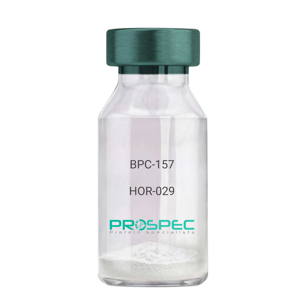
BPC-157Description:
BPC-157 Pentadecapeptide
Product # :
HOR-029Price :
Quantity :
Shipping Method :
Shipped at Room temp
More Info
- description
- formulation
- purity
- More Info
Description
BPC-157 Synthetic is a single, non-glycosylated polypeptide chain containing 15 amino acids, having a molecular mass of 1419.55 Dalton and a Molecular formula of C62H98N16O22.
Formulation
The protein was lyophilized with no additives.
Purity
Greater than 97.0% as determined by analysis by RP-HPLC.
More Info
-
Physical Appearance
Sterile Filtered White lyophilized (freeze-dried) powder.
-
Stability
Lyophilized BPC-157 although stable at room temperature for 3 weeks, should be stored desiccated below -18°C. Upon reconstitution bpc-157 should be stored at 4°C between 2-7 days and for future use below -18°C. For long term storage it is recommended to add a carrier protein (0.1% HSA or BSA). Please prevent freeze-thaw cycles.
-
Solubility
It is recommended to reconstitute the lyophilized BPC-157 in sterile 18MΩ-cm H2O not less than 100 µg/ml, which can then be further diluted to other aqueous solutions.
-
Amino Acid Sequence
H-Gly-Glu-Pro-Pro-Pro-Gly-Lys-Pro-Ala-Asp-Asp-Ala-Gly-Leu-Val-OH
-
Background
BPC-157, short for Body Protection Compound-157, is a synthetic peptide that has garnered significant attention in the field of regenerative medicine and sports science. This peptide, derived from a portion of the human gastric juice protein known as BPC, exhibits remarkable healing and tissue regeneration properties. BPC-157 has shown promise in various preclinical and clinical studies, demonstrating its potential for the treatment of a wide range of injuries and disorders.
The research on BPC-157 encompasses investigations into its mechanisms of action, efficacy, safety, and potential therapeutic applications. Studies have elucidated the peptide's ability to enhance angiogenesis, promote collagen synthesis, modulate inflammatory responses, and protect against oxidative stress. These properties make BPC-157 an intriguing candidate for accelerating tissue healing, reducing inflammation, and improving overall recovery outcomes.
Preclinical studies have revealed the beneficial effects of BPC-157 in several injury models. For instance, BPC-157 has demonstrated its potential in accelerating tendon and ligament healing, mitigating muscle damage, and promoting bone regeneration. These findings suggest that BPC-157 could be a valuable therapeutic tool in orthopedic medicine and sports-related injuries.
Furthermore, BPC-157 has exhibited promising effects on gastrointestinal health. Studies have highlighted its ability to protect and heal the gut lining, reduce ulcer formation, and alleviate symptoms associated with inflammatory bowel disease. These observations open up avenues for BPC-157 as a potential treatment for gastrointestinal disorders.
In addition to its regenerative properties, BPC-157 has shown potential in neurological and psychiatric conditions. Research has indicated its neuroprotective effects, with implications for the treatment of traumatic brain injury, stroke, and neurodegenerative disorders. Preliminary studies also suggest BPC-157's potential as an antidepressant and anxiolytic agent.
Despite the promising findings, further research is needed to fully understand the mechanisms underlying BPC-157's actions and to assess its long-term safety and efficacy. Clinical trials are underway to explore its potential therapeutic applications in humans, including its use in tendon and ligament repair, inflammatory bowel disease, and neurodegenerative disorders.
This comprehensive review aims to summarize the current state of research on BPC-157, providing an overview of its mechanisms of action and therapeutic potential. By examining relevant studies and findings, we aim to shed light on the diverse applications of BPC-157 and its implications for regenerative medicine, sports science, and various disease
ProSpec's products are furnished for LABORATORY RESEARCH USE ONLY. The product may not be used as drugs, agricultural or pesticidal products, food additives or household chemicals.
- View Data Sheet
Name :
PDGFRA HumanDescription:
Platelet-Derived Growth Factor Receptor, Alpha Human Recombinant
Platelet-derived growth factor receptor alpha polypeptide, PDGFR2, PDGF-R-alpha, CD140 antigen-like family member A, CD140a antigen, alpha-type platelet-derived growth factor receptor, RHEPDGFRA, rearranged-in-hypereosinophilia-platelet derived growth factor receptor alpha, PDGFRA/BCR fusion protein, MGC74795, EC 2.7.10.1.
Product # :
CYT-065Price :
Quantity :
Shipping Method :
Shipped with Ice Packs
More Info
- description
- source
- formulation
- purity
- More Info
Description
PDGFRA Human Recombinant produced in E.coli is a single, non-glycosylated polypeptide chain containing 522 amino acids (24-524) and having a molecular mass of 58.4 kDa.The PDGFRA is fused to a 20 amino acid His-Tag at N-terminus and purified by proprietary chromatographic techniques.
Source
E.coli.
Formulation
The PDGFRA solution (0.5mg/ml) contains 20mM Tris-HCl buffer (pH 8.0), 1mM DTT, 0.1M NaCl and 20% glycerol.
Purity
Greater than 85% as determined by SDS-PAGE.
More Info
-
Introduction
PDGFRa is a cell surface tyrosine kinase receptor for PDGF family members. PDGFRa binds to both A and B subunits of PDGF. It is known that PDGFRa is vital for kidney development since mice heterozygous for the receptor display defective kidney phenotypes.
-
Synonyms
Platelet-derived growth factor receptor alpha polypeptide, PDGFR2, PDGF-R-alpha, CD140 antigen-like family member A, CD140a antigen, alpha-type platelet-derived growth factor receptor, RHEPDGFRA, rearranged-in-hypereosinophilia-platelet derived growth factor receptor alpha, PDGFRA/BCR fusion protein, MGC74795, EC 2.7.10.1.
-
Physical Appearance
Sterile Filtered colorless solution.
-
Stability
Store at 4°C if entire vial will be used within 2-4 weeks. Store, frozen at -20°C for longer periods of time. For long term storage it is recommended to add a carrier protein (0.1% HSA or BSA).Avoid multiple freeze-thaw cycles.
-
Amino Acid Sequence
MGSSHHHHHH SSGLVPRGSH MQLSLPSILP NENEKVVQLN SSFSLRCFGE SEVSWQYPMS EEESSDVEIR NEENNSGLFV TVLEVSSASA AHTGLYTCYY NHTQTEENEL EGRHIYIYVP DPDVAFVPLG MTDYLVIVED DDSAIIPCRT TDPETPVTLH NSEGVVPASY DSRQGFNGTF TVGPYICEAT VKGKKFQTIP FNVYALKATS ELDLEMEALK TVYKSGETIV VTCAVFNNEV VDLQWTYPGE VKGKGITMLE EIKVPSIKLV YTLTVPEATV KDSGDYECAA RQATREVKEM KKVTISVHEK GFIEIKPTFS QLEAVNLHEV KHFVVEVRAY PPPRISWLKN NLTLIENLTE ITTDVEKIQE IRYRSKLKLI RAKEEDSGHY TIVAQNEDAV KSYTFELLTQ VPSSILDLVD DHHGSTGGQT VRCTAEGTPL PDIEWMICKD IKKCNNETSW TILANNVSNI ITEIHSRDRS TVEGRVTFAK VEETIAVRCL AKNLLGAENR ELKLVAPTLR SE.
ProSpec's products are furnished for LABORATORY RESEARCH USE ONLY. The product may not be used as drugs, agricultural or pesticidal products, food additives or household chemicals.
- View Data Sheet
Name :
SPP1 Human, HEKDescription:
Osteopontin Human Recombinant, HEK
Secreted Phosphoprotein-1, OPN, BNSP, BSPI, ETA-1, MGC110940, SPP-1, Osteopontin, Bone sialoprotein 1, Urinary stone protein, Nephropontin, Uropontin, SPP1.
Product # :
CYT-047Price :
Quantity :
Shipping Method :
Shipped at Room temp
More Info
- description
- source
- formulation
- purity
- More Info
Description
Osteopontin Human Recombinant is a single, glycosylated, polypeptide chain produced in HEK293 cells, is a full length protein (amino acids 17-314) fused with a polyhistidine tag at the C-terminus, having a total calculated molecular mass of 34.5kDa (The actual molecular mass may be approximately 60-65kDa in SDS-PAGE under reducing conditions due to glycosylation).Osteopontin is purified by proprietary chromatographic techniques.
Source
HEK293 cells.
Formulation
Osteopontin was lyophilized from a 0.2µM filtered solution of 20mM PBS and 150mM NaCl, pH 7.2.
Purity
Greater than 95.0% as determined by SDS-PAGE.
More Info
-
Introduction
Osteopontin is a glycoprotein that was first identified in osteoblasts and is involved in bone remodeling, immune functions in fibroblasts, macrophages, and lymphocytes during inflammation and wound healing. SPP1 binds tightly to hydroxyapatite. SPP1 forms an integral part of the mineralized matrix. SPP1 is vital to cell-matrix interaction.
Secreted Phosphoprotein-1 protects against cardiac ischemia-reperfusion injury via late preconditioning. Expression of both Ostepontin and CD44 in hepatocellular carcinoma is linked with advanced tumor stage and contributes to prognosis information. SPP1 is the most over-expressed gene in intrahepatic cholangiocarcinoma. Secreted Phosphoprotein-1 overexpression is related with interstitial lung diseases. -
Synonyms
Secreted Phosphoprotein-1, OPN, BNSP, BSPI, ETA-1, MGC110940, SPP-1, Osteopontin, Bone sialoprotein 1, Urinary stone protein, Nephropontin, Uropontin, SPP1.
-
Physical Appearance
Sterile Filtered White lyophilized (freeze-dried) powder.
-
Stability
Store at 4°C if entire vial will be used within 2-4 weeks. Store, frozen at -20°C for longer periods of time.Please avoid freeze thaw cycles.
-
Solubility
It is recommended to reconstitute the lyophilized SPP1 in 1xPBS to a concentration no less than 100 µg/ml, which can then be further diluted to other aqueous solutions.
-
Amino Acid Sequence
IPVKQADSGSSEEKQLYNKYPDAVATWLNPDPSQKQNLLAPQNAVSSEETNDFKQETL
PSKSNESHDHMDDMDDEDDDDHVDSQDSIDSNDSDDVDDTDDSHQSDESHHSDESDEL
VTDFPTDLPATEVFTPVVPTVDTYDGRGDSVVYGLRSKSKKFRRPDIQYPDATDEDIT
SHMESEELNGAYKAIPVAQDLNAPSDWDSRGKDSYETSQLDDQSAETHSHKQSRLYKRK
ANDESNEHSDVIDSQELSKVSREFHSHEFHSHEDMLVVDPKSKEEDKHLKFRISHELDS
ASSEVNVDHHHHHH.
ProSpec's products are furnished for LABORATORY RESEARCH USE ONLY. The product may not be used as drugs, agricultural or pesticidal products, food additives or household chemicals.
- View Data Sheet
Name :
IFN tau OvineDescription:
IFN-Tau Ovine Recombinant
IFN-tau1, Trophoblast protein 1, TP-1, Trophoblastin, Antiluteolysin, Trophoblast antiluteolytic protein, IFN-tau, IFN tau-1.
Product # :
CYT-377Price :
Quantity :
Shipping Method :
Shipped at Room temp
More Info
- description
- source
- formulation
- purity
- biological activity
- More Info
Description
IFN-Tau Ovine Recombinant produced in E.Coli is a single, non-glycosylated, polypeptide chain containing 172 amino acids and having a molecular mass of 19914.7 Dalton.The IFN-Tau is purified by proprietary chromatographic techniques.
Source
Escherichia Coli.
Formulation
Lyophilized from (1mg/ml) solution containing PBS pH-7.4.
Purity
Greater than 95.0% as determined by both:
(a) Analysis by RP-HPLC.
(b) Analysis by SDS-PAGE.Biological Activity
The specific activity as determined in a viral resistance assay using bovine kidney MDBK cells was found to be 10,000,000IU/mg.More Info
-
Introduction
IFN-tau is also known as TP-1 (trophoblast protein-1) is a new class of type I IFN that is secreted by the trophoblast and is the signal for maternal recognition of pregnancy in sheep. IFN- tau has potent immunosuppressive and antiviral activities similar to other type I IFN but is less cytotoxic than IFN-alpha and IFN-beta. The current investigation concerns the effect of recombinant ovine IFN- tau (rOIFN- tau) on the modulation of MHC class I and II expression on cloned mouse cerebrovascular endothelial (CVE) cells.
IFN-tau induced tyrosine phosphorylation of Stat1 and upregulated the expression of MHC class I on CVE. One proposed action by which type I IFN reduces the relapse rate in MS is via interference with IFN-?-induced MHC class II expression. IFN- tau was shown to downregulate IFN-?-induced MHC class II expression on CVE and, hence, may be of potential therapeutic value in downregulating inflammation in the central nervous system (CNS). IFN- tau did not upregulate the expression of MHC class II on CVE. IFN- tau also inhibited the replication of Theiler's virus in CVE. -
Synonyms
IFN-tau1, Trophoblast protein 1, TP-1, Trophoblastin, Antiluteolysin, Trophoblast antiluteolytic protein, IFN-tau, IFN tau-1.
-
Physical Appearance
Sterile Filtered White lyophilized (freeze-dried) powder.
-
Stability
Lyophilized IFN-Tau although stable at room temperature for 3 weeks, should be stored desiccated below -18°C. Upon reconstitution IFN-Tau should be stored at 4°C between 2-7 days and for future use below -18°C.For long term storage it is recommended to add a carrier protein (0.1% HSA or BSA).Please prevent freeze-thaw cycles.
-
Solubility
It is recommended to reconstitute the lyophilized IFN Tau in sterile 18MΩ-cm H2O not less than 100µg/ml, which can then be further diluted to other aqueous solutions.
-
Amino Acid Sequence
The sequence of the first five N-terminal amino acids was determined and was found to be Cys-Tyr-Leu-Ser-Arg.
-
Background
What is the molecular weight/Mw of IFN TAU OVINE Protein?
IFN TAU OVINE Protein has a total Mw of 19.9kDa.
What is the source or expression system of IFN TAU OVINE Protein?
Escherichia Coli.
What is the Purity of IFN TAU OVINE Protein?
IFN TAU OVINE Protein is >95% pure as determined by SDS-PAGE.
What is the Biological Activity of IFN TAU OVINE Protein?
The specific activity as determined in a viral resistance assay using bovine kidney MDBK cells was found to be 10,000,000IU/mg.
What is the amino acid sequence of IFN TAU OVINE Protein?
The sequence of the first five N-terminal amino acids was determined and was found to be Cys-Tyr-Leu-Ser-Arg.
What applications can IFN TAU OVINE Protein be used in?
IFN TAU OVINE Protein can probably be used in western blot, ELISA and Lateral Flow.
What is the endotoxin level for IFN TAU OVINE Protein?
The endotoxin level is minimal, IFN TAU OVINE Protein was purified using conventional chromatography techniques.
ProSpec's products are furnished for LABORATORY RESEARCH USE ONLY. The product may not be used as drugs, agricultural or pesticidal products, food additives or household chemicals.
- View Data Sheet
Name :
DDR1 HumanDescription:
Discoidin Domain Receptor Tyrosine Kinase 1 Human Recombinant
Discoidin Domain Receptor Tyrosine Kinase 1, CD167 Antigen-Like Family Member A, Protein-Tyrosine Kinase RTK-6, Mammary Carcinoma Kinase 10, Tyrosine-Protein Kinase CAK, Cell Adhesion Kinase, Tyrosine Kinase DDR, EC 2.7.10.1, EDDR1, NTRK4, PTK3A, HGK2, RTK6, TRKE, CAK, NEP, Neurotrophic Tyrosine Kinase, Receptor, Type 4, Discoidin Domain Receptor Family, Member 1, Epithelial Discoidin Domain Receptor 1, Discoidin Receptor Tyrosine Kinase, PTK3A Protein Tyrosine Kinase 3A, Neuroepithelial Tyrosine Kinase, Protein-Tyrosine Kinase 3A, CD167a Antigen, EC 2.7.10, MCK-10, CD167, MCK10, TRK E, PTK3, DDR.
Product # :
PKA-103Price :
Quantity :
Shipping Method :
Shipped with Ice Packs
More Info
- description
- source
- formulation
- purity
- More Info
Description
DDR1 Human Recombinant produced in Sf9 Baculovirus cells is a single, glycosylated polypeptide chain containing 636 amino acids (21-417a.a.) and having a molecular mass of 71.0kDa (Molecular size on SDS-PAGE will appear at approximately 70-100kDa).DDR1 is expressed with a 239 amino acid hIgG-His tag at C-Terminus and purified by proprietary chromatographic techniques.
Source
Sf9, Baculovirus cells.
Formulation
DDR1 protein solution (0.5mg/ml) contains Phosphate Buffered Saline (pH 7.4) and 10% glycerol.
Purity
Greater than 90.0% as determined by SDS-PAGE.
More Info
-
Introduction
Discoidin Domain Receptor Tyrosine Kinase 1 (DDR1) is a transmembrane glycoprotein which is a part of the discoidin-like domain containing subfamily of receptor tyrosine kinases. DDR1 functions as cell surface receptor for fibrillar collagen and regulates cell attachment to the extracellular matrix, remodeling of the extracellular matrix, cell migration, differentiation, survival and cell proliferation. DDR1 is expressed in epithelial cells, mainly in the kidney, lung, gastrointestinal tract, and brain and is significantly overexpressed in several human tumors.
-
Synonyms
Discoidin Domain Receptor Tyrosine Kinase 1, CD167 Antigen-Like Family Member A, Protein-Tyrosine Kinase RTK-6, Mammary Carcinoma Kinase 10, Tyrosine-Protein Kinase CAK, Cell Adhesion Kinase, Tyrosine Kinase DDR, EC 2.7.10.1, EDDR1, NTRK4, PTK3A, HGK2, RTK6, TRKE, CAK, NEP, Neurotrophic Tyrosine Kinase, Receptor, Type 4, Discoidin Domain Receptor Family, Member 1, Epithelial Discoidin Domain Receptor 1, Discoidin Receptor Tyrosine Kinase, PTK3A Protein Tyrosine Kinase 3A, Neuroepithelial Tyrosine Kinase, Protein-Tyrosine Kinase 3A, CD167a Antigen, EC 2.7.10, MCK-10, CD167, MCK10, TRK E, PTK3, DDR.
-
Physical Appearance
Sterile Filtered colorless solution.
-
Stability
Store at 4°C if entire vial will be used within 2-4 weeks. Store, frozen at -20°C for longer periods of time. For long term storage it is recommended to add a carrier protein (0.1% HSA or BSA). Avoid multiple freeze-thaw cycles.
-
Amino Acid Sequence
DMKGHFDPAK CRYALGMQDR TIPDSDISAS SSWSDSTAAR HSRLESSDGD GAWCPAGSVF PKEEEYLQVD LQRLHLVALV GTQGRHAGGL GKEFSRSYRL RYSRDGRRWM GWKDRWGQEV ISGNEDPEGV VLKDLGPPMV ARLVRFYPRA DRVMSVCLRV ELYGCLWRDG LLSYTAPVGQ TMYLSEAVYL NDSTYDGHTV GGLQYGGLGQ LADGVVGLDD FRKSQELRYV WPGYDYVGWS NHSFSSGYVE MEFEFDRLRA FQAMQVHCNN MHTLGARLPG GVECRFRRGP AMAWEGEPMR HNLGGNLGDP RARAVSVPLG GRVARFLQCR FLFAGPWLLF SEISFISDVV NNSSPALGGT FPPAPWWPPG PPPTNFSSLE LEPRGQQPVA KAEGSPTALE PKSCDKTHTC PPCPAPELLG GPSVFLFPPK PKDTLMISRT PEVTCVVVDV SHEDPEVKFN WYVDGVEVHN AKTKPREEQY NSTYRVVSVL TVLHQDWLNG KEYKCKVSNK ALPAPIEKTI SKAKGQPREP QVYTLPPSRD ELTKNQVSLT CLVKGFYPSD IAVEWESNGQ PENNYKTTPP VLDSDGSFFL YSKLTVDKSR WQQGNVFSCS VMHEALHNHY TQKSLSLSPG KHHHHHH.
ProSpec's products are furnished for LABORATORY RESEARCH USE ONLY. The product may not be used as drugs, agricultural or pesticidal products, food additives or household chemicals.
- View Data Sheet
Name :
EGF Mouse, BiotinDescription:
Epidermal Growth Factor Mouse Recombinant, Biotin
Urogastrone, URG, EGF.
Product # :
CYT-841Price :
Quantity :
Shipping Method :
Shipped with Ice Packs
More Info
- description
- source
- formulation
- purity
- biological activity
- More Info
Description
EGF Mouse Recombinant, Biotin produced in E.Coli is a non-glycosylated polypeptide chain containing 61 amino acids and having a total molecular mass of 7.0kDa. This version of EGF has a N terminal leader sequence hosting a biotin conjugation. There are 0.5 biotins for each EGF protein.
Source
Escherichia Coli.
Formulation
The protein (0.5mg/ml) solution contains sterile PBS.
Purity
Greater than 95.0% as determined by analysis by SDS-PAGE.
Biological Activity
The ED50 as determined by the dose-dependent proliferation of mouse BALB/c 3T3 cells is 0.14-0.2ng/ml, corresponding to a specific activity of 7.1x10⁶units/mg.More Info
-
Introduction
Epidermal growth factor has a profound effect on the differentiation of specific cells in vivo and is a potent mitogenic factor for a variety of cultured cells of both ectodermal and mesodermal origin. The EGF precursor is believed to exist as a membrane-bound molecule which is proteolytically cleaved to generate the 53-amino acid peptide hormone that stimulates cells to divide. EGF stimulates the growth of various epidermal and epithelial tissues in vivo and in vitro and of some fibroblasts in cell culture.
-
Synonyms
Urogastrone, URG, EGF.
-
Physical Appearance
Sterile Filtered clear solution.
-
Stability
Should be stored at 4°C.Please do not freeze.
-
Amino Acid Sequence
MKKIDDDKNS YPGCPSSYDG YCLNGGVCMH IESLDSYTCN CVIGYSGDRC QTRDLEWWEL R.
-
Background
Synergistic Explorations: Epidermal Growth Factor Mouse Recombinant and Biotin Conjugation for Enhanced Therapeutic Potential
Abstract:
This research paper delves into the innovative convergence of Epidermal Growth Factor Mouse Recombinant (EGF-MR) and biotin conjugation, unraveling their intricate interplay, molecular attributes, and therapeutic implications. By employing cutting-edge methodologies involving protein engineering, conjugation chemistry, and cellular assays, this study uncovers the augmented cellular responses driven by EGF-MR-biotin complex. The findings highlight a novel avenue for tailored regenerative medicine and targeted therapy.
Introduction:
Epidermal Growth Factor (EGF) governs pivotal cellular processes. This paper navigates the unexplored realm of Epidermal Growth Factor Mouse Recombinant (EGF-MR) in synergy with biotin conjugation, elucidating their combined molecular attributes and therapeutic potential.
Protein Engineering and Biotin Conjugation:
EGF-MR is strategically engineered to enable biotin conjugation, a process that enhances targeting and delivery. This paper delves into site-specific modification approaches, ensuring precise and controlled conjugation of biotin moieties to EGF-MR.
Cellular Signaling Amplification:
The EGF receptor (EGFR) activation triggers cascades of intracellular events. Structural studies and binding kinetics illuminate how the biotin-conjugated EGF-MR modulates EGFR interactions, amplifying downstream signaling pathways like the MAPK and PI3K/Akt cascades.
Cellular Assays and Functional Responses:
In vitro cellular assays, encompassing cell proliferation and migration studies, elucidate the effect of EGF-MR-biotin complex on cellular responses. Live-cell imaging techniques reveal enhanced cell motility and survival, underpinning the potential therapeutic impact.
Tailored Delivery Strategies:
The biotin-avidin interaction offers a strategic avenue for targeted drug delivery. Employing this interaction, EGF-MR-biotin complex can be directed to specific cell types, revolutionizing precision medicine and enabling tailored therapeutic interventions.
Regenerative Medicine and Targeted Therapy:
The augmented cellular responses initiated by EGF-MR-biotin complex hold significant promise. In regenerative medicine, the complex's potential to accelerate tissue regeneration becomes evident. Furthermore, in targeted therapy, the complex's enhanced cellular uptake offers a novel approach to modulate tumor microenvironments.
Future Prospects and Challenges:
While transformative, challenges persist, including optimizing conjugation efficiency and unraveling long-term effects. Future research should focus on refining delivery strategies and conducting comprehensive long-term studies to harness the full therapeutic potential.
Conclusion:
In a convergence of ingenious methodologies and visionary therapeutic approaches, the synergy between Epidermal Growth Factor Mouse Recombinant and biotin emerges as a captivating frontier. The molecular marriage between EGF-MR and biotin not only amplifies cellular responses but also opens doors for targeted interventions and precision therapies, revolutionizing the landscape of medical advancements.
What is the molecular weight/Mw of MEGF, BIOTIN Protein?
MEGF, BIOTIN Protein has a total Mw of 7kDa.
What is the source or expression system of MEGF, BIOTIN Protein?
Escherichia Coli.
What is the Purity of MEGF, BIOTIN Protein?
MEGF, BIOTIN Protein is >95% pure as determined by SDS-PAGE.
What is the Biological Activity of MEGF, BIOTIN Protein?
The ED50 as determined by the dose-dependent proliferation of mouse BALB/c 3T3 cells is 0.14-0.2ng/ml, corresponding to a specific activity of 7.1x10⁶units/mg.
What is the amino acid sequence of MEGF, BIOTIN Protein?
MKKIDDDKNS YPGCPSSYDG YCLNGGVCMH IESLDSYTCN CVIGYSGDRC QTRDLEWWEL R.
What applications can MEGF, BIOTIN Protein be used in?
MEGF, BIOTIN Protein can probably be used in western blot, ELISA and Lateral Flow.
What is the endotoxin level for MEGF, BIOTIN Protein?
The endotoxin level is minimal, MEGF, BIOTIN Protein was purified using conventional chromatography techniques.
ProSpec's products are furnished for LABORATORY RESEARCH USE ONLY. The product may not be used as drugs, agricultural or pesticidal products, food additives or household chemicals.
- View Data Sheet
Name :
BAFF R HumanDescription:
B-cell Activating Factor Receptor Human Recombinant
TNFRSF13C, CD268, BAFF-R, MGC138235, B cell-activating factor receptor.
Product # :
CYT-429Price :
Quantity :
Shipping Method :
Shipped at Room temp
More Info
- description
- source
- formulation
- purity
- biological activity
- More Info
Description
B Lymphocyte Stimulator Receptor Human Recombinant extracellular produced in E.Coli is a single, non-glycosylated polypeptide chain containing 76 amino acids and having a molecular mass of 7.7 kDa.The BAFF-R is purified by proprietary chromatographic techniques.
Source
Escherichia Coli.
Formulation
Lyophilized from a 0.2μm filtered concentrated (1.0mg/ml) solution in 20mM PB, pH 8.0, 500mM NaCl.
Purity
Greater than 95.0% as determined by:
(a) Analysis by RP-HPLC.
(b) Analysis by SDS-PAGE.Biological Activity
Determined by its ability to block BAFF induced mouse splenocyte survival. The expected ED50 for this effect is 1.0-5.0 µg/ml in the presence of 1.0µg/ml of human soluble BAFF.More Info
-
Introduction
B cell-activating factor (BAFF) enhances B-cell survival in vitro and is a regulator of the peripheral B-cell population. Overexpression of Baff in mice results in mature B-cell hyperplasia and symptoms of systemic lupus erythematosus (SLE). Also, some SLE patients have increased levels of BAFF in serum. Therefore, it has been proposed that abnormally high levels of BAFF may contribute to the pathogenesis of autoimmune diseases by enhancing the survival of autoreactive B cells. The protein encoded by this gene is a receptor for BAFF and is a type III transmembrane protein containing a single extracellular cysteine-rich domain. It is thought that this receptor is the principal receptor required for BAFF-mediated mature B-cell survival.
-
Synonyms
TNFRSF13C, CD268, BAFF-R, MGC138235, B cell-activating factor receptor.
-
Physical Appearance
Sterile Filtered White lyophilized (freeze-dried) powder.
-
Stability
Lyophilized BAFF-R although stable at room temperature for 3 weeks, should be stored desiccated below -18°C. Upon reconstitution B Lymphocyte Stimulator Receptor should be stored at 4°C between 2-7 days and for future use below -18°C.Please prevent freeze-thaw cycles.
-
Solubility
It is recommended to reconstitute the lyophilized B Lymphocyte Stimulator Receptor Recombinant in sterile 18MΩ-cm H2O not less than 100µg/ml, which can then be further diluted to other aqueous solutions.
-
Amino Acid Sequence
MRRGPRSLRGRDAPAPTPCVPAECFDLLVRHCVACGLLRTPRPKPAG
ASSPAPRTALQPQESVGAGAGEAALPLPG. -
Background
B-cell Activating Factor Receptor Human Recombinant: Unlocking the Potential of a Key Immunomodulatory Target
Abstract:
B-cell Activating Factor Receptor (BAFF-R) human recombinant is a critical component of the B-cell immune response, playing a pivotal role in B-cell survival, maturation, and antibody production. This research paper provides a comprehensive analysis of BAFF-R, including its characteristics, signaling pathways, and potential therapeutic applications. Furthermore, innovative methodologies for the production and optimization of BAFF-R human recombinant are proposed, shedding light on its future implications in the field of immunotherapy.
Introduction:
The immune system relies on the precise regulation of B-cell functions, with BAFF-R serving as a key modulator of B-cell development and activation. This paper explores the unique features of BAFF-R and presents novel approaches for its production and optimization, aiming to uncover its therapeutic potential.
Characteristics and Signaling Pathways:
BAFF-R is a type III transmembrane protein expressed primarily on B-cells. It belongs to the tumor necrosis factor receptor superfamily and binds specifically to B-cell activating factor (BAFF). Engagement of BAFF-R by BAFF initiates intracellular signaling cascades, including the activation of nuclear factor-kappa B (NF-κB) and mitogen-activated protein kinase (MAPK) pathways, promoting B-cell survival, proliferation, and differentiation.
Production of BAFF-R Human Recombinant:
Efficient production methodologies are crucial for the therapeutic application of BAFF-R human recombinant. Various expression systems, such as mammalian cell-based platforms, have been explored to ensure proper folding and post-translational modifications of the protein. Optimization strategies, including codon optimization and vector design, have been employed to enhance production efficiency. Purification techniques, such as affinity chromatography and size exclusion chromatography, have been optimized to isolate high-quality BAFF-R recombinant.
Potential Therapeutic Applications:
BAFF-R human recombinant holds great promise in the field of immunotherapy. Dysregulation of the BAFF/BAFF-R signaling axis has been implicated in autoimmune diseases, such as systemic lupus erythematosus and rheumatoid arthritis. Thus, modulating BAFF-R activity using BAFF-R human recombinant may provide a targeted therapeutic approach for these conditions. Additionally, BAFF-R represents a potential target for B-cell malignancies, and BAFF-R human recombinant may serve as an adjuvant therapy in combination with existing treatments.
Conclusion:
BAFF-R human recombinant represents a crucial immunomodulatory target with diverse therapeutic applications in immunotherapy. Optimizing production methodologies and further understanding its signaling pathways will enhance its clinical utility. With its potential implications in autoimmune diseases and B-cell malignancies, BAFF-R human recombinant holds immense promise as an innovative therapeutic tool for immune-related disorders.
What is the molecular weight/Mw of BAFF R Protein?
BAFF R Protein has a total Mw of 7.7kDa.
What is the source or expression system of BAFF R Protein?
Escherichia Coli.
What is the Purity of BAFF R Protein?
BAFF R Protein is >95% pure as determined by SDS-PAGE.
What is the Biological Activity of BAFF R Protein?
Determined by its ability to block BAFF induced mouse splenocyte survival. The expected ED50 for this effect is 1.0-5.0 µg/ml in the presence of 1.0µg/ml of human soluble BAFF.
What is the amino acid sequence of BAFF R Protein?
MRRGPRSLRGRDAPAPTPCVPAECFDLLVRHCVACGLLRTPRPKPAG
ASSPAPRTALQPQESVGAGAGEAALPLPG.
What applications can BAFF R Protein be used in?
BAFF R Protein can probably be used in western blot, ELISA and Lateral Flow.
What is the endotoxin level for BAFF R Protein?
The endotoxin level is minimal, BAFF R Protein was purified using conventional chromatography techniques.
ProSpec's products are furnished for LABORATORY RESEARCH USE ONLY. The product may not be used as drugs, agricultural or pesticidal products, food additives or household chemicals.
- View Data Sheet
Name :
SPARC Human, HisDescription:
Secreted Protein acidic & Rich in Cysteine Human Recombinant, His Tag
Osteonectin, ON, Basement-membrane protein 40, BM-40, SPARC, Secreted Protein acidic and Rich in Cysteine.
Product # :
PRO-582Price :
Quantity :
Shipping Method :
Shipped at Room temp
More Info
- description
- source
- formulation
- purity
- More Info
Description
Osteonectin Human Recombinant fused with 6X His tag produced in E.Coli is a single, non-glycosylated, polypeptide chain containing 295 amino acids and having a molecular mass of 34 kDa.SPARC is expressed with a 6 amino acid His tag at N-Terminus and purified by proprietary chromatographic techniques.
Source
Escherichia Coli.
Formulation
The SPARC (1 mg/ml) was lyophilized after extensive dialyses against 20mM PBS pH-7.4.
Purity
Greater than 95.0% as determined by:
(a) Analysis by RP-HPLC.
(b) Analysis by SDS-PAGE.More Info
-
Introduction
SPARC, an acronym for “secreted protein, acidic and rich in cysteine”, is also known as osteonectin or BM-40. It is the founding member of a family of secreted matricellular proteins with similar domain structure. The 303 amino acid, 43 kDa protein contains a 17 aa signal sequence, an N-terminal acidic region that binds calcium, a follistatin domain containing Kazal-like sequences, and a C-terminal extracellular calcium (EC) binding domain with two EF-hand motifs. SPARC is produced by fibroblasts, capillary endothelial cells, platelets and macrophages, especially in areas of tissue morphogenesis and remodeling. SPARC shows context-specific effects, but generally inhibits adhesion, spreading and proliferation, and promotes collagen matrix formation. For endothelial cells, SPARC disrupts focal adhesions and binds and sequesters PDGF and VEGF. SPARC is abundantly expressed in bone, where it promotes osteoblast differentiation and inhibits adipogenesis.
-
Synonyms
Osteonectin, ON, Basement-membrane protein 40, BM-40, SPARC, Secreted Protein acidic and Rich in Cysteine.
-
Physical Appearance
Sterile Filtered White lyophilized (freeze-dried) powder.
-
Stability
Lyophilized Osteonectin although stable at room temperature for 3 weeks, should be stored desiccated below -18°C. Upon reconstitution BM-40 should be stored at 4°C between 2-7 days and for future use below -18°C.For long term storage it is recommended to add a carrier protein (0.1% HSA or BSA).Please prevent freeze-thaw cycles.
-
Solubility
It is recommended to reconstitute the lyophilized SPARC in sterile 18MΩ-cm H2O not less than 100 µg/ml, which can then be further diluted to other aqueous solutions.
-
Amino Acid Sequence
MSYYHHHHHHPQQEALPDETEVVEETVAEVTEVSVGANPVQVEVGEFD
DGAEETEEEVVAENPCQNHHCKHGKVCELDENNTPMCVCQDPTSCP
APIGEFEKVCSNDNKTFDSSCHFFATKCTLEGTKKGHKLHLDYIGPCK
YIPPCLDSELTEFPLRMRDWLKNVLVTLYERDEDNNLLTKQKLRVKKI
HENEKRLEAGDHPVELLARDFEKNYNMYIFPVHWQFGQLDQHPIDGY
LSHTELAPLRAPLIPMEHCTTRFFETCDLDNDKYIALDEWAGCFGIKQK
DIDKDLVI.
ProSpec's products are furnished for LABORATORY RESEARCH USE ONLY. The product may not be used as drugs, agricultural or pesticidal products, food additives or household chemicals.
- View Data Sheet
Name :
FCER1A Human 201 a.aDescription:
IgE Receptor Subunit A Human Recombinant
Fc Fragment Of IgE, High Affinity I, Receptor For; Alpha Polypeptide, FCE1A, IgE Fc Receptor Subunit Alpha, FcERI, Fc-Epsilon RI-Alpha, Fc Epsilon RI Alpha-Chain, Fc IgE Receptor, Alpha Polypeptide, High Affinity Immunoglobulin Epsilon Receptor Alpha-Subunit, High Affinity Immunoglobulin Epsilon Receptor Subunit Alpha, Immunoglobulin E Receptor, High-Affinity, Of Mast Cells, Alpha Polypeptide.
Product # :
PRO-2357Price :
Quantity :
Shipping Method :
Shipped with Ice Packs
More Info
- description
- source
- formulation
- purity
- More Info
Description
FCER1A Human Recombinant produced in E.Coli is a non-glycosylated polypeptide chain containing 201 amino acids.FCER1Ais fused to a 6 amino acid His-tag at C-terminus & purified by proprietary chromatographic techniques.
Source
Escherichia Coli.
Formulation
1x PBS, 50mM Arginine and 0.05% NaN3.
Purity
Greater than 90.0% as determined by SDS-PAGE.
More Info
-
Introduction
Fc fragment of IgE, high affinity I, receptor for; alpha polypeptide (FCER1A) binds to the Fc region of immunoglobulins epsilon. FCER1A is a high affinity receptor. In addition FCER1A is responsible for starting the allergic response. Binding of allergen to receptor-bound IgE leads to cell activation and the release of mediators such as histamine which is responsible for the manifestations of allergy. This receptor is contains of an alpha subunit, a beta subunit, and two gamma subunits. FCER1A stands for the alpha subunit. Among the diseases associated with FCER1A are mast-cell leukemia, and allergic asthma.
-
Synonyms
Fc Fragment Of IgE, High Affinity I, Receptor For; Alpha Polypeptide, FCE1A, IgE Fc Receptor Subunit Alpha, FcERI, Fc-Epsilon RI-Alpha, Fc Epsilon RI Alpha-Chain, Fc IgE Receptor, Alpha Polypeptide, High Affinity Immunoglobulin Epsilon Receptor Alpha-Subunit, High Affinity Immunoglobulin Epsilon Receptor Subunit Alpha, Immunoglobulin E Receptor, High-Affinity, Of Mast Cells, Alpha Polypeptide.
-
Physical Appearance
Sterile Filtered colorless solution.
-
Stability
The Recombinant FCER1A protein although stable at 4°C for 1 week, should be stored below -18°C. For long term storage it is recommended to add a carrier protein (0.1% HSA or BSA). Please prevent freeze-thaw cycles.
-
Amino Acid Sequence
HMAPAMESPTL LCVALLFFAP DGVLAVPQKP KVSLNPPWNR IFKGENVTLT CNGNNFFEVS STKWFHNGSL SEETNSSLNI VNAKFEDSGE YKCQHQQVNE SEPVYLEVFS DWLLLQASAE VVMEGQPLFL RCHGWRNWDV YKVIYYKDGE ALKYWYENHN ISITNATVED SGTYYCTGKV WQLDYESEPL NITVIKAPLEHHHHHH.
ProSpec's products are furnished for LABORATORY RESEARCH USE ONLY. The product may not be used as drugs, agricultural or pesticidal products, food additives or household chemicals.