Skip to content

High-quality research proteins.

  • 1-800-805-6531

  • Mon - Sat 8.00-18.00, Sun - Closed

  • info@prospecbio.com

  • PO Box 6591 East Brunswick NJ 08816

  • Products
    • Cytokines
    • Growth factors
    • Chemokines
    • Cd antigens
    • Neurotrophins
    • Hormones
    • Enzymes
    • Viral antigens
    • Recombinant proteins
    • Natural proteins
    • Monoclonal antibodies
    • Polyclonal antibodies
    • Thermal Packaging
  • About
    • Company
    • Founder
    • Contribution
  • Customers
    • Eastern Asia
    • Eastern Europe
    • North America
    • South America
    • South East Asia
    • Southern Asia
    • Western Europe
  • Ordering
    • Ordering Method
    • Ordering - Credit Card
    • Ordering - PO
    • Shipping Fees
    • FAQs
  • Services
    • DNA Cloning
    • Protein Expression
    • Monoclonal Antibody
    • Fermentation
    • Technology License
  • Contact us
Log in

Country/region

  • USD $ | Albania
  • USD $ | Andorra
  • USD $ | Argentina
  • USD $ | Armenia
  • USD $ | Australia
  • USD $ | Austria
  • USD $ | Azerbaijan
  • USD $ | Belarus
  • USD $ | Belgium
  • USD $ | Bolivia
  • USD $ | Brazil
  • USD $ | Bulgaria
  • USD $ | Cambodia
  • USD $ | Canada
  • USD $ | Chile
  • USD $ | China
  • USD $ | Colombia
  • USD $ | Costa Rica
  • USD $ | Croatia
  • USD $ | Cyprus
  • USD $ | Czechia
  • USD $ | Denmark
  • USD $ | Estonia
  • USD $ | Finland
  • USD $ | France
  • USD $ | Georgia
  • USD $ | Germany
  • USD $ | Greece
  • USD $ | Hong Kong SAR
  • USD $ | Hungary
  • USD $ | Iceland
  • USD $ | India
  • USD $ | Ireland
  • USD $ | Israel
  • USD $ | Italy
  • USD $ | Japan
  • USD $ | Kazakhstan
  • USD $ | Latvia
  • USD $ | Liechtenstein
  • USD $ | Lithuania
  • USD $ | Madagascar
  • USD $ | Malta
  • USD $ | Mexico
  • USD $ | Moldova
  • USD $ | Monaco
  • USD $ | Netherlands
  • USD $ | New Zealand
  • USD $ | North Macedonia
  • USD $ | Norway
  • USD $ | Panama
  • USD $ | Paraguay
  • USD $ | Peru
  • USD $ | Philippines
  • USD $ | Poland
  • USD $ | Portugal
  • USD $ | Romania
  • USD $ | Singapore
  • USD $ | Slovakia
  • USD $ | Slovenia
  • USD $ | South Africa
  • USD $ | South Korea
  • USD $ | Spain
  • USD $ | Sri Lanka
  • USD $ | Sweden
  • USD $ | Switzerland
  • USD $ | Taiwan
  • USD $ | Thailand
  • USD $ | Türkiye
  • USD $ | Ukraine
  • USD $ | United Kingdom
  • USD $ | United States
  • USD $ | Uruguay
  • USD $ | Vatican City
  • USD $ | Venezuela
  • USD $ | Vietnam
  • Facebook
  • Instagram
logoProSpec - Recombinant Protein Manufacturer for academic and R&D industry
Become a distributor
Account Log in
Wishlist
Cart Cart(0)
  • Products
    +
    • Cytokines
    • Growth factors
    • Chemokines
    • Cd antigens
    • Neurotrophins
    • Hormones
    • Enzymes
    • Viral antigens
    • Recombinant proteins
    • Natural proteins
    • Monoclonal antibodies
    • Polyclonal antibodies
    • Thermal Packaging
  • About
    +
    • Company
    • Founder
    • Contribution
  • Customers
    +
    • Eastern Asia
    • Eastern Europe
    • North America
    • South America
    • South East Asia
    • Southern Asia
    • Western Europe
  • Ordering
    +
    • Ordering Method
    • Ordering - Credit Card
    • Ordering - PO
    • Shipping Fees
    • FAQs
  • Services
    +
    • DNA Cloning
    • Protein Expression
    • Monoclonal Antibody
    • Fermentation
    • Technology License
  • Contact us

Item added to your cart

View cart
View All
  • Cytokines
  • Growth factors
  • Chemokines
  • Cd antigens
  • Neurotrophins
  • Hormones
  • Enzymes
  • Viral antigens
  • Recombinant proteins
  • Natural proteins
  • Monoclonal antibodies
  • Polyclonal antibodies
  • Thermal Packaging
  • Tumor Necrosis Factor

    Tumor Necrosis Factor

    View All
  • 4-1BB

    4-1BB

    View All
  • Adiponectin

    Adiponectin

    View All
  • AIF1

    AIF1

    View All
  • Other Cytokines

    Other Cytokines

    View All
  • AITRL

    AITRL

    View All
  • Angiopoietin

    Angiopoietin

    View All
  • Apolipoprotein

    Apolipoprotein

    View All
  • B-Cell Activating Factor

    B-Cell Activating Factor

    View All
  • Beta Defensin

    Beta Defensin

    View All
  • Bone Morphogenetic Protein

    Bone Morphogenetic Protein

    View All
  • B type Natriuretic Peptide

    B type Natriuretic Peptide

    View All
  • BST

    BST

    View All
  • Betacellulin

    Betacellulin

    View All
  • Cardiotrophin

    Cardiotrophin

    View All
  • Activin

    Activin

    View All
  • Fibroblast Growth Factor

    Fibroblast Growth Factor

    View All
  • Other Growth Factors

    Other Growth Factors

    View All
  • CSF

    CSF

    View All
  • CTGF

    CTGF

    View All
  • IGFBP

    IGFBP

    View All
  • VEGF Protein

    VEGF Protein

    View All
  • EGF

    EGF

    View All
  • Epigen

    Epigen

    View All
  • Erythropoietin

    Erythropoietin

    View All
  • Insulin-Like Growth Factor

    Insulin-Like Growth Factor

    View All
  • Growth Hormone

    Growth Hormone

    View All
  • HDGF

    HDGF

    View All
  • Hepatocyte Growth Factor

    Hepatocyte Growth Factor

    View All
  • Insulin

    Insulin

    View All
  • BCA-1/ BLC (CXCL13)

    BCA-1/ BLC (CXCL13)

    View All
  • BRAK (CXCL14)

    BRAK (CXCL14)

    View All
  • C-10 (CCL6)

    C-10 (CCL6)

    View All
  • Other Chemokines

    Other Chemokines

    View All
  • MEC (CCL28)

    MEC (CCL28)

    View All
  • LD78-beta (CCL3L1)

    LD78-beta (CCL3L1)

    View All
  • CTACK (CCL27)

    CTACK (CCL27)

    View All
  • CXCL16

    CXCL16

    View All
  • CXCL17

    CXCL17

    View All
  • Platelet Factor-4 (CXCL4)

    Platelet Factor-4 (CXCL4)

    View All
  • ENA-78 (CXCL5)

    ENA-78 (CXCL5)

    View All
  • Eotaxin (CCL11,24,26)

    Eotaxin (CCL11,24,26)

    View All
  • Exodus-2 (CCL21)

    Exodus-2 (CCL21)

    View All
  • Fractalkine (CX3CL1)

    Fractalkine (CX3CL1)

    View All
  • CXCL6

    CXCL6

    View All
  • Other CD Antigens

    Other CD Antigens

    View All
  • CD14

    CD14

    View All
  • CD164

    CD164

    View All
  • CD1

    CD1

    View All
  • CD2

    CD2

    View All
  • CD200

    CD200

    View All
  • CD207

    CD207

    View All
  • CD226

    CD226

    View All
  • CD23

    CD23

    View All
  • CD244

    CD244

    View All
  • CD247

    CD247

    View All
  • CD27

    CD27

    View All
  • CD274

    CD274

    View All
  • CD300

    CD300

    View All
  • CD33

    CD33

    View All
  • Other Neurotrophins

    Other Neurotrophins

    View All
  • Beta-NGF

    Beta-NGF

    View All
  • BDNF

    BDNF

    View All
  • CDNF

    CDNF

    View All
  • CNTF

    CNTF

    View All
  • GDNF

    GDNF

    View All
  • Glia Maturation Factor

    Glia Maturation Factor

    View All
  • MANF

    MANF

    View All
  • Midkine

    Midkine

    View All
  • Neuroglobin

    Neuroglobin

    View All
  • Neurotrophic factors

    Neurotrophic factors

    View All
  • Neuregulin

    Neuregulin

    View All
  • Neuropilin

    Neuropilin

    View All
  • Pigment Epithelium-Derived Factor

    Pigment Epithelium-Derived Factor

    View All
  • Pleiotrophin

    Pleiotrophin

    View All
  • Peptide Hormones

    Peptide Hormones

    View All
  • Other Hormones

    Other Hormones

    View All
  • HCG

    HCG

    View All
  • Endothelin

    Endothelin

    View All
  • Exendin

    Exendin

    View All
  • FSH

    FSH

    View All
  • Glucagon

    Glucagon

    View All
  • GHRP

    GHRP

    View All
  • GLP

    GLP

    View All
  • Thyrostimulin

    Thyrostimulin

    View All
  • Inhibin A

    Inhibin A

    View All
  • LHRH

    LHRH

    View All
  • Melanotan

    Melanotan

    View All
  • Procalcitonin

    Procalcitonin

    View All
  • PTH

    PTH

    View All
  • Peroxiredoxin

    Peroxiredoxin

    View All
  • Peptidase

    Peptidase

    View All
  • Synthetase

    Synthetase

    View All
  • Transferase

    Transferase

    View All
  • Hydrolase

    Hydrolase

    View All
  • Dehydrogenase

    Dehydrogenase

    View All
  • ACE2

    ACE2

    View All
  • Esterase

    Esterase

    View All
  • Protein Kinases

    Protein Kinases

    View All
  • Other Enzymes

    Other Enzymes

    View All
  • Phosphatase

    Phosphatase

    View All
  • Deaminase

    Deaminase

    View All
  • Oxygenase

    Oxygenase

    View All
  • Adenylate Kinase

    Adenylate Kinase

    View All
  • Lyase

    Lyase

    View All
  • Chagas

    Chagas

    View All
  • SARS

    SARS

    View All
  • Influenza

    Influenza

    View All
  • ASFV

    ASFV

    View All
  • Astrovirus

    Astrovirus

    View All
  • Borrelia

    Borrelia

    View All
  • Helicobacter Pylori

    Helicobacter Pylori

    View All
  • Chikungunya

    Chikungunya

    View All
  • Chlamydia

    Chlamydia

    View All
  • Cytomegalo

    Cytomegalo

    View All
  • Dengue

    Dengue

    View All
  • EBV

    EBV

    View All
  • Ebola

    Ebola

    View All
  • Feline Leukemia Virus

    Feline Leukemia Virus

    View All
  • Hepatitis A

    Hepatitis A

    View All
  • Other

    Other

    View All
  • Actin

    Actin

    View All
  • ADAM

    ADAM

    View All
  • Complement Component

    Complement Component

    View All
  • Ag85

    Ag85

    View All
  • Eukaryotic Translation Initiation Factor

    Eukaryotic Translation Initiation Factor

    View All
  • Anterior Gradient Protein

    Anterior Gradient Protein

    View All
  • Heat Shock Protein

    Heat Shock Protein

    View All
  • Alpha-2-HS-Glycoprotein

    Alpha-2-HS-Glycoprotein

    View All
  • Hemoglobin

    Hemoglobin

    View All
  • Allergy

    Allergy

    View All
  • Anaplasma

    Anaplasma

    View All
  • Angiogenin

    Angiogenin

    View All
  • Ankyrin Repeat Domain

    Ankyrin Repeat Domain

    View All
  • Annexin

    Annexin

    View All
  • Other Natural Proteins

    Other Natural Proteins

    View All
  • Aprotinin

    Aprotinin

    View All
  • Transferrin

    Transferrin

    View All
  • Avidin

    Avidin

    View All
  • Anti Coagulation Factors

    Anti Coagulation Factors

    View All
  • Natural Albumin

    Natural Albumin

    View All
  • Natural Coagulation Factors

    Natural Coagulation Factors

    View All
  • Fibronectin

    Fibronectin

    View All
  • Thrombin

    Thrombin

    View All
  • Anti Human Enzyme

    Anti Human Enzyme

    View All
  • Other Monoclonal Antibodies

    Other Monoclonal Antibodies

    View All
  • Anti Human Cytokine

    Anti Human Cytokine

    View All
  • Anti Human Heat Shock Protein

    Anti Human Heat Shock Protein

    View All
  • Anti Mouse Lymphocyte

    Anti Mouse Lymphocyte

    View All
  • Anti Human Chemokine

    Anti Human Chemokine

    View All
  • Anti Human Lymphocyte

    Anti Human Lymphocyte

    View All
  • Anti Viral Monoclonal

    Anti Viral Monoclonal

    View All
  • Anti Mouse Cytokine

    Anti Mouse Cytokine

    View All
  • Anti-GST Monoclonal

    Anti-GST Monoclonal

    View All
  • Other Polyclonal Antibodies

    Other Polyclonal Antibodies

    View All
  • Anti Viral

    Anti Viral

    View All
  • Anti-GST Monoclonal

    Anti-GST Monoclonal

    View All
Back

Search results

1000 results found for “Decorin”

Name

Description

Product #

Price

Quantity

Shipping Method

  • View Data Sheet

    Name :

    GAGE12F Human

    Description:

    G Antigen 12F Human Recombinant

    G Antigen 12F, GAGE-12F, G Antigen 12F/G/I.

    Product # :

    PRO-1736

    Price :

    Quantity :

    Shipping Method :

    Ice Icon

    Shipped with Ice Packs

    Add To Cart

    More Info

    • description
    • source
    • formulation
    • purity
    • More Info

    Description

    GAGE12F Human Recombinant produced in E.Coli is a single, non-glycosylated polypeptide chain containing 140 amino acids (1-117aa) and having a molecular mass of 15.4kDa.GAGE12F is fused to a 23 amino acid His-tag at N-terminus & purified by proprietary chromatographic techniques.

    Source

    Escherichia Coli.

    Formulation

    GAGE12F protein solution (0.25mg/ml) containing 20mM Tris-HCl buffer (pH 8.0), 0.15M NaCl and 10% glycerol.

    Purity

    Greater than 85.0% as determined by SDS-PAGE.

    More Info

    • Introduction

      GAGE12F is a member of the GAGE multigene family expressed in a large range of tumors whereas in normal tissues, his expression is restricted to germ cells. These genes are organized in clustered repeats, and have a high degree of forecasted sequence identity, however differ by scattered single nucleotide substitution. Their sequences contain either the antigenic peptide YYWPRPRRY or YRPRPRRY which is recognized by cytotoxic T-cells.

    • Synonyms

      G Antigen 12F, GAGE-12F, G Antigen 12F/G/I.

    • Physical Appearance

      Sterile Filtered clear solution.

    • Stability

      Store at 4°C if entire vial will be used within 2-4 weeks. Store, frozen at -20°C for longer periods of time. For long term storage it is recommended to add a carrier protein (0.1% HSA or BSA).Avoid multiple freeze-thaw cycles.

    • Amino Acid Sequence

      MGSSHHHHHH SSGLVPRGSH MGSMSWRGRS TYYWPRPRRY VQPPEMIGPM RPEQFSDEVE PATPEEGEPA TQRQDPAAAQ EGEDEGASAG QGPKPEAHSQ EQGHPQTGCE CEDGPDGQEM DPPNPEEVKT PEEGEKQSQC

    ProSpec's products are furnished for LABORATORY RESEARCH USE ONLY. The product may not be used as drugs, agricultural or pesticidal products, food additives or household chemicals.

    Gage12F Human
  • View Data Sheet

    Name :

    LDHB Mouse

    Description:

    Lactate Dehydrogenase B Mouse Recombinant

    L-lactate dehydrogenase B chain, LDH-B, LDH heart subunit, LDH-H, Ldh-2, Ldh2, Ldhb.

    Product # :

    ENZ-1052

    Price :

    Quantity :

    Shipping Method :

    Ice Icon

    Shipped with Ice Packs

    Add To Cart

    More Info

    • description
    • source
    • formulation
    • purity
    • biological activity
    • More Info

    Description

    LDHB Mouse Recombinant produced in E.Coli is a single, non-glycosylated polypeptide chain containing 357 amino acids (1-334 a.a) and having a molecular mass of 39kDa.LDHB is fused to a 23 amino acid His-tag at N-terminus & purified by proprietary chromatographic techniques.

    Source

    Escherichia Coli.

    Formulation

    LDHB protein solution (1mg/ml) containing 20mM Tris-HCl (pH8.0), 1mM DTT and 10% glycerol.

    Purity

    Greater than 95.0% as determined by SDS-PAGE.

    Biological Activity

    Specific activity is > 150 units/mg, in which one unit will 1.0 umole of pyruvate to L-lactate and beta-NAD per minute at pH 7.5.

    More Info

    • Introduction

      Lactate dehydrogenase (LDH) is an enzyme(EC1.1.1.27) present in a wide variety of organisms, including plants and animals.
      A tetrameric enzyme that catalyses the interconversion of pyruvateand lactate with concomitant interconversion of NADH and NAD+. At high concentrations of pyruvate, the enzyme exhibits feedback inhibition and the rate of conversion of pyruvate to lactate is decreased. In vertebrates, genes for three different subunits (LDH-A, LDH-B and LDH-C) exist.

    • Synonyms

      L-lactate dehydrogenase B chain, LDH-B, LDH heart subunit, LDH-H, Ldh-2, Ldh2, Ldhb.

    • Physical Appearance

      Sterile filtered colorless solution.

    • Stability

      Store at 4°C if entire vial will be used within 2-4 weeks. Store, frozen at -20°C for longer periods of time. For long term storage, it is recommended to add a carrier protein (0.1% HSA or BSA).Avoid multiple freeze-thaw cycles.

    • Amino Acid Sequence

      MGSSHHHHHH SSGLVPRGSH MGSMATLKEK LIASVADDEA AVPNNKITVV GVGQVGMACA ISILGKSLAD ELALVDVLED KLKGEMMDLQ HGSLFLQTPK IVADKDYSVT ANSKIVVVTA GVRQQEGESR LNLVQRNVNV FKFIIPQIVK YSPDCTIIVV SNPVDILTYV TWKLSGLPKH RVIGSGCNLD SARFRYLMAE KLGIHPSSCH GWILGEHGDS SVAVWSGVNV AGVSLQELNP EMGTDNDSEN WKEVHKMVVD SAYEVIKLKG YTNWAIGLSV ADLIESMLKN LSRIHPVSTM VKGMYGIENE VFLSLPCILN ARGLTSVINQ KLKDDEVAQL RKSADTLWDI QKDLKDL.

    ProSpec's products are furnished for LABORATORY RESEARCH USE ONLY. The product may not be used as drugs, agricultural or pesticidal products, food additives or household chemicals.

    Ldhb Mouse
  • View Data Sheet

    Name :

    CHGA Human, Sf9

    Description:

    Chromogranin A Human Recombinant, Sf9

    CHGA, CGA, Chromogranin-A, Vasostatin I, SP-I, Pituitary secretory protein I.

    Product # :

    PRO-2513

    Price :

    Quantity :

    Shipping Method :

    Ice Icon

    Shipped with Ice Packs

    Add To Cart

    More Info

    • description
    • source
    • formulation
    • purity
    • More Info

    Description

    CHGA produced in Sf9 Baculovirus cells is a single, glycosylated polypeptide chain (19-457 a.a.) and fused to a 6 aa His Tag at C-terminus containing a total of 448 amino acids and having a molecular mass of 50kDa.CHGA shows multiple bands between 50-70kDa on SDS-PAGE, reducing conditions and purified by proprietary chromatographic techniques.

    Source

    Sf9, Baculovirus cells.

    Formulation

    CHGA protein solution (0.25mg/ml) contains Phosphate Buffered Saline (pH 7.4), 20% glycerol & 0.1mM PMSF.

    Purity

    Greater than 85.0% as determined by SDS-PAGE.

    More Info

    • Introduction

      Chromogranin-A Isoform 1 Preproprotein or CHGA is part of the neuroendocrine secretory proteins of the chromogranin/secretogranin family. CHGA is a precursor of numerus enzymes such as pancreastatin, catestatin, vasostatin-1,vasostatin-2, and parastatin. The protein acts as a negative regulator the neuroendocrine activity of autocrine or nearby cells (paracrine). CHGA causes further production of secretory granules that contains insulin in pancreatic islet beta cells.

    • Synonyms

      CHGA, CGA, Chromogranin-A, Vasostatin I, SP-I, Pituitary secretory protein I.

    • Physical Appearance

      Sterile Filtered colorless solution.

    • Stability

      Store at 4°C if entire vial will be used within 2-4 weeks. Store, frozen at -20°C for longer periods of time. For long term storage it is recommended to add a carrier protein (0.1% HSA or BSA). Avoid multiple freeze-thaw cycles.

    • Amino Acid Sequence

      ADLLPVNSPM NKGDTEVMKC IVEVISDTLS KPSPMPVSQE CFETLRGDER ILSILRHQNL LKELQDLALQ GAKERAHQQK KHSGFEDELS EVLENQSSQA ELKEAVEEPS SKDVMEKRED SKEAEKSGEA TDGARPQALP EPMQESKAEG NNQAPGEEEE EEEEATNTHP PASLPSQKYP GPQAEGDSEG LSQGLVDREK GLSAEPGWQA KREEEEEEEE EAEAGEEAVP EEEGPTVVLN PHPSLGYKEI RKGESRSEAL AVDGAGKPGA EEAQDPEGKG EQEHSQQKEE EEEMAVVPQG LFRGGKSGEL EQEEERLSKE WEDSKRWSKM DQLAKELTAE KRLEGQEEEE DNRDSSMKLS FRARAYGFRG PGPQLRRGWR PSSREDSLEA GLPLQVRGYP EEKKEEEGSA NRRPEDQELE SLSAIEAELE KVAHQLQALR RGHHHHHH.

    ProSpec's products are furnished for LABORATORY RESEARCH USE ONLY. The product may not be used as drugs, agricultural or pesticidal products, food additives or household chemicals.

    Chromogranin A
  • View Data Sheet

    Name :

    MCP1 Human, HEK

    Description:

    Monocyte Chemotactic Protein-1/MCAF (CCL2) Human Recombinant, HEK

    CCL2, C-C motif chemokine 2, GDCF-2, HSMCR30, JE, HC11, MCAF, MCP-1, MCP1, Scya2, Sigje, SMC-CF, Immediate-early serum-responsive protein JE, Monocyte chemoattractant protein 1, Small-inducible cytokine A2.

    Product # :

    CHM-048

    Price :

    Quantity :

    Shipping Method :

    Ice Icon

    Shipped with Ice Packs

    Add To Cart

    More Info

    • description
    • source
    • formulation
    • purity
    • biological activity
    • More Info

    Description

    MCP1 Human HEK Recombinant produced in HEK293 Cells is a single, glycosylated polypeptide chain containing 82 amino acids (24-99a.a) and having a molecular mass of 9.5 kDa.MCP1 is expressed with a 6 amino acid His tag at C-Terminus and purified by proprietary chromatographic techniques.

    Source

    HEK293 Cells.

    Formulation

    The MCP1 solution (0.25mg/1ml) contains phosphate buffered saline (pH7.4) and 10% glycerol.

    Purity

    Greater than 90.0% as determined by SDS-PAGE.

    Biological Activity

    Measured its ability to chemoattract using THP-1 human acute monocytic leukemia cells. The ED50 range ≤ 30 ng/ml.

    More Info

    • Introduction

      Chemokine (C-C motif) ligand 2 (CCL2) is a small cytokine belonging to the CC chemokine family that is also known as monocyte chemotactic protein-1 (MCP-1). It is found at the site of tooth eruption and bone degradation. In the bone, CCL2 is expressed by mature osteoclasts and osteoblasts and is under the control of nuclear factor ?B (NF?B).CCL2 recruits immune cells, such as monocytes, to sites of tissue injury and infection.This chemokine is produced as a protein precursor containing signal peptide of 23 amino acids and a mature peptide of 76 amino acids. It is a monomeric polypeptide, with a molecular weight of approximately 13kDa. As with many other CC chemokines, CCL2 is located on chromosome 17 in humans.The cell surface receptors that bind CCL2 are CCR2 and CCR5

    • Synonyms

      CCL2, C-C motif chemokine 2, GDCF-2, HSMCR30, JE, HC11, MCAF, MCP-1, MCP1, Scya2, Sigje, SMC-CF, Immediate-early serum-responsive protein JE, Monocyte chemoattractant protein 1, Small-inducible cytokine A2.

    • Physical Appearance

      Sterile filtered colorless solution.

    • Stability

      Store at 4°C if entire vial will be used within 2-4 weeks.Store, frozen at -20°C for longer periods of time. For long term storage it is recommended to add a carrier protein (0.1% HSA or BSA).Avoid multiple freeze-thaw cycles.

    • Amino Acid Sequence

      QPDAINAPVT CCYNFTNRKI SVQRLASYRR ITSSKCPKEA VIFKTIVAKE ICADPKQKWV QDSMDHLDKQ TQTPKTHHHH HH

    ProSpec's products are furnished for LABORATORY RESEARCH USE ONLY. The product may not be used as drugs, agricultural or pesticidal products, food additives or household chemicals.

    Ccl2 Human
  • View Data Sheet

    Name :

    TFRC Human, SF9

    Description:

    Transferrin Receptor Human Recombinant, Sf9

    Transferrin Receptor, P90, T9, TR, Transferrin Receptor (P90, CD71), Transferrin Receptor Protein 1, CD71 Antigen, IMD46, CD71, TFR1.

    Product # :

    PRO-2587

    Price :

    Quantity :

    Shipping Method :

    Ice Icon

    Shipped with Ice Packs

    Add To Cart

    More Info

    • description
    • source
    • formulation
    • purity
    • More Info

    Description

    TFRC produced in Sf9 Baculovirus cells is a single, glycosylated polypeptide chain containing 669 amino acids (101-760a.a.) and having a molecular mass of 74.9kDa. (Molecular size on SDS-PAGE will appear at approximately 70-100kDa). TFRC is expressed with a 9 amino acid His tag at C-Terminus and purified by proprietary chromatographic techniques

    Source

    Sf9, Baculovirus cells.

    Formulation

    TFRC protein solution (0.5mg/ml) contains Phosphate Buffered Saline (pH 7.4) and 10% glycerol.

    Purity

    Greater than 90.0% as determined by SDS-PAGE.

    More Info

    • Introduction

      The Transferrin glycoproteins are responsible for the blood levels of free iron by binding to iron in the plasma. The transferrin molecules in humans are encoded by a gene called TF. Transferrin binds to iron in a tight but reversible way. The iron levels that binds to transferrin are very low in comparison to the total body iron, nonetheless, it is extremely vital iron levels with the greatest turnover rate.

    • Synonyms

      Transferrin Receptor, P90, T9, TR, Transferrin Receptor (P90, CD71), Transferrin Receptor Protein 1, CD71 Antigen, IMD46, CD71, TFR1.

    • Physical Appearance

      Sterile Filtered clear solution.

    • Stability

      Store at 4°C if entire vial will be used within 2-4 weeks. Store, frozen at -20°C for longer periods of time. For long term storage it is recommended to add a carrier protein (0.1% HSA or BSA).Avoid multiple freeze-thaw cycles.

    • Amino Acid Sequence

      ADPLAGTESP VREEPGEDFP AARRLYWDDL KRKLSEKLDS TDFTGTIKLL NENSYVPREA GSQKDENLAL Recombinant Human TFRC Protein Catalog Number: ATGP3825 YVENQFREFK LSKVWRDQHFVKIQVKDSAQ NSVIIVDKNG RLVYLVENPG GYVAYSKAAT VTGKLVHANF GTKKDFEDLY TPVNGSIVIV RAGKITFAEK VANAESLNAI GVLIYMDQTK FPIVNAELSF FGHAHLGTGD PYTPGFPSFN HTQFPPSRSS GLPNIPVQTI SRAAAEKLFG NMEGDCPSDW KTDSTCRMVTSESKNVKLTV SNVLKEIKIL NIFGVIKGFV EPDHYVVVGA QRDAWGPGAA KSGVGTALLL KLAQMFSDMV LKDGFQPSRS IIFASWSAGD FGSVGATEWL EGYLSSLHLK AFTYINLDKA VLGTSNFKVS ASPLLYTLIE KTMQNVKHPV TGQFLYQDSN WASKVEKLTL DNAAFPFLAYSGIPAVSFCF CEDTDYPYLG TTMDTYKELI ERIPELNKVA RAAAEVAGQF VIKLTHDVEL NLDYERYNSQ LLSFVRDLNQ YRADIKEMGL SLQWLYSARG DFFRATSRLT TDFGNAEKTD RFVMKKLNDR VMRVEYHFLS PYVSPKESPF RHVFWGSGSH TLPALLENLK LRKQNNGAFNETLFRNQLAL ATWTIQGAAN ALSGDVWDID NEFHHHHHH.

    ProSpec's products are furnished for LABORATORY RESEARCH USE ONLY. The product may not be used as drugs, agricultural or pesticidal products, food additives or household chemicals.

    Transferrin Receptor
  • View Data Sheet

    Name :

    Clusterin Rat

    Description:

    Clusterin Rat Recombinant

    CLI, AAG4, KUB1, SGP2, SGP-2, SP-40, TRPM2, MGC24903, Complement-associated protein SP-40,40, Complement cytolysis inhibitor, NA1/NA2, Apolipoprotein J, Apo-J, Testosterone-repressed prostate message 2, TRPM-2.

    Product # :

    CYT-437

    Price :

    Quantity :

    Shipping Method :

    Room Temp Icon

    Shipped at Room temp

    Add To Cart

    More Info

    • description
    • source
    • formulation
    • purity
    • More Info

    Description

    The Clusterin Rat His-Tagged Fusion Protein, produced in E.coli, is 26.5kDa protein containing 215 amino acid residues of the APO-J Rat and 25 additional amino acid residues: N-terminal fusion of T7-Tag (16AA) and C-terminal fusion of His-Tag (9AA). (Underlined).

    Source

    Escherichia Coli.

    Formulation

    Filtered (0.4µm) and lyophilized from 0.5mg/ml in 0.02M Tris buffer and 0.05M NaCl, pH 7.5.

    Purity

    Greater than 90% as determined by SDS PAGE.

    More Info

    • Introduction

      Clusterin also named Apolipoprotein J (APO-J) is a 75-80 kD disulfide-linked heterodimeric protein containing about 30% of N-linked carbohydrate rich in sialic acid but truncated forms targeted to the nucleus have also been identified.
      The precursor polypeptide chain is cleaved proteolytically to remove the 22-mer secretory signal peptide and subsequently between residues 227/228 to generate the a and b chains. These are assembled in anti-parallel to give a heterodimeric molecule in which the cysteine-rich centers are linked by five disulfide bridges and are flanked by two predicted coiled-coil a-helices and three predicted amphipathic a-helices.
      Across a broad range of species clusterin shows a high degree of sequence homology ranging from 70% to 80%. It is nearly ubiquitously expressed in most mammalian tissues and can be found in plasma, milk, urine, cerebrospinal fluid and semen.
      It is able to bind and form complexes with numerous partners such as immunoglobulins, lipids, heparin, bacteria, complement components, paraoxonase, beta amyloid, leptin and others. Clusterin has been ascribed a plethora of functions such as phagocyte recruitment, aggregation induction, complement attack prevention, apoptosis inhibition, membrane remodeling, lipid transport, hormone transport and/or scavenging, matrix metalloproteinase inhibition.
      A genuine function of clusterin has not been defined. One tempting hypothesis says that clusterin is an extracellular chaperone protecting cells from stress induced insults caused by degraded and misfolded protein precipitates.
      Clusterin is up- or down regulated on the mRNA or protein level in many pathological and clinically relevant situations including cancer, organ regeneration, infection, Alzheimer disease, retinitis pigmentosa, myocardial infarction, renal tubular damage, autoimmunity and others.

    • Synonyms

      CLI, AAG4, KUB1, SGP2, SGP-2, SP-40, TRPM2, MGC24903, Complement-associated protein SP-40,40, Complement cytolysis inhibitor, NA1/NA2, Apolipoprotein J, Apo-J, Testosterone-repressed prostate message 2, TRPM-2.

    • Physical Appearance

      White lyophilized (freeze-dried) powder.

    • Stability

      Store lyophilized protein at -20°C. Aliquot the product after reconstitution to avoid repeated freezing/thawing cycles. Reconstituted protein can be stored at 4°C for a limited period of time; it does not show any change after two weeks at 4°C.

    • Solubility

      It is recommended to add deionized water to a working concentration of 0.5mg/ml and let the lyophilized pellet dissolve completely. Product is not sterile! Please filter the product by an appropriate sterile filter before using it in the cell culture.

    • Amino Acid Sequence

      MASMTGGQQM GRDPNSSSPF YFWMNGDRID SLLESDRQQS QVLDAMQDSF TRASGIIDTL FQDRFFTHEPQDIHHFSPMG FPHKRPHLLY PKSRLVRSLM PLSHYGPLSF HNMFQPFFDM IHQAQQAMDV QLHSPALQFPDVDFLKEGED DRTVCKEIRH NSTGCLKMKG QCEKCQEILS VDCSTNNPAQ ANLRQELNDS LQVAERLTQQYNELLHSLQS KMLNTSSLLE QALEHHHHHH.

    • Background

      What is the molecular weight/Mw of CLUSTERIN Protein?
      CLUSTERIN Protein has a total Mw of 26.5kDa.

      What is the source or expression system of CLUSTERIN Protein?
      Escherichia Coli.

      What is the Purity of CLUSTERIN Protein?
      CLUSTERIN Protein is >90% pure as determined by SDS-PAGE.

      What is the Biological Activity of CLUSTERIN Protein?
      The biological functionality of CLUSTERIN Protein will be determined in the future.

      What is the amino acid sequence of CLUSTERIN Protein?
      MASMTGGQQM GRDPNSSSPF YFWMNGDRID SLLESDRQQS QVLDAMQDSF TRASGIIDTL FQDRFFTHEPQDIHHFSPMG FPHKRPHLLY PKSRLVRSLM PLSHYGPLSF HNMFQPFFDM IHQAQQAMDV QLHSPALQFPDVDFLKEGED DRTVCKEIRH NSTGCLKMKG QCEKCQEILS VDCSTNNPAQ ANLRQELNDS LQVAERLTQQYNELLHSLQS KMLNTSSLLE QALEHHHHHH.

      What applications can CLUSTERIN Protein be used in?
      CLUSTERIN Protein can probably be used in western blot, ELISA and Lateral Flow.

      What is the endotoxin level for CLUSTERIN Protein?
      The endotoxin level is minimal, CLUSTERIN Protein was purified using conventional chromatography techniques.

    ProSpec's products are furnished for LABORATORY RESEARCH USE ONLY. The product may not be used as drugs, agricultural or pesticidal products, food additives or household chemicals.

    Clusterin Rat
  • View Data Sheet

    Name :

    Exodus-2 Mouse, Sf9

    Description:

    Exodus-2 (CCL21) Mouse Recombinant, Sf9

    Ccl21a, C-C motif chemokine 21a, 6Ckine, Beta-chemokine exodus-2, Small-inducible cytokine A21a,Thymus-derived chemotactic agent 4, TCA4, Ccl21a, Scya21, Scya21a, 6CKBAC2, 6Ckine, ALP,AW987545, CKb9, plt, Scya21b, SLC.

    Product # :

    CHM-043

    Price :

    Quantity :

    Shipping Method :

    Ice Icon

    Shipped with Ice Packs

    Add To Cart

    More Info

    • description
    • source
    • formulation
    • purity
    • More Info

    Description

    Exodus-2 Mouse Recombinant produced in Sf9 Baculovirus cells is a single, glycosylated polypeptide chain containing 119 amino acids (24-133aa) and having a molecular mass of 13.1kDa.Exodus-2 is fused to a 9 amino acid His tag at C-terminus and purified by proprietary chromatographic techniques.

    Source

    Sf9, Baculovirus cells.

    Formulation

    The Exodus-2 solution (0.5mg/ml) contains Phosphate-Buffered Saline (pH 7.4) and 10% glycerol.

    Purity

    Greater than 90.0% as determined by SDS-PAGE.

    More Info

    • Introduction

      Exodus-2 protein or CCL21 is a cytokine, part of the CC chemokines subgroup. Because of its 6 conserved residues of the cysteine amino acid (instead of the 4 residues we usually see in chemokines), the protein is also named 6Ckine and SLC (secondary lymphoid-tissue chemokine). The gene that codes for this protein is located on chromosome 9 in humans. The Exodus-2 is binding to the CCR7 receptor, which is a chemokine receptor located to the cell’s surface.

    • Synonyms

      Ccl21a, C-C motif chemokine 21a, 6Ckine, Beta-chemokine exodus-2, Small-inducible cytokine A21a,Thymus-derived chemotactic agent 4, TCA4, Ccl21a, Scya21, Scya21a, 6CKBAC2, 6Ckine, ALP,AW987545, CKb9, plt, Scya21b, SLC.

    • Physical Appearance

      Sterile Filtered colorless solution.

    • Stability

      Store at 4°C if entire vial will be used within 2-4 weeks. Store, frozen at -20°C for longer periods of time. For long term storage it is recommended to add a carrier protein (0.1% HSA or BSA).Avoid multiple freeze-thaw cycles.

    • Amino Acid Sequence

      ADPSDGGGQD CCLKYSQKKI PYSIVRGYRK QEPSLGCPIP AILFSPRKHS KPELCANPEE GWVQNLMRRL DQPPAPGKQS PGCRKNRGTS KSGKKGKGSK GCKRTEQTQP SRGHHHHHH

    ProSpec's products are furnished for LABORATORY RESEARCH USE ONLY. The product may not be used as drugs, agricultural or pesticidal products, food additives or household chemicals.

    Exodus 2 Mouse
  • View Data Sheet

    Name :

    HIV-1 NEF Biotin

    Description:

    HIV-1 nef Recombinant Biotin Labeled

    Product # :

    HIV-009

    Price :

    Quantity :

    Shipping Method :

    Ice Icon

    Shipped with Ice Packs

    Add To Cart

    More Info

    • description
    • source
    • formulation
    • purity
    • More Info

    Description

    Recombinant HIV-1 nef Biotin Labeled is a full length protein produced in E.coli and having a molecular mass of 27kDa. HIV-1 nef Biotin is purified by proprietary chromatographic technique.

    Source

    Escherichia Coli.

    Formulation

    HIV-1 nef Biotin protein solution containing PBS & 0.05%(v/v) glycerol.

    Purity

    Greater than 90.0% as determined by SDS-PAGE.

    More Info

    • Introduction

      HIV-1 Nef is anessential factor for efficient viral replication and pathogenesis and therefore is produced shortly after virus infection. HIV-1 Nef is also facilitates virus replication and enhances virions infectivity. Nef exerts pleiotropic effects: decreases cell surface CD4 antigen by interacting with the Src family kinase LCK, down-modulates surface MHC-I molecules and protects the infected cell from apoptosis in order to keep it alive until the next virus generation is mature. Nef protein bypasses host T-cell signaling byinducing a trascriptional program almost identical to that of anti-CD3 cell activation.

    • Physical Appearance

      Sterile Filtered clear solution.

    • Stability

      HIV-1 nef Biotin although stable at 4°C for 1 week, should be stored below -18°C.Please prevent freeze thaw cycles

    • Applications

      Western Blotting, SDS Page

    ProSpec's products are furnished for LABORATORY RESEARCH USE ONLY. The product may not be used as drugs, agricultural or pesticidal products, food additives or household chemicals.

    Hiv 1 Nef
  • View Data Sheet

    Name :

    HCV Core Genotype-3/10

    Description:

    Hepatitis C Virus Core Genotype-3/10 Recombinant

    Product # :

    HCV-201

    Price :

    Quantity :

    Shipping Method :

    Ice Icon

    Shipped with Ice Packs

    Add To Cart

    More Info

    • description
    • source
    • formulation
    • purity
    • More Info

    Description

    The E.coli derived recombinant protein contains the HCV core nucleocapsid immunodominant regions, amino acids 2-119.

    Source

    Escherichia Coli.

    Formulation

    50mM Tris-HCl, pH-8, 60mM NaCl, 10mM glutathione, 0.25% sarkosil & 50% glycerol.

    Purity

    HCV Core Genotype-3/10 protein is >95% pure as determined by 10% PAGE (coomassie staining).

    More Info

    • Introduction

      HCV is a small 50nm, enveloped, single-stranded, positive sense RNAvirus in the family Flaviviridae.
      HCV has a high rate of replication with approximately one trillion particles produced each day in an infected individual. Due to lack of proofreading by the HCV RNA polymerase, the HCV has an exceptionally high mutation rate, a factor that may help it elude the host's immune response. Hepatitis C virus is classified into six genotypes(1-6) with several subtypes within each genotype. The preponderance and distribution of HCV genotypes varies globally. Genotype is clinically important in determining potential response to interferon-based therapy and the required duration of such therapy. Genotypes 1 and 4 are less responsive to interferon-based treatment than are the other genotypes (2, 3, 5 and 6).

    • Stability

      HCV Core Genotype-3/10 although stable at 4°C for 1 week, should be stored below -18°C. Please prevent freeze thaw cycles.

    • Applications

      HCV Core Genotype-3/10 antigen is suitable for ELISA and Western blots, excellent antigen for detection of HCV with minimal specificity problems.

    • Specificity

      Immunoreactive with sera of HCV-infected individuals.

    • Purification Method

      HCV Core Genotype-3/10 protein was purified by proprietary chromatographic technique.

    ProSpec's products are furnished for LABORATORY RESEARCH USE ONLY. The product may not be used as drugs, agricultural or pesticidal products, food additives or household chemicals.

    Hcv Core Genotype 3 10
  • View Data Sheet

    Name :

    BETV4

    Description:

    Polcalcin Bet v 4 Recombinant

    Polcalcin Bet v 4, Calcium-binding pollen allergen Bet v 4, Bet v 4, BETV4.

    Product # :

    ALR-017

    Price :

    Quantity :

    Shipping Method :

    Ice Icon

    Shipped with Ice Packs

    Add To Cart

    More Info

    • description
    • source
    • formulation
    • purity
    • More Info

    Description

    Recombinant BETV4 produced in SF9 is a glycosylated, polypeptide chain having a calculated molecular mass of 10,473 Dalton. BETV4 is expressed with a 6xHis tag at N-terminus and purified by proprietary chromatographic techniques.

    Source

    Sf9 insect cells.

    Formulation

    BETV4 is supplied in 20mM HEPES buffer pH-7.9 and 6M Urea.

    Purity

    Greater than 80.0% as determined by SDS-PAGE.

    More Info

    • Introduction

      BETV4 is primarily expressed in mature birch (Betula verrucosa) pollen and Causes an allergic reaction in human. BETV4 is a calcium-binding protein of 2-EF-hand type, which is exists in pollen of various plant species. This cross-reactivity can serve as a marker allergen for plant polysensitization.

    • Synonyms

      Polcalcin Bet v 4, Calcium-binding pollen allergen Bet v 4, Bet v 4, BETV4.

    • Physical Appearance

      Sterile Filtered clear solution.

    • Stability

      Store at 4°C if entire vial will be used within 2-4 weeks. Store, frozen at -20°C for longer periods of time. Avoid multiple freeze-thaw cycles.

    • Immunological Functions

      1. Binds IgE type human antibodies. 2. Immunodot test with positive/negative sera panels.

    ProSpec's products are furnished for LABORATORY RESEARCH USE ONLY. The product may not be used as drugs, agricultural or pesticidal products, food additives or household chemicals.

    Betv4
  • View Data Sheet

    Name :

    EGF Rat Protein

    Description:

    Epidermal Growth Factor Rat

    Urogastrone, URG, EGF.

    Product # :

    CYT-556

    Price :

    Quantity :

    Shipping Method :

    Room Temp Icon

    Shipped at Room temp

    Add To Cart

    More Info

    • description
    • source
    • formulation
    • purity
    • More Info

    Description

    Epidermal Growth Factor Rat purified from submandibular gland is a single, glycosylated, polypeptide chain having a molecular mass of 6.15 kDa.The EGF is purified by proprietary chromatographic techniques.

    Source

    Adult Male Rat Submandibular Glands.

    Formulation

    The protein was lyophilized from a concentrated (1mg/ml) solution containing 0.01M sodium acetate buffer.

    Purity

    Greater than 99.0% as determined by(a) Analysis by RP-HPLC.
    (b) Analysis by SDS-PAGE.

    More Info

    • Introduction

      Epidermal growth factor has a profound effect on the differentiation of specific cells in vivo and is a potent mitogenic factor for a variety of cultured cells of both ectodermal and mesodermal origin. The EGF precursor is believed to exist as a membrane-bound molecule which is proteolytically cleaved to generate the 53-amino acid peptide hormone that stimulates cells to divide.
      EGF stimulates the growth of various epidermal and epithelial tissues in vivo and in vitro and of some fibroblasts in cell culture.

    • Synonyms

      Urogastrone, URG, EGF.

    • Physical Appearance

      Sterile Filtered White lyophilized (freeze-dried) powder.

    • Stability

      Lyophilized Epidermal Growth Factor Recombinant although stable at room temperature for 3 weeks, should be stored desiccated below -18°C. Upon reconstitution EGF should be stored at 4°C between 2-7 days and for future use below -18°C.For long term storage it is recommended to add a carrier protein (0.1% HSA or BSA).Please prevent freeze-thaw cycles.

    • Solubility

      It is recommended to reconstitute the lyophilized Epidermal Growth Factor in sterile 18MΩ-cm H2O not less than 100µg/ml, which can then be further diluted to other aqueous solutions.

    • Background

      Illuminating Epidermal Growth Factor Rat Recombinant: Deciphering Cellular Signaling and Therapeutic Potential

      Abstract:

      This research paper delves into the enigmatic realm of Epidermal Growth Factor Rat Recombinant (EGF-RR), unraveling its intricate molecular attributes, signaling cascades, and therapeutic prospects. By employing cutting-edge methodologies encompassing protein expression, receptor binding assays, and bioinformatics analyses, this study sheds light on the multifaceted interplay between EGF-RR and cellular responses, offering novel avenues for therapeutic interventions.

      Introduction:

      Epidermal Growth Factor (EGF) is pivotal in cellular regulation. This paper navigates the complexities of Epidermal Growth Factor Rat Recombinant (EGF-RR), focusing on its unique molecular properties and potential therapeutic applications.

      Protein Expression and Purification:

      The study embarks on precise gene optimization to enhance EGF-RR expression. Purification techniques like affinity chromatography yield purified EGF-RR, primed for subsequent analyses.

      Receptor Binding Assays and Ligand Interaction:

      Employing advanced receptor binding assays, the paper deciphers EGF-RR's engagement with its cognate receptor. Quantitative assessments uncover binding kinetics, shedding light on the intricacies of EGF-RR's molecular interaction.

      Cellular Signaling Pathways and Responses:

      In vitro cellular assays unveil the signaling cascades ignited by EGF-RR. Through quantitative phosphoproteomic profiling, the study unravels phosphorylation events triggered by EGF-RR, delineating its role in cellular proliferation, migration, and differentiation.

      Bioinformatics Insights and Structural Modeling:

      Bioinformatics tools facilitate molecular dynamics simulations, offering insights into EGF-RR's receptor interactions and downstream signaling pathways. Structural modeling captures EGF-RR's conformational changes during signaling cascades.

      Therapeutic Implications and Future Prospects:

      EGF-RR's intricate signaling dynamics open avenues for therapeutic exploration. Harnessing its potential in wound healing, tissue regeneration, and cancer modulation emerges as a promising avenue for precision medicine.

      Challenges and Future Directions:

      Challenges, including context-specific responses, beckon further investigation. Future research should delve into cross-talk between signaling pathways and EGF-RR's contributions to diverse disease contexts.

      Conclusion:

      A fusion of advanced methodologies and visionary insights unveils Epidermal Growth Factor Rat Recombinant as an intriguing subject. Its molecular intricacies and complex cellular interplay ignite prospects for therapeutic breakthroughs, ushering in a new era of precision medicine.

      What is the molecular weight/Mw of EGF RAT Protein?
      EGF RAT Protein has a total Mw of 6.15kDa.

      What is the source or expression system of EGF RAT Protein?
      Adult Male Rat Submandibular Glands.

      What is the Purity of EGF RAT Protein?
      EGF RAT Protein is >99% pure as determined by SDS-PAGE.

      What is the Biological Activity of EGF RAT Protein?
      The biological functionality of EGF RAT Protein will be determined in the future.

      What is the amino acid sequence of EGF RAT Protein?
      EGF RAT Protein is composed from 53 amino acids.

      What applications can EGF RAT Protein be used in?
      EGF RAT Protein can probably be used in western blot, ELISA and Lateral Flow.

      What is the endotoxin level for EGF RAT Protein?
      The endotoxin level is minimal, EGF RAT Protein was purified using conventional chromatography techniques.

    ProSpec's products are furnished for LABORATORY RESEARCH USE ONLY. The product may not be used as drugs, agricultural or pesticidal products, food additives or household chemicals.

    Egf Rat
  • View Data Sheet

    Name :

    DKK1 Human Sf9

    Description:

    Dickkopf-Related Protein 1 Human Recombinant, Sf9

    DKK-1, DKK1, HDKK1, HDKK-1, Dickkopf-1, Dickkopf-related protein 1, SK.

    Product # :

    PRO-673

    Price :

    Quantity :

    Shipping Method :

    Ice Icon

    Shipped with Ice Packs

    Add To Cart

    More Info

    • description
    • source
    • formulation
    • purity
    • More Info

    Description

    DKK1 produced in Sf9, Baculovirus cells is a single, glycosylated polypeptide chain containing 253 amino acids (32-266 a.a.) and having a molecular mass of 27.8 kDa (Molecular size on DSD-PAGE will appear at approximately 40kDa). DKK1 is expressed with a 15 amino acid His tag at C-Terminus and purified by proprietary chromatographic techniques.

    Source

    Sf9, Baculovirus cells.

    Formulation

    The DKK1 protein solution (0.25mg/ml) contains PBS (pH 7.4), 50% Glycerol, 2mM DTT, 2mM EDTA, & 0.1mM PMSF.

    Purity

    Greater than 85.0% as determined by SDS-PAGE.

    More Info

    • Introduction

      DKK1 is part of the dickkopf family. DKK1 is a secreted protein with 2 cysteine rich regions and takes part in embryonic development through its inhibition of the WNT signaling pathway. High levels of DKK1 in bone marrow plasma and peripheral blood is associated with the presence of osteolytic bone lesions in patients with multiple myeloma. Induction of DKK1 contributes to the pathological cascade triggered by beta-amyloid and is significantly involved in the development of tau phosphorylation.
      DKK1 reduces the differentiation of osteoblastic precursor cells. DKK1 is an important member in multiple myeloma bone disease. DKK1 expression is reduced in human colon tumors, implying that DKK-1 plays a role as a tumor suppressor gene in this neoplasia; the Wnt/beta-catenin pathway is downregulated by the induction of DKK1 expression, a mechanism that is missing in colon cancer.

    • Synonyms

      DKK-1, DKK1, HDKK1, HDKK-1, Dickkopf-1, Dickkopf-related protein 1, SK.

    • Physical Appearance

      Sterile filtered colorless solution.

    • Stability

      Store at 4°C if entire vial will be used within 2-4 weeks. Store, frozen at -20°C for longer periods of time. For long term storage it is recommended to add a carrier protein (0.1% HSA or BSA). Avoid multiple freeze-thaw cycles.

    • Amino Acid Sequence

      ADPTLNSVLN SNAIKNLPPP LGGAAGHPGS AVSAAPGILY PGGNKYQTID NYQPYPCAED EECGTDEYCA SPTRGGDAGV QICLACRKRR KRCMRHAMCC PGNYCKNGIC VSSDQNHFRG EIEETITESF GNDHSTLDGY SRRTTLSSKM YHTKGQEGSV CLRSSDCASG LCCARHFWSK ICKPVLKEGQ VCTKHRRKGS HGLEIFQRCY CGEGLSCRIQ KDHHQASNSS RLHTCQRHSG RLVPRGSHHH HHH.

    ProSpec's products are furnished for LABORATORY RESEARCH USE ONLY. The product may not be used as drugs, agricultural or pesticidal products, food additives or household chemicals.

    Dkk1 Human
  • View Data Sheet

    Name :

    PCC

    Description:

    Prothrombin Complex

    PCC, Prothrombin Complex.

    Product # :

    PRO-439

    Price :

    Quantity :

    Shipping Method :

    Room Temp Icon

    Shipped at Room temp

    Add To Cart

    More Info

    • description
    • formulation
    • More Info

    Description

    Prothrombin complex is a double virus–inactivated concentrate, containing equal concentrations of Factors II, VIII, IX, X and Heparin.

    Formulation

    Lyophilized Powder from 0.1M sodium chloride and 10mM sodium citrate buffer.

    More Info

    • Introduction

      Prothrombin Complex Concentrate (PCC) is a combination of several blood clotting factors II, VII, IX and X. PCC is used in the treatment of the congenital bleeding disorders associated with Factors II, VII, IX, and X deficiencies. PCC reverses the effect of warfarin (a coumarin anti-coagulant) and is used in cases of significant bleeding in patients with a coagulopathy, for example, gastrointestinal haemorrhage or intracranial haemorrhage. The PCC is also extensively used in treating acquired coagulation abnormalities secondary to vitamin K deficiency, warfarin ingestion, and various types of liver disease.
      Hepatitis, anaphylaxis and thrombosis have been reported as complications of prothrombin complex concentrates administration.

    • Synonyms

      PCC, Prothrombin Complex.

    • Physical Appearance

      Sterile Filtered White lyophilized (freeze-dried) powder.

    • Stability

      Lyophilized Prothrombin-Complex although stable at room temperature for 3 weeks, should be stored desiccated below -18°C. Upon reconstitution Prothrombin-Complex should be stored at 4°C between 2-7 days and for future use below -18°C.For long term storage it is recommended to add a carrier protein (0.1% HSA or BSA).Please prevent freeze-thaw cycles.

    • Solubility

      It is recommended to reconstitute the lyophilized Prothrombin Complex in sterile 18MΩ-cm H2O.

    ProSpec's products are furnished for LABORATORY RESEARCH USE ONLY. The product may not be used as drugs, agricultural or pesticidal products, food additives or household chemicals.

    Prothrombin Complex
  • View Data Sheet

    Name :

    HCV NS4 a+b, Fluoroscein

    Description:

    Hepatitis C Virus NS4 a+b. Fluoroscein Recombinant

    Product # :

    HCV-268

    Price :

    Quantity :

    Shipping Method :

    Ice Icon

    Shipped with Ice Packs

    Add To Cart

    More Info

    • description
    • formulation
    • purity
    • More Info

    Description

    The E.coli derived 19 kDa recombinant protein Fluoroscien labeled contains the HCV NS4 immunodominant regions, amino acids 1658-1863. The protein is fused with b-galactosidase (114 kDa) at N-terminus.

    Formulation

    20mM Tris-Hcl pH 8, 8M urea & 10mM B-ME.

    Purity

    HCV NS4 a+b Fluoroscein protein is >95% pure as determined by 10% PAGE (coomassie staining).

    More Info

    • Introduction

      HCV is a small 50nm, enveloped, single-stranded, positive sense RNAvirus in the family Flaviviridae.
      HCV has a high rate of replication with approximately one trillion particles produced each day in an infected individual. Due to lack of proofreading by the HCV RNA polymerase, the HCV has an exceptionally high mutation rate, a factor that may help it elude the host's immune response. Hepatitis C virus is classified into six genotypes(1-6) with several subtypes within each genotype. The preponderance and distribution of HCV genotypes varies globally. Genotype is clinically important in determining potential response to interferon-based therapy and the required duration of such therapy. Genotypes 1 and 4 are less responsive to interferon-based treatment than are the other genotypes (2, 3, 5 and 6).

    • Stability

      HCV NS4 a+b Fluoroscein although stable at 4°C for 1 week, should be stored below -18°C. Please prevent freeze thaw cycles.

    • Applications

      HCV NS4 a+b Fluoroscein antigen in ELISA and Western blots, excellent antigen for detection of HCV with minimal specificity problems.

    • Specificity

      Immunoreactive with sera of HCV-infected individuals.

    • Purification Method

      HCV NS4 a+b Fluoroscein protein was purified by proprietary chromatographic technique.

    ProSpec's products are furnished for LABORATORY RESEARCH USE ONLY. The product may not be used as drugs, agricultural or pesticidal products, food additives or household chemicals.

    Hcv Ns4 Ab Fluoroscein
  • View Data Sheet

    Name :

    Fibronectin Rat

    Description:

    Fibronectin Rat

    Product # :

    PRO-131

    Price :

    Quantity :

    Shipping Method :

    Room Temp Icon

    Shipped at Room temp

    Add To Cart

    More Info

    • description
    • source
    • formulation
    • purity
    • More Info

    Description

    Plasma fibronectin level is elevated in severe coronary artery disease. Increased plasma fibronectin levels are related with venous thromboembolism (VTE) particularly in males, and extend the probable association between biomarkers and risk factors for arterial atherothrombosis and VTE. Fibronectin plays a role in several cellular processes, including tissue repair, embryogenesis, blood clotting, and cell migration/adhesion. Fibronectin consists in two main forms: 1) as an insoluble glycoprotein dimer that serves as a linker in the etracellular matrix and 2) as a soluble disulphide linked dimer found in the plasma. The plasma form is produced by hepatocytes, and the ECM form is synthesized by fibroblasts, chondrocytes, endothelial cells, macrophages, as well as certain epithelial cells. Fibronectin alos takes part as a general cell adhesion molecule by anchoring cells to collagen or proteoglycan substrates. Fibronectin organizes cellular interaction with the ECM by binding to different components of the extracellular matrix and to membrane-bound Fibronectin receptors on cell surfaces. Molecular Weight 220kDa.

    Source

    Rat Plasma.

    Formulation

    The Rat Fibronectin was lyophilized from a concentrated 1mg/ml solution containing 20mM Tris Cl pH-7.5.

    Purity

    Greater than 95.0% as determined by SDS-PAGE.

    More Info

    • Physical Appearance

      Sterile Filtered White lyophilized (freeze-dried) powder.

    • Stability

      Store the lyophilized fibronectin at 4°C. Upon reconstitution fibronectin should be stored at 4°C for 2 weeks and for future use below -18°C.Please prevent freeze-thaw cycles.

    • Solubility

      It is recommended to reconstitute the lyophilized Rat Fibronectin in sterile 18M-cm H2O not less than 100µg/ml, which can then be further diluted to other aqueous solutions.

    ProSpec's products are furnished for LABORATORY RESEARCH USE ONLY. The product may not be used as drugs, agricultural or pesticidal products, food additives or household chemicals.

    Fibronectin Rat
  • View Data Sheet

    Name :

    Clusterin Canine

    Description:

    Clusterin Canine Recombinant

    CLI, AAG4, KUB1, SGP2, SGP-2, SP-40, TRPM2, MGC24903, Glycoprotein 80, Gp80, CLU, Clusterin, Apolipoprotein J, Apo-J.

    Product # :

    CYT-549

    Price :

    Quantity :

    Shipping Method :

    Room Temp Icon

    Shipped at Room temp

    Add To Cart

    More Info

    • description
    • source
    • formulation
    • purity
    • More Info
    • sds-page

    Description

    Apolipoprotein-J canine Recombinant produced in E.Coli is a single, non-glycosylated, Polypeptide chain (Asn227~Glu445) and having a molecular mass of 30 kDa.
    The protein is fused to His tag at N-Terminus.
    The Apolipoprotein-J canine is purified by proprietary chromatographic techniques.

    Source

    Escherichia Coli.

    Formulation

    Canine Clusterin was lyophilized from 20mM Tris, 150mM NaCl, pH8.0, 0.01% skl and 5%Trehalose.

    Purity

    Greater than 90% as determined by SDS PAGE.

    sds-page

    Clusterin Canine sds-page - Product image 1

    More Info

    • Introduction

      Clusterin also named Apolipoprotein J (APO-J) is a 75-80 kD disulfide-linked heterodimeric protein containing about 30% of N-linked carbohydrate rich in sialic acid but truncated forms targeted to the nucleus have also been identified.
      The precursor polypeptide chain is cleaved proteolytically to remove the 22-mer secretory signal peptide and subsequently between residues 227/228 to generate the a and b chains. These are assembled in anti-parallel to give a heterodimeric molecule in which the cysteine-rich centers are linked by five disulfide bridges and are flanked by two predicted coiled-coil a-helices and three predicted amphipathic a-helices.
      Across a broad range of species clusterin shows a high degree of sequence homology ranging from 70% to 80%. It is nearly ubiquitously expressed in most mammalian tissues and can be found in plasma, milk, urine, cerebrospinal fluid and semen.
      It is able to bind and form complexes with numerous partners such as immunoglobulins, lipids, heparin, bacteria, complement components, paraoxonase, beta amyloid, leptin and others. Clusterin has been ascribed a plethora of functions such as phagocyte recruitment, aggregation induction, complement attack prevention, apoptosis inhibition, membrane remodeling, lipid transport, hormone transport and/or scavenging, matrix metalloproteinase inhibition.
      A genuine function of clusterin has not been defined. One tempting hypothesis says that clusterin is an extracellular chaperone protecting cells from stress induced insults caused by degraded and misfolded protein precipitates.
      Clusterin is up- or down regulated on the mRNA or protein level in many pathological and clinically relevant situations including cancer, organ regeneration, infection, Alzheimer disease, retinitis pigmentosa, myocardial infarction, renal tubular damage, autoimmunity and others.

    • Synonyms

      CLI, AAG4, KUB1, SGP2, SGP-2, SP-40, TRPM2, MGC24903, Glycoprotein 80, Gp80, CLU, Clusterin, Apolipoprotein J, Apo-J.

    • Physical Appearance

      Sterile Filtered White lyophilized (freeze-dried) powder.

    • Stability

      Store lyophilized protein at -20°C. Aliquot the product after reconstitution to avoid repeated freezing/thawing cycles. Reconstituted protein can be stored at 4°C for a limited period of time; it does not show any change after two weeks at 4°C.

    • Solubility

      Reconstitute in 20mM Tris and 150mM NaCl (pH8.0) to a concentration of 0.1-1.0 mg/mL and let the lyophilized pellet dissolve completely.

    • Amino Acid Sequence

      NIIPFP RFQPLNFHDM FQPFFDMIHQ AQQAMDVNLH RIPYHFPIEF PEEDNRTVCK EIRHNSTGCL KMKDQCEKCQ EILSVDCSSN NPAQVQLRQE LSNSLQIAEK FTKLYDELLQ SYQEKMFNTS SLLKQLNEQF SWVSQLANLT QSEDPFYLQV TTVGSQTSDS NVPVGFTKVV VKLFDSDPIT VMIPEAVSRN NPKFMETVAE KALQEYRQKHREE.

    • Background

      What is the molecular weight/Mw of CLUSTERIN Protein?
      CLUSTERIN Protein has a total Mw of 30kDa.

      What is the source or expression system of CLUSTERIN Protein?
      Escherichia Coli.

      What is the Purity of CLUSTERIN Protein?
      CLUSTERIN Protein is >90% pure as determined by SDS-PAGE.

      What is the Biological Activity of CLUSTERIN Protein?
      The biological functionality of CLUSTERIN Protein will be determined in the future.

      What is the amino acid sequence of CLUSTERIN Protein?
      NIIPFP RFQPLNFHDM FQPFFDMIHQ AQQAMDVNLH RIPYHFPIEF PEEDNRTVCK EIRHNSTGCL KMKDQCEKCQ EILSVDCSSN NPAQVQLRQE LSNSLQIAEK FTKLYDELLQ SYQEKMFNTS SLLKQLNEQF SWVSQLANLT QSEDPFYLQV TTVGSQTSDS NVPVGFTKVV VKLFDSDPIT VMIPEAVSRN NPKFMETVAE KALQEYRQKHREE.

      What applications can CLUSTERIN Protein be used in?
      CLUSTERIN Protein can probably be used in western blot, ELISA and Lateral Flow.

      What is the endotoxin level for CLUSTERIN Protein?
      The endotoxin level is minimal, CLUSTERIN Protein was purified using conventional chromatography techniques

    ProSpec's products are furnished for LABORATORY RESEARCH USE ONLY. The product may not be used as drugs, agricultural or pesticidal products, food additives or household chemicals.

    Clusterin Canine
  • View Data Sheet

    Name :

    Ecotin E.Coli

    Description:

    Ecotin E.Coli Recombinant

    E. coli serine protease inhibitor.

    Product # :

    ENZ-058

    Price :

    Quantity :

    Shipping Method :

    Ice Icon

    Shipped with Ice Packs

    Add To Cart

    More Info

    • description
    • source
    • formulation
    • purity
    • More Info

    Description

    Ecotin produced in E.Coli is a single, non-glycosylated polypeptide chain containing 163 amino acids (21-162a.a.) and having a molecular mass of 18.3kDa.Ecotin is fused to a 20 amino acid His-tag at N-terminus & purified by proprietary chromatographic techniques.

    Source

    Escherichia Coli.

    Formulation

    The Ecotin protein solution (1mg/ml) is formulated in 20mM Tris-HCl buffer (pH8.0), 1mM DTT, 50mM NaCl and 10% glycerol.

    Purity

    Greater than 95% as determined by SDS-PAGE.

    More Info

    • Introduction

      Ecotin inhibits pancreatic serine proteases. Ecotin protein inhibits chymotrypsin, trypsin, elastases, factor X, kallikrein as well as a variety of other proteases. The power of inhibition is not linked to a specific protease specificity.

    • Synonyms

      E. coli serine protease inhibitor.

    • Physical Appearance

      Sterile Filtered clear solution.

    • Stability

      Store at 4°C if entire vial will be used within 2-4 weeks. Store, frozen at -20°C for longer periods of time.Please avoid freeze thaw cycles.

    • Amino Acid Sequence

      MGSSHHHHHH SSGLVPRGSH MAESVQPLEK IAPYPQAEKG MKRQVIQLTP QEDESTLKVE LLIGQTLEVD CNLHRLGGKL ENKTLEGWGY DYYVFDKVSS PVSTMMACPD GKKEKKFVTA YLGDAGMLRY NSKLPIVVYT PDNVDVKYRV WKAEEKIDNA VVR

    ProSpec's products are furnished for LABORATORY RESEARCH USE ONLY. The product may not be used as drugs, agricultural or pesticidal products, food additives or household chemicals.

    Ecotin Ecoli
  • View Data Sheet

    Name :

    EPCAM Human

    Description:

    Epithelial Cell Adhesion Molecule Human Recombinant

    Epithelial cell adhesion molecule, Ep-CAM, Adenocarcinoma-associated antigen, Cell surface glycoprotein Trop-1, Epithelial cell surface antigen, Epithelial glycoprotein, EGP, Epithelial glycoprotein 314, EGP314, hEGP314, KS 1/4 antigen, KSA, Major gastrointestinal tumor-associated protein GA733-2, Tumor-associated calcium signal transducer 1, CD326, EPCAM, GA733-2, M1S2, M4S1, MIC18, TACSTD1, TROP1, ESA, MK-1, DIAR5, EGP-2, EGP40, KS1/4, HNPCC8.

    Product # :

    PRO-1322

    Price :

    Quantity :

    Shipping Method :

    Ice Icon

    Shipped with Ice Packs

    Add To Cart

    More Info

    • description
    • source
    • formulation
    • purity
    • More Info

    Description

    EPCAM Human Recombinant produced in E.Coli is a single, non-glycosylated polypeptide chain containing 267 amino acids (24-265 a.a.) and having a molecular mass of 30.1kDa.EPCAM is fused to a 25 amino acid His-tag at N-terminus & purified by proprietary chromatographic techniques.

    Source

    Escherichia Coli.

    Formulation

    EPCAM protein solution (1mg/ml) containing 20mM Tris-HCl buffer (pH 8.0), 0.4M urea and 10% glycerol.

    Purity

    Greater than 85.0% as determined by SDS-PAGE.

    More Info

    • Introduction

      EPCAM is a carcinoma-associated antigen and belongs to a family which includes at least 2 type I membrane proteins. The EPCAM protein has a role in embryonic stem cells proliferation and differentiation. EPCAM is used as a target for immunotherapy treatment of human carcinomas. EPCAM is expressed on most normal epithelial cells and gastrointestinal carcinomas and acts as a homotypic calcium-independent cell adhesion molecule. Epithelial cell adhesion molecules (EPCAM) can act as a physical homophilic interaction molecule between intestinal epithelial cells (IECs) and intraepithelial lymphocytes (IELs) at the mucosal epithelium for supplying immunological barrier as a first line of defense against mucosal infection. EPCAM gene mutations result in congenital tufting enteropathy.

    • Synonyms

      Epithelial cell adhesion molecule, Ep-CAM, Adenocarcinoma-associated antigen, Cell surface glycoprotein Trop-1, Epithelial cell surface antigen, Epithelial glycoprotein, EGP, Epithelial glycoprotein 314, EGP314, hEGP314, KS 1/4 antigen, KSA, Major gastrointestinal tumor-associated protein GA733-2, Tumor-associated calcium signal transducer 1, CD326, EPCAM, GA733-2, M1S2, M4S1, MIC18, TACSTD1, TROP1, ESA, MK-1, DIAR5, EGP-2, EGP40, KS1/4, HNPCC8.

    • Physical Appearance

      Sterile Filtered colorless solution.

    • Stability

      Store at 4°C if entire vial will be used within 2-4 weeks. Store, frozen at -20°C for longer periods of time. For long term storage it is recommended to add a carrier protein (0.1% HSA or BSA).Avoid multiple freeze-thaw cycles.

    • Amino Acid Sequence

      MGSSHHHHHH SSGLVPRGSH MGSHMQEECV CENYKLAVNC FVNNNRQCQC TSVGAQNTVI CSKLAAKCLV MKAEMNGSKL GRRAKPEGALQNNDGLYDPD CDESGLFKAK QCNGTSMCWC VNTAGVRRTD KDTEITCSER VRTYWIIIEL KHKAREKPYD SKSLRTALQK EITTRYQLDP KFITSILYEN NVITIDLVQN SSQKTQNDVD IADVAYYFEK DVKGESLFHS KKMDLTVNGE QLDLDPGQTL IYYVDEKAPE FSMQGLK.

    ProSpec's products are furnished for LABORATORY RESEARCH USE ONLY. The product may not be used as drugs, agricultural or pesticidal products, food additives or household chemicals.

    Epcam Human
  • View Data Sheet

    Name :

    EPCAM Human, sf9

    Description:

    Epithelial Cell Adhesion Molecule Human Recombinant, Sf9

    Epithelial Cell Adhesion Molecule , Tumor-Associated Calcium Signal Transducer 1, Major Gastrointestinal Tumor-Associated Protein GA733-2, Adenocarcinoma-Associated Antigen, Cell Surface Glycoprotein Trop-1, Epithelial Glycoprotein 314, TACSTD1, EGP314, MIC18, TROP1, M4S1, KSA, Membrane Component, Chromosome 4, Surface Marker (35kD Glycoprotein), Antigen Identified By Monoclonal Antibody AUA1, Human Epithelial Glycoprotein-2, Epithelial Cell Surface Antigen, Epithelial Glycoprotein, KS 1/4 Antigen, CD326 Antigen, GA733-2, HEGP314, HNPCC8, Ep-CAM, DIAR5, EGP-2, EGP40, KS1/4, MK-1, M1S2, ESA, EGP, EPCAM.

    Product # :

    PRO-2238

    Price :

    Quantity :

    Shipping Method :

    Ice Icon

    Shipped with Ice Packs

    Add To Cart

    More Info

    • description
    • source
    • formulation
    • purity
    • More Info

    Description

    EPCAM produced in Sf9 Baculovirus cells is a single, glycosylated polypeptide chain (24-265 a.a.) and fused to a 6 aa His Tag at C-terminus containing a total of 248 amino acids and having a molecular mass of 28.2kDa.EPCAM shows multiple bands between 28-40kDa on SDS-PAGE, reducing conditions and purified by proprietary chromatographic techniques.

    Source

    Sf9, Baculovirus cells.

    Formulation

    EPCAM protein solution (1mg/ml) contains Phosphate buffered saline (pH7.4) and 10% glycerol.

    Purity

    Greater than 95.0% as determined by SDS-PAGE.

    More Info

    • Introduction

      EPCAM is a carcinoma-associated antigen and belongs to a family which includes at least 2 type I membrane proteins. The EPCAM protein has a role in embryonic stem cells proliferation and differentiation. EPCAM is used as a target for immunotherapy treatment of human carcinomas. EPCAM is expressed on most normal epithelial cells and gastrointestinal carcinomas and acts as a homotypic calcium-independent cell adhesion molecule. Epithelial cell adhesion molecules (EPCAM) can act as a physical homophilic interaction molecule between intestinal epithelial cells (IECs) and intraepithelial lymphocytes (IELs) at the mucosal epithelium for supplying immunological barrier as a first line of defense against mucosal infection. EPCAM gene mutations result in congenital tufting enteropathy.

    • Synonyms

      Epithelial Cell Adhesion Molecule , Tumor-Associated Calcium Signal Transducer 1, Major Gastrointestinal Tumor-Associated Protein GA733-2, Adenocarcinoma-Associated Antigen, Cell Surface Glycoprotein Trop-1, Epithelial Glycoprotein 314, TACSTD1, EGP314, MIC18, TROP1, M4S1, KSA, Membrane Component, Chromosome 4, Surface Marker (35kD Glycoprotein), Antigen Identified By Monoclonal Antibody AUA1, Human Epithelial Glycoprotein-2, Epithelial Cell Surface Antigen, Epithelial Glycoprotein, KS 1/4 Antigen, CD326 Antigen, GA733-2, HEGP314, HNPCC8, Ep-CAM, DIAR5, EGP-2, EGP40, KS1/4, MK-1, M1S2, ESA, EGP, EPCAM.

    • Physical Appearance

      Sterile filtered colorless solution.

    • Stability

      Store at 4°C if entire vial will be used within 2-4 weeks. Store, frozen at -20°C for longer periods of time. For long term storage it is recommended to add a carrier protein (0.1% HSA or BSA).Avoid multiple freeze-thaw cycles.

    • Amino Acid Sequence

      QEECVCENYK LAVNCFVNNN RQCQCTSVGA QNTVICSKLA AKCLVMKAEM NGSKLGRRAK PEGALQNNDG LYDPDCDESG LFKAKQCNGT STCWCVNTAG VRRTDKDTEI TCSERVRTYW IIIELKHKAR EKPYDSKSLR TALQKEITTR YQLDPKFITS ILYENNVITI DLVQNSSQKT QNDVDIADVA YYFEKDVKGE SLFHSKKMDL TVNGEQLDLD PGQTLIYYVD EKAPEFSMQG LKHHHHHH

    ProSpec's products are furnished for LABORATORY RESEARCH USE ONLY. The product may not be used as drugs, agricultural or pesticidal products, food additives or household chemicals.

    Epcam Human Sf9
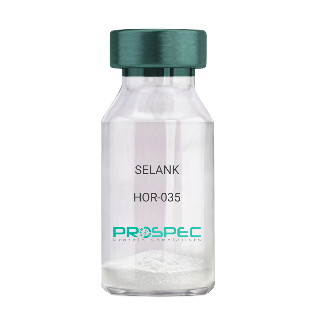
  • View Data Sheet

    Name :

    SELANK

    Description:

    SELANK

    Product # :

    HOR-035

    Price :

    Quantity :

    Shipping Method :

    Room Temp Icon

    Shipped at Room temp

    Add To Cart

    More Info

    • description
    • formulation
    • purity
    • More Info

    Description

    SELANK Synthetic is a single, non-glycosylated polypeptide chain containing 7 amino acids, having a molecular mass of 751.89 Dalton and a Molecular formula of C33H57N11O9.

    Formulation

    The protein was lyophilized with no additives.

    Purity

    Greater than 97.0% as determined by analysis by RP-HPLC.

    More Info

    • Physical Appearance

      Sterile Filtered White lyophilized (freeze-dried) powder.

    • Stability

      Lyophilized SELANK although stable at room temperature for 3 weeks, should be stored desiccated below -18°C. Upon reconstitution SELANK should be stored at 4°C between 2-7 days and for future use below -18°C. For long term storage it is recommended to add a carrier protein (0.1% HSA or BSA). Please prevent freeze-thaw cycles.

    • Solubility

      It is recommended to reconstitute the lyophilized SELANK in sterile 18MΩ-cm H2O not less than 100 µg/ml, which can then be further diluted to other aqueous solutions.

    • Amino Acid Sequence

      H-Thr-Lys-Pro-Arg-Pro-Gly-Pro-OH.

    • Background

      Selank, also known as TP-7, is a synthetic peptide that has garnered significant attention in the scientific community due to its potential anxiolytic, neuroprotective, and nootropic effects. This heptapeptide, derived from the naturally occurring human tetrapeptide Tuftsin, has been shown to possess a wide range of biological activities, including enhancing memory, reducing anxiety, and potentially mitigating the effects of neurodegenerative diseases.

      Selank's primary mechanism of action involves its interaction with the GABAergic system, the body's main inhibitory neurotransmitter system. By modulating the activity of GABA receptors, Selank can exert a calming effect on the brain, reducing anxiety and enhancing cognitive function. Studies by Semenova et al. (2014) have demonstrated that Selank can enhance memory and learning in rats, suggesting potential applications in cognitive enhancement and the treatment of cognitive disorders.

      In addition to its anxiolytic and nootropic effects, Selank has been shown to possess neuroprotective properties. Research by Ershov et al. (2016) found that Selank could protect neurons from oxidative stress, suggesting potential applications in the treatment of neurodegenerative diseases such as Alzheimer's and Parkinson's disease.

      Given its anxiolytic, nootropic, and neuroprotective effects, Selank has been proposed as a potential therapeutic agent for a variety of conditions, including anxiety disorders, cognitive disorders, and neurodegenerative diseases. For instance, a study by Medvedev et al. (2017) found that Selank could improve symptoms in patients with generalized anxiety disorder, indicating its potential as a therapeutic agent in the treatment of anxiety disorders.

      While research on Selank is promising, it is important to note that most studies have been conducted in animals or in vitro. More research is needed to fully understand the potential effects and applications of Selank in humans. However, the existing body of research suggests that Selank could be a promising tool in the treatment of anxiety disorders, cognitive disorders, and neurodegenerative diseases.

    ProSpec's products are furnished for LABORATORY RESEARCH USE ONLY. The product may not be used as drugs, agricultural or pesticidal products, food additives or household chemicals.

    Selank
  • View Data Sheet

    Name :

    Leptin qA Ovine, PEG

    Description:

    Leptin Quadruple Antagonist Pegylated Ovine Recombinant

    Product # :

    CYT-1246

    Price :

    Quantity :

    Shipping Method :

    Room Temp Icon

    Shipped at Room temp

    Add To Cart

    More Info

    • description
    • source
    • formulation
    • purity
    • biological activity
    • More Info

    Description

    Leptin Antagonist Quadruple Mutant Ovine Recombinant is a single non-glycosilated polypeptide chain containing 146 amino acids and additional Ala at N-terminus. The Ovine Leptin antagonist was mutated, resulting in D23L/L39A/D40A/F41A mutant. The Ovine Leptin antagonist is bound to 20 kDa mono-PEG at N-terminus, resulting in 35.6 kDa. The Ovine Leptin Quadruple anatagonist Pegylated runs as a 48 kDa due to enlarged hydrodymanic volume. Leptin Antagonist Quadruple Mutant Ovine Recombinant was purified by proprietary chromatographic techniques.

    Source

    Escherichia coli.

    Formulation

    The Ovine Leptin Quadruple anatagonist was lyophilized from a concentrated (0.65mg/ml) solution with 0.003mM NaHCO3.

    Purity

    Greater than 95.0% as determined by:

    (a) Gel filtration analysis.

    (b) Analysis by SDS-PAGE.

    Biological Activity

    Pegylated Ovine Leptin Quadruple Antagonist inhibits leptin-induced proliferation of BAF/3 cells stably transfected with the long form of human leptin receptor. Pegylated recombinant Ovine leptin antagonist in vitro activity is 6-8 fold lower than the non-pegylated recombinant super Ovine leptin antagonist but is 15 fold higher as compared to pegylated recombinant super active ovine leptin antagonist.

    More Info

    • Physical Appearance

      White lyophilized (freeze-dried) powder.

    • Stability

      Lyophilized Leptin Antagonist Quadruple Mutant Ovine Recombinant although stable at room temperature for several weeks, should be stored desiccated below -18°C. Upon reconstitution at > 0.1 mg/ml and up to 2mM and filter sterilization LEP mutant can be stored at 4°C or even room temperature for several weeks making it suitable for long term infusion studies using osmotic pumps. At lower concentration addition of a carrier protein (0.1% HSA or BSA) is suggested. Please prevent freeze-thaw cycles.

    • Solubility

      It is recommended to reconstitute the lyophilized Leptin Antagonist Quadruple Mutant Ovine Recombinant in sterile water or sterile 0.4% NaHCO3adjusted to pH 8-9, not less than 100µg/ml, which can then be further diluted with other aqueous solutions.

    • Background

      Leptin is mainly produced by adipocytes. Leptin’s main part is to regulate long-term energy balance. Leptin is encoded by the LEP gene. Leptin effects mostly on leptin receptors in the cell mambrane of various cells in the human body. The leptin receptor can be found on a various cell types. The leptin receptor is a single-transmembrane-domain type 1 cytokine receptor. leptin levels influence satiety, appetite and triggers behaviours which save energy. High leptin levels are interpreted by the brain that energy reserves are high, whereas low leptin levels means that energy reserves are low, in the process adapting the organism to starvation through a variety of metabolic, neurobiochemical, endocrine and behavioral change.

    • Protein content

      Protein quantization was carried out by UV spectroscopy at 280 nm using the absorbency value of 0.2 as the extinction coefficient for a 0.1% (1mg/ml) solution at pH 8.0. This value is calculated by the PC GENE computer analysis program of protein sequences (IntelliGenetics).

    ProSpec's products are furnished for LABORATORY RESEARCH USE ONLY. The product may not be used as drugs, agricultural or pesticidal products, food additives or household chemicals.

    Leptin Antagonist Peg Ovine
  • View Data Sheet

    Name :

    Rantes Rat

    Description:

    Rantes Rat Recombinant (CCL5)

    Small inducible cytokine A5, CCL5, T-cell-specific RANTES protein, SIS-delta, T cell- specific protein P228, TCP228, chemokine (C-C motif) ligand 5, SISd, SCYA5, RANTES, D17S136E, MGC17164.

    Product # :

    CHM-352

    Price :

    Quantity :

    Shipping Method :

    Room Temp Icon

    Shipped at Room temp

    Add To Cart

    More Info

    • description
    • source
    • formulation
    • purity
    • biological activity
    • More Info

    Description

    Rantes Rat Recombinant produced in E.Coli is a single, non-glycosylated polypeptide chain containing 68 amino acids and having a molecular mass of 7876 Dalton. The Rat Rantes is purified by proprietary chromatographic techniques.

    Source

    Escherichia Coli.

    Formulation

    Lyophilized from a concentrated (1mg/ml) solution in water containing no additives.

    Purity

    Greater than 95.0% as determined by:
    (a) Analysis by RP-HPLC.
    (b) Analysis by SDS-PAGE.

    Biological Activity

    The activity is determined by the ability to chemoattract total human lymphocytes and murine T-cells at a concentration between 1-10ng/ml corresponding to a Specific Activity of 100,000-1,000,000IU/mg.

    More Info

    • Introduction

      Regulated upon Activation, Normal T-cell Expressed, and Secreted or RANTES is an 8 kDa protein classified as a chemotactic cytokine or chemokine. It has recently been renamed CCL5. RANTES is chemotactic for T cells, eosinophils and basophils and plays an active role in recruiting leukocytes into inflammatory sites. With the help of particular cytokines (i.e. IL-2 and IFN-?) that are released by T cells, RANTES also induces the proliferation and activation of certain natural killer (NK) cells to form CHAK (CC-Chemokine-activated killer) cells. It is also a HIV-suppressive factor released from CD8+ T cells. This chemokine has been localized to chromosome 17 in humans.

    • Synonyms

      Small inducible cytokine A5, CCL5, T-cell-specific RANTES protein, SIS-delta, T cell- specific protein P228, TCP228, chemokine (C-C motif) ligand 5, SISd, SCYA5, RANTES, D17S136E, MGC17164.

    • Physical Appearance

      Sterile Filtered White lyophilized (freeze-dried) powder.

    • Stability

      Lyophilized Rat Rantes although stable at room temperature for 3 weeks, should be stored desiccated below -18°C. Upon reconstitution Rat CCL5 should be stored at 4°C between 2-7 days and for future use below -18°C.For long term storage it is recommended to add a carrier protein (0.1% HSA or BSA).Please prevent freeze-thaw cycles.

    • Solubility

      It is recommended to reconstitute the lyophilized Rantes in sterile 18MΩ-cm H2O not less than 100µg/ml, which can then be further diluted to other aqueous solutions.

    • Amino Acid Sequence

      SPYGSDTTPC CFAYLSLALP RAHVKEYFYT SSKCSNLAVV FVTRRNRQVC ANPEKKWVQE YINYLEMS.

    ProSpec's products are furnished for LABORATORY RESEARCH USE ONLY. The product may not be used as drugs, agricultural or pesticidal products, food additives or household chemicals.

    Rantes Rat
  • View Data Sheet

    Name :

    CTHRC1 Human, HEK

    Description:

    Collagen Triple Helix Repeat Containing 1 Human Recombinant, HEK

    Collagen Triple Helix Repeat Containing 1, Protein NMTC1, CTHRC1.

    Product # :

    PRO-2028

    Price :

    Quantity :

    Shipping Method :

    Room Temp Icon

    Shipped at Room temp

    Add To Cart

    More Info

    • description
    • source
    • formulation
    • purity
    • More Info

    Description

    CTHRC1 Human Recombinant produced in HEK cells is a single, glycosylated, polypeptide chain (Ser31-Lys243) containing a total of 219 amino acids, having a calculated molecular mass of 23.9kDa and fused to a 6 aa His tag at C-Terminus.

    Source

    HEK 293.

    Formulation

    CTHRC1 was filtered (0.4µm) and lyophilized in phosphate buffered saline pH 7.5.

    Purity

    Greater than 95.0% as determined by SDS-PAGE.

    More Info

    • Introduction

      Collagen triple helix repeat-containing protein 1 (CTHRC1) functions as a negative regulator of collagen matrix deposition. CTHRC1 is a secreted 28kDa protein which is glycosylated and highly conserved from lower chordates to mammals. CTHRC1 is highly connected with calcified tissues and cartilaginous matrix, but not with endothelial cells. CTHRC1 is detected qualitatively in plasma of healthy human subjects. CTHRC1 plasma levels are also significantly elevated during pregnancy, in diabetes, in inflammatory and infectious conditions, in subjects with acute myeloid leukemia but not in subjects with solid cancers. The hormonal functions of CTHRC1 include regulation of lipid storage and cellular glycogen levels with potentially far-reaching implications for cell metabolism and physiology. CTHRC1 gene deletion leads to fatty liver (steatosis) formation in mice while others exhibited inactivation of the CTHRC1 gene also results in low bone mass.

    • Synonyms

      Collagen Triple Helix Repeat Containing 1, Protein NMTC1, CTHRC1.

    • Physical Appearance

      Filtered White lyophilized (freeze-dried) powder.

    • Stability

      Store lyophilized protein at -20°C. Aliquot the product after reconstitution to avoid repeated freezing/thawing cycles. Reconstituted protein can be stored at 4°C for a limited period of time; it does not show any change after two weeks at 4°C.

    • Solubility

      It is recommended to add deionized water to prepare a working stock solution of approximately 0.5 mg/ml and let the lyophilized pellet dissolve completely. CTHRC1 is not sterile! Please filter the product by an appropriate sterile filter before using it in the cell culture.

    • Amino Acid Sequence

      SEIPKGKQKA QLRQREVVDL YNGMCLQGPA GVPGRDGSPG ANGIPGTPGI PGRDGFKGEK GECLRESFEE SWTPNYKQCS WSSLNYGIDL GKIAECTFTK MRSNSALRVL FSGSLRLKCR NACCQRWYFT FNGAECSGPL PIEAIIYLDQ GSPEMNSTIN IHRTSSVEGL CEGIGAGLVD VAIWVGTCSD YPKGDASTGW NSVSRIIIEE LPK HHHHHH.

    ProSpec's products are furnished for LABORATORY RESEARCH USE ONLY. The product may not be used as drugs, agricultural or pesticidal products, food additives or household chemicals.

    Cthrc1 Human Hek
  • View Data Sheet

    Name :

    DAG1 Human

    Description:

    Dystroglycan 1 Human Recombinant

    Dystroglycan, Dystrophin-associated glycoprotein 1, DAG1, A3a, DAG, AGRNR, 156DAG, MDDGC7, MDDGC9.

    Product # :

    PRO-1571

    Price :

    Quantity :

    Shipping Method :

    Room Temp Icon

    Shipped at Room temp

    Add To Cart

    More Info

    • description
    • source
    • formulation
    • purity
    • More Info

    Description

    DAG1 Human Recombinant produced in E.Coli is a single, non-glycosylated, polypeptide chain (a.a 30-312) containing 293 amino acids including a 10 a.a N-terminal His tag. The total molecular mass is 31.87kDa (calculated).

    Source

    Escherichia Coli.

    Formulation

    DAG1 filtered (0.4 µm) and lyophilized from 0.5mg/ml in 0.05M phosphate buffer and 0.075M NaCl, pH 7.4.

    Purity

    Greater than 90.0% as determined by SDS-PAGE.

    More Info

    • Introduction

      Dystroglycan 1 (DAG1) is a laminin binding component of the dystrophin-glycoprotein complex which provides a connection between the subsarcolemmal cytoskeleton and the extracellular matrix. The N-terminal domain of alpha-dystroglycan is secreted into the cerebrospinal fluid. The effect of DAG1 on the nervous system remains vague. The complete dystroglycan complex is expressed in a various tissues and has a role in processes such as laminin and basement membrane assembly, sarcolemmal stability, cell survival, peripheral nerve myelination, nodal structure, cell migration, and epithelial polarization. DAG1 is a candidate gene for the site of the mutation in autosomal recessive muscular dystrophies. The dramatic decrease of DAG1 in Duchenne muscular dystrophy leads to a loss of linkage between the sarcolemma and extracellular matrix, making muscle fibers more susceptible to necrosis.

    • Synonyms

      Dystroglycan, Dystrophin-associated glycoprotein 1, DAG1, A3a, DAG, AGRNR, 156DAG, MDDGC7, MDDGC9.

    • Physical Appearance

      Filtered White lyophilized (freeze-dried) powder.

    • Stability

      Store lyophilized protein at -20°C. Aliquot the product after reconstitution to avoid repeated freezing/thawing cycles. Reconstituted protein can be stored at 4°C for a limited period of time; it does not show any change after two weeks at 4°C.

    • Solubility

      It is recommended to add deionized water to prepare a working stock solution of approximately 0.5mg/ml and let the lyophilized pellet dissolve completely. DAG1 is not sterile! Please filter the product by an appropriate sterile filter before using it in the cell culture.

    • Amino Acid Sequence

      MKHHHHHHASHWPSEPSEAV RDWENQLEAS MHSVLSDLHE AVPTVVGIPD GTAVVGRSFR VTIPTDLIAS SGDIIKVSAA GKEALPSWLH WDSQSHTLEG LPLDTDKGVH YISVSATRLG ANGSHIPQTS SVFSIEVYPE DHSELQSVRT ASPDPGEVVS SACAADEPVT VLTVILDADL TKMTPKQRID LLHRMRSFSE VELHNMKLVP VVNNRLFDMS AFMAGPGNAK KVVENGALLS WKLGCSLNQN SVPDIHGVEA PAREGAMSAQ LGYPVVGWHI ANKKPPLPKR VRR.

    ProSpec's products are furnished for LABORATORY RESEARCH USE ONLY. The product may not be used as drugs, agricultural or pesticidal products, food additives or household chemicals.

    Dag1 Human
  • 1
  • …
  • 24
  • 25
  • 26
  • 27
  • 28
  • …
  • 42
Your browser does not support the video tag.

Products

  • Cytokines
  • Growth Factors
  • Chemokines
  • CD Antigens
  • Neurotrophins
  • Hormones
  • Enzymes
  • Viral Antigens
  • Recombinant Proteins
  • Natural Proteins
  • Monoclonal Antibodies
  • Polyclonal Antibodies
  • Thermal Packaging

About

  • Company
  • Founder
  • Contribution
  • Contact Us
  • My Account

Ordering

  • Ordering Method
  • Ordering - Credit Card
  • Ordering PO
  • Shipping Fees
  • FAQ's

Legal

  • Terms & Conditions
  • Privacy
  • Site Map
  • info@prospecbio.com
  • Facebook
  • Instagram
  • Linkedin Profile

© 2026 ProSpec-Tany TechnoGene Ltd. All Rights Reserved.

  • Choosing a selection results in a full page refresh.
  • Opens in a new window.