Search results
1000 results found for “neuritin”
Name
Description
Product #
Price
Quantity
Shipping Method
- View Data Sheet
Name :
EREG Human, HEKDescription:
Epiregulin Human Recombinant, HEK
EPR, Epiregulin, Ep, ER, Proepiregulin, EREG.
Product # :
CYT-1206Price :
Quantity :
Shipping Method :
Shipped with Ice Packs
More Info
- description
- source
- formulation
- purity
- biological activity
- More Info
Description
EREG Human Recombinant produced in HEK293 cells is a single, glycosylated polypeptide chain (63-108a.a) containing 289 amino acids and having a molecular mass of 32.6 kDa.EREG is fused to a 239 amino acid hIgG-His-Tag at C-terminus & purified by proprietary chromatographic techniques.
Source
HEK293 cells.
Formulation
EREG protein (0.25mg/ml) contains 10% glycerol and Phosphate-Buffered Saline (pH 7.4).
Purity
Greater than 90.0% as determined by SDS-PAGE.
Biological Activity
Measured in a cell proliferation assay using Balb/3T3 mouse embryonic fibroblast cells. The ED50 range ≤ 1ug/ml.
More Info
-
Introduction
"Epiregulin is part of the EGF family. Epiregulin functions as a ligand of EGFR, as well as a ligand of most members of the ERBB (v-erb-b2 oncogene homolog) family of tyrosine-kinase receptors.Epiregulin is expressed mostly in the placenta and peripheral blood leukocytes and in specific carcinomas of the bladder, lung, kidney and colon. Epiregulin stimulates the proliferation of keratinocytes, hepatocytes, fibroblasts and vascular smooth muscle cells. Epiregulin inhibits the growth of several tumor-derived epithelial cell lines. Human Epiregulin is initially synthesized as a glycosylated 19.0 kDa transmembrane precursor protein, which is processed by proteolytic cleavage to produce a 6.0 kDa mature secreted sequence."
-
Synonyms
EPR, Epiregulin, Ep, ER, Proepiregulin, EREG.
-
Physical Appearance
Sterile filtered colorless solution.
-
Stability
Store at 4°C if entire vial will be used within 2-4 weeks.Store, frozen at -20°C for longer periods of time.For long term storage it is recommended to add a carrier protein (0.1% HSA or BSA).Avoid multiple freeze-thaw cycles.
-
Amino Acid Sequence
DGSMVSITKC SSDMNGYCLH GQCIYLVDMS QNYCRCEVGY TGVRCEHFFL LEPKSCDKTH TCPPCPAPEL LGGPSVFLFP PKPKDTLMIS RTPEVTCVVV DVSHEDPEVK FNWYVDGVEV HNAKTKPREE QYNSTYRVVS VLTVLHQDWL NGKEYKCKVS NKALPAPIEK TISKAKGQPR EPQVYTLPPS RDELTKNQVS LTCLVKGFYP SDIAVEWESN GQPENNYKTT PPVLDSDGSF FLYSKLTVDK SRWQQGNVFS CSVMHEALHN HYTQKSLSLS PGKHHHHHH
-
Background
What is the molecular weight/Mw of EREG Protein?
EREG Protein has a total Mw of 32.6kDa.
What is the source or expression system of EREG Protein?
HEK293 cells.
What is the Purity of EREG Protein?
EREG Protein is >90% pure as determined by SDS-PAGE.
What is the Biological Activity of EREG Protein?
Measured in a cell proliferation assay using Balb/3T3 mouse embryonic fibroblast cells. The ED50 range ≤ 1ug/ml.
What is the amino acid sequence of EREG Protein?
DGSMVSITKC SSDMNGYCLH GQCIYLVDMS QNYCRCEVGY TGVRCEHFFL LEPKSCDKTH TCPPCPAPEL LGGPSVFLFP PKPKDTLMIS RTPEVTCVVV DVSHEDPEVK FNWYVDGVEV HNAKTKPREE QYNSTYRVVS VLTVLHQDWL NGKEYKCKVS NKALPAPIEK TISKAKGQPR EPQVYTLPPS RDELTKNQVS LTCLVKGFYP SDIAVEWESN GQPENNYKTT PPVLDSDGSF FLYSKLTVDK SRWQQGNVFS CSVMHEALHN HYTQKSLSLS PGKHHHHHH
What applications can EREG Protein be used in?
EREG Protein can probably be used in western blot, ELISA and Lateral Flow.
What is the endotoxin level for EREG Protein?
The endotoxin level is minimal, EREG Protein was purified using conventional chromatography techniques.
ProSpec's products are furnished for LABORATORY RESEARCH USE ONLY. The product may not be used as drugs, agricultural or pesticidal products, food additives or household chemicals.
- View Data Sheet
Name :
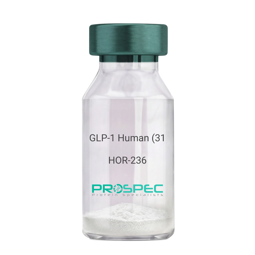
GLP-1 Human (31 a.a.)Description:
Glucagon Like Peptide-1 (31 a.a.) Human Recombinant
GLP1, GLP2, GRPP.
Product # :
HOR-236Price :
Quantity :
Shipping Method :
Shipped at Room temp
More Info
- description
- source
- formulation
- purity
- More Info
Description
Glucagon Like Peptide-1 Human Recombinant produced in E.Coli is a single, non-glycosylated, polypeptide chain containing 31 amino acids and having a molecular mass of 3.3 kDa. The GLP-1 is purified by proprietary chromatographic techniques.
Source
Escherichia Coli.
Formulation
The Recombinant GLP-1 concentrated solution (1mg/ml) was lyophilized after extensive dialyses against 0.1mg sodium phosphate monobasic & 1.6mg sodium phosphate dibasic.
Purity
Greater than 98.0% as determined by:
(a) Analysis by RP-HPLC.
(b) Analysis by SDS-PAGE.More Info
-
Introduction
Glucagon-like peptide-1 (GLP-1) is derived from the transcription product of the proglucagon gene. The major source of GLP-1 in the body is the intestinal L cell that secretes GLP-1 as a guthormone. The biologically active forms of GLP-1 are: GLP-1-(7-37) and GLP-1-(7-36)NH2.
GLP-1 secretion by L cells is dependent on the presence of nutrients in the lumen of the small intestine. The secretagogues (agents that causes or stimulates secretion) of this hormone include major nutrients like carbohydrate, proteinand lipid. Once in the circulation, GLP-1 has a half life of less than 2 minutes, due to rapid degradation by the enzyme dipeptidyl peptidase-4.
GLP-1 possesses several physiological properties that make it a subject of intensive investigation as a potential treatment of diabetes mellitus. The known physiological functions of GLP-1 include: Increases insulin secretion from the pancreas in a glucose-dependent manner, decreases glucagon secretion from the pancreas, increases beta cells mass and insulin gene expression, inhibits acid secretion and gastric emptying in the stomach, decreases food intake by increasing satiety. -
Synonyms
GLP1, GLP2, GRPP.
-
Physical Appearance
Sterile Filtered White lyophilized (freeze-dried) powder.
-
Stability
Lyophilized Glucagon Like Peptide-1 although stable at room temperature for 3 weeks, should be stored desiccated below -18°C. Upon reconstitution GLP-1 should be stored at 4°C between 2-7 days and for future use below -18°C. For long term storage it is recommended to add a carrier protein (0.1% HSA or BSA).Please prevent freeze-thaw cycles.
-
Solubility
It is recommended to reconstitute the lyophilized Glucagon Like Peptide-1 in sterile 18MΩ-cm H2O not less than 100µg/ml, which can then be further diluted to other aqueous solutions.
-
Amino Acid Sequence
HAEGTFTSDV SSYLEGQAAK EFIAWLVKGR G.
ProSpec's products are furnished for LABORATORY RESEARCH USE ONLY. The product may not be used as drugs, agricultural or pesticidal products, food additives or household chemicals.
- View Data Sheet
Name :
ProInsulin HumanDescription:
ProInsulin C-Peptide Analogue Human Recombinant
Insulin, Insulin-Dependent Diabetes Mellitus 2, Preproinsulin, Proinsulin, MODY10, IDDM1, IDDM2, IDDM, ILPR, IRDN.
Product # :
CYT-1120Price :
Quantity :
Shipping Method :
Shipped at Room temp
More Info
- description
- source
- formulation
- purity
- More Info
Description
ProInsulin C-Peptide Analogue Human Recombinant produced in E.Coli is a non-glycosylated polypeptide chain containing 35 amino acid and having a molecular mass of approximately 3.6kDa.ProInsulin is purified by proprietary chromatographic techniques.
Source
Escherichia Coli.
Formulation
Lyophilized from a 0.2μm filtered concentrated solution in PBS, pH 7.4.
Purity
Greater than 95.0% as determined by:
(a) Analysis by RP-HPLC.
(b) Analysis by SDS-PAGE.More Info
-
Introduction
Insulin decreases blood glucose concentration. Insulin increases cell permeability to monosaccharides, amino acids and fatty acids. Insulin accelerates glycolysis, the pentose phosphate cycle, and glycogen synthesis in liver.
-
Synonyms
Insulin, Insulin-Dependent Diabetes Mellitus 2, Preproinsulin, Proinsulin, MODY10, IDDM1, IDDM2, IDDM, ILPR, IRDN.
-
Physical Appearance
Sterile Filtered White lyophilized (freeze-dried) powder.
-
Stability
Lyophilized ProInsulin although stable at room temperature for 3 weeks, should be stored desiccated below -18°C. Upon reconstitution ProInsulin C-Peptide Analogue should be stored at 4°C between 2-7 days and for future use below -18°C.Please prevent freeze-thaw cycles.
-
Solubility
It is recommended to reconstitute the lyophilized ProInsulin C-Peptide Analogue in sterile 18MΩ-cm H2O not less than 100µg/ml, which can then be further diluted to other aqueous solutions.
-
Amino Acid Sequence
RREAEDLQVG QVELGGGPGA GSLQPLALEG SLQKR.
ProSpec's products are furnished for LABORATORY RESEARCH USE ONLY. The product may not be used as drugs, agricultural or pesticidal products, food additives or household chemicals.
- View Data Sheet
Name :
CHGA Human, Sf9Description:
Chromogranin A Human Recombinant, Sf9
CHGA, CGA, Chromogranin-A, Vasostatin I, SP-I, Pituitary secretory protein I.
Product # :
PRO-2513Price :
Quantity :
Shipping Method :
Shipped with Ice Packs
More Info
- description
- source
- formulation
- purity
- More Info
Description
CHGA produced in Sf9 Baculovirus cells is a single, glycosylated polypeptide chain (19-457 a.a.) and fused to a 6 aa His Tag at C-terminus containing a total of 448 amino acids and having a molecular mass of 50kDa.CHGA shows multiple bands between 50-70kDa on SDS-PAGE, reducing conditions and purified by proprietary chromatographic techniques.
Source
Sf9, Baculovirus cells.
Formulation
CHGA protein solution (0.25mg/ml) contains Phosphate Buffered Saline (pH 7.4), 20% glycerol & 0.1mM PMSF.
Purity
Greater than 85.0% as determined by SDS-PAGE.
More Info
-
Introduction
Chromogranin-A Isoform 1 Preproprotein or CHGA is part of the neuroendocrine secretory proteins of the chromogranin/secretogranin family. CHGA is a precursor of numerus enzymes such as pancreastatin, catestatin, vasostatin-1,vasostatin-2, and parastatin. The protein acts as a negative regulator the neuroendocrine activity of autocrine or nearby cells (paracrine). CHGA causes further production of secretory granules that contains insulin in pancreatic islet beta cells.
-
Synonyms
CHGA, CGA, Chromogranin-A, Vasostatin I, SP-I, Pituitary secretory protein I.
-
Physical Appearance
Sterile Filtered colorless solution.
-
Stability
Store at 4°C if entire vial will be used within 2-4 weeks. Store, frozen at -20°C for longer periods of time. For long term storage it is recommended to add a carrier protein (0.1% HSA or BSA). Avoid multiple freeze-thaw cycles.
-
Amino Acid Sequence
ADLLPVNSPM NKGDTEVMKC IVEVISDTLS KPSPMPVSQE CFETLRGDER ILSILRHQNL LKELQDLALQ GAKERAHQQK KHSGFEDELS EVLENQSSQA ELKEAVEEPS SKDVMEKRED SKEAEKSGEA TDGARPQALP EPMQESKAEG NNQAPGEEEE EEEEATNTHP PASLPSQKYP GPQAEGDSEG LSQGLVDREK GLSAEPGWQA KREEEEEEEE EAEAGEEAVP EEEGPTVVLN PHPSLGYKEI RKGESRSEAL AVDGAGKPGA EEAQDPEGKG EQEHSQQKEE EEEMAVVPQG LFRGGKSGEL EQEEERLSKE WEDSKRWSKM DQLAKELTAE KRLEGQEEEE DNRDSSMKLS FRARAYGFRG PGPQLRRGWR PSSREDSLEA GLPLQVRGYP EEKKEEEGSA NRRPEDQELE SLSAIEAELE KVAHQLQALR RGHHHHHH.
ProSpec's products are furnished for LABORATORY RESEARCH USE ONLY. The product may not be used as drugs, agricultural or pesticidal products, food additives or household chemicals.
- View Data Sheet
Name :
YOD1 HumanDescription:
YOD1 Human Recombinant
DUBA8, OTUD2, PRO0907, RP11-164O23.1, Ubiquitin thioesterase OTU1, DUBA-8, HIN-7, HsHIN7, OTU domain-containing protein 2.
Product # :
ENZ-696Price :
Quantity :
Shipping Method :
Shipped with Ice Packs
More Info
- description
- source
- formulation
- purity
- More Info
Description
YOD1 Human Recombinant produced in E.coli is a single, non-glycosylated polypeptide chain containing 371 amino acids (1-348) and having a molecular mass of 40.7kDa.YOD1 is fused to a 23 amino acid His-tag at N-terminus & purified by proprietary chromatographic techniques.
Source
Escherichia Coli.
Formulation
The YOD1 solution (0.25mg/ml) contains 20mM Tris-HCl buffer (pH 8.0), 0.15M NaCl, 30% glycerol and 1mM DTT.
Purity
Greater than 90.0% as determined by SDS-PAGE.
More Info
-
Introduction
YOD1 is a Hydrolase which removes conjugated ubiquitin from proteins and takes part in endoplasmic reticulum-associated degradation (ERAD) for misfolded lumenal proteins. YOD1 is a highly conserved deubiquitinating enzyme belonging to the ovarian tumor (otubain) family, whose function has yet to be determined in mammalian cells. YOD1 is a component of a multiprotein complex with p97 as its nucleus, proposing a functional link to a pathway responsible for the dislocation of misfolded proteins from the endoplasmic reticulum. YOD1 variant xpression deprived of its deubiquitinating activity compels a halt on the dislocation reaction, as concluded by the stabilization of various dislocation substrates.
-
Synonyms
DUBA8, OTUD2, PRO0907, RP11-164O23.1, Ubiquitin thioesterase OTU1, DUBA-8, HIN-7, HsHIN7, OTU domain-containing protein 2.
-
Physical Appearance
Sterile Filtered colorless solution.
-
Stability
Store at 4°C if entire vial will be used within 2-4 weeks. Store, frozen at -20°C for longer periods of time. For long term storage it is recommended to add a carrier protein (0.1% HSA or BSA).Avoid multiple freeze-thaw cycles.
-
Amino Acid Sequence
MGSSHHHHHH SSGLVPRGSH MGSMFGPAKG RHFGVHPAPG FPGGVSQQAA GTKAGPAGAW PVGSRTDTMW RLRCKAKDGT HVLQGLSSRT RVRELQGQIA AITGIAPGGQ RILVGYPPEC LDLSNGDTIL EDLPIQSGDM LIIEEDQTRP RSSPAFTKRG ASSYVRETLP VLTRTVVPAD NSCLFTSVYY VVEGGVLNPA CAPEMRRLIA QIVASDPDFY SEAILGKTNQ EYCDWIKRDD TWGGAIEISI LSKFYQCEIC VVDTQTVRID RFGEDAGYTK RVLLIYDGIH YDPLQRNFPD PDTPPLTIFS SNDDIVLVQA LELADEARRR RQFTDVNRFT LRCMVCQKGL TGQAEAREHA KETGHTNFGE V.
ProSpec's products are furnished for LABORATORY RESEARCH USE ONLY. The product may not be used as drugs, agricultural or pesticidal products, food additives or household chemicals.
- View Data Sheet
Name :
PEDF Human, HEKDescription:
Pigment Epithelium-Derived Factor Human Recombinant, HEK
Pigment epithelium-derived factor, PEDF, Serpin-F1, SerpinF1, EPC-1, EPC1, PIG35.
Product # :
CYT-553Price :
Quantity :
Shipping Method :
Shipped at Room temp
More Info
- description
- source
- formulation
- purity
- More Info
Description
PEDF Human Recombinant produced in HEK cells is a single, glycosylated, polypeptide chain containing a total of 410 amino acids, having a molecular mass of 45.6 kDa and fused to an 11 aa FLAG tag at C-Terminus.The Human PEDF is purified by proprietary chromatographic techniques.
Source
HEK 293.
Formulation
The filtered (0.4µm) concentrated (0.5mg/ml) protein solution was lyophilized with 20mM Tris & 20mM NaCl pH-7.5.
Purity
Greater than 95% as determined by SDS-PAGE.
More Info
-
Introduction
PEDF is a neurotrophic protein that induces extensive neuronal differentiation in retinoblastoma cells. SerpinF1 is a potent inhibitor of angiogenesis. EPC1 doesn’t undergo the stressed to relaxed conformation transition characteristic as of the active serpins since it exhibits no serine protease inhibitory activity.
Aqueous humour level of asymmetric dimethylarginine is correlated with PEDF in humans. ADMA and PEDF levels are increased in response to inflammation in uveitis.
Lack of PEDF expression is a potent factor for the enhancement of tumor growth and angiogenesis in breast cancer.
PEDF & VEGF genes contribute to the development of diabetic retinopathy.
PEDF and VEGF structural changes in blood vessel wall play an important role in the pathophysiology of PD patients.
PEDF-overexpressing tumors exhibited reduced intratumoral angiogenesis.
SerpinF1 is a new promising approach for the treatment of osteosarcoma.
Levels of the natural ocular anti-angiogenic factor SentrinF1 (PEDF) is associated with proliferative retinopathy.
VEGF secreted by retinal pigment epithelial cells upregulates PEDF expression via VEGFR-1 in an autocrine manner.
Sentrin-F1 concentration in the aqueous humor of diabetic patients predicts who will develop progression of retinopathy.
PEDF blocks angiogenic effects of leptin through its anti-oxidative properties. -
Synonyms
Pigment epithelium-derived factor, PEDF, Serpin-F1, SerpinF1, EPC-1, EPC1, PIG35.
-
Physical Appearance
Filtered White lyophilized (freeze-dried) powder.
-
Stability
Store lyophilized protein at -20°C. Aliquot the product after reconstitution to avoid repeated freezing/thawing cycles. Reconstituted protein can be stored at 4°C for a limited period of time.
-
Solubility
It is recomnded to add deionized water to a working concentration of 0.5mg/ml and let the lyophilized pellet dissolve completely. Product is not sterile! Please filter the product by an appropriate sterile filter before using it in the cell culture.
-
Amino Acid Sequence
QNPASPPEEG SPDPDSTGAL VEEEDPFFKV PVNKLAAAVS NFGYDLYRVR SSTSPTTNVL LSPLSVATAL SALSLGAEQR TESIIHRALY YDLISSPDIH GTYKELLDTV TAPQKNLKSA SRIVFEKKLR IKSSFVAPLE KSYGTRPRVL TGNPRLDLQE INNWVQAQMK GKLARSTKEI PDEISILLLG VAHFKGQWVT KFDSRKTSLE DFYLDEERTV RVPMMSDPKA VLRYGLDSDL SCKIAQLPLT GSMSIIFFLP LKVTQNLTLI EESLTSEFIH DIDRELKTVQ AVLTVPKLKL SYEGEVTKSL QEMKLQSLFD SPDFSKITGK PIKLTQVEHR AGFEWNEDGA GTTPSPGLQP AHLTFPLDYH LNQPFIFVLR DTDTGALLFI GKILDPRGPAAADYKDDDDK.
ProSpec's products are furnished for LABORATORY RESEARCH USE ONLY. The product may not be used as drugs, agricultural or pesticidal products, food additives or household chemicals.
- View Data Sheet
Name :
Leptin qA Ovine, PEGDescription:
Leptin Quadruple Antagonist Pegylated Ovine Recombinant
Product # :
CYT-1246Price :
Quantity :
Shipping Method :
Shipped at Room temp
More Info
- description
- source
- formulation
- purity
- biological activity
- More Info
Description
Leptin Antagonist Quadruple Mutant Ovine Recombinant is a single non-glycosilated polypeptide chain containing 146 amino acids and additional Ala at N-terminus. The Ovine Leptin antagonist was mutated, resulting in D23L/L39A/D40A/F41A mutant. The Ovine Leptin antagonist is bound to 20 kDa mono-PEG at N-terminus, resulting in 35.6 kDa. The Ovine Leptin Quadruple anatagonist Pegylated runs as a 48 kDa due to enlarged hydrodymanic volume. Leptin Antagonist Quadruple Mutant Ovine Recombinant was purified by proprietary chromatographic techniques.
Source
Escherichia coli.
Formulation
The Ovine Leptin Quadruple anatagonist was lyophilized from a concentrated (0.65mg/ml) solution with 0.003mM NaHCO3.
Purity
Greater than 95.0% as determined by:
(a) Gel filtration analysis.
(b) Analysis by SDS-PAGE.
Biological Activity
Pegylated Ovine Leptin Quadruple Antagonist inhibits leptin-induced proliferation of BAF/3 cells stably transfected with the long form of human leptin receptor. Pegylated recombinant Ovine leptin antagonist in vitro activity is 6-8 fold lower than the non-pegylated recombinant super Ovine leptin antagonist but is 15 fold higher as compared to pegylated recombinant super active ovine leptin antagonist.
More Info
-
Physical Appearance
White lyophilized (freeze-dried) powder.
-
Stability
Lyophilized Leptin Antagonist Quadruple Mutant Ovine Recombinant although stable at room temperature for several weeks, should be stored desiccated below -18°C. Upon reconstitution at > 0.1 mg/ml and up to 2mM and filter sterilization LEP mutant can be stored at 4°C or even room temperature for several weeks making it suitable for long term infusion studies using osmotic pumps. At lower concentration addition of a carrier protein (0.1% HSA or BSA) is suggested. Please prevent freeze-thaw cycles.
-
Solubility
It is recommended to reconstitute the lyophilized Leptin Antagonist Quadruple Mutant Ovine Recombinant in sterile water or sterile 0.4% NaHCO3adjusted to pH 8-9, not less than 100µg/ml, which can then be further diluted with other aqueous solutions.
-
Background
Leptin is mainly produced by adipocytes. Leptin’s main part is to regulate long-term energy balance. Leptin is encoded by the LEP gene. Leptin effects mostly on leptin receptors in the cell mambrane of various cells in the human body. The leptin receptor can be found on a various cell types. The leptin receptor is a single-transmembrane-domain type 1 cytokine receptor. leptin levels influence satiety, appetite and triggers behaviours which save energy. High leptin levels are interpreted by the brain that energy reserves are high, whereas low leptin levels means that energy reserves are low, in the process adapting the organism to starvation through a variety of metabolic, neurobiochemical, endocrine and behavioral change.
-
Protein content
Protein quantization was carried out by UV spectroscopy at 280 nm using the absorbency value of 0.2 as the extinction coefficient for a 0.1% (1mg/ml) solution at pH 8.0. This value is calculated by the PC GENE computer analysis program of protein sequences (IntelliGenetics).
ProSpec's products are furnished for LABORATORY RESEARCH USE ONLY. The product may not be used as drugs, agricultural or pesticidal products, food additives or household chemicals.
- View Data Sheet
Name :
sRANKL HumanDescription:
RANK Ligand Soluble Human Recombinant
Soluble Receptor Activator of NFkB Ligand, TNFSF11, TRANCE, TNF-related activation-induced cytokine, OPGL, ODF, Osteoclast differentiation factor, Tumor necrosis factor ligand superfamily member 11, Receptor activator of nuclear factor kappa B ligand, RANKL, Osteoprotegerin ligand, CD254 antigen, sRANKL, sOdf, hRANKL2.
Product # :
CYT-334Price :
Quantity :
Shipping Method :
Shipped at Room temp
More Info
- description
- source
- formulation
- purity
- biological activity
- More Info
- HPLC, SDS-PAGE
Description
sRANKL Human Recombinant produced in E.Coli is a single, non-glycosylated polypeptide chain containing 175 amino acids and having a molecular mass of 19.7kDa.
Source
Escherichia Coli.
Formulation
The protein was lyophilized from a concentrated (1mg/ml) solution containing 10mM Sodium phosphate, pH-7.5.
Purity
Greater than 95.0% as determined by analysis by SDS-PAGE.
Biological Activity
The activity of RAW-Blue was measured to be 46.96 ng/ml, corresponding to a specific activity of 2.1x10⁴ units/mg.
HPLC, SDS-PAGE
More Info
-
Introduction
RANKL binds to tnfrsf11b/opg and to tnfrsf11a/rank. Osteoclast differentiation and activation factor. Augments the ability of dendritic cells to stimulate naive t-cell proliferation. May be an important regulator of interactions between t-cells and dendritic cells and may play a role in the regulation of the t-cell-dependent immune response. sRANKL may also play an important role in enhanced bone-resorption in humoral hypercalcemia of malignancy.
-
Synonyms
Soluble Receptor Activator of NFkB Ligand, TNFSF11, TRANCE, TNF-related activation-induced cytokine, OPGL, ODF, Osteoclast differentiation factor, Tumor necrosis factor ligand superfamily member 11, Receptor activator of nuclear factor kappa B ligand, RANKL, Osteoprotegerin ligand, CD254 antigen, sRANKL, sOdf, hRANKL2.
-
Physical Appearance
Sterile Filtered White lyophilized (freeze-dried) powder.
-
Stability
Lyophilized TNFSF11 although stable at room temperature for 3 weeks, should be stored desiccated below -18°C. Upon reconstitution sRANKL should be stored at 4°C between 2-7 days and for future use below -18°C.For long term storage it is recommended to add a carrier protein (0.1% HSA or BSA).Please prevent freeze-thaw cycles.
-
Solubility
It is recommended to reconstitute the lyophilized sRANKL in sterile 18MΩ-cm H2O at a concentration of 100µg/ml, which can then be further diluted to other aqueous solutions.
-
Amino Acid Sequence
EKAMVDGSW LDLAKRSKLE AQPFAHLTIN ATDIPSGSHK VSLSSWYHDR GWAKISNMTF SNGKLIVNQD GFYYLYANIC FRHHETSGDL ATEYLQLMVY VTKTSIKIPS SHTLMKGGST KYWSGNSEFH FYSINVGGFF KLRSGEEISI EVSNPSLLDP DQDATYFGAF KVRDID.
ProSpec's products are furnished for LABORATORY RESEARCH USE ONLY. The product may not be used as drugs, agricultural or pesticidal products, food additives or household chemicals.
- View Data Sheet
Name :
CRYGD MouseDescription:
Crystallin, Gamma D Mouse Recombinant
Gamma-crystallin D, Gamma-D-crystallin , Gamma-crystallin 1, CRYGD.
Product # :
PRO-2484Price :
Quantity :
Shipping Method :
Shipped with Ice Packs
More Info
- description
- source
- formulation
- purity
- More Info
Description
CRYGD Mouse Recombinant produced in E.Coli is a single, non-glycosylated polypeptide chain containing 197 amino acids (1-174 a.a.) and having a molecular mass of 23.5kDa.CRYGD is fused to a 23 amino acid His-tag at N-terminus & purified by proprietary chromatographic techniques.
Source
Escherichia Coli.
Formulation
CRYGD protein solution (1mg/ml) containing 20mM Tris-HCl buffer (pH 8.0), 0.1M NaCl and 10% glycerol.
Purity
Greater than 95.0% as determined by SDS-PAGE.
More Info
-
Introduction
CRYGD is a member of the beta/gamma-crystallin family. Crystallins are the principal structural components of the vertebrate eye lens. The mammalian lens crystallins are divided into alpha, beta, and gamma families. Gamma-crystallins are involved in cataract formation. Defects in the CRYGD gene are responsible for cataract autosomal dominant (ADC), cataract congenital non-nuclear polymorphic autosomal dominant (CCP), cataract congenital cerulean type 3 (CCA3) and cataract crystalline aculeiform (CACA).
-
Synonyms
Gamma-crystallin D, Gamma-D-crystallin , Gamma-crystallin 1, CRYGD.
-
Physical Appearance
Sterile filtered colorless solution.
-
Stability
Store at 4°C if entire vial will be used within 2-4 weeks. Store, frozen at -20°C for longer periods of time. For long term storage it is recommended to add a carrier protein (0.1% HSA or BSA).Avoid multiple freeze-thaw cycles.
-
Amino Acid Sequence
MGSSHHHHHH SSGLVPRGSH MGSMGKITFY EDRGFQGRHY ECSTDHSNLQ PYFSRCNSVR VDSGCWMLYE QPNFTGCQYF LRRGDYPDYQ QWMGFSDSVR SCRLIPHAGS HRIRLYEREE YRGQMIEFTE DCPSLQDRFH FNEIYSLNVL EGCWVLYDMT NYRGRQYLLR PGEYRRYHDW
GAMNARVGSL RRVMDFY
ProSpec's products are furnished for LABORATORY RESEARCH USE ONLY. The product may not be used as drugs, agricultural or pesticidal products, food additives or household chemicals.
- View Data Sheet
Name :
ANGPT1 HumanDescription:
Angiopoietin-1 Human Recombinant
Angiopoietin 1, KIAA0003, ANG-1, AGP1, AGPT.
Product # :
CYT-074Price :
Quantity :
Shipping Method :
Shipped with Ice Packs
More Info
- description
- source
- formulation
- purity
- More Info
- sds-page
Description
ANGPT1 Human Recombinant produced in E.coli is a single, non-glycosylated polypeptide chain containing 480 amino acids (20-498) and having a molecular mass of 55.6 kDa.The ANGPT1 is purified by proprietary chromatographic techniques.
Source
E.coli.
Formulation
The ANGPT1 solution (1mg/ml) contains 20mM Tris-HCl buffer (pH 8.0), 1M Urea and 5% glycerol.
Purity
Greater than 85% as determined by SDS-PAGE.
sds-page
More Info
-
Introduction
ANGPT1 is an angiogenic factor which intervenes in blood vessel maturation and takes part in endothelial development. ANGPT1 is a secreted ligand for Tie-2, a cell surface receptor tyrosine kinase expressed in endothelial and hemopoietic cells. The glycosylated ANGPT1 protein has a coiled-coil region in the amino terminus and a fibrinogen-like domain at the carboxy terminus.
-
Synonyms
Angiopoietin 1, KIAA0003, ANG-1, AGP1, AGPT.
-
Physical Appearance
Sterile Filtered colorless solution.
-
Stability
Store at 4°C if entire vial will be used within 2-4 weeks. Store, frozen at -20°C for longer periods of time. For long term storage it is recommended to add a carrier protein (0.1% HSA or BSA).Avoid multiple freeze-thaw cycles.
-
Amino Acid Sequence
MSNQRRSPEN SGRRYNRIQH GQCAYTFILP EHDGNCREST TDQYNTNALQ RDAPHVEPDF SSQKLQHLEH VMENYTQWLQ KLENYIVENM KSEMAQIQQN AVQNHTATML EIGTSLLSQT AEQTRKLTDV ETQVLNQTSR LEIQLLENSL STYKLEKQLL QQTNEILKIH EKNSLLEHKI LEMEGKHKEE LDTLKEEKEN LQGLVTRQTY IIQELEKQLN RATTNNSVLQ KQQLELMDTV HNLVNLCTKE GVLLKGGKRE EEKPFRDCAD VYQAGFNKSG IYTIYINNMP EPKKVFCNMD VNGGGWTVIQ HREDGSLDFQ RGWKEYKMGF GNPSGEYWLG NEFIFAITSQ RQYMLRIELM DWEGNRAYSQ YDRFHIGNEK QNYRLYLKGH TGTAGKQSSL ILHGADFSTK DADNDNCMCK CALMLTGGWW FDACGPSNLN GMFYTAGQNH GKLNGIKWHY FKGPSYSLRS TTMMIRPLDF
-
Background
This factor was found in the conditioned medium of the human neuroepithelioma cell line SHEP1 and the mouse myoblast cell line C2C12ras. The cDNA encoding a protein of 498 amino acids was isolated by using a secretion-trap expression cloning procedure exploiting the presence of a signal sequence present in growth factors that are secreted by producer cells (Davis et al, 1996). Murine and human factors show 97 % identity at the amino acid level. For a related factor see: CDT6. The human gene has been mapped to chromosome 8q22.3-q23 (Cheung et al, 1998).
The expression of angiopoietin-1 mRNA is downregulated by PDGF, EGF, IL1-beta, and TGF-beta (Enholm et al, 1997).
Angiopoietin-1 is a ligand for the receptor-like tyrosine kinase designated TIE-2 (Davis et al, 1996). Binding of Angiopoietin-1 to its receptor induces tyrosine phosphorylation of the cytoplasmic receptor domain. A naturally occuring antagonist of Angiopoietin-1 binding to TIE-2 is Angiopoietin-2. Angiopoietin-1 also binds to the TIE-1 receptor and this interaction appears to be critical for the development of the right-hand side venous system. It is dispensable for the left-hand side venous system, suggesting that right-hand and left-hand side vascular networks are established early before asymmetrical features of the network become morphologically discernible Loughna and Sato, 2001).
Angiopoietin-1 does not directly promote the growth of cultured endothelial cells. Angiopoietin-1 is chemotactic for endothelial cells (Witzenbichler et al, 1998). Excess soluble TIE-2 receptors abolish the chemotactic response of endothelial cells toward angiopoietin-1. Angiopoietin-1 has been shown to counteract cell death by apoptosis in cultured endothelial cells (Holash et al, 1999). Angiopoietin-1 also acts as an apoptosis survival factor for endothelial cells and this effect is augmented by the presence of VEGF (Kwak et al, 1999).
Angiopoietin-2 dose-dependently blocks directed migration toward Angiopoietin-1. Carlson et al (2001) have shown that Ang-1 binds rather selectively to vitronectin and that Ang-1 can directly support adhesion of human umbilical vein endothelial cells and fibroblasts in a process mediated by integrins.
The physiologic roles of Angiopoietin-1 and its receptor are limited to angiogenic processes that occur subsequent to the earlier vasculogenic and angiogenic actions of the VEGF family and their receptors. However, it is unlike most of the known angiogenesis factors such as VEGF and other classical endothelial cell growth factors in that addition of the factor to cultures of endothelial cells does not directly promote cell growth even though the TIE-2 receptor becomes activated. Angiopoietin-1 also appears to be incapable of inducing the formation of tubules by endothelial cells.
Angiopoietins can potentiate the effects of other angiogenic cytokines. An investigation of the impact of angiopoietins on neovascularization in vivo in the cornea micropocket assay of neovascularization demonstrates that neither Angiopoietin-1 nor Angiopoietin-2 alone promote neovascularization. Holash et al (1999) have shown that a subset of tumors initially grows by coopting existing host vessels. Regression of these vessels via a process that involves disruption of interactions between endothelial cells and smooth muscle cells as well as cell death by apoptosis of endothelial cells first causes loss of tumour cells before angiogenesis begins at the tumor margin and the tumor is rescued under the influence of VEGF, Angiopoietin-1, and probably other angiogenic stimuli.
The embryonic expression pattern of Angiopoietin-1 suggests that it plays a particularly important role in the developing heart. Angiopoietin-1 is expressed highly in the myocardial wall surrounding the endocardium expressing TIE-2. Expression of Angiopoietin-1 becomes much more widespread later in development.
ANGPT1 Protein has a total Mw of 55.6kDa.
What is the source or expression system of ANGPT1 Protein?
Escherichia Coli.
What is the Purity of ANGPT1 Protein?
ANGPT1 Protein is >85% pure as determined by SDS-PAGE.
What is the Biological Activity of ANGPT1 Protein?
The biological functionality of ANGPT1 Protein will be determined in the future.
What is the amino acid sequence of ANGPT1 Protein?
MSNQRRSPEN SGRRYNRIQH GQCAYTFILP EHDGNCREST TDQYNTNALQ RDAPHVEPDF SSQKLQHLEH VMENYTQWLQ KLENYIVENM KSEMAQIQQN AVQNHTATML EIGTSLLSQT AEQTRKLTDV ETQVLNQTSR LEIQLLENSL STYKLEKQLL QQTNEILKIH EKNSLLEHKI LEMEGKHKEE LDTLKEEKEN LQGLVTRQTY IIQELEKQLN RATTNNSVLQ KQQLELMDTV HNLVNLCTKE GVLLKGGKRE EEKPFRDCAD VYQAGFNKSG IYTIYINNMP EPKKVFCNMD VNGGGWTVIQ HREDGSLDFQ RGWKEYKMGF GNPSGEYWLG NEFIFAITSQ RQYMLRIELM DWEGNRAYSQ YDRFHIGNEK QNYRLYLKGH TGTAGKQSSL ILHGADFSTK DADNDNCMCK CALMLTGGWW FDACGPSNLN GMFYTAGQNH GKLNGIKWHY FKGPSYSLRS TTMMIRPLDF
What applications can ANGPT1 Protein be used in?
ANGPT1 Protein can probably be used in western blot, ELISA and Lateral Flow.
What is the endotoxin level for ANGPT1 Protein?
The endotoxin level is minimal, ANGPT1 Protein was purified using conventional chromatography techniques.
ProSpec's products are furnished for LABORATORY RESEARCH USE ONLY. The product may not be used as drugs, agricultural or pesticidal products, food additives or household chemicals.
- View Data Sheet
Name :
NAP 2 RatDescription:
Neutrophil Activating Protein-2 Rat Recombinant (CXCL7)
Platelet basic protein, PBP, Small inducible cytokine B7, CXCL7, Leukocyte-derived growth factor, LDGF, Macrophage-derived growth factor, MDGF, pro-platelet basic protein (chemokine (C-X-C motif) ligand 7), TC1, TC2, TGB, TGB1, B-TG1, CTAP3, NAP-2, SCYB7, THBGB, LA-PF4, THBGB1, Beta-TG, CTAPIII, CTAP-III.
Product # :
CHM-269Price :
Quantity :
Shipping Method :
Shipped at Room temp
More Info
- description
- source
- formulation
- purity
- biological activity
- More Info
Description
NAP-2 Rat Recombinant produced in E.coli is a single, non-glycosylated polypeptide chain containing 62 amino acids and having a molecular mass of 6.8kDa.The NAP 2 is purified by proprietary chromatographic techniques.
Source
Escherichia Coli.
Formulation
NAP-2 protein was lyophilized from a 0.2µm filtered concentrated solution in 1×PBS, pH 7.4.
Purity
Greater than 97.0% as determined by:
(a) Analysis by RP-HPLC.
(b) Analysis by SDS-PAGE.Biological Activity
Measured by its ability to chemoattract BaF3 mouse pro-B cells transfected with human CXCR2. The ED50 for this effect is less than 10ng/ml, corresponding to a specific activity of 100,000units/mg.More Info
-
Introduction
Chemokine (C-X-C motif) ligand (CXCL7) is a small cytokine belonging to the CXC chemokine family. It is a protein that is released in large amounts from platelets following their activation. It stimulates various processes including mitogenesis, synthesis of extracellular matrix, glucose metabolism and synthesis of plasminogen activator.
-
Synonyms
Platelet basic protein, PBP, Small inducible cytokine B7, CXCL7, Leukocyte-derived growth factor, LDGF, Macrophage-derived growth factor, MDGF, pro-platelet basic protein (chemokine (C-X-C motif) ligand 7), TC1, TC2, TGB, TGB1, B-TG1, CTAP3, NAP-2, SCYB7, THBGB, LA-PF4, THBGB1, Beta-TG, CTAPIII, CTAP-III.
-
Physical Appearance
Sterile Filtered White lyophilized (freeze-dried) powder.
-
Stability
Lyophilized NAP-2 although stable at room temperature for 3 weeks, should be stored desiccated below -18°C. Upon reconstitution CXCL7 should be stored at 4°C between 2-7 days and for future use below -18°C. For long term storage it is recommended to add a carrier protein (0.1% HSA or BSA). Please prevent freeze-thaw cycles.
-
Solubility
It is recommended to reconstitute the lyophilized NAP-2 in sterile 18M-cm H2O not less than 100µg/ml, which can then be further diluted to other aqueous solutions.
-
Amino Acid Sequence
IELRCRCTNT LSGIPLNSIS RVNVFRPGAH CDNVEVIATL KNGKEVCLDP TAPMIKKIVK KI.
ProSpec's products are furnished for LABORATORY RESEARCH USE ONLY. The product may not be used as drugs, agricultural or pesticidal products, food additives or household chemicals.
- View Data Sheet
Name :
NFKBIB HumanDescription:
NF-kappa-B Inhibitor Beta Human Recombinant
NF-kappa-B inhibitor beta, NF-kappa-BIB, I-kappa-B-beta, IkB-B, IkB-beta, IkappaBbeta, Thyroid receptor-interacting protein 9, TR-interacting protein 9, TRIP-9, NFKBIB, IKBB, TRIP9.
Product # :
PRO-1046Price :
Quantity :
Shipping Method :
Shipped with Ice Packs
More Info
- description
- source
- formulation
- purity
- More Info
Description
NFKBIB Human Recombinant produced in E.Coli is a single, non-glycosylated polypeptide chain containing 380 amino acids (1-356 a.a) and having a molecular mass of 40.3kDa (Molecular weight on SDS-PAGE will appear higher).NFKBIB is fused to a 24 amino acid His-tag at N-terminus & purified by proprietary chromatographic techniques.
Source
Escherichia Coli.
Formulation
NFKBIB protein solution (1mg/ml) containing 20mM Tris-HCl buffer (pH8.0), 20% glycerol and 1mM DTT.
Purity
Greater than 90.0% as determined by SDS-PAGE.
More Info
-
Introduction
NF-kappa-B inhibitor beta (NFKBIB) is a member of the NF-kappa-B inhibitor family, which inhibit NF-kappa-B by complexing with, and trapping it in the cytoplasm. Phosphorylation of serine residues on these proteins by kinases marks them for destruction via the ubiquitination pathway, thus allowing activation of the NF-kappa-B, which translocates to the nucleus to act as a transcription factor.
-
Synonyms
NF-kappa-B inhibitor beta, NF-kappa-BIB, I-kappa-B-beta, IkB-B, IkB-beta, IkappaBbeta, Thyroid receptor-interacting protein 9, TR-interacting protein 9, TRIP-9, NFKBIB, IKBB, TRIP9.
-
Physical Appearance
Sterile Filtered clear solution.
-
Stability
Store at 4°C if entire vial will be used within 2-4 weeks. Store, frozen at -20°C for longer periods of time. For long term storage it is recommended to add a carrier protein (0.1% HSA or BSA).Avoid multiple freeze-thaw cycles.
-
Amino Acid Sequence
MGSSHHHHHH SSGLVPRGSH MGSHMAGVAC LGKAADADEW CDSGLGSLGP DAAAPGGPGL GAELGPGLSW APLVFGYVTE DGDTALHLAV IHQHEPFLDF LLGFSAGTEY MDLQNDLGQT ALHLAAILGE TSTVEKLYAA GAGLCVAERR GHTALHLACR VGAHACARAL LQPRPRRPRE
APDTYLAQGP DRTPDTNHTP VALYPDSDLE KEEEESEEDW KLQLEAENYE GHTPLHVAVI HKDVEMVRLL RDAGADLDKP EPTCGRSPLH LAVEAQAADV LELLLRAGAN PAARMYGGRT PLGSAMLRPN PILARLLRAH GAPEPEGEDE KSGPCSSSSD SDSGDEGDEY DDIVVHSSRS QTRLPPTPAS KPLPDDPRPV.
ProSpec's products are furnished for LABORATORY RESEARCH USE ONLY. The product may not be used as drugs, agricultural or pesticidal products, food additives or household chemicals.
- View Data Sheet
Name :
BAIAP2 HumanDescription:
BAI1-Associated Protein 2 Human Recombinant
Brain-specific angiogenesis inhibitor 1-associated protein 2, BAI1-associated protein 2, Protein BAP2, Fas ligand-associated factor 3, FLAF3, IRS-58, IRSp53/58, IRSP53.
Product # :
PRO-1022Price :
Quantity :
Shipping Method :
Shipped with Ice Packs
More Info
- description
- source
- formulation
- purity
- More Info
Description
BAIAP2 Human Recombinant produced in E.coli is a single, non-glycosylated polypeptide chain containing 530 amino acids (1-522) and having a molecular mass of 58.4kDa.BAIAP2 is fused to an 8 amino acid His-tag at C-terminus & purified by proprietary chromatographic techniques.
Source
E.coli.
Formulation
The BAIAP2 solution (0.5mg/ml) contains 20mM Tris-HCl buffer (pH 8.0), 100mM NaCl, 1mM DTT and 30% glycerol.
Purity
Greater than 90% as determined by SDS-PAGE.
More Info
-
Introduction
BAIAP2 is a ubiquitous regulator of the actin cytoskeleton. Controled by the Rho-family GTPases BAIAP2 facilitates filopodia development. BAIAP2 is expressed in the cytoplasm and binds small membrane-bound G-proteins to cytoplasmic effector proteins. BAIAP2 was identified as interacting with the dentatorubral-pallidoluysian atrophy gene, which is related to an autosomal dominant neurodegenerative disease.
-
Synonyms
Brain-specific angiogenesis inhibitor 1-associated protein 2, BAI1-associated protein 2, Protein BAP2, Fas ligand-associated factor 3, FLAF3, IRS-58, IRSp53/58, IRSP53.
-
Physical Appearance
Sterile Filtered colorless solution.
-
Stability
Store at 4°C if entire vial will be used within 2-4 weeks. Store, frozen at -20°C for longer periods of time. For long term storage it is recommended to add a carrier protein (0.1% HSA or BSA).Avoid multiple freeze-thaw cycles.
-
Amino Acid Sequence
MSLSRSEEMH RLTENVYKTI MEQFNPSLRN FIAMGKNYEK ALAGVTYAAK GYFDALVKMG ELASESQGSK ELGDVLFQMA EVHRQIQNQL EEMLKSFHNE LLTQLEQKVE LDSRYLSAAL KKYQTEQRSK GDALDKCQAE LKKLRKKSQG SKNPQKYSDK ELQYIDAISN KQGELENYVS DGYKTALTEE RRRFCFLVEK QCAVAKNSAA YHSKGKELLA QKLPLWQQAC ADPSKIPERA VQLMQQVASN GATLPSALSA SKSNLVISDP IPGAKPLPVP PELAPFVGRM SAQESTPIMN GVTGPDGEDY SPWADRKAAQ PKSLSPPQSQ SKLSDSYSNT LPVRKSVTPK NSYATTAENK TLPRSSSMAA GLERNGRMRV KAIFSHAAGD NSTLLSFKEG DLITLLVPEA RDGWHYGESE KTKMRGWFPF SYTRVLDSDG SDRLHMSLQQ GKSSSTGNLL DKDDLAIPPP DYGAASRAFP AQTASGFKQR PYSVAVPAFS QGLDDYGARS MSSGSGTLVS TVVEHHHHHH
ProSpec's products are furnished for LABORATORY RESEARCH USE ONLY. The product may not be used as drugs, agricultural or pesticidal products, food additives or household chemicals.
- View Data Sheet
Name :
ELAVL2 HumanDescription:
ELAV Like Neuron-Specific RNA Binding Protein 2 Human Recombinant
ELAV Like Neuron-Specific RNA Binding Protein 2, HUB, ELAV (Embryonic Lethal, Abnormal Vision, Drosophila)-Like 2 (Hu Antigen B), Nervous System-Specific RNA-Binding Protein Hel-N1, Hu-Antigen B, HELN1, ELAV (Embryonic Lethal, Abnormal Vision, Drosophila)-Like 2, ELAV-Like Neuronal Protein 1 Isoform Hel-N2, ELAV-Like Neuronal Protein 1, ELAV-Like Protein 2, Hu Antigen B, HEL-N1, ELAV-like protein 2, ELAV-like neuronal protein 1, Hu-antigen B.
Product # :
PRO-2155Price :
Quantity :
Shipping Method :
Shipped with Ice Packs
More Info
- description
- source
- formulation
- purity
- More Info
Description
ELAVL2 Human Recombinant produced in E.Coli is a single, non-glycosylated polypeptide chain containing 369 amino acids (1-346 a.a) and having a molecular mass of 40.4kDa. ELAVL2 is fused to a 23 amino acid His-tag at N-terminus & purified by proprietary chromatographic techniques.
Source
Escherichia Coli.
Formulation
ELAVL2 protein solution (0.5mg/ml) containing Phosphate buffered saline (pH7.4), 30% glycerol and 1mM DTT.
Purity
Greater than 85% as determined by SDS-PAGE.
More Info
-
Introduction
ELAV Like Neuron-Specific RNA Binding Protein 2, also known as ELAVL2 is a neural-specific RNA-binding protein which is recognized with his ability to bind to several 3' UTRs, as well as its own and in addition those of FOS and ID. ELAVL2 identify a GAAA motif in the RNA. One of the diseases associated with ELAVL2 is contagious pustular dermatitis.
-
Synonyms
ELAV Like Neuron-Specific RNA Binding Protein 2, HUB, ELAV (Embryonic Lethal, Abnormal Vision, Drosophila)-Like 2 (Hu Antigen B), Nervous System-Specific RNA-Binding Protein Hel-N1, Hu-Antigen B, HELN1, ELAV (Embryonic Lethal, Abnormal Vision, Drosophila)-Like 2, ELAV-Like Neuronal Protein 1 Isoform Hel-N2, ELAV-Like Neuronal Protein 1, ELAV-Like Protein 2, Hu Antigen B, HEL-N1, ELAV-like protein 2, ELAV-like neuronal protein 1, Hu-antigen B.
-
Physical Appearance
Sterile filtered colorless solution.
-
Stability
Store at 4°C if entire vial will be used within 2-4 weeks. Store, frozen at -20°C for longer periods of time. For long term storage it is recommended to add a carrier protein (0.1% HSA or BSA).Avoid multiple freeze-thaw cycles.
-
Amino Acid Sequence
MGSSHHHHHH SSGLVPRGSH MGSMETQLSN GPTCNNTANG PTTINNNCSS PVDSGNTEDS KTNLIVNYLP QNMTQEELKS LFGSIGEIES CKLVRDKITG QSLGYGFVNY IDPKDAEKAI NTLNGLRLQT KTIKVSYARP SSASIRDANL YVSGLPKTMT QKELEQLFSQ YGRIITSRIL VDQVTGISRG VGFIRFDKRI EAEEAIKGLN GQKPPGATEP ITVKFANNPS QKTNQAILSQ LYQSPNRRYP GPLAQQAQRF RFSPMTIDGM TSLAGINIPG HPGTGWCIFV YNLAPDADES ILWQMFGPFG AVTNVKVIRD FNTNKCKGFG FVTMTNYDEA AMAIASLNGY RLGDRVLQVS FKTNKTHKA.
ProSpec's products are furnished for LABORATORY RESEARCH USE ONLY. The product may not be used as drugs, agricultural or pesticidal products, food additives or household chemicals.
- View Data Sheet
Name :
NUDT16L1 HumanDescription:
Nudix Type Motif 16 Like-1 Human Recombinant
Protein syndesmos, NUDT16-like protein 1, NUDT16L1, SDOS, MGC11275.
Product # :
ENZ-079Price :
Quantity :
Shipping Method :
Shipped with Ice Packs
More Info
- description
- source
- formulation
- purity
- More Info
Description
NUDT16L1 Human Recombinant fused with a 20 amino acid His tag at N-terminus produced in E.Coli is a single, non-glycosylated, polypeptide chain containing 231 amino acids (1-211 a.a.) and having a molecular mass of 25.5kDa. The NUDT16L1 is purified by proprietary chromatographic techniques.
Source
Escherichia Coli.
Formulation
The NUDT16L1 solution (0.5 mg/ml) contains 20mM Tris-HCl buffer (pH8.0), 20% glycerol, 0.1M NaCl and 1mM DTT.
Purity
Greater than 95.0% as determined by SDS-PAGE.
More Info
-
Introduction
Syndesmos (NUDT16L1) is a cytoplasmic protein which interacts specifically with the cytoplasmic domain of syndecan-4, and co-localizes with syndecan-4 in focal contacts. NUDT16L1 interacts with the focal adhesion adaptor protein paxillin. The binding of NUDT16L1 to paxillin is direct, and these interactions are triggered by the activation of protein kinase C.
-
Synonyms
Protein syndesmos, NUDT16-like protein 1, NUDT16L1, SDOS, MGC11275.
-
Physical Appearance
Sterile Filtered colorless solution.
-
Stability
Store at 4°C if entire vial will be used within 2-4 weeks. Store, frozen at -20°C for longer periods of time. For long term storage it is recommended to add a carrier protein (0.1% HSA or BSA).Please prevent freeze-thaw cycles.
-
Amino Acid Sequence
MGSSHHHHHH SSGLVPRGSH MSTAAVPELK QISRVEAMRL GPGWSHSCHA MLYAANPGQL FGRIPMRFSV LMQMRFDGLL GFPGGFVDRR FWSLEDGLNR VLGLGLGCLR LTEADYLSSH LTEGPHRVVA HLYARQLTLE QLHAVEISAV HSRDHGLEVL GLVRVPLYTQ KDRVGGFPNF LSNAFVSTAK CQLLFALKVL NMMPEEKLVE ALAAATEKQK KALEKLLPAS S.
ProSpec's products are furnished for LABORATORY RESEARCH USE ONLY. The product may not be used as drugs, agricultural or pesticidal products, food additives or household chemicals.
- View Data Sheet
Name :
NUDT2 HumanDescription:
Nudix Type Motif 2 Human Recombinant
nudix (nucleoside diphosphate linked moiety X)-type motif 2, APAH1, Diadenosine 5',5'''-P1,P4-tetraphosphate asymmetrical hydrolase, Diadenosine tetraphosphatase, Nucleoside diphosphate-linked moiety X motif 2, Ap4Aase, EC 3.6.1.17, MGC10404, Ap4A hydrolase 1, bis(5'-nucleosyl)-tetraphosphatase (asymmetrical), diadenosine 5',5''-P1,P4-tetraphosphate pyrophosphohydrolase.
Product # :
ENZ-063Price :
Quantity :
Shipping Method :
Shipped with Ice Packs
More Info
- description
- source
- formulation
- purity
- More Info
Description
NUDT2 produced in E.Coli is a single, non-glycosylated polypeptide chain containing 167 amino acids (1-147a.a.) and having a molecular mass of 19.0kDa.NUDT2 is fused to a 20 amino acid His-tag at N-terminus & purified by proprietary chromatographic techniques.
Source
Escherichia Coli.
Formulation
The NUDT2 protein solution (1mg/1ml) is formulated in 20mM Tris-HCl buffer (pH8.0) 1mM DTT, 0.1M NaCl and 10% glycerol.
Purity
Greater than 90% as determined by SDS-PAGE.
More Info
-
Introduction
NUDT2 belongs to the MutT family of nucleotide pyrophosphatases, a subset of the larger NUDIX hydrolase family. NUDT2 conserves homeostasis by using water to cleave the metabolite NUDT symmetrically back into its original ATP and AMP molecules. In addition, NUDT2 is active towards other adenosine and diadenosine polyphosphates with four or more phosphate groups, however, not towards diadenosine triphosphate. NUDT2 has a role in heat shock and metabolic stress by regulating intracellular dinucleoside polyphosphate concentrations.
-
Synonyms
nudix (nucleoside diphosphate linked moiety X)-type motif 2, APAH1, Diadenosine 5',5'''-P1,P4-tetraphosphate asymmetrical hydrolase, Diadenosine tetraphosphatase, Nucleoside diphosphate-linked moiety X motif 2, Ap4Aase, EC 3.6.1.17, MGC10404, Ap4A hydrolase 1, bis(5'-nucleosyl)-tetraphosphatase (asymmetrical), diadenosine 5',5''-P1,P4-tetraphosphate pyrophosphohydrolase.
-
Physical Appearance
Sterile Filtered clear solution.
-
Stability
Store at 4°C if entire vial will be used within 2-4 weeks. Store, frozen at -20°C for longer periods of time.Please avoid freeze thaw cycles.
-
Amino Acid Sequence
MGSSHHHHHH SSGLVPRGSH MALRACGLII FRRCLIPKVD NNAIEFLLLQ ASDGIHHWTP PKGHVEPGED DLETALRETQ EEAGIEAGQL TIIEGFKREL NYVARNKPKT VIYWLAEVKD YDVEIRLSHE HQAYRWLGLE EACQLAQFKE MKAALQEGHQ FLCSIEA
ProSpec's products are furnished for LABORATORY RESEARCH USE ONLY. The product may not be used as drugs, agricultural or pesticidal products, food additives or household chemicals.
- View Data Sheet
Name :
STMN1 HumanDescription:
Stathmin-1 Human Recombinant
Metablastin, Phosphoprotein p19, Oncoprotein p18, PP17, PP19, PR22, Prosolin, C1orf215, FLJ32206, MGC138869, MGC138870, STMN1, Stathmin, Leukemia-associated phosphoprotein p18, Op18, Protein Pr22, LAP18, Lag, SMN.
Product # :
PRO-676Price :
Quantity :
Shipping Method :
Shipped with Ice Packs
More Info
- description
- source
- formulation
- purity
- More Info
Description
Recombinant Human Stathmin-1 produced in E.Coli is a single, non-glycosylated polypeptide chain containing 169 amino acids (1-149 a.a) and having a molecular mass of 19.4 kDa. STMN1 is fused to 20 amino acid His Tga at N-Terminus and purified by conventional chromatography techniques.
Source
Escherichia Coli.
Formulation
The STMN1 protein solution contains 20mM Tris-HCl, pH-8 and 10% Glycerol.
Purity
Greater than 95.0% as determined by SDS-PAGE.
More Info
-
Introduction
STMN1 takes part in the regulation of the microtubule filament structure by destabilizing microtubules. STMN1 promotes the disassembly of microtubules and prevents assembly. STMN1 is a ubiquitous cytosolic phosphoprotein which functions as an intracellular relay integrating regulatory signals of the cellular environment. Stathmin functions as an Oncoprotein in regulation of the cell cycle. Stathmin mutation results in uncontrolled cell proliferation. Stathmin expression associates with cervical carcinogenesis and tumor progression, thus Stathmin is an important prognostic marker in cervical carcinoma. STMN1, takes part in the prevenative progression of ER-positive primary in breast cancer. Somatic mutation in STMN1 effects cell homeostasis that leads to tumorigenicity.
-
Synonyms
Metablastin, Phosphoprotein p19, Oncoprotein p18, PP17, PP19, PR22, Prosolin, C1orf215, FLJ32206, MGC138869, MGC138870, STMN1, Stathmin, Leukemia-associated phosphoprotein p18, Op18, Protein Pr22, LAP18, Lag, SMN.
-
Physical Appearance
Sterile filtered colorless solution.
-
Stability
Store at 4°C if entire vial will be used within 2-4 weeks. Store, frozen at -20°C for longer periods of time. For long term storage it is recommended to add a carrier protein (0.1% HSA or BSA). Avoid multiple freeze-thaw cycles.
-
Amino Acid Sequence
MGSSHHHHHH SSGLVPRGSH MASSDIQVKE LEKRASGQAF ELILSPRSKE SVPEFPLSPP KKKDLSLEEI QKKLEAAEER RKSHEAEVLK QLAEKREHEK EVLQKAIEEN NNFSKMAEEK LTHKMEANKE NREAQMAAKL ERLREKDKHI EEVRKNKESK DPADETEAD.
ProSpec's products are furnished for LABORATORY RESEARCH USE ONLY. The product may not be used as drugs, agricultural or pesticidal products, food additives or household chemicals.
- View Data Sheet
Name :
Streptavidin (37-159), HisDescription:
Streptavidin (37-159 a.a) Recombinant, His Tag
Product # :
PRO-1495Price :
Quantity :
Shipping Method :
Shipped with Ice Packs
More Info
- description
- source
- formulation
- purity
- More Info
Description
Streptavidin Recombinant produced in E. coli is a single polypeptide chain containing 148 amino acids (37-159) and having a molecular mass of 15.6kDa. Streptavidin is fused to a 25 amino acid His-tag at N-terminus & purified by proprietary chromatographic techniques.
Source
E.coli.
Formulation
The Streptavidin solution (0.25mg/ml) contains 20mM Tris-HCl buffer (pH 8.0), 0.15M NaCl and 20% glycerol.
Purity
Greater than 90% as determined by SDS-PAGE.
More Info
-
Introduction
Streptavidin is a tetrameric protein secreted by Streptomyces avidinii which binds firmly to biotin. Streptavidin is widely used in molecular biology through its unique high affinity for the vitamin biotin. The dissociation constant (Kd) of the biotin-streptavidin complex is about ~10-15 mol/L. The strong affinity recognition of biotin and biotinylated molecules has made streptavidin one of the most important components in diagnostics and laboratory kits. The streptavidin/biotin system has one of the biggest free energies of association of yet observed for noncovalent binding of a protein and small ligand in aqueous solution (K_assoc = 10**14). The complexes are also extremely stable over a wide range of temperature and pH.
-
Physical Appearance
Sterile Filtered colorless solution.
-
Stability
Store at 4°C if entire vial will be used within 2-4 weeks. Store, frozen at -20°C for longer periods of time. For long term storage it is recommended to add a carrier protein (0.1% HSA or BSA).Avoid multiple freeze-thaw cycles.
-
Amino Acid Sequence
MGSSHHHHHH SSGLVPRGSH MGSHMAEAGI TGTWYNQLGS TFIVTAGADG ALTGTYESAV GNAESRYVLT GRYDSAPATD GSGTALGWTV AWKNNYRNAH SATTWSGQYV GGAEARINTQ WLLTSGTTEA NAWKSTLVGH DTFTKVKP.
ProSpec's products are furnished for LABORATORY RESEARCH USE ONLY. The product may not be used as drugs, agricultural or pesticidal products, food additives or household chemicals.
- View Data Sheet
Name :
OTUB2 HumanDescription:
Ubiquitin Aldehyde Binding 2 Human Recombinant
Ubiquitin thioesterase OTUB2, Deubiquitinating enzyme OTUB2, OTU domain-containing ubiquitin aldehyde-binding protein 2, Otubain-2, Ubiquitin-specific-processing protease OTUB2, OTUB2, C14orf137, OTB2, OTU2.
Product # :
PRO-214Price :
Quantity :
Shipping Method :
Shipped with Ice Packs
More Info
- description
- source
- formulation
- purity
- More Info
Description
OTUB2 produced in E.Coli is a single, non-glycosylated polypeptide chain containing 254 amino acids (1-234 a.a.) and having a molecular mass of 29.4kDa.OTUB2 is fused to a 20 amino acid His-tag at N-terminus & purified by proprietary chromatographic techniques.
Source
Escherichia Coli.
Formulation
OTUB2 protein solution (1mg/ml) containing 20mM Tris-HCl buffer (pH 8.0), 1mM DTT, 10% glycerol and 50mM NaCl.
Purity
Greater than 95.0% as determined by SDS-PAGE.
More Info
-
Introduction
Ubiquitin thioesterase OTUB2 (OTUB2) is a member of the peptidase C65 family. OTUB2 functions as a hydrolase which can remove conjugated ubiquitin from proteins in vitro and may thus play a key regulatory role at the level of protein turnover by preventing degradation.
-
Synonyms
Ubiquitin thioesterase OTUB2, Deubiquitinating enzyme OTUB2, OTU domain-containing ubiquitin aldehyde-binding protein 2, Otubain-2, Ubiquitin-specific-processing protease OTUB2, OTUB2, C14orf137, OTB2, OTU2.
-
Physical Appearance
Sterile Filtered colorless solution.
-
Stability
Store at 4°C if entire vial will be used within 2-4 weeks. Store, frozen at -20°C for longer periods of time. For long term storage it is recommended to add a carrier protein (0.1% HSA or BSA).Avoid multiple freeze-thaw cycles.
-
Amino Acid Sequence
MGSSHHHHHH SSGLVPRGSH MSETSFNLIS EKCDILSILR DHPENRIYRR KIEELSKRFT AIRKTKGDGN CFYRALGYSY LESLLGKSRE IFKFKERVLQ TPNDLLAAGF EEHKFRNFFN AFYSVVELVE KDGSVSSLLK VFNDQSASDH IVQFLRLLTS AFIRNRADFF RHFIDEEMDI KDFCTHEVEP MATECDHIQI TALSQALSIA LQVEYVDEMD TALNHHVFPE AATPSVYLLY KTSHYNILYA ADKH.
ProSpec's products are furnished for LABORATORY RESEARCH USE ONLY. The product may not be used as drugs, agricultural or pesticidal products, food additives or household chemicals.
- View Data Sheet
Name :
FANK1 HumanDescription:
Fibronectin Type III And Ankyrin Repeat Domains 1 Human Recombinant
Fibronectin Type 3 And Ankyrin Repeat Domains 1, 1700007B22Rik.
Product # :
PRO-1768Price :
Quantity :
Shipping Method :
Shipped with Ice Packs
More Info
- description
- source
- formulation
- purity
- More Info
Description
FANK1 Human Recombinant produced in E.Coli is a single, non-glycosylated polypeptide chain containing 368 amino acids (1-345 a.a) and having a molecular mass of 40.7kDa.FANK1 is fused to a 23 amino acid His-tag at N-terminus & purified by proprietary chromatographic techniques.
Source
Escherichia Coli.
Formulation
FANK1 protein solution (1mg/ml) containing 20mM Tris-HCl buffer (pH8.0), 0.15M NaCl, 1mM DTT, 0.1mM PMSF and 20% glycerol.
Purity
Greater than 90.0% as determined by SDS-PAGE.
More Info
-
Introduction
FANK1 was localized in the nuclei of identical cells in the seminiferous epithelium. Depending on its nuclear localization, a gene ontology analysis proposes that FANK1 has a DNA binding activity therefor operates as a transcription factor. The extremely controlled expression of FANK1, suggests that it takes part in regulating gene expression in the transition from the meiotic phase to the haploid phase throughout spermatogenesis.
-
Synonyms
Fibronectin Type 3 And Ankyrin Repeat Domains 1, 1700007B22Rik.
-
Physical Appearance
Sterile Filtered clear solution.
-
Stability
Store at 4°C if entire vial will be used within 2-4 weeks. Store, frozen at -20°C for longer periods of time.For long term storage it is recommended to add a carrier protein (0.1% HSA or BSA).Please avoid freeze thaw cycles.
-
Amino Acid Sequence
MGSSHHHHHH SSGLVPRGSH MGSMEPQKIM PPSKPHPPVV GKVTHHSIEL YWDLEKKAKR QGPQEQWFRF SIEEEDPKMH TYGIIYTGYA TKHVVEGLEP RTLYRFRLKV TSPSGECEYS PLVSVSTTRE PISSEHLHRA VSVNDEDLLV RILQGGRVKV DVPNKFGFTA LMVAAQKGYT RLVKILVSNG TDVNLKNGSG KDSLMLACYA GHLDVVKYLR RHGASWQARD LGGCTALHWA ADGGHCSVIE WMIKDGCEVD VVDTGSGWTP LMRVSAVSGN QRVASLLIDA GANVNVKDRN GKTPLMVAVL NNHEELVQLL LDKGADASVK NEFGKGVLEM ARVFDRQSVV SLLEERKKKQ RPKKSCVC
ProSpec's products are furnished for LABORATORY RESEARCH USE ONLY. The product may not be used as drugs, agricultural or pesticidal products, food additives or household chemicals.
- View Data Sheet
Name :
PFN1 RatDescription:
Profilin-1 Rat Recombinant
Profilin-1, Profilin I.
Product # :
PRO-2231Price :
Quantity :
Shipping Method :
Shipped with Ice Packs
More Info
- description
- source
- formulation
- purity
- More Info
Description
PFN1 Rat Recombinant produced in E.Coli is a single, non-glycosylated polypeptide chain containing 164 amino acids (1-140 a.a) and having a molecular mass of 17.5kDa. PFN1 is fused to a 24 amino acid His-tag at N-terminus & purified by proprietary chromatographic techniques.
Source
Escherichia Coli.
Formulation
PFN1 protein solution (1mg/ml) containing Phosphate Buffered Saline (pH 7.4) and 10% glycerol 1mM DTT.
Purity
Greater than 95.0% as determined by SDS-PAGE.
More Info
-
Introduction
Profilin-1 also known as Pfn1 is a ubiquitous actin monomer-binding protein which is a member of the profilin family. Pfn1 significantly enhances skin wound healing in-vitro as well as in-vivo which is mediated by purinergic receptors. Furthermore, Pfn1 is also active in endothelial cell migration and vessel sprouting. Pfn1 is considered to regulate actin polymerization in response to extracellular signals.
-
Synonyms
Profilin-1, Profilin I.
-
Physical Appearance
Sterile Filtered colorless solution.
-
Stability
Store at 4°C if entire vial will be used within 2-4 weeks. Store, frozen at -20°C for longer periods of time. For long term storage it is recommended to add a carrier protein (0.1% HSA or BSA).Avoid multiple freeze-thaw cycles.
-
Amino Acid Sequence
MGSSHHHHHH SSGLVPRGSH MGSHMAGWNA YIDSLMADGT CQDAAIVGYK DSPSVWAAVP GKTFVSITPA EVGVLVGKDR SSFFVNGLTL GGQKCSVIRD SLLQDGEFTM DLRTKSTGGA PTFNVTVTMT AKTLVLLMGK EGVHGGLINK KCYEMASHLR RSQY.
ProSpec's products are furnished for LABORATORY RESEARCH USE ONLY. The product may not be used as drugs, agricultural or pesticidal products, food additives or household chemicals.
- View Data Sheet
Name :
FilaminDescription:
Filamin
Product # :
PRO-521Price :
Quantity :
Shipping Method :
Shipped at Room temp
More Info
- description
- source
- formulation
- purity
- More Info
Description
Ultra Pure Filamin having a Molecular mass of 250 kDa.
Source
Chicken Gizzard.
Formulation
The protein was lyophilized from a 1mg/ml solution containing 20mM Tris / acetate buffer pH 7.6, 0.1mM EDTA, 2mM DTT, 9M urea and 20mM NaCl.
Purity
Greater than 90.0% as determined by SDS-PAGE.
More Info
-
Introduction
Filamin is a large (270kd) dimeric actin crosslinking protein from a variety of sources, which helps to stabilize the 3D cortical actin network. The fundamental structure of filamin is well conserved and consists of an actin binding domain at the N-terminus followed by a C-terminal rod domain consisting of numerous repeat segments ranging from 4 in C. elegans to 24 in mammalian cells. Each repeat in the rod domain consists of roughly 100 residues and forms an immunoglobulin like fold. Such immunoglobulin folds have been found in a variety of proteins and are responsible for protein-protein interactions. Filamin Human actin-binding protein (ABP), aka filamin, crosslinks actin filaments into orthogonal networks in cortical cytoplasm and participates in the anchoring of membrane proteins for the actin cytoskeleton. Mammalian filamin interacts directly with at least 30 proteins such as transmembrane receptors, second messenger-associated proteins, protein kinases, phosphatases and cytoskeletal proteins and these interactions have been shown to require one or more of the repeat elements in the rod domain.
-
Physical Appearance
Sterile Filtered White lyophilized (freeze-dried) powder.
-
Stability
Lyophilized Filamin although stable at room temperature for 3 weeks, should be stored desiccated below -18°C. Upon reconstitution Filamin should be stored at 4°C between 2-7 days and for future use below -18°C.For long term storage it is recommended to add a carrier protein (0.1% HSA or BSA).Please prevent freeze-thaw cycles.
-
Solubility
It is recommended to reconstitute the lyophilized Filamin protein at a concentration of 0.5mg/ml in water.
ProSpec's products are furnished for LABORATORY RESEARCH USE ONLY. The product may not be used as drugs, agricultural or pesticidal products, food additives or household chemicals.
- View Data Sheet
Name :
PITPNB HumanDescription:
Phosphatidylinositol Transfer Protein Beta Human Recombinant
Phosphatidylinositol transfer protein beta isoform, PI-TP-beta, PtdIns transfer protein beta, PtdInsTP beta, PITPNB, VIB1B, PtdInsTP.
Product # :
PRO-003Price :
Quantity :
Shipping Method :
Shipped with Ice Packs
More Info
- description
- source
- formulation
- purity
- More Info
Description
PITPNB Human Recombinant fused with a 20 amino acid His tag at N-terminus produced in E.Coli is a single, non-glycosylated, polypeptide chain containing 291 amino acids (1-271 a.a.) and having a molecular mass of 33.7kDa. The PITPNB is purified by proprietary chromatographic techniques.
Source
Escherichia Coli.
Formulation
The PITPNB solution (1 mg/ml) contains 20mM Tris-HCl buffer (pH 8.0), 1mM DTT and 10% glycerol.
Purity
Greater than 95.0% as determined by SDS-PAGE.
More Info
-
Introduction
Phosphatidylinositol transfer protein beta isoform (PITPNB) is found in the cytoplasm, where it catalyzes the transfer of phosphatidylinositol (PI) and phosphatidylcholine (PC) between membranes. PITPNB mobilizes PI from the endoplasmic reticulum and regulates its release from stored vesicles in the Golgi network. PITPNB is extensively expressed in various tissues.
-
Synonyms
Phosphatidylinositol transfer protein beta isoform, PI-TP-beta, PtdIns transfer protein beta, PtdInsTP beta, PITPNB, VIB1B, PtdInsTP.
-
Physical Appearance
Sterile Filtered colorless solution.
-
Stability
Store at 4°C if entire vial will be used within 2-4 weeks. Store, frozen at -20°C for longer periods of time. For long term storage it is recommended to add a carrier protein (0.1% HSA or BSA).Avoid multiple freeze-thaw cycles.
-
Amino Acid Sequence
MGSSHHHHHH SSGLVPRGSH MVLIKEFRVV LPCSVQEYQV GQLYSVAEAS KNETGGGEGI EVLKNEPYEK DGEKGQYTHK IYHLKSKVPA FVRMIAPEGS LVFHEKAWNA YPYCRTIVTN EYMKDDFFIK IETWHKPDLG TLENVHGLDP NTWKTVEIVH IDIADRSQVE PADYKADEDP ALFQSVKTKR GPLGPNWKKE LANSPDCPQM CAYKLVTIKF KWWGLQSKVE NFIQKQEKRI FTNFHRQLFC WIDKWIDLTM EDIRRMEDET QKELETMRKR GSVRGTSAAD V.
ProSpec's products are furnished for LABORATORY RESEARCH USE ONLY. The product may not be used as drugs, agricultural or pesticidal products, food additives or household chemicals.
- View Data Sheet
Name :
CX3CL1 MouseDescription:
Fractalkine Mouse Recombinant (CX3CL1)
Fractalkine, C-X3-C motif chemokine 1, CX3C membrane-anchored chemokine, Neurotactin, Small-inducible cytokine D1, Cx3cl1, Cx3c, Fkn, Scyd1, CX3C, ABCD-3, AB030188, AI848747, D8Bwg0439e.
Product # :
CHM-017Price :
Quantity :
Shipping Method :
Shipped at Room temp
More Info
- description
- source
- formulation
- purity
- biological activity
- More Info
Description
Fractalkine Mouse Recombinant produced in E.Coli is a single, non-glycosylated, polypeptide chain containing 76 amino acids and having a molecular mass of 8.7kDa. The CX3CL1 is purified by proprietary chromatographic techniques.
Source
Escherichia Coli.
Formulation
Lyophilized from a 0.2µm filtered concentrated solution in PBS, pH 7.4.
Purity
Greater than 97.0% as determined by:
(a) Analysis by RP-HPLC.
(b) Analysis by SDS-PAGE.Biological Activity
The ED50as determined by a cell proliferation assay using human peripheral blood lymphocytes (PBL) is less than 0.5 µg/ml, corresponding to a specific activity of > 2000IU/mg.More Info
-
Introduction
Fractalkine soluble form is chemotactic for t-cells and monocytes, but not for neutrophils. Fractalkine membrane-bound form promotes adhesion of those leukocytes to endothelial cells. Fractalkine regulates leukocyte adhesion and migration processes at the endothelium and binds to CX3CR1. Natural Human Fractalkine is produced as a long protein (373-amino acid) with an extended mucin-like stalk and a chemokine domain on top. The mucin-like stalk permits it to bind to the cell surface. Fractalkine gene is located on human chromosome 16 along with some CC chemokines known as CCL17 and CCL22.
-
Synonyms
Fractalkine, C-X3-C motif chemokine 1, CX3C membrane-anchored chemokine, Neurotactin, Small-inducible cytokine D1, Cx3cl1, Cx3c, Fkn, Scyd1, CX3C, ABCD-3, AB030188, AI848747, D8Bwg0439e.
-
Physical Appearance
Sterile Filtered White lyophilized (freeze-dried) powder.
-
Stability
Lyophilized CX3CL1 although stable at room temperature for 3 weeks, should be stored desiccated below -18°C. Upon reconstitution CX3CL1 should be stored at 4°C between 2-7 days and for future use below -18°C.For long term storage it is recommended to add a carrier protein (0.1% HSA or BSA).Please prevent freeze-thaw cycles.
-
Solubility
It is recommended to reconstitute the lyophilized CX3CL1 in sterile 18MΩ-cm H2O not less than 100µg/ml, which can then be further diluted to other aqueous solutions.
-
Amino Acid Sequence
QHLGMTKCEI MCGKMTSRIP VALLIRYQLN QESCGKRAIV LETTQHRRFC ADPKEKWVQD AMKHLDHQAA ALTKNG.
-
Background
What is the molecular weight/Mw of CX3CL1 MOUSE Protein?
CX3CL1 MOUSE Protein has a total Mw of 8.7kDa.
What is the source or expression system of CX3CL1 MOUSE Protein?
Escherichia Coli.
What is the Purity of CX3CL1 MOUSE Protein?
CX3CL1 MOUSE Protein is >97% pure as determined by SDS-PAGE.
What is the Biological Activity of CX3CL1 MOUSE Protein?
The ED50as determined by a cell proliferation assay using human peripheral blood lymphocytes (PBL) is less than 0.5 µg/ml, corresponding to a specific activity of > 2000IU/mg.
What is the amino acid sequence of CX3CL1 MOUSE Protein?
QHLGMTKCEI MCGKMTSRIP VALLIRYQLN QESCGKRAIV LETTQHRRFC ADPKEKWVQD AMKHLDHQAA ALTKNG.
What applications can CX3CL1 MOUSE Protein be used in?
CX3CL1 MOUSE Protein can probably be used in western blot, ELISA and Lateral Flow.
What is the endotoxin level for CX3CL1 MOUSE Protein?
The endotoxin level is minimal, CX3CL1 MOUSE Protein was purified using conventional chromatography techniques.
ProSpec's products are furnished for LABORATORY RESEARCH USE ONLY. The product may not be used as drugs, agricultural or pesticidal products, food additives or household chemicals.