Skip to content

High-quality research proteins.

  • 1-800-805-6531

  • Mon - Sat 8.00-18.00, Sun - Closed

  • info@prospecbio.com

  • PO Box 6591 East Brunswick NJ 08816

  • Products
    • Cytokines
    • Growth factors
    • Chemokines
    • Cd antigens
    • Neurotrophins
    • Hormones
    • Enzymes
    • Viral antigens
    • Recombinant proteins
    • Natural proteins
    • Monoclonal antibodies
    • Polyclonal antibodies
    • Thermal Packaging
  • About
    • Company
    • Founder
    • Contribution
  • Customers
    • Eastern Asia
    • Eastern Europe
    • North America
    • South America
    • South East Asia
    • Southern Asia
    • Western Europe
  • Ordering
    • Ordering Method
    • Ordering - Credit Card
    • Ordering - PO
    • Shipping Fees
    • FAQs
  • Services
    • DNA Cloning
    • Protein Expression
    • Monoclonal Antibody
    • Fermentation
    • Technology License
  • Contact us
Log in

Country/region

  • USD $ | Albania
  • USD $ | Andorra
  • USD $ | Argentina
  • USD $ | Armenia
  • USD $ | Australia
  • USD $ | Austria
  • USD $ | Azerbaijan
  • USD $ | Belarus
  • USD $ | Belgium
  • USD $ | Bolivia
  • USD $ | Brazil
  • USD $ | Bulgaria
  • USD $ | Cambodia
  • USD $ | Canada
  • USD $ | Chile
  • USD $ | China
  • USD $ | Colombia
  • USD $ | Costa Rica
  • USD $ | Croatia
  • USD $ | Cyprus
  • USD $ | Czechia
  • USD $ | Denmark
  • USD $ | Estonia
  • USD $ | Finland
  • USD $ | France
  • USD $ | Georgia
  • USD $ | Germany
  • USD $ | Greece
  • USD $ | Hong Kong SAR
  • USD $ | Hungary
  • USD $ | Iceland
  • USD $ | India
  • USD $ | Ireland
  • USD $ | Israel
  • USD $ | Italy
  • USD $ | Japan
  • USD $ | Kazakhstan
  • USD $ | Latvia
  • USD $ | Liechtenstein
  • USD $ | Lithuania
  • USD $ | Madagascar
  • USD $ | Malta
  • USD $ | Mexico
  • USD $ | Moldova
  • USD $ | Monaco
  • USD $ | Netherlands
  • USD $ | New Zealand
  • USD $ | North Macedonia
  • USD $ | Norway
  • USD $ | Panama
  • USD $ | Paraguay
  • USD $ | Peru
  • USD $ | Philippines
  • USD $ | Poland
  • USD $ | Portugal
  • USD $ | Romania
  • USD $ | Singapore
  • USD $ | Slovakia
  • USD $ | Slovenia
  • USD $ | South Africa
  • USD $ | South Korea
  • USD $ | Spain
  • USD $ | Sri Lanka
  • USD $ | Sweden
  • USD $ | Switzerland
  • USD $ | Taiwan
  • USD $ | Thailand
  • USD $ | Türkiye
  • USD $ | Ukraine
  • USD $ | United Kingdom
  • USD $ | United States
  • USD $ | Uruguay
  • USD $ | Vatican City
  • USD $ | Venezuela
  • USD $ | Vietnam
  • Facebook
  • Instagram
logoProSpec - Recombinant Protein Manufacturer for academic and R&D industry
Become a distributor
Account Log in
Wishlist
Cart Cart(0)
  • Products
    +
    • Cytokines
    • Growth factors
    • Chemokines
    • Cd antigens
    • Neurotrophins
    • Hormones
    • Enzymes
    • Viral antigens
    • Recombinant proteins
    • Natural proteins
    • Monoclonal antibodies
    • Polyclonal antibodies
    • Thermal Packaging
  • About
    +
    • Company
    • Founder
    • Contribution
  • Customers
    +
    • Eastern Asia
    • Eastern Europe
    • North America
    • South America
    • South East Asia
    • Southern Asia
    • Western Europe
  • Ordering
    +
    • Ordering Method
    • Ordering - Credit Card
    • Ordering - PO
    • Shipping Fees
    • FAQs
  • Services
    +
    • DNA Cloning
    • Protein Expression
    • Monoclonal Antibody
    • Fermentation
    • Technology License
  • Contact us

Item added to your cart

View cart
View All
  • Cytokines
  • Growth factors
  • Chemokines
  • Cd antigens
  • Neurotrophins
  • Hormones
  • Enzymes
  • Viral antigens
  • Recombinant proteins
  • Natural proteins
  • Monoclonal antibodies
  • Polyclonal antibodies
  • Thermal Packaging
  • Tumor Necrosis Factor

    Tumor Necrosis Factor

    View All
  • 4-1BB

    4-1BB

    View All
  • Adiponectin

    Adiponectin

    View All
  • AIF1

    AIF1

    View All
  • Other Cytokines

    Other Cytokines

    View All
  • AITRL

    AITRL

    View All
  • Angiopoietin

    Angiopoietin

    View All
  • Apolipoprotein

    Apolipoprotein

    View All
  • B-Cell Activating Factor

    B-Cell Activating Factor

    View All
  • Beta Defensin

    Beta Defensin

    View All
  • Bone Morphogenetic Protein

    Bone Morphogenetic Protein

    View All
  • B type Natriuretic Peptide

    B type Natriuretic Peptide

    View All
  • BST

    BST

    View All
  • Betacellulin

    Betacellulin

    View All
  • Cardiotrophin

    Cardiotrophin

    View All
  • Activin

    Activin

    View All
  • Fibroblast Growth Factor

    Fibroblast Growth Factor

    View All
  • Other Growth Factors

    Other Growth Factors

    View All
  • CSF

    CSF

    View All
  • CTGF

    CTGF

    View All
  • IGFBP

    IGFBP

    View All
  • VEGF Protein

    VEGF Protein

    View All
  • EGF

    EGF

    View All
  • Epigen

    Epigen

    View All
  • Erythropoietin

    Erythropoietin

    View All
  • Insulin-Like Growth Factor

    Insulin-Like Growth Factor

    View All
  • Growth Hormone

    Growth Hormone

    View All
  • HDGF

    HDGF

    View All
  • Hepatocyte Growth Factor

    Hepatocyte Growth Factor

    View All
  • Insulin

    Insulin

    View All
  • BCA-1/ BLC (CXCL13)

    BCA-1/ BLC (CXCL13)

    View All
  • BRAK (CXCL14)

    BRAK (CXCL14)

    View All
  • C-10 (CCL6)

    C-10 (CCL6)

    View All
  • Other Chemokines

    Other Chemokines

    View All
  • MEC (CCL28)

    MEC (CCL28)

    View All
  • LD78-beta (CCL3L1)

    LD78-beta (CCL3L1)

    View All
  • CTACK (CCL27)

    CTACK (CCL27)

    View All
  • CXCL16

    CXCL16

    View All
  • CXCL17

    CXCL17

    View All
  • Platelet Factor-4 (CXCL4)

    Platelet Factor-4 (CXCL4)

    View All
  • ENA-78 (CXCL5)

    ENA-78 (CXCL5)

    View All
  • Eotaxin (CCL11,24,26)

    Eotaxin (CCL11,24,26)

    View All
  • Exodus-2 (CCL21)

    Exodus-2 (CCL21)

    View All
  • Fractalkine (CX3CL1)

    Fractalkine (CX3CL1)

    View All
  • CXCL6

    CXCL6

    View All
  • Other CD Antigens

    Other CD Antigens

    View All
  • CD14

    CD14

    View All
  • CD164

    CD164

    View All
  • CD1

    CD1

    View All
  • CD2

    CD2

    View All
  • CD200

    CD200

    View All
  • CD207

    CD207

    View All
  • CD226

    CD226

    View All
  • CD23

    CD23

    View All
  • CD244

    CD244

    View All
  • CD247

    CD247

    View All
  • CD27

    CD27

    View All
  • CD274

    CD274

    View All
  • CD300

    CD300

    View All
  • CD33

    CD33

    View All
  • Other Neurotrophins

    Other Neurotrophins

    View All
  • Beta-NGF

    Beta-NGF

    View All
  • BDNF

    BDNF

    View All
  • CDNF

    CDNF

    View All
  • CNTF

    CNTF

    View All
  • GDNF

    GDNF

    View All
  • Glia Maturation Factor

    Glia Maturation Factor

    View All
  • MANF

    MANF

    View All
  • Midkine

    Midkine

    View All
  • Neuroglobin

    Neuroglobin

    View All
  • Neurotrophic factors

    Neurotrophic factors

    View All
  • Neuregulin

    Neuregulin

    View All
  • Neuropilin

    Neuropilin

    View All
  • Pigment Epithelium-Derived Factor

    Pigment Epithelium-Derived Factor

    View All
  • Pleiotrophin

    Pleiotrophin

    View All
  • Peptide Hormones

    Peptide Hormones

    View All
  • Other Hormones

    Other Hormones

    View All
  • HCG

    HCG

    View All
  • Endothelin

    Endothelin

    View All
  • Exendin

    Exendin

    View All
  • FSH

    FSH

    View All
  • Glucagon

    Glucagon

    View All
  • GHRP

    GHRP

    View All
  • GLP

    GLP

    View All
  • Thyrostimulin

    Thyrostimulin

    View All
  • Inhibin A

    Inhibin A

    View All
  • LHRH

    LHRH

    View All
  • Melanotan

    Melanotan

    View All
  • Procalcitonin

    Procalcitonin

    View All
  • PTH

    PTH

    View All
  • Peroxiredoxin

    Peroxiredoxin

    View All
  • Peptidase

    Peptidase

    View All
  • Synthetase

    Synthetase

    View All
  • Transferase

    Transferase

    View All
  • Hydrolase

    Hydrolase

    View All
  • Dehydrogenase

    Dehydrogenase

    View All
  • ACE2

    ACE2

    View All
  • Esterase

    Esterase

    View All
  • Protein Kinases

    Protein Kinases

    View All
  • Other Enzymes

    Other Enzymes

    View All
  • Phosphatase

    Phosphatase

    View All
  • Deaminase

    Deaminase

    View All
  • Oxygenase

    Oxygenase

    View All
  • Adenylate Kinase

    Adenylate Kinase

    View All
  • Lyase

    Lyase

    View All
  • Chagas

    Chagas

    View All
  • SARS

    SARS

    View All
  • Influenza

    Influenza

    View All
  • ASFV

    ASFV

    View All
  • Astrovirus

    Astrovirus

    View All
  • Borrelia

    Borrelia

    View All
  • Helicobacter Pylori

    Helicobacter Pylori

    View All
  • Chikungunya

    Chikungunya

    View All
  • Chlamydia

    Chlamydia

    View All
  • Cytomegalo

    Cytomegalo

    View All
  • Dengue

    Dengue

    View All
  • EBV

    EBV

    View All
  • Ebola

    Ebola

    View All
  • Feline Leukemia Virus

    Feline Leukemia Virus

    View All
  • Hepatitis A

    Hepatitis A

    View All
  • Other

    Other

    View All
  • Actin

    Actin

    View All
  • ADAM

    ADAM

    View All
  • Complement Component

    Complement Component

    View All
  • Ag85

    Ag85

    View All
  • Eukaryotic Translation Initiation Factor

    Eukaryotic Translation Initiation Factor

    View All
  • Anterior Gradient Protein

    Anterior Gradient Protein

    View All
  • Heat Shock Protein

    Heat Shock Protein

    View All
  • Alpha-2-HS-Glycoprotein

    Alpha-2-HS-Glycoprotein

    View All
  • Hemoglobin

    Hemoglobin

    View All
  • Allergy

    Allergy

    View All
  • Anaplasma

    Anaplasma

    View All
  • Angiogenin

    Angiogenin

    View All
  • Ankyrin Repeat Domain

    Ankyrin Repeat Domain

    View All
  • Annexin

    Annexin

    View All
  • Other Natural Proteins

    Other Natural Proteins

    View All
  • Aprotinin

    Aprotinin

    View All
  • Transferrin

    Transferrin

    View All
  • Avidin

    Avidin

    View All
  • Anti Coagulation Factors

    Anti Coagulation Factors

    View All
  • Natural Albumin

    Natural Albumin

    View All
  • Natural Coagulation Factors

    Natural Coagulation Factors

    View All
  • Fibronectin

    Fibronectin

    View All
  • Thrombin

    Thrombin

    View All
  • Anti Human Enzyme

    Anti Human Enzyme

    View All
  • Other Monoclonal Antibodies

    Other Monoclonal Antibodies

    View All
  • Anti Human Cytokine

    Anti Human Cytokine

    View All
  • Anti Human Heat Shock Protein

    Anti Human Heat Shock Protein

    View All
  • Anti Mouse Lymphocyte

    Anti Mouse Lymphocyte

    View All
  • Anti Human Chemokine

    Anti Human Chemokine

    View All
  • Anti Human Lymphocyte

    Anti Human Lymphocyte

    View All
  • Anti Viral Monoclonal

    Anti Viral Monoclonal

    View All
  • Anti Mouse Cytokine

    Anti Mouse Cytokine

    View All
  • Anti-GST Monoclonal

    Anti-GST Monoclonal

    View All
  • Other Polyclonal Antibodies

    Other Polyclonal Antibodies

    View All
  • Anti Viral

    Anti Viral

    View All
  • Anti-GST Monoclonal

    Anti-GST Monoclonal

    View All
Back

Search results

1000 results found for “Leptin”

Name

Description

Product #

Price

Quantity

Shipping Method

  • View Data Sheet

    Name :

    CLMP Human

    Description:

    CXADR-Like Membrane Protein Human Recombinant

    CXADR-Like Membrane Protein, CLMP, Adipocyte Adhesion Molecule, Coxsackie- And Adenovirus Receptor-Like Membrane Protein, ACAM, ASAM, Adipocyte-Specific Adhesion Molecule, CAR-Like Membrane Protein, CSBS, CSBM.

    Product # :

    PRO-1941

    Price :

    Quantity :

    Shipping Method :

    Ice Icon

    Shipped with Ice Packs

    Add To Cart

    More Info

    • description
    • source
    • formulation
    • purity
    • More Info

    Description

    CLMP Human Recombinant produced in E.coli is a single, non-glycosylated polypeptide chain containing 233 amino acids (19-235) and having a molecular mass of 26.1 kDa.CLMP is fused to a 16 amino acid His-tag at N-terminus & purified by proprietary chromatographic techniques.

    Source

    Escherichia Coli.

    Formulation

    The CLMP solution (0.5mg/ml) contains 20mM Tris-HCl buffer (pH 8.0), 0.4M UREA and 10% glycerol.

    Purity

    Greater than 90.0% as determined by SDS-PAGE.

    More Info

    • Introduction

      The CTX family of proteins (including ASAM) are type I transmembrane proteins within the Ig superfamily, which localize to junctional complexes between endothelial and epithelial cells and may play a role in cell-cell adhesion. CLMP has a role in adipocyte differentiation and development of obesity. CLMP is required for normal small intestine development.

    • Synonyms

      CXADR-Like Membrane Protein, CLMP, Adipocyte Adhesion Molecule, Coxsackie- And Adenovirus Receptor-Like Membrane Protein, ACAM, ASAM, Adipocyte-Specific Adhesion Molecule, CAR-Like Membrane Protein, CSBS, CSBM.

    • Physical Appearance

      Sterile Filtered clear solution.

    • Stability

      Store at 4°C if entire vial will be used within 2-4 weeks. Store, frozen at -20°C for longer periods of time. For long term storage it is recommended to add a carrier protein (0.1% HSA or BSA).Avoid multiple freeze-thaw cycles.

    • Amino Acid Sequence

      MASMTGGQQM GRGSHMTHTE IKRVAEEKVT LPCHHQLGLP EKDTLDIEWL LTDNEGNQKV VITYSSRHVY NNLTEEQKGR VAFASNFLAG DASLQIEPLK PSDEGRYTCK VKNSGRYVWS HVILKVLVRP SKPKCELEGE LTEGSDLTLQ CESSSGTEPI VYYWQRIREK EGEDERLPPK SRIDYNHPGR VLLQNLTMSY SGLYQCTAGN EAGKESCVVR VTVQYVQSIG MVA.

    ProSpec's products are furnished for LABORATORY RESEARCH USE ONLY. The product may not be used as drugs, agricultural or pesticidal products, food additives or household chemicals.

    Clmp Human
  • View Data Sheet

    Name :

    CTF1 Rat

    Description:

    Cardiotrophin-1 Rat Recombinant

    Cardiotrophin-1, CT-1, Ctf1.

    Product # :

    CYT-199

    Price :

    Quantity :

    Shipping Method :

    Room Temp Icon

    Shipped at Room temp

    Add To Cart

    More Info

    • description
    • source
    • formulation
    • purity
    • biological activity
    • More Info

    Description

    Cardiotrophin-1 Rat Recombinant produced in E.Coli is a single, non-glycosylated, polypeptide chain containing 203 amino acids and having a molecular mass of 21.4kDa.The CTF1 Rat is purified by proprietary chromatographic techniques.

    Source

    Escherichia Coli.

    Formulation

    Lyophilized from a 0.2µm filtered concentrated solution in PBS, pH 7.4.

    Purity

    Greater than 95.0% as determined by SDS-PAGE and HPLC analyses.

    Biological Activity

    The ED50 as determined by the dose-dependent proliferation of TF-1 cells was found to be < 0.5ng/ml, corresponding to a specific activity of > 2.0×10⁶ units/mg.

    More Info

    • Introduction

      Cardiotrophin 1 (CT-1) is a 201 amino acid member of the interleukin-6 superfamily. It was identified by its ability to induce hypertrophic response in cardiac myocytes. CT-1 mRNA levels were found both in cardiac myocytes and in cardiac nonmyocytes. CT 1 was also detected in abundance in normal adult human lung and was expressed in both fetal and adult airway smooth muscle cells. CT 1 activates gp130 dependent signaling and stimulates the Janus kinase/signal transducers and activators of transcription (JAK/STAT) pathway to transduce hypertrophic and cytoprotective signals in cardiac myocytes.
      CT 1 has also a neurotrophic function. CTF1 deficiency causes increased motoneuron cell death in spinal cord and brainstem nuclei of mice during a period between embryonic day 14 and the first postnatal week. Moreover, CT-1 is a hepatocyte survival factor that efficiently reduces hepatocellular damage in animal models of acute liver injury. Cardiotrophin 1 expression is augmented after hypoxic stimulation and it can protect cardiac cells when added either prior to simulated ischaemia or at the time of reoxygenation following simulated ischaemia. Cardiotrophin 1 can induce expression of the protective heat shock proteins (hsps) in cardiac cells.
      Cardiotrophin-1 increased ventricular expression of ANP, brain natriuretic peptide (BNP) and angiotensinogen mRNA.
      Cardiophin 1 levels were significantly elevated in patients with heart failure, patients with dilatative cardiomyopathy, moderate/severe mitral regurgitation, stable and unstable angina and after acute myocardial infarction.

    • Synonyms

      Cardiotrophin-1, CT-1, Ctf1.

    • Physical Appearance

      Sterile Filtered White lyophilized (freeze-dried) powder.

    • Stability

      Lyophilized Cardiotrophin-1 Rat although stable at room temperature for 3 weeks, should be stored desiccated below -18°C. Upon reconstitution CTF1 Rat should be stored at 4°C between 2-7 days and for future use below -18°C.Please prevent freeze-thaw cycles.

    • Solubility

      It is recommended to reconstitute the lyophilized CTF1 in sterile 4mM HCl not less than 100µg/ml, which can then be further diluted to other aqueous solutions.

    • Amino Acid Sequence

      MSQREGSLED HQTDSSFSFL PHLEAKIRQT HNLARLLTKY ADQLLEEYVQ QQGEPFGLPG FSPPRLPLAG LSGPAPSHAG LPVSERLRQD AAALSALPAL LDAVRRRQAE LNPRAPRLLR SLEDAARQVR ALGAAVETVL AALGAAARGP VPEPVATSAL FTSNSAAGVF SAKVLGLHVC GLYGEWVSRT EGDLGQLVPG GVA.

    • Background

      What is the molecular weight/Mw of CTF1 Protein?
      CTF1 Protein has a total Mw of 21.4kDa.

      What is the source or expression system of CTF1 Protein?
      Escherichia Coli.

      What is the Purity of CTF1 Protein?
      CTF1 Protein is >95% pure as determined by SDS-PAGE.

      What is the Biological Activity of CTF1 Protein?
      The ED50 as determined by the dose-dependent proliferation of TF-1 cells was found to be < 0.5ng/ml, corresponding to a specific activity of > 2.0×10⁶ units/mg.

      What is the amino acid sequence of CTF1 Protein?
      MSQREGSLED HQTDSSFSFL PHLEAKIRQT HNLARLLTKY ADQLLEEYVQ QQGEPFGLPG FSPPRLPLAG LSGPAPSHAG LPVSERLRQD AAALSALPAL LDAVRRRQAE LNPRAPRLLR SLEDAARQVR ALGAAVETVL AALGAAARGP VPEPVATSAL FTSNSAAGVF SAKVLGLHVC GLYGEWVSRT EGDLGQLVPG GVA.

      What applications can CTF1 Protein be used in?
      CTF1 Protein can probably be used in western blot, ELISA and Lateral Flow.

      What is the endotoxin level for CTF1 Protein?
      The endotoxin level is minimal, CTF1 Protein was purified using conventional chromatography techniques.

    ProSpec's products are furnished for LABORATORY RESEARCH USE ONLY. The product may not be used as drugs, agricultural or pesticidal products, food additives or household chemicals.

    Ct 1 Rat
  • View Data Sheet

    Name :

    Activin B Human Active

    Description:

    Activin-B Human Recombinant, Active

    Inhibin beta B (activin AB beta polypeptide), Inhibin, beta-2, Activin beta-B chain, MGC157939.

    Product # :

    CYT-057

    Price :

    Quantity :

    Shipping Method :

    Room Temp Icon

    Shipped at Room temp

    Add To Cart

    More Info

    • description
    • source
    • formulation
    • purity
    • biological activity
    • More Info

    Description

    Activin B human Recombinant produced in Nicotiana benthamiana plant is a beta-B single chain (aa 293-406) containing 123 amino acids (molecular formula C615H910N178O177S12). Activin B is fused to a 10-His-tag at the N-terminal having the total molecular mass of 14kDa and purified by standard chromatographic techniques.

    Source

    Nicotiana benthamiana plant

    Formulation

    Lyophilized from 1mg/ml solution in 0.05M Tris-HCl buffer pH 7.4.

    Purity

    Greater than 97.0% as determined by Analysis by SDS-PAGE.

    Biological Activity

    The biological activity of Activin B is measured by its ability to inhibit mouse plasmacytoma cell line (MPC-11) cells proliferation. EC50 <5ng/ml is required to stimulate a half-maximal response at cytokine saturation. Note: Since applications vary, each investigator should titrate the reagent to obtain optimal results.

    More Info

    • Synonyms

      Inhibin beta B (activin AB beta polypeptide), Inhibin, beta-2, Activin beta-B chain, MGC157939.

    • Physical Appearance

      Sterile Filtered White lyophilized (freeze-dried) powder.

    • Stability

      Lyophilized Activin B although stable at room temperature for 3 weeks, should be stored desiccated below -18°C. Upon reconstitution Activin B should be stored at 4°C between 2-7 days and for future use below -18°C. For long term storage it is recommended to add a carrier protein (0.1% HSA or BSA). Please prevent freeze-thaw cycles.

    • Solubility

      It is recommended to reconstitute the lyophilized Activin B in sterile 18M-cm H2O not less than 100µg/ml, which can then be further diluted to other aqueous solutions.

    • Amino Acid Sequence

      HHHHHHHHHH GLECDGRTNL CCRQQFFIDF RLIGWNDWII APTGYYGNYC EGSCPAYLAG VPGSASSFHT AVVNQYRMRG LNPGTVNSCC IPTKLSTMSM LYFDDEYNIV KRDVPNMIVE ECG.

    • Background

      An Investigation into the Functional Roles and Therapeutic Potential of Activin-B Human Recombinant, Active

      1. Abstract

      Activin-B Human Recombinant, Active, also referred to as beta-2, Activin beta-B chain, or MGC157939, is a crucial component of the Transforming Growth Factor-beta (TGF-beta) superfamily. The multifaceted nature of this protein implicates it in numerous physiological processes. This paper delves into the bioactivity of Activin-B, exploring its role in cellular proliferation, differentiation, apoptosis, and its potential for therapeutic applications, especially in the realms of regenerative medicine, reproductive health, and cancer therapy.

      2. Introduction

      The TGF-beta superfamily, of which Activin-B is a member, is renowned for its far-reaching implications in cell and developmental biology. This superfamily boasts members that control cell growth, differentiation, and apoptosis, thus playing vital roles in organogenesis, bone growth, and reproductive functions. This research paper aims to shed light on the characteristics and potential therapeutic applications of Activin-B.

      3. Structure and Synthesis of Activin-B

      Activin-B is a dimeric protein, composed of two identical beta-B chains. This homodimer undergoes multiple stages of synthesis, starting as a precursor protein, which then experiences proteolytic processing to eventually form the mature peptide. It is this coordinated activity of various enzymes and molecular chaperones that ensure the accurate biosynthesis of Activin-B.

      4. Biological Functions of Activin-B

      Activin-B's roles extend from embryogenesis and organogenesis to the modulation of reproductive functions. Its influence over cellular proliferation, differentiation, and apoptosis has significant repercussions in physiological and pathological scenarios. Its regulatory functions also encompass immunomodulation and wound healing, underpinning its extensive biological reach.

      5. Activin-B in Regenerative Medicine

      Regenerative medicine's primary focus is the repair and regeneration of tissues, and it is here that the potential of Activin-B shines. The protein's capacity to regulate cellular processes positions it as a possible agent in tissue repair, making it an intriguing research topic for therapeutic applications in regenerative medicine.

      6. Activin-B and Reproductive Health

      Activin-B’s role in reproductive health is undeniable, having been implicated in follicular development, ovulation, and pregnancy maintenance. Its potent influence on reproductive functions indicates the possibility of its use in the treatment of reproductive disorders, providing a potential pathway for further therapeutic development.

      7. Activin-B in Cancer

      Recent research has connected the deregulation of Activin-B to various types of cancer. Deciphering the mechanisms through which Activin-B affects cancer cell proliferation and survival could open up new avenues for targeted cancer therapy. This critical linkage emphasizes the need for comprehensive studies on Activin-B's role in oncogenesis.

      8. Conclusion and Future Perspectives

      Our understanding of Activin-B's biological functions has grown immensely, but many mysteries remain. The continued exploration of the molecular mechanisms through which Activin-B operates will undoubtedly yield more insights into its potential therapeutic uses, guiding the development of new treatments for a myriad of diseases.

      What is the molecular weight / Mw of Activin B Protein?
      Activin A Protein has a total Mw of 14 kDa.

      What is the source or expression system of Activin B Protein?
      Nicotinia

      What is the Purity of Activin B Protein?
      Activin B Protein is >97% pure as determined by SDS-PAGE.

      What is the Biological Activity of Activin B Protein?
      The biological activity of Activin B is measured by its ability to inhibit mouse plasmacytoma cell line (MPC-11) cells proliferation. EC50 <5ng/ml is required to stimulate a half-maximal response at cytokine saturation. Note: Since applications vary, each investigator should titrate the reagent to obtain optimal results.

      What is the endotoxin level for Activin B Protein?
      The endotoxin level is minimal, ACTIVIN B Protein was purified using conventional chromatography techniques.

      What is the amino acid sequence of ACTIVIN B Protein?
      HHHHHHHHHH GLECDGRTNL CCRQQFFIDF RLIGWNDWII APTGYYGNYC EGSCPAYLAG VPGSASSFHT AVVNQYRMRG LNPGTVNSCC IPTKLSTMSM LYFDDEYNIV KRDVPNMIVE ECG

      What applications can ACTIVIN B Protein be used in?
      ACTIVIN A Protein can probably be used in western blot, ELISA and Lateral Flow.

    ProSpec's products are furnished for LABORATORY RESEARCH USE ONLY. The product may not be used as drugs, agricultural or pesticidal products, food additives or household chemicals.

    Activin B Human Active
  • View Data Sheet

    Name :

    Histone Bovine

    Description:

    Bovine Histone

    Product # :

    PRO-2558

    Price :

    Quantity :

    Shipping Method :

    Ice Icon

    Shipped with Ice Packs

    Add To Cart

    More Info

    • description
    • source
    • formulation
    • purity
    • More Info

    Description

    Histone Bovine is purified from bovine tissues by proprietary protein-chemical techniques.

    Source

    Bovine tissues.

    Formulation

    Histone Bovine is supplied in 20mM HEPES buffer pH-7.6, 250mM NaCl and 20% glycerol.

    Purity

    Greater than 80.0% as determined by SDS-PAGE.

    More Info

    • Introduction

      Histone is vastly alkaline protein exists in eukaryotic cell nuclei which set and direct the DNA into structural units named nucleosomes. 5 key families of histones are: H1/H5, H2A, H2B, H3, including H4. The core histones are H2A, H2B, H3 and H4, whereas histones H1/H5 are identified as the linker histones. Moreover the dynamics of chromatin structure depend on posttranslational modification of histones in addition to the appearance of different histone variants. Histone H3 as well as H4 are modified covalently at several residues. The histone code is constitute by these and the H2A/H2B modifications.

    • Physical Appearance

      Sterile Filtered clear solution.

    • Stability

      Store at 4°C if entire vial will be used within 2-4 weeks. Store, frozen at -20°C for longer periods of time. Avoid multiple freeze-thaw cycles.

    • Immunological Functions

      1. Binds IgG type human auto antibodies.2. Checkerboard/immunodot analysis of positive/negative samples.

    • coating concentration

      0.2-0.5 μg/ml (depending on the type of ELISA plate and coating buffer). Suitable for labeling of functional groups.

    ProSpec's products are furnished for LABORATORY RESEARCH USE ONLY. The product may not be used as drugs, agricultural or pesticidal products, food additives or household chemicals.

    Histone Bovine
  • View Data Sheet

    Name :

    HBsAg adw

    Description:

    Hepatitis B Surface Antigen, adw Recombinant

    Product # :

    HBS-872

    Price :

    Quantity :

    Shipping Method :

    Ice Icon

    Shipped with Ice Packs

    Add To Cart

    More Info

    • description
    • source
    • formulation
    • purity
    • More Info

    Description

    HbsAg adw produced Pichia Pastoris, having a molecular weight of approximately 24.0 kDa as shown on SDS-PAGE.

    Source

    Pichia Pastoris.

    Formulation

    Sterile Filtered solution containing 20mM Phosphate Buffer, 154mM sodium chloride, pH 7.1.

    Purity

    Greater than 95.0% as determined by SDS-PAGE.

    More Info

    • Introduction

      HBsAg is the surface antigenof the Hepatitis-B-Virus (HBV). The capsidof a virus has different surface proteins from the rest of the virus. The antigen is a protein that binds specifically on one of these surface proteins. It is commonly referred to as the Australian Antigen.

    • Physical Appearance

      Sterile Filtered pale solution.

    • Stability

      HBsAg Should be stored at 4°C.DO NOT FREEZE.

    ProSpec's products are furnished for LABORATORY RESEARCH USE ONLY. The product may not be used as drugs, agricultural or pesticidal products, food additives or household chemicals.

    Hbsag Adw
  • View Data Sheet

    Name :

    Clusterin Human

    Description:

    Clusterin Human Recombinant

    CLI, AAG4, KUB1, SGP2, SGP-2, SP-40, TRPM2, MGC24903, Clusterin, Apolipoprotein J, Apo-J.

    Product # :

    CYT-278

    Price :

    Quantity :

    Shipping Method :

    Room Temp Icon

    Shipped at Room temp

    Add To Cart

    More Info

    • description
    • source
    • formulation
    • purity
    • More Info

    Description

    Clusterin Human Recombinant produced in HEK is a glycosylated, polypeptide chain containing 438 amino acids and having a molecular mass of 51.27 kDa. Clusterin (1-427 a.a.) is fused to 11 a.a. flag tag at c-terminal and purified by proprietary chromatographic techniques.

    Source

    293 cell line (Human embryonic kidney).

    Formulation

    Filtered (0.4 micron) and lyophilized PBS, pH 7.5.

    Purity

    Greater than 95% as determined by SDS PAGE.

    More Info

    • Introduction

      Clusterin also named Apolipoprotein J (APO-J) is a 75-80 kD disulfide-linked heterodimeric protein containing about 30% of N-linked carbohydrate rich in sialic acid but truncated forms targeted to the nucleus have also been identified.
      The precursor polypeptide chain is cleaved proteolytically to remove the 22-mer secretory signal peptide and subsequently between residues 227/228 to generate the a and b chains. These are assembled in anti-parallel to give a heterodimeric molecule in which the cysteine-rich centers are linked by five disulfide bridges and are flanked by two predicted coiled-coil a-helices and three predicted amphipathic a-helices.
      Across a broad range of species clusterin shows a high degree of sequence homology ranging from 70% to 80%. It is nearly ubiquitously expressed in most mammalian tissues and can be found in plasma, milk, urine, cerebrospinal fluid and semen.
      It is able to bind and form complexes with numerous partners such as immunoglobulins, lipids, bacteria, complement components, paraoxonase, beta amyloid, leptin and others. Clusterin has been ascribed a plethora of functions such as phagocyte recruitment, aggregation induction, complement attack prevention, apoptosis inhibition, membrane remodeling, lipid transport, hormone transport and/or scavenging, matrix metalloproteinase inhibition.
      A genuine function of clusterin has not been defined. One tempting hypothesis says that clusterin is an extracellular chaperone protecting cells from stress induced insults caused by degraded and misfolded protein precipitates.
      Clusterin is up- or down regulated on the mRNA or protein level in many pathological and clinically relevant situations including cancer, organ regeneration, infection, Alzheimer disease, retinitis pigmentosa, myocardial infarction, renal tubular damage, autoimmunity and others.

    • Synonyms

      CLI, AAG4, KUB1, SGP2, SGP-2, SP-40, TRPM2, MGC24903, Clusterin, Apolipoprotein J, Apo-J.

    • Physical Appearance

      Filtered, White, Lyophilized powder.

    • Stability

      Store lyophilized protein at -20°C. Aliquot the product after reconstitution to avoid repeated freezing/thawing cycles. Reconstituted protein can be stored at 4°C for a limited period of time; it does not show any change after two weeks at 4°C.

    • Solubility

      Add deionized water to prepare a working stock solution of approximately 0.5mg/ml and let the lyophilized pellet dissolve completely. Product not sterile! Please filter the product by an appropriate sterile filter before using it in cell culture.

    • Amino Acid Sequence

      DQTVSDNELQ EMSNQGSKYV NKEIQNAVNG VKQIKTLIEK TNEERKTLLS NLEEAKKKKE DALNETRESE TKLKELPGVC NETMMALWEE CKPCLKQTCM KFYARVCRSGS GLVGRQLEE FLNQSSPFYF WMNGDRIDSL LENDRQQTHM LDVMQDHFSRA SSIIDELFQ DRFFTREPQD TYHYLPFSLP HRRPHFFFPK SRIVRSLMPF SPYEPLNFHA MFQPFLEMIH EAQQAMDIHF HSPAFQHPPT EFIREGDDDR TVCREIRHNS TGCLRMKDQC DKCREILSVD CSTNNPSQAKLRRELDESLQ VAERLTRKYN ELLKSYQWKM LNTSSLLEQL NEQFNWVSRL ANLTQGEDQYYLRVTTVASH TSDSDVPSGV TEVVVKLFDS DPITVTVPVE VSRKNPKFME TVAEKALQEY RKKHREEAAA DYKDDDDK.

    • Background

      What is the molecular weight/Mw of CLUSTERIN Protein?
      CLUSTERIN Protein has a total Mw of 51.27kDa.

      What is the source or expression system of CLUSTERIN Protein?
      293 cell line
      What is the Purity of CLUSTERIN Protein?
      CLUSTERIN Protein is >95% pure as determined by SDS-PAGE.

      What is the Biological Activity of CLUSTERIN Protein?
      The biological functionality of CLUSTERIN Protein will be determined in the future.

      What is the amino acid sequence of CLUSTERIN Protein?
      DQTVSDNELQ EMSNQGSKYV NKEIQNAVNG VKQIKTLIEK TNEERKTLLS NLEEAKKKKE DALNETRESE TKLKELPGVC NETMMALWEE CKPCLKQTCM KFYARVCRSGS GLVGRQLEE FLNQSSPFYF WMNGDRIDSL LENDRQQTHM LDVMQDHFSRA SSIIDELFQ DRFFTREPQD TYHYLPFSLP HRRPHFFFPK SRIVRSLMPF SPYEPLNFHA MFQPFLEMIH EAQQAMDIHF HSPAFQHPPT EFIREGDDDR TVCREIRHNS TGCLRMKDQC DKCREILSVD CSTNNPSQAKLRRELDESLQ VAERLTRKYN ELLKSYQWKM LNTSSLLEQL NEQFNWVSRL ANLTQGEDQYYLRVTTVASH TSDSDVPSGV TEVVVKLFDS DPITVTVPVE VSRKNPKFME TVAEKALQEY RKKHREEAAA DYKDDDDK.

      What applications can CLUSTERIN Protein be used in?
      CLUSTERIN Protein can probably be used in western blot, ELISA and Lateral Flow.

      What is the endotoxin level for CLUSTERIN Protein?
      The endotoxin level is minimal, CLUSTERIN Protein was purified using conventional chromatography techniques.

    ProSpec's products are furnished for LABORATORY RESEARCH USE ONLY. The product may not be used as drugs, agricultural or pesticidal products, food additives or household chemicals.

    Clusterin Human Recombinant
  • View Data Sheet

    Name :

    IFI30 Human

    Description:

    IFN Gamma-Inducible protein 30 Human Recombinant

    IFI30, Gamma-IFN-Inducible Lysosomal Thiol Reductase, IFN Gamma-Inducible Protein 30 Preproprotein, Gamma-IFN-Inducible Protein IP-30, Legumaturain, GILT, IP30, IFI-30, MGC32056, EC 1.8.

    Product # :

    CYT-183

    Price :

    Quantity :

    Shipping Method :

    Ice Icon

    Shipped with Ice Packs

    Add To Cart

    More Info

    • description
    • source
    • formulation
    • purity
    • More Info
    • SDS-PAGE

    Description

    IFI30 Human Recombinant produced in E. coli is a single polypeptide chain containing 199 amino acids (58-232) and having a molecular mass of 22.5 kDa. IFI30 is fused to a 24 amino acid His-tag at N-terminus & purified by proprietary chromatographic techniques.

    Source

    E.coli.

    Formulation

    The IFI30 solution (1mg/1ml) contains 20mM Tris-HCl buffer (pH 8.0), 0.1M NaCl, 1mM DTT and 10% glycerol.

    Purity

    Greater than 90% as determined by SDS-PAGE.

    SDS-PAGE

    IFI30 Human - Product image 1

    More Info

    • Introduction

      IFNI30 inducible lysosomal thiol reductase (IFI30), is a part of the GILT family. IFI30 is a lysosomal thiol reductase which at low pH is capable of decreasing protein’s disulfide bonds. IFI30 is expressed constitutively in antigen-presenting cells and induced by gamma-IFN in other cell types. Also, IFI30 plays an important role in MHC class II-restricted antigen processing. IFI30 facilitates the generation of MHC class II-restricted epitopes from disulfide bond-containing antigen by the endocytic reduction of disulfide bonds and Also facilitates MHC class I-restricted recognition of exogenous antigens containing disulfide bonds by CD8+ T-cells or cross-presentation.

    • Synonyms

      IFI30, Gamma-IFN-Inducible Lysosomal Thiol Reductase, IFN Gamma-Inducible Protein 30 Preproprotein, Gamma-IFN-Inducible Protein IP-30, Legumaturain, GILT, IP30, IFI-30, MGC32056, EC 1.8.

    • Physical Appearance

      Sterile Filtered colorless solution.

    • Stability

      Store at 4°C if entire vial will be used within 2-4 weeks. Store, frozen at -20°C for longer periods of time. For long term storage it is recommended to add a carrier protein (0.1% HSA or BSA).Avoid multiple freeze-thaw cycles.

    • Amino Acid Sequence

      MGSSHHHHHH SSGLVPRGSH MGSMNAPLVN VTLYYEALCG GCRAFLIREL FPTWLLVMEI LNVTLVPYGN AQEQNVSGRW EFKCQHGEEE CKFNKVEACV LDELDMELAF LTIVCMEEFE DMERSLPLCL QLYAPGLSPD TIMECAMGDR GMQLMHANAQ RTDALQPPHE YVPWVTVNGK PLEDQTQLLT LVCQLYQGK.

    • Background

      What is the molecular weight/Mw of IFI30 HUMAN Protein?
      IFI30 HUMAN Protein has a total Mw of 22.5kDa.

      What is the source or expression system of IFI30 HUMAN Protein?
      Escherichia Coli.

      What is the Purity of IFI30 HUMAN Protein?
      IFI30 HUMAN Protein is >90% pure as determined by SDS-PAGE.

      What is the Biological Activity of IFI30 HUMAN Protein?
      The biological functionality of IFI30 HUMAN Protein will be determined in the future.

      What is the amino acid sequence of IFI30 HUMAN Protein?
      MGSSHHHHHH SSGLVPRGSH MGSMNAPLVN VTLYYEALCG GCRAFLIREL FPTWLLVMEI LNVTLVPYGN AQEQNVSGRW EFKCQHGEEE CKFNKVEACV LDELDMELAF LTIVCMEEFE DMERSLPLCL QLYAPGLSPD TIMECAMGDR GMQLMHANAQ RTDALQPPHE YVPWVTVNGK PLEDQTQLLT LVCQLYQGK.
      What applications can IFI30 HUMAN Protein be used in?
      IFI30 HUMAN Protein can probably be used in western blot, ELISA and Lateral Flow.

      What is the endotoxin level for IFI30 HUMAN Protein?
      The endotoxin level is minimal, IFI30 HUMAN Protein was purified using conventional chromatography techniques.


    ProSpec's products are furnished for LABORATORY RESEARCH USE ONLY. The product may not be used as drugs, agricultural or pesticidal products, food additives or household chemicals.

    Ifi30 Human
  • View Data Sheet

    Name :

    BD 3 Rat

    Description:

    Beta Defensin-3 Rat Recombinant

    Beta-defensin 3, BD-3, Defensin beta 3, Defb3.

    Product # :

    CYT-063

    Price :

    Quantity :

    Shipping Method :

    Room Temp Icon

    Shipped at Room temp

    Add To Cart

    More Info

    • description
    • source
    • formulation
    • purity
    • biological activity
    • More Info

    Description

    BD-3 Rat Recombinant produced in E.coli is a single, non-glycosylated polypeptide chain containing 41 amino acids and having a molecular mass of 4.5kDa.The BD-3 is purified by proprietary chromatographic techniques.

    Source

    Escherichia Coli.

    Formulation

    BD-3 protein was lyophilized from a 0.2µm filtered concentrated solution in PBS, pH 7.4.

    Purity

    Greater than 98.0% as determined by:
    (a) Analysis by RP-HPLC.
    (b) Analysis by SDS-PAGE.

    Biological Activity

    Measured by its antimicrobial activity against E. coli. The ED50 for this effect is typically 4-20µg/ml, corresponding to a specific activity of 50,000-250,000units/mg.

    More Info

    • Introduction

      Defensins form a family of microbicidal and cytotoxic peptides made by neutrophils. Members of the defensin family are highly similar in protein sequence. This gene encodes defensin, beta 103A, which has broad spectrum antimicrobial activity and may play an important role in innate epithelial defense.

    • Synonyms

      Beta-defensin 3, BD-3, Defensin beta 3, Defb3.

    • Physical Appearance

      Sterile Filtered White lyophilized (freeze-dried) powder.

    • Stability

      Lyophilized BD-3 although stable at room temperature for 3 weeks, should be stored desiccated below -18°C. Upon reconstitution BD-3 should be stored at 4°C between 2-7 days and for future use below -18°C. For long term storage it is recommended to add a carrier protein (0.1% HSA or BSA). Please prevent freeze-thaw cycles.

    • Solubility

      It is recommended to reconstitute the lyophilized BD-3 in sterile 18M-cm H2O not less than 100µg/ml, which can then be further diluted to other aqueous solutions.

    • Amino Acid Sequence

      KKVYNAVSCM TNGGICWLKC SGTFREIGSC GTRQLKCCKK K.

    • Background

      What is the molecular weight/Mw of BD3 Protein?
      BD3 Protein has a total Mw of 4.5kDa.

      What is the source or expression system of BD3 Protein?
      Escherichia Coli.

      What is the Purity of BD3 Protein?
      BD3 Protein is >98% pure as determined by SDS-PAGE.

      What is the Biological Activity of BD3 Protein?
      Measured by its antimicrobial activity against E. coli. The ED50 for this effect is typically 4-20µg/ml, corresponding to a specific activity of 50,000-250,000units/mg.

      What is the amino acid sequence of BD3 Protein?
      KKVYNAVSCM TNGGICWLKC SGTFREIGSC GTRQLKCCKK K.

      What applications can BD3 Protein be used in?
      BD3 Protein can probably be used in western blot, ELISA and Lateral Flow.

      What is the endotoxin level for BD3 Protein?
      The endotoxin level is minimal, BD3 Protein was purified using conventional chromatography techniques.

    ProSpec's products are furnished for LABORATORY RESEARCH USE ONLY. The product may not be used as drugs, agricultural or pesticidal products, food additives or household chemicals.

    Bd 3 Rat
  • View Data Sheet

    Name :

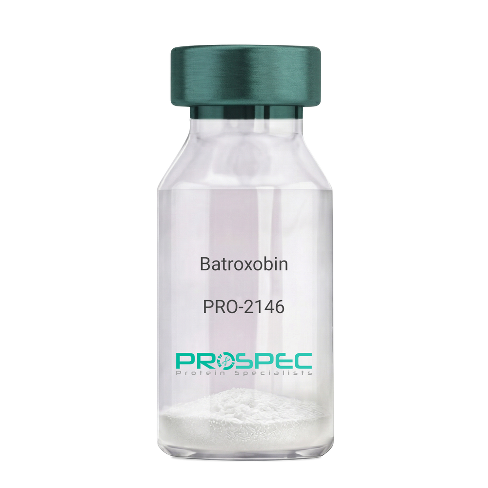
    Batroxobin

    Description:

    Batroxobin

    Thrombin-like enzyme batroxobin, EC 3.4.21.74, BX, Bothrops atrox serine proteinase, Venombin-A, Batroxobin.

    Product # :

    PRO-2146

    Price :

    Quantity :

    Shipping Method :

    Room Temp Icon

    Shipped at Room temp

    Add To Cart

    More Info

    • description
    • formulation
    • More Info

    Description

    Batroxobin, isolated from Bothrops atrox snake venom, has an Mw of approximately 43kDa.

    Formulation

    The Batroxobin protein was lyophilized from a concentrated (1mg/ml) solution with no additives.

    More Info

    • Introduction

      Batroxobin is a serin protease that reduces fibronogen levels and is originally extracted from snake venom of Bothrops Atrox. Batroxobin is used in defibrinogenation and thrombolysis and also has an effect on c-fos gene and growth factor.
      Batroxobin can efficiently restrain proliferation of VSMCs, by blocking the release and uptake of Ca2+, thus influencing [Ca2+]i.
      Batroxobin converts fibrinogen to fibrin through the restricted release of fibrinopeptide-A from fibrinogen to promote blood to clot. Unlike thrombin, it is not affected by heparin and hirudin.

    • Synonyms

      Thrombin-like enzyme batroxobin, EC 3.4.21.74, BX, Bothrops atrox serine proteinase, Venombin-A, Batroxobin.

    • Physical Appearance

      Sterile Filtered white lyophilized powder.

    • Stability

      Store the lyophilized Batroxobin between 2-8°C. Do not freeze!

    • Solubility

      It is recommended to reconstitute the lyophilized Batroxobin in sterile 18MΩ-cm H2O not less than 100µg/ml or more than 10mg/ml solutions.

    • Unit Definition

      100BU [Batroxobin Units]=1mg.

    ProSpec's products are furnished for LABORATORY RESEARCH USE ONLY. The product may not be used as drugs, agricultural or pesticidal products, food additives or household chemicals.

    Batroxobin Native
  • View Data Sheet

    Name :

    CTSW Human

    Description:

    Cathepsin-W Human Recombinant

    Cathepsin W (Lymphopain), LYPN, lymphopain, EC 3.4.22.-.

    Product # :

    ENZ-762

    Price :

    Quantity :

    Shipping Method :

    Ice Icon

    Shipped with Ice Packs

    Add To Cart

    More Info

    • description
    • source
    • formulation
    • purity
    • More Info

    Description

    CTSW Human Recombinant produced in E.coli is a single, non-glycosylated polypeptide chain containing 378 amino acids (22-376) and having a molecular mass of 42.0 kDa. CTSW is fused to a 23 amino acid His-tag at N-terminus.

    Source

    Escherichia Coli.

    Formulation

    The CTSW solution (0.5mg/ml) contains 20mM Tris-HCl buffer (pH 8.0), 0.4M Urea and 10% glycerol.

    Purity

    Greater than 85% as determined by SDS-PAGE.

    More Info

    • Introduction

      CTSW protein belongs to the peptidase C1 family. CTSW is a cysteine proteinase with a detailed role in the regulation and mechanism of T-cell cytolytic activity. The encoded CTSW is linked to the membrane inside the endoplasmic reticulum of natural killer and cytotoxic T-cells. CTSW expression is up-regulated by interleukin-2.

    • Synonyms

      Cathepsin W (Lymphopain), LYPN, lymphopain, EC 3.4.22.-.

    • Physical Appearance

      Sterile Filtered clear solution.

    • Stability

      Store at 4°C if entire vial will be used within 2-4 weeks. Store, frozen at -20°C for longer periods of time. For long term storage it is recommended to add a carrier protein (0.1% HSA or BSA).Avoid multiple freeze-thaw cycles.

    • Amino Acid Sequence

      MGSSHHHHHH SSGLVPRGSH MGSIRGPLRA QDLGPQPLEL KEAFKLFQIQ FNRSYLSPEE HAHRLDIFAH NLAQAQRLQE EDLGTAEFGV TPFSDLTEEE FGQLYGYRRA AGGVPSMGRE IRSEEPEESV PFSCDWRKVA GAISPIKDQK NCNCCWAMAA AGNIETLWRI SFWDFVDVSV QELLDCGRCG DGCHGGFVWD AFITVLNNSG LASEKDYPFQ GKVRAHRCHP KKYQKVAWIQ DFIMLQNNEH RIAQYLATYG PITVTINMKP LQLYRKGVIK ATPTTCDPQL VDHSVLLVGF GSVKSEEGIW AETVSSQSQP QPPHPTPYWI LKNSWGAQWG EKGYFRLHRG SNTCGITKFP LTARVQKPDM KPRVSCPP.

    ProSpec's products are furnished for LABORATORY RESEARCH USE ONLY. The product may not be used as drugs, agricultural or pesticidal products, food additives or household chemicals.

    Ctsw Human
  • View Data Sheet

    Name :

    ELOB Mouse

    Description:

    Elongin B Mouse Recombinant

    Transcription elongation factor B polypeptide 2, TCEB2, RNA polymerase IItranscription factor SIII subunit B, SIII p18, EloB, Elongin 18 kDa subunit.

    Product # :

    PRO-2550

    Price :

    Quantity :

    Shipping Method :

    Ice Icon

    Shipped with Ice Packs

    Add To Cart

    More Info

    • description
    • source
    • formulation
    • purity
    • More Info

    Description

    ELOB Mouse Recombinant produced in E.Coli is a single, non-glycosylated polypeptide chain of 141 amino acids ( 1-118 a.a.) having a molecular mass of 15.6 kDa. The Recombinant Mouse ELOB is fused to a 23 amino acid His-tag at N-terminus & purified by proprietary chromatographic techniques.

    Source

    Escherichia Coli.

    Formulation

    The protein (1mg/ml) contains PBS pH-7.4 containing 1mM DTT and 10% Glycerol.

    Purity

    Greater than 95.0% as determined by SDS-PAGE.

    More Info

    • Introduction

      Elongin B (Elob) is a subunit of the transcription factor B (SIII) complex. SIII complex is a general transcription elongation factor that increases the RNA polymerase II transcription elongation past template-encoded arresting sites. The SIII complex comprised of a transcriptionally active subunit (A) and 2 regulatory subunits (B and C). Subunit A is transcriptionally active and its transcription activity is enhanced by binding to the dimeric complex of the SIII regulatory subunits B and C. The von Hippel-Lindau tumor suppressor protein binds to elongin B and C and inhibits transcription elongation.

    • Synonyms

      Transcription elongation factor B polypeptide 2, TCEB2, RNA polymerase IItranscription factor SIII subunit B, SIII p18, EloB, Elongin 18 kDa subunit.

    • Physical Appearance

      Sterile filtered colorless solution.

    • Stability

      Store at 4°C if entire vial will be used within 2-4 weeks. Store, frozen at -20°C for longer periods of time. For long term storage it is recommended to add a carrier protein (0.1% HSA or BSA). Avoid multiple freeze-thaw cycles.

    • Amino Acid Sequence

      MGSSHHHHHH SSGLVPRGSH MGSMDVFLMI RRHKTTIFTD AKESSTVFEL KRIVEGILKR PPEEQRLYKD DQLLDDGKTL GECGFTSQTA RPQAPATVGL AFRADDTFEA LRIEPFSSPP ELPDVMKPQD SGGSANEQAV Q

    ProSpec's products are furnished for LABORATORY RESEARCH USE ONLY. The product may not be used as drugs, agricultural or pesticidal products, food additives or household chemicals.

    Elongin B Human
  • View Data Sheet

    Name :

    CRABP1 Human

    Description:

    Cellular Retinoic Acid binding Protein 1 Human Recombinant

    Cellular retinoic acid-binding protein 1, Cellular retinoic acid-binding protein I, CRABP-I, CRABP1, RBP5, CRABP, CRABPI.

    Product # :

    PRO-710

    Price :

    Quantity :

    Shipping Method :

    Ice Icon

    Shipped with Ice Packs

    Add To Cart

    More Info

    • description
    • source
    • formulation
    • purity
    • More Info

    Description

    CRABP1 Human Recombinant produced in E.Coli is a single, non-glycosylated polypeptide chain containing 137 amino acids and having a molecular mass of 15.5kDa. The CRABP1 is purified by proprietary chromatographic techniques.

    Source

    Escherichia Coli.

    Formulation

    The CRABP1 (1mg/ml) protein solution contains 20mM Tris-HCl buffer (pH8.0) and 10% glycerol.

    Purity

    Greater than 95.0% as determined by SDS-PAGE.

    More Info

    • Introduction

      CRABP1 is a member of special carrier proteins for members of the vitamin A family. It is believed that CRABP1 has an essential role in retinoic acid-mediated differentiation and proliferation processes. Though, CRABP1 is structurally similar to the cellular retinol-binding proteins, it binds only retinoic acid at specific sites within the nucleus, which may contribute to vitamin A-directed differentiation in epithelial tissue. CRABP1 is constitutively expressed and is thought to have different functions in the cell than the related CRABP2. CRABP1 forms a beta-barrel structure which accommodates hydrophobic ligands in its interior.
      Loss of CRABP1 function as a result of hypermethylation of its promoter leads to pathogenesis of papillary thyroid carcinoma. Furthermore, frequent methylation-associated silencing of CRABP1 is linked to esophageal squamous-cell carcinoma.

    • Synonyms

      Cellular retinoic acid-binding protein 1, Cellular retinoic acid-binding protein I, CRABP-I, CRABP1, RBP5, CRABP, CRABPI.

    • Physical Appearance

      Sterile Filtered colorless solution.

    • Stability

      Store at 4°C if entire vial will be used within 2-4 weeks. Store, frozen at -20°C for longer periods of time. For long term storage it is recommended to add a carrier protein (0.1% HSA or BSA).Avoid multiple freeze-thaw cycles.

    • Amino Acid Sequence

      MPNFAGTWKM RSSENFDELL KALGVNAMLR KVAVAAASKP HVEIRQDGDQ FYIKTSTTVR TTEINFKVGE GFEEETVDGR KCRSLATWEN ENKIHCTQTL LEGDGPKTYW TRELANDELI LTFGADDVVC TRIYVRE.

    ProSpec's products are furnished for LABORATORY RESEARCH USE ONLY. The product may not be used as drugs, agricultural or pesticidal products, food additives or household chemicals.

    Crabp1 Human
  • View Data Sheet

    Name :

    RELT Human

    Description:

    RELT Human Recombinant

    RELT, TNF Receptor, Receptor Expressed In Lymphoid Tissues, Tumor Necrosis Factor Receptor Superfamily Member 19L, RELT Tumor Necrosis Factor Receptor, TNFRSF19L, Tumor Necrosis Factor Receptor Superfamily, Member 19-Like, TRLT.

    Product # :

    CYT-1101

    Price :

    Quantity :

    Shipping Method :

    Ice Icon

    Shipped with Ice Packs

    Add To Cart

    More Info

    • description
    • source
    • formulation
    • purity
    • More Info

    Description

    RELT produced in Sf9 Baculovirus cells is a single, glycosylated polypeptide chain containing 376 amino acids (26-162a.a.) and having a molecular mass of 41.4kDa. (Molecular size on SDS-PAGE will appear at approximately 40-57kDa).RELT is expressed with a 239 amino acid hIgG-His-tag at C-Terminus and purified by proprietary chromatographic techniques.

    Source

    Sf9, Baculovirus cells.

    Formulation

    RELT protein solution (0.5mg/ml) contains 10% glycerol & Phosphate Buffered Saline (pH 7.4).

    Purity

    Greater than 90% as determined by SDS-PAGE.

    More Info

    • Introduction

      RELTis expressed in hematopoietic tissues and peripheral blood leukocytesand is a part of thetumor necrosis factor receptor superfamily. RELT mediates activation of NF-kappa-B and takes part in T-cell activation.overexpression of RELT in HEK-293 cells induces p38 and JNK signalling and leads to apoptosis. it can also costimulate T-cell proliferation in the presence of CD3 signalling.

    • Synonyms

      RELT, TNF Receptor, Receptor Expressed In Lymphoid Tissues, Tumor Necrosis Factor Receptor Superfamily Member 19L, RELT Tumor Necrosis Factor Receptor, TNFRSF19L, Tumor Necrosis Factor Receptor Superfamily, Member 19-Like, TRLT.

    • Physical Appearance

      Sterile filtered colorless solution.

    • Stability

      Store at 4°C if entire vial will be used within 2-4 weeks. Store, frozen at -20°C for longer periods of time. For long term storage it is recommended to add a carrier protein (0.1% HSA or BSA).Avoid multiple freeze-thaw cycles.

    • Amino Acid Sequence

      STTLWQCPPG EEPDLDPGQG TLCRPCPPGT FSAAWGSSPC QPHARCSLWR RLEAQVGMAT
      RDTLCGDCWP GWFGPWGVPR VPCQPCSWAPLGTHGCDEWG RRARRGVEVA AGASSGGETR QPGNGTRAGG PEETAAQVEP KSCDKTHTCP PCPAPELLGG PSVFLFPPKP KDTLMISRTP EVTCVVVDVS HEDPEVKFNW YVDGVEVHNA KTKPREEQYN STYRVVSVLT VLHQDWLNGK EYKCKVSNKA LPAPIEKTIS KAKGQPREPQ VYTLPPSRDE LTKNQVSLTC LVKGFYPSDI AVEWESNGQP ENNYKTTPPV LDSDGSFFLY SKLTVDKSRW QQGNVFSCSV MHEALHNHYT QKSLSLSPGK HHHHHH

    ProSpec's products are furnished for LABORATORY RESEARCH USE ONLY. The product may not be used as drugs, agricultural or pesticidal products, food additives or household chemicals.

    Relt Human
  • View Data Sheet

    Name :

    Midkine Mouse

    Description:

    Midkine Mouse Recombinant

    NEGF-2, Neurite Growth-Promoting Factor 2, MK, Neurite outgrowth-promoting protein, Midgestation and kidney protein, Amphiregulin-associated protein, ARAP, Neurite outgrowth-promoting factor 2, FLJ27379, Midkine, MK1, NEGF2.

    Product # :

    CYT-178

    Price :

    Quantity :

    Shipping Method :

    Room Temp Icon

    Shipped at Room temp

    Add To Cart

    More Info

    • description
    • source
    • formulation
    • purity
    • biological activity
    • More Info

    Description

    Midkine Mouse Recombinant produced in E.Coli is a single, non-glycosylated, polypeptide chain containing 120 amino acids and having a molecular mass of 13.3kDa.The Midkine Mouse is purified by proprietary chromatographic techniques.

    Source

    Escherichia Coli.

    Formulation

    Lyophilized from a 0.2um filtered concentrated solution in PBS, pH 7.4.

    Purity

    Greater than 95.0% as determined by HPLC and SDS-PAGE.

    Biological Activity

    Fully biologically active when compared to standard. Determined by its ability to chemoattract human neutrophils using a concentration range of 10-100 ng/ml corresponding to a specific activity of 10,000-100,000IU/mg.

    More Info

    • Introduction

      Midkine (MK) is the product of a retinoic acid responsive gene, MK, and is a member of a family of heparin binding factors. It contains 121 amino acid residues including 10 conserved cysteine residues, all of which appear to be disulphide linked.
      Midkine is expressed during embryogenesis, showing an expression pattern that suggests functions in neurogenesis, cell migration, secondary organogenetic induction, and mesoderm-epithelial interaction.
      The widespread downregulation of MK in the adult human is reverted in a number of cancers, in which polypeptides are able to act as both transforming growth factors and promoters of angiogenesis.
      Midkine (MK), induces chemotaxis of human neutrophils and was found to trigger mobilization of intracellular calcium of these cells.
      Midkine induces histamine release from rat peritoneal mast cells with a rapid response in a dose dependent manner.
      Midkine is also a potent stimulator of collagen and glycosaminoglycan synthesis.

    • Synonyms

      NEGF-2, Neurite Growth-Promoting Factor 2, MK, Neurite outgrowth-promoting protein, Midgestation and kidney protein, Amphiregulin-associated protein, ARAP, Neurite outgrowth-promoting factor 2, FLJ27379, Midkine, MK1, NEGF2.

    • Physical Appearance

      Sterile Filtered White lyophilized (freeze-dried) powder.

    • Stability

      Lyophilized Midkine Mouse although stable at room temperature for 3 weeks, should be stored desiccated below -18°C. Upon reconstitution Midkine Mouse should be stored at 4°C between 2-7 days and for future use below -18°C.Please prevent freeze-thaw cycles.

    • Solubility

      It is recommended to reconstitute the lyophilized Midkine Mouse in sterile 18M-cm H2O not less than 100µg/ml, which can then be further diluted to other aqueous solutions.

    • Amino Acid Sequence

      VAKKKEKVKK GSECSEWTWG PCTPSSKDCG MGFREGTCGA QTQRVHCKVP CNWKKEFGAD CKYKFESWGA CDGSTGTKAR QGTLKKARYN AQCQETIRVT KPCTSKTKSK TKAKKGKGKD

    ProSpec's products are furnished for LABORATORY RESEARCH USE ONLY. The product may not be used as drugs, agricultural or pesticidal products, food additives or household chemicals.

    Midkine Mouse
  • View Data Sheet

    Name :

    S100A8 Human

    Description:

    S100 Calcium Binding Protein A8 Human Recombinant

    Calgranulin A, MRP8, CAGA, CGLA, CFAG, Protein S100-A8, S100 calcium-binding protein A8, Migration inhibitory factor-related protein 8, MRP-8, p8, Cystic fibrosis antigen, Leukocyte L1 complex light chain, Calprotectin L1L subunit, Urinary stone protein band A, S100A8, MIF, NIF, L1Ag, CP-10, MA387, 60B8AG.

    Product # :

    PRO-800

    Price :

    Quantity :

    Shipping Method :

    Ice Icon

    Shipped with Ice Packs

    Add To Cart

    More Info

    • description
    • source
    • formulation
    • purity
    • More Info

    Description

    S100A8 Human Recombinant produced in E.Coli is a single, non-glycosylated, polypeptide chain containing 93 amino acids (1-93 a.a.) and having a molecular mass of 10.8 kDa. The S100A8 is purified by proprietary chromatographic techniques.

    Source

    Escherichia Coli.

    Formulation

    The S100A8 solution (0.5mg/ml) contains 20mM Tris-HCl pH-8, 1mM DTT and 10% glycerol.

    Purity

    Greater than 90% as determined by SDS-PAGE.

    More Info

    • Introduction

      S100A8 is a part of the S100 family of proteins containing 2 EF-hand calcium-binding motifs. S100 proteins are localized in the cytoplasm and/or nucleus of a broad range of cells, and participate in the regulation of cellular processes such as cell cycle progression and differentiation. S100A8 plays a role in the inhibition of casein kinase and as a cytokine. S100A8 altered expression is related with cystic fibrosis disease. S100A8 is a calcium-binding protein that has antimicrobial activity against bacteria and fungi.S100A8 is crucial for resistance towards invasion by pathogenic bacteria. S100A8 up-regulates transcription of genes that are under the control of NF-kappa-B. S100A8 plays a role in the development of endotoxic shock in response to bacterial lipopolysaccharide. S100A8 endorses tubulin polymerization and promotes phagocyte migration and infiltration of granulocytes at sites of wounding. S100A8 takes part as a pro-inflammatory mediator in acute and chronic inflammation and up-regulates the release of IL8 and cell-surface expression of ICAM1.

    • Synonyms

      Calgranulin A, MRP8, CAGA, CGLA, CFAG, Protein S100-A8, S100 calcium-binding protein A8, Migration inhibitory factor-related protein 8, MRP-8, p8, Cystic fibrosis antigen, Leukocyte L1 complex light chain, Calprotectin L1L subunit, Urinary stone protein band A, S100A8, MIF, NIF, L1Ag, CP-10, MA387, 60B8AG.

    • Physical Appearance

      Sterile Filtered clear colorless solution.

    • Stability

      Store at 4°C if entire vial will be used within 2-4 weeks.Store, frozen at -20°C for longer periods of time. For long term storage it is recommended to add a carrier protein (0.1% HSA or BSA).Avoid multiple freeze-thaw cycles.

    • Amino Acid Sequence

      MLTELEKALN SIIDVYHKYS LIKGNFHAVY RDDLKKLLET ECPQYIRKKG ADVWFKELDI NTDGAVNFQE FLILVIKMGV AAHKKSHEES HKE.

    ProSpec's products are furnished for LABORATORY RESEARCH USE ONLY. The product may not be used as drugs, agricultural or pesticidal products, food additives or household chemicals.

    S100A8 Human
  • View Data Sheet

    Name :

    CUTC Human

    Description:

    cutC Copper Transporter Homolog Human Recombinant

    Copper homeostasis protein cutC homolog, CUTC, CGI-32, RP11-483F11.3.

    Product # :

    PRO-911

    Price :

    Quantity :

    Shipping Method :

    Ice Icon

    Shipped with Ice Packs

    Add To Cart

    More Info

    • description
    • source
    • formulation
    • purity
    • More Info

    Description

    CUTC Human Recombinant produced in E.Coli is a single, non-glycosylated polypeptide chain containing 293 amino acids (1-273 a.a) and having a molecular mass of 31.5kDa.CUTC is fused to a 20 amino acid His-tag at N-terminus & purified by proprietary chromatographic techniques.

    Source

    Escherichia Coli.

    Formulation

    CUTC protein solution (0.5mg/ml) containing 20mM Tris-HCl buffer (pH 8.0), 5mM DTT, 10% glycerol and 200mM NaCl.

    Purity

    Greater than 95.0% as determined by SDS-PAGE.

    More Info

    • Introduction

      CUTC belongs to the Cut family and may be involved in efflux trafficking of cuprous ion. The Cut family is linked with the copper homeostasis and involved in several vital metabolisms, such as uptake, storage, delivery, and efflux of copper. Copper which is an essential heavy metal trace element has an imperative role in cell physiology.

    • Synonyms

      Copper homeostasis protein cutC homolog, CUTC, CGI-32, RP11-483F11.3.

    • Physical Appearance

      Sterile filtered colorless solution.

    • Stability

      Store at 4°C if entire vial will be used within 2-4 weeks. Store, frozen at -20°C for longer periods of time. For long term storage it is recommended to add a carrier protein (0.1% HSA or BSA).Avoid multiple freeze-thaw cycles.

    • Amino Acid Sequence

      MGSSHHHHHH SSGLVPRGSH MKRQGASSER KRARIPSGKA GAANGFLMEV CVDSVESAVN AERGGADRIE LCSGLSEGGT TPSMGVLQVV KQSVQIPVFV MIRPRGGDFL YSDREIEVMK ADIRLAKLYG ADGLVFGALT EDGHIDKELC MSLMAICRPL PVTFHRAFDM VHDPMAALET LLTLGFERVL TSGCDSSALE GLPLIKRLIE QAKGRIVVMP GGGITDRNLQ RILEGSGATE FHCSARSTRD SGMKFRNSSV AMGASLSCSE YSLKVTDVTK VRTLNAIAKN ILV.

    ProSpec's products are furnished for LABORATORY RESEARCH USE ONLY. The product may not be used as drugs, agricultural or pesticidal products, food additives or household chemicals.

    Cutc Human
  • View Data Sheet

    Name :

    CXCL17 Human, His

    Description:

    VEGF Co-regulated Chemokine 1, His Tag Human Recombinant

    Dcip1, DMC, MGC138300, UNQ473, VCC-1, VCC1, VEGF coregulated chemokine 1, C-X-C motif chemokine 17, Dendritic cell and monocyte chemokine-like protein.

    Product # :

    CHM-024

    Price :

    Quantity :

    Shipping Method :

    Ice Icon

    Shipped with Ice Packs

    Add To Cart

    More Info

    • description
    • source
    • formulation
    • purity
    • More Info
    • SDS-PAGE

    Description

    CXCL17 Human Recombinant produced in E.Coli is a single, non-glycosylated polypeptide chain containing 119 amino acids (22-119 a.a) and having a molecular mass of 13.7kDa.CXCL17 is fused to a 21 amino acid His-tag at N-terminus & purified by proprietary chromatographic techniques.

    Source

    Escherichia Coli.

    Formulation

    CXCL17 protein solution (0.5mg/ml) contains 20mM Tris-HCl buffer (pH 8.0), 0.4M Urea and 10% glycerol.

    Purity

    Greater than 85.0% as determined by SDS-PAGE.

    SDS-PAGE

    CXCL17 Human, His-SDS-PAGE - Product image 1

    More Info

    • Introduction

      Dendritic cell and monocyte chemokinelike protein (DMC/CXCL17/VEGF-correlated chemokine 1/VCC1), is a secreted molecule with a size and predicted 3-dimensional folding pattern similar to that of chemokines CXCL8/IL8 and CXCL14/BRAK. CXCL17 is constitutively generated by airway and intestinal epithelium. CXCL17 induces the chemotaxis of quiescent, but not LPS-activated peripheral blood monocytes and dendritic cells, and it also binds these cells specifically. The expression of CXCL17 is increased in endothelial cells when they are induced to form tubes in vitro. CXCL17, CXCL1/GRO and CXCL8/IL8 which have roles in angiogenesis, show significantly correlated expression with that of VEGF in primary lung, breast and esophageal tumors. Therefore, CXCL17 is suggested to have a role in tumor angiogenesis. The mature Rat CXCL17 shares 82%, 71% amino acid sequence identity with mouse, human CXCL17, respectively.

    • Synonyms

      Dcip1, DMC, MGC138300, UNQ473, VCC-1, VCC1, VEGF coregulated chemokine 1, C-X-C motif chemokine 17, Dendritic cell and monocyte chemokine-like protein.

    • Physical Appearance

      Sterile Filtered clear solution.

    • Stability

      Store at 4°C if entire vial will be used within 2-4 weeks. Store, frozen at -20°C for longer periods of time. For long term storage it is recommended to add a carrier protein (0.1% HSA or BSA).Avoid multiple freeze-thaw cycles.

    • Amino Acid Sequence

      MGSSHHHHHH SSGLVPRGSH MSSLNPGVAR GHRDRGQASR RWLQEGGQEC ECKDWFLRAP RRKFMTVSGL PKKQCPCDHF KGNVKKTRHQ RHHRKPNKHS RACQQFLKQC QLRSFALPL.

    • Background

      What is the molecular weight/Mw of CXCL17 HUMAN, HIS Protein?
      CXCL17 HUMAN, HIS Protein has a total Mw of 13.7kDa.

      What is the source or expression system of CXCL17 HUMAN, HIS Protein?
      Escherichia Coli.

      What is the Purity of CXCL17 HUMAN, HIS Protein?
      CXCL17 HUMAN, HIS Protein is >85% pure as determined by SDS-PAGE.

      What is the Biological Activity of CXCL17 HUMAN, HIS Protein?
      The biological functionality of CXCL17 HUMAN, HIS Protein will be determined in the future.

      What is the amino acid sequence of CXCL17 HUMAN, HIS Protein?
      MGSSHHHHHH SSGLVPRGSH MSSLNPGVAR GHRDRGQASR RWLQEGGQEC ECKDWFLRAP RRKFMTVSGL PKKQCPCDHF KGNVKKTRHQ RHHRKPNKHS RACQQFLKQC QLRSFALPL.

      What applications can CXCL17 HUMAN, HIS Protein be used in?
      CXCL17 HUMAN, HIS Protein can probably be used in western blot, ELISA and Lateral Flow.

      What is the endotoxin level for CXCL17 HUMAN, HIS Protein?
      The endotoxin level is minimal, CXCL17 HUMAN, HIS Protein was purified using conventional chromatography techniques.


    ProSpec's products are furnished for LABORATORY RESEARCH USE ONLY. The product may not be used as drugs, agricultural or pesticidal products, food additives or household chemicals.

    Cxcl17 Human His
  • View Data Sheet

    Name :

    CYTH1 Human

    Description:

    Cytohesin 1 Human Recombinant

    Cytohesin 1, D17S811E, PH, SEC7 And Coiled-Coil Domain-Containing Protein 1, Pleckstrin Homology, Sec7 And Coiled-Coil Domains 1, SEC7 Homolog B2-1, PSCD1, SEC7, Homolog Of Secretory Protein SEC7, Cytoadhesin 1, CYTOHESIN-1, B2-1, Cytohesin-1.

    Product # :

    PRO-2215

    Price :

    Quantity :

    Shipping Method :

    Ice Icon

    Shipped with Ice Packs

    Add To Cart

    More Info

    • description
    • source
    • formulation
    • purity
    • More Info

    Description

    CYTH1 Human Recombinant produced in E.Coli is a single, non-glycosylated polypeptide chain containing 421 amino acids (1-398 a.a) and having a molecular mass of 48.8kDa. CYTH1 is fused to a 23 amino acid His-tag at N-terminus & purified by proprietary chromatographic techniques.

    Source

    E.coli.

    Formulation

    CYTH1 protein solution (0.5mg/ml) containing Phosphate buffered saline (pH7.4), 10% glycerol and 1mM DTT.

    Purity

    Greater than 85% as determined by SDS-PAGE.

    More Info

    • Introduction

      Cytohesin 1 also known as CYTH1 belongs to the PSCD family. CYTH1 is responsible for promoting guanine-nucleotide exchange on ARF1 and ARF5 and also promotes the activation of ARF factors by the replacement of GDP with GTP.

    • Synonyms

      Cytohesin 1, D17S811E, PH, SEC7 And Coiled-Coil Domain-Containing Protein 1, Pleckstrin Homology, Sec7 And Coiled-Coil Domains 1, SEC7 Homolog B2-1, PSCD1, SEC7, Homolog Of Secretory Protein SEC7, Cytoadhesin 1, CYTOHESIN-1, B2-1, Cytohesin-1.

    • Physical Appearance

      Sterile Filtered colorless solution.

    • Stability

      Store at 4°C if entire vial will be used within 2-4 weeks. Store, frozen at -20°C for longer periods of time. For long term storage it is recommended to add a carrier protein (0.1% HSA or BSA).Avoid multiple freeze-thaw cycles.

    • Amino Acid Sequence

      MGSSHHHHHH SSGLVPRGSH MGSMEEDDSY VPSDLTAEER QELENIRRRK QELLADIQRL KDEIAEVANE IENLGSTEER KNMQRNKQVA MGRKKFNMDP KKGIQFLIEN DLLKNTCEDI AQFLYKGEGL NKTAIGDYLG ERDEFNIQVL HAFVELHEFT DLNLVQALRQ FLWSFRLPGE AQKIDRMMEA FAQRYCQCNN GVFQSTDTCY VLSFAIIMLN TSLHNPNVKD KPTVERFIAM NRGINDGGDL PEELLRNLYE SIKNEPFKIP EDDGNDLTHT FFNPDREGWL LKLGGGRVKT WKRRWFILTD NCLYYFEYTT DKEPRGIIPL ENLSIREVED SKKPNCFELY IPDNKDQVIK ACKTEADGRV VEGNHTVYRI SAPTPEEKEE WIKCIKAAIS RDPFYEMLAA RKKKVSSTKR H.

    ProSpec's products are furnished for LABORATORY RESEARCH USE ONLY. The product may not be used as drugs, agricultural or pesticidal products, food additives or household chemicals.

    Cyth1 Human
  • View Data Sheet

    Name :

    APOD Human, HEK

    Description:

    Apolipoprotein-D Human Recombinant, HEK

    Apolipoprotein D, Apo-D, ApoD.

    Product # :

    CYT-768

    Price :

    Quantity :

    Shipping Method :

    Room Temp Icon

    Shipped at Room temp

    Add To Cart

    More Info

    • description
    • source
    • formulation
    • purity
    • More Info

    Description

    Apolipoprotein-D Human Recombinant produced in HEK cells is a single, glycosylated, polypeptide chain (aa 21-189) containing a total of 175 amino acids, having a molecular mass of 20.1kDa (calculated) and fused to a 6 aa His tag at C-Terminus.The Human APOD is purified by proprietary chromatographic techniques.

    Source

    HEK 293.

    Formulation

    Filtered (0.4µm) and lyophilized from 0.5mg/ml in 0.05M phosphate buffer and 0.075M NaCl, pH 7.4.

    Purity

    Greater than 80.0% as determined by SDS-PAGE.

    More Info

    • Introduction

      Apolipoprotein-D is mainly associated with high density lipoproteins in human plasma. Apolipoprotein-D is an atypical apolipoprotein and, based on its primary structure, Apolipoprotein-D is a member of the lipocalin family. Lipocalins adopt a beta-barrel tertiary structure and transport small hydrophobic ligands. Apolipoprotein-D binds cholesterol, progesterone, pregnenolone, bilirubin and arachidonic acid.
      Apolipoprotein-D is expressed in numerous tissues having high levels of expression in spleen, testes and brain. Apolipoprotein-D is present at high concentrations in the cyst fluid of women with gross cystic disease of the breast, a condition associated with increased risk of breast cancer. Apolipoprotein-D accumulates in regenerating peripheral nerves and in the cerebrospinal fluid of patients with neurodegenerative conditions, such as Alzheimer's disease. Apolipoprotein-D participates in maintenance and repair within the central and peripheral nervous systems. Apolipoprotein-D is a multi-ligand, multi-functional transporter and transports a ligand from 1 cell to another within an organ, scavenge a ligand within an organ for transport to the blood or could transport a ligand from the circulation to specific cells within a tissue.

    • Synonyms

      Apolipoprotein D, Apo-D, ApoD.

    • Physical Appearance

      Filtered White lyophilized (freeze-dried) powder.

    • Stability

      Store lyophilized protein at -20°C. Aliquot the product after reconstitution to avoid repeated freezing/thawing cycles. Reconstituted protein can be stored at 4°C for a limited period of time.

    • Solubility

      It is recommended to add deionized water to a working concentration of 0.5mg/ml and let the lyophilized pellet dissolve completely. Product is not sterile! Please filter the product by an appropriate sterile filter before using it in the cell culture.

    • Amino Acid Sequence

      QAFHLGKCPN PPVQENFDVN KYLGRWYEIE KIPTTFENGR CIQANYSLME NGKIKVLNQE LRADGTVNQI EGEATPVNLT EPAKLEVKFS WFMPSAPYWI LATDYENYAL VYSCTCIIQL FHVDFAWILA RNPNLPPETV DSLKNILTSN NIDVKKMTVT DQVNCPKLSH HHHHH.

    • Background

      What is the molecular weight/Mw of APO D Protein?
      APO D Protein has a total Mw of 20.1kDa.

      What is the source or expression system of APO D Protein?
      HEK 293.

      What is the Purity of APO D Protein?
      APO D Protein is >80% pure as determined by SDS-PAGE.

      What is the Biological Activity of APO D Protein?
      The biological functionality of APO D Protein will be determined in the future.

      What is the amino acid sequence of APO D Protein?
      QAFHLGKCPN PPVQENFDVN KYLGRWYEIE KIPTTFENGR CIQANYSLME NGKIKVLNQE LRADGTVNQI EGEATPVNLT EPAKLEVKFS WFMPSAPYWI LATDYENYAL VYSCTCIIQL FHVDFAWILA RNPNLPPETV DSLKNILTSN NIDVKKMTVT DQVNCPKLSH HHHHH.

      What applications can APO D Protein be used in?
      APO D Protein can probably be used in western blot, ELISA and Lateral Flow.

      What is the endotoxin level for APO D Protein?
      The endotoxin level is minimal, APO D Protein was purified using conventional chromatography techniques.

    ProSpec's products are furnished for LABORATORY RESEARCH USE ONLY. The product may not be used as drugs, agricultural or pesticidal products, food additives or household chemicals.

    Apod Human Hek
  • View Data Sheet

    Name :

    EMAP II Human

    Description:

    Endothelial-Monocyte Activating Polypeptide II Human Recombinant

    AIMP1, EMAP2, EMAP-2, EMAPII, SCYE1, Multisynthetase complex auxiliary component p43, Endothelial monocyte-activating polypeptide 2, EMAP-II, p43.

    Product # :

    CYT-607

    Price :

    Quantity :

    Shipping Method :

    Room Temp Icon

    Shipped at Room temp

    Add To Cart

    More Info

    • description
    • source
    • formulation
    • purity
    • biological activity
    • More Info

    Description

    EMAP-II Recombinant Human produced in E.Coli is a single, non-glycosylated polypeptide chain containing 166 amino acids and having a molecular mass of 18.3 kDa. The EMAP-II is purified by proprietary chromatographic techniques.

    Source

    Escherichia Coli.

    Formulation

    Lyophilized from a concentrated (1mg/ml) solution in water containing 20mM sodium Phosphate buffer pH=7.5 and 130mM sodium chloride.

    Purity

    Greater than 98.0% as determined by(a) Analysis by RP-HPLC.
    (b) Analysis by SDS-PAGE.

    Biological Activity

    Determined by the apoptotic effect on MCF-7 cells using a concentration of 20-30 ng/ml.

    More Info

    • Introduction

      EMAP-II also called SCYE1 is a tumor derived cytokine that plays a role in a wide variety of activities on endothelial cells, monocytes and neutrophils. EMAP-II inhibits endothelial cell proliferation, vasculogenesis, neovessel formation, and can induce apoptosis. It is also chemotactic towards neutrophils and monocytes and induces myeloperoxidase activity from neutrophils. EMAP-II clinical value is inhibiting angiogenesis of vascular beds and suppressing the growth of primary and secondary tumors with no affect to normal tissues. SCYE1is specifically induced by apoptosis, and it is involved in the control of angiogenesis, inflammation, and wound healing. The release of this SCYE1 renders the tumor-associated vasculature sensitive to tumor necrosis factor. The precursor protein is identical to the p43 subunit, which is associated with the multi-tRNA synthetase complex, and it modulates aminoacylation activity of tRNA synthetase in normal cells. EMAP-2 plays a role in in the stimulation of inflammatory responses after proteolytic cleavage in tumor cells.

    • Synonyms

      AIMP1, EMAP2, EMAP-2, EMAPII, SCYE1, Multisynthetase complex auxiliary component p43, Endothelial monocyte-activating polypeptide 2, EMAP-II, p43.

    • Physical Appearance

      Sterile Filtered White lyophilized (freeze-dried) powder.

    • Stability

      Lyophilized EMAP-II although stable at room temperature for 3 weeks, should be stored desiccated below -18°C. Upon reconstitution EMAP-II should be stored at 4°C between 2-7 days and for future use below -18°C.Please prevent freeze-thaw cycles.

    • Solubility

      It is recommended to reconstitute the lyophilized EMAP-II in sterile 18MΩ-cm H2O not less than 100µg/ml, which can then be further diluted to other aqueous solutions.

    • Amino Acid Sequence

      SKPIDVSRLD LRIGCIITAR KHPDADSLYV EEVDVGEIAP RTVVSGLVNH VPLEQM QNRM VILLCNLKPA KMRGVLSQAM VMCASSPEKI EILAPPNGSV PGDRITFDAF PGEPDKELNP KKKIWEQIQP DLHTNDECVA TYKGVPFEVK GKGVCRAQTM SNSGIK.

    • Background

      What is the molecular weight/Mw of EMAP II HUMAN Protein?
      EMAP II HUMAN Protein has a total Mw of 18.3kDa.

      What is the source or expression system of EMAP II HUMAN Protein?
      Escherichia Coli.

      What is the Purity of EMAP II HUMAN Protein?
      EMAP II HUMAN Protein is >98% pure as determined by SDS-PAGE.

      What is the Biological Activity of EMAP II HUMAN Protein?
      Determined by the apoptotic effect on MCF-7 cells using a concentration of 20-30 ng/ml.

      What is the amino acid sequence of EMAP II HUMAN Protein?
      SKPIDVSRLD LRIGCIITAR KHPDADSLYV EEVDVGEIAP RTVVSGLVNH VPLEQM QNRM VILLCNLKPA KMRGVLSQAM VMCASSPEKI EILAPPNGSV PGDRITFDAF PGEPDKELNP KKKIWEQIQP DLHTNDECVA TYKGVPFEVK GKGVCRAQTM SNSGIK.

      What applications can EMAP II HUMAN Protein be used in?
      EMAP II HUMAN Protein can probably be used in western blot, ELISA and Lateral Flow.

      What is the endotoxin level for EMAP II HUMAN Protein?
      The endotoxin level is minimal, EMAP II HUMAN Protein was purified using conventional chromatography techniques

    ProSpec's products are furnished for LABORATORY RESEARCH USE ONLY. The product may not be used as drugs, agricultural or pesticidal products, food additives or household chemicals.

    Emap Ii
  • View Data Sheet

    Name :

    Eotaxin Human

    Description:

    Eotaxin Human Recombinant (CCL11)

    Small inducible cytokine A11, CCL11, Eosinophil chemotactic protein, chemokine (C-C motif) ligand 11, SCYA11, MGC22554.

    Product # :

    CHM-256

    Price :

    Quantity :

    Shipping Method :

    Room Temp Icon

    Shipped at Room temp

    Add To Cart

    More Info

    • description
    • source
    • formulation
    • purity
    • biological activity
    • More Info

    Description

    Eotaxin Human Recombinant produced in E.Coli is a single, non-glycosylated polypeptide chain containing 74 amino acids and having a molecular mass of 8345.9 Dalton. The CCL11 is purified by proprietary chromatographic techniques.

    Source

    Escherichia Coli.

    Formulation

    Lyophilized from a 0.2µm filtered concentrated (1.0mg/ml) solution in 20mM PB, pH 7.4, 150mM NaCl.

    Purity

    Greater than 97.0% as determined by:
    (a) Analysis by RP-HPLC.
    (b) Analysis by SDS-PAGE.

    Biological Activity

    The activity is determined by the chemoattract of human PBE (peripheral blood eosinophils) at a concentration between 0.1-10 ng/ml corresponding to a Specific Activity of 100,000-10,000,000IU/mg.

    More Info

    • Introduction

      Chemokine (C-C motif) ligand 11 (CCL11) is a small cytokine belonging to the CC chemokine family that is also known as eotaxin. CCL11 selectively recruits eosinophils by inducing their chemotaxis, and therefore, is implicated in allergic responses. The effects of CCL11 are mediated by its binding to a G-protein-linked receptor known as a chemokine receptor. Chemokine receptors for which CCL11 is a ligand include CCR2, CCR3 and CCR5. The gene for human CCL11 (scya11) is encoded on three exons and is located on chromosome 17.

    • Synonyms

      Small inducible cytokine A11, CCL11, Eosinophil chemotactic protein, chemokine (C-C motif) ligand 11, SCYA11, MGC22554.

    • Physical Appearance

      Sterile Filtered White lyophilized (freeze-dried) powder.

    • Stability

      Lyophilized Eotaxin although stable at room temperature for 3 weeks, should be stored desiccated below -18°C. Upon reconstitution CCL11 should be stored at 4°C between 2-7 days and for future use below -18°C.For long term storage it is recommended to add a carrier protein (0.1% HSA or BSA).Please prevent freeze-thaw cycles.

    • Solubility

      It is recommended to reconstitute the lyophilized Eotaxin Human Recombinant in sterile 18MΩ-cm H2O not less than 100µg/ml, which can then be further diluted to other aqueous solutions.

    • Amino Acid Sequence

      GPASVPTTCC FNLANRKIPL QRLESYRRIT SGKCPQKAVI FKTKLAKDICADPKKKWVQD

      SMKYLDQKSP TPKP.

    • Background

      What is the molecular weight/Mw of EOTAXIN HUMAN Protein?
      EOTAXIN HUMAN Protein has a total Mw of 8.3459kDa.

      What is the source or expression system of EOTAXIN HUMAN Protein?
      Escherichia Coli.

      What is the Purity of EOTAXIN HUMAN Protein?
      EOTAXIN HUMAN Protein is >97% pure as determined by SDS-PAGE.

      What is the Biological Activity of EOTAXIN HUMAN Protein?
      The activity is determined by the chemoattract of human PBE (peripheral blood eosinophils) at a concentration between 0.1-10 ng/ml corresponding to a Specific Activity of 100,000-10,000,000IU/mg.

      What is the amino acid sequence of EOTAXIN HUMAN Protein?
      GPASVPTTCC FNLANRKIPL QRLESYRRIT SGKCPQKAVI FKTKLAKDICADPKKKWVQD
      SMKYLDQKSP TPKP.

      What applications can EOTAXIN HUMAN Protein be used in?
      EOTAXIN HUMAN Protein can probably be used in western blot, ELISA and Lateral Flow.

      What is the endotoxin level for EOTAXIN HUMAN Protein?
      The endotoxin level is minimal, EOTAXIN HUMAN Protein was purified using conventional chromatography techniques.


    ProSpec's products are furnished for LABORATORY RESEARCH USE ONLY. The product may not be used as drugs, agricultural or pesticidal products, food additives or household chemicals.

    Eotaxin Human
  • View Data Sheet

    Name :

    PBLD Human

    Description:

    Phenazine Biosynthesis-Like Protein Domain Containing Human Recombinant

    Phenazine biosynthesis-like domain-containing protein, MAWD-binding protein, Unknown protein 32 from 2D-page of liver tissue, PBLD, MAWBP, MAWDBP, FLJ14767, FLJ35507.

    Product # :

    PRO-010

    Price :

    Quantity :

    Shipping Method :

    Ice Icon

    Shipped with Ice Packs

    Add To Cart

    More Info

    • description
    • source
    • formulation
    • purity
    • More Info

    Description

    PBLD Human Recombinant fused with a 20 amino acid His tag at N-terminus produced in E.Coli is a single, non-glycosylated, polypeptide chain containing 308 amino acids (1-288 a.a.) and having a molecular mass of 33.9kDa. The PBLD is purified by proprietary chromatographic techniques.

    Source

    Escherichia Coli.

    Formulation

    The PBLD solution (1 mg/ml) contains 20mM Tris-HCl buffer(pH 8.0), 10% glycerol, 2mM DTT and 0.1M NaCl.

    Purity

    Greater than 95.0% as determined by SDS-PAGE.

    More Info

    • Introduction

      PBLD is member of the phenazine biosynthesis-like protein (PhzF) family. PBLD which is expressed in most tissues is the only representative of the PhzF family in the human genome. PBLD participates in the MAPK signaling pathway. PBLD is involved in multiple basic cellular functions, its expression is elevated in several disease processes, including folate deficiency and hypotension.

    • Synonyms

      Phenazine biosynthesis-like domain-containing protein, MAWD-binding protein, Unknown protein 32 from 2D-page of liver tissue, PBLD, MAWBP, MAWDBP, FLJ14767, FLJ35507.

    • Physical Appearance

      Sterile Filtered colorless solution.

    • Stability

      Store at 4°C if entire vial will be used within 2-4 weeks. Store, frozen at -20°C for longer periods of time. For long term storage it is recommended to add a carrier protein (0.1% HSA or BSA).Avoid multiple freeze-thaw cycles.

    • Amino Acid Sequence

      MGSSHHHHHH SSGLVPRGSH MKLPIFIADA FTARAFRGNP AAVCLLENEL DEDMHQKIAR EMNLSETAFI RKLHPTDNFA QSSCFGLRWF TPASEVPLCG HATLASAAVL FHKIKNMNST LTFVTLSGEL RARRAEDGIV LDLPLYPAHP QDFHEVEDLI KTAIGNTLVQ DICYSPDTQK LLVRLSDVYN RSFLENLKVN TENLLQVENT GKVKGLILTL KGEPGGQTQA FDFYSRYFAP WVGVAEDPVT GSAHAVLSSY WSQHLGKKEM HAFQCSHRGG ELGISLRPDG RVDIRGGAAV VLEGTLTA.

    ProSpec's products are furnished for LABORATORY RESEARCH USE ONLY. The product may not be used as drugs, agricultural or pesticidal products, food additives or household chemicals.

    Pbld Human
  • View Data Sheet

    Name :

    BST1 Human

    Description:

    Bone Marrow Stromal Cell Antigen 1 Human Recombinant

    Bone Marrow Stromal Cell Antigen 1, ADP-Ribosyl Cyclase 2, Bone Marrow Stromal Antigen 1, Cyclic ADP-Ribose Hydrolase 2, NAD(+) Nucleosidase, CADPr Hydrolase 2, ADP-Ribosyl Cyclase/Cyclic ADP-Ribose Hydrolase 2, CD157 Antigen, EC 3.2.2.6, CD157, BST-1, ADP-ribosyl cyclase/cyclic ADP-ribose hydrolase 2, ADP-ribosyl cyclase 2, Bone marrow stromal antigen 1, Cyclic ADP-ribose hydrolase 2, cADPr hydrolase 2.

    Product # :

    CYT-1071

    Price :

    Quantity :

    Shipping Method :

    Ice Icon

    Shipped with Ice Packs

    Add To Cart

    More Info

    • description
    • source
    • formulation
    • purity
    • More Info

    Description

    BST1 produced in Sf9 Baculovirus cells is a single, glycosylated polypeptide chain containing 267 amino acids (33-293a.a.) and having a molecular mass of 30.5kDa. (Molecular size on SDS-PAGE will appear at approximately 40-57kDa).BST1 is expressed with a 6 amino acid His tag at C-Terminus and purified by proprietary chromatographic techniques.

    Source

    Sf9, Baculovirus cells.

    Formulation

    BST1 protein solution (0.5mg/ml) contains 10% glycerol & Phosphate Buffered Saline (pH 7.4).

    Purity

    Greater than 95.0% as determined by SDS-PAGE.

    More Info

    • Introduction

      BST1 (Bone Marrow Stromal Cell Antigen 1), is a GPI (glycosylphosphatidylinositol) anchored membrane protein which is part of the CD38 family. BST1 was initially recognized as a bone marrow stromal cell molecule. BST1 is an ectoenzyme sharing more than a few features with ADP-ribosyl cyclase CD38. BST1 together with CD38, exhibit both DP-ribosyl cyclase and cyclinc ADP ribose hydrolase activities. BST1 participates in rheumatoid arthritis due to its enhanced expression in RA-derived bone marrow stromal cell lines. Moreover, BST1 is expressed by cells of the myeloid lineage and could perform as a receptor with a signal transduction capability.

    • Synonyms

      Bone Marrow Stromal Cell Antigen 1, ADP-Ribosyl Cyclase 2, Bone Marrow Stromal Antigen 1, Cyclic ADP-Ribose Hydrolase 2, NAD(+) Nucleosidase, CADPr Hydrolase 2, ADP-Ribosyl Cyclase/Cyclic ADP-Ribose Hydrolase 2, CD157 Antigen, EC 3.2.2.6, CD157, BST-1, ADP-ribosyl cyclase/cyclic ADP-ribose hydrolase 2, ADP-ribosyl cyclase 2, Bone marrow stromal antigen 1, Cyclic ADP-ribose hydrolase 2, cADPr hydrolase 2.

    • Physical Appearance

      Sterile filtered colorless solution.

    • Stability

      Store at 4°C if entire vial will be used within 2-4 weeks. Store, frozen at -20°C for longer periods of time. For long term storage it is recommended to add a carrier protein (0.1% HSA or BSA).Avoid multiple freeze-thaw cycles.

    • Amino Acid Sequence

      RWRGEGTSAH LRDIFLGRCA EYRALLSPEQ RNKNCTAIWE AFKVALDKDP CSVLPSDYDL FINLSRHSIP RDKSLFWENS HLLVNSFADN TRRFMPLSDV LYGRVADFLS WCRQKNDSGL DYQSCPTSED CENNPVDSFW KRASIQYSKD SSGVIHVMLN GSEPTGAYPI KGFFADYEIP NLQKEKITRI EIWVMHEIGG PNVESCGEGS MKVLEKRLKD MGFQYSCIND YRPVKLLQCV DHSTHPDCAL KSAAAATQRK AHHHHHH.

    • Background

      The Emerging Role of Bone Marrow Stromal Cell Antigen 1 Human Recombinant in the Theater of Regenerative Medicine

      Introduction

      In the ever-evolving panorama of medical science, regenerative medicine is graduating from a fantastical dream into an operational reality. Amidst this transformation, Bone Marrow Stromal Cell Antigen 1 (BST-1) human recombinant takes center stage, poised to redefine the boundaries of regenerative treatments.

      BST-1: A Versatile Player

      BST-1, fondly known as CD157, is a familiar actor on the cellular stage, choreographing the ballet of monocyte differentiation and survival. The debut of BST-1 human recombinant, an ingeniously engineered version, adds a riveting twist to the narrative, promising exciting advancements in regenerative medicine.

      Engineering a Cellular Conductor

      With E. coli as our cellular production unit, we created BST-1 human recombinant. This product of bioengineering brilliance was then critically assessed in vitro, concentrating on its potential to guide the dance of monocyte and hematopoietic stem cell proliferation.

      Entering the Biological Stage

      Moving from the controlled in vitro environment, we ventured into a more complex, in vivo study with a mouse model. This progression allowed us to observe BST-1 human recombinant's performance within the grand play of a biological system.

      An Enthusiastic Applause for Results

      Our exploratory journey, spanning the laboratory and the biological stage, unveiled encouraging results. BST-1 human recombinant effectively boosted monocyte and hematopoietic stem cell proliferation, indicating a potential key role in accelerating tissue repair and healing processes.

      Conclusion

      The unfolding narrative of BST-1 human recombinant inspires hope for a bright future in regenerative medicine. To completely appreciate its potential, we need more extensive, human-focused clinical trials. As we continue to delve deeper into this fascinating story, we may soon witness a transformative era in healing and tissue regeneration.

      What is the molecular weight/Mw of BST1 Protein?
      BST1 Protein has a total Mw of 30.5kDa.

      What is the source or expression system of BST1 Protein?
      Sf9, Baculovirus cells.

      What is the Purity of BST1 Protein?
      BST1 Protein is >95% pure as determined by SDS-PAGE.

      What is the Biological Activity of BST1 Protein?
      The biological functionality of BST1 Protein will be determined in the future.

      What is the amino acid sequence of BST1 Protein?
      RWRGEGTSAH LRDIFLGRCA EYRALLSPEQ RNKNCTAIWE AFKVALDKDP CSVLPSDYDL FINLSRHSIP RDKSLFWENS HLLVNSFADN TRRFMPLSDV LYGRVADFLS WCRQKNDSGL DYQSCPTSED CENNPVDSFW KRASIQYSKD SSGVIHVMLN GSEPTGAYPI KGFFADYEIP NLQKEKITRI EIWVMHEIGG PNVESCGEGS MKVLEKRLKD MGFQYSCIND YRPVKLLQCV DHSTHPDCAL KSAAAATQRK AHHHHHH.

      What applications can BST1 Protein be used in?
      BST1 Protein can probably be used in western blot, ELISA and Lateral Flow.

      What is the endotoxin level for BST1 Protein?
      The endotoxin level is minimal, BST1 Protein was purified using conventional chromatography techniques.

    ProSpec's products are furnished for LABORATORY RESEARCH USE ONLY. The product may not be used as drugs, agricultural or pesticidal products, food additives or household chemicals.

    Bst1 Human
  • View Data Sheet

    Name :

    FABP6 Human, His

    Description:

    Fatty Acid Binding Protein 6 Human Recombinant, His Tag

    I-BABP, ILBP, I-15P, I-BAP, ILBP3, ILLBP, I-BABP, I-BALB, FABP-6, Gastrotropin, Ileal lipid-binding protein, Intestinal 15 kDa protein, Intestinal bile acid-binding protein, Fatty acid-binding protein 6, FABP6.

    Product # :

    PRO-667

    Price :

    Quantity :

    Shipping Method :

    Ice Icon

    Shipped with Ice Packs

    Add To Cart

    More Info

    • description
    • source
    • formulation
    • purity
    • More Info

    Description

    FABP6 Human Recombinant produced in E.Coli is a single, non-glycosylated polypeptide chain containing 128 amino acids and having a molecular mass of 18 kDa. FABP6 is fused to His tag at N-terminus and purified by standard chromatography techniques.

    Source

    Escherichia Coli.

    Formulation

    FABP6 His-Tag is supplied in 20mM Tris HCL pH=8, 0.5mM DTT and 50% glycerol.

    Purity

    Greater than 95% as determined by SDS-PAGE.

    More Info

    • Introduction

      FABP6 also called ileal fatty acid binding protein, is part of the small family of highly conserved, cytoplasmic proteins that bind long-chain fatty acids and other hydrophobic ligands. FABP6 cytosolic protein binds bile acid. FABP6 plays a role in fatty acid uptake, transport, and metabolism. FABP6 stimulates gastric acid and pepsinogen secretion. Seems to be able to bind to bile salts and bilirubins. FABP6 expression is restricted in the small intestine to the ileum where it is involved in the enterohepatic circulation of bile acids. Alternate transcription promoters generate 2 transcript variants, encoding a 128 aa and a 177 aa residue protein. Human FABP6 isoform 2 contains 128 amino acid residues and is acetylated on Ala2. FABP6 binds together fatty acids and bile acids and is directly involved in fatty acid transport and metabolism.

    • Synonyms

      I-BABP, ILBP, I-15P, I-BAP, ILBP3, ILLBP, I-BABP, I-BALB, FABP-6, Gastrotropin, Ileal lipid-binding protein, Intestinal 15 kDa protein, Intestinal bile acid-binding protein, Fatty acid-binding protein 6, FABP6.

    • Physical Appearance

      Sterile Filtered colorless liquid formulation.

    • Stability

      Store at 4°C if entire vial will be used within 2-4 weeks. Store, frozen at -20°C for longer periods of time.Please avoid freeze thaw cycles.

    ProSpec's products are furnished for LABORATORY RESEARCH USE ONLY. The product may not be used as drugs, agricultural or pesticidal products, food additives or household chemicals.

    Fabp6 Human His
  • 1
  • …
  • 25
  • 26
  • 27
  • 28
  • 29
  • …
  • 42
Your browser does not support the video tag.

Products

  • Cytokines
  • Growth Factors
  • Chemokines
  • CD Antigens
  • Neurotrophins
  • Hormones
  • Enzymes
  • Viral Antigens
  • Recombinant Proteins
  • Natural Proteins
  • Monoclonal Antibodies
  • Polyclonal Antibodies
  • Thermal Packaging

About

  • Company
  • Founder
  • Contribution
  • Contact Us
  • My Account

Ordering

  • Ordering Method
  • Ordering - Credit Card
  • Ordering PO
  • Shipping Fees
  • FAQ's

Legal

  • Terms & Conditions
  • Privacy
  • Site Map
  • info@prospecbio.com
  • Facebook
  • Instagram
  • Linkedin Profile

© 2026 ProSpec-Tany TechnoGene Ltd. All Rights Reserved.

  • Choosing a selection results in a full page refresh.
  • Opens in a new window.