Skip to content

High-quality research proteins.

  • 1-800-805-6531

  • Mon - Sat 8.00-18.00, Sun - Closed

  • info@prospecbio.com

  • PO Box 6591 East Brunswick NJ 08816

  • Products
    • Cytokines
    • Growth factors
    • Chemokines
    • Cd antigens
    • Neurotrophins
    • Hormones
    • Enzymes
    • Viral antigens
    • Recombinant proteins
    • Natural proteins
    • Monoclonal antibodies
    • Polyclonal antibodies
    • Thermal Packaging
  • About
    • Company
    • Founder
    • Contribution
  • Customers
    • Eastern Asia
    • Eastern Europe
    • North America
    • South America
    • South East Asia
    • Southern Asia
    • Western Europe
  • Ordering
    • Ordering Method
    • Ordering - Credit Card
    • Ordering - PO
    • Shipping Fees
    • FAQs
  • Services
    • DNA Cloning
    • Protein Expression
    • Monoclonal Antibody
    • Fermentation
    • Technology License
  • Contact us
Log in

Country/region

  • USD $ | Albania
  • USD $ | Andorra
  • USD $ | Argentina
  • USD $ | Armenia
  • USD $ | Australia
  • USD $ | Austria
  • USD $ | Azerbaijan
  • USD $ | Belarus
  • USD $ | Belgium
  • USD $ | Bolivia
  • USD $ | Brazil
  • USD $ | Bulgaria
  • USD $ | Cambodia
  • USD $ | Canada
  • USD $ | Chile
  • USD $ | China
  • USD $ | Colombia
  • USD $ | Costa Rica
  • USD $ | Croatia
  • USD $ | Cyprus
  • USD $ | Czechia
  • USD $ | Denmark
  • USD $ | Estonia
  • USD $ | Finland
  • USD $ | France
  • USD $ | Georgia
  • USD $ | Germany
  • USD $ | Greece
  • USD $ | Hong Kong SAR
  • USD $ | Hungary
  • USD $ | Iceland
  • USD $ | India
  • USD $ | Ireland
  • USD $ | Israel
  • USD $ | Italy
  • USD $ | Japan
  • USD $ | Kazakhstan
  • USD $ | Latvia
  • USD $ | Liechtenstein
  • USD $ | Lithuania
  • USD $ | Madagascar
  • USD $ | Malta
  • USD $ | Mexico
  • USD $ | Moldova
  • USD $ | Monaco
  • USD $ | Netherlands
  • USD $ | New Zealand
  • USD $ | North Macedonia
  • USD $ | Norway
  • USD $ | Panama
  • USD $ | Paraguay
  • USD $ | Peru
  • USD $ | Philippines
  • USD $ | Poland
  • USD $ | Portugal
  • USD $ | Romania
  • USD $ | Singapore
  • USD $ | Slovakia
  • USD $ | Slovenia
  • USD $ | South Africa
  • USD $ | South Korea
  • USD $ | Spain
  • USD $ | Sri Lanka
  • USD $ | Sweden
  • USD $ | Switzerland
  • USD $ | Taiwan
  • USD $ | Thailand
  • USD $ | Türkiye
  • USD $ | Ukraine
  • USD $ | United Kingdom
  • USD $ | United States
  • USD $ | Uruguay
  • USD $ | Vatican City
  • USD $ | Venezuela
  • USD $ | Vietnam
  • Facebook
  • Instagram
logoProSpec - Recombinant Protein Manufacturer for academic and R&D industry
Become a distributor
Account Log in
Wishlist
Cart Cart(0)
  • Products
    +
    • Cytokines
    • Growth factors
    • Chemokines
    • Cd antigens
    • Neurotrophins
    • Hormones
    • Enzymes
    • Viral antigens
    • Recombinant proteins
    • Natural proteins
    • Monoclonal antibodies
    • Polyclonal antibodies
    • Thermal Packaging
  • About
    +
    • Company
    • Founder
    • Contribution
  • Customers
    +
    • Eastern Asia
    • Eastern Europe
    • North America
    • South America
    • South East Asia
    • Southern Asia
    • Western Europe
  • Ordering
    +
    • Ordering Method
    • Ordering - Credit Card
    • Ordering - PO
    • Shipping Fees
    • FAQs
  • Services
    +
    • DNA Cloning
    • Protein Expression
    • Monoclonal Antibody
    • Fermentation
    • Technology License
  • Contact us

Item added to your cart

View cart
View All
  • Cytokines
  • Growth factors
  • Chemokines
  • Cd antigens
  • Neurotrophins
  • Hormones
  • Enzymes
  • Viral antigens
  • Recombinant proteins
  • Natural proteins
  • Monoclonal antibodies
  • Polyclonal antibodies
  • Thermal Packaging
  • Tumor Necrosis Factor

    Tumor Necrosis Factor

    View All
  • 4-1BB

    4-1BB

    View All
  • Adiponectin

    Adiponectin

    View All
  • AIF1

    AIF1

    View All
  • Other Cytokines

    Other Cytokines

    View All
  • AITRL

    AITRL

    View All
  • Angiopoietin

    Angiopoietin

    View All
  • Apolipoprotein

    Apolipoprotein

    View All
  • B-Cell Activating Factor

    B-Cell Activating Factor

    View All
  • Beta Defensin

    Beta Defensin

    View All
  • Bone Morphogenetic Protein

    Bone Morphogenetic Protein

    View All
  • B type Natriuretic Peptide

    B type Natriuretic Peptide

    View All
  • BST

    BST

    View All
  • Betacellulin

    Betacellulin

    View All
  • Cardiotrophin

    Cardiotrophin

    View All
  • Activin

    Activin

    View All
  • Fibroblast Growth Factor

    Fibroblast Growth Factor

    View All
  • Other Growth Factors

    Other Growth Factors

    View All
  • CSF

    CSF

    View All
  • CTGF

    CTGF

    View All
  • IGFBP

    IGFBP

    View All
  • VEGF Protein

    VEGF Protein

    View All
  • EGF

    EGF

    View All
  • Epigen

    Epigen

    View All
  • Erythropoietin

    Erythropoietin

    View All
  • Insulin-Like Growth Factor

    Insulin-Like Growth Factor

    View All
  • Growth Hormone

    Growth Hormone

    View All
  • HDGF

    HDGF

    View All
  • Hepatocyte Growth Factor

    Hepatocyte Growth Factor

    View All
  • Insulin

    Insulin

    View All
  • BCA-1/ BLC (CXCL13)

    BCA-1/ BLC (CXCL13)

    View All
  • BRAK (CXCL14)

    BRAK (CXCL14)

    View All
  • C-10 (CCL6)

    C-10 (CCL6)

    View All
  • Other Chemokines

    Other Chemokines

    View All
  • MEC (CCL28)

    MEC (CCL28)

    View All
  • LD78-beta (CCL3L1)

    LD78-beta (CCL3L1)

    View All
  • CTACK (CCL27)

    CTACK (CCL27)

    View All
  • CXCL16

    CXCL16

    View All
  • CXCL17

    CXCL17

    View All
  • Platelet Factor-4 (CXCL4)

    Platelet Factor-4 (CXCL4)

    View All
  • ENA-78 (CXCL5)

    ENA-78 (CXCL5)

    View All
  • Eotaxin (CCL11,24,26)

    Eotaxin (CCL11,24,26)

    View All
  • Exodus-2 (CCL21)

    Exodus-2 (CCL21)

    View All
  • Fractalkine (CX3CL1)

    Fractalkine (CX3CL1)

    View All
  • CXCL6

    CXCL6

    View All
  • Other CD Antigens

    Other CD Antigens

    View All
  • CD14

    CD14

    View All
  • CD164

    CD164

    View All
  • CD1

    CD1

    View All
  • CD2

    CD2

    View All
  • CD200

    CD200

    View All
  • CD207

    CD207

    View All
  • CD226

    CD226

    View All
  • CD23

    CD23

    View All
  • CD244

    CD244

    View All
  • CD247

    CD247

    View All
  • CD27

    CD27

    View All
  • CD274

    CD274

    View All
  • CD300

    CD300

    View All
  • CD33

    CD33

    View All
  • Other Neurotrophins

    Other Neurotrophins

    View All
  • Beta-NGF

    Beta-NGF

    View All
  • BDNF

    BDNF

    View All
  • CDNF

    CDNF

    View All
  • CNTF

    CNTF

    View All
  • GDNF

    GDNF

    View All
  • Glia Maturation Factor

    Glia Maturation Factor

    View All
  • MANF

    MANF

    View All
  • Midkine

    Midkine

    View All
  • Neuroglobin

    Neuroglobin

    View All
  • Neurotrophic factors

    Neurotrophic factors

    View All
  • Neuregulin

    Neuregulin

    View All
  • Neuropilin

    Neuropilin

    View All
  • Pigment Epithelium-Derived Factor

    Pigment Epithelium-Derived Factor

    View All
  • Pleiotrophin

    Pleiotrophin

    View All
  • Peptide Hormones

    Peptide Hormones

    View All
  • Other Hormones

    Other Hormones

    View All
  • HCG

    HCG

    View All
  • Endothelin

    Endothelin

    View All
  • Exendin

    Exendin

    View All
  • FSH

    FSH

    View All
  • Glucagon

    Glucagon

    View All
  • GHRP

    GHRP

    View All
  • GLP

    GLP

    View All
  • Thyrostimulin

    Thyrostimulin

    View All
  • Inhibin A

    Inhibin A

    View All
  • LHRH

    LHRH

    View All
  • Melanotan

    Melanotan

    View All
  • Procalcitonin

    Procalcitonin

    View All
  • PTH

    PTH

    View All
  • Peroxiredoxin

    Peroxiredoxin

    View All
  • Peptidase

    Peptidase

    View All
  • Synthetase

    Synthetase

    View All
  • Transferase

    Transferase

    View All
  • Hydrolase

    Hydrolase

    View All
  • Dehydrogenase

    Dehydrogenase

    View All
  • ACE2

    ACE2

    View All
  • Esterase

    Esterase

    View All
  • Protein Kinases

    Protein Kinases

    View All
  • Other Enzymes

    Other Enzymes

    View All
  • Phosphatase

    Phosphatase

    View All
  • Deaminase

    Deaminase

    View All
  • Oxygenase

    Oxygenase

    View All
  • Adenylate Kinase

    Adenylate Kinase

    View All
  • Lyase

    Lyase

    View All
  • Chagas

    Chagas

    View All
  • SARS

    SARS

    View All
  • Influenza

    Influenza

    View All
  • ASFV

    ASFV

    View All
  • Astrovirus

    Astrovirus

    View All
  • Borrelia

    Borrelia

    View All
  • Helicobacter Pylori

    Helicobacter Pylori

    View All
  • Chikungunya

    Chikungunya

    View All
  • Chlamydia

    Chlamydia

    View All
  • Cytomegalo

    Cytomegalo

    View All
  • Dengue

    Dengue

    View All
  • EBV

    EBV

    View All
  • Ebola

    Ebola

    View All
  • Feline Leukemia Virus

    Feline Leukemia Virus

    View All
  • Hepatitis A

    Hepatitis A

    View All
  • Other

    Other

    View All
  • Actin

    Actin

    View All
  • ADAM

    ADAM

    View All
  • Complement Component

    Complement Component

    View All
  • Ag85

    Ag85

    View All
  • Eukaryotic Translation Initiation Factor

    Eukaryotic Translation Initiation Factor

    View All
  • Anterior Gradient Protein

    Anterior Gradient Protein

    View All
  • Heat Shock Protein

    Heat Shock Protein

    View All
  • Alpha-2-HS-Glycoprotein

    Alpha-2-HS-Glycoprotein

    View All
  • Hemoglobin

    Hemoglobin

    View All
  • Allergy

    Allergy

    View All
  • Anaplasma

    Anaplasma

    View All
  • Angiogenin

    Angiogenin

    View All
  • Ankyrin Repeat Domain

    Ankyrin Repeat Domain

    View All
  • Annexin

    Annexin

    View All
  • Other Natural Proteins

    Other Natural Proteins

    View All
  • Aprotinin

    Aprotinin

    View All
  • Transferrin

    Transferrin

    View All
  • Avidin

    Avidin

    View All
  • Anti Coagulation Factors

    Anti Coagulation Factors

    View All
  • Natural Albumin

    Natural Albumin

    View All
  • Natural Coagulation Factors

    Natural Coagulation Factors

    View All
  • Fibronectin

    Fibronectin

    View All
  • Thrombin

    Thrombin

    View All
  • Anti Human Enzyme

    Anti Human Enzyme

    View All
  • Other Monoclonal Antibodies

    Other Monoclonal Antibodies

    View All
  • Anti Human Cytokine

    Anti Human Cytokine

    View All
  • Anti Human Heat Shock Protein

    Anti Human Heat Shock Protein

    View All
  • Anti Mouse Lymphocyte

    Anti Mouse Lymphocyte

    View All
  • Anti Human Chemokine

    Anti Human Chemokine

    View All
  • Anti Human Lymphocyte

    Anti Human Lymphocyte

    View All
  • Anti Viral Monoclonal

    Anti Viral Monoclonal

    View All
  • Anti Mouse Cytokine

    Anti Mouse Cytokine

    View All
  • Anti-GST Monoclonal

    Anti-GST Monoclonal

    View All
  • Other Polyclonal Antibodies

    Other Polyclonal Antibodies

    View All
  • Anti Viral

    Anti Viral

    View All
  • Anti-GST Monoclonal

    Anti-GST Monoclonal

    View All
Back

Search results

1000 results found for “ercc”

Name

Description

Product #

Price

Quantity

Shipping Method

  • View Data Sheet

    Name :

    LIN7C Human

    Description:

    LIN7C Human Recombinant

    LIN-7-C, LIN-7C, MALS-3, MALS3, VELI3, Lin-7 homolog C, Protein lin-7 homolog C, Mammalian lin-seven protein 3, MALS-3, Veli-3, LIN7C, Vertebrate lin-7 homolog 3.

    Product # :

    PRO-1305

    Price :

    Quantity :

    Shipping Method :

    Ice Icon

    Shipped with Ice Packs

    Add To Cart

    More Info

    • description
    • source
    • formulation
    • purity
    • More Info

    Description

    LIN7C Human Recombinant produced in E.Coli is a single, non-glycosylated polypeptide chain containing 220 amino acids (1-197 a.a.) and having a molecular mass of 24.2 kDa. LIN7C is fused to a 23 amino acid His-tag at N-terminus & purified by proprietary chromatographic techniques.

    Source

    Escherichia Coli.

    Formulation

    LIN7C protein solution (0.5mg/ml) contains 20mM Tris-HCl buffer (pH 8.0), 0.2M NaCl and 30% glycerol.

    Purity

    Greater than 90.0% as determined by SDS-PAGE

    More Info

    • Introduction

      LIN7C has a part in establishing and preserving the asymmetric distribution of channels and receptors at the plasma membrane of polarized cells. LIN7C forms membrane-associated multiprotein complexes which regulate distribution and recycling of proteins to the appropriate membrane domains.

    • Synonyms

      LIN-7-C, LIN-7C, MALS-3, MALS3, VELI3, Lin-7 homolog C, Protein lin-7 homolog C, Mammalian lin-seven protein 3, MALS-3, Veli-3, LIN7C, Vertebrate lin-7 homolog 3.

    • Physical Appearance

      Sterile filtered colorless solution.

    • Stability

      Store at 4°C if entire vial will be used within 2-4 weeks. Store, frozen at -20°C for longer periods of time. For long term storage it is recommended to add a carrier protein (0.1% HSA or BSA). Avoid multiple freeze-thaw cycles.

    • Amino Acid Sequence

      MGCSHHHHHH SSGLVPRGSH MGSMAALGEP VRLERDICRA IELLEKLQRS GEVPPQKLQA LQRVLQSEFC NAVREVYEHV YETVDISSSP EVRANATAKA TVAAFAASEG HSHPRVVELP KTEEGLGFNI MGGKEQNSPI YISRIIPGGI ADRHGGLKRG DQLLSVNGVS VEGEHHEKAV ELLKAAQGKV KLVVRYTPKV LEEMESRFEK MRSAKRRQQT.

    ProSpec's products are furnished for LABORATORY RESEARCH USE ONLY. The product may not be used as drugs, agricultural or pesticidal products, food additives or household chemicals.

    Lin7C Human
  • View Data Sheet

    Name :

    LXN Mouse

    Description:

    Latexin Mouse Recombinant

    Latexin, Endogenous carboxypeptidase inhibitor, ECI, Tissue carboxypeptidase inhibitor, TCI.

    Product # :

    PRO-2218

    Price :

    Quantity :

    Shipping Method :

    Ice Icon

    Shipped with Ice Packs

    Add To Cart

    More Info

    • description
    • source
    • formulation
    • purity
    • More Info

    Description

    LXN Mouse Recombinant produced in E.Coli is a single, non-glycosylated polypeptide chain containing 245 amino acids (1-222 a.a) and having a molecular mass of 27.9kDa. LXN is fused to a 23 amino acid His-tag at N-terminus & purified by proprietary chromatographic techniques.

    Source

    Escherichia Coli.

    Formulation

    LXN protein solution (1mg/ml) containing Phosphate Buffer Saline (pH7.4), 30% glycerol and 1mM DTT.

    Purity

    Greater than 95.0% as determined by SDS-PAGE.

    More Info

    • Introduction

      Lxn also known as Latexin is a member of the protease inhibitor I47 (latexin) family. Lxn's function is hardly reversible, non-competitive, as well as potent inhibitor of CPA1, CPA2 and CPA4. Furthermore, Lxn plays a role in inflammation. Among the diseases associated with LXN are endocervicitis and listeriosis.

    • Synonyms

      Latexin, Endogenous carboxypeptidase inhibitor, ECI, Tissue carboxypeptidase inhibitor, TCI.

    • Physical Appearance

      Sterile Filtered colorless solution.

    • Stability

      Store at 4°C if entire vial will be used within 2-4 weeks. Store, frozen at -20°C for longer periods of time. For long term storage it is recommended to add a carrier protein (0.1% HSA or BSA).Avoid multiple freeze-thaw cycles.

    • Amino Acid Sequence

      MGSSHHHHHH SSGLVPRGSH MGSMEIPPTH YAASRAASVA ENCINYQQGT PHKLFLVQTV QQASKEDIPG RGHKYHLKFS VEEIIQKQVT VNCTAEVLYP QMGQGSAPEV NFTFEGEIGK NPDEEDNTFY QSLMSLKRPL EAQDIPDNFG NVSPQMKPVQ HLAWVACGYV MWQNSTEDTW YKMLKIQTVK QVQRNDDFIE LDYTILLHDI ASQEIIPWQM QVLWHPQYGT KVKHNSRLPK EGQAE.

    ProSpec's products are furnished for LABORATORY RESEARCH USE ONLY. The product may not be used as drugs, agricultural or pesticidal products, food additives or household chemicals.

    Lxn Mouse
  • View Data Sheet

    Name :

    VAMP8 Human

    Description:

    Endobrevin Human Recombinant

    VAMP8, VAMP-8, Endobrevin, Vesicle-Associated Membrane Protein 8, EDB.

    Product # :

    PRO-660

    Price :

    Quantity :

    Shipping Method :

    Ice Icon

    Shipped with Ice Packs

    Add To Cart

    More Info

    • description
    • source
    • formulation
    • purity
    • More Info

    Description

    VAMP8 Human Recombinant produced in E.Coli is a single, non-glycosylated polypeptide chain containing 96 amino acids (1-76 a.a.) and having a molecular mass of 10.9 kDa. The VAMP8 is fused to a 20 amino acid His Tag at N-terminus and purified by proprietary chromatography techniques.

    Source

    Escherichia Coli.

    Formulation

    The VAMP8 protein solution (0.25mg/ml) contains 20mM Tris pH-8, 0.1mM PMSF, 0.2M NaCl and 50% glycerol.

    Purity

    Greater than 85.0% as determined by SDS-PAGE.

    More Info

    • Introduction

      VAMP8 also called endobrevin, is the main component of a SNARE complex involved in the docking and fusion of synaptic vesicles with the presynaptic membrane. VAMP8 protein is involved in the regulatation of enzyme secretion in pancreatic acinar cells and plays a part in the abscission of the midbody during cell division, which leads to completely separate daughter cells. VAMP8 is essential for dense-granule secretion in platelets. VAMP8 is related with the perinuclear vesicular structures of the early endocytic compartment. VAMP8 interacts particularly with the soluble NSF-attachment protein (alpha-SNAP), through an VAMP8-containing SNARE complex.

    • Synonyms

      VAMP8, VAMP-8, Endobrevin, Vesicle-Associated Membrane Protein 8, EDB.

    • Physical Appearance

      Sterile Filtered colorless solution.

    • Stability

      Store at 4°C if entire vial will be used within 2-4 weeks. Store, frozen at -20°C for longer periods of time. For long term storage it is recommended to add a carrier protein (0.1% HSA or BSA).Please prevent freeze-thaw cycles.

    • Amino Acid Sequence

      MGSSHHHHHH SSGLVPRGSH MEEASEGGGN DRVRNLQSEV EGVKNIMTQN VERILARGEN LEHLRNKTED LEATSEHFKT TSQKVARKFW WKNVKM.

    ProSpec's products are furnished for LABORATORY RESEARCH USE ONLY. The product may not be used as drugs, agricultural or pesticidal products, food additives or household chemicals.

    Vamp8 Human
  • View Data Sheet

    Name :

    DACT3 Antibody

    Description:

    Dapper homolog 3, Mouse Anti Human

    Dapper homolog 3, Antagonist of beta-catenin Dapper homolog 3, Arginine-rich region 1 protein, DACT3, RRR1, MGC15476.

    Product # :

    ANT-432

    Price :

    Quantity :

    Shipping Method :

    Ice Icon

    Shipped with Ice Packs

    Add To Cart

    More Info

    • formulation
    • More Info

    Formulation

    1mg/ml containing PBS, pH-7.4, & 0.1% Sodium Azide.

    More Info

    • Introduction

      DACT3 which belongs to the DACT (Dpr/Frodo) gene family plays an imperative role in a negative regulator of Wnt/-catenin signaling. Unlike other Wnt signaling inhibitors that are silenced by DNA methylation, DACT3 repression is linked to bivalent histone modifications.

    • Synonyms

      Dapper homolog 3, Antagonist of beta-catenin Dapper homolog 3, Arginine-rich region 1 protein, DACT3, RRR1, MGC15476.

    • Immunogen

      Anti-human DACT3 mAb, is derived from hybridization of mouse F0 myeloma cells with spleen cells from BALB/c mice immunized with recombinant human DACT3 amino acids 89-202 purified from E. coli.

    • Ig Subclass

      Mouse IgG1 heavy chain and κ light chain.

    • Clone

      P2A5AT.

    • Applications

      DACT3 antibody has been tested by ELISA, Western blot and immunohistochemistry analysis to assure specificity and reactivity. Since application varies, however, each investigation should be titrated by the reagent to obtain optimal results. Recommended dilution range for Western blot is 1:250 ~ 1:500 and immunohistochemistry analysis is 1:50. Recommended starting dilution for Western blot is 1:250 and Immunohistochemistry is 1:50.

    • Type

      Mouse Anti Human Monoclonal.

    • Storage Procedures

      For periods up to 1 month store at 4°C, for longer periods of time, store at -20°C. Prevent freeze thaw cycles.

    • Purification Method

      DACT3 antibody was purified from mouse ascitic fluids by protein-G affinity chromatography.

    ProSpec's products are furnished for LABORATORY RESEARCH USE ONLY. The product may not be used as drugs, agricultural or pesticidal products, food additives or household chemicals.

    Dact3 Antibody
  • View Data Sheet

    Name :

    ANGPTL3 (PAT23D8AT) Antibody

    Description:

    Mouse Anti Human Angiopoietin Like Protein 3 Clone PAT23D8AT

    Angiopoietin Like 3, Angiopoietin 5, ANGPT5, ANG-5, Angiopoietin-Related Protein 3 , Angiopoietin-Like Protein 3, Angiopoietin-Like 3, Angiopoietin-5, FHBL2, ANL3.

    Product # :

    ANT-709

    Price :

    Quantity :

    Shipping Method :

    Ice Icon

    Shipped with Ice Packs

    Add To Cart

    More Info

    • formulation
    • More Info

    Formulation

    1mg/ml containing PBS, pH-7.4, 10% Glycerol and 0.02% Sodium Azide.

    More Info

    • Introduction

      ANGPTL3 and ANGPTL4 are angiopoietin-like proteins secreted and expressed mainly by the liver, their role being the regulation of triglyceride metabolism by inhibiting the lipolysis of triglyceride-rich lipoproteins. During different nutritional states (feeding/fasting) the levels of the circulating triglycerides are regulated by Angptl3 and Angptl4 through differential inhibition of Lipoprotein lipase (LPL) as shown by the experimental data. The molecular structure of ANGPTL3 is similar to that of the angiopoietins (vascular endothelial growth factors). Deletion mutants of human Angiopoietin 5 were used in order to demonstrate that the N-terminal domain (fragment 17-207) and not the C-terminal fibrinogen-like domain (fragment 207-460) increased the plasma triglyceride levels in mice.

    • Synonyms

      Angiopoietin Like 3, Angiopoietin 5, ANGPT5, ANG-5, Angiopoietin-Related Protein 3 , Angiopoietin-Like Protein 3, Angiopoietin-Like 3, Angiopoietin-5, FHBL2, ANL3.

    • Physical Appearance

      Sterile filtered colorless solution.

    • Immunogen

      Anti-human ANGPTL3 mAb, is derived from hybridization of mouse F0 myeloma cells with spleen cells from BALB/c mice immunized with a recombinant human ANGPTL3 protein 243-460 amino acids purified from E. coli.

    • Ig Subclass

      Mouse IgG2a heavy chain and κ light chain.

    • Clone

      PAT23D8AT.

    • Applications

      ANGPTL3 antibody has been tested by ELISA, Western blot analysis and ICC/IF to assure specificity and reactivity. Since application varies, however, each investigation should be titrated by the reagent to obtain optimal results.

    • Type

      Mouse Anti Human Monoclonal.

    • Storage Procedures

      For periods up to 1 month store at 4°C, for longer periods of time, store at -20°C. Prevent freeze thaw cycles.

    • Purification Method

      ANGPTL3 antibody was purified by protein-A affinity chromatography.

    ProSpec's products are furnished for LABORATORY RESEARCH USE ONLY. The product may not be used as drugs, agricultural or pesticidal products, food additives or household chemicals.

    Anti Angptl3
  • View Data Sheet

    Name :

    EPHB4 Mouse

    Description:

    EPH Receptor B4 Mouse Recombinant

    Ephrin type-B receptor 4 (EC:2.7.10.1), Developmental kinase 2, mDK-2, Hepatoma transmembrane kinase, Tyrosine kinase MYK-1, Ephb4, Htk, Mdk2, Myk1.

    Product # :

    PRO-2344

    Price :

    Quantity :

    Shipping Method :

    Ice Icon

    Shipped with Ice Packs

    Add To Cart

    More Info

    • description
    • source
    • formulation
    • purity
    • More Info

    Description

    EPHB4 Mouse Recombinant produced in Sf9 Baculovirus cells is a single, glycosylated polypeptide chain containing 532 amino acids (16-539 a.a.) and having a molecular mass of 58.7kDa (Migrates at 50-70kDa on SDS-PAGE under reducing conditions).EPHB4 is expressed with an 8 amino acid His tag at C-Terminus and purified by proprietary chromatographic techniques.

    Source

    Sf9, Baculovirus cells.

    Formulation

    EPHB4 protein solution (0.25mg/ml) contains Phosphate Buffered Saline (pH 7.4) and 10% glycerol.

    Purity

    Greater than 85.0% as determined by analysis by SDS-PAGE.

    More Info

    • Introduction

      Ephrin type-B receptor 4 isoform b (Ephb4) belongs to the Eph receptor tyrosine kinase family which functions in neuronal guidance and mediate venal/arterial separation. EPHB4-mediated forward signaling regulates cellular repulsion and segregation form EFNB2-expressing cells. Furthermore, Ephb4 has a role in postnatal blood vessel remodeling, morphogenesis and permeability and is therefore essential in the context of tumor angiogenesis.

    • Synonyms

      Ephrin type-B receptor 4 (EC:2.7.10.1), Developmental kinase 2, mDK-2, Hepatoma transmembrane kinase, Tyrosine kinase MYK-1, Ephb4, Htk, Mdk2, Myk1.

    • Physical Appearance

      Sterile Filtered clear solution.

    • Stability

      Store at 4°C if entire vial will be used within 2-4 weeks. Store, frozen at -20°C for longer periods of time. For long term storage it is recommended to add a carrier protein (0.1% HSA or BSA).Avoid multiple freeze-thaw cycles.

    • Amino Acid Sequence

      LEETLLNTKLETADLKWVTYPQAEGQWEELSGLDEEQHSVRTYEVCDMKRPGGQAHWLRT
      GWVPRRGAVHVYATIRFTMMECLSLPRASRSCKETFTVFYYESEADTATAHTPAWMENPY
      IKVDTVAAEHLTRKRPGAEATGKVNIKTLRLGPLSKAGFYLAFQDQGACMALLSLHLFYK
      KCSWLITNLTYFPETVPRELVVPVAGSCVANAVPTANPSPSLYCREDGQWAEQQVTGCSC
      APGYEAAESNKVCRACGQGTFKPQIGDESCLPCPANSHSNNIGSPVCLCRIGYYRARSDP
      RSSPCTTPPSAPRSVVHHLNGSTLRLEWSAPLESGGREDLTYAVRCRECRPGGSCLPCGG
      DMTFDPGPRDLVEPWVAIRGLRPDVTYTFEVAALNGVSTLATGPPPFEPVNVTTDREVPP
      AVSDIRVTRSSPSSLILSWAIPRAPSGAVLDYEVKYHEKGAEGPSSVRFLKTSENRAELR
      GLKRGASYLVQVRARSEAGYGPFGQEHHSQTQLDESESWREQLAVEHHHHHH.

    ProSpec's products are furnished for LABORATORY RESEARCH USE ONLY. The product may not be used as drugs, agricultural or pesticidal products, food additives or household chemicals.

    Ephb4 Mouse
  • View Data Sheet

    Name :

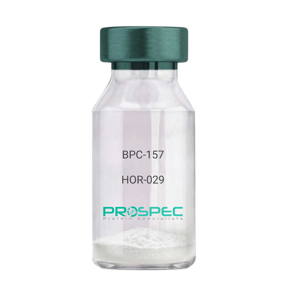
    BPC-157

    Description:

    BPC-157 Pentadecapeptide

    Product # :

    HOR-029

    Price :

    Quantity :

    Shipping Method :

    Room Temp Icon

    Shipped at Room temp

    Add To Cart

    More Info

    • description
    • formulation
    • purity
    • More Info

    Description

    BPC-157 Synthetic is a single, non-glycosylated polypeptide chain containing 15 amino acids, having a molecular mass of 1419.55 Dalton and a Molecular formula of C62H98N16O22.

    Formulation

    The protein was lyophilized with no additives.

    Purity

    Greater than 97.0% as determined by analysis by RP-HPLC.

    More Info

    • Physical Appearance

      Sterile Filtered White lyophilized (freeze-dried) powder.

    • Stability

      Lyophilized BPC-157 although stable at room temperature for 3 weeks, should be stored desiccated below -18°C. Upon reconstitution bpc-157 should be stored at 4°C between 2-7 days and for future use below -18°C. For long term storage it is recommended to add a carrier protein (0.1% HSA or BSA). Please prevent freeze-thaw cycles.

    • Solubility

      It is recommended to reconstitute the lyophilized BPC-157 in sterile 18MΩ-cm H2O not less than 100 µg/ml, which can then be further diluted to other aqueous solutions.

    • Amino Acid Sequence

      H-Gly-Glu-Pro-Pro-Pro-Gly-Lys-Pro-Ala-Asp-Asp-Ala-Gly-Leu-Val-OH

    • Background

      BPC-157, short for Body Protection Compound-157, is a synthetic peptide that has garnered significant attention in the field of regenerative medicine and sports science. This peptide, derived from a portion of the human gastric juice protein known as BPC, exhibits remarkable healing and tissue regeneration properties. BPC-157 has shown promise in various preclinical and clinical studies, demonstrating its potential for the treatment of a wide range of injuries and disorders.

      The research on BPC-157 encompasses investigations into its mechanisms of action, efficacy, safety, and potential therapeutic applications. Studies have elucidated the peptide's ability to enhance angiogenesis, promote collagen synthesis, modulate inflammatory responses, and protect against oxidative stress. These properties make BPC-157 an intriguing candidate for accelerating tissue healing, reducing inflammation, and improving overall recovery outcomes.

      Preclinical studies have revealed the beneficial effects of BPC-157 in several injury models. For instance, BPC-157 has demonstrated its potential in accelerating tendon and ligament healing, mitigating muscle damage, and promoting bone regeneration. These findings suggest that BPC-157 could be a valuable therapeutic tool in orthopedic medicine and sports-related injuries.

      Furthermore, BPC-157 has exhibited promising effects on gastrointestinal health. Studies have highlighted its ability to protect and heal the gut lining, reduce ulcer formation, and alleviate symptoms associated with inflammatory bowel disease. These observations open up avenues for BPC-157 as a potential treatment for gastrointestinal disorders.

      In addition to its regenerative properties, BPC-157 has shown potential in neurological and psychiatric conditions. Research has indicated its neuroprotective effects, with implications for the treatment of traumatic brain injury, stroke, and neurodegenerative disorders. Preliminary studies also suggest BPC-157's potential as an antidepressant and anxiolytic agent.

      Despite the promising findings, further research is needed to fully understand the mechanisms underlying BPC-157's actions and to assess its long-term safety and efficacy. Clinical trials are underway to explore its potential therapeutic applications in humans, including its use in tendon and ligament repair, inflammatory bowel disease, and neurodegenerative disorders.

      This comprehensive review aims to summarize the current state of research on BPC-157, providing an overview of its mechanisms of action and therapeutic potential. By examining relevant studies and findings, we aim to shed light on the diverse applications of BPC-157 and its implications for regenerative medicine, sports science, and various disease

    ProSpec's products are furnished for LABORATORY RESEARCH USE ONLY. The product may not be used as drugs, agricultural or pesticidal products, food additives or household chemicals.

    Bpc 157
  • View Data Sheet

    Name :

    INHBC Human

    Description:

    Inhibin-Beta C Chain Human Recombinant

    Inhibin Beta C, Actv Beta-C Chain, IHBC, Inhibin Beta C Chain.

    Product # :

    HOR-010

    Price :

    Quantity :

    Shipping Method :

    Ice Icon

    Shipped with Ice Packs

    Add To Cart

    More Info

    • description
    • source
    • formulation
    • purity
    • More Info

    Description

    INHBC Human Recombinant produced in E.coli is a single, non-glycosylated polypeptide chain containing 139 amino acids (237-352) and having a molecular mass of 14.9kDa.INHBC is fused to a 23 amino acid His-tag at N-terminus & purified by proprietary chromatographic techniques.

    Source

    E.coli.

    Formulation

    The INHBC solution (0.5mg/ml) contains 20mM Tris-HCl buffer (pH 8.0), 0.15M NaCl, 1mM DTT and 10% glycerol.

    Purity

    Greater than 85% as determined by SDS-PAGE.

    More Info

    • Introduction

      INHBC, the beta C chain of inhibin, belongs to the TGF-beta superfamily. INHBC formulates heterodimers with beta A and beta B subunits. Other members of the TGF-beta superfamily are Actv's and Inhibins, hormones with contradictory roles which take part in pituitary, hypothalamic, and gonadal hormone secretion, as well as differentiation and growth of numerous cell types.

    • Synonyms

      Inhibin Beta C, Actv Beta-C Chain, IHBC, Inhibin Beta C Chain.

    • Physical Appearance

      Sterile Filtered colorless solution.

    • Stability

      Store at 4°C if entire vial will be used within 2-4 weeks. Store, frozen at -20°C for longer periods of time. For long term storage it is recommended to add a carrier protein (0.1% HSA or BSA).Avoid multiple freeze-thaw cycles.

    • Amino Acid Sequence

      MGSSHHHHHH SSGLVPRGSH MGSGIDCQGG SRMCCRQEFF VDFREIGWHD WIIQPEGYAM NFCIGQCPLH IAGMPGIAAS FHTAVLNLLK ANTAAGTTGG GSCCVPTARR PLSLLYYDRD SNIVKTDIPD MVVEACGCS

    ProSpec's products are furnished for LABORATORY RESEARCH USE ONLY. The product may not be used as drugs, agricultural or pesticidal products, food additives or household chemicals.

    Inhbc Human
  • View Data Sheet

    Name :

    FLRT3 Human, HEK

    Description:

    Fibronectin Leucine Rich Transmembrane Protein 3 Human Recombinant, HEK

    Fibronectin Leucine Rich Transmembrane Protein 3, Fibronectin-Like Domain, Containing Leucine-Rich Transmembrane Protein 3, HH21, Leucine-Rich Repeat, Transmembrane Protein FLRT3, KIAA1469.

    Product # :

    PRO-2805

    Price :

    Quantity :

    Shipping Method :

    Ice Icon

    Shipped with Ice Packs

    Add To Cart

    More Info

    • description
    • source
    • formulation
    • purity
    • biological activity
    • More Info

    Description

    FLRT3 Human Recombinant is a single, glycosylated, polypeptide chain (29-528 a.a) containing a total of 506 amino acids and having a molecular mass of 57.3 kDa. FLRT3 is fused to a 6 a.a his-Tag at C-terminus and is purified by proprietary chromatographic techniques.

    Source

    HEK293 Cells.

    Formulation

    FLRT3 protein solution (0.25mg/ml) contains Phosphate Buffered Saline (pH 7.4) and 10% glycerol.

    Purity

    Greater than 95.0% as determined by SDS-PAGE.

    Biological Activity

    >40%.  Measured by the ability of the immobilized protein to support the adhesion of Neuro-2a neuroblast cells. When cells are added to human FLRT3 coated plates 5 ug/ml.

    More Info

    • Synonyms

      Fibronectin Leucine Rich Transmembrane Protein 3, Fibronectin-Like Domain, Containing Leucine-Rich Transmembrane Protein 3, HH21, Leucine-Rich Repeat, Transmembrane Protein FLRT3, KIAA1469.

    • Physical Appearance

      Sterile filtered colorless solution.

    • Stability

      Store at 4°C if entire vial will be used within 2-4 weeks.Store, frozen at -20°C for longer periods of time. For long term storage it is recommended to add a carrier protein (0.1% HSA or BSA).Avoid multiple freeze-thaw cycles.

    • Amino Acid Sequence

      KSCPSVCRCD AGFIYCNDRF LTSIPTGIPE DATTLYLQNN QINNAGIPSD LKNLLKVERI YLYHNSLDEF PTNLPKYVKE LHLQENNIRT ITYDSLSKIP YLEELHLDDN SVSAVSIEEG AFRDSNYLRL LFLSRNHLST IPWGLPRTIE ELRLDDNRIS TISSPSLQGL TSLKRLVLDG NLLNNHGLGD KVFFNLVNLT ELSLVRNSLT AAPVNLPGTN LRKLYLQDNH INRVPPNAFS
      YLRQLYRLDM SNNNLSNLPQ GIFDDLDNIT QLILRNNPWY CGCKMKWVRD WLQSLPVKVN VRGLMCQAPE KVRGMAIKDL NAELFDCKDS GIVSTIQITT AIPNTVYPAQ GQWPAPVTKQ PDIKNPKLTK DHQTTGSPSR KTITITVKSV TSDTIHISWK LALPMTALRL SWLKLGHSPA FGSITETIVT GERSEYLVTA LEPDSPYKVC MVPMETSNLY LFDETPVCIE TETAPLRMYN
      PTTTLNREQE KEPYKNPNLP HHHHHH.

    • Background

      Fibronectin leucine-rich transmembrane protein 3, commonly known as FLRT3, stands as a molecular architect in the intricate landscape of neural development. Its roles, initially discovered in the embryonic nervous system, have expanded to encompass various physiological and pathological processes in both the brain and beyond. This research endeavors to unravel the enigma of FLRT3 protein, exploring its structural intricacies, physiological functions, and its far-reaching implications in neurobiology, embryogenesis, and disease. By delving into FLRT3's multifaceted roles, scientists aim to decipher the underlying mechanisms that govern its diverse functions and explore potential therapeutic avenues in the realms of neuroscience and beyond.

      Structural Complexities of FLRT3:

      FLRT3 belongs to the FLRT family, characterized by extracellular leucine-rich repeats (LRRs) and a transmembrane domain. These structural motifs enable FLRT3 to participate in a myriad of interactions, including binding with cell adhesion molecules and guidance cues. Understanding the three-dimensional architecture of FLRT3 is fundamental for unraveling its molecular partnerships, biological activities, and its contributions to cell adhesion and signaling.

      Physiological Functions in Neural Development:

      In the developing nervous system, FLRT3 acts as a guidance molecule, steering growing axons and dendrites to their precise destinations. Through interactions with other cell surface receptors and ligands, FLRT3 modulates axon pathfinding, synapse formation, and neuronal migration. Its presence in growth cones and developing neural circuits underscores its significance in sculpting the intricate neural networks essential for proper brain function.

      Beyond Neural Development:

      Beyond its canonical roles in neurodevelopment, FLRT3 has emerged as a versatile player in various physiological processes. It participates in tissue morphogenesis, modulates cell adhesion, and influences immune responses. Recent studies have also implicated FLRT3 in cancer progression, highlighting its involvement in pathological conditions and making it a potential target for therapeutic interventions in cancer therapy.

      FLRT3 as a Therapeutic Target:

      The diverse roles of FLRT3 in neural development and diseases position it as an attractive target for therapeutic interventions. Modulating FLRT3 interactions offers novel avenues for neurological disorder treatments, including neurodevelopmental disorders and neurodegenerative diseases. Moreover, understanding FLRT3's involvement in cancer biology opens doors for innovative cancer therapies, making it a promising target for precision medicine approaches.

      FLRT3, with its intricate structural features and diverse functional roles, stands as a linchpin in the realms of neuroscience, embryogenesis, and disease. Its multifaceted contributions to neural development, tissue morphogenesis, and disease pathogenesis underscore its significance in both health and pathology. As researchers continue to unravel FLRT3’s complexities, they not only deepen our understanding of fundamental biological processes but also pave the way for groundbreaking discoveries in neuroscience and therapeutic interventions, ultimately shaping the future landscape of medicine and scientific inquiry.

    ProSpec's products are furnished for LABORATORY RESEARCH USE ONLY. The product may not be used as drugs, agricultural or pesticidal products, food additives or household chemicals.

    Flrt3 Protein
  • View Data Sheet

    Name :

    CD52 Human

    Description:

    CD52 Human Recombinant

    CD52 Molecule, Human Epididymis-Specific Protein 5, CD52 Antigen, Epididymal Secretory Protein E5, Cambridge Pathology 1 Antigen, CDW52, He5, Epididymis Secretory Sperm Binding Protein Li 171mP, CDW52 Antigen, CD52 Antigen, HEL-S-171mP, EDDM5, CDw52, HE5.

    Product # :

    PRO-2525

    Price :

    Quantity :

    Shipping Method :

    Ice Icon

    Shipped with Ice Packs

    Add To Cart

    More Info

    • description
    • source
    • formulation
    • purity
    • More Info

    Description

    CD52 produced in Sf9 Baculovirus cells is a single, glycosylated polypeptide chain containing 251 amino acids (25-36 a.a.) and having a molecular mass of 28.2kDa. (Molecular size on SDS-PAGE will appear at approximately 28-40kDa).CD52 is expressed with a 239 amino acid hIgG-His-tag at C-Terminus and purified by proprietary chromatographic techniques.

    Source

    Sf9, Baculovirus cells.

    Formulation

    CD52 protein solution (0.5mg/ml) contains 10% glycerol & Phosphate Buffered Saline (pH 7.4).

    Purity

    Greater than 95.0% as determined by SDS-PAGE.

    More Info

    • Introduction

      CD52 is a small GPI (glycosylphosphatidylinositol) anchored glycoprotein which is tied to the cell surface with a GPI linkage. CD52 induces immune suppression by complement-meditated cell lysis. Moreover, CD52 is expressed on lymphocytes, eosinophils, monocytes-derived dendritic cells, monocytes, as well as neutrophils, however not on the stem cells from which these lymphocytes were derived. Furthermore, CD52 is a significant goal for therapeutic interventions intended for leukocyte depletion in post-transplant immunosuppression & hematological malignancies.

    • Synonyms

      CD52 Molecule, Human Epididymis-Specific Protein 5, CD52 Antigen, Epididymal Secretory Protein E5, Cambridge Pathology 1 Antigen, CDW52, He5, Epididymis Secretory Sperm Binding Protein Li 171mP, CDW52 Antigen, CD52 Antigen, HEL-S-171mP, EDDM5, CDw52, HE5.

    • Physical Appearance

      Sterile filtered colorless solution.

    • Stability

      Store at 4°C if entire vial will be used within 2-4 weeks. Store, frozen at -20°C for longer periods of time. For long term storage it is recommended to add a carrier protein (0.1% HSA or BSA).Avoid multiple freeze-thaw cycles.

    • Amino Acid Sequence

      GQNDTSQTSS PSLEPKSCDK THTCPPCPAP ELLGGPSVFL FPPKPKDTLM ISRTPEVTCV VVDVSHEDPE VKFNWYVDGV EVHNAKTKPR EEQYNSTYRV VSVLTVLHQD WLNGKEYKCK VSNKALPAPI EKTISKAKGQ PREPQVYTLP PSRDELTKNQ VSLTCLVKGF YPSDIAVEWE
      SNGQPENNYK TTPPVLDSDG SFFLYSKLTV DKSRWQQGNV FSCSVMHEAL HNHYTQKSLS LSPGKHHHHH H.

    ProSpec's products are furnished for LABORATORY RESEARCH USE ONLY. The product may not be used as drugs, agricultural or pesticidal products, food additives or household chemicals.

    Cd52 Human
  • View Data Sheet

    Name :

    BMP 4 Human

    Description:

    Bone Morphogenetic Protein-4 Human Recombinant

    BMP4, ZYME, BMP2B, BMP2B1.

    Product # :

    CYT-361

    Price :

    Quantity :

    Shipping Method :

    Room Temp Icon

    Shipped at Room temp

    Add To Cart

    More Info

    • description
    • source
    • formulation
    • purity
    • More Info

    Description

    Bone Morphogenetic Protein-4 Human Recombinant produced in E.Coli is a monomeric, non-glycosylated, Polypeptide chain containing 116 amino acids and having a molecular mass of 13kDa. The BMP-4 is purified by proprietary chromatographic techniques.

    Source

    Escherichia Coli.

    Formulation

    BMP-4 was lyophilized from a 0.2µm filtered concentrated (1mg/ml) solution in 20mM Na2CO3 buffer, pH 9.0.

    Purity

    Greater than 95.0% as determined by:
    (a) Analysis by RP-HPLC.
    (b) Analysis by SDS-PAGE.

    More Info

    • Introduction

      The protein encoded by this gene is a member of the bone morphogenetic protein family which is part of the transforming growth factor-beta superfamily. The superfamily includes large families of growth and differentiation factors. Bone morphogenetic proteins were originally identified by an ability of demineralized bone extract to induce endochondral osteogenesis in vivo in an extraskeletal site. This particular family member plays an important role in the onset of endochondral bone formation in humans, and a reduction in expression has been associated with a variety of bone diseases, including the heritable disorder Fibrodysplasia Ossificans Progressiva. Alternative splicing in the 5' untranslated region of this gene has been described and three variants are described, all encoding an identical protein.

    • Synonyms

      BMP4, ZYME, BMP2B, BMP2B1.

    • Physical Appearance

      Sterile Filtered White lyophilized (freeze-dried) powder.

    • Stability

      Lyophilized Bone Morphogenetic Protein-4 although stable at room temperature for 3 weeks, should be stored desiccated below -18°C. Upon reconstitution BMP4 should be stored at 4°C between 2-7 days and for future use below -18°C.For long term storage it is recommended to add a carrier protein (0.1% HSA or BSA).Please prevent freeze-thaw cycles.

    • Solubility

      It is recommended to reconstitute the lyophilized Bone Morphogenetic Protein-4 in sterile 18M-cm H2O not less than 100µg/ml, which can then be further diluted to other aqueous solutions.

    • Amino Acid Sequence

      SPKHHSQRAR KKNKNCRRHS LYVDFSDVGW NDWIVAPPGY QAFYCHGDCP FPLADHLNST NHAIVQTLVN SVNSSIPKAC CVPTELSAIS MLYLDEYDKV VLKNYQEMVV EGCGCR.

    • Background

      What You Should Know About Bone Morphogenetic Protein-4 (BMP-4) Human Recombinant

      As part of the transforming growth factor-beta (TGF-β) superfamily, Bone morphogenetic protein-4 (BMP-4) participates in multiple developmental processes, from embryogenesis to bone and cartilage formation.

      Since this signaling protein is involved in many physiological processes, its laboratory-produced version has been studied for different medical applications. Additionally, a reduction in BMP-4 expression has been associated with multiple diseases, leading to further research into its potential therapeutic benefits.

      Are you interested in learning more about Bone Morphogenetic Protein-4 (BMP-4) human recombinant? Read on to find more information!

      How Does Bone Morphogenetic Protein-4 (BMP-4) Work?

      Bone Morphogenetic Protein-4 (BMP-4) regulates microRNAs miR-494 and miR-126-5p expression, controlling endothelial cells' involvement and function in angiogenesis. As such, it has diverse effects on cell growth, differentiation, and survival.

      The Role of BMP-4

      This protein emits signals that promote the formation of different tissues and organs, including the bones and cartilage, kidneys, teeth, and the neural tube. In other words, it's essential for the development of the heart, skeleton, and central nervous system.

      However, the role of BMP-4 goes beyond these processes. It participates in different physiological activities, such as:

      • Embryonic development
      • Wound healing
      • Bone remodeling
      • Immune response modulation
      • Tissue repair
      • Cardiac development and function

      What Is Bone Morphogenetic Protein-4 (BMP-4) Human Recombinant?

      To replicate the effects of the BMP4 found in humans and explore its possible therapeutic applications, many laboratories have started producing this protein in Chinese hamster ovary (CHO) cells.

      As mentioned, decreased BMP-4 expression has been associated with different diseases, including bone disorders, fibrosis, and cancer, which can cause other conditions, such as organ dysfunction.

      More research is needed, but BMP-4 human recombinant (rhBMP4) produced in CHO has the potential to address these diseases and could be used for other medical applications. These are some examples:

      • Cancer therapy
      • Development of engineered tissues and organs
      • Bone regeneration for the treatment of osteoporosis and nonunion fractures
      • Bone growth and fusion in spinal fusion surgeries (the U.S. Food and Drug Administration approved some bone morphogenetic proteins for these procedures)
      • Promotion of tissue repair and regeneration

      Final Thoughts BMP-4

      Although BMP-4 human recombinant produced in CHO offers potential benefits, several challenges remain, including possible side effects, as high doses can cause inflammation, bone overgrowth, and other issues.

      However, the long-term effects of rhBMP4 are still under investigation. Further research will provide solutions to address these challenges and allow experts to explore this laboratory-produced protein's power in different medical fields.

      What is the molecular weight/Mw of BMP4 Protein?
      BMP4 Protein has a total Mw of 13kDa.

      What is the source or expression system of BMP4 Protein?
      Escherichia Coli.

      What is the Purity of BMP4 Protein?
      BMP4 Protein is >95% pure as determined by SDS-PAGE.

      What is the Biological Activity of BMP4 Protein?
      The biological functionality of BMP4 Protein will be determined in the future.

      What is the amino acid sequence of BMP4 Protein?
      SPKHHSQRAR KKNKNCRRHS LYVDFSDVGW NDWIVAPPGY QAFYCHGDCP FPLADHLNST NHAIVQTLVN SVNSSIPKAC CVPTELSAIS MLYLDEYDKV VLKNYQEMVV EGCGCR.

      What applications can BMP4 Protein be used in?
      BMP4 Protein can probably be used in western blot, ELISA and Lateral Flow.

      What is the endotoxin level for BMP4 Protein?
      The endotoxin level is minimal, BMP4 Protein was purified using conventional chromatography techniques.

    ProSpec's products are furnished for LABORATORY RESEARCH USE ONLY. The product may not be used as drugs, agricultural or pesticidal products, food additives or household chemicals.

    Bmp4 Human
  • View Data Sheet

    Name :

    M. Tuberculosis RV1681 Antibody

    Description:

    Mycobacterium Tuberculosis RV1681, Polyclonal Rabbit Antibody

    Product # :

    ANT-493

    Price :

    Quantity :

    Shipping Method :

    Ice Icon

    Shipped with Ice Packs

    Add To Cart

    More Info

    • description
    • formulation
    • purity
    • More Info

    Description

    The polyclonal antibody to Mycobacterium Tuberculosis RV1681 was collected from the rabbit igG and immunized with recombinant M. tuberculosis t RV-1681.

    Formulation

    Glycine-Tris buffer, pH7.5.

    Purity

    Pure Rabbit IgG, >95% pure as determined by 12% PAGE (coomassie staining).

    More Info

    • Introduction

      Mycobacterium tuberculosis, a member of the Mycobacteriaceae family, is an important infectious pathogenic bacteria and the cause for most cases of tuberculosis. It is the second common cause of death from an infectious disease in the world. Diagnosis using patient’s specimen culture of M. tuberculosis are still considered gold standard, but it is expensive and time-consuming. RV-1681 is a protein released from M. tuberculosis on infected organ, which filtrates through into the urine after circulating in the blood flow, to the kidney.RV-1681 is considered to be a reliable marker for M. tuberculosis diagnosis allowing easy low cost and time saving urine testing to screen large population for M. tuberculosis infection.

    • Physical Appearance

      Sterile Filtered clear colorless solution.

    • Stability

      Store at 4°C if entire vial will be used within 2-4 weeks. Store, frozen at -20°C for longer periods of time. For long term storage it is recommended to add a carrier protein (0.1% HSA or BSA).Avoid multiple freeze-thaw cycles.

    • Purification Method

      Purified by affinity chromatographic technique.

    ProSpec's products are furnished for LABORATORY RESEARCH USE ONLY. The product may not be used as drugs, agricultural or pesticidal products, food additives or household chemicals.

    M Tuberculosis Rv1681 Antibody
  • View Data Sheet

    Name :

    PPM1A Human

    Description:

    Protein Phosphatase 1A Alpha Isoform Human Recombinant

    Protein phosphatase 1A, EC 3.1.3.16, Protein phosphatase 2C isoform alpha, PP2C-alpha, IA, PPM1A, PP2CA, MGC9201.

    Product # :

    PKA-222

    Price :

    Quantity :

    Shipping Method :

    Ice Icon

    Shipped with Ice Packs

    Add To Cart

    More Info

    • description
    • source
    • formulation
    • purity
    • More Info

    Description

    PPM1A Human Recombinant produced in E.Coli is a single, non-glycosylated polypeptide chain containing 418 amino acids (1-382a.a) and having a molecular mass of 46.6kDa. PPM1A is fused to a 36 amino acid His-tag at N-terminus & purified by proprietary chromatographic techniques.

    Source

    Escherichia Coli.

    Formulation

    PPM1A protein solution (1mg/ml) contains 10mM Tris-HCl, pH7.5, 50mM NaCl, 2mM DTT, 1mM MnCl2 and 20% glycerol.

    Purity

    Greater than 95.0% as determined by SDS-PAGE.

    More Info

    • Introduction

      Protein Phosphatase 2C alpha is a member of the PP2C family of Ser/Thr protein phosphatases. PP2C family members are known to be negative regulators of cell stress response pathways. This phosphatase dephosphorylates, and negatively regulates the activities of, MAP kinases and MAP kinase kinases. It has been shown to inhibit the activation of p38 and JNK kinase cascades induced by environmental stresses. This phosphatase can also dephosphorylate cyclin-dependent kinases, and thus may be involved in cell cycle control. Overexpression of this phosphatase is reported to activate the expression of the tumor suppressor gene TP53/p53, which leads to G2/M cell cycle arrest and apoptosis. Three alternatively spliced transcript variants encoding two distinct isoforms have been described.
      Protein phosphatase 2C(PP2C?) is a Mn2+- or Mg2+-dependent protein serine/threonine phosphatase that is essential for regulating cellular stress response in eukaryotes.

    • Synonyms

      Protein phosphatase 1A, EC 3.1.3.16, Protein phosphatase 2C isoform alpha, PP2C-alpha, IA, PPM1A, PP2CA, MGC9201.

    • Physical Appearance

      Sterile filtered colorless solution.

    • Stability

      Store at 4°C if entire vial will be used within 2-4 weeks. Store, frozen at -20°C for longer periods of time. For long term storage it is recommended to add a carrier protein (0.1% HSA or BSA). Avoid multiple freeze-thaw cycles.

    • Amino Acid Sequence

      MRGSHHHHHH GMASMTGGQQ MGRDLYDDDD KDRWILMGAF LDKPKMEKHN AQGQGNGLRY GLSSMQGWRV EMEDAHTAVI GLPSGLESWS FFAVYDGHAG SQVAKYCCEH LLDHITNNQD FKGSAGAPSV ENVKNGIRTG FLEIDEHMRV MSEKKHGADR SGSTAVGVLI SPQHTYFINC GDSRGLLCRN RKVHFFTQDH KPSNPLEKER IQNAGGSVMI QRVNGSLAVS RALGDFDYKC VHGKGPTEQL VSPEPEVHDI ERSEEDDQFI ILACDGIWDV MGNEELCDFV RSRLEVTDDL EKVCNEVVDT CLYKGSRDNM SVILICFPNA PKVSPEAVKK EAELDKYLEC RVEEIIKKQG EGVPDLVHVM RTLASENIPS LPPGGELASK RNVIEAVYNR LNPYKNDDTD STSTDDMW.

    • Unit Definition

      One unit will hydrolyze 1 nanomole of p-nitrophenyl phosphatate (pNPP) per minute at pH 7.5 at 37°C.

    • Specific Activity

      >1,400 U/mg.

    ProSpec's products are furnished for LABORATORY RESEARCH USE ONLY. The product may not be used as drugs, agricultural or pesticidal products, food additives or household chemicals.

    Ppm1A Human
  • View Data Sheet

    Name :

    Acrp30 Antibody

    Description:

    Adiponectin, Mouse Anti Human

    Acrp30, AdipoQ, GBP-28, APM-1, ACDC.

    Product # :

    ANT-232

    Price :

    Quantity :

    Shipping Method :

    Ice Icon

    Shipped with Ice Packs

    Add To Cart

    More Info

    • formulation
    • More Info

    Formulation

    containing PBS, pH-7.4, & 0.1% Sodium Azide

    More Info

    • Introduction

      Human Adiponectin is a secreted protein expressed exclusively in differentiated adipocyte (adipokine). Adiponectin contains a modular structure comprising an N-terminal collagenous domain followed by a C-terminal globular domain. APM-1 plays a role in various physiological processes such as energy homeostasis and obesity. Plasma levels of adiponectin are reduced in obese humans, and decreased levels are associated with insulin resistance and hyperinsulinemia.

    • Synonyms

      Acrp30, AdipoQ, GBP-28, APM-1, ACDC.

    • Physical Appearance

      Sterile Filtered solution.

    • Immunogen

      Anti-human adiponectin mAb, is derived from hybridization of mouse SP2/O myeloma cells with spleen cells from BALB/c mice immunized with Recombinant human adiponectin amino acids 15-244 purified from E. coli.

    • Ig Subclass

      Mouse IgG1 heavy chain and k light chain.

    • Clone

      P1G12AT.

    • Applications

      Adiponectin antibody has been tested by ELISA and Western blot analysis to assure specificity and reactivity. Recommended dilution range for Western blot analysis is 1:250 ~ 1:1,000. The antibody has the specificity against globular domain of adiponectin. Recommended starting dilution is 1:500.

    • Type

      Mouse Anti Human Monoclonal.

    • Storage Procedures

      For periods up to 1 month store at 4°C, for longer periods of time, store at -20°C. Prevent freeze thaw cycles.

    • Purification Method

      Adiponectin antibody was purified from mouse ascitic fluids by protein-G affinity chromatography.

    ProSpec's products are furnished for LABORATORY RESEARCH USE ONLY. The product may not be used as drugs, agricultural or pesticidal products, food additives or household chemicals.

    Acrp30 Antibody
  • View Data Sheet

    Name :

    BAG2 Antibody

    Description:

    BCL2-Associated Athanogene 2, Mouse Anti Human

    BAG family molecular chaperone regulator 2, BAG-2, Bcl-2-associated athanogene 2, BAG2, KIAA0576, MGC149462, dJ417I1.2.

    Product # :

    ANT-669

    Price :

    Quantity :

    Shipping Method :

    Ice Icon

    Shipped with Ice Packs

    Add To Cart

    More Info

    • formulation
    • More Info

    Formulation

    1mg/ml containing PBS, pH-7.4, 10% Glycerol and 0.02% Sodium Azide.

    More Info

    • Introduction

      BCL2-associated athanogene 2 (BAG2) belongs to the Bag family of proteins. BAG2 is a chief component of the HSC 70/CHIP chaperone-dependent ubiquitin ligase complex and acts to upset CHIP-mediated ubiquitylation. BAG proteins compete with Hip for attaching to the Hsc70/Hsp70 ATPase domain and promote substrate discharge. All the BAG proteins have a roughly 45 amino acid BAG domain by the C-terminus but vary clearly in their N-terminal regions. The BAG domains of BAG1, BAG2, and BAG3 act together expressly with the Hsc70 ATPase domain in vitro and in mammalian cells. All three proteins attach with high affinity to the ATPase domain of Hsc70 and obstruct its chaperone activity in a Hip-repressible manner.

    • Synonyms

      BAG family molecular chaperone regulator 2, BAG-2, Bcl-2-associated athanogene 2, BAG2, KIAA0576, MGC149462, dJ417I1.2.

    • Physical Appearance

      Sterile filtered colorless solution.

    • Immunogen

      Anti-human BAG2 mAb, is derived from hybridization of mouse F0 myeloma cells with spleen cells from BALB/c mice immunized with a recombinant human BAG2 protein 1-211 amino acids purified from E. coli.

    • Ig Subclass

      Mouse IgG2a heavy chain and k light chain.

    • Clone

      PAT29E9AT.

    • Applications

      The antibody has been tested by ELISA, Western blot analysis, Flow cytometry and ICC/IF to assure specificity and reactivity. Since application varies, however, each investigation should be titrated by the reagent to obtain optimal results.

    • Type

      Mouse Anti Human Monoclonal.

    • Storage Procedures

      For periods up to 1 month store at 4°C, for longer periods of time, store at -20°C. Prevent freeze thaw cycles.

    • Purification Method

      BAG2 antibody was purified from mouse ascitic fluids by protein-A affinity chromatography.

    ProSpec's products are furnished for LABORATORY RESEARCH USE ONLY. The product may not be used as drugs, agricultural or pesticidal products, food additives or household chemicals.

    Bag2 Antibody
  • View Data Sheet

    Name :

    EPHA2 PAT66G9AT Antibody

    Description:

    EPH receptor A2 PAT66G9AT, Mouse Anti Human

    EPHA2, EPH Receptor A2, ECK, Tyrosine-Protein Kinase Receptor ECK, EC 2.7.10.1, CTRCT6, ARCC2, CTPP1, CTPA, Epithelial Cell Receptor Protein Tyrosine Kinase, Ephrin Type-A Receptor 2, Soluble EPHA2 Variant 1, Epithelial Cell Kinase, EC 2.7.10, EphA2.

    Product # :

    ANT-672

    Price :

    Quantity :

    Shipping Method :

    Ice Icon

    Shipped with Ice Packs

    Add To Cart

    More Info

    • formulation
    • More Info

    Formulation

    1mg/ml containing PBS, pH-7.4, 10% Glycerol and 0.02% Sodium Azide.

    More Info

    • Introduction

      EPH Receptor A2 (EPHA2) is a member of the ephrin receptor subfamily of the protein-tyrosine kinase family. EPHA2 is a protein which binds ephrin-A ligands. EPH and EPH-related receptors are associated with mediating developmental events, particularly in the nervous system. Receptors in the EPH subfamily normally have a single kinase domain and an extracellular region containing a Cys-rich domain and 2 fibronectin type III repeats. The ephrin receptors are divided into two groups based on the similarity of their extracellular domain sequences and their affinities for binding ephrin-A and ephrin-B ligands. EPHA2 gene mutations are the cause of certain genetically-related cataract disorders.

    • Synonyms

      EPHA2, EPH Receptor A2, ECK, Tyrosine-Protein Kinase Receptor ECK, EC 2.7.10.1, CTRCT6, ARCC2, CTPP1, CTPA, Epithelial Cell Receptor Protein Tyrosine Kinase, Ephrin Type-A Receptor 2, Soluble EPHA2 Variant 1, Epithelial Cell Kinase, EC 2.7.10, EphA2.

    • Physical Appearance

      Sterile filtered colorless solution.

    • Immunogen

      Anti-human EPHA2 mAb, is derived from hybridization of mouse F0 myeloma cells with spleen cells from BALB/c mice immunized with a recombinant human EPHA2 protein 27-537 amino acids purified from E. coli.

    • Ig Subclass

      Mouse IgG2b heavy chain and k light chain.

    • Clone

      PAT66G9AT.

    • Applications

      The antibody has been tested by ELISA, Western blot analysis, Flow cytometry and ICC/IF to assure specificity and reactivity. Since application varies, however, each investigation should be titrated by the reagent to obtain optimal results.

    • Type

      Mouse Anti Human Monoclonal.

    • Storage Procedures

      For periods up to 1 month store at 4°C, for longer periods of time, store at -20°C. Prevent freeze thaw cycles.

    • Purification Method

      EPHA2 antibody was purified from mouse ascitic fluids by protein-A affinity chromatography.

    ProSpec's products are furnished for LABORATORY RESEARCH USE ONLY. The product may not be used as drugs, agricultural or pesticidal products, food additives or household chemicals.

    Epha2 Pat66G9At Antibody
  • View Data Sheet

    Name :

    KCTD15 Antibody

    Description:

    Potassium channel tetramerisation domain containing 15, Mouse Anti Human

    BTB/POZ domain-containing protein KCTD15, Potassium channel tetramerisation domain containing 15, KCTD15, MGC2628, MGC25497.

    Product # :

    ANT-028

    Price :

    Quantity :

    Shipping Method :

    Ice Icon

    Shipped with Ice Packs

    Add To Cart

    More Info

    • formulation
    • More Info

    Formulation

    1mg/ml containing PBS, pH-7.4, & 0.1% Sodium Azide.

    More Info

    • Introduction

      KCTD15 protein is encoded in humans by the KCTD15 gene. KCTD15 is expressed at a high level in the brain and the hypothalamus. The potassium channel KCTD15 was identified as a genetic loci linked to higher than normal BMI in humans along with genes such as GNPDA2, MTCH2, FTO, and TMEM18. SNPs (Single nucleotide polymorphisms) in non-diabetic and diabetic patients showed that FTO was most strongly associated with obesity while MTCH2 and GNPDA2 were still notably associated with higher than normal BMI levels.

    • Synonyms

      BTB/POZ domain-containing protein KCTD15, Potassium channel tetramerisation domain containing 15, KCTD15, MGC2628, MGC25497.

    • Immunogen

      Anti-human KCTD15 mAb, is derived from hybridization of mouse F0 myeloma cells with spleen cells from BALB/c mice immunized with recombinant human KCTD15 amino acids 1-234 purified from E. coli.

    • Ig Subclass

      Mouse IgG3 heavy chain and L light chain.

    • Clone

      PAT4C3AT.

    • Applications

      KCTD15 antibody has been tested by ELISA, Western blot and Immunofluorescence analysis to assure specificity and reactivity. Since application varies, however, each investigation should be titrated by the reagent to obtain optimal results. Recommended dilution range for Western blot analysis and Immunofluorescence is 1:250 ~ 500.
      Recommended starting dilution is 1:250.

    • Type

      Mouse Anti Human Monoclonal.

    • Storage Procedures

      For periods up to 1 month store at 4°C, for longer periods of time, store at -20°C. Prevent freeze thaw cycles.

    • Purification Method

      KCTD15 antibody was purified from mouse ascitic fluids by protein-G affinity chromatography.

    ProSpec's products are furnished for LABORATORY RESEARCH USE ONLY. The product may not be used as drugs, agricultural or pesticidal products, food additives or household chemicals.

    Kctd15 Antibody
  • View Data Sheet

    Name :

    C1R Human

    Description:

    Complement C1r Human

    Complement C1r subcomponent, Complement component 1 subcomponent r, C1R.

    Product # :

    ENZ-1163

    Price :

    Quantity :

    Shipping Method :

    Ice Icon

    Shipped with Ice Packs

    Add To Cart

    More Info

    • description
    • source
    • formulation
    • purity
    • More Info

    Description

    Human Complement C1r produced in Human plasma having a molecular mass of 92 kDa.

    Source

    Human Plasma.

    Formulation

    (1mg/ml) 140mM NaCl, 10mM Imidazole and 8mM EDTA, pH 7.4.

    Purity

    Greater than 90.0% as determined by SDS-PAGE.

    More Info

    • Introduction

      C1r enzyme is the activated form of C1r proenzyme which is inactive zymogen until C1 is activated.C1r is a subunit of the C1 complex which is the first complement component in the classical pathway of complement. C1 is a non-covalent calcium-dependent complex containing1 C1q, 2 C1r and 2 C1s molecules.Each C1q bindsthe Fc domains of IgG or IgM by using2 or more of its six arms. The binding of multiple arms to immune complexes causesthe production of 2 proteases that cleave and activate the 2 C1s protease zymogens in the complex by the C1r proteins. The activation of C1r results from cleavage of C1r into 2 fragments of 57,000 and 35,000 Dalton.

    • Synonyms

      Complement C1r subcomponent, Complement component 1 subcomponent r, C1R.

    • Physical Appearance

      Sterile Filtered solution.

    • Stability

      Human C1r is stable at 4°C if entire vial will be used within 2-4 weeks.Store, frozen below -20°C for longer periods of time.For long term storage it is recommended to add a carrier protein (0.1% HSA or BSA).Avoid multiple freeze-thaw cycles

    • Human Virus Test

      Plasma from each donor has been tested and found negative for antibody to HIV-1, HIV-2, HCV, HTLV-I/II, STS and HBSAG.

    ProSpec's products are furnished for LABORATORY RESEARCH USE ONLY. The product may not be used as drugs, agricultural or pesticidal products, food additives or household chemicals.

    C1R Human
  • View Data Sheet

    Name :

    C3 Human

    Description:

    Complement C3 Human

    Complement C3, C3 and PZP-like alpha-2-macroglobulin domain-containing protein 1, C3, CPAMD1.

    Product # :

    PRO-2684

    Price :

    Quantity :

    Shipping Method :

    Ice Icon

    Shipped with Ice Packs

    Add To Cart

    More Info

    • description
    • source
    • formulation
    • purity
    • More Info

    Description

    Human Complement C3 produced in Human plasma having a molecular mass of 185 kDa.

    Source

    Human Plasma.

    Formulation

    C3 solution contains phosphate buffer saline.

    Purity

    Greater than 94.0% as determined by SDS-PAGE.

    More Info

    • Introduction

      Native human C3 is a naturally glycosylated polypeptide containing two disulfide-linked chains. C3 is central to the activation of all 3 pathways of complement activation. Initiation of each pathway generates proteolytic enzyme complexes which binds the target surface. These enzymes cleave a peptide bond in C3 releasing the anaphylatoxin C3a and activating C3b. Most of the C3 activated during complement activation never attaches to the surface due to its thioester reaction with water forming fluid phase C3b which is rapidly inactivated by factors H and I forming iC3b. Surface-bound C3b is necessary in all 3 pathways for efficient activation of C5 and formation of C5b-9 complexes that lyse the target cell membrane.

    • Synonyms

      Complement C3, C3 and PZP-like alpha-2-macroglobulin domain-containing protein 1, C3, CPAMD1.

    • Physical Appearance

      Sterile Filtered solution.

    • Stability

      C3 Human is stable at 4°C if entire vial will be used within 2-4 weeks.Store, frozen below -20°C for longer periods of time.For long term storage it is recommended to add a carrier protein (0.1% HSA or BSA).Avoid multiple freeze-thaw cycles.

    • Human Virus Test

      Plasma from each donor has been tested and found negative for antibody to HIV-1,HIV-2, HCV, HTLV-I &II, STS and HBSAG.

    ProSpec's products are furnished for LABORATORY RESEARCH USE ONLY. The product may not be used as drugs, agricultural or pesticidal products, food additives or household chemicals.

    C3 Human
  • View Data Sheet

    Name :

    EBV p18 M

    Description:

    Epstein -Barr virus (HHV-4) p18 Mosaic Recombinant

    Product # :

    EBV-275

    Price :

    Quantity :

    Shipping Method :

    Ice Icon

    Shipped with Ice Packs

    Add To Cart

    More Info

    • description
    • source
    • formulation
    • purity
    • More Info

    Description

    The E.Coli derived recombinant artificial mosaic protein contains two epitopes from C-end associated in one molecule from the HHV-4 p18 regions.

    Source

    Escherichia Coli.

    Formulation

    50mM Tris-Hcl pH 8.0, 60mM NaCl, 10mM glutathione and 50% glycerol.

    Purity

    EBV-p18 protein is >95% pure as determined by 10% PAGE (coomassie staining).

    More Info

    • Introduction

      The Epstein-Barr virus (EBV), also called Human herpes virus 4 (HHV-4), is a virusof the herpes family(which includes Herpes simplex virusand Cytomegalovirus. On infecting the B-lymphocyte, the linear virus genome circularizes and the virus subsequently persists within the cell as an episome. The virus can execute several distinct programs of gene expressionwhich can be broadly categorized as being lytic cycle or latent cycle. The lytic cycleor productive infection results in staged expression of a host of viral proteinswith the ultimate objective of producing infectious virions. Formally, this phase of infection does not inevitably lead to lysis of the host cellas EBV virions are produced by budding from the infected cell. The latent cycle(lysogenic) programs are those that do not result in production of virions. A very limited, distinct set of viral proteins are produced during latent cycle infection. These include Epstein-Barr nuclear antigen(EBNA)-1, EBNA-2, EBNA-3A, EBNA-3B, EBNA-3C, EBNA-leader protein (EBNA-LP) and latent membrane proteins(LMP)-1, LMP-2A and LMP-2B and the Epstein-Barr encoded RNAs(EBERs).

    • Stability

      EBV p18 M although stable at 4°C for 1 week, should be stored below -18°C. Please prevent freeze thaw cycles.

    • Applications

      EBV-p18 Mosaic antigen is suitable for ELISA and Western blots, excellent antigen for detection of HHV-4 (EBV) with minimal specificity problems.

    • Specificity

      Immunoreactive with sera of EBV-infected individuals.

    • Purification Method

      EBV-p18 Mosaic was purified by proprietary chromatographic technique.

    ProSpec's products are furnished for LABORATORY RESEARCH USE ONLY. The product may not be used as drugs, agricultural or pesticidal products, food additives or household chemicals.

    Ebv Mosaic
  • View Data Sheet

    Name :

    HAV P3C

    Description:

    Hepatitis A Virus P3C Recombinant

    Product # :

    HAV-231

    Price :

    Quantity :

    Shipping Method :

    Ice Icon

    Shipped with Ice Packs

    Add To Cart

    More Info

    • description
    • source
    • formulation
    • purity
    • More Info

    Description

    The E.Coli derived 40 kDa recombinant protein contains the P3C immunodominant regions, amino acids 1643-1743.

    Source

    Escherichia Coli.

    Formulation

    10mM CBB, pH9.6, 0.1% SDS and 50% glycerol.

    Purity

    HAV P3C protein is >90% pure as determined by 10% PAGE(coomassie staining).

    More Info

    • Introduction

      Forty-two antigenic domains were identified across the hepatitis A virus (HAV) polyprotein by using a set of 237 overlapping 20-mer synthetic peptides spanning the entire HAV polyprotein. Nineteen antigenic domains were found within the structural proteins, and 22 were found within the nonstructural proteins, with 1 domain spanning the junction of VP1 and P2A proteins. Five of these domains were considered immunodominant, as judged by both the breadth and the strength of their immunoreactivity. One domain is located within the VP2 protein at position 57-90 aa. A second domain, located at position 767-842 aa, contains the C-terminal part of the VP1 protein and the entire P2A protein. A third domain, located at position 1403-1456 aa, comprises the C-terminal part of the P2C protein and the N-terminal half of the P3A protein. The fourth domain, located at position 1500-1519 aa, includes almost the entire P3B, and the last domain, located at position 1719-1764 aa, contains the C-terminal region of the P3C protein and the N-terminal region of the P3D protein. Four of the five most immunoreactive domains are derived from small HAV proteins and/or encompass protein cleavage sites separating different HAV proteins.

    • Stability

      HAV P3C protein although stable at 4°C for 1 week, should be stored below -18°C. Please prevent freeze thaw cycles.

    • Applications

      HAV P3C antigen is suitable for ELISA and Western blots, excellent antigen for detection of HAV with minimal specificity problems.

    • Specificity

      Immunoreactive with sera HAV-infected individuals.

    • Purification Method

      HAV P3C protein was purified by proprietary chromatographic technique.

    ProSpec's products are furnished for LABORATORY RESEARCH USE ONLY. The product may not be used as drugs, agricultural or pesticidal products, food additives or household chemicals.

    Hepatitis A Virus P3C
  • View Data Sheet

    Name :

    Rubella Capsid-C

    Description:

    Rubella Virus Capsid-C Recombinant

    Product # :

    RUB-293

    Price :

    Quantity :

    Shipping Method :

    Ice Icon

    Shipped with Ice Packs

    Add To Cart

    More Info

    • description
    • formulation
    • purity
    • More Info

    Description

    The E.Coli derived recombinant protein contains the Rubella Virus Capsid C regions, 1-123 amino acids and fused to a GST-Tag at C-terminus.

    Formulation

    25mM Tris-HCl pH 8, 10mM glutation, 0.2% sarcosyl and 50% glycerol.

    Purity

    Rubella protein is >95% pure as determined by 10% PAGE (coomassie staining).

    More Info

    • Introduction

      Rubella virus is an enveloped positive-strand RNA virus of the family TOGAVIRIDAE: Virions are composed of three structural proteins: a capsid and two membrane-spanning glycoproteins, E2 and E1. During virus assembly, the capsid interacts with genomic RNA to form nucleocapsids.

    • Stability

      Rubella Capsid protein although stable at 4°C for 1 week, should be stored below -18°C. Please prevent freeze thaw cycles.

    • Specificity

      Immunoreactive with sera of Rubella Virus infected individuals.

    • Purification Method

      Rubella protein was purified by proprietary chromatographic technique.

    ProSpec's products are furnished for LABORATORY RESEARCH USE ONLY. The product may not be used as drugs, agricultural or pesticidal products, food additives or household chemicals.

    product_image.jpg
  • View Data Sheet

    Name :

    EBV p23

    Description:

    Epstein-Barr Virus (HHV-4) p23 Recombinant

    Product # :

    EBV-274

    Price :

    Quantity :

    Shipping Method :

    Ice Icon

    Shipped with Ice Packs

    Add To Cart

    More Info

    • description
    • source
    • formulation
    • purity
    • More Info

    Description

    The E.Coli derived recombinant protein contains the HHV-4 p23 regions, 1-162 amino acids and fused to a GST-Tag at C-terminus.

    Source

    Escherichia Coli.

    Formulation

    25mM glycine pH-9.6 and 50% glycerol.

    Purity

    EBV- p23 protein is >95% pure as determined by 10% PAGE (coomassie staining).

    More Info

    • Introduction

      The Epstein-Barr virus (EBV), also called Human herpes virus 4 (HHV-4), is a virusof the herpes family(which includes Herpes simplex virusand Cytomegalovirus. On infecting the B-lymphocyte, the linear virus genome circularizes and the virus subsequently persists within the cell as an episome. The virus can execute several distinct programs of gene expressionwhich can be broadly categorized as being lytic cycle or latent cycle. The lytic cycleor productive infection results in staged expression of a host of viral proteinswith the ultimate objective of producing infectious virions. Formally, this phase of infection does not inevitably lead to lysis of the host cellas EBV virions are produced by budding from the infected cell. The latent cycle(lysogenic) programs are those that do not result in production of virions. A very limited, distinct set of viral proteins are produced during latent cycle infection. These include Epstein-Barr nuclear antigen(EBNA)-1, EBNA-2, EBNA-3A, EBNA-3B, EBNA-3C, EBNA-leader protein (EBNA-LP) and latent membrane proteins(LMP)-1, LMP-2A and LMP-2B and the Epstein-Barr encoded RNAs(EBERs).

    • Stability

      EBV-p23 protein although stable at 4°C for 1 week, should be stored below -18°C. Please prevent freeze thaw cycles.

    • Applications

      EBV-p23 antigen is suitable for ELISA and Western blots, excellent antigen for detection of HHV-4 (EBV) with minimal specificity problems.

    • Specificity

      Immunoreactive with sera of EBV-infected individuals.

    • Purification Method

      EBV-p23 was purified by proprietary chromatographic technique.

    ProSpec's products are furnished for LABORATORY RESEARCH USE ONLY. The product may not be used as drugs, agricultural or pesticidal products, food additives or household chemicals.

    Ebv P23
  • View Data Sheet

    Name :

    SARS MERS RBD

    Description:

    SARS MERS Spike Receptor Binding Domain Recombinant

    Middle East respiratory syndrome coronavirus, Human betacoronavirus 2c EMC/2012, MERS-CoV, MERS, MERSCoV RBD, MERS RBD, receptor binding domain, RBD, Spike RBD protein, Spike glycoprotein, S glycoprotein, E2, Peplomer protein

    Product # :

    SARS-054

    Price :

    Quantity :

    Shipping Method :

    Ice Icon

    Shipped with Ice Packs

    Add To Cart

    More Info

    • description
    • source
    • formulation
    • purity
    • More Info

    Description

    SARS MERS RBD Recombinant produced in Sf9 Baculovirus cells is a single, glycosylated polypeptide chain containing 258 amino acids (358-606 aa) and having a molecular mass of 28.2kDa.SARS MERS RBD is fused to a 6 amino acid His-tag at C-terminus & purified by proprietary chromatographic techniques.

    Source

    Sf9, Baculovirus cells.

    Formulation

    The SARS MERS RBD solution (0.5mg/ml) contains 10% glycerol and Phosphate-Buffered Saline (pH 7.4).

    Purity

    Greater than 90.0% as determined by SDS-PAGE.

    More Info

    • Introduction

      Since April 2012, cases of the Middle East Respiratory Syndrome Coronavirus (MERS-CoV) have been identified in various countries. Coronaviruses are the cause of the common cold, SARS (severe acute respiratory syndrome) and other severe illnesses with high mortality rates, all are classified into coronavirus family. MERS-CoV is a new type of SARS found in the coronavirus family causing severe pneumonia with sudden and serious respiratory illness with high mortality rates as well. Since January 27th 2015, the WHO has reported 956 human cases, including 351 deaths. More cases of the new coronavirus strain are expected. Like in other coronaviruses, large surface spike glycoprotein is a central structural protein of this virus; it is located above the virion surface to bind and enter into the target cell. Spike protein has 2 domains- S1 and S2. The S1 domain is responsible for cellular tropism and interaction with target cell, while the S2 domain is responsible for membrane fusion. The C-terminal of S1 domain contains a receptor binding domain, and is also a potential target for vaccine development and an antigen for diagnosis.

    • Synonyms

      Middle East respiratory syndrome coronavirus, Human betacoronavirus 2c EMC/2012, MERS-CoV, MERS, MERSCoV RBD, MERS RBD, receptor binding domain, RBD, Spike RBD protein, Spike glycoprotein, S glycoprotein, E2, Peplomer protein

    • Physical Appearance

      Sterile filtered colorless solution.

    • Stability

      Store at 4°C if entire vial will be used within 2-4 weeks.Store, frozen at -20°C for longer periods of time.For long term storage it is recommended to add a carrier protein (0.1% HSA or BSA).Avoid multiple freeze-thaw cycles.

    • Amino Acid Sequence

      ADPSGVYSVS SFEAKPSGSV VEQAEGVECD FSPLLSGTPP QVYNFKRLVF TNCNYNLTKL LSLFSVNDFT CSQISPAAIA SNCYSSLILD YFSYPLSMKS DLSVSSAGPI SQFNYKQSFS NPTCLILATV PHNLTTITKP LKYSYINKCS RLLSDDRTEV PQLVNANQYS PCVSIVPSTV WEDGDYYRKQ LSPLEGGGWL VASGSTVAMT EQLQMGFGIT VQYGTDTNSV CPKLEFANDT KIASQLGNCV EYHHHHHH

    ProSpec's products are furnished for LABORATORY RESEARCH USE ONLY. The product may not be used as drugs, agricultural or pesticidal products, food additives or household chemicals.

    product_image.jpg
  • 1
  • …
  • 24
  • 25
  • 26
  • 27
  • 28
  • …
  • 42
Your browser does not support the video tag.

Products

  • Cytokines
  • Growth Factors
  • Chemokines
  • CD Antigens
  • Neurotrophins
  • Hormones
  • Enzymes
  • Viral Antigens
  • Recombinant Proteins
  • Natural Proteins
  • Monoclonal Antibodies
  • Polyclonal Antibodies
  • Thermal Packaging

About

  • Company
  • Founder
  • Contribution
  • Contact Us
  • My Account

Ordering

  • Ordering Method
  • Ordering - Credit Card
  • Ordering PO
  • Shipping Fees
  • FAQ's

Legal

  • Terms & Conditions
  • Privacy
  • Site Map
  • info@prospecbio.com
  • Facebook
  • Instagram
  • Linkedin Profile

© 2026 ProSpec-Tany TechnoGene Ltd. All Rights Reserved.

  • Choosing a selection results in a full page refresh.
  • Opens in a new window.