Search results
1000 results found for “Tumor Necrosis Factor”
Name
Description
Product #
Price
Quantity
Shipping Method
- View Data Sheet
Name :
CCL2 RatDescription:
Monocyte Chemotactic Protein-1 Rat Recombinant (CCL2)
Small inducible cytokine A2, CCL2, Monocyte chemotactic protein 1, MCP-1, Monocyte chemoattractant protein 1, Monocyte chemotactic and activating factor, MCAF, Monocyte secretory protein JE, HC11, chemokine (C-C motif) ligand 2, MCP1, SCYA2, GDCF-2, SMC-CF, HSMCR30, MGC9434, GDCF-2 HC11, Immediate-early serum-responsive JE protein.
Product # :
CHM-315Price :
Quantity :
Shipping Method :
Shipped at Room temp
More Info
- description
- source
- formulation
- purity
- biological activity
- More Info
- Citations
Description
Monocyte Chemotactic Protein-1 Rat Recombinant produced in E.Coli is a non-glycosylated, Polypeptide chain containing 125 amino acids and having a molecular mass of 14.1 kDa. The MCP-1 is purified by proprietary chromatographic techniques.
Source
Escherichia Coli.
Formulation
The protein was lyophilized from a concentrated (1mg/ml) sterile solution containing no additives.
Purity
Greater than 98.0% as determined by SDS-PAGE.
Biological Activity
ED50 =1-10ng/ml corresponding to a Specific Activity of 100,000-1,000,000IU/mg. The biological activity was determined by measuring the dose dependent chemotaxis with human THP-1 cells. The optimal concentration should be determined for each specific application by an initial dose-response assay.Citations
More Info
-
Introduction
Chemokine (C-C motif) ligand 2 (CCL2) is a small cytokine belonging to the CC chemokine family that is also known as monocyte chemotactic protein-1 (MCP-1). It is found at the site of tooth eruption and bone degradation. In the bone, CCL2 is expressed by mature osteoclasts and osteoblasts and is under the control of nuclear factor ?B (NF?B). CCL2 recruits immune cells, such as monocytes, to sites of tissue injury and infection. This chemokine is produced as a protein precursor containing signal peptide of 23 amino acids and a mature peptide of 76 amino acids. It is a monomeric polypeptide, with a molecular weightof approximately 13kDa. As with many other CC chemokines, CCL2 is located on chromosome 17 in humans. The cell surface receptors that bind CCL2 are CCR2 and CCR5.
-
Synonyms
Small inducible cytokine A2, CCL2, Monocyte chemotactic protein 1, MCP-1, Monocyte chemoattractant protein 1, Monocyte chemotactic and activating factor, MCAF, Monocyte secretory protein JE, HC11, chemokine (C-C motif) ligand 2, MCP1, SCYA2, GDCF-2, SMC-CF, HSMCR30, MGC9434, GDCF-2 HC11, Immediate-early serum-responsive JE protein.
-
Physical Appearance
Sterile Filtered White lyophilized (freeze-dried) powder.
-
Stability
Lyophilized MCP-1 although stable at room temperature for 3 weeks, should be stored desiccated below -18°C. Upon reconstitution CCL2 should be stored at 4°C between 2-7 days and for future use below -18°C.For long term storage it is recommended to add a carrier protein (0.1% HSA or BSA).Please prevent freeze-thaw cycles.
-
Solubility
It is recommended to reconstitute the lyophilized Monocyte Chemotactic Protein-1 in sterile 18MΩ-cm H2O not less than 100µg/ml, which can then be further diluted to other aqueous solutions.
-
Amino Acid Sequence
QPDAVNAPLT CCYSFTGKMI PMSRLENYKR ITSSRCPKEA VVFVTKLKRE ICADPNKEWV QKYIRKLDQN QVRSETTVFY KIASTLRTSA PLNVNLTHKS EANASTLFST TTSSTSVEVT SMTEN.
-
Background
What is the molecular weight/Mw of CCL2 RAT Protein?
CCL2 RAT Protein has a total Mw of 14.1kDa.
What is the source or expression system of CCL2 RAT Protein?
Escherichia Coli.
What is the Purity of CCL2 RAT Protein?
CCL2 RAT Protein is >98% pure as determined by SDS-PAGE.
What is the Biological Activity of CCL2 RAT Protein?
ED50 =1-10ng/ml corresponding to a Specific Activity of 100,000-1,000,000IU/mg. The biological activity was determined by measuring the dose dependent chemotaxis with human THP-1 cells. The optimal concentration should be determined for each specific application by an initial dose-response assay.
What is the amino acid sequence of CCL2 RAT Protein?
QPDAVNAPLT CCYSFTGKMI PMSRLENYKR ITSSRCPKEA VVFVTKLKRE ICADPNKEWV QKYIRKLDQN QVRSETTVFY KIASTLRTSA PLNVNLTHKS EANASTLFST TTSSTSVEVT SMTEN.
What applications can CCL2 RAT Protein be used in?
CCL2 RAT Protein can probably be used in western blot, ELISA and Lateral Flow.
What is the endotoxin level for CCL2 RAT Protein?
The endotoxin level is minimal, CCL2 RAT Protein was purified using conventional chromatography techniques.
Who supplies recombinant CCL2 RAT that is in stock and available to order?
ProSpec has the protein in stock and available for immediate delivery.
Where can I order bulk mg quantities of CCL2 RAT?
ProSpec supplies bulk mg quantities at affordable prices that meets research and academic budgets.
ProSpec's products are furnished for LABORATORY RESEARCH USE ONLY. The product may not be used as drugs, agricultural or pesticidal products, food additives or household chemicals.
- View Data Sheet
Name :
IL36RN MouseDescription:
Interleukin-36 Receptor Antagonist Mouse Recombinant
Product # :
CYT-154Price :
Quantity :
Shipping Method :
Shipped at Room temp
More Info
- description
- source
- formulation
- purity
- biological activity
- More Info
- Citations
Description
IL36RN Mouse Recombinant produced in E.Coli is a single, non-glycosylated, polypeptide chain containing 154 amino acids and having a molecular mass of 17.0kDa.The IL36RN is purified by proprietary chromatographic techniques.
Source
Escherichia Coli.
Formulation
Lyophilized from a 0.2µm filtered concentrated solution in PBS, pH 7.4.
Purity
Greater than 95.0% as determined by SDS-PAGE.
Biological Activity
Fully biologically active when compared to standard. Measured by its ability to inhibit IL-36a, IL-36b or IL-36g induced IL-6 secretion by NIH-3T3 mouse embryonic fibroblast cells. The ED50 for this effect is typically 0.8-4ug/ml (corresponding to a specific activity of 250-125units/mg) in the presence of 15ng/ml of recombinant mouse IL-36b.Citations
More Info
-
Introduction
IL36RA belongs to the IL-1 family of proteins. IL36RA is produced as a 156 aa protein which holds no signal sequence, no prosegment and no potential N-linked glycosylation site but, IL36RA is secreted as a 17 kDa monomer. Cells known to express IL36Ra/IL1F5 include monocytes, B cells, keratinocytes, dendritic cells/Langerhans cells and gastric fundus Parietal and Chief cells.
-
Physical Appearance
Sterile Filtered White lyophilized (freeze-dried) powder.
-
Stability
Lyophilized IL 36RN although stable at room temperature for 3 weeks, should be stored desiccated below -18°C. Upon reconstitution IL 36RN should be stored at 4°C between 2-7 days and for future use below -18°C.Please prevent freeze-thaw cycles.
-
Solubility
It is recommended to reconstitute the lyophilized IL36RN in sterile 18M-cm H2O not less than 100µg/ml, which can then be further diluted to other aqueous solutions.
-
Amino Acid Sequence
VLSGALCFRM KDSALKVLYL HNNQLLAGGL HAEKVIKGEE ISVVPNRALD ASLSPVILGV QGGSQCLSCG TEKGPILKLE PVNIMELYLG AKESKSFTFY RRDMGLTSSF ESAAYPGWFL CTSPEADQPV RLTQIPEDPA WDAPITDFYF QQCD
ProSpec's products are furnished for LABORATORY RESEARCH USE ONLY. The product may not be used as drugs, agricultural or pesticidal products, food additives or household chemicals.
- View Data Sheet
Name :
SAA EquineDescription:
Serum Amyloid A (APO-SAA) Equine Recombinant
Product # :
CYT-1222Price :
Quantity :
Shipping Method :
Shipped at Room temp
More Info
- description
- source
- formulation
- purity
- More Info
- Citations
Description
SAA Equine produced in E.coli is a single, non-glycosylated polypeptide chain (1-110 a.a) containing 120 a.a and having a molecular mass of 13,580 Dalton. SAA s fused with a 10 amino acids affinity tag at N-Terminus and purified by proprietary chromatographic techniques.
Source
Escherichia Coli.
Formulation
SAA was lyophilized from 0.01M HCl (pH 2.0).
Purity
Greater than 95.0% as determined by SDS-PAGE.
Citations
More Info
-
Physical Appearance
Sterile Filtered White lyophilized (freeze-dried) powder.
-
Stability
Lyophilized SAA although stable at room temperature for 3 weeks, should be stored desiccated below -18°C. Upon reconstitution Serum Amyloid A (APO-SAA) should be stored at 4°C between 2-7 days and for future use below -18°C. For long term storage it is recommended to add a carrier protein (0.1% HSA or BSA). Please prevent freeze-thaw cycles.
-
Solubility
It is recommended to reconstitute the lyophilized SAA in 0.01M HCl (pH 2.0) not less than 100µg/ml, which can then be further diluted to other aqueous solutions.
-
Background
Elevated SAA levels serve as sensitive markers for various inflammatory disorders, including infections, autoimmune diseases, and cancer. SAA assays, often utilizing recombinant SAA proteins, are invaluable in clinical diagnostics, aiding in disease monitoring and treatment evaluation. Moreover, the targeted modulation of SAA activities presents novel therapeutic strategies, offering potential interventions in conditions characterized by excessive inflammation.
SAA Recombinant Protein, as a representative of the intricate SAA family, embodies the complexities of inflammation, immune regulation, and lipid metabolism. Its versatile roles in health and disease underscore its significance in biology and medicine. By unraveling the molecular intricacies of SAA, scientists pave the way for innovative diagnostic methods, therapeutic interventions, and a deeper understanding of the intricate interplay between inflammation and various physiological processes. This research not only illuminates the multifaceted nature of SAA but also holds the promise of transformative advancements in both clinical practice and scientific discovery.
ProSpec's products are furnished for LABORATORY RESEARCH USE ONLY. The product may not be used as drugs, agricultural or pesticidal products, food additives or household chemicals.
- View Data Sheet
Name :
IL1A RatDescription:
Interleukin-1 alpha Rat Recombinant
Hematopoietin-1, Lymphocyte-activating factor (LAF), Endogenous Pyrogen (EP), Leukocyte Endogenous Mediator (LEM), Mononuclear Cell Factor (MCF), IL-1 alpha,IL1, IL-1A, IL1F1.
Product # :
CYT-381Price :
Quantity :
Shipping Method :
Shipped at Room temp
More Info
- description
- source
- formulation
- purity
- biological activity
- More Info
- Citations
Description
Interleukin-1A Rat Recombinant produced in E.Coli is single, a non-glycosylated, Polypeptide chain containing 155 amino acids and having a molecular mass of 17703 Dalton. The IL-1A is purified by proprietary chromatographic techniques.
Source
Escherichia Coli.
Formulation
The protein was lyophilized from a concentrated (1mg/ml) sterile solution containing 50mM Tris-HCl, pH=8.
Purity
Greater than 98.0% as determined by:
(a) Analysis by RP-HPLC.
(b) Analysis by SDS-PAGE.Biological Activity
The ED50 as determined by the dose-dependant stimulation of murine D10S cells is < 0.005 ng/ml, corresponding to a Specific Activity of 200,000,000IU/mg.Citations
More Info
-
Introduction
Interleukin-1 alpha is a proinflammatory cytokine produced by a wide variety of cell types, including macrophages, osteoblasts, monocytes and hepatocytes. Circulating levels of are normally low and only rise after stimulation by agents such as those produced byinflammation, infection or microbial endotoxins. IL-1 alpha possesses a wide variety of biological activities and exerts its effects by binding to specific cell surface receptors.
-
Synonyms
Hematopoietin-1, Lymphocyte-activating factor (LAF), Endogenous Pyrogen (EP), Leukocyte Endogenous Mediator (LEM), Mononuclear Cell Factor (MCF), IL-1 alpha,IL1, IL-1A, IL1F1.
-
Physical Appearance
Sterile Filtered White lyophilized (freeze-dried) powder.
-
Stability
Lyophilized Interleukin-1a although stable at room temperature for 3 weeks, should be stored desiccated below -18°C. Upon reconstitution IL1A should be stored at 4°C between 2-7 days and for future use below -18°C. For long term storage it is recommended to add a carrier protein (0.1% HSA or BSA).Please prevent freeze-thaw cycles.
-
Solubility
It is recommended to reconstitute the lyophilized Interleukin 1a in sterile 18MΩ-cm H2O not less than 100µg/ml, which can then be further diluted to other aqueous solutions.
-
Amino Acid Sequence
The sequence of the first five N-terminal amino acids was determined and was found to be Ala-Pro-His-Ser-Phe.
-
Background
What is the molecular weight/Mw of IL1A RAT Protein?
IL1A RAT Protein has a total Mw of 17.7kDa.
What is the source or expression system of IL1A RAT Protein?
Escherichia Coli.
What is the Purity of IL1A RAT Protein?
IL1A RAT Protein is >98% pure as determined by SDS-PAGE.
What is the Biological Activity of IL1A RAT Protein?
The ED50 as determined by the dose-dependant stimulation of murine D10S cells is < 0.005 ng/ml, corresponding to a Specific Activity of 200,000,000IU/mg.
What is the amino acid sequence of IL1A RAT Protein?
The sequence of the first five N-terminal amino acids was determined and was found to be Ala-Pro-His-Ser-Phe.
What applications can IL1A RAT Protein be used in?
IL1A RAT Protein can probably be used in western blot, ELISA and Lateral Flow.
What is the endotoxin level for IL1A RAT Protein?
The endotoxin level is minimal, IL1A RAT Protein was purified using conventional chromatography techniques.
ProSpec's products are furnished for LABORATORY RESEARCH USE ONLY. The product may not be used as drugs, agricultural or pesticidal products, food additives or household chemicals.
- View Data Sheet
Name :
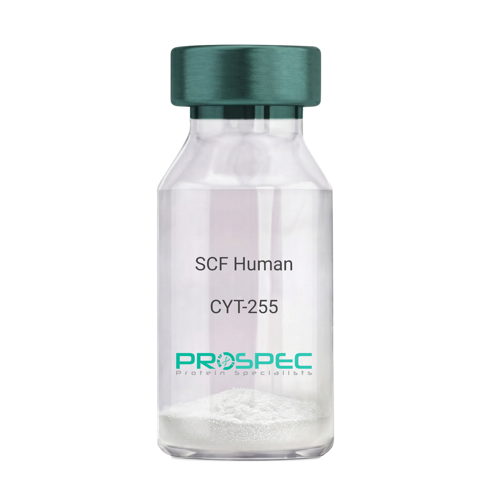
SCF HumanDescription:
Stem Cell Factor Human Recombinant
Kit ligand Precursor, C-kit ligand, SCF, Mast cell growth factor, MGF, SF, KL-1, Kitl, DKFZp686F2250.
Product # :
CYT-255Price :
Quantity :
Shipping Method :
Shipped at Room temp
More Info
- description
- source
- formulation
- purity
- biological activity
- More Info
- Citations
Description
Stem Cell Factor Human Recombinant produced in E.Coli is a single, non-glycosylated polypeptide chain containing 165 amino acids and having a molecular mass of 18409 Dalton. The SCF is purified by proprietary chromatographic techniques.
Source
Escherichia Coli.
Formulation
Lyophilized from a concentrated (1mg/ml) solution in water containing 0.02% NaHCO3.
Purity
Greater than 98.0% as determined by:
(a) Analysis by SEC-HPLC.
(b) Analysis by SDS-PAGE.Biological Activity
The ED50 as determined by the dose-dependant stimulation of Human TF-1 cells is < 2 ng/ml, corresponding to a Specific Activity of 500,000 IU/mg.Citations
More Info
-
Introduction
Stem cell factor / KIT ligand (SCF) is a cytokine which binds CD117(c-Kit). SCF is also known as "steel factor" or "c-kit ligand". SCF exists in two forms, cell surface bound SCF and soluble (or free) SCF. Soluble SCF is produced by the cleavage of surface bound SCF by metalloproteases. SCF is a growth factor important for the survival, proliferation, and differentiation of hematopoietic stem cells and other hematopoietic progenitor cells. One of its roles is to change the BFU-E (burst-forming unit-erythroid) cells, which are the earliest erythrocyte precursors in the erythrocytic series, into the CFU-E (colony-forming unit-erythroid).
-
Synonyms
Kit ligand Precursor, C-kit ligand, SCF, Mast cell growth factor, MGF, SF, KL-1, Kitl, DKFZp686F2250.
-
Physical Appearance
Sterile Filtered White lyophilized (freeze-dried) powder.
-
Stability
Lyophilized KIT ligand although stable at room temperature for 3 weeks, should be stored desiccated below -18°C. Upon reconstitution SCF should be stored at 4°C between 2-7 days and for future use below -18°C.For long term storage it is recommended to add a carrier protein (0.1% HSA or BSA).Please prevent freeze-thaw cycles.
-
Solubility
It is recommended to reconstitute the lyophilized Stem Cell Factor in sterile 18MΩ-cm H2O not less than 100µg/ml, which can then be further diluted to other aqueous solutions.
-
Amino Acid Sequence
The sequence of the first five N-terminal amino acids was determined and was found to be Met-Glu-Gly-Ile-Cys.
-
Protein content
Protein quantitation was carried out by two independent methods1. UV spectroscopy at 280 nm using the absorbency value of 0.52 as the extinction coefficient for a 0.1% (1mg/ml) solution. This value is calculated by the PC GENE computer analysis program of protein sequences (IntelliGenetics). 2. Analysis by RP-HPLC, using a standard solution of Stem Cell Factor as a Reference Standard.
ProSpec's products are furnished for LABORATORY RESEARCH USE ONLY. The product may not be used as drugs, agricultural or pesticidal products, food additives or household chemicals.
- View Data Sheet
Name :
b NGF Human, HEKDescription:
beta Nerve Growth Factor Human Recombinant, HEK
Beta Polypeptide, NGF, NGFB, HSAN5, Beta-NGF, MGC161426, MGC161428.
Product # :
CYT-079Price :
Quantity :
Shipping Method :
Shipped at Room temp
More Info
- description
- source
- formulation
- purity
- biological activity
- More Info
- Citations
Description
BNGF Human Recombinant produced in HEK293 is a noncovalently disulfide linked homodimer, glycosylated, polypeptide chain (Ser122-Arg239) containing 2 identical 118 amino acids and having a molecular mass of 26.5 kDa.
Source
HEK293 cells.
Formulation
The b-NGF was lyophilized from 1mg/ml in 20mM PB and 0.25M NaCl pH-7.5.
Purity
Greater than 97% as obsereved by SDS-PAGE.
Biological Activity
The specific activity was determined by the dose-dependent stimulation of the proliferation of human TF-1 cells (human erythroleukemic indicator cell line), the ED50 is <0.04-0.4ng/ml.
Citations
More Info
-
Introduction
NGF-beta has nerve growth stimulating activity and the complex is involved in the regulation of growth and the differentiation of sympathetic and certain sensory neurons. Mutations in this gene have been associated with hereditary sensory and autonomic neuropathy, type 5 (HSAN5), and dysregulation of this gene's expression is associated with allergic rhinitis.
-
Synonyms
Beta Polypeptide, NGF, NGFB, HSAN5, Beta-NGF, MGC161426, MGC161428.
-
Physical Appearance
Sterile Filtered White lyophilized (freeze-dried) powder.
-
Stability
Lyophilized b-NGF although stable at room temperature for 3 weeks, should be stored desiccated below -18°C. Upon reconstitution b-NGF should be stored at 4°C between 2-7 days and for future use below -18°C.For long term storage it is recommended to add a carrier protein (0.1% HSA or BSA).Please prevent freeze-thaw cycles.
-
Solubility
It is recommended to reconstitute the lyophilized b-NGF in sterile distilled pyrogen free water at a concentration of 0.25mg/ml.
-
Background
What is the molecular weight/Mw of B NGF Protein?
B NGF Protein has a total Mw of 26.5kDa.
What is the source or expression system of B NGF Protein?
HEK293 cells.
What is the Purity of B NGF Protein?
B NGF Protein is >97% pure as determined by SDS-PAGE.
What is the Biological Activity of B NGF Protein?
The specific activity was determined by the dose-dependent stimulation of the proliferation of human TF-1 cells (human erythroleukemic indicator cell line), the ED50 is <0.04-0.4ng/ml.
What is the amino acid sequence of B NGF Protein?
B NGF Protein is composed from 118 amino acids.
What applications can B NGF Protein be used in?
B NGF Protein can probably be used in western blot, ELISA and Lateral Flow.
What is the endotoxin level for B NGF Protein?
The endotoxin level is minimal, B NGF Protein was purified using conventional chromatography techniques.
ProSpec's products are furnished for LABORATORY RESEARCH USE ONLY. The product may not be used as drugs, agricultural or pesticidal products, food additives or household chemicals.
- View Data Sheet
Name :
GM- CSF HumanDescription:
Granulocyte Macrophage-Colony Stimulating Factor Human Recombinant
CSF-2, MGI-1GM, GM-CSF, Pluripoietin-alpha, MGC131935, MGC138897.
Product # :
CYT-221Price :
Quantity :
Shipping Method :
Shipped at Room temp
More Info
- description
- source
- formulation
- purity
- biological activity
- More Info
- Citations
Description
Granulocyte Macrophage Colony Stimulating Factor Human Recombinant produced in E.Coli is a single, non-glycosylated, polypeptide chain containing 127 amino acids and having a molecular mass of 14477 Dalton. GM-CSF is purified by proprietary chromatographic techniques.
Source
Escherichia Coli.
Formulation
GM-CSF was lyophilized after extensive dialysis against 2mM sodium phosphate buffer pH= 7.4±0.1.
Purity
Greater than 98.0% as determined by:
1. Analysis by RP-HPLC.
2. Analysis by SDS-PAGE.Biological Activity
The ED50 as determined by the dose-dependant stimulation of the proliferation of human TF-1 cells (human erythroleukemic indicator cell line) is < 0.1 ng/ml, corresponding to a Specific Activity of 11,100,000 IU/mg.Citations
More Info
-
Introduction
Granulocyte Macrophage Colony Stimulating Factor (GM-CSF) was first characterized as a growth factor that supports the in-vitro colony formation of granulocytes-macrophages progenitor cells. It is a pleiotropic cytokine and a member of a family of endogenous cytokines of the hematopoietic system. GM-CSF is produced as a response to immune or inflammatory stimuli by activated cells of the hematopoietic system such as T cells, B cells, macrophages, mast cells and also fibroblasts and alveolar epithelial cells. It plays an important role in regulating the proliferation, differentiation, survival and activation of hematopoietic cells such as granulocytes and monocytes ,neutrophiles, basophiles and eosonophoiles, erythroid cells, megakaryocytes and T cells.
Human and mouse GM-CSF have about 56% homology and are species specific. Human GM-CSF is not active on mouse cells and vice versa. It is active on canine and feline cells.
GMCSF is 144 amino acids, 22kDa glycoprotein. It is composed of four bundles alpha helices. Its receptor is heterodimers with a ligand-specific alpha subunit and a betac subunit that is shared with the interleukin IL-3 and IL-5 receptors. This unusual form of receptor assembly likely applies also to IL-3 and IL-5 receptors. Cross-linking the two receptor subunits is required for receptor activation and signaling .
GMCSF has been shown to be involved in maturation, mobilization and antigen presentation of myeloid dentritic cells (DCs) in-vivo or ex-vivo. This function promotes Th1 immune responses, cytotoxcity, anti-angiogenesis as well as allergic inflammation, and the development of autoimmunity. Therefore GMCSF can be used in immunotherapy for the treatment of immune suppressed and immune-compromised patients as well as in veterinary medicine for the same purpose. GM-CSF is also important in regulation of embryo development and pregnancy and specifically in embryo implantation and subsequent development . -
Synonyms
CSF-2, MGI-1GM, GM-CSF, Pluripoietin-alpha, MGC131935, MGC138897.
-
Physical Appearance
Sterile Filtered White lyophilized (freeze-dried) powder.
-
Stability
Lyophilized Granulocyte Macrophage Colony Stimulating Factor although stable at room temperature for 3 weeks, should be stored desiccated below -18°C. Upon reconstitution GMCSF should be stored at 4°C between 2-7 days and for future use below -18°C.For long term storage it is recommended to add a carrier protein (0.1% HSA or BSA).Please prevent freeze-thaw cycles.
-
Solubility
It is recommended to reconstitute the lyophilized Granulocyte Macrophage Colony Stimulating Factor in sterile 18MΩ-cm H2O not less than 100µg/ml, which can then be further diluted to other aqueous solutions.
-
Amino Acid Sequence
The sequence of the first five N-terminal amino acids was determined and was found to be Ala-Pro-Ala-Arg-Ser.
N-terminal methionine has been completely removed enzymatically. -
Background
What is the molecular weight/Mw of GM CSF HUMAN Protein?
GM CSF HUMAN Protein has a total Mw of 14.47kDa.
What is the source or expression system of GM CSF HUMAN Protein?
Escherichia Coli.
What is the Purity of GM CSF HUMAN Protein?
GM CSF HUMAN Protein is >98% pure as determined by SDS-PAGE.
What is the Biological Activity of GM CSF HUMAN Protein?
The ED50 as determined by the dose-dependant stimulation of the proliferation of human TF-1 cells (human erythroleukemic indicator cell line) is < 0.1 ng/ml, corresponding to a Specific Activity of 11,100,000 IU/mg.
What is the amino acid sequence of GM CSF HUMAN Protein?
The sequence of the first five N-terminal amino acids was determined and was found to be Ala-Pro-Ala-Arg-Ser.
N-terminal methionine has been completely removed enzymatically.
What applications can GM CSF HUMAN Protein be used in?
GM CSF HUMAN Protein can probably be used in western blot, ELISA and Lateral Flow.
What is the endotoxin level for GM CSF HUMAN Protein?
The endotoxin level is minimal, GM CSF HUMAN Protein was purified using conventional chromatography techniques.
-
Protein content
GM-CSF quantitation was carried out by two independent methods:1. UV spectroscopy at 280 nm using the absorbency value of 0.963 as the extinction coefficient for a 0.1% (1mg/ml) solution. This value is calculated by the PC GEN computer analysis program of protein sequences (IntelliGenetics). 2. Analysis by RP-HPLC, using a standard solution of GM-CSF as a Reference Standard.
ProSpec's products are furnished for LABORATORY RESEARCH USE ONLY. The product may not be used as drugs, agricultural or pesticidal products, food additives or household chemicals.
- View Data Sheet
Name :
Adiponectin Human, HisDescription:
Adiponectin Human Recombinant, His tag
Acrp30, AdipoQ, GBP-28, APM-1, ACDC.
Product # :
CYT-433Price :
Quantity :
Shipping Method :
Shipped at Room temp
More Info
- description
- source
- formulation
- purity
- More Info
- Citations
Description
The Acrp30 Human is created as a recombinant protein with N-terminal fusion of His Tag. The Adiponectin His-Tagged Fusion Protein, produced in E. coli, is 26.4 kDa protein containing 230 amino acid residues of the Acrp30 Human and 12 additional amino acid residues - HisTag (underlined).
Source
Escherichia Coli.
Formulation
Acrp30 Human was filtered (0.4µm) and lyophilized from 0.5 mg/ml in 0.02M Tris buffer pH7.5, 0.15M NaCl.
Purity
Acrp30 Human purity is greater than 95% as determined by SDS-PAGE.
Citations
More Info
-
Introduction
Adiponectin, also referred to as Acrp30, AdipoQ and GBP-28, is a recently discovered 244 amino acid protein, the product of the apM1 gene, which is physiologically active and specifically and highly expressed in adipose cells (Adipokine). The protein belongs to the soluble defense collagen super family; it has a collagen-like domain structurally homologous with collagen VIII and X and complement factor C1q-like globular domain. APM-1 forms homotrimers, which are the building blocks for higher order complexes found circulating in serum.
-
Synonyms
Acrp30, AdipoQ, GBP-28, APM-1, ACDC.
-
Physical Appearance
Filtered White lyophilized (freeze-dried) powder.
-
Stability
Store lyophilized Acrp30 Human at -20°C. Aliquot the product after reconstitution to avoid repeated freezing/thawing cycles. Reconstituted Acrp30 Human can be stored at 4°C for a limited period of time; it does not show any change after two weeks at 4°C.
-
Solubility
It is recommended to add deionized water to prepare a working stock solution of approximately 0.5mg/ml and let the lyophilized pellet dissolve completely. Product is not sterile! Please filter the product by an appropriate sterile filter before using it on cell culture.
-
Amino Acid Sequence
MRGSHHHHHHGSGHDQETTT QGPGVLLPLP KGACTGWMAG IPGHPGHNGA PGRDGRDGTP GEKGEKGDPG LIGPKGDIGE TGVPGAEGPR GFPGIQGRKG EPGEGAYVYR SAFSVGLETY VTIPNMPIRF TKIFYNQQNH YDGSTGKFHC NIPGLYYFAY HITVYMKDVK VSLFKKDKAM LFTYDQYQEN NVDQASGSVL LHLEVGDQVW LQVYGEGERN GLYADNDNDS TFTGFLLYHD TN.
-
Background
What is the molecular weight / Mw of ADIPONECTIN Protein?
ADIPONECTIN Protein has a total Mw of 26.4kDa.
What is the source or expression system of ADIPONECTIN Protein?
Escherichia Coli.
What is the Purity of ADIPONECTIN Protein?
ADIPONECTIN Protein is >95% pure as determined by SDS-PAGE.
What is the Biological Activity of ADIPONECTIN Protein?
The biological functionality of ADIPONECTIN Protein will be determined in the future.
What is the amino acid sequence of ADIPONECTIN Protein?
MRGSHHHHHHGSGHDQETTT QGPGVLLPLP KGACTGWMAG IPGHPGHNGA PGRDGRDGTP GEKGEKGDPG LIGPKGDIGE TGVPGAEGPR GFPGIQGRKG EPGEGAYVYR SAFSVGLETY VTIPNMPIRF TKIFYNQQNH YDGSTGKFHC NIPGLYYFAY HITVYMKDVK VSLFKKDKAM LFTYDQYQEN NVDQASGSVL LHLEVGDQVW LQVYGEGERN GLYADNDNDS TFTGFLLYHD TN.
What applications can ADIPONECTIN Protein be used in?
ADIPONECTIN Protein can probably be used in western blot, ELISA and Lateral Flow.
What is the endotoxin level for ADIPONECTIN Protein?
The endotoxin level is minimal, ADIPONECTIN Protein was purified using conventional chromatography techniques
ProSpec's products are furnished for LABORATORY RESEARCH USE ONLY. The product may not be used as drugs, agricultural or pesticidal products, food additives or household chemicals.
- View Data Sheet
Name :
SUMF1 HumanDescription:
Sulfatase Modifying Factor 1 Human Recombinant
Sulfatase modifying factor 1, FGE, C-alpha-formylglycine-generating enzyme 1, FGly-generating enzyme, UNQ3037, AAPA3037, EC 1.8.99.
Product # :
PRO-986Price :
Quantity :
Shipping Method :
Shipped with Ice Packs
More Info
- description
- source
- formulation
- purity
- More Info
- Citations
Description
SUMF1 Human Recombinant produced in E.Coli is a single, non-glycosylated polypeptide chain containing 304 amino acids (91-374 a.a.) and having a molecular mass of 34.1kDa.SUMF1 is fused to a 20 amino acid His-tag at N-terminus & purified by proprietary chromatographic techniques.
Source
E.coli.
Formulation
SUMF1 protein solution (0.5mg/ml) containing 20mM Tris-HCl buffer (pH8.0), 2M UREA, 2mM DTT and 20% glycerol.
Purity
Greater than 85% as determined by SDS-PAGE.
Citations
More Info
-
Introduction
SUMF1 is a member of the SUMF family. SUMF1 catalyzes the hydrolysis of sulfate esters by oxidizing a cysteine residue in the substrate sulfatase to an active site 3-oxoalanine residue called C-alpha-formylglycine. Alterations in this gene result in multiple sulfatase deficiency which is a lysosomal storage disorder.
-
Synonyms
Sulfatase modifying factor 1, FGE, C-alpha-formylglycine-generating enzyme 1, FGly-generating enzyme, UNQ3037, AAPA3037, EC 1.8.99.
-
Physical Appearance
Sterile Filtered colorless solution.
-
Stability
Store at 4°C if entire vial will be used within 2-4 weeks. Store, frozen at -20°C for longer periods of time. For long term storage it is recommended to add a carrier protein (0.1% HSA or BSA).Avoid multiple freeze-thaw cycles.
-
Amino Acid Sequence
MGSSHHHHHH SSGLVPRGSH MVPIPAGVFT MGTDDPQIKQ DGEAPARRVT IDAFYMDAYE VSNTEFEKFV NSTGYLTEAE KFGDSFVFEG MLSEQVKTNI QQAVAAAPWW LPVKGANWRH PEGPDSTILH RPDHPVLHVS WNDAVAYCTW AGKRLPTEAE WEYSCRGGLH NRLFPWGNKL QPKGQHYANI WQGEFPVTNT GEDGFQGTAP VDAFPPNGYG LYNIVGNAWE TSDWWTVHH SVEETLNPKG PPSGKDRVKK GGSYMCHRSY CYRYRCAARS QNTPDSSASN LGFRCAADRL PTMD
ProSpec's products are furnished for LABORATORY RESEARCH USE ONLY. The product may not be used as drugs, agricultural or pesticidal products, food additives or household chemicals.
- View Data Sheet
Name :
NTRK2 MouseDescription:
Neurotrophic Receptor Tyrosine Kinase 2 Mouse Recombinant
TrkB, NTRK2, NTRK-2, Tyrosine Kinase Receptor B, BDNF/NT-3 Growth Factors Receptor
Product # :
CYT-919Price :
Quantity :
Shipping Method :
Shipped with Ice Packs
More Info
- description
- source
- formulation
- purity
- More Info
- Citations
Description
NTRK2 Mouse produced in Sf9 Insect cells is a single, glycosylated polypeptide chain containing 409 amino acids (32-429a.a.) and having a molecular mass of 45.7kDa (Molecular size on SDS-PAGE will appear at approximately 57-70kDa under reducing conditions).NTRK2 Mouse is expressed with an 11 amino acid His tag at C-Terminus and purified by proprietary chromatographic techniques.
Source
Sf9, Insect cells.
Formulation
NTRK2 Mouse protein solution (0.5mg/ml) contains PBS pH 7.4 and 10% glycerol.
Purity
Greater than 95.0% as determined by SDS-PAGE.
Citations
More Info
-
Introduction
NTRK2 plays a role in the growth and the development of the central and the peripheral nervous systems using the regulation of neuron proliferation, migration, survival, differentiation, and synapse production. NTRK2 is a receptor for B-DNF and NT4. NTRK2 binds to neurotrophin-3 which regulates neuron survival through NTRK2. NTRK-2 activates numerous effectors such as SHC-1, SH2B-1, PLCG-1 and SH2B-2 that control overlapping signaling cascades.
-
Synonyms
TrkB, NTRK2, NTRK-2, Tyrosine Kinase Receptor B, BDNF/NT-3 Growth Factors Receptor
-
Physical Appearance
Sterile filtered colorless solution.
-
Stability
Store at 4°C if entire vial will be used within 2-4 weeks.Store, frozen at -20°C for longer periods of time. For long term storage it is recommended to add a carrier protein (0.1% HSA or BSA).Avoid multiple freeze-thaw cycles.
-
Amino Acid Sequence
CPTSCKCSSA RIWCTEPSPG IVAFPRLEPN SVDPENITEI LIANQKRLEI INEDDVEAYV GLRNLTIVDS GLKFVAYKAF LKNSNLRHIN FTRNKLTSLS RRHFRHLDLS DLILTGNPFT CSCDIMWLKT LQETKSSPDT QDLYCLNESS KNMPLANLQI PNCGLPSARL AAPNLTVEEG KSVTLSCSVG GDPLPTLYWD VGNLVSKHMN ETSHTQGSLR ITNISSDDSG KQISCVAENL VGEDQDSVNL TVHFAPTITF LESPTSDHHW CIPFTVRGNP KPALQWFYNG AILNESKYIC TKIHVTNHTE YHGCLQLDNP THMNNGDYTL MAKNEYGKDE RQISAHFMGR PGVDYETNPN YPEVLYEDWT TPTDIGDTTN KSNEIPSTDV ADQSNREHAA ALEHHHHHH.
ProSpec's products are furnished for LABORATORY RESEARCH USE ONLY. The product may not be used as drugs, agricultural or pesticidal products, food additives or household chemicals.
- View Data Sheet
Name :
IL3 Mouse, sf9Description:
Interleukin-3 Mouse Recombinant, sf9
Interleukin-3, Interleukin-3, sf9, IL3 sf9, L-3, Hematopoietic growth factor, Mast cell growth factor, MCGF, Multipotential colony-stimulating factor, P-cell-stimulating factor, Il3, Csfmu, Il-3, BPA, Csfmu, HCGF, Il-3, MCGF, PSF.
Product # :
CYT-922Price :
Quantity :
Shipping Method :
Shipped with Ice Packs
More Info
- description
- source
- formulation
- purity
- biological activity
- More Info
- SDS-PAGE
- Citations
Description
IL3 Mouse Recombinant produced in Sf9 Baculovirus cells is a single, glycosylated polypeptide chain containing 148 amino acids (27-166 a.a.) and having a molecular mass of 16.7kDa (Migrates at 18-28kDa on SDS-PAGE under reducing conditions).IL3 is expressed with a 6 amino acid His tag at C-Terminus and purified by proprietary chromatographic techniques.
Source
Sf9, Baculovirus cells.
Formulation
IL3 protein solution (0.25mg/ml) contains Phosphate Buffered Saline (pH 7.4) and 10% glycerol.
Purity
Greater than 90.0% as determined by SDS-PAGE.
Biological Activity
The ED50 is Measured in a cell proliferation assay using TF-1 human erythroleukemic and is ≤ 0.1 ng/ml.
SDS-PAGE
Citations
More Info
-
Introduction
Interleukin-3 is a pleiotropic cytokine produced primarily by activated T cells.
IL-3 is thought to function via specific cell surface receptors to stimulate the proliferation, differentiation and survival of haematopoietic cell lines. IL-3 has also been shown to affect the functional activity of a variety of other cell types including mast cells, eosinophils, megakaryocytes and basophils. -
Synonyms
Interleukin-3, Interleukin-3, sf9, IL3 sf9, L-3, Hematopoietic growth factor, Mast cell growth factor, MCGF, Multipotential colony-stimulating factor, P-cell-stimulating factor, Il3, Csfmu, Il-3, BPA, Csfmu, HCGF, Il-3, MCGF, PSF.
-
Physical Appearance
Sterile Filtered colorless solution.
-
Stability
Store at 4°C if entire vial will be used within 2-4 weeks. Store, frozen at -20°C for longer periods of time. For long term storage it is recommended to add a carrier protein (0.1% HSA or BSA). Avoid multiple freeze-thaw cycles.
-
Amino Acid Sequence
ASISGRDTHR LTRTLNCSSI VKEIIGKLPE PELKTDDEGP SLRNKSFRRV NLSKFVESQG EVDPEDRYVI KSNLQKLNCC LPTSANDSAL PGVFIRDLDD FRKKLRFYMV HLNDLETVLT SRPPQPASGS VSPNRGTVEC LEHHHHHH.
-
Background
What is the molecular weight/Mw of IL3 MOUSE, SF9 Protein?
IL3 MOUSE, SF9 Protein has a total Mw of 16.7kDa.
What is the source or expression system of IL3 MOUSE, SF9 Protein?
Sf9, Baculovirus cells.
What is the Purity of IL3 MOUSE, SF9 Protein?
IL3 MOUSE, SF9 Protein is >90% pure as determined by SDS-PAGE.
What is the Biological Activity of IL3 MOUSE, SF9 Protein?
The ED50 is Measured in a cell proliferation assay using TF-1 human erythroleukemic and is ≤ 0.1 ng/ml.
What is the amino acid sequence of IL3 MOUSE, SF9 Protein?
ASISGRDTHR LTRTLNCSSI VKEIIGKLPE PELKTDDEGP SLRNKSFRRV NLSKFVESQG EVDPEDRYVI KSNLQKLNCC LPTSANDSAL PGVFIRDLDD FRKKLRFYMV HLNDLETVLT SRPPQPASGS VSPNRGTVEC LEHHHHHH.
What applications can IL3 MOUSE, SF9 Protein be used in?
IL3 MOUSE, SF9 Protein can probably be used in western blot, ELISA and Lateral Flow.
What is the endotoxin level for IL3 MOUSE, SF9 Protein?
The endotoxin level is minimal, IL3 MOUSE, SF9 Protein was purified using conventional chromatography techniques.
Who supplies recombinant IL3 MOUSE, SF9 that is in stock and available to order?
ProSpec has the protein in stock and available for immediate delivery.
Where can I order bulk mg quantities of IL3 MOUSE, SF9?
ProSpec supplies bulk mg quantities at affordable prices that meets research and academic budgets.
ProSpec's products are furnished for LABORATORY RESEARCH USE ONLY. The product may not be used as drugs, agricultural or pesticidal products, food additives or household chemicals.
- View Data Sheet
Name :
EGF Mouse, HisDescription:
Epidermal Growth Factor Mouse Recombinant, His Tag
Urogastrone, URG, EGF.
Product # :
CYT-138Price :
Quantity :
Shipping Method :
Shipped with Ice Packs
More Info
- description
- source
- formulation
- purity
- More Info
- SDS-PAGE
- Citations
Description
EGF mouse Recombinant produced in E. coli is a single polypeptide chain containing 77 amino acids (977-1029) and having a molecular mass of 8.6kDa.EGF is fused to a 24 amino acid His-tag at N-terminus & purified by proprietary chromatographic techniques.
Source
E.coli.
Formulation
The EGF solution (0.5mg/1ml) contains 20mM Tris-HCl buffer (pH 8.0), 100mM NaCl, 2mM DTT and 10% glycerol.
Purity
Greater than 90% as determined by SDS-PAGE.
SDS-PAGE
Citations
More Info
-
Introduction
Epidermal growth factor has a profound effect on the differentiation of specific cells in vivo and is a potent mitogenic factor for a variety of cultured cells of both ectodermal and mesodermal origin. The EGF precursor is believed to exist as a membrane-bound molecule which is proteolytically cleaved to generate the 53-amino acid peptide hormone that stimulates cells to divide.
EGF stimulates the growth of various epidermal and epithelial tissues in vivo and in vitro and of some fibroblasts in cell culture. -
Synonyms
Urogastrone, URG, EGF.
-
Physical Appearance
Sterile Filtered colorless solution.
-
Stability
Store at 4°C if entire vial will be used within 2-4 weeks. Store, frozen at -20°C for longer periods of time. For long term storage it is recommended to add a carrier protein (0.1% HSA or BSA).Avoid multiple freeze-thaw cycles.
-
Amino Acid Sequence
MGSSHHHHHH SSGLVPRGSH MGSMNSYPGC PSSYDGYCLN GGVCMHIESL DSYTCNCVIG YSGDRCQTRD LRWWELR.
-
Background
Unveiling Epidermal Growth Factor Mouse Recombinant: Harnessing His Tag for Enhanced Insights and Therapeutic Prospects
Abstract:
This research paper delves into the realm of Epidermal Growth Factor Mouse Recombinant (EGF-MR), focusing on the strategic integration of a Histidine (His) Tag. By employing sophisticated methodologies encompassing protein engineering, chromatographic techniques, and cellular assays, this study unveils the multifaceted molecular attributes of EGF-MR with His Tag. The findings not only enhance our understanding of EGF-MR's behavior but also illuminate potential avenues for therapeutic interventions.
Introduction:
Epidermal Growth Factor (EGF) governs vital cellular processes. This paper delves into Epidermal Growth Factor Mouse Recombinant (EGF-MR) with a specific emphasis on the incorporation of a Histidine (His) Tag, unraveling its molecular intricacies and therapeutic implications.
Protein Engineering and His Tag Integration:
The paper navigates the tailored engineering of EGF-MR to accommodate a His Tag, a peptide sequence that facilitates protein purification. The process involves strategic modification of the EGF-MR gene to ensure proper folding and presentation of the His Tag.
Chromatographic Purification and His Tag Affinity:
Chromatographic techniques, specifically immobilized metal ion affinity chromatography (IMAC), are employed to purify the His-tagged EGF-MR. The His Tag's high affinity for metal ions facilitates efficient purification, yielding a highly purified and bioactive protein product.
Structural and Functional Insights:
The presence of the His Tag is not just for purification; it serves as a molecular handle to investigate EGF-MR's structural dynamics. High-resolution structural analyses coupled with biophysical assays unravel how the His Tag affects EGF-MR's conformation and binding interactions.
Cellular Assays and Bioactivity Assessment:
In vitro cellular assays, including proliferation and migration studies, provide insights into the impact of His Tag on EGF-MR's bioactivity. Comparative analyses shed light on the functionality of His-tagged EGF-MR and its potential implications in cellular responses.
Therapeutic Prospects and Targeted Delivery:
The incorporation of a His Tag presents a unique avenue for tailored drug delivery. The His Tag can serve as a docking site for targeted therapies, enabling precise interactions with specific receptors on target cells.
Future Directions and Challenges:
While promising, challenges such as potential steric hindrance from the His Tag require consideration. Future research should focus on optimizing the positioning of the His Tag to maintain EGF-MR's full biological activity.
Conclusion:
In a harmonious synthesis of advanced methodologies and innovative insights, the integration of His Tag into Epidermal Growth Factor Mouse Recombinant emerges as a transformative paradigm. The His Tag not only facilitates purification but also offers a molecular window into EGF-MR's behavior, potentially redefining targeted therapies and precision medicine.
What is the molecular weight/Mw of EGF MOUSE, HIS Protein?
EGF MOUSE, HIS Protein has a total Mw of 8.6kDa.
What is the source or expression system of EGF MOUSE, HIS Protein?
Escherichia Coli.
What is the Purity of EGF MOUSE, HIS Protein?
EGF MOUSE, HIS Protein is >90% pure as determined by SDS-PAGE.
What is the Biological Activity of EGF MOUSE, HIS Protein?
The biological functionality of EGF MOUSE, HIS Protein will be determined in the future.
What is the amino acid sequence of EGF MOUSE, HIS Protein?
MGSSHHHHHH SSGLVPRGSH MGSMNSYPGC PSSYDGYCLN GGVCMHIESL DSYTCNCVIG YSGDRCQTRD LRWWELR.
What applications can EGF MOUSE, HIS Protein be used in?
EGF MOUSE, HIS Protein can probably be used in western blot, ELISA and Lateral Flow.
What is the endotoxin level for EGF MOUSE, HIS Protein?
The endotoxin level is minimal, EGF MOUSE, HIS Protein was purified using conventional chromatography techniques
ProSpec's products are furnished for LABORATORY RESEARCH USE ONLY. The product may not be used as drugs, agricultural or pesticidal products, food additives or household chemicals.
- View Data Sheet
Name :
IL33 AntibodyDescription:
Interleukin-33, Mouse Anti Human
Interleukin 33, DVS27, NF-HEV, NKHEV, C9orf26, Interleukin-1 family member 11, IL- 1F11, Nuclear factor from high endothelial venules, NFEHEV, DKFZp586H0523, RP11-575C20.2, IL-33.
Product # :
ANT-369Price :
Quantity :
Shipping Method :
Shipped with Ice Packs
More Info
- formulation
- More Info
- Citations
Formulation
1mg/ml containing PBS, pH-7.4, & 0.1% Sodium Azide.
Citations
More Info
-
Introduction
Interleukin 33 (IL-33) is a 32kDa proinflammatory cytokine that may also regulate gene transcription in producer cells. IL-33 is structurally related to IL-1, which induces helper T cells to produce type 2 cytokines and acts through the receptor IL1RL-1 (IL1 receptor-like-1), which is known also as ST2. Binding of IL-33 to this receptor activates NF-kappa-B and MAP kinases and induces in vitro Th2 cells to produce cytokines. In vivo, IL-33 induces expression of IL-4, IL-5, IL-13 and leads to severe pathological changes in mucosal organs and in vitro, it can be divided to N-terminal fragment of 12kDa and C-terminal fragment of 18kDa by cleavage of caspase-1.
-
Synonyms
Interleukin 33, DVS27, NF-HEV, NKHEV, C9orf26, Interleukin-1 family member 11, IL- 1F11, Nuclear factor from high endothelial venules, NFEHEV, DKFZp586H0523, RP11-575C20.2, IL-33.
-
Physical Appearance
Sterile Filtered colorless solution.
-
Immunogen
Anti-human IL33 mAb, is derived from hybridization of mouse F0 myeloma cells with spleen cells from BALB/c mice immunized with Recombinant human IL33 amino acids 112-270 purified from E. coli.
-
Ig Subclass
Mouse IgG2b heavy chain and κ light chain.
-
Clone
P4E9AT.
-
Applications
IL33 antibody has been tested by ELISA and Western blot analysis to assure specificity and reactivity. Since application varies, however, each investigation should be titrated by the reagent to obtain optimal results. Recommended dilution range for Western blot analysis is 1:500 ~ 1,000. Recommended starting dilution is 1:500.
-
Type
Mouse Anti Human Monoclonal.
-
Storage Procedures
For periods up to 1 month store at 4°C, for longer periods of time, store at -20°C. Prevent freeze thaw cycles.
-
Purification Method
IL33 antibody was purified from mouse ascitic fluids by protein-G affinity chromatography.
ProSpec's products are furnished for LABORATORY RESEARCH USE ONLY. The product may not be used as drugs, agricultural or pesticidal products, food additives or household chemicals.
- View Data Sheet
Name :
TLR3 HumanDescription:
Toll Like Receptor 3 Human Recombinant
Toll Like Receptor 3, Toll-Like Receptor 3, CD283 Antigen, CD283, IIAE2, Toll-like receptor 3.
Product # :
PRO-2511Price :
Quantity :
Shipping Method :
Shipped with Ice Packs
More Info
- description
- source
- formulation
- purity
- More Info
- Citations
Description
TLR3 produced in Sf9 Baculovirus cells is a single, glycosylated polypeptide chain containing 690 amino acids (23-704 a.a.) and having a molecular mass of 78.5kDa. (Molecular size on SDS-PAGE will appear at approximately 70-100kDa).TLR3 is expressed with an 8 amino acid His tag at C-Terminus and purified by proprietary chromatographic techniques.
Source
Sf9, Baculovirus cells.
Formulation
TLR3 protein solution (0.5 mg/ml) contains 20% glycerol, 20mM Tris-HCl Buffer (pH 8.0), 0.1M NaCl & 1mM DTT.
Purity
Greater than 90.0% as determined by SDS-PAGE.
Citations
More Info
-
Introduction
TLR3 (Toll-like receptor 3) belongs to the toll-like receptor family which takes an essential part in pathogen recognition as well as activation of innate immunity. TLR3 has innate immune receptors for sensing microbial molecules in addition to damage-associated molecular patterns released from host cells. TLR3 recognizes dsRNA and activation of the receptor which induces the activation of NF-kappaB and the production of type I interferons. TLR3 signaling activation is related with ischemic preconditioning-induced protection against brain ischemia and attenuation of reactive astrogliosis. TLR3 is most plentifully expressed in the placenta & pancreas, and is limited to the dendritic subpopulation of the leukocytes.
-
Synonyms
Toll Like Receptor 3, Toll-Like Receptor 3, CD283 Antigen, CD283, IIAE2, Toll-like receptor 3.
-
Physical Appearance
Sterile Filtered colorless solution.
-
Stability
Store at 4°C if entire vial will be used within 2-4 weeks. Store, frozen at -20°C for longer periods of time. For long term storage it is recommended to add a carrier protein (0.1% HSA or BSA).Avoid multiple freeze-thaw cycles.
-
Amino Acid Sequence
SSTTKCTVSH EVADCSHLKL TQVPDDLPTN ITVLNLTHNQ LRRLPAANFT RYSQLTSLDV GFNTISKLEP ELCQKLPMLK VLNLQHNELS QLSDKTFAFC TNLTELHLMS NSIQKIKNNP FVKQKNLITL DLSHNGLSST KLGTQVQLEN LQELLLSNNK IQALKSEELD IFANSSLKKL ELSSNQIKEF SPGCFHAIGR LFGLFLNNVQ LGPSLTEKLC LELANTSIRN LSLSNSQLST TSNTTFLGLK WTNLTMLDLS YNNLNVVGND SFAWLPQLEY FFLEYNNIQH LFSHSLHGLF NVRYLNLKRS FTKQSISLAS LPKIDDFSFQ WLKCLEHLNM EDNDIPGIKS NMFTGLINLK YLSLSNSFTS LRTLTNETFV SLAHSPLHIL NLTKNKISKI ESDAFSWLGH LEVLDLGLNE IGQELTGQEW RGLENIFEIY LSYNKYLQLT RNSFALVPSL QRLMLRRVAL KNVDSSPSPF QPLRNLTILD LSNNNIANIN DDMLEGLEKL EILDLQHNNL ARLWKHANPG GPIYFLKGLS HLHILNLESN GFDEIPVEVF KDLFELKIID LGLNNLNTLP ASVFNNQVSL KSLNLQKNLI TSVEKKVFGP AFRNLTELDM RFNPFDCTCE SIAWFVNWIN ETHTNIPELS SHYLCNTPPH YHGFPVRLFD TSSCKDSAPF ELLEHHHHHH.
ProSpec's products are furnished for LABORATORY RESEARCH USE ONLY. The product may not be used as drugs, agricultural or pesticidal products, food additives or household chemicals.
- View Data Sheet
Name :
FGF23 HumanDescription:
Fibroblast Growth Factor-23 Human Recombinant
Tumor-derived hypophosphatemia-inducing factor, HYPF, ADHR, HPDR2, PHPTC, FGF23, FGF-23, Fibroblast Growth Factor-23.
Product # :
CYT-020Price :
Quantity :
Shipping Method :
Shipped at Room temp
More Info
- description
- source
- formulation
- purity
- biological activity
- More Info
- Citations
Description
Fibroblast Growth Factor-23 Human Recombinant produced in E.Coli is a single, non-glycosylated polypeptide chain containing a total of 228 amino acids and having a molecular mass of 22.5kDa. The FGF-23 is and purified by chromatographic techniques.
Source
Escherichia Coli.
Formulation
The FGF-23 protein (0.5mg/ml) was lyophilized from a 0.2µm filtered concentrated solution in PBS, pH 7.4.
Purity
Greater than 95.0% as determined by SDS-PAGE.
Biological Activity
The biological activity of FGF-23 was measured in a cell proliferation assay using NIH/3T3 mouse embryonic fibroblasts. The ED50 for this effect is typically 0.05-0.5µg/ml in the presence of 5µg/ml of Recombinant Mouse Klotho and 10 µg/ml of HPR.
Citations
More Info
-
Introduction
FGF-23 is a member of the fibroblast growth factor (FGF) family. FGF family members possess broad mitogenic and cell survival activities and are involved in a variety of biological processes including embryonic development, cell growth, morphogenesis, tissue repair, tumor growth and invasion. FGF-23 inhibits renal tubular phosphate transport. The FGF-23 gene was identified by its mutations associated with autosomal dominant hypophosphatemic rickets (ADHR), an inherited phosphate wasting disorder. Abnormally high level expression of FGF-23 was found in oncogenic hypophosphatemic osteomalacia (OHO), a phenotypically similar disease caused by abnormal phosphate metabolism. FGF-23 mutations have also been shown to cause familial tumoral calcinosis with hyperphosphatemia.
-
Synonyms
Tumor-derived hypophosphatemia-inducing factor, HYPF, ADHR, HPDR2, PHPTC, FGF23, FGF-23, Fibroblast Growth Factor-23.
-
Physical Appearance
Sterile Filtered white lyophilized powder.
-
Stability
Lyophilized FGF-23 although stable at room temperature for 3 weeks, should be stored desiccated below -18°C. Upon reconstitution FGF-23 should be stored at 4°C between 2-7 days and for future use below -18°C.For long term storage it is recommended to add a carrier protein (0.1% HSA or BSA).Please prevent freeze-thaw cycles.
-
Solubility
It is recommended to reconstitute the lyophilized FGF-23 in sterile 18M-cm H2O not less than 100µg/ml, which can then be further diluted to other aqueous solutions.
-
Amino Acid Sequence
MYPNASPLLG SSWGGLIHLY TATARNSYHL QIHKNGHVDG APHQTIYSAL MIRSEDAGFV VITGVMSRRY LCMDFRGNIF GSHYFDPENC RFQHQTLENG YDVYHSPQYH FLVSLGRAKR AFLPGMNPPP YSQFLSRRNE IPLIHFNTPI PRRHTRSAED DSERDPLNVL KPRARMTPAP ASCSQELPSA EDNSPMASDP LGVVRGGRVN THAGGTGPEG CRPFAKFI.
-
Background
Before it was discovered in 2000, there was a hypothesis that a similar type of protein existed that performed many of the functions we see in FGF23. This was originally referred to as phosphatonin. Various effects were described and noted by researchers including inhibition of production and inhibition of secretion of parathyroid hormone. Derived from the bone Fibroblast Growth Factor 23 is a phosphaturic hormone. It increases phosphate excretion when acting on the kidney and also suppresses the biosynthesis of 1,25(OH)2D3.
Mechanism
Despite various research studies into the topic, many of the mechanisms for the regulations of FGF23 production remain a mystery to the scientific community. While we know that mutations in PHEX, ENPP1 and DMP1 result in the increased expression of FGF23, it is unclear why this occurs. This also means that currently, it is not possible to regulate the production of FGF23 either. We also do not know how signals from FGF23 regulate vitamin D metabolism. However, understanding these types of mechanisms could offer information needed to provide better treatment for deranged bone and mineral metabolism.Interactions
Molecular interactions involving FGF-23, vitamin D and klotho do provide the solution needed to regulate phosphate levels within the body. Furthermore, an interaction between Vitamin D and FGF3 can have an impact on renal phosphate balance. As well as this, when in the presence of klotho, FGF3 does actually increase bioactivity and begins to change systemic phosphate homeostasis.Function
Based on research it seems that the main function for FGF23 is the regulation of phosphate concentration in plasma. It seems to be secreted from the osteocytes due to elevated levels . When acting upon the kidneys, the hormone reduces the expression of NPT2. This is a sodium-phosphate cotransporter found in the proximal tube.
As such, it appears as though FGF23 is able to reduce the reabsorption, all the while maxing the excretion of phosphate. It has also been suggested that the hormone is able to suppress 1-alpha-hydroxylase. If this is the case, it can limit its potential to activate vitamin D and thus impair the ability for calcium absorption.Structure
FGF243 is located on the chromosome 12. It is composed of three exons. The crystal structure of FGF23 is completely different from the common conformation typically adopted by paracrine-acting FGFs. Instead, there is a conformation of the HPR region between beta strands 10 and 12. As well as this, there is a cleft between the other HPR-binding region, the beta1-beta12 loop and this one. This comes before a direct interaction between HPR sulfate and FGF23s backbone atoms. Due to this, endocrine function is benefitted and HPR-binding affinity is reduced for the lipangs.
Certain mutations that cause the protein to be completely resistant to proteolytic cleavage does trigger a surge in activity of the protein and the renal phosphate loss typically found in certain human diseases including hypophosphatemic rickets.
Studies have also revealed that FGF23 is overproduced in certain tumors including phosphaturic mesenchymal tumors. Furthermore a reduced level of activity for this protein is believed to lead to higher phosphate levels and familial tumor calcinosis clinical syndrome.What is the molecular weight/Mw of FGF23 HUMAN Protein?
FGF23 HUMAN Protein has a total Mw of 22.5kDa.
What is the source or expression system of FGF23 HUMAN Protein?
Escherichia Coli.
What is the Purity of FGF23 HUMAN Protein?
FGF23 HUMAN Protein is >95X% pure as determined by SDS-PAGE.
What is the Biological Activity of FGF23 HUMAN Protein?
The biological activity of FGF-23 was measured in a cell proliferation assay using NIH/3T3 mouse embryonic fibroblasts. The ED50 for this effect is typically 0.05-0.5µg/ml in the presence of 5µg/ml of Recombinant Mouse Klotho and 10 µg/ml of HPR.
What is the amino acid sequence of FGF23 HUMAN Protein?
MYPNASPLLG SSWGGLIHLY TATARNSYHL QIHKNGHVDG APHQTIYSAL MIRSEDAGFV VITGVMSRRY LCMDFRGNIF GSHYFDPENC RFQHQTLENG YDVYHSPQYH FLVSLGRAKR AFLPGMNPPP YSQFLSRRNE IPLIHFNTPI PRRHTRSAED DSERDPLNVL KPRARMTPAP ASCSQELPSA EDNSPMASDP LGVVRGGRVN THAGGTGPEG CRPFAKFI.
What applications can FGF23 HUMAN Protein be used in?
FGF23 HUMAN Protein can probably be used in western blot, ELISA and Lateral Flow.
What is the endotoxin level for FGF23 HUMAN Protein?
The endotoxin level is minimal, FGF23 HUMAN Protein was purified using conventional chromatography techniques.
ProSpec's products are furnished for LABORATORY RESEARCH USE ONLY. The product may not be used as drugs, agricultural or pesticidal products, food additives or household chemicals.
- View Data Sheet
Name :
CXCL4 MouseDescription:
Platelet Factor-4 Mouse Recombinant (CXCL4)
CXCL4, PF-4, PF4, Iroplact, Oncostatin-A, SCYB4, MGC138298.
Product # :
CHM-245Price :
Quantity :
Shipping Method :
Shipped at Room temp
More Info
- description
- source
- formulation
- purity
- biological activity
- More Info
- Citations
Description
CXCL4 Mouse Recombinant produced in E.Coli is a single, non-glycosylated polypeptide chain containing 76 amino acids and having a molecular mass of 8.2kDa.
Source
Escherichia Coli.
Formulation
The Mouse CXCL4 protein was lyophilized from a 0.2µm filtered concentrated solution in 20mM PB, pH 7.4 and 1.5M NaCl.
Purity
Greater than 97.0% as determined by:
(a) Analysis by RP-HPLC.
(b) Analysis by SDS-PAGE.Biological Activity
Determined by its ability to chemoattract human neutrophils using a concentration range of 10-100ng/ml.
Citations
More Info
-
Introduction
Platelet factor-4 is a 70-amino acid protein that is released from the alpha-granules of activated platelets. Its major physiologic role appears to be neutralization of molecules on the endothelial surface of blood vessels, thereby inhibiting local activity and promoting coagulation. As a strong chemoattractant for neutrophils and fibroblasts, PF4 probably has a role in inflammation and wound repair. Oncostatin-A is a member of the CXC chemocinfamily.
-
Synonyms
CXCL4, PF-4, PF4, Iroplact, Oncostatin-A, SCYB4, MGC138298.
-
Physical Appearance
Sterile Filtered white lyophilized powder.
-
Stability
Lyophilized CXCL4 Mouse Recombinant although stable at room temperature for 3 weeks, should be stored desiccated below -18°C. Upon reconstitution Mouse CXCL4 should be stored at 4°C between 2-7 days and for future use below -18°C.For long term storage it is recommended to add a carrier protein (0.1% HSA or BSA).Please prevent freeze-thaw cycles.
-
Solubility
It is recommended to reconstitute the lyophilized CXCL4 in sterile 18MΩ-cm H2O not less than 100µg/ml, which can then be further diluted to other aqueous solutions.
-
Amino Acid Sequence
VTSAGPEESD GDLSCVCVKT ISSGIHLKHI TSLEVIKAGR HCAVPQLIAT LKNGRKICLD RQAPLYKKVI KKILES.
-
Background
What is the molecular weight/Mw of PLATELET FACTOR 4 MOUSE Protein?
PLATELET FACTOR 4 MOUSE Protein has a total Mw of 8.2kDa.
What is the source or expression system of PLATELET FACTOR 4 MOUSE Protein?
Escherichia Coli.
What is the Purity of PLATELET FACTOR 4 MOUSE Protein?
PLATELET FACTOR 4 MOUSE Protein is >97% pure as determined by SDS-PAGE.
What is the Biological Activity of PLATELET FACTOR 4 MOUSE Protein?
Determined by its ability to chemoattract human neutrophils using a concentration range of 10-100ng/ml.
What is the amino acid sequence of PLATELET FACTOR 4 MOUSE Protein?
VTSAGPEESD GDLSCVCVKT ISSGIHLKHI TSLEVIKAGR HCAVPQLIAT LKNGRKICLD RQAPLYKKVI KKILES.
What applications can PLATELET FACTOR 4 MOUSE Protein be used in?
PLATELET FACTOR 4 MOUSE Protein can probably be used in western blot, ELISA and Lateral Flow.
What is the endotoxin level for PLATELET FACTOR 4 MOUSE Protein?
The endotoxin level is minimal, PLATELET FACTOR 4 MOUSE Protein was purified using conventional chromatography techniques.
ProSpec's products are furnished for LABORATORY RESEARCH USE ONLY. The product may not be used as drugs, agricultural or pesticidal products, food additives or household chemicals.
- View Data Sheet
Name :
TXN1, HisDescription:
Thioredoxin Recombinant, His Tag
Thioredoxin-1, Trx-1, trxA, fipA, tsnC, b3781, JW5856.
Product # :
PRO-784Price :
Quantity :
Shipping Method :
Shipped with Ice Packs
More Info
- description
- source
- formulation
- purity
- biological activity
- More Info
- Citations
Description
Recombinant Thioredoxin produced in E.Coli is a single, non-glycosylated polypeptide chain containing 117 amino acids (2-109 a.a.) and having a molecular mass of 12.8kDa. TRX contains 9 amino acid His Tag N-terminus and is purified by proprietary chromatographic techniques.
Source
Escherichia Coli.
Formulation
The TRX His Tag protein solution (0.5mg/ml) contains 20mM Tris-HCl buffer (pH 8.0), 10% glycerol and 1mM DTT.
Purity
Greater than 95.0% as determined by SDS-PAGE.
Biological Activity
Specific activity is >70 A650/cm/min/mg, detected by measuring the increase of insulin precipitation in absorbance at 650 nm resulting from the reduction of insulin.
Citations
More Info
-
Introduction
Thioredoxins are small disulphide-containing redox proteins (within the conserved Cys-Gly-Pro-Cys active site) that have been found in all the kingdoms of living organisms. Thioredoxin contains a single disulfide active site and serves as a general protein disulphide oxidoreductase. Thioredoxins are involved in the first unique step in DNA synthesis. It interacts with a broad range of proteins by a redox mechanism based on reversible oxidation of two cysteine thiol groups to a disulphide, accompanied by the transfer of two electrons and two protons. The net result is the covalent interconversion of a disulphide and a dithiol. Trx also provides control over a number of transcription factors affecting cell proliferation and death through a mechanism referred to as redox regulation. It has been suggested that thioredoxin may catalyze the formation of correct disulfides during protein folding because of its ability to act as an efficient oxidoreductant. This could be especially useful in
-
Synonyms
Thioredoxin-1, Trx-1, trxA, fipA, tsnC, b3781, JW5856.
-
Physical Appearance
Sterile Filtered colorless solution.
-
Stability
Store at 4°C if entire vial will be used within 2-4 weeks. Store, frozen at -20°C for longer periods of time. For long term storage it is recommended to add a carrier protein (0.1% HSA or BSA).Avoid multiple freeze-thaw cycles.
-
Amino Acid Sequence
MHHHHHHMGS DKIIHLTDDS FDTDVLKADG AILVDFWAEW CGPCKMIAPI LDEIADEYQG KLTVAKLNID QNPGTAPKYG IRGIPTLLLF KNGEVAATKV GALSKGQLKE FLDANLAGS.
ProSpec's products are furnished for LABORATORY RESEARCH USE ONLY. The product may not be used as drugs, agricultural or pesticidal products, food additives or household chemicals.
- View Data Sheet
Name :
MIA Human, HisDescription:
Melanoma Inhibitory Activity Protein Human Recombinant, His Tag
Melanoma-derived growth regulatory protein precursor, Cartilage-derived retinoic acid-sensitive protein, CD-RAP, MIA.
Product # :
CYT-810Price :
Quantity :
Shipping Method :
Shipped with Ice Packs
More Info
- description
- source
- formulation
- purity
- More Info
- Citations
Description
MIA Human Recombinant produced in E.coli is a single, non-glycosylated polypeptide chain containing 128 amino acids (25-131) and having a molecular mass of 14.4 kDa.MIA is fused to a 21 amino acid His-tag at N-terminus.
Source
Escherichia Coli.
Formulation
The MIA solution (0.25mg/ml) contains 20mM Tris-HCl buffer (pH 8.0), 0.4M Urea and 10% glycerol.
Purity
Greater than 90% as determined by SDS-PAGE.
Citations
More Info
-
Introduction
The Melanoma Inhibitory protein (MIA) was identified as an inhibitor of in vitro growth of malignant melanoma cells. The protein contains a SH3 domain. MIA acts as a potent tumor cell growth inhibitor for malignant melanoma cells and some other neuroectodermal tumors, including gliomas, in an autocrine fashion. In a study of human melanoma cell lines with different metastatic capacity MIA mRNA expression appeared to be inversely correlated with pigmentation. MIA has been shown to represent a very sensitive and specific serum marker for systemic malignant melanoma that might be useful for staging of primary melanomas, detection of progression from localized to metastatic disease during follow-up, and monitoring therapy of advanced melanomas.
-
Synonyms
Melanoma-derived growth regulatory protein precursor, Cartilage-derived retinoic acid-sensitive protein, CD-RAP, MIA.
-
Physical Appearance
Sterile Filtered clear solution.
-
Stability
Store at 4°C if entire vial will be used within 2-4 weeks. Store, frozen at -20°C for longer periods of time. For long term storage it is recommended to add a carrier protein (0.1% HSA or BSA).Avoid multiple freeze-thaw cycles.
-
Amino Acid Sequence
MGSSHHHHHH SSGLVPRGSH MGPMPKLADR KLCADQECSH PISMAVALQD YMAPDCRFLT IHRGQVVYVF SKLKGRGRLF WGGSVQGDYY GDLAARLGYF PSSIVREDQT LKPGKVDVKT DKWDFYCQ
ProSpec's products are furnished for LABORATORY RESEARCH USE ONLY. The product may not be used as drugs, agricultural or pesticidal products, food additives or household chemicals.
- View Data Sheet
Name :
TCEAL1 HumanDescription:
Transcription Elongation Factor A (SII)-Like 1 Human Recombinant
Transcription elongation factor A protein-like 1, TCEA-like protein 1, Nuclear phosphoprotein p21/SIIR, Transcription elongation factor S-II protein-like 1, TCEAL1, SIIR, p21, pp21.
Product # :
PRO-507Price :
Quantity :
Shipping Method :
Shipped with Ice Packs
More Info
- description
- source
- formulation
- purity
- More Info
- Citations
Description
TCEAL1 Human Recombinant fused with an 8 amino acid His tag at C-terminus produced in E.Coli is a single, non-glycosylated, polypeptide chain containing 167 amino acids (1-159 a.a.) and having a molecular mass of 19.7kDa. The TCEAL1 is purified by proprietary chromatographic techniques.
Source
Escherichia Coli.
Formulation
The TCEAL1 solution (1mg/ml) contains 20mM Tris-HCl buffer (pH 8.0), 2mM DTT, 20% glycerol and 0.1M NaCl.
Purity
Greater than 90.0% as determined by SDS-PAGE.
Citations
More Info
-
Introduction
Transcription elongation factor A protein-like 1 (TCEAL1) is a member of the transcription elongation factor A (SII)-like (TCEAL) family, which may function as nuclear phosphoproteins that modulate transcription in a promoter context-dependent manner. TCEAL1 is involved in transcriptional regulation. TCEAL1 is expressed in all tissues, especially highly expressed in the heart, ovary, prostate and skeletal muscle.
-
Synonyms
Transcription elongation factor A protein-like 1, TCEA-like protein 1, Nuclear phosphoprotein p21/SIIR, Transcription elongation factor S-II protein-like 1, TCEAL1, SIIR, p21, pp21.
-
Physical Appearance
Sterile Filtered colorless solution.
-
Stability
Store at 4°C if entire vial will be used within 2-4 weeks. Store, frozen at -20°C for longer periods of time. For long term storage it is recommended to add a carrier protein (0.1% HSA or BSA).Avoid multiple freeze-thaw cycles.
-
Amino Acid Sequence
MDKPRKENEE EPQSAPKTDE ERPPVEHSPE KQSPEEQSSE EQSSEEEFFP EELLPELLPE MLLSEERPPQ EGLSRKDLFE GRPPMEQPPC GVGKHKLEEG SFKERLARSR PQFRGDIHGR NLSNEEMIQA ADELEEMKRV RNKLMIMHWK AKRSRPYPIL EHHHHHH.
ProSpec's products are furnished for LABORATORY RESEARCH USE ONLY. The product may not be used as drugs, agricultural or pesticidal products, food additives or household chemicals.
- View Data Sheet
Name :
FLT4 HumanDescription:
Vascular Endothelial Growth Factor Receptor-3 Human Recombinant
Tyrosine-protein kinase receptor FLT4, PCL, FLT41, FMS-LIKE TYROSINE KINASE 4, VEGFR-3, VEGFR3.
Product # :
PKA-244Price :
Quantity :
Shipping Method :
Shipped at Room temp
More Info
- description
- source
- formulation
- purity
- biological activity
- More Info
- Citations
Description
Soluble FLT4 Human Recombinant fused with a carboxy-terminal 6X histidine-tag produced in baculovirus is a monomeric, glycosylated, polypeptide containing the extracellular part, 25-774 amino acids and having a total molecular mass of 120 kDa. The soluble receptor protein contains only the first 7 extracellular domains, which contain all the information necessary for ligand binding. The FLT4 is purified by proprietary chromatographic techniques.
Source
Insect Cells.
Formulation
FLT4 was lyophilized from a concentrated (1mg/ml) sterile solution containing 1xPBS.
Purity
Greater than 90.0% as determined by SDS-PAGE.
Biological Activity
Measured by its ability to bind recombinant rat VEGF-C in a functional solid phase binding assay. Immobilised recombinant human VEGFR-3/FLT-4 at 5 µg/ml can bind recombinant rat VEGF-C in a linear range of 8-500 ng/ml.Citations
More Info
-
Introduction
All three VEGF receptors belong to the class III subfamily of receptor tyrosine kinases (RTKs) characterised by the seven immunoglobulin-like loops in the extracellular domain. The expression of VEGFR-1 to -3 is almost exclusively restricted to hematopoietic precursor cells, vascular and lymphatic endothelial cells and to the monocyte/macrophage lineage. They play key roles in vasculogenesis, hematopoiesis, angiogenesis and lymphangiogenesis. The FLT-4 cDNA encodes a 1298 amino acid (aa) residue precursor protein with a 23 aa residue signal peptide. Mature VEGFR-3/FLT-4 is composed of a 751 aa residue extracellular domain, a 22 aa transmembrane domain and a 482 aa residue cytoplasmic domain. Both VEGF family members VEGF-C and VEGF-D have been shown to bind and activate VEGFR-3/FLT-4. The Flt-4 gene is widely expressed in the early embryo but becomes restricted to the lymphatic endothelial a latter stages of development. It is important for lymphangiogenesis.
-
Synonyms
Tyrosine-protein kinase receptor FLT4, PCL, FLT41, FMS-LIKE TYROSINE KINASE 4, VEGFR-3, VEGFR3.
-
Physical Appearance
Sterile Filtered White lyophilized (freeze-dried) powder.
-
Stability
Lyophilized FLT4 although stable at room temperature for 3 weeks, should be stored desiccated below -18°C. Upon reconstitution FLT4 should be stored at 4°C between 2-7 days and for future use below -18°C.For long term storage it is recommended to add a carrier protein (0.1% HSA or BSA).Please prevent freeze-thaw cycles.
-
Solubility
It is recommended to reconstitute the lyophilized FLT4 in sterile water not less than 100 µg/ml, which can then be further diluted to other aqueous solutions.
ProSpec's products are furnished for LABORATORY RESEARCH USE ONLY. The product may not be used as drugs, agricultural or pesticidal products, food additives or household chemicals.
- View Data Sheet
Name :
Adiponectin Human, Sf9Description:
Adiponectin Human Recombinant, Sf9
Adiponectin, C1Q And Collagen Domain Containing, Adipose Most Abundant Gene Transcript 1 Protein, Adipocyte Complement-Related 30 KDa Protein, 30 KDa Adipocyte Complement-Related Protein, ACRP30, APM-1, GBP28, ACDC, APM1, Adipocyte, C1q And Collagen Domain-Containing Protein, Adipocyte, C1Q And Collagen Domain Containing, Adipose Most Abundant Gene Transcript 1, Adipose Specific Collagen-Like Factor, Gelatin-Binding Protein 28, Gelatin-Binding Protein, Adiponectin Precursor, Adiponectin, ADIPQTL1, ADPN, 30 kDa adipocyte complement-related protein, dipocyte complement-related 30 kDa protein, ACRP30, Adipocyte, C1q and collagen domain-containing protein, Adipose most abundant gene transcript 1 protein, apM-1, Gelatin-binding protein.
Product # :
CYT-1002Price :
Quantity :
Shipping Method :
Shipped with Ice Packs
More Info
- description
- source
- formulation
- purity
- More Info
- sds-page
- Citations
Description
Acrp30 Human Recombinant produced in Sf9 Baculovirus cells is a single, glycosylated polypeptide chain containing 146 amino acids (106-242a.a.) and having a molecular mass of 16.9kDa. (Molecular size on SDS-PAGE will appear at approximately 13.5-18kDa). Acrp30 is expressed with a 6 amino acid His tag at C-Terminus and purified by proprietary chromatographic techniques.
Source
Sf9, Baculovirus cells.
Formulation
Acrp30 protein solution (0.5mg/ml) contains Phosphate Buffered Saline (pH 7.4) and 10% glycerol.
Purity
Greater than 95.0% as determined by SDS-PAGE.
sds-page
Citations
More Info
-
Introduction
The adipose tissue exclusively expresses and secretes Adiponectin (Acrp30). Acrp30 is involved in various physiological processes such as energy homeostasis, insulin sensitivity, hormonal processes, fatty acid metabolism and obesity.
Adiponectin circulates in the plasma. Decreased levels of Adiponectin are associated with insulin resistance and hyperinsulinemia, as seen in people with obesity insulin resistance, and diabetes type 2, whose plasma levels of adiponectin are reduced.
The modular structure of Acrp30 is comprised of N-terminal collagenous domain followed by a C-terminal globular domain.
Acrp30 also acts as a significant negative regulator in hematopoiesis and immune systems; it may be involved in ending inflammatory responses through its inhibitory functions. Adiponectin inhibits endothelial NF-kappa-b signaling through a cAMP-dependent pathway, it also inhibits TNF-alpha- induced expression of endothelial adhesion molecules. -
Synonyms
Adiponectin, C1Q And Collagen Domain Containing, Adipose Most Abundant Gene Transcript 1 Protein, Adipocyte Complement-Related 30 KDa Protein, 30 KDa Adipocyte Complement-Related Protein, ACRP30, APM-1, GBP28, ACDC, APM1, Adipocyte, C1q And Collagen Domain-Containing Protein, Adipocyte, C1Q And Collagen Domain Containing, Adipose Most Abundant Gene Transcript 1, Adipose Specific Collagen-Like Factor, Gelatin-Binding Protein 28, Gelatin-Binding Protein, Adiponectin Precursor, Adiponectin, ADIPQTL1, ADPN, 30 kDa adipocyte complement-related protein, dipocyte complement-related 30 kDa protein, ACRP30, Adipocyte, C1q and collagen domain-containing protein, Adipose most abundant gene transcript 1 protein, apM-1, Gelatin-binding protein.
-
Physical Appearance
Sterile Filtered clear solution.
-
Stability
Store at 4°C if entire vial will be used within 2-4 weeks. Store, frozen at -20°C for longer periods of time. For long term storage it is recommended to add a carrier protein (0.1% HSA or BSA).Avoid multiple freeze-thaw cycles.
-
Amino Acid Sequence
ADPEGAYVYR SAFSVGLETY VTIPNMPIRF TKIFYNQQNH YDGSTGKFHC NIPGLYYFAY HITVYMKDVK VSLFKKDKAM LFTYDQYQEN NVDQASGSVL LHLEVGDQVW LQVYGEGERN GLYADNDNDS TFTGFLLYHD HHHHHH.
-
Background
What is the molecular weight/Mw of ADIPONECTIN Protein?
ADIPONECTIN Protein has a total Mw of 16.9kDa.
What is the source or expression system of ADIPONECTIN Protein?
Sf9, Baculovirus cells.
What is the Purity of ADIPONECTIN Protein?
ADIPONECTIN Protein is >95% pure as determined by SDS-PAGE.
What is the Biological Activity of ADIPONECTIN Protein?
The biological functionality of ADIPONECTIN Protein will be determined in the future.
What is the amino acid sequence of ADIPONECTIN Protein?
ADPEGAYVYR SAFSVGLETY VTIPNMPIRF TKIFYNQQNH YDGSTGKFHC NIPGLYYFAY HITVYMKDVK VSLFKKDKAM LFTYDQYQEN NVDQASGSVL LHLEVGDQVW LQVYGEGERN GLYADNDNDS TFTGFLLYHD HHHHHH.
What applications can ADIPONECTIN Protein be used in?
ADIPONECTIN Protein can probably be used in western blot, ELISA and Lateral Flow.
What is the endotoxin level for ADIPONECTIN Protein?
The endotoxin level is minimal, ADIPONECTIN Protein was purified using conventional chromatography techniques.
ProSpec's products are furnished for LABORATORY RESEARCH USE ONLY. The product may not be used as drugs, agricultural or pesticidal products, food additives or household chemicals.
- View Data Sheet
Name :
CD131 HumanDescription:
GM-CSF Receptor Beta Human Recombinant
CSF2RB,Colony Stimulating Factor 2 Receptor, Beta, Low-Affinity (Granulocyte-Macrophage), GM-CSF/IL-3/IL-5 Receptor Common Beta Subunit, CDw131, IL3RB, SMDP5, IL5RB, Interleukin 3 Receptor/Granulocyte-Macrophage Colony Stimulating Factor 3 Receptor, Beta (High Affinity), Colony-Stimulating Factor-2 Receptor, Beta, Low-Affinity, GM-CSF/IL-3/IL-5 Receptor Common Beta-Chain, Cytokine Receptor Common Subunit Beta, CD131 Antigen, CD131.
Product # :
CYT-928Price :
Quantity :
Shipping Method :
Shipped with Ice Packs
More Info
- description
- source
- formulation
- purity
- More Info
- sds-page
- Citations
Description
CSF2RB Human Recombinant produced in Sf9 Baculovirus cells is a single, glycosylated polypeptide chain containing 435 amino acids (17-443 a.a) and having a molecular mass of 49.7kDa. CSF2RB is fused to an 8 amino acid His-tag at C-terminus & purified by proprietary chromatographic techniques.
Source
Sf9, Baculovirus cells.
Formulation
CSF2RB protein solution (0.5mg/ml) containing Phosphate Buffered Saline(pH7.4) and 10% glycerol.
Purity
Greater than 95.0% as determined by SDS-PAGE.
sds-page
Citations
More Info
-
Introduction
GM-CSF Receptor Beta, also known as CSF2RB is a member of the type I cytokine receptor family. CSF2RB is a high affinity receptor for interleukin-3, interleukin-5 as well as granulocyte-macrophage colony-stimulating factor. CSF2RB unique form of receptor assembly applies also to IL-3 and IL-5 receptors, providing a structural basis for understanding their activation mechanism which is essential for the development of therapeutics.
-
Synonyms
CSF2RB,Colony Stimulating Factor 2 Receptor, Beta, Low-Affinity (Granulocyte-Macrophage), GM-CSF/IL-3/IL-5 Receptor Common Beta Subunit, CDw131, IL3RB, SMDP5, IL5RB, Interleukin 3 Receptor/Granulocyte-Macrophage Colony Stimulating Factor 3 Receptor, Beta (High Affinity), Colony-Stimulating Factor-2 Receptor, Beta, Low-Affinity, GM-CSF/IL-3/IL-5 Receptor Common Beta-Chain, Cytokine Receptor Common Subunit Beta, CD131 Antigen, CD131.
-
Physical Appearance
Sterile Filtered clear solution.
-
Stability
Store at 4°C if entire vial will be used within 2-4 weeks. Store, frozen at -20°C for longer periods of time. For long term storage it is recommended to add a carrier protein (0.1% HSA or BSA). Avoid multiple freeze-thaw cycles.
-
Amino Acid Sequence
WERSLAGAEE TIPLQTLRCY NDYTSHITCR WADTQDAQRL VNVTLIRRVN EDLLEPVSCD LSDDMPWSAC PHPRCVPRRC VIPCQSFVVT DVDYFSFQPD RPLGTRLTVT LTQHVQPPEP RDLQISTDQD HFLLTWSVAL GSPQSHWLSP GDLEFEVVYK RLQDSWEDAA ILLSNTSQAT LGPEHLMPSS TYVARVRTRL APGSRLSGRP SKWSPEVCWD SQPGDEAQPQ NLECFFDGAA VLSCSWEVRK EVASSVSFGL FYKPSPDAGE EECSPVLREG LGSLHTRHHC QIPVPDPATH GQYIVSVQPR RAEKHIKSSV NIQMAPPSLN VTKDGDSYSL RWETMKMRYE HIDHTFEIQY RKDTATWKDS KTETLQNAHS MALPALEPST RYWARVRVRT SRTGYNGIWS EWSEARSWDT ESVLPMWLEH HHHHH.
-
Background
Title: GM-CSF Receptor Beta Human Recombinant: A Key Receptor in Immunological Research
Abstract:
Granulocyte-macrophage colony-stimulating factor receptor beta (GM-CSF Rβ) is a crucial receptor involved in immune cell development and function. This research paper provides a comprehensive analysis of human recombinant GM-CSF Rβ, focusing on its production, characterization, and applications in immunological research. The paper discusses the significance of GM-CSF Rβ in immune cell signaling and its role in various immune-related disorders. Furthermore, it elucidates the potential therapeutic implications of recombinant GM-CSF Rβ in immunotherapy and highlights ongoing research in the field. The information presented in this paper aims to enhance the understanding of human recombinant GM-CSF Rβ and its utility as a research tool in immunological studies.Introduction:
Granulocyte-macrophage colony-stimulating factor receptor beta (GM-CSF Rβ) is a high-affinity receptor that binds to granulocyte-macrophage colony-stimulating factor (GM-CSF). It plays a critical role in immune cell development, differentiation, and activation. Human recombinant GM-CSF Rβ, produced through genetic engineering techniques, enables researchers to investigate its biological functions and therapeutic potential.Production and Characterization:
Recombinant GM-CSF Rβ is typically produced using expression systems such as mammalian cells or bacteria. The protein is then purified and characterized to ensure its structural integrity and functional activity. Quality control measures are implemented to confirm the specificity and binding affinity of the recombinant receptor.Immunological Significance:
GM-CSF Rβ is expressed on various immune cells, including myeloid cells, dendritic cells, and macrophages. It plays a crucial role in cell signaling pathways, promoting cell proliferation, survival, and activation. The dysregulation of GM-CSF Rβ signaling has been implicated in autoimmune diseases, inflammatory disorders, and hematological malignancies. Recombinant GM-CSF Rβ provides a valuable tool for investigating these immune-related processes and deciphering the underlying mechanisms.Therapeutic Implications:
The dysregulation of GM-CSF Rβ signaling in immune-related disorders has prompted the exploration of recombinant GM-CSF Rβ as a potential therapeutic target. Targeted therapies, such as monoclonal antibodies and small-molecule inhibitors, are being developed to modulate GM-CSF Rβ signaling and restore immune homeostasis. Ongoing research focuses on optimizing these therapeutic approaches and identifying novel treatment strategies.Conclusion:
Human recombinant GM-CSF Rβ is a critical research tool in the field of immunology. Its production, characterization, and applications in immune cell signaling contribute to our understanding of immune responses and the development of novel therapeutics. Continued research and advancements in GM-CSF Rβ-based immunotherapy hold promise for improving treatment outcomes in various immune-related disorders.What is the molecular weight/Mw of CD131 Protein?
CD131 Protein has a total Mw of 49.7kDa.
What is the source or expression system of CD131 Protein?
Sf9, Baculovirus cells.
What is the Purity of CD131 Protein?
CD131 Protein is >95% pure as determined by SDS-PAGE.
What is the Biological Activity of CD131 Protein?
The biological functionality of CD131 Protein will be determined in the future.
What is the amino acid sequence of CD131 Protein?
WERSLAGAEE TIPLQTLRCY NDYTSHITCR WADTQDAQRL VNVTLIRRVN EDLLEPVSCD LSDDMPWSAC PHPRCVPRRC VIPCQSFVVT DVDYFSFQPD RPLGTRLTVT LTQHVQPPEP RDLQISTDQD HFLLTWSVAL GSPQSHWLSP GDLEFEVVYK RLQDSWEDAA ILLSNTSQAT LGPEHLMPSS TYVARVRTRL APGSRLSGRP SKWSPEVCWD SQPGDEAQPQ NLECFFDGAA VLSCSWEVRK EVASSVSFGL FYKPSPDAGE EECSPVLREG LGSLHTRHHC QIPVPDPATH GQYIVSVQPR RAEKHIKSSV NIQMAPPSLN VTKDGDSYSL RWETMKMRYE HIDHTFEIQY RKDTATWKDS KTETLQNAHS MALPALEPST RYWARVRVRT SRTGYNGIWS EWSEARSWDT ESVLPMWLEH HHHHH.
What applications can CD131 Protein be used in?
CD131 Protein can probably be used in western blot, ELISA and Lateral Flow.
What is the endotoxin level for CD131 Protein?
The endotoxin level is minimal, CD131 Protein was purified using conventional chromatography techniques.
ProSpec's products are furnished for LABORATORY RESEARCH USE ONLY. The product may not be used as drugs, agricultural or pesticidal products, food additives or household chemicals.
- View Data Sheet
Name :
EIF1 HumanDescription:
Eukaryotic Translation Initiation Factor 1 Human Recombinant
A121, EIF-1, EIF1A, ISO1, SUI1, Eukaryotic translation initiation factor 1, eIF1, Protein translation factor SUI1 homolog, Sui1iso1, EIF1.
Product # :
PRO-773Price :
Quantity :
Shipping Method :
Shipped with Ice Packs
More Info
- description
- source
- formulation
- purity
- More Info
- Citations
Description
EIF1 Human Recombinant produced in E.Coli is a single, non-glycosylated polypeptide chain containing 150 amino acids (1-113 a.a.) and having a molecular weight of 16.9kDa.The EIF1 is fused to 37 a.a. His-Tag at N-terminus and purified by proprietary chromatographic techniques.
Source
Escherichia Coli.
Formulation
The EIF1 protein solution contains 20mM Tris-HCl, pH-8, 100mM NaCl, 1mM DTT and 10% glycerol.
Purity
Greater than 90.0% as determined by SDS-PAGE.
Citations
More Info
-
Introduction
EIF1 is important for scanning and plays a role in initiation site selection. EIF1 promotes the assembly of 48S ribosomal at the authentic initiation codon of a conventional capped mRNA.
-
Synonyms
A121, EIF-1, EIF1A, ISO1, SUI1, Eukaryotic translation initiation factor 1, eIF1, Protein translation factor SUI1 homolog, Sui1iso1, EIF1.
-
Physical Appearance
Sterile filtered colorless solution.
-
Stability
Store at 4°C if entire vial will be used within 2-4 weeks. Store, frozen at -20°C for longer periods of time. For long term storage it is recommended to add a carrier protein (0.1% HSA or BSA). Avoid multiple freeze-thaw cycles.
-
Amino Acid Sequence
MRGSHHHHHH GMASMTGGQQ MGRDLYDDDD KDRWAGSMSA IQNLHSFDPF ADASKGDDLL PAGTEDYIHI RIQQRNGRKT LTTVQGIADD YDKKKLVKAF KKKFACNGTV IEHPEYGEVI QLQGDQRKNI CQFLVEIGLA KDDQLKVHGF.
ProSpec's products are furnished for LABORATORY RESEARCH USE ONLY. The product may not be used as drugs, agricultural or pesticidal products, food additives or household chemicals.
- View Data Sheet
Name :
BD 3 AntibodyDescription:
Beta Defensin-3, Mouse Anti Human
HBD3, HBP3, DEFB3, HBD-3, HBP-3, DEFB103.
Product # :
ANT-050Price :
Quantity :
Shipping Method :
Shipped with Ice Packs
More Info
- formulation
- More Info
- Citations
Formulation
1mg/ml in PBS (after reconstitution).
Citations
More Info
-
Introduction
Defensins form a family of microbicidal and cytotoxic peptides made by neutrophils. Members of the defensin family are highly similar in protein sequence. This gene encodes defensin, beta 103A, which has broad spectrum antimicrobial activity and may play an important role in innate epithelial defense.
-
Synonyms
HBD3, HBP3, DEFB3, HBD-3, HBP-3, DEFB103.
-
Solubility
Reconstitute with H20. Mix gently, wash the sides of the vial and wait 30-60 seconds before use.
-
Immunogen
r.Human beta-defensin-3
-
Ig Subclass
mouse IgG2a
-
Clone
hb-def3
-
Applications
Direct ELISA, Immuneprecipitation, blotting , Immunohistochemistry.
-
Titer
By direct ELISA, 1:7,000 dilution will yield 0.4 O.D using alkaline phosphatase conjugated rabbit anti-mouse Ig (Jackson Laboratories).
-
Shipping Conditions
Antibody is shipped lyophilized at ambient temperature.
-
Type
Mouse Anti Human Monoclonal.
-
Storage Procedures
In lyophilized form, for long periods, store at 4°C in a dry environment. After reconstitution, if not intended for use within a month, aliquot and store at -20°C.
-
Purification Method
Ion exchange
ProSpec's products are furnished for LABORATORY RESEARCH USE ONLY. The product may not be used as drugs, agricultural or pesticidal products, food additives or household chemicals.