Skip to content

High-quality research proteins.

  • 1-800-805-6531

  • Mon - Sat 8.00-18.00, Sun - Closed

  • info@prospecbio.com

  • PO Box 6591 East Brunswick NJ 08816

  • Products
    • Cytokines
    • Growth factors
    • Chemokines
    • Cd antigens
    • Neurotrophins
    • Hormones
    • Enzymes
    • Viral antigens
    • Recombinant proteins
    • Natural proteins
    • Monoclonal antibodies
    • Polyclonal antibodies
    • Thermal Packaging
  • About
    • Company
    • Founder
    • Contribution
  • Customers
    • Eastern Asia
    • Eastern Europe
    • North America
    • South America
    • South East Asia
    • Southern Asia
    • Western Europe
  • Ordering
    • Ordering Method
    • Ordering - Credit Card
    • Ordering - PO
    • Shipping Fees
    • FAQs
  • Services
    • DNA Cloning
    • Protein Expression
    • Monoclonal Antibody
    • Fermentation
    • Technology License
  • Contact us
Log in

Country/region

  • USD $ | Albania
  • USD $ | Andorra
  • USD $ | Argentina
  • USD $ | Armenia
  • USD $ | Australia
  • USD $ | Austria
  • USD $ | Azerbaijan
  • USD $ | Belarus
  • USD $ | Belgium
  • USD $ | Bolivia
  • USD $ | Brazil
  • USD $ | Bulgaria
  • USD $ | Cambodia
  • USD $ | Canada
  • USD $ | Chile
  • USD $ | China
  • USD $ | Colombia
  • USD $ | Costa Rica
  • USD $ | Croatia
  • USD $ | Cyprus
  • USD $ | Czechia
  • USD $ | Denmark
  • USD $ | Estonia
  • USD $ | Finland
  • USD $ | France
  • USD $ | Georgia
  • USD $ | Germany
  • USD $ | Greece
  • USD $ | Hong Kong SAR
  • USD $ | Hungary
  • USD $ | Iceland
  • USD $ | India
  • USD $ | Ireland
  • USD $ | Israel
  • USD $ | Italy
  • USD $ | Japan
  • USD $ | Kazakhstan
  • USD $ | Latvia
  • USD $ | Liechtenstein
  • USD $ | Lithuania
  • USD $ | Madagascar
  • USD $ | Malta
  • USD $ | Mexico
  • USD $ | Moldova
  • USD $ | Monaco
  • USD $ | Netherlands
  • USD $ | New Zealand
  • USD $ | North Macedonia
  • USD $ | Norway
  • USD $ | Panama
  • USD $ | Paraguay
  • USD $ | Peru
  • USD $ | Philippines
  • USD $ | Poland
  • USD $ | Portugal
  • USD $ | Romania
  • USD $ | Singapore
  • USD $ | Slovakia
  • USD $ | Slovenia
  • USD $ | South Africa
  • USD $ | South Korea
  • USD $ | Spain
  • USD $ | Sri Lanka
  • USD $ | Sweden
  • USD $ | Switzerland
  • USD $ | Taiwan
  • USD $ | Thailand
  • USD $ | Türkiye
  • USD $ | Ukraine
  • USD $ | United Kingdom
  • USD $ | United States
  • USD $ | Uruguay
  • USD $ | Vatican City
  • USD $ | Venezuela
  • USD $ | Vietnam
  • Facebook
  • Instagram
logoProSpec - Recombinant Protein Manufacturer for academic and R&D industry
Become a distributor
Account Log in
Wishlist
Cart Cart(0)
  • Products
    +
    • Cytokines
    • Growth factors
    • Chemokines
    • Cd antigens
    • Neurotrophins
    • Hormones
    • Enzymes
    • Viral antigens
    • Recombinant proteins
    • Natural proteins
    • Monoclonal antibodies
    • Polyclonal antibodies
    • Thermal Packaging
  • About
    +
    • Company
    • Founder
    • Contribution
  • Customers
    +
    • Eastern Asia
    • Eastern Europe
    • North America
    • South America
    • South East Asia
    • Southern Asia
    • Western Europe
  • Ordering
    +
    • Ordering Method
    • Ordering - Credit Card
    • Ordering - PO
    • Shipping Fees
    • FAQs
  • Services
    +
    • DNA Cloning
    • Protein Expression
    • Monoclonal Antibody
    • Fermentation
    • Technology License
  • Contact us

Item added to your cart

View cart
View All
  • Cytokines
  • Growth factors
  • Chemokines
  • Cd antigens
  • Neurotrophins
  • Hormones
  • Enzymes
  • Viral antigens
  • Recombinant proteins
  • Natural proteins
  • Monoclonal antibodies
  • Polyclonal antibodies
  • Thermal Packaging
  • Tumor Necrosis Factor

    Tumor Necrosis Factor

    View All
  • 4-1BB

    4-1BB

    View All
  • Adiponectin

    Adiponectin

    View All
  • AIF1

    AIF1

    View All
  • Other Cytokines

    Other Cytokines

    View All
  • AITRL

    AITRL

    View All
  • Angiopoietin

    Angiopoietin

    View All
  • Apolipoprotein

    Apolipoprotein

    View All
  • B-Cell Activating Factor

    B-Cell Activating Factor

    View All
  • Beta Defensin

    Beta Defensin

    View All
  • Bone Morphogenetic Protein

    Bone Morphogenetic Protein

    View All
  • B type Natriuretic Peptide

    B type Natriuretic Peptide

    View All
  • BST

    BST

    View All
  • Betacellulin

    Betacellulin

    View All
  • Cardiotrophin

    Cardiotrophin

    View All
  • Activin

    Activin

    View All
  • Fibroblast Growth Factor

    Fibroblast Growth Factor

    View All
  • Other Growth Factors

    Other Growth Factors

    View All
  • CSF

    CSF

    View All
  • CTGF

    CTGF

    View All
  • IGFBP

    IGFBP

    View All
  • VEGF Protein

    VEGF Protein

    View All
  • EGF

    EGF

    View All
  • Epigen

    Epigen

    View All
  • Erythropoietin

    Erythropoietin

    View All
  • Insulin-Like Growth Factor

    Insulin-Like Growth Factor

    View All
  • Growth Hormone

    Growth Hormone

    View All
  • HDGF

    HDGF

    View All
  • Hepatocyte Growth Factor

    Hepatocyte Growth Factor

    View All
  • Insulin

    Insulin

    View All
  • BCA-1/ BLC (CXCL13)

    BCA-1/ BLC (CXCL13)

    View All
  • BRAK (CXCL14)

    BRAK (CXCL14)

    View All
  • C-10 (CCL6)

    C-10 (CCL6)

    View All
  • Other Chemokines

    Other Chemokines

    View All
  • MEC (CCL28)

    MEC (CCL28)

    View All
  • LD78-beta (CCL3L1)

    LD78-beta (CCL3L1)

    View All
  • CTACK (CCL27)

    CTACK (CCL27)

    View All
  • CXCL16

    CXCL16

    View All
  • CXCL17

    CXCL17

    View All
  • Platelet Factor-4 (CXCL4)

    Platelet Factor-4 (CXCL4)

    View All
  • ENA-78 (CXCL5)

    ENA-78 (CXCL5)

    View All
  • Eotaxin (CCL11,24,26)

    Eotaxin (CCL11,24,26)

    View All
  • Exodus-2 (CCL21)

    Exodus-2 (CCL21)

    View All
  • Fractalkine (CX3CL1)

    Fractalkine (CX3CL1)

    View All
  • CXCL6

    CXCL6

    View All
  • Other CD Antigens

    Other CD Antigens

    View All
  • CD14

    CD14

    View All
  • CD164

    CD164

    View All
  • CD1

    CD1

    View All
  • CD2

    CD2

    View All
  • CD200

    CD200

    View All
  • CD207

    CD207

    View All
  • CD226

    CD226

    View All
  • CD23

    CD23

    View All
  • CD244

    CD244

    View All
  • CD247

    CD247

    View All
  • CD27

    CD27

    View All
  • CD274

    CD274

    View All
  • CD300

    CD300

    View All
  • CD33

    CD33

    View All
  • Other Neurotrophins

    Other Neurotrophins

    View All
  • Beta-NGF

    Beta-NGF

    View All
  • BDNF

    BDNF

    View All
  • CDNF

    CDNF

    View All
  • CNTF

    CNTF

    View All
  • GDNF

    GDNF

    View All
  • Glia Maturation Factor

    Glia Maturation Factor

    View All
  • MANF

    MANF

    View All
  • Midkine

    Midkine

    View All
  • Neuroglobin

    Neuroglobin

    View All
  • Neurotrophic factors

    Neurotrophic factors

    View All
  • Neuregulin

    Neuregulin

    View All
  • Neuropilin

    Neuropilin

    View All
  • Pigment Epithelium-Derived Factor

    Pigment Epithelium-Derived Factor

    View All
  • Pleiotrophin

    Pleiotrophin

    View All
  • Peptide Hormones

    Peptide Hormones

    View All
  • Other Hormones

    Other Hormones

    View All
  • HCG

    HCG

    View All
  • Endothelin

    Endothelin

    View All
  • Exendin

    Exendin

    View All
  • FSH

    FSH

    View All
  • Glucagon

    Glucagon

    View All
  • GHRP

    GHRP

    View All
  • GLP

    GLP

    View All
  • Thyrostimulin

    Thyrostimulin

    View All
  • Inhibin A

    Inhibin A

    View All
  • LHRH

    LHRH

    View All
  • Melanotan

    Melanotan

    View All
  • Procalcitonin

    Procalcitonin

    View All
  • PTH

    PTH

    View All
  • Peroxiredoxin

    Peroxiredoxin

    View All
  • Peptidase

    Peptidase

    View All
  • Synthetase

    Synthetase

    View All
  • Transferase

    Transferase

    View All
  • Hydrolase

    Hydrolase

    View All
  • Dehydrogenase

    Dehydrogenase

    View All
  • ACE2

    ACE2

    View All
  • Esterase

    Esterase

    View All
  • Protein Kinases

    Protein Kinases

    View All
  • Other Enzymes

    Other Enzymes

    View All
  • Phosphatase

    Phosphatase

    View All
  • Deaminase

    Deaminase

    View All
  • Oxygenase

    Oxygenase

    View All
  • Adenylate Kinase

    Adenylate Kinase

    View All
  • Lyase

    Lyase

    View All
  • Chagas

    Chagas

    View All
  • SARS

    SARS

    View All
  • Influenza

    Influenza

    View All
  • ASFV

    ASFV

    View All
  • Astrovirus

    Astrovirus

    View All
  • Borrelia

    Borrelia

    View All
  • Helicobacter Pylori

    Helicobacter Pylori

    View All
  • Chikungunya

    Chikungunya

    View All
  • Chlamydia

    Chlamydia

    View All
  • Cytomegalo

    Cytomegalo

    View All
  • Dengue

    Dengue

    View All
  • EBV

    EBV

    View All
  • Ebola

    Ebola

    View All
  • Feline Leukemia Virus

    Feline Leukemia Virus

    View All
  • Hepatitis A

    Hepatitis A

    View All
  • Other

    Other

    View All
  • Actin

    Actin

    View All
  • ADAM

    ADAM

    View All
  • Complement Component

    Complement Component

    View All
  • Ag85

    Ag85

    View All
  • Eukaryotic Translation Initiation Factor

    Eukaryotic Translation Initiation Factor

    View All
  • Anterior Gradient Protein

    Anterior Gradient Protein

    View All
  • Heat Shock Protein

    Heat Shock Protein

    View All
  • Alpha-2-HS-Glycoprotein

    Alpha-2-HS-Glycoprotein

    View All
  • Hemoglobin

    Hemoglobin

    View All
  • Allergy

    Allergy

    View All
  • Anaplasma

    Anaplasma

    View All
  • Angiogenin

    Angiogenin

    View All
  • Ankyrin Repeat Domain

    Ankyrin Repeat Domain

    View All
  • Annexin

    Annexin

    View All
  • Other Natural Proteins

    Other Natural Proteins

    View All
  • Aprotinin

    Aprotinin

    View All
  • Transferrin

    Transferrin

    View All
  • Avidin

    Avidin

    View All
  • Anti Coagulation Factors

    Anti Coagulation Factors

    View All
  • Natural Albumin

    Natural Albumin

    View All
  • Natural Coagulation Factors

    Natural Coagulation Factors

    View All
  • Fibronectin

    Fibronectin

    View All
  • Thrombin

    Thrombin

    View All
  • Anti Human Enzyme

    Anti Human Enzyme

    View All
  • Other Monoclonal Antibodies

    Other Monoclonal Antibodies

    View All
  • Anti Human Cytokine

    Anti Human Cytokine

    View All
  • Anti Human Heat Shock Protein

    Anti Human Heat Shock Protein

    View All
  • Anti Mouse Lymphocyte

    Anti Mouse Lymphocyte

    View All
  • Anti Human Chemokine

    Anti Human Chemokine

    View All
  • Anti Human Lymphocyte

    Anti Human Lymphocyte

    View All
  • Anti Viral Monoclonal

    Anti Viral Monoclonal

    View All
  • Anti Mouse Cytokine

    Anti Mouse Cytokine

    View All
  • Anti-GST Monoclonal

    Anti-GST Monoclonal

    View All
  • Other Polyclonal Antibodies

    Other Polyclonal Antibodies

    View All
  • Anti Viral

    Anti Viral

    View All
  • Anti-GST Monoclonal

    Anti-GST Monoclonal

    View All
Back

Search results

1000 results found for “Influenza”

Name

Description

Product #

Price

Quantity

Shipping Method

  • View Data Sheet

    Name :

    CD21 Antibody

    Description:

    CD21, Mouse Anti-Human

    Complement receptor type 2, Cr2, Complement C3d receptor, Epstein-Barr virus receptor, EBV receptor, CD21 antigen, CR2, C3DR, CD21, SLEB9.

    Product # :

    ANT-257

    Price :

    Quantity :

    Shipping Method :

    Room Temp Icon

    Shipped at Room temp

    Add To Cart

    More Info

    • formulation
    • More Info

    Formulation

    1mg/ml in PBS (after reconstitution).

    More Info

    • Introduction

      CD21 is expressed strongly on mature B cells, follicular dentritic cells and weakly on immature thymocytes and T lymphocytes. In B-cell ontogeny, CD21 appears after the pre-B-stage, is maintained during peripheral B-cell development and is lost upon terminal differentiation into plasma cells. CD21 expression is also gradually lost after stimulation of B cells in vitro. CD21 functions as receptor for C3d, C3dg and iC3b Complement components, for EBV and for IFNalpha. CD21 binds to CD23 and associates with CD19, CD81 and Leu13 to form a large signal-transduction complex involved in B cell activation.

    • Synonyms

      Complement receptor type 2, Cr2, Complement C3d receptor, Epstein-Barr virus receptor, EBV receptor, CD21 antigen, CR2, C3DR, CD21, SLEB9.

    • Solubility

      Reconstitute with of H20. Mix gently, wash the sides of the vial and wait 30-60 seconds before use.

    • Immunogen

      Purified human B-Cells.

    • Ig Subclass

      Mouse IgG2a.

    • Clone

      hCD21.

    • Applications

      Staining antibody. For staining, use 10µl/1,000,000 cells.

    • Available Conjugates

      This antibody is also available conjugated to biotin and FITC. For staining with biotin or FITC-conjugated antibody use 5-10µl/106 cells.

    • Type

      Mouse Anti Human Monoclonal.

    • Storage Procedures

      Lyophilized: store at 4°C. After reconstitution, if not intended for use within a month, aliquot and store at -20°C.

    • Purification Method

      Protein-A.

    ProSpec's products are furnished for LABORATORY RESEARCH USE ONLY. The product may not be used as drugs, agricultural or pesticidal products, food additives or household chemicals.

    Cd21 Antibody
  • View Data Sheet

    Name :

    HIV-2 gp36

    Description:

    Synthetic HIV-2 gp36

    Product # :

    HIV-104

    Price :

    Quantity :

    Shipping Method :

    Ice Icon

    Shipped with Ice Packs

    Add To Cart

    More Info

    • description
    • source
    • formulation
    • purity
    • More Info

    Description

    HIV-2 gp36 is full length chemically synthesized polypeptide sequence of HIV-2 envelope immunodominant regions.

    Source

    Synthetic.

    Formulation

    (1mg/1ml) in H2O.

    Purity

    Greater than 95.0% as determined by HPLC.

    More Info

    • Introduction

      HIV-1 and HIV-2 appear to package their RNA differently. HIV-1 binds to any appropriate RNA whereas HIV-2 preferentially binds to mRNA which creates the Gag protein itself. This means that HIV-1 is better able to mutate. HIV-2 is transmitted in the same ways as HIV-1: Through exposure to bodily fluids such as blood, semen, tears and vaginal fluids. Immunodeficiency develops more slowly with HIV-2.
      HIV-2 is less infectious in the early stages of the virus than with HIV-1.
      The infectiousness of HIV-2 increases as the virus progresses.
      Major differences include reduced pathogenicity of HIV-2 relative to HIV-1, enhanced immune control of HIV-2 infection and often some degree of CD4-independence. Despite considerable sequence and phenotypic differences between HIV-1 and 2 envelopes, structurally they are quite similar. Both membrane-anchored proteins eventually form the 6-helix bundles from the N-terminal and C-terminal regions of the ectodomain, which is common to many viral and cellular fusion proteins and which seems to drive fusion.
      HIV-1 gp41 helical regions can form more stable 6-helix bundles than HIV-2 gp41 helical regions however HIV-2 fusion occurs at a lower threshold temperature (25°C), does not require Ca2+ in the medium, is insensitive to treatment of target cells with cytochalasin B, and is not affected by target membrane glycosphingolipid composition.

    • Physical Appearance

      Sterile filtered colorless clear solution.

    • Stability

      HIV-2 gp36 although stable at 4°C for 1 week, should be stored below -18°C. Please prevent freeze thaw cycles.

    • Applications

      HIV-2 gp36 antigen is suitable for ELISA and Western blots, excellent antigen for early detection of HIV seroconvertors with minimal specificity problems.

    • Specificity

      Immunoreactive with all sera of HIV-2 infected individuals.

    ProSpec's products are furnished for LABORATORY RESEARCH USE ONLY. The product may not be used as drugs, agricultural or pesticidal products, food additives or household chemicals.

    Hiv 2 Gp 36 Synthetic
  • View Data Sheet

    Name :

    HSV-8 M

    Description:

    Herpes Simplex Virus-8 Mosaic Recombinant

    Product # :

    HSV-225

    Price :

    Quantity :

    Shipping Method :

    Ice Icon

    Shipped with Ice Packs

    Add To Cart

    More Info

    • description
    • formulation
    • purity
    • More Info

    Description

    The E.Coli derived recombinant protein contains the C-terminal immunodominant regions from ORF65 140-170 a.a. and N-terminal regions from ORF8 32-62 a.a. The protein is fused with a GST tag.

    Formulation

    100mM NaCl, 0.1% SDS and 50% glycerol.

    Purity

    HSV-8 Mosaic protein is >95% pure as determined by 10% PAGE (coomassie staining).

    More Info

    • Introduction

      Entry of HSV into the host cell involves interactions of several viral glycoproteins with cell surface receptors. The virus particle is covered by an envelope which, when bound to specific receptors on the cell surface, will fuse with the cell membrane and create an opening, or pore, through which the virus enters the host cell. The sequential stages of HSV entry are analagous to those of other viruses. At first, complementary receptors on the virus and cell surface bring the two membranes into proximity. In an intermediate state, the two membranes begin to merge, forming a hemifusion state. Finally, a stable entry pore is formed through which the viral envelope contents are introduced to the host cell.

    • Physical Appearance

      Sterile Filtered clear solution.

    • Stability

      HSV-8 Mosaic protein although stable at 4°C for 1 week, should be stored below -18°C. Please prevent freeze thaw cycles.

    • Applications

      HSV-8 Mosaic antigen is suitable for ELISA and Western blots, excellent antigen for detection of HSV with minimal specificity problems.

    • Specificity

      Immunoreactive with sera of HSV-8 infected individuals.

    • Purification Method

      HSV-8 Mosaic was purified by proprietary chromatographic technique.

    ProSpec's products are furnished for LABORATORY RESEARCH USE ONLY. The product may not be used as drugs, agricultural or pesticidal products, food additives or household chemicals.

    Herpes Simplex Virus 8 Mosaic
  • View Data Sheet

    Name :

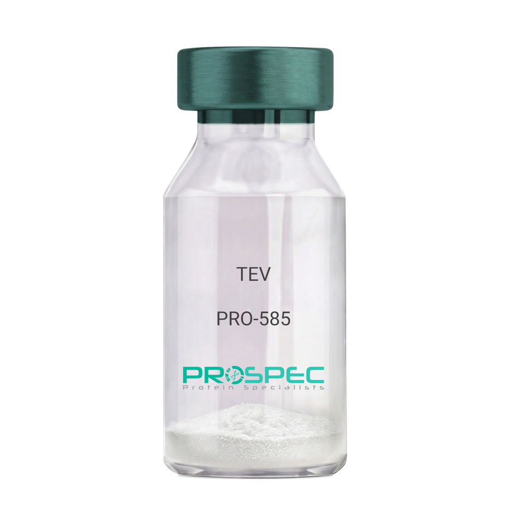
    TEV

    Description:

    Tobacco Etch Virus Protease Recombinant

    rTEV, TEV, P1 protease.

    Product # :

    PRO-585

    Price :

    Quantity :

    Shipping Method :

    Ice Icon

    Shipped with Ice Packs

    Add To Cart

    More Info

    • description
    • source
    • formulation
    • purity
    • More Info

    Description

    Recombinant TEV Protease (rTEV) is a site-specific protease purified from E. coli. The protease can be used for the removal of affinity tags from fusion proteins. The seven-amino-acid recognition site for rTEV is Glu-Asn-Leu-Tyr-Phe-Gln-Gly with cleavage occurring between Gln and Gly. The optimal temperature for cleavage is 30°C; however, the enzyme can be used at temperatures as low as 4°C. The rTEV contains His tag.The rTEV is purified by proprietary chromatographic techniques.

    Source

    Escherichia Coli.

    Formulation

    The rTEV contains 25mM Tris, Ph 8.0, 75mM NaCl, 5mM EDTA, 10mM GSH, 50% Glycerol.

    Purity

    Greater than 90.0% as determined by analysis by SDS-PAGE.

    More Info

    • Introduction

      TEV protease is the common name for the 27 kDa catalytic domain of the Nuclear Inclusion a (NIa) protein encoded by the tobacco etch virus (TEV). Because its sequence specificity is far more stringent than that of factor Xa, thrombin, or enterokinase, TEV protease is a very useful reagent for cleaving fusion proteins. TEV protease recognizes a linear epitope of the general form E-Xaa-Xaa-Y -Xaa-Q-(G/S), with cleavage occurring between Q and G or Q and S. The most commonly used sequence is ENLYFQG.

    • Synonyms

      rTEV, TEV, P1 protease.

    • Physical Appearance

      Sterile liquid formulation.

    • Stability

      rTEV although stable at 4°C for 1 week, should be stored below -18°C. Please prevent freeze thaw cycles.

    • Cleavage Conditions

      A number of variables can be changed to optimize the cleavage of any specific protein. The amount of rTEV, the temperature of the incubation, and the time needed for cleavage may be examined. If the protein of interest is heat-labile, then 4°C incubations are recommended. Reactions at 4°C will require longer incubation times and/or more rTEV.

    • Unit Definition

      One unit is defined as the amount of enzyme needed to cleave 3 ug of fusion protein in 1 hour to 85 % completion at 30°C in a buffer containing 50 mM Tris-HCl, pH 8.0, 0.5 mM EDTA, and 1 mM DTT.

    ProSpec's products are furnished for LABORATORY RESEARCH USE ONLY. The product may not be used as drugs, agricultural or pesticidal products, food additives or household chemicals.

    Tev Protease
  • View Data Sheet

    Name :

    Bet v 1.0101, Sf9

    Description:

    Major pollen allergen Bet v 1-A Recombinant, Sf9

    Major pollen allergen Bet v 1-A, Allergen Bet v I-A, Bet v 1-A, BETVIA, BETVI.

    Product # :

    PRO-2282

    Price :

    Quantity :

    Shipping Method :

    Ice Icon

    Shipped with Ice Packs

    Add To Cart

    More Info

    • description
    • source
    • formulation
    • purity
    • More Info

    Description

    Recombinant Major pollen allergen Bet v 1-A produced in SF9 is a glycosylated, polypeptide chain having a calculated molecular mass of 18,942 Dalton. Bet v 1.0101 is expressed with a 10xHis tag at N-terminus and purified by proprietary chromatographic techniques.

    Source

    Sf9 insect cells.

    Formulation

    Bet v 1.0101 protein solution is supplied in 20mM HEPES buffer pH-8.0, 200mM NaCl and 20% glycerol.

    Purity

    Greater than 95.0% as determined by SDS-PAGE.

    More Info

    • Introduction

      Major pollen allergen Bet v 1-A (Bet v 1.0101) causes an allergic reaction in humans. Bet v 1.0101 is a cause of type I allergic reactions in Europe, North America and the former Soviet Union states, observed in early spring.

    • Synonyms

      Major pollen allergen Bet v 1-A, Allergen Bet v I-A, Bet v 1-A, BETVIA, BETVI.

    • Physical Appearance

      Sterile Filtered clear solution.

    • Stability

      Store at 4°C if entire vial will be used within 2-4 weeks. Store, frozen at -20°C for longer periods of time. Avoid multiple freeze-thaw cycles.

    • Immunological Functions

      1. Binds IgE type human antibodies. 2. Immunodot test with positive/negative sera panels.

    ProSpec's products are furnished for LABORATORY RESEARCH USE ONLY. The product may not be used as drugs, agricultural or pesticidal products, food additives or household chemicals.

    Bet V 10101
  • View Data Sheet

    Name :

    AIMP1 Antibody

    Description:

    Aminoacyl tRNA Synthetase Complex-Interacting Multifunctional Protein 1, Mouse Anti Human

    Aminoacyl tRNA synthase complex-interacting multifunctional protein 1, Multisynthase complex auxiliary component p43, AIMP1, EMAP2, SCYE1, p43, EMAPII.

    Product # :

    ANT-619

    Price :

    Quantity :

    Shipping Method :

    Ice Icon

    Shipped with Ice Packs

    Add To Cart

    More Info

    • formulation
    • More Info

    Formulation

    1mg/ml containing PBS, pH-7.4, 10% Glycerol and 0.02% Sodium Azide.

    More Info

    • Introduction

      AIMP1 (EMPA2 or p43) is a cytokine that is specifically induced by apoptosis, and it is involved in the control of angiogenesis, inflammation, and wound healing. The release of the AIMP1 cytokine renders the tumor-associated vasculature sensitive to tumor necrosis factor. Furthermore, AIMP1 is involved in the stimulation of inflammatory responses after proteolytic cleavage in tumor cells.

    • Synonyms

      Aminoacyl tRNA synthase complex-interacting multifunctional protein 1, Multisynthase complex auxiliary component p43, AIMP1, EMAP2, SCYE1, p43, EMAPII.

    • Physical Appearance

      Sterile filtered colorless solution.

    • Immunogen

      Anti-human AIMP1 mAb, is derived from hybridization of mouse F0 myeloma cells with spleen cells from BALB/c mice immunized with a recombinant human AIMP1 protein 1-336 amino acids purified from E. coli.

    • Ig Subclass

      Mouse IgG2a heavy chain and Kappa light chain.

    • Clone

      PAT6D10AT.

    • Applications

      The antibody has been tested by ELISA, Western blot analysis, Flow cytometry and ICC/IF to assure specificity and reactivity. Since application varies, however, each investigation should be titrated by the reagent to obtain optimal results.

    • Type

      Mouse Anti Human Monoclonal.

    • Storage Procedures

      For periods up to 1 month store at 4°C, for longer periods of time, store at -20°C. Prevent freeze thaw cycles.

    • Purification Method

      AIMP1 antibody was purified by protein-A affinity chromatography.

    ProSpec's products are furnished for LABORATORY RESEARCH USE ONLY. The product may not be used as drugs, agricultural or pesticidal products, food additives or household chemicals.

    Aimp1 Antibody
  • View Data Sheet

    Name :

    HTATIP2 Antibody

    Description:

    HIV-1 Tat Interactive Protein 2, Mouse Anti Human

    TIP30, CC3, SDR44U1, HTATIP2, EC=1.1.1.-, HIV-1 TAT-interactive protein 2, FLJ26963.

    Product # :

    ANT-522

    Price :

    Quantity :

    Shipping Method :

    Ice Icon

    Shipped with Ice Packs

    Add To Cart

    More Info

    • formulation
    • More Info

    Formulation

    1mg/ml containing PBS, pH-7.4, 10% Glycerol and 0.02% Sodium Azide.

    More Info

    • Introduction

      HTATIP2 is part of the short-chain dehydrogenases/reductases (SDR) family which acts as a tumor suppressor in metabolic suppression, inhibition of angiogenesis and induces the expression of apoptosis related genes Bad and Siva. HTATIP2 cooperates with the activation domain of HIV-1 TAT and enhances its transcription by phosphorylating RNA polymerase II (Pol II). Defects in HTATIP2 are related with hepatocellular carcinomas and apoptotic resistant tumor cells, implicating a probable use for HTATIP2 in antitumor therapy.

    • Synonyms

      TIP30, CC3, SDR44U1, HTATIP2, EC=1.1.1.-, HIV-1 TAT-interactive protein 2, FLJ26963.

    • Physical Appearance

      Sterile filtered colorless solution.

    • Immunogen

      Anti-human HTATIP2 mAb, is derived from hybridization of mouse F0 myeloma cells with spleen cells from BALB/c mice immunized with recombinant human HTATIP2 amino acids 1-242 purified from E. coli.

    • Ig Subclass

      Mouse IgG2a heavy chain and ? light chain.

    • Clone

      PAT5D6AT.

    • Applications

      HTATIP2 antibody has been tested by ELISA, Western blot analysis, Flow cytometry and ICC/IF to assure specificity and reactivity. Since application varies, however, each investigation should be titrated by the reagent to obtain optimal results.

    • Type

      Mouse Anti Human Monoclonal.

    • Storage Procedures

      For periods up to 1 month store at 4°C, for longer periods of time, store at -20°C. Prevent freeze thaw cycles.

    • Purification Method

      HTATIP2 antibody was purified from mouse ascitic fluids by protein-A affinity chromatography.

    ProSpec's products are furnished for LABORATORY RESEARCH USE ONLY. The product may not be used as drugs, agricultural or pesticidal products, food additives or household chemicals.

    Htatip2 Antibody
  • View Data Sheet

    Name :

    IL15 PAT7G9AT Antibody

    Description:

    Interleukin-15 PAT7G9AT Mouse Anti-Human

    IL-15, MGC9721, Interleukin-15.

    Product # :

    ANT-776

    Price :

    Quantity :

    Shipping Method :

    Ice Icon

    Shipped with Ice Packs

    Add To Cart

    More Info

    • formulation
    • More Info

    Formulation

    1mg/ml containing PBS, pH-7.4, 10% Glycerol and 0.02% Sodium Azide.

    More Info

    • Synonyms

      IL-15, MGC9721, Interleukin-15.

    • Physical Appearance

      Sterile filtered colorless solution.

    • Immunogen

      Anti-human IL15 mAb, is derived from hybridization of mouse F0 myeloma cells with spleen cells from BALB/c mice immunized with a recombinant human IL15 protein 49-162 amino acids purified from E. coli.

    • Ig Subclass

      Mouse IgG2a heavy chain and Kappa light chain.

    • Clone

      PAT7G9T.

    • Applications

      The antibody has been tested by ELISA, Western blot analysis, FACS and ICC/IF to assure specificity and reactivity. Since application varies, however, each investigation should be titrated by the reagent to obtain optimal results.

    • Background

      Interleukin-15 which is produced by a wide variety of cells and tissues is a pleiotropic cytokine which takes part in innate and adaptive immunity. IL- 15 is well known due to its involvement in a trans-signaling mechanism where IL-15 receptor alpha from 1 subset of cells presents IL-15 to close cells that express IL-2Rβ/γc. IL-15 like IL-2, stimulates the proliferation of Tlymphocytes and induces the generation of cytotoxic T-lymphocytes (CTLs) and lymphokine-activated killer (LAK) cells. IL-15 and IL-2 may also compete for the same receptor, and thus negatively regulate each other's activity.

    • Type

      Mouse Anti Human Monoclonal.

    • Storage Procedures

      For periods up to 1 month store at 4°C, for longer periods of time, store at -20°C. Prevent freeze thaw cycles.

    • Purification Method

      IL15 antibody was purified by protein-A affinity chromatography.

    ProSpec's products are furnished for LABORATORY RESEARCH USE ONLY. The product may not be used as drugs, agricultural or pesticidal products, food additives or household chemicals.

    Il15 Antibody
  • View Data Sheet

    Name :

    CFL1 Antibody

    Description:

    Cofilin-1, Mouse Anti Human

    CFL-1, CFL1, Cofilin1, Cofilin-1, Cofilin non-muscle isoform, 18 kDa phosphoprotein, p18.

    Product # :

    ANT-354

    Price :

    Quantity :

    Shipping Method :

    Ice Icon

    Shipped with Ice Packs

    Add To Cart

    More Info

    • formulation
    • More Info

    Formulation

    1mg/ml containing PBS, pH-7.4, & 0.1% Sodium Azide.

    More Info

    • Introduction

      Cofilin is a family of actin-binding proteins which disassembles actin filaments. It is a widely distributed intracellular actin-modulating protein that binds and depolymerizes filamentous F-actin and inhibits the polymerization of monomeric G-actin in a pH-dependent manner. It is involved in the translocation of actin-cofilin complex from cytoplasm to nucleus.

    • Synonyms

      CFL-1, CFL1, Cofilin1, Cofilin-1, Cofilin non-muscle isoform, 18 kDa phosphoprotein, p18.

    • Immunogen

      Anti-human CFL1 mAb is derived from hybridization of mouse F0 myeloma cells with spleen cells from BALB/c mice immunized with recombinant human CFL1 amino acids 1-166 purified from E. coli.

    • Ig Subclass

      Mouse IgG1 heavy chain and κ light chain.

    • Clone

      PAT1C1AT.

    • Applications

      CFL1 antibody has been tested by ELISA and Western blot analysis to assure specificity and reactivity. Since application varies, however, each investigation should be titrated by the reagent to obtain optimal results. Recommended dilution range for Western blot analysis is 1:1,000 ~ 2,000.Recommended starting dilution is 1:1,000.

    • Type

      Mouse Anti Human Monoclonal.

    • Storage Procedures

      For periods up to 1 month store at 4°C, for longer periods of time, store at -20°C. Prevent freeze thaw cycles.

    • Purification Method

      CFL1 antibody was purified from mouse ascitic fluids by protein-G affinity chromatography.

    ProSpec's products are furnished for LABORATORY RESEARCH USE ONLY. The product may not be used as drugs, agricultural or pesticidal products, food additives or household chemicals.

    Cfl1 Antibody
  • View Data Sheet

    Name :

    IL1RL1 Human

    Description:

    Interleukin-1 receptor-like 1 Human Recombinant

    Interleukin-1 receptor-like 1, Protein ST2, IL1RL1, DER4, ST2, T1, ST2L, ST2V, FIT-1, MGC32623.

    Product # :

    CYT-671

    Price :

    Quantity :

    Shipping Method :

    Ice Icon

    Shipped with Ice Packs

    Add To Cart

    More Info

    • description
    • source
    • formulation
    • purity
    • More Info

    Description

    IL1RL1 Human Recombinant produced in E.Coli is a single, non-glycosylated, Polypeptide chain containing 310 amino acids fragment (19-328) corresponding to the soluble IL1RL1 coding sequence, having a molecular weight of 39.5kDa and fused with a 4.5kDa amino-terminal hexahistidine tag. The IL1RL1 is purified by proprietary chromatographic techniques.

    Source

    Escherichia Coli.

    Formulation

    IL1RL1 protein solution is supplied in 25mM Sodium Acetate pH 4.8 and 50% glycerol.

    Purity

    Greater than 95.0% as determined by SDS-PAGE.

    More Info

    • Introduction

      The IL1RL1 gene is a member of the IL-1 receptor family, encoding a transmembrane protein with a structure similar to IL-1R1. IL1RL1 is a receptor for interleukin-33, its stimulation recruits MYD88, IRAK1, IRAK4, and TRAF6, followed by phosphorylation of MAPK3/ERK1 and/or MAPK1/ERK2, MAPK14, and MAPK8. IL1RL1 may possibly be involved in helper T-cell function. IL1RL1 is highly expressed in kidney, lung, placenta, stomach, skeletal muscle, colon and small intestine.
      A soluble form of the IL1RL1 is produced from the same gene by alternative splicing and was shown to be expressed in several cell types including fibroblasts and mast cells. Soluble IL1RL1 also acts as a negative regulator of Th2 cytokine production and high levels have been reported in several disease states and conditions including asthma, sepsis and myocardial infarction.
      Analysis of the similar gene in mouse suggested that the IL1RL1 receptor can be induced by proinflammatory stimuli, and may be involved in the function of helper T cells.

    • Synonyms

      Interleukin-1 receptor-like 1, Protein ST2, IL1RL1, DER4, ST2, T1, ST2L, ST2V, FIT-1, MGC32623.

    • Physical Appearance

      Sterile Filtered clear solution.

    • Stability

      Store at 4°C if entire vial will be used within 2-4 weeks. Store, frozen at -20°C for longer periods of time. Please avoid freeze thaw cycles.

    ProSpec's products are furnished for LABORATORY RESEARCH USE ONLY. The product may not be used as drugs, agricultural or pesticidal products, food additives or household chemicals.

    Il1Rl1 Human
  • View Data Sheet

    Name :

    ASF1A Human

    Description:

    ASF1 Anti-Silencing Function 1 Homolog A Human Recombinant

    CGI-98, HSPC146, DKFZp547E2110, ASF1A, Histone chaperone ASF1A, Anti-silencing function protein 1 homolog A, hAsf1, hAsf1a, CCG1-interacting factor A, CIA, hCIA.

    Product # :

    PRO-682

    Price :

    Quantity :

    Shipping Method :

    Ice Icon

    Shipped with Ice Packs

    Add To Cart

    More Info

    • description
    • source
    • formulation
    • purity
    • More Info

    Description

    ASF1A Human Recombinant produced in E.Coli is a single, non-glycosylated polypeptide chain containing 240 amino acids (1-204 a.a.) and having a molecular mass of 27kDa.ASF1A is fused to a 36 amino acid His Tag and purified by proprietary chromatographic techniques.

    Source

    Escherichia Coli.

    Formulation

    The ASF1A protein solution contains 20mM Tris-HCl, pH-8, 1mM DTT and 10% glycerol.

    Purity

    Greater than 90.0% as determined by SDS-PAGE.

    More Info

    • Introduction

      ASF1A is part of the H3/H4 family of histone chaperone proteins corresponding to the anti-silencing function-1 gene in yeast. ASF1A is an important element of the histone donor complex that functions in nucleosome assembly. ASF1A interacts with histones H3 and H4, and functions together with a chromatin assembly factor during DNA replication and repair. Deletion of ASF1A in yeast and Drosophila confers sensitivity to various DNA damaging agents and inhibitors of DNA replication, increases genomic instability and sister chromatid exchange, and activates the DNA damage checkpoint.

    • Synonyms

      CGI-98, HSPC146, DKFZp547E2110, ASF1A, Histone chaperone ASF1A, Anti-silencing function protein 1 homolog A, hAsf1, hAsf1a, CCG1-interacting factor A, CIA, hCIA.

    • Physical Appearance

      Sterile filtered colorless solution.

    • Stability

      Store at 4°C if entire vial will be used within 2-4 weeks. Store, frozen at -20°C for longer periods of time. For long term storage it is recommended to add a carrier protein (0.1% HSA or BSA).Avoid multiple freeze-thaw cycles.

    • Amino Acid Sequence

      MRGSHHHHHH GMASMTGGQQ MGRDLYDDDD KDRWGSMAKV QVNNVVVLDN PSPFYNPFQF EITFECIEDL SEDLEWKIIY VGSAESEEYD QVLDSVLVGP VPAGRHMFVF QADAPNPGLI PDADAVGVTV VLITCTYRGQ EFIRVGYYVN NEYTETELRE NPPVKPDFSK LQRNILASNPRVTRFHINWE DNTEKLEDAE SSNPNLQSLL STDALPSASK GWSTSENSLN VMLESHMDCM.

    ProSpec's products are furnished for LABORATORY RESEARCH USE ONLY. The product may not be used as drugs, agricultural or pesticidal products, food additives or household chemicals.

    Asf1A Human
  • View Data Sheet

    Name :

    HCV NS5B

    Description:

    Hepatitis C Virus NS5B Recombinant

    Product # :

    HCV-269

    Price :

    Quantity :

    Shipping Method :

    Ice Icon

    Shipped with Ice Packs

    Add To Cart

    More Info

    • description
    • formulation
    • purity
    • More Info

    Description

    The E.coli derived recombinant protein contains the full length of the NS5B immunodominant regions Genotype 1B and fused to a 6xHis tag at C-Terminus.

    Formulation

    1.5M urea, 25mM Tris-HCl pH 8.0, 0.2% Triton-X and 50% Glycerol.

    Purity

    Protein is >95% pure as determined by SDS-PAGE.

    More Info

    • Introduction

      HCV is a small 50nm, enveloped, single-stranded, positive sense RNAvirus in the family Flaviviridae.
      HCV has a high rate of replication with approximately one trillion particles produced each day in an infected individual. Due to lack of proofreading by the HCV RNA polymerase, the HCV has an exceptionally high mutation rate, a factor that may help it elude the host's immune response. Hepatitis C virus is classified into six genotypes(1-6) with several subtypes within each genotype. The preponderance and distribution of HCV genotypes varies globally. Genotype is clinically important in determining potential response to interferon-based therapy and the required duration of such therapy. Genotypes 1 and 4 are less responsive to interferon-based treatment than are the other genotypes (2, 3, 5 and 6).

    • Stability

      HCV NS5B although stable at 4°C for 1 week, should be stored below -18°C. Please prevent freeze thaw cycles.

    • Applications

      HCV NS5B antigen in ELISA and Western blots, excellent antigen for detection of HCV with minimal specificity problems.

    • Specificity

      Immunoreactive with sera of HCV-infected individuals.

    • Purification Method

      HCV NS5B Genotype-1b protein was purified by proprietary chromatographic technique.

    ProSpec's products are furnished for LABORATORY RESEARCH USE ONLY. The product may not be used as drugs, agricultural or pesticidal products, food additives or household chemicals.

    Hcv Ns5B
  • View Data Sheet

    Name :

    Anaplasma p44

    Description:

    Anaplasma phagocytophilum p44 Recombinant

    Product # :

    PRO-2566

    Price :

    Quantity :

    Shipping Method :

    Ice Icon

    Shipped with Ice Packs

    Add To Cart

    More Info

    • description
    • source
    • formulation
    • purity
    • More Info

    Description

    Recombinant Anaplasma p44 produced in E.Coli is a single, non-glycosylated polypeptide chain having a molecular mass of 49kDa. Anaplasma p44 is expressed with a -10x His tag at N-terminus and purified by proprietary chromatographic techniques.

    Source

    Escherichia Coli.

    Formulation

    Anaplasma p44 is supplied at a 20mM HEPES buffer pH-8.0, 250mM NaCl and 6M Urea.

    Purity

    Greater than 95% as determined by SDS-PAGE.

    More Info

    • Introduction

      Anaplasma p44 which belongs to the outer membrane antigen superfamily (OMP1/Msp2/p44), is a serodiagnostic antigen for HGA. Anaplasma p44 allows the bacterium to adhere to the host cell and prevents host immune surveillance.

    • Physical Appearance

      Sterile Filtered solution.

    • Stability

      Store at 4°C if entire vial will be used within 2-4 weeks. Store, frozen at -20°C for longer periods of time. Avoid multiple freeze-thaw cycles.

    • Immunological Functions

      1. Binds IgG- and IgM-type human antibodies. 2. Immunodot test with positive/negative samples.

    ProSpec's products are furnished for LABORATORY RESEARCH USE ONLY. The product may not be used as drugs, agricultural or pesticidal products, food additives or household chemicals.

    Anaplasma Phagocytophilum P44
  • View Data Sheet

    Name :

    PhI p 6

    Description:

    Pollen allergen Phl p 6 Recombinant

    Pollen allergen Phl p 6, Allergen Phl p VI, Phl p 6, PHLPVI.

    Product # :

    PRO-2308

    Price :

    Quantity :

    Shipping Method :

    Ice Icon

    Shipped with Ice Packs

    Add To Cart

    More Info

    • description
    • source
    • formulation
    • purity
    • More Info

    Description

    Recombinant Pollen allergen Phl p 6 produced in SF9 is a glycosylated, polypeptide chain having a calculated molecular mass of 13,162 Dalton. PhI p 6 is expressed with a 10xHis tag at N-terminus and purified by proprietary chromatographic techniques.

    Source

    Sf9 insect cells.

    Formulation

    PhI p 6 is supplied in 20mM HEPES buffer pH-7.9 and 6M Urea.

    Purity

    Greater than 80.0% as determined by SDS-PAGE.

    More Info

    • Introduction

      Pollen allergen Phl p 6 (PhI p 6) causes an allergic reaction in humans.

    • Synonyms

      Pollen allergen Phl p 6, Allergen Phl p VI, Phl p 6, PHLPVI.

    • Physical Appearance

      Sterile Filtered clear solution.

    • Stability

      Store at 4°C if entire vial will be used within 2-4 weeks. Store, frozen at -20°C for longer periods of time. Avoid multiple freeze-thaw cycles.

    • Immunological Functions

      1. Binds IgE type human antibodies. 2. Immunodot test with positive/negative sera panels.

    • Molar extinction coefficient

      5960; A280(1mg/ml)=0.453

    ProSpec's products are furnished for LABORATORY RESEARCH USE ONLY. The product may not be used as drugs, agricultural or pesticidal products, food additives or household chemicals.

    Phi P 6
  • View Data Sheet

    Name :

    IL-4 Porcine

    Description:

    Interleukin-4 Porcine Recombinant

    BCGF, BCDF, B cell stimulating factor, BSF-1, Lymphocyte stimulatory factor 1, IL-4, MGC79402, Binetrakin, Pitrakinra.

    Product # :

    CYT-1098

    Price :

    Quantity :

    Shipping Method :

    Room Temp Icon

    Shipped at Room temp

    Add To Cart

    More Info

    • description
    • source
    • formulation
    • purity
    • biological activity
    • More Info

    Description

    Interleukin-4 Porcine Recombinant produced in E. coli is a non-glycosylated monomer chain containing 110 amino acids and having a molecular mass of 12.7kDa. IL-4 is purified by proprietary chromatographic techniques.

    Source

    Escherichia Coli.

    Formulation

    The protein was lyophilized from a sterile (0.2µm) filtered solution containing 10 mM sodium phosphate, pH 7.5.

    Purity

    Greater than 95.0% as determined by SDS-PAGE.

    Biological Activity

    The ED50, as determined by TF-1 cell proliferation is 0.504ng/ml corresponding to a specific activity which is 2.0 x 10^6 units/mg.

    More Info

    • Introduction

      IL4 is a pleiotropic cytokine produced by activated T cells. IL4 is a ligand for interleukin 4 receptor. The interleukin 4 receptor also binds to IL13, which contributes to various overlapping roles of this cytokine and IL13. STAT6, a signal transducer and activator of transcription plays a main role in mediating the immune regulatory signal of this cytokine. IL4, IL3, IL5, IL13, and CSF2 form a cytokine gene cluster on chromosome 5q, with this gene particularly close to IL13. IL4, IL13 and IL5 are regulated co-ordinately by several long-range regulatory elements in an over 120 kilobase range on the chromosome.

    • Synonyms

      BCGF, BCDF, B cell stimulating factor, BSF-1, Lymphocyte stimulatory factor 1, IL-4, MGC79402, Binetrakin, Pitrakinra.

    • Physical Appearance

      Sterile Filtered White lyophilized (freeze-dried) powder.

    • Stability

      Lyophilized IL-4 although stable at room temperature for 3 weeks, should be stored desiccated below -18°C. Upon reconstitution IL-4 should be stored at 4°C between 2-7 days and for future use below -18°C. Please prevent freeze-thaw cycles.

    • Solubility

      It is recommended to reconstitute the lyophilized IL-4 in sterile 18MΩ-cm H2O not less than 100µg/ml, which can then be further diluted to other aqueous solutions.

    • Amino Acid Sequence

      MHKCDITLQE IIKTLNILTA RKNSCMELPV TDVFAAPENT TEKETFCRAS TVLRHIYRHH TCMKSLLSGL DRNLSSMANM TCSVHEAKKS TLKDFLERLK TIMKEKYSKC.

    ProSpec's products are furnished for LABORATORY RESEARCH USE ONLY. The product may not be used as drugs, agricultural or pesticidal products, food additives or household chemicals.

    Il4 Porcine
  • View Data Sheet

    Name :

    SARS Nucleocapsid Biotin Antibody

    Description:

    Mouse Anti SARS Nucleocapsid Biotinylated

    Product # :

    ANT-764

    Price :

    Quantity :

    Shipping Method :

    Ice Icon

    Shipped with Ice Packs

    Add To Cart

    More Info

    • description
    • formulation
    • purity
    • More Info

    Description

    SARS Nucleoprotein Biotinylated antibody clone PT3851 is specific for the nucleocapsid SARS-CoV protein and recognizes the nucleoprotein of SARS-CoV-2 by ELISA.

    Formulation

    1mg/ml in DPBS, 1% BSA and 0.1% Proclin 950.

    Purity

    Greater than 90% as determined by SDS-PAGE gel.

    More Info

    • Introduction

      SARS Coronavirus is an enveloped virus containing three outer structural proteins, namely the membrane (M), envelope (E), and spike (S) proteins. Spike (S)-glycoprotein of the virus interacts with a cellular receptor and mediates membrane fusion to allow viral entry into susceptible target cells. Accordingly, S-protein plays an important role in virus infection cycle and is the primary target of neutralizing antibodies.It has recently been shown that SARS is caused by a human coronavirus. Human coronaviruses are the major cause of upper respiratory tract illness in humans, such as the common cold. Coronaviruses are positive-stranded RNA viruses, featuring the largest viral RNA genomes known to date (27-31 kb). The first step in coronavirus infection is binding of the viral spike protein, a 139-kDa protein, to certain receptors on host cells. The spike protein is the main surface antigen of the coronavirus. The most prominent protein in the culture supernatants infected with SARS virus is a 46 kDa nucleocapsid protein. This suggests that the nucleocapsid protein is a major immunogen that may be useful for early diagnostics.

    • Physical Appearance

      Sterile Filtered liquid formulation.

    • Stability

      Store at 4°C, stable for 2 weeks. For long-term storage, store at -20°C.

    • Immunogen

      Recombinant protein fragment 1-49 a.a. of the SARS nucleoprotein.

    • Clone

      PT3851

    • Applications

      ELISA.

    • Isotype

      Mouse IgG2b.

    • Type

      Mouse antibody Monoclonal.

    • Purification Method

      Protein A affinity purified.

    ProSpec's products are furnished for LABORATORY RESEARCH USE ONLY. The product may not be used as drugs, agricultural or pesticidal products, food additives or household chemicals.

    Cov Nucleocprotein Antibody
  • View Data Sheet

    Name :

    HCV NS5B (2634-2752 a.a)

    Description:

    Hepatitis C Virus NS5B (2634-2752 a.a) Recombinant

    Product # :

    HCV-003

    Price :

    Quantity :

    Shipping Method :

    Ice Icon

    Shipped with Ice Packs

    Add To Cart

    More Info

    • description
    • source
    • formulation
    • purity
    • More Info

    Description

    Recombinant Hepatitis C Virus NS5B produced in E. coli is a single polypeptide chain containing 155 amino acids (aa 2634-2752) and having a molecular mass of 17kDa (NCBI Accession # NP_671491).Recombinant HCV NS5B is fused to a 36 amino acid His-tag at N-terminus & purified by proprietary chromatographic techniques.

    Source

    Escherichia Coli.

    Formulation

    The HCV NS5B protein solution (0.25mg/ml) contains 20mM Tris-HCl buffer (pH 8.0), 0.15M NaCl, 10% glycerol and 1mM DTT.

    Purity

    Greater than 90% as determined by SDS-PAGE.

    More Info

    • Introduction

      HCV is a small 50nm, enveloped, single-stranded, positive sense RNAvirus in the family Flaviviridae. HCV has a high rate of replication with approximately one trillion particles produced each day in an infected individual. Due to lack of proofreading by the HCV RNA polymerase, the HCV has an exceptionally high mutation rate, a factor that may help it elude the host's immune response. Hepatitis C virus is classified into six genotypes(1-6) with several subtypes within each genotype. The preponderance and distribution of HCV genotypes varies globally. Genotype is clinically important in determining potential response to interferon-based therapy and the required duration of such therapy. Genotypes 1 and 4 are less responsive to interferon-based treatment than are the other genotypes (2, 3, 5 and 6).

    • Physical Appearance

      Sterile filtered colorless solution.

    • Stability

      Store at 4°C if entire vial will be used within 2-4 weeks. Store, frozen at -20°C for longer periods of time. For long term storage it is recommended to add a carrier protein (0.1% HSA or BSA).Avoid multiple freeze-thaw cycles.

    • Amino Acid Sequence

      MRGSHHHHHH GMASMTGGQQ MGRDLYDDDD KDRWGSPMGF SYDTRCFDST VTESDIRTEE AIYQCCDLDP QARVAIKSLT ERLYVGGPLT NSRGENCGYR RCRASGVLTT SCGNTLTCYI KARAACRAAG LQDCTMLVCG DDLVVICESA GVQED.

    ProSpec's products are furnished for LABORATORY RESEARCH USE ONLY. The product may not be used as drugs, agricultural or pesticidal products, food additives or household chemicals.

    Hcv Ns5B 2634 2752 Aa
  • View Data Sheet

    Name :

    Lymphotactin Mouse

    Description:

    Lymphotactin (XCL1) Mouse Recombinant

    XCL1, Cytokine SCM-1, ATAC, Lymphotaxin, SCM-1-alpha, Small inducible cytokine C1, XC chemokine ligand 1, LTN, LPTN, SCM1, SCM-1, SCYC1, SCM-1a.

    Product # :

    CHM-037

    Price :

    Quantity :

    Shipping Method :

    Room Temp Icon

    Shipped at Room temp

    Add To Cart

    More Info

    • description
    • source
    • formulation
    • purity
    • biological activity
    • More Info

    Description

    Lymphotactin (XCL1) Mouse Recombinant produced in E.Coli is a non-glycosylated polypeptide chain containing 93 amino acids and having a molecular mass of approximately 10.1kDa.Lymphotactin is purified by proprietary chromatographic techniques.

    Source

    Escherichia Coli.

    Formulation

    Lyophilized from a 0.2μm filtered concentrated solution in PBS, pH 7.4.

    Purity

    reater than 97.0% as determined by:
    (a) Analysis by RP-HPLC.
    (b) Analysis by SDS-PAGE.

    Biological Activity

    The ED50 as determined by a chemotaxis bioassay using human XCR1 transfected murine BaF3 cells is < 100 ng/ml, corresponding to a specific activity of > 1.0 × 10⁴ IU/mg.

    More Info

    • Introduction

      XCL1 is a small cytokine belongs to the XC chemokine family that is also known as lymphotactin. XCL1 is found in high levels in spleen, thymus, intestine and peripheral blood leukocytes, and at lower levels in lung, prostate gland and ovary. Cellular sources for XCL1 include activated thymic and peripheral blood CD8+ T cells. This chemokine attracts T cells. In humans, XCL1 is closely related to XCL2, whose gene is found at the same locus on chromosome 1. XCL1 induces it chemotactic function by binding to a chemokine receptor called XCR1.

    • Synonyms

      XCL1, Cytokine SCM-1, ATAC, Lymphotaxin, SCM-1-alpha, Small inducible cytokine C1, XC chemokine ligand 1, LTN, LPTN, SCM1, SCM-1, SCYC1, SCM-1a.

    • Physical Appearance

      Sterile Filtered White lyophilized (freeze-dried) powder.

    • Stability

      Lyophilized XCL1 although stable at room temperature for 3 weeks, should be stored desiccated below -18°C. Upon reconstitution Lymphotactin should be stored at 4°C between 2-7 days and for future use below -18°C.Please prevent freeze-thaw cycles.

    • Solubility

      It is recommended to reconstitute the lyophilized Lymphotactin in sterile 18MΩ-cm H2O not less than 100µg/ml, which can then be further diluted to other aqueous solutions.

    • Amino Acid Sequence

      VGTEVLEESS CVNLQTQRLP VQKIKTYIIW EGAMRAVIFV TKRGLKICAD PEAKWVKAAI KTVDGRASTR KNMAETVPTG AQRSTSTAIT LTG.

    ProSpec's products are furnished for LABORATORY RESEARCH USE ONLY. The product may not be used as drugs, agricultural or pesticidal products, food additives or household chemicals.

    Lymphotactin Mouse
  • View Data Sheet

    Name :

    VEGF E (Orf Virus)

    Description:

    Vascular Endothelial Growth Factor-E Recombinant (Orf Virus)

    Product # :

    CYT-263

    Price :

    Quantity :

    Shipping Method :

    Room Temp Icon

    Shipped at Room temp

    Add To Cart

    More Info

    • description
    • source
    • formulation
    • purity
    • biological activity
    • More Info

    Description

    A DNA sequence encoding the mature variant of ovVEGF-E isolate D1701 (Dehio et al., 1999; GenBank accession No. AF106020) was expressed in E. coli as a 132 amino acid residue fusion protein with an N-terminal His-tag sequence and a thrombin cleavage site. Recombinant VEGF-E homodimer was dimerized in vitro and has a predicted mass of approximately 35 kDa.

    Source

    Escherichia Coli.

    Formulation

    The protein was lyophilized from a concentrated (1mg/ml) solution containing PBS.

    Purity

    Greater than 90.0% as determined by SDS-PAGE.

    Biological Activity

    The biological activity was determined (1) by the ability to induce VEGFR-2/KDR receptor phosphorylation in PAE/KDR cells and (2) in a cell proliferation assay using primary HUVECs. The ED50 for this effect is typically 1-5ng/ml.

    More Info

    • Introduction

      Based on sequence similarity to VEGF-A, a gene encoding a VEGF homologue has recently been discovered in the genome of Orf virus (OV) (Lyttle et al., 1994). Different isolates of Orf virus show significant amino acid sequence similarity to VEGF-A and described as a viral virulence factor that appears to be derived from captured host genes. All eight cysteine residues of the central cysteine knot motif characteristic of members of the VEGF family are conserved among other residues in the VEGF-E proteins (Dehio et al., 1999; Wise et al., 1999). Alignment of all mammalian VEGF sequences indicated that VEGF-E is distinct from the previously described VEGFs but most closely related to VEGF-A. Like VEGF-A, VEGF-E was found to bind with high affinity to VEGF receptor-2 (KDR) resulting in receptor autophosphorylation, whilst in contrast to VEGF-A, VEGF-E can not bind to VEGF receptor-1 (Flt-1). Furthermore VEGF-E can also not bind to VEGF receptor-3 (FLT-4). Therefore VEGF-E is a potent angiog

    • Physical Appearance

      Sterile Filtered White lyophilized (freeze-dried) powder.

    • Stability

      Lyophilized Vascular Endothelial Growth Factor-E Orf Virus although stable at room temperature for 3 weeks, should be stored desiccated below -18°C. Upon reconstitution VEGF E -OV should be stored at 4°C between 2-7 days and for future use below -18°C.Please prevent freeze-thaw cycles.

    • Solubility

      The lyophilized oVEGF-E Orf Virus should be reconstituted in water or medium to a concentration not lower than 50µg/ml. For long term storage we would recommend to add at least 0.1% human or bovine serum albumin.

    • Amino Acid Sequence

      MGSSHHHHHH SSGLVPRGSH DSTKTWSEVF ENSGCKPRPM VFRVHDEHPE LTSQRFNPPC VTLMRCGGCC NDESLECVPT EEANVTMQLM GASVSGGNGM QHLSFVEHKK CDCKPPLTTT PPTTTRPPRR RR

    ProSpec's products are furnished for LABORATORY RESEARCH USE ONLY. The product may not be used as drugs, agricultural or pesticidal products, food additives or household chemicals.

    Vegf E Orf Virus
  • View Data Sheet

    Name :

    Borrelia Garinii OspC

    Description:

    Borrelia Garinii OspC Recombinant

    Product # :

    BOR-021

    Price :

    Quantity :

    Shipping Method :

    Ice Icon

    Shipped with Ice Packs

    Add To Cart

    More Info

    • description
    • source
    • formulation
    • purity
    • More Info

    Description

    Recombinant Borrelia Garinii Outer Surface Protein C produced in E.coli is a non-glycosylated, polypeptide chain having a calculated molecular mass of 22kDa.Borrelia Garinii OspC is expressed with a 10xHis tag at N-terminus and purified by proprietary chromatographic techniques.

    Source

    Escherichia Coli.

    Formulation

    Borrelia Garinii OspC is supplied in 20mM HEPES buffer pH-8.0, 200mM NaCl and 20% glycerol.

    Purity

    Greater than 95.0% as determined by SDS-PAGE.

    More Info

    • Introduction

      Borrelia belongs to a genus of bacteria of the spirochete phylum. Borrelia causes borreliosis, which is a zoonotic, vector-borne disease transmitted mainly by ticks and some by lice, depending on the species. Of the 36 known species of Borrelia, 12 are distinguished to cause Lyme disease or borreliosis and are transmitted by ticks. The main Borrelia species causing Lyme disease are Borrelia burgdorferi, Borrelia afzelii, and Borrelia garinii. The Borrelia genus members have a linear chromosome which is about 900 kbp in length as well as an excess of both linear and circular plasmids in the 5-220 kbp size range. The plasmids are atypical, as compared to most bacterial plasmids, since they contain many paralogous sequences, a large number of pseudogenes and, in some cases, essential genes. Moreover, a number of the plasmids have features suggesting that they are prophages.

    • Physical Appearance

      Sterile Filtered clear solution.

    • Stability

      Store at 4°C if entire vial will be used within 2-4 weeks. Store, frozen at -20°C for longer periods of time. Avoid multiple freeze-thaw cycles.

    • Immunological Functions

      1. Binds IgG- and IgM-type human antibodies.2. Immunodot test with Lyme disease positive/negative plasma.

    ProSpec's products are furnished for LABORATORY RESEARCH USE ONLY. The product may not be used as drugs, agricultural or pesticidal products, food additives or household chemicals.

    Borrelia Garinii Ospc
  • View Data Sheet

    Name :

    Dengue 1 NS1 Antibody

    Description:

    Polyclonal Rabbit Anti Dengue 1 NS1

    Product # :

    ANT-083

    Price :

    Quantity :

    Shipping Method :

    Ice Icon

    Shipped with Ice Packs

    Add To Cart

    More Info

    • description
    • source
    • formulation
    • purity
    • More Info

    Description

    Polyclonal antibody to dengue-1 NS1 IgG was derived from the rabbit immunized with full length recombinant dengue NS1, reactive to all dengue serotype NS1.

    Source

    Rabbit.

    Formulation

    0.2M glycine pH 7.5 and 0.02% NaN3.

    Purity

    Protein is >95% pure as determined by 12% PAGE (coomassie staining). Purified by protein A chromatography.

    More Info

    • Stability

      Should be stored below -18°C. Please prevent freeze thaw cycles.

    • Applications

      Immunoassay.

    • Isotype

      Purified IgG.

    ProSpec's products are furnished for LABORATORY RESEARCH USE ONLY. The product may not be used as drugs, agricultural or pesticidal products, food additives or household chemicals.

    Dengue 1 Ns1 Antibody
  • View Data Sheet

    Name :

    T.pallidum p41

    Description:

    Treponema pallidum p41 Recombinant

    Product # :

    TRP-242

    Price :

    Quantity :

    Shipping Method :

    Ice Icon

    Shipped with Ice Packs

    Add To Cart

    More Info

    • description
    • source
    • formulation
    • purity
    • More Info

    Description

    The E.coli derived recombinant protein contains the outer membrane T.Pallidum p41 immunodominant regions. The protein is fused with a Beta-galactosidase tag.

    Source

    Escherichia Coli.

    Formulation

    10mM Tris-HCl pH8.0, 1mM EDTA, 1mMDTT and 8M urea.

    Purity

    Treponema pallidum protein is >90% pure as determined by 10% PAGE (coomassie staining).

    More Info

    • Introduction

      Treponema pallidum is a gram-negative spirochaete bacterium and is considered to be metabolically crippled. There are at least four known subspecies: T. pallidum pallidum, T. pallidum pertenue, T. pallidum carateum and T. pallidum endemicum. The helical structure of T. pallidum pallidum allows it to move in a corkscrew motion through viscous mediums such as mucus. Treponema pallidum sub sp. pallidum has one of the smallest bacterial genomes at 1.14 million base pairs (Mb) and has limited metabolic capabilities, reflecting its adaptation through genome reduction to the rich environment of mammalian tissue.

    • Stability

      T.pallidum p41 although stable at 4°C for 1 week, should be stored below -18°C. Please prevent freeze thaw cycles.

    • Applications

      Treponema pallidum is suitable for ELISA and Western blots, excellent antigen for detection of T. Pallidum with minimal specificity problems.

    • Specificity

      Immunoreactive with sera of T.Pallidum infected individuals.

    • Purification Method

      Treponema pallidum protein was purified by proprietary chromatographic technique.

    ProSpec's products are furnished for LABORATORY RESEARCH USE ONLY. The product may not be used as drugs, agricultural or pesticidal products, food additives or household chemicals.

    Tpallidum P41
  • View Data Sheet

    Name :

    HCV E2

    Description:

    Hepatitis C Virus E2 Recombinant

    Product # :

    HCV-005

    Price :

    Quantity :

    Shipping Method :

    Ice Icon

    Shipped with Ice Packs

    Add To Cart

    More Info

    • description
    • source
    • formulation
    • purity
    • More Info

    Description

    Recombinant Hepatitis C Virus E2 Genotype-1 produced in E. coli is a single polypeptide chain containing 226 amino acids (aa 482-671) and having a molecular mass of 25.4kDa (NCBI Accession#NP_671491).HCV E2 is fused to a 36 amino acid His-tag at N-terminus.

    Source

    Escherichia Coli.

    Formulation

    The HCV E2 solution (0.25mg/ml) contains 20mM Tris-HCl buffer (pH 8.0), 0.4M Urea and 10% glycerol.

    Purity

    Greater than 80.0% as determined by SDS-PAGE.

    More Info

    • Introduction

      E1 and E2 glycoproteins form a heterodimer which is involved in virus attachment to the host cell, virion internalization via clathrin-dependent endocytosis and fusion with host membrane. E1/E2 heterodimer binds to human LDLR, CD81 and SCARB1/SR-BI receptors, however this binding insufficient for infection, some additional liver specific cofactors may be required. The fusion function may perhaps be conducted by E1. E2 hinders human EIF2AK2/PKR activation, preventing the establishment of an antiviral state. E2 is a viral ligand for CD209/DC-SIGN and CLEC4M/DC-SIGNR, which are respectively located on dendritic cells (DCs), and on liver sinusoidal endothelial cells and macrophage-like cells of lymph node sinuses. These interactions allow seizure of circulating HCV particles by these cells and subsequent diffusion to permissive cells.

    • Physical Appearance

      Sterile Filtered colorless solution.

    • Stability

      Store at 4°C if entire vial will be used within 2-4 weeks. Store, frozen at -20°C for longer periods of time. For long term storage it is recommended to add a carrier protein (0.1% HSA or BSA).Avoid multiple freeze-thaw cycles.

    • Amino Acid Sequence

      MRGSHHHHHH GMASMTGGQQ MGRDLYDDDD KDRWGSERPY CWHYPPRPCG IVPAKSVCGP VYCFTPSPVV VGTTDRSGAP TYSWGANDTD VFVLNNTRPP LGNWFGCTWM NSTGFTKVCG APPCVIGGVG NNTLLCPTDC FRKHPEATYS RCGSGPWITP RCMVDYPYRL WHYPCTINYT IFKVRMYVGG VEHRLEAACN WTRGERCDLE DRDRSELSPL LLSTTQ.

    ProSpec's products are furnished for LABORATORY RESEARCH USE ONLY. The product may not be used as drugs, agricultural or pesticidal products, food additives or household chemicals.

    Hcv E2
  • View Data Sheet

    Name :

    CLMP Human

    Description:

    CXADR-Like Membrane Protein Human Recombinant

    CXADR-Like Membrane Protein, CLMP, Adipocyte Adhesion Molecule, Coxsackie- And Adenovirus Receptor-Like Membrane Protein, ACAM, ASAM, Adipocyte-Specific Adhesion Molecule, CAR-Like Membrane Protein, CSBS, CSBM.

    Product # :

    PRO-1941

    Price :

    Quantity :

    Shipping Method :

    Ice Icon

    Shipped with Ice Packs

    Add To Cart

    More Info

    • description
    • source
    • formulation
    • purity
    • More Info

    Description

    CLMP Human Recombinant produced in E.coli is a single, non-glycosylated polypeptide chain containing 233 amino acids (19-235) and having a molecular mass of 26.1 kDa.CLMP is fused to a 16 amino acid His-tag at N-terminus & purified by proprietary chromatographic techniques.

    Source

    Escherichia Coli.

    Formulation

    The CLMP solution (0.5mg/ml) contains 20mM Tris-HCl buffer (pH 8.0), 0.4M UREA and 10% glycerol.

    Purity

    Greater than 90.0% as determined by SDS-PAGE.

    More Info

    • Introduction

      The CTX family of proteins (including ASAM) are type I transmembrane proteins within the Ig superfamily, which localize to junctional complexes between endothelial and epithelial cells and may play a role in cell-cell adhesion. CLMP has a role in adipocyte differentiation and development of obesity. CLMP is required for normal small intestine development.

    • Synonyms

      CXADR-Like Membrane Protein, CLMP, Adipocyte Adhesion Molecule, Coxsackie- And Adenovirus Receptor-Like Membrane Protein, ACAM, ASAM, Adipocyte-Specific Adhesion Molecule, CAR-Like Membrane Protein, CSBS, CSBM.

    • Physical Appearance

      Sterile Filtered clear solution.

    • Stability

      Store at 4°C if entire vial will be used within 2-4 weeks. Store, frozen at -20°C for longer periods of time. For long term storage it is recommended to add a carrier protein (0.1% HSA or BSA).Avoid multiple freeze-thaw cycles.

    • Amino Acid Sequence

      MASMTGGQQM GRGSHMTHTE IKRVAEEKVT LPCHHQLGLP EKDTLDIEWL LTDNEGNQKV VITYSSRHVY NNLTEEQKGR VAFASNFLAG DASLQIEPLK PSDEGRYTCK VKNSGRYVWS HVILKVLVRP SKPKCELEGE LTEGSDLTLQ CESSSGTEPI VYYWQRIREK EGEDERLPPK SRIDYNHPGR VLLQNLTMSY SGLYQCTAGN EAGKESCVVR VTVQYVQSIG MVA.

    ProSpec's products are furnished for LABORATORY RESEARCH USE ONLY. The product may not be used as drugs, agricultural or pesticidal products, food additives or household chemicals.

    Clmp Human
  • 1
  • …
  • 23
  • 24
  • 25
  • 26
  • 27
  • …
  • 42
Your browser does not support the video tag.

Products

  • Cytokines
  • Growth Factors
  • Chemokines
  • CD Antigens
  • Neurotrophins
  • Hormones
  • Enzymes
  • Viral Antigens
  • Recombinant Proteins
  • Natural Proteins
  • Monoclonal Antibodies
  • Polyclonal Antibodies
  • Thermal Packaging

About

  • Company
  • Founder
  • Contribution
  • Contact Us
  • My Account

Ordering

  • Ordering Method
  • Ordering - Credit Card
  • Ordering PO
  • Shipping Fees
  • FAQ's

Legal

  • Terms & Conditions
  • Privacy
  • Site Map
  • info@prospecbio.com
  • Facebook
  • Instagram
  • Linkedin Profile

© 2026 ProSpec-Tany TechnoGene Ltd. All Rights Reserved.

  • Choosing a selection results in a full page refresh.
  • Opens in a new window.