Search results
1000 results found for “erythropoietin”
Name
Description
Product #
Price
Quantity
Shipping Method
- View Data Sheet
Name :
HBG2 HumanDescription:
Hemoglobin Gamma G Human Recombinant
TNCY, Hemoglobin subunit gamma-2, Gamma-2-globin, Hb F Gamma, Hemoglobin gamma-2 chain, Hemoglobin gamma-G chain.
Product # :
PRO-021Price :
Quantity :
Shipping Method :
Shipped with Ice Packs
More Info
- description
- source
- formulation
- purity
- More Info
Description
HBG2 Human Recombinant produced in E.Coli is a single, non-glycosylated polypeptide chain containing 170 amino acids (1-147 a.a.) and having a molecular mass of 18.5kDa.HBG2 is fused to a 23 amino acid His-tag at N-terminus & purified by proprietary chromatographic techniques.
Source
Escherichia Coli.
Formulation
HBG2 protein solution (0.5mg/ml) contains 20mM Tris-HCl buffer (pH 8.0), 0.1M NaCl,20% glycerol and 1mM DTT.
Purity
Greater than 90.0% as determined by SDS-PAGE.
More Info
-
Introduction
Hemoglobin subunit gamma-2 (HBG2), is a member of the globin family. HBG2 belongs to the fetal hemoglobin subunit, which comprised of two alpha chains organized with 2 gamma chains. HBG2 Amplified fetal hemoglobin production in adults and improve the clinical severity of sickle cell disease and beta-thalassemia major.
-
Synonyms
TNCY, Hemoglobin subunit gamma-2, Gamma-2-globin, Hb F Gamma, Hemoglobin gamma-2 chain, Hemoglobin gamma-G chain.
-
Physical Appearance
Sterile filtered reddish solution.
-
Stability
Store at 4°C if entire vial will be used within 2-4 weeks. Store, frozen at -20°C for longer periods of time. For long term storage it is recommended to add a carrier protein (0.1% HSA or BSA).Avoid multiple freeze-thaw cycles.
-
Amino Acid Sequence
MGSSHHHHHH SSGLVPRGSH MGSMGHFTEE DKATITSLWG KVNVEDAGGE TLGRLLVVYP WTQRFFDSFG NLSSASAIMG NPKVKAHGKK VLTSLGDAIK HLDDLKGTFA QLSELHCDKL HVDPENFKLL GNVLVTVLAI HFGKEFTPEV QASWQKMVTG VASALSSRYH.
ProSpec's products are furnished for LABORATORY RESEARCH USE ONLY. The product may not be used as drugs, agricultural or pesticidal products, food additives or household chemicals.
- View Data Sheet
Name :
RND3 HumanDescription:
Rho Family GTPase 3 Human Recombinant
ARHE, memB, Rho8, RhoE, Rho-related GTP-binding protein RhoE, Protein MemB, Rho family GTPase 3, Rho-related GTP-binding protein Rho8, Rnd3.
Product # :
PRO-487Price :
Quantity :
Shipping Method :
Shipped with Ice Packs
More Info
- description
- source
- formulation
- purity
- More Info
Description
RND3 Recombinant Human produced in E.Coli is a single, non-glycosylated polypeptide chain containing 261 amino acids (1-241 a.a.) and having a molecular mass of 29.2 kDa. The RND3 is fused to a 20 amino acid His-Tag at N-terminus and purified by proprietary chromatographic techniques.
Source
Escherichia Coli.
Formulation
RND3 Human solution containing 20mM Tris-HCl pH-8, 1mM DTT, 1mM EDTA, 0.1M NaCl & 40% glycerol.
Purity
Greater than 90.0% as determined by SDS-PAGE.
More Info
-
Introduction
RND3 is a small GTPase which is part of the Ras superfamily that controls the association of the actin cytoskeleton in reaction to extracellular growth factors. RND3 activity causes a decrease in stress fibers and an increase in cell migration, at least in part through regulation of the Rho-dependent kinase ROCK1. RND3 inhibits DNA-damage induced apoptosis and cell cycle arrest. RND3 binds GTP and is resistant to Rho-specific GTPase-activating proteins.
-
Synonyms
ARHE, memB, Rho8, RhoE, Rho-related GTP-binding protein RhoE, Protein MemB, Rho family GTPase 3, Rho-related GTP-binding protein Rho8, Rnd3.
-
Physical Appearance
Sterile filtered colorless solution.
-
Stability
Store at 4°C if entire vial will be used within 2-4 weeks. Store, frozen at -20°C for longer periods of time. For long term storage it is recommended to add a carrier protein (0.1% HSA or BSA).Avoid multiple freeze-thaw cycles.
-
Amino Acid Sequence
MGSSHHHHHH SSGLVPRGSH MKERRASQKL SSKSIMDPNQ NVKCKIVVVG DSQCGKTALL HVFAKDCFPE NYVPTVFENY TASFEIDTQR IELSLWDTSG SPYYDNVRPL SYPDSDAVLI CFDISRPETL DSVLKKWKGE IQEFCPNTKM LLVGCKSDLR TDVSTLVELS NHRQTPVSYD QGANMAKQIG AATYIECSAL QSENSVRDIF HVATLACVNK TNKNVKRNKS QRATKRISHM PSRPELSAVA TDLRKDKAKS C.
ProSpec's products are furnished for LABORATORY RESEARCH USE ONLY. The product may not be used as drugs, agricultural or pesticidal products, food additives or household chemicals.
- View Data Sheet
Name :
RPE HumanDescription:
Ribulose-5-Phosphate-3-Epimerase Human Recombinant
Ribulose-phosphate 3-epimerase, Ribulose-5-phosphate-3-epimerase, RPE, HUSSY-17, RPE2-1.
Product # :
ENZ-584Price :
Quantity :
Shipping Method :
Shipped with Ice Packs
More Info
- description
- source
- formulation
- purity
- More Info
Description
RPE Human Recombinant produced in E.coli is a single, non-glycosylated polypeptide chain containing 252 amino acids (1-228) and having a molecular mass of 27.5kDa.RPE is fused to a 24 amino acid His-tag at N-terminus & purified by proprietary chromatographic techniques.
Source
Escherichia Coli.
Formulation
The RPE solution (0.5mg/ml) contains 20mM Tris-HCl buffer (pH 8.0), 2mM DTT, 30% glycerol and 200mM NaCl.
Purity
Greater than 95% as determined by SDS-PAGE.
More Info
-
Introduction
Ribulose-phosphate 3-epimerase (RPE) is a member of the ribulose-phosphate 3-epimerase family. RPE is the enzyme which converts D-ribulose 5-phosphate into D-xylulose 5-phosphate in Calvin's reductive pentose phosphate cycle. Therefore, the RPE enzyme has one substrate, D-ribulose 5-phosphate, and one product, D-xylulose 5-phosphate. RPE participates in three metabolic pathways: pentose phosphate pathway, pentose and glucuronate interconversions, and carbon fixation.
-
Synonyms
Ribulose-phosphate 3-epimerase, Ribulose-5-phosphate-3-epimerase, RPE, HUSSY-17, RPE2-1.
-
Physical Appearance
Sterile Filtered clear solution.
-
Stability
Store at 4°C if entire vial will be used within 2-4 weeks.Store, frozen at -20°C for longer periods of time. For long term storage it is recommended to add a carrier protein (0.1% HSA or BSA).Avoid multiple freeze-thaw cycles.
-
Amino Acid Sequence
MGSSHHHHHH SSGLVPRGSH MGSHMASGCK IGPSILNSDL ANLGAECLRM LDSGADYLHL DVMDGHFVPN ITFGHPVVES LRKQLGQDPF FDMHMMVSKP EQWVKPMAVA GANQYTFHLE ATENPGALIK DIRENGMKVG LAIKPGTSVE YLAPWANQID MALVMTVEPG FGGQKFMEDM MPKVHWLRTQ FPSLDIEVDG GVGPDTVHKC AEAGANMIVS GSAIMRSEDP RSVINLLRNV CSEAAQKRSL DR.
ProSpec's products are furnished for LABORATORY RESEARCH USE ONLY. The product may not be used as drugs, agricultural or pesticidal products, food additives or household chemicals.
- View Data Sheet
Name :
LIF Human, Sf9Description:
Leukemia Inhibitory Factor Human Recombinant, Sf9
Leukemia Inhibitory Factor, Differentiation Inhibitory Activity, Cholinergic Differentiation Factor, Differentiation-Stimulating Factor, Hepatocyte-Stimulating Factor III, Differentiation-Inducing Factor, Melanoma-Derived LPL Inhibitor, Human Interleukin In DA Cells, D Factor, HILDA, MLPLI, Emfilermin, DIA, CDF, Leukemia inhibitory factor, LIF, Differentiation-stimulating factor, D factor, Melanoma-derived LPL inhibitor, MLPLI, Emfilermin.
Product # :
CYT-1003Price :
Quantity :
Shipping Method :
Shipped with Ice Packs
More Info
- description
- source
- formulation
- purity
- biological activity
- More Info
Description
LIF Human Recombinant produced in Sf9 Baculovirus cells is a single, glycosylated polypeptide chain containing 189 amino acids (23-202a.a.) and having a molecular mass of 20.8kDa (Molecular size on SDS-PAGE will appear at approximately 18-40kDa). LIF is expressed with a 6 amino acid His tag at C-Terminus and purified by proprietary chromatographic techniques.
Source
Sf9, Baculovirus cells.
Formulation
LIF protein solution (0.25mg/ml) contains Phosphate Buffered Saline (pH 7.4) and 10% glycerol.
Purity
Greater than 90.0% as determined by SDS-PAGE.
Biological Activity
Measured in a cell proliferation assay using TF-1 human erythroleukemic cell. The ED50 for this effects is less or equal to 0.5 ng/ml.
More Info
-
Introduction
Leukemia Inhibitory Factor also called LIF is a lymphoid factor that promotes long-term maintenance of embryonic stem cells by suppressing spontaneous differentiation. Leukemia Inhibitory Factor has several functions such as cholinergic neuron differentiation, control of stem cell pluripotency, bone & fat metabolism, mitogenesis of factor dependent cell lines & promotion of megakaryocyte production in vivo. Human and mouse LIF exhibit a 78% identity in its amino acid sequence.
-
Synonyms
Leukemia Inhibitory Factor, Differentiation Inhibitory Activity, Cholinergic Differentiation Factor, Differentiation-Stimulating Factor, Hepatocyte-Stimulating Factor III, Differentiation-Inducing Factor, Melanoma-Derived LPL Inhibitor, Human Interleukin In DA Cells, D Factor, HILDA, MLPLI, Emfilermin, DIA, CDF, Leukemia inhibitory factor, LIF, Differentiation-stimulating factor, D factor, Melanoma-derived LPL inhibitor, MLPLI, Emfilermin.
-
Physical Appearance
Sterile filtered colorless solution.
-
Stability
Store at 4°C if entire vial will be used within 2-4 weeks. Store, frozen at -20°C for longer periods of time. For long term storage it is recommended to add a carrier protein (0.1% HSA or BSA). Avoid multiple freeze-thaw cycles.
-
Amino Acid Sequence
ADPSPLPITP VNATCAIRHP CHNNLMNQIR SQLAQLNGSA NALFILYYTA QGEPFPNNLD KLCGPNVTDF PPFHANGTEK AKLVELYRIV VYLGTSLGNI TRDQKILNPS ALSLHSKLNA TADILRGLLS NVLCRLCSKY HVGHVDVTYG PDTSGKDVFQ KKKLGCQLLG KYKQIIAVLA
QAFHHHHHH.
ProSpec's products are furnished for LABORATORY RESEARCH USE ONLY. The product may not be used as drugs, agricultural or pesticidal products, food additives or household chemicals.
- View Data Sheet
Name :
PTHrP N15 HumanDescription:
Parathyroid Hormone Related Protein N15 Labeled Human Recombinant
Parathyroid Hormone 2, PTH2, TIPF39, Tuberoinfundibular 39 Residue Protein.
Product # :
HOR-005Price :
Quantity :
Shipping Method :
Shipped at Room temp
More Info
- description
- source
- formulation
- purity
- More Info
Description
PTHrP N15 Human Recombinant produced in E.Coli is a single, non-glycosylated, polypeptide chain containing 86 amino acids, having an MW of 10033 Da labeled by the stable isotope N15.The PTHrP is purified by proprietary chromatographic techniques.
Source
Escherichia Coli.
Formulation
PthRp N15 protein was lyophilized from a 0.2µm filtered concentrated solution in 1xPBS,
pH 7.4.Purity
Greater than 95.0% as determined by:
(a) Analysis by RP-HPLC.
(b) Analysis by SDS-PAGE.More Info
-
Introduction
PTHrP is a powerful and discriminating agonist of PTH2R which takes part in adenyl cyclase activation and intracellular calcium levels elevation. PTHrP encourages protein kinase C beta activation, recruitment of beta-arrestin and PTH2R internalization. Additionally, PTHrP inhibits cell proliferation through its contribution to PTH2R activation, activates nociceptors and nociceptive circuits and acts as a neuropeptide in spermatogenesis.
-
Synonyms
Parathyroid Hormone 2, PTH2, TIPF39, Tuberoinfundibular 39 Residue Protein.
-
Physical Appearance
Sterile Filtered White lyophilized (freeze-dried) powder.
-
Stability
Lyophilized PTHrP N15 although stable at room temperature for 3 weeks, should be stored desiccated below -18C. Upon reconstitution PTHrP N15 should be stored at 4C between 2-7 days and for future use below -18C.For long term storage it is recommended to add a carrier protein (0.1% HSA or BSA).Please prevent freeze-thaw cycles.
-
Solubility
It is recommended to be briefly centrifuged prior to opening to bring the contents to the bottom. Reconstitute in 10mM HAc to a concentration of 0.1-1.0 mg/mL. Further dilutions should be made in appropriate buffered solutions.
-
Amino Acid Sequence
AVSEHQLLHD KGKSIQDLRR RFFLHHLIAE IHTAEIRATS EVSPNSKPSP NTKNHPVRFG SDDEGRYLTQ ETNKVETYKE QPLKTP.
ProSpec's products are furnished for LABORATORY RESEARCH USE ONLY. The product may not be used as drugs, agricultural or pesticidal products, food additives or household chemicals.
- View Data Sheet
Name :
RhD HumanDescription:
Rh Blood Group D Antigen Human Recombinant
SLC42A5, CD240D, DIIIc, RhPI, RhII, Rh4.
Product # :
PRO-2822Price :
Quantity :
Shipping Method :
Shipped at Room temp
More Info
- description
- source
- formulation
- purity
- More Info
Description
The RhD Human is created as a recombinant protein with a 4kda N-terminal fusion of His Tag. The RhD His-Tagged Fusion Protein, produced in E. coli, is a 13kDa protein containing 58 amino acid residues of the RhD Human, 108-165 amino acids.
Source
Escherichia Coli.
Formulation
Each mg was lyophilized with 1xPBS, 0.4% SDS and 4mM DTT.
Purity
Greater than 90% as determined by SDS-PAGE.
More Info
-
Synonyms
SLC42A5, CD240D, DIIIc, RhPI, RhII, Rh4.
-
Physical Appearance
Filtered White lyophilized (freeze-dried) powder.
-
Stability
Store lyophilized RhD at -20°C. Aliquot the product after reconstitution to avoid repeated freezing/thawing cycles. Reconstituted protein can be stored at 4°C for a limited period of time; it does not show any change after two weeks at 4°C.
-
Solubility
It is recommended to add deionized water to prepare a working stock solution of approximately 0.5mg/ml and let the lyophilized pellet dissolve completely. Product is not sterile!
Please filter the product by an appropriate sterile filter before using it on cell culture.
-
Background
The Rh blood group system is one of the major blood group systems in humans and the D antigen is the main component of this system.
D Antigen
The presence of the D antigen on the surface of red blood cells indicates a positive Rh factor. Individuals with this antigen are classified as Rh-positive (Rh+), while those without it are Rh-negative (Rh-).
Genetics
The Rh factor is inherited in a Mendelian manner, with the D antigen being monitored by the RHD gene located on chromosome 1. The presence of the D allele (RHD) results in Rh positivity.
Immunization
Rh-negative individuals can develop antibodies against the D antigen if exposed to Rh-positive blood. In order to prevent such interaction, Rh-negative mothers are often given Rh immunoglobulin (Rho(D) immune globulin) during and after pregnancy.
Clinical Significance
The Rh D antigen is vital in blood transfusions and pregnancy. Rh incompatibility can occur when an Rh- mother carries an Rh+ fetus, which may lead to hemolytic disease of the newborn (HDN).
Blood Transfusions
Rh compatibility is very important in transfusions. Rh- individuals should receive Rh- blood to avoid an immune reaction.
ProSpec's products are furnished for LABORATORY RESEARCH USE ONLY. The product may not be used as drugs, agricultural or pesticidal products, food additives or household chemicals.
- View Data Sheet
Name :
Visfatin Human, HisDescription:
Visfatin Recombinant Human, His Tag
PBEF, Pre-B cell colony-enhancing factor, Nicotinamide phosphoribosyltransferase NAmPRTase, Nampt, MGC117256, DKFZP666B131, 1110035O14Rik.
Product # :
CYT-563Price :
Quantity :
Shipping Method :
Shipped with Ice Packs
More Info
- description
- source
- formulation
- purity
- More Info
Description
Visfatin Human Recombinant produced in E.Coli is a single, non-glycosylated, polypeptide chain containing 511 amino acids and having a molecular mass of 57 kDa. The recombinant human Visfatin is fused to His tag at N-Terminus.
Source
Escherichia Coli.
Formulation
Visfatin His tag protein contains 20mM Tris pH-8, 0.1mM DTT & 10% glycerol.
Purity
Greater than 95.0% as determined by(a) Analysis by RP-HPLC.
(b) Analysis by SDS-PAGE.More Info
-
Introduction
Excess adiposity is the most important risk in the development of insulin resistance and type 2 diabetes mellitus (T2DM). Adipose tissue produces several proteins (adipocytokines) such as leptin, adiponectin, resistin, tumor necrosis factor-?, and IL-6, that modulate insulin sensitivity and appear to play an important role in the pathogenesis of insulin resistance, diabetes, dyslipidemia, inflammation, and atherosclerosis. However, the mechanisms by which fat tissue induces insulin resistance and the role of adipocytokines in the pathogenesis of T2DM have not been well established.
Visfatin, also known as pre-B cell colony-enhancing factor (PBEF), is a cytokine that is highly expressed in visceral fat and was originally isolated as a secreted factor that synergizes with IL-7 and stem cell factors to promote the growth of B cell precursors. Visfatin homologs have been identified in carp, invertebrate mollusks, and bacteria, as well as in vertebrates, including humans and the mouse. It has been postulated to play a role in innate immunity.
Visfatin exerts insulin-mimetic effects that are dose-dependent and quantitatively similar to those of insulin in stimulating muscle and adipocyte glucose transport, and in inhibiting hepatocyte glucose production. Intravenous injection of recombinant visfatin in mice decreased plasma glucose in a dose-dependent fashion. In keeping with its insulin-mimetic effects, visfatin was as effective as insulin in reducing hyperglycemia in insulin-deficient diabetic mice. Visfatin was also found to be bound to and activate insulin receptor, causing receptor phosphorylation and the activation of downstream signaling molecules. However, visfatin and insulin did not compete for binding to the insulin receptor, indicating that the two proteins were recognized by different regions of the receptor. Thus, visfatin might play a role in glucose homeostasis and dysregulation in biosynthesis or signal transduction, and might contribute to the pathogenesis of diabetes. -
Synonyms
PBEF, Pre-B cell colony-enhancing factor, Nicotinamide phosphoribosyltransferase NAmPRTase, Nampt, MGC117256, DKFZP666B131, 1110035O14Rik.
-
Physical Appearance
Sterile Filtered solution at a concentration of 1mg/ml.
-
Stability
Store at 4°C if entire vial will be used within 2-4 weeks. Store, frozen at -20°C for longer periods of time. Please avoid freeze thaw cycles.
-
Amino Acid Sequence
MGSSHHHHHH SSGLVPRGSH MNPAAEAEFN ILLATDSYKV THYKQYPPNT SKVYSYFECR EKKTENSKLR KVKYEETVFY GLQYILNKYL KGKVVTKEKI QEAKDVYKEH FQDDVFNEKG WNYILEKYDG HLPIEIKAVP EGFVIPRGNV LFTVENTDPE CYWLTNWIET ILVQSWYPIT VATNSREQKK ILAKYLLETS GNLDGLEYKL HDFGYRGVSS QETAGIGASA HLVNFKGTDT VAGLALIKKY YGTKDPVPGY SVPAAEHSTI TAWGKDHEKD AFEHIVTQFS SVPVSVVSDS YDIYNACEKI WGEDLRHLIV SRSTQAPLII RPDSGNPLDT VLKVLEILGK KFPVTENSKG YKLLPPYLRV IQGDGVDINT LQEIVEGMKQ KMWSIENIAF GSGGGLLQKL TRDLLNCSFK CSYVVTNGLG INVFKDPVAD PNKRSKKGRL SLHRTPAGNF VTLEEGKGDL EEYGQDLLHT VFKNGKVTKS YSFDEIRKNA QLNIELEAAH H.
ProSpec's products are furnished for LABORATORY RESEARCH USE ONLY. The product may not be used as drugs, agricultural or pesticidal products, food additives or household chemicals.
- View Data Sheet
Name :
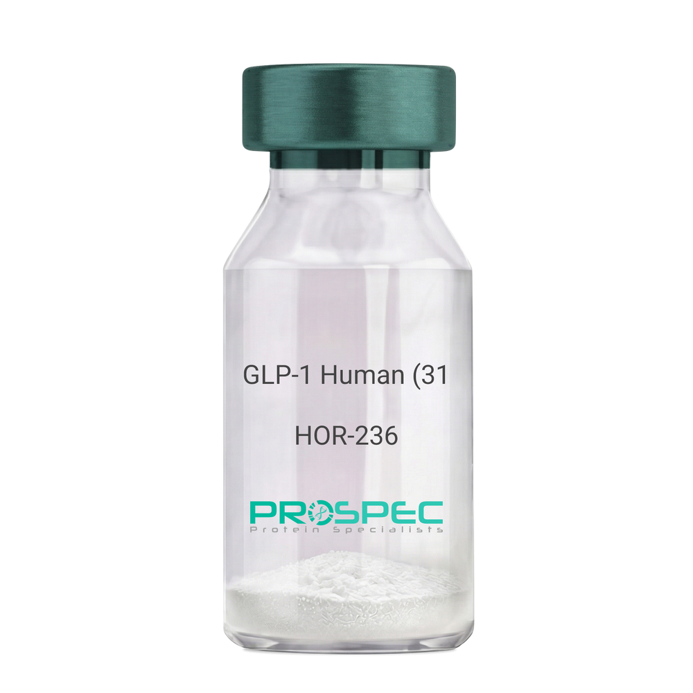
GLP-1 Human (31 a.a.)Description:
Glucagon Like Peptide-1 (31 a.a.) Human Recombinant
GLP1, GLP2, GRPP.
Product # :
HOR-236Price :
Quantity :
Shipping Method :
Shipped at Room temp
More Info
- description
- source
- formulation
- purity
- More Info
Description
Glucagon Like Peptide-1 Human Recombinant produced in E.Coli is a single, non-glycosylated, polypeptide chain containing 31 amino acids and having a molecular mass of 3.3 kDa. The GLP-1 is purified by proprietary chromatographic techniques.
Source
Escherichia Coli.
Formulation
The Recombinant GLP-1 concentrated solution (1mg/ml) was lyophilized after extensive dialyses against 0.1mg sodium phosphate monobasic & 1.6mg sodium phosphate dibasic.
Purity
Greater than 98.0% as determined by:
(a) Analysis by RP-HPLC.
(b) Analysis by SDS-PAGE.More Info
-
Introduction
Glucagon-like peptide-1 (GLP-1) is derived from the transcription product of the proglucagon gene. The major source of GLP-1 in the body is the intestinal L cell that secretes GLP-1 as a guthormone. The biologically active forms of GLP-1 are: GLP-1-(7-37) and GLP-1-(7-36)NH2.
GLP-1 secretion by L cells is dependent on the presence of nutrients in the lumen of the small intestine. The secretagogues (agents that causes or stimulates secretion) of this hormone include major nutrients like carbohydrate, proteinand lipid. Once in the circulation, GLP-1 has a half life of less than 2 minutes, due to rapid degradation by the enzyme dipeptidyl peptidase-4.
GLP-1 possesses several physiological properties that make it a subject of intensive investigation as a potential treatment of diabetes mellitus. The known physiological functions of GLP-1 include: Increases insulin secretion from the pancreas in a glucose-dependent manner, decreases glucagon secretion from the pancreas, increases beta cells mass and insulin gene expression, inhibits acid secretion and gastric emptying in the stomach, decreases food intake by increasing satiety. -
Synonyms
GLP1, GLP2, GRPP.
-
Physical Appearance
Sterile Filtered White lyophilized (freeze-dried) powder.
-
Stability
Lyophilized Glucagon Like Peptide-1 although stable at room temperature for 3 weeks, should be stored desiccated below -18°C. Upon reconstitution GLP-1 should be stored at 4°C between 2-7 days and for future use below -18°C. For long term storage it is recommended to add a carrier protein (0.1% HSA or BSA).Please prevent freeze-thaw cycles.
-
Solubility
It is recommended to reconstitute the lyophilized Glucagon Like Peptide-1 in sterile 18MΩ-cm H2O not less than 100µg/ml, which can then be further diluted to other aqueous solutions.
-
Amino Acid Sequence
HAEGTFTSDV SSYLEGQAAK EFIAWLVKGR G.
ProSpec's products are furnished for LABORATORY RESEARCH USE ONLY. The product may not be used as drugs, agricultural or pesticidal products, food additives or household chemicals.
- View Data Sheet
Name :
PTH (7-84) HumanDescription:
Parathyroid Hormone (7-84) Human Recombinant
Parathyrin, PTH, Parathormone.
Product # :
HOR-011Price :
Quantity :
Shipping Method :
Shipped at Room temp
More Info
- description
- source
- formulation
- purity
- More Info
Description
PTH (7-84) Human Recombinant produced in E.Coli is a single, non-glycosylated, polypeptide chain containing 76 amino acids, having an MW of 8.8kDa. The PTH (7-84) is purified by proprietary chromatographic techniques.
Source
Escherichia Coli.
Formulation
Lyophilized from a 0.2 µm filtered concentrated solution in PBS, pH 7.4.
Purity
Greater than 97.0% as determined by:
(a) Analysis by RP-HPLC.
(b) Analysis by SDS-PAGE.More Info
-
Introduction
Parathyroid hormone (PTH), or parathormone, is secreted by the parathyroid glands as a polypeptide containing 84 amino acids. It acts to increase the concentration of calcium in the blood, whereas calcitonin (a hormone produced by the parafollicular cells of the thyroid gland) acts to decrease calcium concentration. PTH acts to increase the concentration of calcium in the blood by acting upon parathyroid hormone receptor in three parts of the body: In the bones- It enhances the release of calcium from the large reservoir contained in the bones. Bone resorption is the normal destruction of bone by osteoclasts, which are indirectly stimulated by PTH. Stimulation is indirect since osteoclasts do not have a receptor for PTH; rather, PTH binds to osteoblasts, the cells responsible for creating bone. Binding stimulates osteoblasts to increase their expression of RANKL, which can bind to osteoclast precursors containing RANK, a receptor for RANKL. The binding of RANKL to RANK stimulates these precursors to fuse, forming new osteoclasts which ultimately enhances the resorption of bone. In the kidney- It enhances active reabsorption of calcium from distal tubules and the thick ascending limb. In the intestine- It enhances the absorption of calcium in the intestine by increasing the production of vitamin D and upregulating the enzyme responsible for 1-alpha hydroxylation of 25-hydroxy vitamin D, converting vitamin D to its active form (1,25-dihydroxy vitamin D) which effects the actual absorption of calcium (as Ca2+ ions) by the intestine via calbindin.
-
Synonyms
Parathyrin, PTH, Parathormone.
-
Physical Appearance
Sterile Filtered White lyophilized (freeze-dried) powder.
-
Stability
Lyophilized PTH (7-84) although stable at room temperature for 3 weeks, should be stored desiccated below -18C. Upon reconstitution PTH (7-84) should be stored at 4C between 2-7 days and for future use below -18C.For long term storage it is recommended to add a carrier protein (0.1% HSA or BSA).Please prevent freeze-thaw cycles.
-
Solubility
It is recommended to reconstitute the lyophilized PTH (7-84) in sterile 18MΩ-cm H2O not less than 100 µg/ml, which can then be further diluted to other aqueous solutions.
-
Amino Acid Sequence
LMHNLGKHLN SMERVEWLRK KLQDVHNFVA LGAPLAPRDA GSQRPRKKED NVLVESHEKS LGEADKADVN VLTKAKSQ.
ProSpec's products are furnished for LABORATORY RESEARCH USE ONLY. The product may not be used as drugs, agricultural or pesticidal products, food additives or household chemicals.
- View Data Sheet
Name :
YWHAE Human, HisDescription:
Tyr-3/Trp-5 Monooxygenase Activation Protein, Epsilon Human Recombinant, His Tag
YWHAE, MDS, MDCR, KCIP-1, 14-3-3E, 14-3-3 Epsilon, FLJ45465, Tyr-3/Trp- 5 Monooxygenase Activation Protein Epsilon, HEL2, KCIP-1, MDCR, MDS.
Product # :
PKA-095Price :
Quantity :
Shipping Method :
Shipped with Ice Packs
More Info
- description
- source
- formulation
- purity
- More Info
Description
YWHAE Human Recombinant produced in E. coli is a single polypeptide chain containing 275 amino acids (1-255) and having a molecular mass of 31.3kDa. YWHAE is fused to a 20 amino acid His-tag at N-terminus & purified by proprietary chromatographic techniques.
Source
Escherichia Coli.
Formulation
The YWHAE solution (1mg/1ml) contains phosphate buffered Saline (pH7.4), 1mM DTT and 10% glycerol.
Purity
Greater than 90.0% as determined by SDS-PAGE.
More Info
-
Introduction
The 14-3-3 family of proteins plays a key regulatory role in signal transduction, checkpoint control, apoptotic and nutrient-sensing pathways. 14-3-3 proteins are highly conserved and ubiquitously expressed. There are at least seven isoforms, β, γ, ε, σ, ζ, τ and η that have been identified in mammals. The 14-3-3 epsilon, a subtype of the 14-3-3 family of proteins, was thought to be brain and neuron-specific. It has been shown to interact with CDC25 phosphatases, RAF1 and IRS1 proteins, suggesting its role in diverse biochemical activities related to signal transduction, such as cell division and regulation of insulin sensitivity. It has also been implicated in the pathogenesis of small cell lung cancer.
-
Synonyms
YWHAE, MDS, MDCR, KCIP-1, 14-3-3E, 14-3-3 Epsilon, FLJ45465, Tyr-3/Trp- 5 Monooxygenase Activation Protein Epsilon, HEL2, KCIP-1, MDCR, MDS.
-
Physical Appearance
Sterile Filtered colorless solution.
-
Stability
Store at 4°C if entire vial will be used within 2-4 weeks.Store, frozen at -20°C for longer periods of time. For long term storage it is recommended to add a carrier protein (0.1% HSA or BSA).Avoid multiple freeze-thaw cycles.
-
Amino Acid Sequence
MGSSHHHHHH SSGLVPRGSH MDDREDLVYQ AKLAEQAERY DEMVESMKKV AGMDVELTVE ERNLLSVAYK NVIGARRASW RIISSIEQKE ENKGGEDKLK MIREYRQMVE TELKLICCDI LDVLDKHLIP AANTGESKVF YYKMKGDYHR YLAEFATGND RKEAAENSLV AYKAASDIAM TELPPTHPIR LGLALNFSVF YYEILNSPDR ACRLAKAAFD DAIAELDTLS EESYKDSTLI MQLLRDNLTL WTSDMQGDGE EQNKEALQDV EDENQ.
ProSpec's products are furnished for LABORATORY RESEARCH USE ONLY. The product may not be used as drugs, agricultural or pesticidal products, food additives or household chemicals.
- View Data Sheet
Name :
Ferritin Human, FTLDescription:
Ferritin Human Recombinant, Light Chain
Ferritin, FTL, MGC71996, Ferritin light chain.
Product # :
PRO-650Price :
Quantity :
Shipping Method :
Shipped with Ice Packs
More Info
- description
- source
- formulation
- purity
- More Info
Description
Ferritin Human Recombinant Light Chain produced in E.Coli is a single, non-glycosylated polypeptide chain containing 175 amino acids and having a molecular mass of 20 kDa.
Source
Escherichia Coli.
Formulation
The protein solution (1mg/ml) contains 20mM Tris-HCl pH-7.5.
Purity
Greater than 90.0% as determined by SDS-PAGE.
More Info
-
Introduction
Ferritin is a fairly large, iron-storage heteropolymeric protein composed of 2 subunit types, light Ferritin & heavy Ferritin polypeptides, which is expressed in most kinds of cells and co-assemble in different proportion in a tissue-specific manner. Ferritin is composed of 24 self-assembled polypeptide subunits of the heavy and light ferritin chains and is characterized by the capacity to remove Fe from solution in the presence of oxygen.
Ferritin light polypeptide protein is the main intracellular iron storage protein in prokaryotes and eukaryotes. Variation in ferritin subunit composition influence the rates of iron uptake and release in various tissues. A key function of ferritin is the storage of iron in a soluble and nontoxic state. Defects in this light chain ferritin gene are associated with several neurodegenerative diseases and hyper ferrit anemia-cataract syndrome.
Ferritin stores iron in a soluble, nontoxic, readily accessible form. Ferritin is needed for iron homeostasis. Iron is taken up in the ferrous form and deposited as ferric hydroxides after it has been oxidized. -
Synonyms
Ferritin, FTL, MGC71996, Ferritin light chain.
-
Physical Appearance
Sterile Filtered solution.
-
Stability
Store at 4°C if entire vial will be used within 2-4 weeks. Store, frozen at -20°C for longer periods of time. For long term storage it is recommended to add a carrier protein (0.1% HSA or BSA).Avoid multiple freeze-thaw cycles.
-
Amino Acid Sequence
MSSQIRQNYS TDVEAAVNSL VNLYLQASYT YLSLGFYFDR DDVALEGVSH FFRELAEEKR EGYERLLKMQ NQRGGRALFQ DIKKPAEDEW GKTPDAMKAA MALEKKLNQA LLDLHALGSA RTDPHLCDFL ETHFLDEEVK LIKKMGDHLT NLHRLGGPEA GLGEYLFERL TLKHD.
ProSpec's products are furnished for LABORATORY RESEARCH USE ONLY. The product may not be used as drugs, agricultural or pesticidal products, food additives or household chemicals.
- View Data Sheet
Name :
il 18 HumanDescription:
Interleukin-18 Human Recombinant
IGIF, IL-1g, IL-18, IL1F4, MGC12320, IFN-gamma-inducing factor, Interleukin-1 gamma, IL-1 gamma, Iboctadekin.
Product # :
CYT-269Price :
Quantity :
Shipping Method :
Shipped at Room temp
More Info
- description
- source
- formulation
- purity
- More Info
Description
Interleukin-18 Human Recombinant produced in E.Coli is a single, non-glycosylated polypeptide chain containing 157 amino acids and having a molecular mass of 18.2 kDa. The IL-18 is purified by proprietary chromatographic techniques.
Source
Escherichia Coli.
Formulation
Lyophilized from a 0.2µm filtered concentrated solution in PBS, pH 7.0.
Purity
Greater than 95.0% as determined by SDS-PAGE.
More Info
-
Introduction
IL-18 is a proinflammatory cytokine. This cytokine can induce the IFN-gamma production of T cells. The combination of this cytokine and IL12 has been shown to inhibit IL4 dependent IgE and IgG1 production, and enhance IgG2a production of B cells. IL-18 binding protein (IL18BP) can specifically interact with this cytokine, and thus negatively regulate its biological activity.
-
Synonyms
IGIF, IL-1g, IL-18, IL1F4, MGC12320, IFN-gamma-inducing factor, Interleukin-1 gamma, IL-1 gamma, Iboctadekin.
-
Physical Appearance
Sterile Filtered White lyophilized (freeze-dried) powder.
-
Stability
Lyophilized Interleukin 18 although stable at room temperature for 3 weeks, should be stored desiccated below -18°C. Upon reconstitution IL18 should be stored at 4°C between 2-7 days and for future use below -18°C.For long term storage it is recommended to add a carrier protein (0.1% HSA or BSA).Please prevent freeze-thaw cycles.
-
Solubility
It is recommended to reconstitute the lyophilized Interleukin 18 in sterile PBS at 0.1mg/ml, which can then be further diluted to other aqueous solutions.
-
Amino Acid Sequence
YFGKLESKLS VIRNLNDQVL FIDQGNRPLF EDMTDSDCRD NAPRTIFIIS MYKDSQPRGM AVTISVKCEK ISTLSCENKI ISFKEMNPPD NIKDTKSDII FFQRSVPGHD NKMQFESSSY EGYFLACEKE RDLFKLILKK EDELGDRSIM FTVQNED
-
Background
Also known as IFN-gamma inducing factor, Interleukin-18 or IL18 is a protein. In humans this protein is encoded by the IL18 gene. The protein is a proinflammatory cytokine.
Mechanism
The levels of IL-18 in the human body are increased at sites of inflammation. This includes cases of rheumatoid arthritis as well as other similar conditions. Osteoblastic cells express the protein and it is capable of inhibiting osteoclast formation. It is able to do this through a variety of mechanisms.
For instance, it is able to stimulate GM-CSF. This is created by T cells and is a response to treatment using IL-18. As well as this, the cytokine does stimulate INF-y production through vivo in bone. Furthermore, the impact on bone resorption and osteoclastogenesis is increased when used in conjunction with IL-12 treatment. Studies have shown that IL-18 provides an indirect stimulus on osteoclastogenesis due to the effect it has on T lymphocytes.
Furthermore, evidence has shown that IL-18 does increase the production of OPG. This was studied in research on transgenic mice that overexpressed IL-18. In these cases osteoclasts decreased as did bone mass. This suggested that IL-18 also has an impact on bone growth.
Interactions
Research has also explored the different interactions of IL-18 on other proteins. This includes the interaction between IL-18 and IL-18R. This has been shown to decrease the power of protective immunity and increase pathogenic responses during an infection involving intracellular bacteria. This interaction suggests that the presence or absence of IL-18R signal does impact the pathogenic compared to protective immunity.
Another interaction between interleukin 19 and Astrocyte has shown that it can improve neuropathic pain processing following nerve injury. It is proposed this is due to the fact that the nociceptive signals in the spinal cord are augmented due to this reaction.
Function
Belonging to the IL-1 superfamily, this cytokine is produced by macrophages as well as various other cells. It operates after binding with the interleukin-18 receptor. Working with IL-12, the protein is then able to induce-cell mediated immunity after an infection from lipopolysaccharide and other microbial products.
Once stimulated by IL-18 other cells including natural killer and T cells then release IFN-y. This type II IFN plays a crucial part in activating the macrophages of various other cells.
Together IL12 and IL-18 are able to successfully inhibit IgE and IG1 production that is dependent on IL-4. As well as this, the protein is also able to increase IgG2a production through B cells. IL-18 will interact specifically with this type of cytokine and has a negative impact on regulation of biological activity.
Structure
Many researchers have suggested that the structure of IL-18 is a key way to understand it’s receptor activation mechanism. The structure of IL-18 closely resembles of IL-1 and has various similarities. It is folded into a beta-trefoil structure and three sites have been shown to be important for receptor activation. These were revealed through extensive mutagenesis. Two of the sites provide binding sites for the IL-18 receptor and are located in positions similar to IL-1. The third structure seems to be used for IL-18 receptor beta binding.
ProSpec's products are furnished for LABORATORY RESEARCH USE ONLY. The product may not be used as drugs, agricultural or pesticidal products, food additives or household chemicals.
- View Data Sheet
Name :
RetatrutideDescription:
Retatrutide
Product # :
HOR-058Price :
Quantity :
Shipping Method :
Shipped at Room temp
More Info
- description
- formulation
- purity
- More Info
Description
Retatrutide is a synthetic single, non-glycosylated polypeptide chain containing 39 amino acids, having a molecular mass of 4731 Dalton and a Molecular formula of C221H342N46O68.
Formulation
The protein was lyophilized with no additives.
Purity
Greater than 97.0% as determined by analysis by RP-HPLC.
More Info
-
Physical Appearance
Sterile Filtered White lyophilized (freeze-dried) powder.
-
Stability
Lyophilized Retatrutide although stable at room temperature for 3 weeks, should be stored desiccated below -18°C. Upon reconstitution Retatrutide should be stored at 4°C between 2-7 days and for future use below -18°C.
For long term storage it is recommended to add a carrier protein (0.1% HSA or BSA).
Please prevent freeze-thaw cycles.
-
Solubility
It is recommended to reconstitute the lyophilized Retatrutide in sterile 18MΩ-cm H2O not less than 100 µg/ml, which can then be further diluted to other aqueous solutions.
-
Amino Acid Sequence
H-Tyr-Aib-Gln-Gly-Thr-Phe-Thr-SerAsp-Tyr-Ser-Ile-α-Me-Leu-Leu-Asp-Lys-{diacid-C20-gamma-Glu- (AEEA)-Lys}-Ala-Gln-Aib-Ala-Phe-Ile-Glu-Tyr-Leu-Leu-Glu-Gly-GlyPro-Ser-Ser-Gly-Ala-Pro-Pro-Pro-Ser-NH2.
-
Background
Retatrutide is atriple agonist targeting the GIP, GLP-1, and glucagon receptors which offers a distinct metabolic profile that goes beyond previous dual agonists like Tirzepatide. Retatrutide takes a very important part in metabolic pharmacotherapy, achieving high weight loss of nearly 30% in Phase 3 trials. Retatrutide’s triple-receptor agonism—mainly the inclusion of the glucagon receptor—provides a superior metabolic advantage in clearing liver fat and increasing thermogenesis compared to dual agonists.
ProSpec's products are furnished for LABORATORY RESEARCH USE ONLY. The product may not be used as drugs, agricultural or pesticidal products, food additives or household chemicals.
- View Data Sheet
Name :
EnfuvirtideDescription:
Enfuvirtide (T-20)
Product # :
PRO-376Price :
Quantity :
Shipping Method :
Shipped at Room temp
More Info
- description
- formulation
- purity
- More Info
Description
Enfuvirtide has an amino acid sequence Ac-Tyr-Thr-Ser-Leu-Ile-His-Ser-Leu-Ile-Glu-Glu-Ser-Gln-Asn-Gln-Gln-Glu-Lys-Asn-Glu-Gln-Glu-Leu-Leu-Glu-Leu-Asp-Lys-Trp-Ala-Ser-Leu-Trp-Asn-Trp-Phe-NH2, molecular formula of C204H301N51O64 and a molecular weight of 4488.9 Dalton.
Formulation
The protein (1mg/ml) was lyophilized with no additives.
Purity
Greater than 98.0% as determined by RP-HPLC.
More Info
-
Introduction
Enfuvirtide also called human immunodeficiency fusion inhibitor blocks HIV’s ability to infect healthy CD4 cells. It can reduce the amount of HIV in the blood and increase the number of CD4 cells. Enfuvirtide operates by disrupting the HIV-1 molecular machinery at the final phase of fusion with the target cell, preventing uninfected cells from becoming infected. Enfuvirtide mimics components of the HIV-1 fusion apparatus and displace them, preventing normal fusion. HIV attaches to the host CD4+ cell receptor using the viral protein GP120; upon binding, GP120 deforms allowing the viral protein GP41 to insert itself into the host cell's plasma membrane. Entry inhibitors bind to GP41 preventing the formation of an entry pore for the capsid of the virus, keeping it out of the cell.
-
Physical Appearance
Sterile Filtered White lyophilized (freeze-dried) powder.
-
Stability
Lyophilized Enfuvirtide although stable at room temperature for 3 weeks, should be stored desiccated below -18°C. Upon reconstitution Enfuvirtide should be stored at 4°C between 2-7 days and for future use below -18°C.For long term storage it is recommended to add a carrier protein (0.1% HSA or BSA).Please prevent freeze-thaw cycles.
-
Solubility
Enfuvirtide lyophilized powder is soluble in water at a concentration of 1mg/ml.
ProSpec's products are furnished for LABORATORY RESEARCH USE ONLY. The product may not be used as drugs, agricultural or pesticidal products, food additives or household chemicals.
- View Data Sheet
Name :
Transferrin Human, CHODescription:
Transferrin Human Recombinant, CHO
Serotransferrin, Transferrin, Siderophilin, Beta-1-metal-binding globulin, TF, PRO1557, PRO2086, DKFZp781D0156, HTF.
Product # :
PRO-2782Price :
Quantity :
Shipping Method :
Shipped at Room temp
More Info
- description
- source
- formulation
- purity
- More Info
Description
Recombinant Human Transferrin produced in CHO cells is a glycosylated, polypeptide chain containing having a molecular mass of 76 kDa. Human Transferrin has homologous C and N-terminal domains, each of which binds one ion of ferric iron.
Source
Chinese Hamster Ovary cells.
Formulation
Transferrin solution contains 0.05% NaN3 and PBS.
Purity
Protein is >95% pure as determined by 10% PAGE (coomassie staining).
More Info
-
Synonyms
Serotransferrin, Transferrin, Siderophilin, Beta-1-metal-binding globulin, TF, PRO1557, PRO2086, DKFZp781D0156, HTF.
-
Physical Appearance
Sterile Filtered clear solution.
-
Stability
Store at 4°C if entire vial will be used within 2-4 weeks. Store, frozen at -20°C for longer periods of time. For long term storage it is recommended to add a carrier protein (0.1% HSA or BSA). Avoid multiple freeze-thaw cycles.
-
Applications
Immunoassay, cell culture.
-
Background
Human recombinant transferrin, a glycoprotein responsible for iron transport in the body, has gained increasing attention in the fields of biomedicine and health sciences. This multifaceted protein serves as an essential carrier of iron and is crucial for cellular growth, immunity, and various physiological processes. Its recombinant form, produced through advanced biotechnological methods, offers several advantages for therapeutic and research purposes. This study aims to provide a comprehensive exploration of human recombinant transferrin, shedding light on its various functions and potential applications in health and biomedicine.
The primary objective of this research is to elucidate the essential role of transferrin in iron homeostasis and its significance for human health. In vitro and in vivo experiments will be conducted to investigate how recombinant transferrin interacts with cellular receptors, regulates iron uptake, and influences cellular proliferation. Understanding these mechanisms is fundamental for deciphering the complexities of iron metabolism and its impact on health and disease.
The second objective is to assess the clinical relevance of human recombinant transferrin in medical interventions. Clinical trials and studies involving individuals with iron-related disorders, such as iron-deficiency anemia, will be conducted to evaluate the efficacy and safety of recombinant transferrin supplementation. These investigations may provide insights into the use of recombinant transferrin as a therapeutic agent in various clinical settings.
The third objective is to explore the broader implications of human recombinant transferrin in biomedicine and research. Research will investigate its potential roles in areas beyond iron transport, such as drug delivery, tissue engineering, and cell culture. Understanding the multifaceted properties of recombinant transferrin may open new avenues for innovative approaches in various medical specialties and scientific research.
By delving into the diverse functions of human recombinant transferrin, this research aims to expand our understanding of its physiological roles and clinical applications. The findings may contribute to the development of innovative strategies for the treatment of iron-related disorders and the advancement of biomedicine and scientific research.
ProSpec's products are furnished for LABORATORY RESEARCH USE ONLY. The product may not be used as drugs, agricultural or pesticidal products, food additives or household chemicals.
- View Data Sheet
Name :
APOM Human, HEKDescription:
Apolipoprotein-M Human Recombinant, HEK
G3a, HSPC336, NG20, Apolipoprotein M, APOM, Apo-M, MGC22400.
Product # :
CYT-026Price :
Quantity :
Shipping Method :
Shipped at Room temp
More Info
- description
- source
- formulation
- purity
- More Info
Description
APOM HEK Human Recombinant Protein is 20 kDa protein containing 179 amino acid residues of the APOM Human and 13 additional amino acid residues of flag Tag.
Source
HEK293.
Formulation
APOM Human was filtered (0.4µm) and lyophilized from 0.5 mg/ml supplied in 20mM TRIS and 50mM NaCl, pH 7.5.
Purity
Greater than 90% as determined by SDS-PAGE.
More Info
-
Introduction
APOM is belongs to the lipocalin protein family and is associated with high density lipoproteins and to a lesser extent with low density lipoproteins and triglyceride-rich lipoproteins. APOM is secreted through the plasma membrane but remains membrane-bound, where it takes part in lipid transport.
-
Synonyms
G3a, HSPC336, NG20, Apolipoprotein M, APOM, Apo-M, MGC22400.
-
Physical Appearance
White lyophilized (freeze-dried) powder.
-
Stability
Store lyophilized APOM Human at -20°C. Aliquot the product after reconstitution to avoid repeated freezing/thawing cycles. Reconstituted APOM Human can be stored at 4°C for a limited period of time; it does not show any change after two weeks at 4°C.
-
Solubility
It is recommended to add deionized water to prepare a working stock solution of approximately 0.5mg/ml and let the lyophilized pellet dissolve completely. Product is not sterile! Please filter the product by an appropriate sterile filter before using it on cell culture.
-
Amino Acid Sequence
HVDYKDDDDK PAGCPEHSQL TTLGVDGKEF PEVHLGQWYF IAGAAPTKEE LATFDPVDNI VFNMAAGSAP MQLHLRATIR MKDGLCVPRK WIYHLTEGST DLRTEGRPDM KTELFSSSCP GGIMLNETGQ GYQRFLLYNR SPHPPEKCVE EFKSLTSCLD SKAFLLTPRN QEACELSNN
-
Applications
Cell culture and/or animal studies, ELISA and Western blotting.
-
Background
What is the molecular weight/Mw of APOM Protein?
APOM Protein has a total Mw of 20kDa.
What is the source or expression system of APOM Protein?
HEK293.
What is the Purity of APOM Protein?
APOM Protein is >90% pure as determined by SDS-PAGE.
What is the Biological Activity of APOM Protein?
The biological functionality of APOM Protein will be determined in the future.
What is the amino acid sequence of APOM Protein?
HVDYKDDDDK PAGCPEHSQL TTLGVDGKEF PEVHLGQWYF IAGAAPTKEE LATFDPVDNI VFNMAAGSAP MQLHLRATIR MKDGLCVPRK WIYHLTEGST DLRTEGRPDM KTELFSSSCP GGIMLNETGQ GYQRFLLYNR SPHPPEKCVE EFKSLTSCLD SKAFLLTPRN QEACELSNN
What applications can APOM Protein be used in?
APOM Protein can probably be used in western blot, ELISA and Lateral Flow.
What is the endotoxin level for APOM Protein?
The endotoxin level is minimal, APOM Protein was purified using conventional chromatography techniques.
ProSpec's products are furnished for LABORATORY RESEARCH USE ONLY. The product may not be used as drugs, agricultural or pesticidal products, food additives or household chemicals.
- View Data Sheet
Name :
F3 MouseDescription:
Coagulation Factor III Mouse Recombinant
Tissue factor, TF, Coagulation factor III, CD142.
Product # :
PRO-2316Price :
Quantity :
Shipping Method :
Shipped with Ice Packs
More Info
- description
- source
- formulation
- purity
- More Info
Description
F3 produced in Sf9 Baculovirus cells is a single, glycosylated polypeptide chain containing 232 amino acids (29-251 a.a.) and having a molecular mass of 26.4kDa (Migrates at 28-40kDa on SDS-PAGE under reducing conditions). F3 is expressed with a 6 amino acid His tag at C-Terminus and purified by proprietary chromatographic techniques.
Source
Sf9, Baculovirus cells.
Formulation
F3 protein solution (0.5mg/ml) contains Phosphate Buffered Saline (pH 7.4) and 10% glycerol.
Purity
Greater than 95.0% as determined by SDS-PAGE.
More Info
-
Introduction
Tissue factor is well-known as the main cellular initiator of blood coagulation. The Tissue factor gene encodes coagulation factor III which is a cell surface glycoprotein that enables cells to initiate the blood coagulation cascades, and functions as the high-affinity receptor for the coagulation factor VII. Following vessel injury, the Tissue Factor and Factor VIIa complex activates the coagulation protease cascade, which leads to fibrin deposition and activation of platelets. The ensuing complex presents a catalytic event, which is responsible for initiation of the coagulation protease cascades by specific limited proteolysis. Therefore, Tissue factor has a role in normal hemostasis by initiating the cell-surface assembly and propagation of the coagulation protease cascade. Tissue Factor can also be stimulated by the inflammatory mediators interleukin 1 and TNF, as well as by endotoxin, to appear on monocytes and vascular endothelial cells as a component of cellular immune response.
Tissue factor is the only one in the coagulation pathway for which a congenital deficiency has not been described. Certain levels of Tissue Factor are essential for the maintained viability and growth of endothelium and Tissue Factor-expressing tumor cells. Additionally, abnormal Tissue Factor expression inside the vasculature initiates life threatening thrombosis in various diseases, for example sepsis, atherosclerosis, and cancer. Alternative spliced Tissue Factor expression advances tumor growth, and is linked to increased tumor cell proliferation and angiogenesis in pancreatic cancer. -
Synonyms
Tissue factor, TF, Coagulation factor III, CD142.
-
Physical Appearance
Sterile Filtered colorless solution.
-
Stability
Store at 4°C if entire vial will be used within 2-4 weeks. Store, frozen at -20°C for longer periods of time. For long term storage it is recommended to add a carrier protein (0.1% HSA or BSA). Avoid multiple freeze-thaw cycles.
-
Amino Acid Sequence
ADPAGIPEKA FNLTWISTDF KTILEWQPKP TNYTYTVQIS DRSRNWKNKC FSTTDTECDL TDEIVKDVTW AYEAKVLSVP RRNSVHGDGD QLVIHGEEPP FTNAPKFLPY RDTNLGQPVI QQFEQDGRKL NVVVKDSLTL VRKNGTFLTL RQVFGKDLGY IITYRKGSST GKKTNITNTN EFSIDVEEGV SYCFFVQAMI FSRKTNQNSP GSSTVCTEQW KSFLGEHHHH HH.
ProSpec's products are furnished for LABORATORY RESEARCH USE ONLY. The product may not be used as drugs, agricultural or pesticidal products, food additives or household chemicals.
- View Data Sheet
Name :
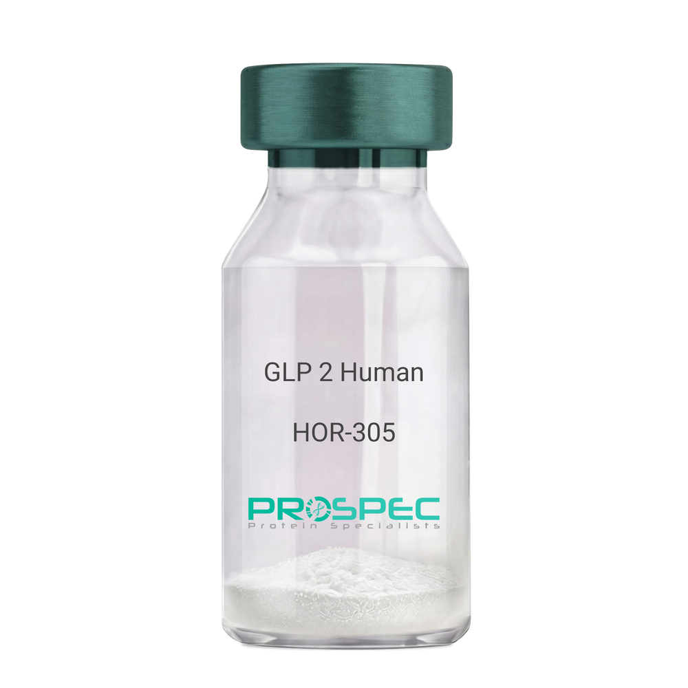
GLP 2 HumanDescription:
Human GLP-2
GLP2, GLP-2, GLP 2.
Product # :
HOR-305Price :
Quantity :
Shipping Method :
Shipped at Room temp
More Info
- description
- formulation
- purity
- More Info
Description
GLP-2 contains 34 amino acids having a molecular mass of 3922.35 Dalton.
Formulation
The protein (1mg/ml) was lyophilized with no additives.
Purity
Greater than 98.0% as determined by RP-HPLC.
More Info
-
Introduction
GLP-2 functions as an intestinal growth factor, which stimulates intestinal epithelial growth. GLP2 is involved in diabetes-associated bowel growth. GLP2 enhances cell differentiation, playing a role as a cytokine and in tissue regeneration, and mediating cytoprotection. GLP2 is invloded numerous therapeutic applications. GLP2 regulates signaling pathways coupled to cell proliferation and cell death by apoptosis.
GLP-2 is produced by specific post-translational proteolytic cleavage of proGLP. GLP-2 is manufactured by the intestinal endocrine L cell and by several neurons in the central nervous system. -
Synonyms
GLP2, GLP-2, GLP 2, Glucagon Like Peptide-2.
-
Physical Appearance
Sterile Filtered White lyophilized (freeze-dried) powder.
-
Stability
Lyophilized GLP2 although stable at room temperature for 3 weeks, should be stored desiccated below -18°C. Upon reconstitution GLP2 should be stored at 4°C between 2-7 days and for future use below -18°C.For long term storage it is recommended to add a carrier protein (0.1% HSA or BSA).Please prevent freeze-thaw cycles.
-
Solubility
It is recommended to reconstitute the lyophilized GLP-2 in sterile 18MΩ-cm H2O not less than 100 µg/ml, which can then be further diluted to other aqueous solutions.
-
Amino Acid Sequence
His-Ala-Asp-Gly-Ser-Phe-Ser-Asp-Glu-Met-Asn-Thr-Ile-Leu-Asp-Asn-Leu-Ala-Ala-Arg-Asp-Phe-Ile-Asn-Trp-Leu-Ile-Gln-Thr-Lys-Ile-Thr-Asp-Arg.
-
Background
What is the molecular weight/Mw of GLP 2 HUMAN Protein?
GLP 2 HUMAN Protein has a total Mw of 3.92kDa.
What is the Purity of GLP 2 HUMAN Protein?
GLP 2 HUMAN Protein is >95% pure as determined by SDS-PAGE.
What is the Biological Activity of GLP 2 HUMAN Protein?
The biological functionality of GLP 2 HUMAN Protein will be determined in the future.
What is the amino acid sequence of GLP 2 HUMAN Protein?
His-Ala-Asp-Gly-Ser-Phe-Ser-Asp-Glu-Met-Asn-Thr-Ile-Leu-Asp-Asn-Leu-Ala-Ala-Arg-Asp-Phe-Ile-Asn-Trp-Leu-Ile-Gln-Thr-Lys-Ile-Thr-Asp-Arg.
What applications can GLP 2 HUMAN Protein be used in?
GLP 2 HUMAN Protein can probably be used in western blot, ELISA and Lateral Flow.
What is the endotoxin level for GLP 2 HUMAN Protein?
The endotoxin level is minimal, GLP 2 HUMAN Protein was purified using conventional chromatography techniques.
ProSpec's products are furnished for LABORATORY RESEARCH USE ONLY. The product may not be used as drugs, agricultural or pesticidal products, food additives or household chemicals.
- View Data Sheet
Name :
Adiponectin Mouse, HisDescription:
Adiponectin Mouse Recombinant, His Tag
Acrp30, AdipoQ, GBP-28, APM-1, ACDC.
Product # :
CYT-537Price :
Quantity :
Shipping Method :
Shipped with Ice Packs
More Info
- description
- source
- formulation
- purity
- More Info
- sds-page
Description
The Adiponectin Mouse is created as a recombinant protein with a 21 a.a N-terminal fusion of His Tag. The Adiponectin His-Tagged Fusion Protein, produced in E. coli, is a 27.2kDa protein containing 251 amino acid residues of the Acrp30 Mouse, 18-247 amino acids.
Source
Escherichia Coli.
Formulation
Acrp30 Mouse is a sterile filtered liquid formulation containing (1mg/ml) 20mM Tris-HCl pH-8, 1mM DTT and 10% Glycerol.
Purity
Acrp30 Mouse purity is greater than 90% as determined by SDS-PAGE.
sds-page
More Info
-
Introduction
Adiponectin is an adipocyte specific secreted protein that circulates in the plasma. It is induced during adipocyte differentiation and its secretion is stimulated by insulin. Mouse adiponectin shares about 83% amino acid identity with that human. Adiponectin plays a role in various physiological processes such as energy homeostasis and obesity. Adiponectin is reduced in obese humans, and decreased level is associated with insulin resistance and hyperinsulinemia.
-
Synonyms
Acrp30, AdipoQ, GBP-28, APM-1, ACDC.
-
Stability
Store at 4°C if entire vial will be used within 2-4 weeks. Store, frozen at -20°C for longer periods of time. For long term storage it is recommended to add a carrier protein (0.1% HSA or BSA).Avoid multiple freeze-thaw cycles.
-
Amino Acid Sequence
MGSSHHHHHH SSGLVPRGSH MEDDVTTTEE LAPALVPPPK GTCAGWMAGI PGHPGHNGTP GRDGRDGTPG EKGEKGDAGL LGPKGETGDV GMTGAEGPRG FPGTPGRKGE PGEAAYVYRS AFSVGLETRV TVPNVPIRFT KIFYNQQNHY DGSTGKFYCN IPGLYYFSYH ITVYMKDVKV SLFKKDKAVL FTYDQYQEKN VDQASGSVLL HLEVGDQVWL QVYGDGDHNG LYADNVNDST FTGFLLYHDT N.
-
Background
What is the molecular weight/Mw of ADIPONECTIN Protein?
ADIPONECTIN Protein has a total Mw of 27.2kDa.
What is the source or expression system of ADIPONECTIN Protein?
Escherichia Coli.
What is the Purity of ADIPONECTIN Protein?
ADIPONECTIN Protein is >90% pure as determined by SDS-PAGE.
What is the Biological Activity of ADIPONECTIN Protein?
The biological functionality of ADIPONECTIN Protein will be determined in the future.
What is the amino acid sequence of ADIPONECTIN Protein?
MGSSHHHHHH SSGLVPRGSH MEDDVTTTEE LAPALVPPPK GTCAGWMAGI PGHPGHNGTP GRDGRDGTPG EKGEKGDAGL LGPKGETGDV GMTGAEGPRG FPGTPGRKGE PGEAAYVYRS AFSVGLETRV TVPNVPIRFT KIFYNQQNHY DGSTGKFYCN IPGLYYFSYH ITVYMKDVKV SLFKKDKAVL FTYDQYQEKN VDQASGSVLL HLEVGDQVWL QVYGDGDHNG LYADNVNDST FTGFLLYHDT N.
What applications can ADIPONECTIN Protein be used in?
ADIPONECTIN Protein can probably be used in western blot, ELISA and Lateral Flow.
What is the endotoxin level for ADIPONECTIN Protein?
The endotoxin level is minimal, ADIPONECTIN Protein was purified using conventional chromatography techniques.
ProSpec's products are furnished for LABORATORY RESEARCH USE ONLY. The product may not be used as drugs, agricultural or pesticidal products, food additives or household chemicals.
- View Data Sheet
Name :
IL 9 MouseDescription:
Interleukin-9 Mouse Recombinant
P40, HP40, T-cell growth factor p40, IL-9, P40 cytokine.
Product # :
CYT-373Price :
Quantity :
Shipping Method :
Shipped at Room temp
More Info
- description
- source
- formulation
- purity
- biological activity
- More Info
Description
Interleukin-9 Mouse Recombinant produced in E.Coli is a single, non-glycosylated single polypeptide chain containing 127 amino acids and having a molecular mass of 14.3kDa. The IL-9 is purified by proprietary chromatographic techniques.
Source
Escherichia Coli.
Formulation
Lyophilized from a concentrated (1mg/ml) solution 10mM Na2PO4, pH 7.5.
Purity
Greater than 96.0% as determined by SDS-PAGE.
Biological Activity
The ED50 as determined by the dose-dependant stimulation of human MO7e cells is < 0.5 ng/ml, corresponding to a Specific Activity of 2,000,000IU/mg.More Info
-
Introduction
Factor that is thought to be a regulator of hematopoiesis. It has been shown to enhance the growth of human mast cells and megakaryoblastic leukemic cells as well as murine helper t-cell clones. IL-9 is a glycoprotein with a molecular weight of 32-39 that is derived from T-cells, and maps to human chromosome 5.
-
Synonyms
P40, HP40, T-cell growth factor p40, IL-9, P40 cytokine.
-
Physical Appearance
Sterile Filtered White lyophilized (freeze-dried) powder.
-
Stability
Lyophilized Interleukin-9 although stable at room temperature for 3 weeks, should be stored desiccated below -18°C. Upon reconstitution IL9 should be stored at 4°C between 2-7 days and for future use below -18°C.For long term storage it is recommended to add a carrier protein (0.1% HSA or BSA).Please prevent freeze-thaw cycles.
-
Solubility
It is recommended to reconstitute the lyophilized Interleukin 9 in sterile 18MΩ-cm H2O not less than 100µg/ml, which can then be further diluted to other aqueous solutions.
-
Amino Acid Sequence
MQRCSTTWGI RDTNYLIENL KDDPPSKCSC SGNVTSCLCL SVPTDDCTTP CYREGLLQLT NATQKSRLLP VFHRVKRIVE VLKNITCPSF SCEKPCNQTM AGNTMSFLKS LLGTFQKTEM QRQKSRP.
-
Protein content
Protein quantitation was carried out by two independent methods:1. UV spectroscopy at 280 nm using the absorbency value of 0.6 as the extinction coefficient for a 0.1% (1mg/ml) solution. This value is calculated by the PC GENE computer analysis program of protein sequences (IntelliGenetics). 2. Analysis by RP-HPLC, using a standard solution of IL-9 as a Reference Standard.
ProSpec's products are furnished for LABORATORY RESEARCH USE ONLY. The product may not be used as drugs, agricultural or pesticidal products, food additives or household chemicals.
- View Data Sheet
Name :
HTATIP2 HumanDescription:
HIV-1 Tat Interactive Protein 2 Human Recombinant
TIP30, CC3, SDR44U1, HTATIP2, EC=1.1.1.-, HIV-1 TAT-interactive protein 2, FLJ26963.
Product # :
ENZ-546Price :
Quantity :
Shipping Method :
Shipped with Ice Packs
More Info
- description
- source
- formulation
- purity
- More Info
Description
HTATIP2 Recombinant Human produced in E.Coli is a single, non-glycosylated polypeptide chain containing 262 amino acids (1-242 a.a.) and having a molecular mass of 29.3 kDa. The HTATIP2 is fused to a 20 amino acid His-Tag at N-terminus and purified by proprietary chromatographic techniques.
Source
Escherichia Coli.
Formulation
HTATIP2 protein solution (1mg/ml) containing 20mM Tris-HCl pH-8, 1mM DTT & 10% glycerol.
Purity
Greater than 95.0% as determined by SDS-PAGE.
More Info
-
Introduction
HTATIP2 is part of the short-chain dehydrogenases/reductases (SDR) family which acts as a tumor suppressor in metabolic suppression, inhibition of angiogenesis and induces the expression of apoptosis related genes Bad and Siva. HTATIP2 cooperates with the activation domain of HIV-1 TAT and enhances its transcription by phosphorylating RNA polymerase II (Pol II). Defects in HTATIP2 are related with hepatocellular carcinomas and apoptotic resistant tumor cells, implicating a probable use for HTATIP2 in antitumor therapy.
-
Synonyms
TIP30, CC3, SDR44U1, HTATIP2, EC=1.1.1.-, HIV-1 TAT-interactive protein 2, FLJ26963.
-
Physical Appearance
Sterile filtered colorless solution.
-
Stability
Store at 4°C if entire vial will be used within 2-4 weeks. Store, frozen at -20°C for longer periods of time. For long term storage it is recommended to add a carrier protein (0.1% HSA or BSA).Avoid multiple freeze-thaw cycles.
-
Amino Acid Sequence
MGSSHHHHHH SSGLVPRGSH MAETEALSKL REDFRMQNKS VFILGASGET GRVLLKEILE QGLFSKVTLI GRRKLTFDEE AYKNVNQEVV DFEKLDDYAS AFQGHDVGFC CLGTTRGKAG AEGFVRVDRD YVLKSAELAK AGGCKHFNLL SSKGADKSSN FLYLQVKGEV EAKVEELKFD RYSVFRPGVL LCDRQESRPG EWLVRKFFGS LPDSWARGHS VPVVTVVRAM LNNVVRPRDK QMELLENKAI HDLGKAHGSL KP.
ProSpec's products are furnished for LABORATORY RESEARCH USE ONLY. The product may not be used as drugs, agricultural or pesticidal products, food additives or household chemicals.
- View Data Sheet
Name :
Ebola Zaire GPDescription:
Ebola Zaire Glycoprotein Recombinant
Product # :
EVD-077Price :
Quantity :
Shipping Method :
Shipped with Ice Packs
More Info
- description
- source
- formulation
- purity
- More Info
Description
Recombinant Ebola Zaire Glycoprotein is a mucin like domain containing 181 amino acids was derived from Zaire Ebola Virus (strain Kikwit-95) gp mucin sequence produced in E. coli, and fused to a 6xHis tag at its C-terminus, having a molecular weight 38kDa.Ebola Zaire GP is purified by a proprietary chromatographic technique.
Source
Escherichia Coli.
Formulation
Ebola Zaire GP protein solution is supplied in Phosphate buffer with 25mM arginine and 0.02% sodium azide.
Purity
>95% pure as determined by 12% SDS-PAGE (Coomassie blue stain).
More Info
-
Introduction
Ebolavirus (EVD) belongs to the Filoviridae family of proteins which is comprised of a single-strand, non-infectious RNA genome. EVD genome is about 19,000 base pairs long and covers 7 genes in the order 3'-UTR-NP-VP35-VP40-GP-VP30-VP24-L-5'-UTR. There are 4 different ebolaviruses such as: Zaire (EBO-Z), Sudan (EBO-S), Cote d’Ivoire (EBO-CI) and Reston (EBO-R) that differ in amino acid sequence and location of where the gene overlaps. The EBOV glycoprotein is the only virally expressed protein above the virion surface. The EBOV glycoprotein is essential for virus attachment to host cell and fusion with target cell. Mucin like domain within Ebola virus glycoprotein contains multiple glycosylated amino acids. It has been shown that antibodies frequently develop against its mucin like domain. Recombinant mucin like domain is a suitable antigen to test specific antibodies from Ebola virus infected patients.
-
Stability
Store at 4°C if entire vial will be used within 2-4 weeks. Store, frozen at -20°C for longer periods of time. For long term storage it is recommended to add a carrier protein (0.1% HSA or BSA).Avoid multiple freeze-thaw cycles.
-
Applications
Immunoassay.
ProSpec's products are furnished for LABORATORY RESEARCH USE ONLY. The product may not be used as drugs, agricultural or pesticidal products, food additives or household chemicals.
- View Data Sheet
Name :
GH AntibodyDescription:
Growth Hormone, Mouse Anti Human
GH1, GH, GHN, GH-N, hGH-N, Pituitary growth hormone, Growth hormone 1, Somatotropin.
Product # :
ANT-197Price :
Quantity :
Shipping Method :
Shipped at Room temp
More Info
- formulation
- More Info
Formulation
1mg/ml in PBS (after reconstitution).
More Info
-
Introduction
GH is a member of the somatotropin/prolactin family of hormones which play an important role in growth control. The gene, along with four other related genes, is located at the growth hormone locus on chromosome 17 where they are interspersed in the same transcriptional orientation; an arrangement which is thought to have evolved by a series of gene duplications. The five genes share a remarkably high degree of sequence identity. Alternative splicing generates additional isoforms of each of the five growth hormones, leading to further diversity and potential for specialization. This particular family member is expressed in the pituitary but not in placental tissue as is the case for the other four genes in the growth hormone locus. Mutations in or deletions of the gene lead to growth hormone deficiency and short stature.
-
Synonyms
GH1, GH, GHN, GH-N, hGH-N, Pituitary growth hormone, Growth hormone 1, Somatotropin.
-
Solubility
Reconstitute with sterile H20. Mix gently, wash the sides of the vial and wait 30-60 seconds before use.
-
Immunogen
r.Human Growth Hormone.
-
Ig Subclass
Mouse IgG1.
-
Clone
NYRhGH.
-
Applications
Direct ELISA, Western Blot, Immuneprecipitation.
-
Titer
By direct ELISA, 1:10,000 dilution will yield 0.5 O.D using alkaline phosphatase conjugated rabbit anti-mouse Ig (Jackson Laboratories).
-
Shipping Conditions
Antibody is shipped lyophilized at ambient temperature.
-
Type
Mouse Anti Human Monoclonal.
-
Storage Procedures
In lyophilized form, for long periods, store at 4C in a dry environment. After reconstitution, if not intended for use within a month, aliquot and store at -20C.
-
Purification Method
Ion exchange.
ProSpec's products are furnished for LABORATORY RESEARCH USE ONLY. The product may not be used as drugs, agricultural or pesticidal products, food additives or household chemicals.
- View Data Sheet
Name :
MX2 HumanDescription:
Myxovirus Resistance 2 Human Recombinant
Myxovirus (Influenza Virus) Resistance 2 (Homolog Of Mouse), Myxovirus Resistance Protein 2, P78-Related Protein.
Product # :
PRO-1837Price :
Quantity :
Shipping Method :
Shipped with Ice Packs
More Info
- description
- source
- formulation
- purity
- More Info
Description
MX2 Human Recombinant produced in E.coli is a single, non-glycosylated polypeptide chain containing 126 amino acids (626-715) and having a molecular mass of 14.9 kDa. MX2 is fused to a 36 amino acid His-tag at N-terminus.
Source
Escherichia Coli.
Formulation
The MX2 solution (1mg/ml) contains 20mM Tris-HCl buffer (pH 8.0), 0.4M Urea and 10% glycerol.
Purity
Greater than 90% as determined by SDS-PAGE.
More Info
-
Introduction
MX2 is a member of 2 protein families: the dynamin family and the large GTPases family. MX2 has a cytoplasmic and nuclear structure which is found in a granular pattern in the heterochromatin region under the nuclear envelope. The nuclear form has a NLS (nuclear localization signal) at the amino terminal end however it is missing in the cytoplasmic form as a result of the use of a different translation start codon. MX2 is upregulated by IFN-a, but unlike myxovirus resistance protein 1 has no antiviral activity.
-
Synonyms
Myxovirus (Influenza Virus) Resistance 2 (Homolog Of Mouse), Myxovirus Resistance Protein 2, P78-Related Protein.
-
Physical Appearance
Sterile Filtered clear solution.
-
Stability
Store at 4°C if entire vial will be used within 2-4 weeks. Store, frozen at -20°C for longer periods of time. For long term storage it is recommended to add a carrier protein (0.1% HSA or BSA).Avoid multiple freeze-thaw cycles.
-
Amino Acid Sequence
MRGSHHHHHH GMASMTGGQQ MGRDLYDDDD KDRWGSIGIH LNAYFLETSK RLANQIPFII QYFMLRENGD SLQKAMMQIL QEKNRYSWLL QEQSETATKR RILKERIYRL TQARHALCQF SSKEIH
ProSpec's products are furnished for LABORATORY RESEARCH USE ONLY. The product may not be used as drugs, agricultural or pesticidal products, food additives or household chemicals.