Search results
1000 results found for “ahcy”
Name
Description
Product #
Price
Quantity
Shipping Method
- View Data Sheet
Name :
PTPRC HumanDescription:
Protein Tyrosine Phosphatase Receptor Type C Human Recombinant
LCA, LY5, B220, CD45, T200, GP180, Receptor-type tyrosine-protein phosphatase C, Leukocyte common antigen, L-CA, PTPRC.
Product # :
ENZ-775Price :
Quantity :
Shipping Method :
Shipped with Ice Packs
More Info
- description
- source
- formulation
- purity
- More Info
Description
PTPRC Human Recombinant produced in E.coli is a single, non-glycosylated polypeptide chain containing 257 amino acids (1031-1251a.a) and having a molecular mass of 29.6kDa. PTPRC is fused to a 36 amino acid His-tag at N-terminus & purified by proprietary chromatographic techniques.
Source
Escherichia Coli.
Formulation
The PTPRC solution (0.25mg/ml) contains 20mM Tris-HCl buffer (pH 8.0), 0.15M NaCl, 10% glycerol and 1mM DTT.
Purity
Greater than 95% as determined by SDS-PAGE.
More Info
-
Introduction
Protein Tyrosine Phosphatase Receptor Type C, also known as PTPRC, which is a vital regulator of T- and B-cell antigen receptor signaling, is also a part of the protein tyrosine phosphatase (PTP) family. PTPs are signaling molecules which regulate a variety of cellular processes including differentiation, cell growth, mitosis, and oncogenic transformation. PTPRC contains an extracellular domain, a single transmembrane segment and 2 tandem intracytoplasmic catalytic domains, and therefore is classified as a receptor type PTP.
-
Synonyms
LCA, LY5, B220, CD45, T200, GP180, Receptor-type tyrosine-protein phosphatase C, Leukocyte common antigen, L-CA, PTPRC.
-
Physical Appearance
Sterile Filtered colorless solution.
-
Stability
Store at 4°C if entire vial will be used within 2-4 weeks. Store, frozen at -20°C for longer periods of time. For long term storage it is recommended to add a carrier protein (0.1% HSA or BSA). Avoid multiple freeze-thaw cycles.
-
Amino Acid Sequence
MRGSHHHHHH GMASMTGGQQ MGRDLYDDDD KDRWGSVMIA AQGPLKETIG DFWQMIFQRK VKVIVMLTEL KHGDQEICAQ YWGEGKQTYG DIEVDLKDTD KSSTYTLRVF ELRHSKRKDS RTVYQYQYTN WSVEQLPAEP KELISMIQVV KQKLPQKNSS EGNKHHKSTP LLIHCRDGSQ QTGIFCALLN LLESAETEEV VDIFQVVKAL RKARPGMVST FEQYQFLYDV IASTYPAQNG QVKKNNHQED KIEFDNE.
ProSpec's products are furnished for LABORATORY RESEARCH USE ONLY. The product may not be used as drugs, agricultural or pesticidal products, food additives or household chemicals.
- View Data Sheet
Name :
GHRP2Description:
Growth Hormone Releasing Peptide-2
GHRP-2, GHRP.
Product # :
HOR-271Price :
Quantity :
Shipping Method :
Shipped at Room temp
More Info
- description
- formulation
- purity
- More Info
Description
Growth Hormone Releasing Peptide-2 Synthetic is a single, non-glycosylated polypeptide chain containing 6 amino acids, having a molecular mass of 817.9 Dalton and a Molecular formula of C45H55N9O6.
Formulation
The GHRP-2 hormone was lyophilized with no additives.
Purity
Greater than 99.0% as determined by analysis by RP-HPLC.
More Info
-
Introduction
GH-releasing peptides (GHRPs) are synthetic peptides that like GHRH act directly on pituitary somatotrophs to stimulate GH release. GHRP-2, an investigational drug, is one of the most potent members of the GHRP family. It has been shown to be effective in adults via the oral and intranasal as well as the iv route of administration.
-
Synonyms
GHRP-2, GHRP.
-
Physical Appearance
Sterile Filtered White lyophilized (freeze-dried) powder.
-
Stability
Lyophilized Growth Hormone Releasing Peptide-2 although stable at room temperature for 3 weeks, should be stored desiccated below -18°C. Upon reconstitution GHRP-2 should be stored at 4°C between 2-7 days and for future use below -18°C.For long term storage it is recommended to add a carrier protein (0.1% HSA or BSA).Please prevent freeze-thaw cycles.
-
Solubility
It is recommended to reconstitute the lyophilized GHRP-2 in sterile 18MΩ-cm H2O not less than 100 µg/ml, which can then be further diluted to other aqueous solutions.
-
Amino Acid Sequence
H-D-Ala-D-2-Nal-Ala-Trp-D-Phe-Lys-NH2.
ProSpec's products are furnished for LABORATORY RESEARCH USE ONLY. The product may not be used as drugs, agricultural or pesticidal products, food additives or household chemicals.
- View Data Sheet
Name :
AZGP1 HumanDescription:
Alpha-2-Glycoprotein 1 Zinc-Binding Human
Zn-alpha-2-glycoprotein, Zn-alpha-2-GP, AZGP1, ZAG, Zinc-alpha-2-glycoprotein, ZNGP1, ZA2G.
Product # :
PRO-1605Price :
Quantity :
Shipping Method :
Shipped at Room temp
More Info
- description
- source
- formulation
- purity
- More Info
Description
The Human Zinc-Alpha 2 Glycoprotein produced from Human Serum has a molecular mass of 32.14kDa (calculated without glycosylation) containing 278 amino acid residues.
Source
Human Serum.
Formulation
ZA2G protein filtered (0.4µm) and lyophilized in 0.5mg/ml in 20mM TRIS and 50mM NaCl, pH 8.0.
Purity
Greater than 80% as determined by SDS-PAGE.
More Info
-
Introduction
Zinc-alpha-2-glycoprotein (ZAG) is found in body fluids such as serum, sweat, and seminal and breast cyst fluids. It is identical in amino acid sequence to tumor-derived lipid mobilizing factor (LMF), a protein associated with the dramatic loss of adipose body stores in cancer cachexia, and has been shown to stimulate lipolysis by adipocytes in vivo and in vitro. A role for ZAG has been proposed in the regulation of body weight, and age-dependent changes in genetically influenced obesity, and also it regulates melanin production by normal and malignant melanocytes. It has also recently been classified as a novel adipokine in that it is produced by both white and brown fat adipocytes and may act in a local autocrine fashion in the reduction of adiposity in cachexia. Controlling ZAG/LMF's activity could be life-saving in the management of certain cancers and other cachexiainducing conditions, and its possible normal role in body fat store homeostasis is deserving of understanding in its own right. ZAG exhibits a class I major histocompatibility complex (MHC) fold but is a soluble protein rather than being anchored to plasma membranes and does not associate with alpha-2-microglobulin in humans. Like antigen-presenting MHC class I proteins, ZAG has an open apical groove, and X-ray crystallography of human derived ZAG revealed an unidentifiable electron density in a similar position to that occupied by antigenic peptides in classical MHC proteins and glycolipids in isoforms of CD1. This presumptive ligand is not a peptide, and the groove is too small to hold a glycolipid such as is presented by CD1 isoforms. By analogy with all other MHC class I-related proteins that have an open apical groove [some do not ], occupancy by a ligand is probably crucial to ZAG's biological function. Despite all of the structural and biochemical evidence that ZAG binds a ligand, none has so far been found by extraction from protein isolated from biological fluids. This difficulty could be because the ligand is labile, heterogeneous, or readily lost during purification procedures. Knowing more about how ZAG interacts with the compounds it has been found to bind, both natural and artificial, will inform searches for the elusive ligand(s) and its/their role in ZAG's signaling function.
-
Synonyms
Zn-alpha-2-glycoprotein, Zn-alpha-2-GP, AZGP1, ZAG, Zinc-alpha-2-glycoprotein, ZNGP1, ZA2G.
-
Physical Appearance
Filtered White lyophilized (freeze-dried) powder.
-
Stability
Store lyophilized protein at -20°C. Aliquot the product after reconstitution to avoid repeated freezing/thawing cycles. Reconstituted protein can be stored at 4°C for a limited period of time; it does not show any change after two weeks at 4°C.
-
Solubility
It is recommended to add deionized water to prepare a working stock solution of approximately 0.5 mg/ml and let the lyophilized pellet dissolve completely. Product is not sterile! Please filter the product by an appropriate sterile filter before using it in the cell culture.
-
Amino Acid Sequence
QENQDGRYSL TYIYTGLSKH VEDVPAFQAL GSLNDLQFFR YNSKDRKSQP MGLWRQVEGM EDWKQDSQLQ KAREDIFMET LKDIVEYYND SNGSHVLQGR FGCEIENNRS SGAFWKYYYD GKDYIEFNKE IPAWVPFDPA AQITKQKWEA EPVYVQRAKA YLEEECPATL RKYLKYSKNI LDRQDPPSVV VTSHQAPGEK KKLKCLAYDF YPGKIDVHWT RAGEVQEPEL RGDVLHNGNG TYQSWVVVAV PPQDTAPYSC HVQHSSLAQP LVVPWEAS.
-
Human Virus Test
Blood samples from each donor have been tested and found negative for HBsAg, anti-HCV, HIV Ag/Ab and syphilis.
ProSpec's products are furnished for LABORATORY RESEARCH USE ONLY. The product may not be used as drugs, agricultural or pesticidal products, food additives or household chemicals.
- View Data Sheet
Name :
PLGF2 Human, HEKDescription:
Placental Growth Factor-2, HEK Human Recombinant
PIGF, PGF, PlGF-2, PLGF-2.
Product # :
CYT-1228Price :
Quantity :
Shipping Method :
Shipped with Ice Packs
More Info
- description
- source
- formulation
- purity
- biological activity
- More Info
Description
PLGF2 Human Recombinant produced in HEK293 cells is a single, glycosylated polypeptide chain (a.a 19-170) containing 158 amino acids and having a molecular mass of 18.1kDa. PLGF2 is fused to a 6 amino acid His tag at C-terminus and purified by proprietary chromatographic techniques.
Source
HEK293 cells.
Formulation
PLGF2 protein (0.5mg/ml) contains Phosphate-Buffered Saline (pH 7.4) and 10% glycerol.
Purity
Greater than 90.0% as determined by SDS-PAGE.
Biological Activity
Measured by its binding ability in a functional ELISA with Human VEGFR1/Flt-1.
More Info
-
Synonyms
PIGF, PGF, PlGF-2, PLGF-2.
-
Physical Appearance
Sterile Filtered colorless solution.
-
Stability
Store at 4°C if entire vial will be used within 2-4 weeks. Store, frozen at -20°C for longer periods of time. For long term storage it is recommended to add a carrier protein (0.1% HSA or BSA). Avoid multiple freeze-thaw cycles.
-
Amino Acid Sequence
LPAVPPQQWA LSAGNGSSEV EVVPFQEVWG RSYCRALERL VDVVSEYPSE VEHMFSPSCV SLLRCTGCCG DENLHCVPVE TANVTMQLLK IRSGDRPSYV ELTFSQHVRC ECRPLREKMK PERRRPKGRG KRRREKQRPT DCHLCGDAVP RRHHHHHH.
-
Background
PLGF2, a homodimeric glycoprotein, exhibits a unique structural configuration essential for its interactions with VEGF receptors and other signaling molecules. The human recombinant form provides a controlled platform for studying the three-dimensional structure of PLGF2, elucidating its binding affinities and conformational dynamics. Understanding its structure is fundamental for deciphering how PLGF2 mediates angiogenic signaling and vascular remodeling.
ProSpec's products are furnished for LABORATORY RESEARCH USE ONLY. The product may not be used as drugs, agricultural or pesticidal products, food additives or household chemicals.
- View Data Sheet
Name :
LIF HumanDescription:
Leukemia Inhibitory Factor Human Recombinant
CDF, HILDA, D-FACTOR, Differentiation- stimulating factor, Melanoma-derived LPL inhibitor, MLPLI, Emfilermin, Leukemia inhibitory factor, LIF, DIA.
Product # :
CYT-644Price :
Quantity :
Shipping Method :
Shipped at Room temp
More Info
- description
- source
- formulation
- purity
- biological activity
- More Info
Description
Leukemia Inhibitory Factor (LIF) Human Recombinant produced in E.Coli is a single, non-glycosylated, polypeptide chain containing 180 amino acids and having a molecular mass of 19.7kDa. The Leukemia Inhibitory Factor (LIF) is purified by proprietary chromatographic techniques.
Source
Escherichia Coli.
Formulation
Leukemia Inhibitory Factor (LIF) was lyophilized from a concentrated (1mg/ml) sterile solution containing 1xPBS pH 7.4.
Purity
Greater than 98.0% as determined by:
(a) Analysis by RP-HPLC.
(b) Analysis by SDS-PAGE.Biological Activity
The ED50 was determined by the M1 cell differentiation assay is < 0.01 ng/ml, corresponding to a specific activity of 100,000,000IU/mg.More Info
-
Introduction
Leukemia Inhibitory Factor also called LIF is a lymphoid factor that promotes long-term maintenance of embryonic stem cells by suppressing spontaneous differentiation. Leukemia Inhibitory Factor has several functions such as cholinergic neuron differentiation, control of stem cell pluripotency, bone & fat metabolism, mitogenesis of factor dependent cell lines & promotion of megakaryocyte production in vivo. Human and mouse LIF exhibit a 78% identity in its amino acid sequence.
-
Synonyms
CDF, HILDA, D-FACTOR, Differentiation- stimulating factor, Melanoma-derived LPL inhibitor, MLPLI, Emfilermin, Leukemia inhibitory factor, LIF, DIA.
-
Physical Appearance
Sterile Filtered White lyophilized (freeze-dried) powder.
-
Stability
Lyophilized Leukemia Inhibitory Factor (LIF) although stable at room temperature for 3 weeks, should be stored desiccated below -18°C. Upon reconstitution Leukemia Inhibitory Factor (LIF) should be stored at 4°C between 2-7 days and for future use below -18°C.For long term storage it is recommended to add a carrier protein (0.1% HSA or BSA).Please prevent freeze-thaw cycles.
-
Solubility
It is recommended to reconstitute the lyophilized Leukemia Inhibitory Factor (LIF) in sterile water not less than 100µg/ml, which can then be further diluted to other aqueous solutions.
-
Amino Acid Sequence
SPLPITPVNA TCAIRHPCHN NLMNQIRSQL AQLNGSANAL FILYYTAQGE PFPNNLDKLC GPNVTDFPPF HANGTEKAKL VELYRIVVYL GTSLGNITRD QKILNPSALS LHSKLNATAD ILRGLLSNVL CRLCSKYHVG HVDVTYGPDT SGKDVFQKKK LGCQLLGKYK QIIAVLAQAF.
-
Background
Leukemia Inhibitory Factor (LIF) Background
Leukemia Inhibitory Factor (LIF) is a cytokine within the interleukin-6 family. It is influential in the hypothalamus and involved in energy balance.
LIF stimulates ACTH secretion and affects the stress and immune response in the body. Administration of LIF has been shown to correct low plasma ACTH and repair hypothalamic-pituitary-adrenal (HPA) axis responses.
Function and Applications of LIF
LIF prevents the proliferation of myeloid leukemia cells by inducing their terminal differentiation. Beyond cancer, LIF influences bone metabolism, embryogenesis, and inflammation.
Moreover, it supports the self-renewal of stem cells in culture by activating Stat3, thus preventing spontaneous differentiation, and is studied for potential benefits in fertility treatments.
Structure and Interactions
LIF's structure consists of a four alpha-helix bundle similar to other hematopoietic cytokines. It interacts with components such as the ciliary neurotrophic factor (CNTF) through multimeric receptors, influencing both cytokine's effects on cells. This interaction is crucial for forming high-affinity binding sites that mediate their biological activities.
ProSpec's products are furnished for LABORATORY RESEARCH USE ONLY. The product may not be used as drugs, agricultural or pesticidal products, food additives or household chemicals.
- View Data Sheet
Description:
MHC Class II, (I-A) Mouse Antibody, FITC
Product # :
ANT-270Price :
Quantity :
Shipping Method :
Shipped at Room temp
More Info
- formulation
- More Info
Formulation
1mg/ml in PBS (after reconstitution).
More Info
-
Introduction
MHC Class II is consists of 2 membrane spanning proteins. Each of these proteins is roughly 30 kDa, and has of two Globular domains, Alpha-1, Alpha-2, Beta-1 and Beta-2. The two regions farthest away from the membrane are alpha-1 and beta-1. The two proteins link without Covalent Bonds. The MHC Class II protein mainly presents peptides, which have been digested from external sources. MHC Class II is normally only expressed by Antigen Presenting Cells (APC) which digest foreign protein. Within the RER the alpha and beta proteins of the molecule, associate with each other, while a third protein called the "invariant chain," is necessary for stabilizing the complex, without it, they will not associate. The MHC-invariant complex is then passed from the RER, into and out of, the Golgi body. It fuses with an Endocytic compartment, where an external protein has been sampled and degraded.
-
Solubility
Reconstitute with of H20. Mix gently, wash the sides of the vial and wait 30-60 seconds before use.
-
Immunogen
Purified mouse LN B cells (C3H anti-C57Bl6).
-
Ig Subclass
Mouse IgG2a.
-
Clone
NYRmI-A.
-
Applications
Cytotoxic and staining antibody. For staining, use 10ml/1,000,000 cells. Titer for cytotoxicity should be determined by the investigator.
-
Specificity
Recognizes most MHC class II haplotypes (b,f,p,q,r,s,u,v), but weak against I-Ak.
-
Available Conjugates
This antibody is also available unconjugated and conjugated to Biotin.
-
Type
Mouse Antibody Monoclonal.
-
Storage Procedures
Lyophilized: store at 4°C. After reconstitution, if not intended for use within a month, aliquot and store at -20°C.
-
Purification Method
Ion exchange column.
ProSpec's products are furnished for LABORATORY RESEARCH USE ONLY. The product may not be used as drugs, agricultural or pesticidal products, food additives or household chemicals.
- View Data Sheet
Name :
CD2 FITC AntibodyDescription:
CD2-FITC (T11, LFA-2), Mouse Anti-Human FITC
T11, SRBC, LFA-2, LFA-3 receptor, Erythrocyte receptor, Rosette receptor.
Product # :
ANT-188Price :
Quantity :
Shipping Method :
Shipped at Room temp
More Info
- formulation
- More Info
Formulation
1mg/ml in PBS (after reconstitution).
More Info
-
Introduction
CD-2 interacts with lymphocyte function-associated antigen (lfa-3) and cd48/bcm1 to mediate adhesion between t-cells and other cell types. CD2 is implicated in the triggering of t- cells, the cytoplasmic domain is implicated in the signaling function.
-
Synonyms
T11, SRBC, LFA-2, LFA-3 receptor, Erythrocyte receptor, Rosette receptor.
-
Solubility
Reconstitute with of H20. Mix gently, wash the sides of the vial and wait 30-60 seconds before use.
-
Immunogen
Purified human PBL T cells.
-
Ig Subclass
Mouse IgG2a.
-
Clone
hCD2.
-
Applications
Staining antibody. For staining, use 10µl/1,000,000 cells. Titer for blocking/activating T cells should be determined by the investigator.
-
Type
Mouse Anti Human Monoclonal.
-
Storage Procedures
Lyophilized: store at 4°C. After reconstitution, if not intended for use within a month, aliquot and store at -20°C.
-
Purification Method
Ion exchange column.
ProSpec's products are furnished for LABORATORY RESEARCH USE ONLY. The product may not be used as drugs, agricultural or pesticidal products, food additives or household chemicals.
- View Data Sheet
Name :
CD44 AntibodyDescription:
CD44, Mouse Anti-Human
MDU2, MDU3, MIC4, CDW44, CSPG8, HCELL, HUTCH-I, Phagocytic glycoprotein I, PGP-1, Extracellular matrix receptor-III, ECMR-III, Hermes antigen, Hyaluronate receptor, Heparan sulfate proteoglycan, Epican) CDw44.
Product # :
ANT-242Price :
Quantity :
Shipping Method :
Shipped at Room temp
More Info
- formulation
- More Info
Formulation
1mg/ml in PBS (after reconstitution).
More Info
-
Introduction
CD44 is a cell-surface glycoprotein that plays a role in cell-cell interactions, cell adhesion and migration. CD44 is a receptor for hyaluronic acid and also interacts with other ligands, such as osteopontin, collagens, and matrix metalloproteinases. CD44 participates in a wide variety of cellular functions such as lymphocyte activation, recirculation and homing, hematopoiesis, and tumor metastasis.
CD44 and CD49d are putative activity markers and CD44 a potential novel therapeutic target in multiple sclerosis. Increased CD44 antigen is associated with relapses in non-small cell lung cancers. -
Synonyms
MDU2, MDU3, MIC4, CDW44, CSPG8, HCELL, HUTCH-I, Phagocytic glycoprotein I, PGP-1, Extracellular matrix receptor-III, ECMR-III, Hermes antigen, Hyaluronate receptor, Heparan sulfate proteoglycan, Epican) CDw44.
-
Solubility
Reconstitute with of H20. Mix gently, wash the sides of the vial and wait 30-60 seconds before use.
-
Immunogen
Purified human T-Cells.
-
Ig Subclass
Mouse IgG1.
-
Clone
hCD44.
-
Applications
Staining antibody. For staining, use 10µl/1,000,000 cells.
-
Available Conjugates
This antibody is also available conjugated to biotin and FITC. For staining with biotin or FITC-conjugated antibody use 5-10µl/106 cells.
-
Type
Mouse Anti Human Monoclonal.
-
Storage Procedures
Lyophilized: store at 4°C. After reconstitution, if not intended for use within a month, aliquot and store at -20°C.
-
Purification Method
Protein-A.
ProSpec's products are furnished for LABORATORY RESEARCH USE ONLY. The product may not be used as drugs, agricultural or pesticidal products, food additives or household chemicals.
- View Data Sheet
Name :
LY6G AntibodyDescription:
LY6G, Rat Anti Mouse Antibody
Lymphocyte antigen 6G, Ly-6G, Ly-6G.1, Ly6g, Gr1, Gr-1.
Product # :
ANT-158Price :
Quantity :
Shipping Method :
Shipped at Room temp
More Info
- formulation
- More Info
Formulation
1mg/ml in PBS (after reconstitution).
More Info
-
Introduction
Myeloid differentiation antigen Gr1 (Ly6G) is a GPI-anchored protein, which is briefly expressed on monocytes in the bone marrow. The level of the Ly6G expression in the bone marrow completely correlates with granulocyte differentiation and maturation. Ly6G ligation on murine neutrophils inhibits neutrophil recruitment, thus providing the first evidence of a function for the Ly6G molecule. Ly6G is seen primarily on neutrophils, also in a subgroup of eosinophils, differentiating pre-monocytes and plasmacytoid dendritic cells.
-
Synonyms
Lymphocyte antigen 6G, Ly-6G, Ly-6G.1, Ly6g, Gr1, Gr-1.
-
Solubility
Reconstitute with H20. Mix gently, wash the sides of the vial and wait 30-60 seconds before use.
-
Immunogen
Ly-6G-transfected cell line.
-
Ig Subclass
Rat IgG2a.
-
Clone
YRmLy-6G.
-
Applications
Flow cytometry, immunohistochemistry, cell depletion.
This antibody recognizes only Ly-6G which is expressed on most myeloid cells in the bone marrow and all peripheral granulocytes. For flow cytometry, use 10 µl/106 cells. Exact titer for cell depletion in vivo (cytotoxicity) should be determined by the investigator. -
Shipping Conditions
Antibody is shipped lyophilized at ambient temperature.
-
Available Conjugates
This antibody is also available conjugated to biotin and FITC.
-
Type
Rat Anti Mouse Monoclonal.
-
Storage Procedures
In lyophilized form, for long periods, store at 4°C in a dry environment. After reconstitution, if not intended for use within a month, aliquot and store at -20°C.
-
Purification Method
Protein A column.
ProSpec's products are furnished for LABORATORY RESEARCH USE ONLY. The product may not be used as drugs, agricultural or pesticidal products, food additives or household chemicals.
- View Data Sheet
Name :
AGO2 HumanDescription:
Argonaute 2 Human Recombinant
Protein argonaute-2, Argonaute2, hAgo2, Argonaute RISC catalytic component 2, Eukaryotic translation initiation factor 2C 2, eIF-2C 2, eIF2C 2, PAZ Piwi domain protein, PPD, AGO2, EIF2C2, Protein slicer.
Product # :
PRO-2577Price :
Quantity :
Shipping Method :
Shipped with Ice Packs
More Info
- description
- source
- formulation
- purity
- More Info
Description
AGO2 Human Recombinant produced in E.Coli is a single, non-glycosylated, polypeptide chain (a.a 1-859) containing 869 amino acids including a 10 a.a N-terminal His tag. The total molecular mass is 98.4kDa (calculated).
Source
Escherichia Coli.
Formulation
AGO2 filltered solution in 50mM acetate buffer, pH 4.0 and 20% (w/v) glycerol.
Purity
Greater than 90.0% as determined by SDS-PAGE.
More Info
-
Introduction
The Argonaute protein is part of the RISC or RNA-induced silencing complex, as so, the protein has a key part in the slicing processes of RNA. The RNA interference (RNAi) is being held by RISC. Small non-coding RNA fragments bond to the Argonaute proteins, through base pairing, eventually leads to the cleavage of messenger RNA or translation suppression.
-
Synonyms
Protein argonaute-2, Argonaute2, hAgo2, Argonaute RISC catalytic component 2, Eukaryotic translation initiation factor 2C 2, eIF-2C 2, eIF2C 2, PAZ Piwi domain protein, PPD, AGO2, EIF2C2, Protein slicer.
-
Physical Appearance
Filtered colorless solution.
-
Stability
Store at 4°C if entire vial will be used within 2-4 weeks. Store, frozen at -20°C for longer periods of time. For long term storage it is recommended to add a carrier protein (0.1% HSA or BSA). Avoid multiple freeze-thaw cycles.
-
Amino Acid Sequence
MKHHHHHHAS MYSGAGPALA PPAPPPPIQG YAFKPPPRPD FGTSGRTIKL QANFFEMDIP KIDIYHYELD IKPEKCPRRV NREIVEHMVQ HFKTQIFGDR KPVFDGRKNL YTAMPLPIGR DKVELEVTLP GEGKDRIFKV SIKWVSCVSL QALHDALSGR LPSVPFETIQ ALDVVMRHLP SMRYTPVGRS FFTASEGCSN PLGGGREVWF GFHQSVRPSL WKMMLNIDVS ATAFYKAQPV IEFVCEVLDF KSIEEQQKPL TDSQRVKFTK EIKGLKVEIT HCGQMKRKYR VCNVTRRPAS HQTFPLQQES GQTVECTVAQ YFKDRHKLVL RYPHLPCLQV GQEQKHTYLP LEVCNIVAGQ RCIKKLTDNQ TSTMIRATAR SAPDRQEEIS KLMRSASFNT DPYVREFGIM VKDEMTDVTG RVLQPPSILY GGRNKAIATP VQGVWDMRNK QFHTGIEIKV WAIACFAPQR QCTEVHLKSF TEQLRKISRD AGMPIQGQPC FCKYAQGADS VEPMFRHLKN TYAGLQLVVV ILPGKTPVYA EVKRVGDTVL GMATQCVQMK NVQRTTPQTL SNLCLKINVK LGGVNNILLP QGRPPVFQQP VIFLGADVTH PPAGDGKKPS IAAVVGSMDA HPNRYCATVR VQQHRQEIIQ DLAAMVRELL IQFYKSTRFK PTRIIFYRDG VSEGQFQQVL HHELLAIREA CIKLEKDYQP GITFIVVQKR HHTRLFCTDK NERVGKSGNI PAGTTVDTKI THPTEFDFYL CSHAGIQGTS RPSHYHVLWD DNRFSSDELQ ILTYQLCHTY VRCTRSVSIP APAYYAHLVA FRARYHLVDK EHDSAEGSHT SGQSNGRDHQ ALAKAVQVHQ DTLRTMYFA.
ProSpec's products are furnished for LABORATORY RESEARCH USE ONLY. The product may not be used as drugs, agricultural or pesticidal products, food additives or household chemicals.
- View Data Sheet
Name :
HDGF2 HumanDescription:
Hepatoma-Derived Growth Factor-2 Human Recombinant
Hepatoma-derived growth factor-related protein 3, HRP-3, Hepatoma-derived growth factor 2, HDGF-2, HDGFRP3, HDGF2, CGI-142.
Product # :
CYT-132Price :
Quantity :
Shipping Method :
Shipped with Ice Packs
More Info
- description
- source
- formulation
- purity
- More Info
- SDS-PAGE
Description
HDGF2 Human Recombinant produced in E.Coli is a single, non-glycosylated polypeptide chain containing 227 amino acids (1-203 a.a.) and having a molecular mass of 25.1kDa (Molecular weight on SDS-PAGE will appear higher). HDGF2 is fused to a 24 amino acid His-tag at N-terminus & purified by proprietary chromatographic techniques.
Source
Escherichia Coli.
Formulation
HDGF2 protein solution (0.5mg/ml) containing 20mM Tris-HCl buffer (pH 8.0), 1mM DTT, 10% glycerol and 0.1M NaCl.
Purity
Greater than 90% as determined by SDS-PAGE.
SDS-PAGE
More Info
-
Introduction
Hepatoma-derived growth factor, related protein 3 (HDGFRP3/HDGF2) is the original member of a family of polypeptides labeled HDGF related proteins (HRPs). The HDGF2 protein augments DNA synthesis and may play a part in cell proliferation. HDGF2 is expressed primarily in the testis and the brain, to an intermediate extent in the heart, and to a slight extent in the ovaries, kidneys, spleen, and liver in humans.
-
Synonyms
Hepatoma-derived growth factor-related protein 3, HRP-3, Hepatoma-derived growth factor 2, HDGF-2, HDGFRP3, HDGF2, CGI-142.
-
Physical Appearance
Sterile Filtered clear solution.
-
Stability
Store at 4°C if entire vial will be used within 2-4 weeks. Store, frozen at -20°C for longer periods of time. For long term storage it is recommended to add a carrier protein (0.1% HSA or BSA).Avoid multiple freeze-thaw cycles.
-
Amino Acid Sequence
MGSSHHHHHH SSGLVPRGSH MGSHMARPRP REYKAGDLVF AKMKGYPHWP ARIDELPEGA VKPPANKYPI FFFGTHETAF LGPKDLFPYK EYKDKFGKSN KRKGFNEGLW EIENNPGVKF TGYQAIQQQS SSETEGEGGN TADASSEEEG DRVEEDGKGK RKNEKAGSKR KKSYTSKKSS
KQSRKSPGDE DDKDCKEEEN KSSSEGGDAG NDTRNTTSDL QKTSEGT. -
Background
What is the molecular weight/Mw of HDGF2 HUMAN Protein?
HDGF2 HUMAN Protein has a total Mw of 25.1kDa.
What is the source or expression system of HDGF2 HUMAN Protein?
Escherichia Coli.
What is the Purity of HDGF2 HUMAN Protein?
HDGF2 HUMAN Protein is >90% pure as determined by SDS-PAGE.
What is the Biological Activity of HDGF2 HUMAN Protein?
The biological functionality of HDGF2 HUMAN Protein will be determined in the future.
What is the amino acid sequence of HDGF2 HUMAN Protein?
MGSSHHHHHH SSGLVPRGSH MGSHMARPRP REYKAGDLVF AKMKGYPHWP ARIDELPEGA VKPPANKYPI FFFGTHETAF LGPKDLFPYK EYKDKFGKSN KRKGFNEGLW EIENNPGVKF TGYQAIQQQS SSETEGEGGN TADASSEEEG DRVEEDGKGK RKNEKAGSKR KKSYTSKKSS
KQSRKSPGDE DDKDCKEEEN KSSSEGGDAG NDTRNTTSDL QKTSEGT.
What applications can HDGF2 HUMAN Protein be used in?
HDGF2 HUMAN Protein can probably be used in western blot, ELISA and Lateral Flow.
What is the endotoxin level for HDGF2 HUMAN Protein?
The endotoxin level is minimal, HDGF2 HUMAN Protein was purified using conventional chromatography techniques.
ProSpec's products are furnished for LABORATORY RESEARCH USE ONLY. The product may not be used as drugs, agricultural or pesticidal products, food additives or household chemicals.
- View Data Sheet
Name :
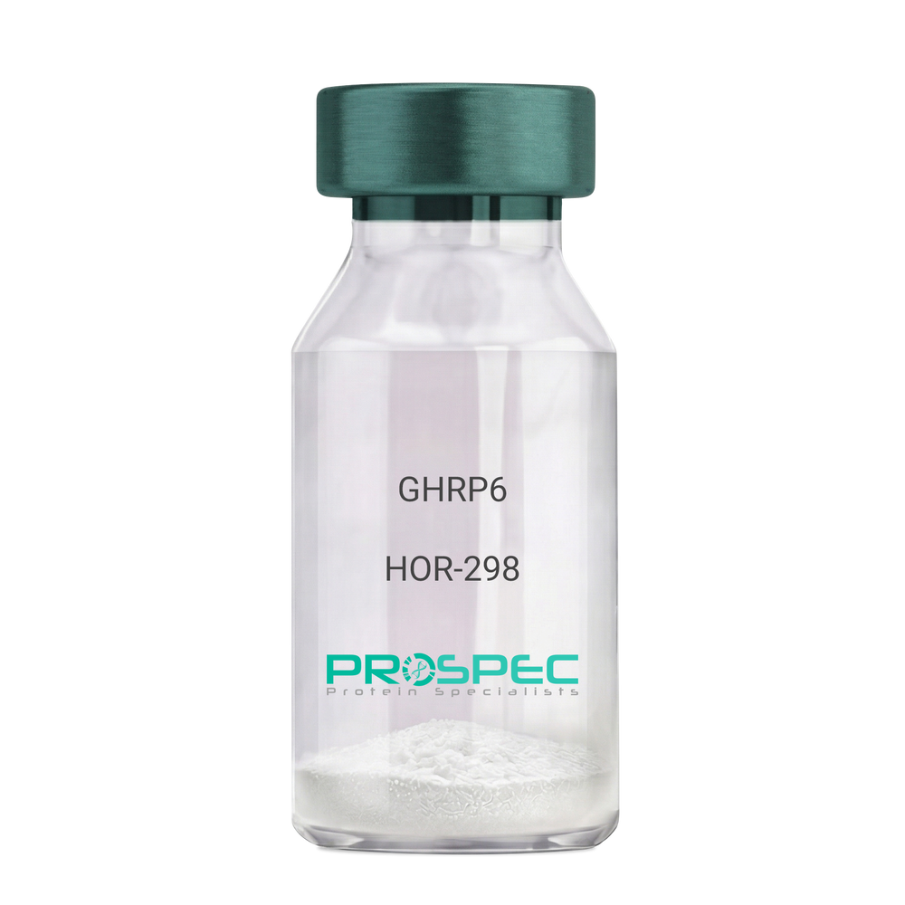
GHRP6Description:
Growth Hormone Releasing Peptide-6
GHRP-6, GHRP6
Product # :
HOR-298Price :
Quantity :
Shipping Method :
Shipped at Room temp
More Info
- description
- formulation
- purity
- More Info
Description
Growth Hormone Releasing Peptide-6 Synthetic is a single, non-glycosylated polypeptide chain containing 6 amino acids, having a molecular mass of 873 Dalton and a Molecular formula of C46H56N12O6.
Formulation
The GHRP-6 peptide was lyophilized with no additives.
Purity
Greater than 98.0% as determined by analysis by RP-HPLC.
More Info
-
Introduction
GH-releasing peptides (GHRPs) are synthetic peptides that like GHRH act directly on pituitary somatotrophs to stimulate GH release. Growth hormone (GH) release is stimulated by a variety of synthetic secretagogues, of which growth hormone-releasing hexapeptide (GHRP-6) has been most thoroughly studied; it is thought to have actions at both pituitary and hypothalamic site.
-
Synonyms
GHRP-6, GHRP6
-
Physical Appearance
Sterile Filtered White lyophilized (freeze-dried) powder.
-
Stability
Lyophilized Growth Hormone Releasing Peptide-6 although stable at room temperature for 3 weeks, should be stored desiccated below -18°C. Upon reconstitution GHRP-6 should be stored at 4°C between 2-7 days and for future use below -18°C. For long term storage it is recommended to add a carrier protein (0.1% HSA or BSA).Please prevent freeze-thaw cycles.
-
Solubility
It is recommended to reconstitute the lyophilized GHRP-6 in sterile 18MΩ-cm H2O not less than 100 µg/ml, which can then be further diluted to other aqueous solutions.
-
Amino Acid Sequence
His-D-Trp-Ala-Trp-D-Phe-Lys-NH2.
ProSpec's products are furnished for LABORATORY RESEARCH USE ONLY. The product may not be used as drugs, agricultural or pesticidal products, food additives or household chemicals.
- View Data Sheet
Name :
ADI1 AntibodyDescription:
Acireductone Dioxygenase 1, Mouse Anti Human
APL1, ARD, FLJ10913, HMFT1638, MTCBP-1, SIPL, 1,2-dihydroxy-3-keto-5-methylthiopentene dioxygenase, Acireductone dioxygenase, Fe-ARD, Membrane-type 1 matrix metalloproteinase cytoplasmic tail-binding protein 1, Submergence-induced protein-like factor.
Product # :
ANT-466Price :
Quantity :
Shipping Method :
Shipped with Ice Packs
More Info
- formulation
- More Info
Formulation
1mg/ml containing PBS, pH-7.4, 10% Glycerol and 0.02% Sodium Azide.
More Info
-
Introduction
Acireductone dioxygenase 1 (ADI1) is a part of the acireductone dioxygenase family of metal-binding enzymes, which are involved in methionine salvage. ADI1 regulates mRNA processing in the nucleus, and carries out different functions depending on its localization. Related pseudogenes have been defined on chromosomes 8 and 20. ADI1 down-regulates cell migration arbitrated by MMP14.
-
Synonyms
APL1, ARD, FLJ10913, HMFT1638, MTCBP-1, SIPL, 1,2-dihydroxy-3-keto-5-methylthiopentene dioxygenase, Acireductone dioxygenase, Fe-ARD, Membrane-type 1 matrix metalloproteinase cytoplasmic tail-binding protein 1, Submergence-induced protein-like factor.
-
Physical Appearance
Sterile filtered colorless solution.
-
Immunogen
Anti-human ADI1 mAb, clone PAT27E8A, is derived from hybridization of mouse F0 myeloma cells with spleen cells from BALB/c mice immunized with a recombinant human ADI1 protein 1-179 amino acids purified from E. coli.
-
Ig Subclass
Mouse IgG1 heavy chain and Kappa light chain.
-
Clone
PAT27E8A.
-
Applications
The antibody has been tested by ELISA, Western blot analysis to assure specificity and reactivity. Since application varies, however, each investigation should be titrated by the reagent to obtain optimal results. Recommended dilution range for Western blot analysis is 1:500 ~ 1:5000. Recommended starting dilution is 1:1000.
-
Type
Mouse Anti Human Monoclonal.
-
Storage Procedures
For periods up to 1 month store at 4°C, for longer periods of time, store at -20°C. Prevent freeze thaw cycles.
ProSpec's products are furnished for LABORATORY RESEARCH USE ONLY. The product may not be used as drugs, agricultural or pesticidal products, food additives or household chemicals.
- View Data Sheet
Name :
His Tag AntibodyDescription:
Polyhistidine Tag, Mouse Antibody
Product # :
ANT-753Price :
Quantity :
Shipping Method :
Shipped with Ice Packs
More Info
- description
- formulation
- More Info
Description
Monoclonal antibodies are produced by immunizing mouse with synthesized polypeptide 6 x His. This antibody was purified via protein-A affinity chromatography.
Formulation
1mg/ml in PBS (pH 7.4) 0.02% Sodium Azide & 10% Glycerol
More Info
-
Introduction
His tag peptide also called polyhistidine-tag is chain of amino acids that are attached to proteins and consist of at least six histidine (6xHis, Hexa Histidine) residues, often at the N- or C-terminus of the protein. Polyhistidine-tags are used for affinity purification of genetically engineered proteins.
-
Physical Appearance
Sterile Filtered solution.
-
Ig Subclass
Kappa IgG1.
-
Clone
PAT1D6AT.
-
Applications
ELISA and W.B. The researcher should titrate to obtain optimal results.
-
Type
Mouse Antibody Monoclonal.
ProSpec's products are furnished for LABORATORY RESEARCH USE ONLY. The product may not be used as drugs, agricultural or pesticidal products, food additives or household chemicals.
- View Data Sheet
Name :
AKR1C1 AntibodyDescription:
Aldo-Keto Reductase Family 1 Member C1, Mouse Anti Human
DDH1, DDH, HAKRC, 20-alpha-HSD, DD1/DD2, HBAB, C9, DD1, H-37, MBAB, MGC8954, 2-ALPHA-HSD, AKR1C1, Aldo-keto reductase family 1 member C1, 20-alpha-hydroxysteroid dehydrogenase, Trans-1,2-dihydrobenzene-1,2-diol dehydrogenase, Indanol dehydrogenase, Dihydrodiol dehydrogenase 1/2, Chlordecone reductase homolog HAKRC, High-affinity hepatic bile acid-binding protein.
Product # :
ANT-044Price :
Quantity :
Shipping Method :
Shipped with Ice Packs
More Info
- formulation
- More Info
Formulation
1mg/ml containing PBS, pH-7.4, & 0.1% Sodium Azide.
More Info
-
Introduction
AKR1C1 transfers progesterone to its inactive state or in other words catalyzes the reaction of 20-alpha-hydroxy progesterone (20-alpha-OHP). In the liver and intestine. AKR1C1 transfers bile and monitors the intrahepatic bile acid concentration though it has a low bile-binding ability. AKR1C1 participates in myelin formation. AKR1C1 is part of the aldo/keto reductase superfamily, which has over 40 known enzymes which catalyze the conversion of aldehydes and ketones to their corresponding alcohols by utilizing NADH and/or NADPH as cofactors thus display overlapping but distinct substrate specificity.
-
Synonyms
DDH1, DDH, HAKRC, 20-alpha-HSD, DD1/DD2, HBAB, C9, DD1, H-37, MBAB, MGC8954, 2-ALPHA-HSD, AKR1C1, Aldo-keto reductase family 1 member C1, 20-alpha-hydroxysteroid dehydrogenase, Trans-1,2-dihydrobenzene-1,2-diol dehydrogenase, Indanol dehydrogenase, Dihydrodiol dehydrogenase 1/2, Chlordecone reductase homolog HAKRC, High-affinity hepatic bile acid-binding protein.
-
Immunogen
Anti-human AKR1C1 mAb, is derived from hybridization of mouse FO myeloma cells with spleen cells from BALB/c mice immunized with recombinant human AKR1C1 amino acids 1-323 purified from E. coli.
-
Ig Subclass
Mouse IgG1 heavy chain and k light chain.
-
Clone
PAT6D10AT.
-
Applications
AKR1C1 antibody has been tested by ELISA and Western blot analysis to assure specificity and reactivity. Since application varies, however, each investigation should be titrated by the reagent to obtain optimal results. Recommended dilution range for Western blot analysis is 1:1000.
Recommended starting dilution is 1:1000. -
Type
Mouse Anti Human Monoclonal.
-
Storage Procedures
For periods up to 1 month store at 4°C, for longer periods of time, store at -20°C. Prevent freeze thaw cycles.
-
Purification Method
AKR1C1 antibody was purified from mouse ascitic fluids by protein-G affinity chromatography.
ProSpec's products are furnished for LABORATORY RESEARCH USE ONLY. The product may not be used as drugs, agricultural or pesticidal products, food additives or household chemicals.
- View Data Sheet
Name :
NANS AntibodyDescription:
N-acetylneuraminic acid synthase, Mouse Anti Human
Sialic acid synthase, N-acetylneuraminate synthase, N-acetylneuraminate-9-phosphate synthase, N-acetylneuraminic acid phosphate synthase, N-acetylneuraminic acid synthase, NANS, SAS.
Product # :
ANT-054Price :
Quantity :
Shipping Method :
Shipped with Ice Packs
More Info
- formulation
- More Info
Formulation
1mg/ml containing PBS, pH-7.4, & 0.1% Sodium Azide.
More Info
-
Introduction
NANS is a 359 amino acid protein that contains one AFP (antifreeze proteins)-like domain and functions in the biosynthesis of sialic acids. The ubiquitously expressed NANS enzymatically catalyzes the H2O-dependent formation of N-acetylneuraminic acid (Neu5Ac) and 2-keto-3-deoxy-D-glycero-D-galacto-nononic acid (KDN), both of which are sialic acids. NANS uses N-acetylmannosamine 6-phosphate as a substrate for Neu5Ac synthesis and mannose 6-phosphate as a substrate for KDN synthesis.
-
Synonyms
Sialic acid synthase, N-acetylneuraminate synthase, N-acetylneuraminate-9-phosphate synthase, N-acetylneuraminic acid phosphate synthase, N-acetylneuraminic acid synthase, NANS, SAS.
-
Immunogen
Anti-human NANS mAb, is derived from hybridization of mouse FO myeloma cells with spleen cells from BALB/c mice immunized with recombinant human NANS amino acids 1-359 purified from E. coli.
-
Ig Subclass
Mouse IgG1 heavy chain and k light chain.
-
Clone
PAT1G6AT.
-
Applications
NANS antibody has been tested by ELISA, Western blot analysis to assure specificity and reactivity. Since application varies, however, each investigation should be titrated by the reagent to obtain optimal results. Recommended dilution range for Western blot analysis is 1:1,000.
-
Type
Mouse Anti Human Monoclonal.
-
Storage Procedures
For periods up to 1 month store at 4°C, for longer periods of time, store at -20°C. Prevent freeze thaw cycles.
-
Purification Method
NANS antibody was purified from mouse ascitic fluids by protein-G affinity chromatography.
ProSpec's products are furnished for LABORATORY RESEARCH USE ONLY. The product may not be used as drugs, agricultural or pesticidal products, food additives or household chemicals.
- View Data Sheet
Name :
HSP90 Alpha AntibodyDescription:
Heat shock protein HSP 90-alpha, Mouse Anti Human
HSPN, LAP2, HSP86, HSPC1, HSPCA, Hsp89, Hsp90, HSP90A, HSP90N, HSPCAL1, HSPCAL4, FLJ31884, Heat shock protein HSP 90-alpha, Renal carcinoma antigen NY-REN-38, HSP 86, HSP90AA1.
Product # :
ANT-398Price :
Quantity :
Shipping Method :
Shipped with Ice Packs
More Info
- formulation
- More Info
Formulation
1mg/ml containing PBS, pH-7.4, 0.02% Sodium Azide and 10% Glycerol.
More Info
-
Introduction
HSP90 has been identified in the cytosol, nucleus and endoplasmic reticulum, and is reported to exist in many tissue types. In several tissues, including smooth muscle, HSP90 comprises up to 2% of total cellular protein. HSP90 functions as a dimer, assembled as part of heterocomplex. It possesses ATP-binding site and low ATPase activity. HSP90 is able to associate with actin filaments in certain conditions. Another important property of HSP90 is the binding of unoccupied steroid hormone receptors. HSP90 has been characterized as a molecular chaperone able to keep the target protein in a folding-competent state. It has an enhanced chaperone activity in oligomeric form at high temperatures. HSP90 function is sensitive to bivalent cations concentration.
-
Synonyms
HSPN, LAP2, HSP86, HSPC1, HSPCA, Hsp89, Hsp90, HSP90A, HSP90N, HSPCAL1, HSPCAL4, FLJ31884, Heat shock protein HSP 90-alpha, Renal carcinoma antigen NY-REN-38, HSP 86, HSP90AA1.
-
Physical Appearance
Sterile Filtered clear solution.
-
Immunogen
Anti-human HSP90A mAb, is derived from hybridization of mouse SP2/0 myeloma cells with spleen cells from BALB/c mice immunized with recombinant human HSP90A amino acids 1-732 purified from E. coli.
-
Ig Subclass
Mouse IgG2b heavy chain and κ light chain.
-
Clone
P4F10AT.
-
Applications
HSP90A antibody has been tested by ELISA, Western blot and immunohistochemistry analysis to assure specificity and reactivity. Since application varies, however, each investigation should be titrated by the reagent to obtain optimal results. Recommended dilution range for Western blot is 1:500 ~ 1:2,000 and immunohistochemistry analysis is 1:100~200. Recommended starting dilution for Western blot is 1:1,000 and Immunohistochemistry is 1:100.
-
Type
Mouse Anti Human Monoclonal.
-
Storage Procedures
For periods up to 1 month store at 4°C, for longer periods of time, store at -20°C. Prevent freeze thaw cycles.
-
Purification Method
HSP90A antibody was purified from mouse ascitic fluids by protein-G affinity chromatography.
ProSpec's products are furnished for LABORATORY RESEARCH USE ONLY. The product may not be used as drugs, agricultural or pesticidal products, food additives or household chemicals.
- View Data Sheet
Name :
HTATIP2 AntibodyDescription:
HIV-1 Tat Interactive Protein 2, Mouse Anti Human
TIP30, CC3, SDR44U1, HTATIP2, EC=1.1.1.-, HIV-1 TAT-interactive protein 2, FLJ26963.
Product # :
ANT-522Price :
Quantity :
Shipping Method :
Shipped with Ice Packs
More Info
- formulation
- More Info
Formulation
1mg/ml containing PBS, pH-7.4, 10% Glycerol and 0.02% Sodium Azide.
More Info
-
Introduction
HTATIP2 is part of the short-chain dehydrogenases/reductases (SDR) family which acts as a tumor suppressor in metabolic suppression, inhibition of angiogenesis and induces the expression of apoptosis related genes Bad and Siva. HTATIP2 cooperates with the activation domain of HIV-1 TAT and enhances its transcription by phosphorylating RNA polymerase II (Pol II). Defects in HTATIP2 are related with hepatocellular carcinomas and apoptotic resistant tumor cells, implicating a probable use for HTATIP2 in antitumor therapy.
-
Synonyms
TIP30, CC3, SDR44U1, HTATIP2, EC=1.1.1.-, HIV-1 TAT-interactive protein 2, FLJ26963.
-
Physical Appearance
Sterile filtered colorless solution.
-
Immunogen
Anti-human HTATIP2 mAb, is derived from hybridization of mouse F0 myeloma cells with spleen cells from BALB/c mice immunized with recombinant human HTATIP2 amino acids 1-242 purified from E. coli.
-
Ig Subclass
Mouse IgG2a heavy chain and ? light chain.
-
Clone
PAT5D6AT.
-
Applications
HTATIP2 antibody has been tested by ELISA, Western blot analysis, Flow cytometry and ICC/IF to assure specificity and reactivity. Since application varies, however, each investigation should be titrated by the reagent to obtain optimal results.
-
Type
Mouse Anti Human Monoclonal.
-
Storage Procedures
For periods up to 1 month store at 4°C, for longer periods of time, store at -20°C. Prevent freeze thaw cycles.
-
Purification Method
HTATIP2 antibody was purified from mouse ascitic fluids by protein-A affinity chromatography.
ProSpec's products are furnished for LABORATORY RESEARCH USE ONLY. The product may not be used as drugs, agricultural or pesticidal products, food additives or household chemicals.
- View Data Sheet
Name :
SNCG AntibodyDescription:
Gamma-Synuclein, Polyclonal Rabbit Anti-Human Antibody
Gamma-synuclein, Persyn, Breast cancer-specific gene 1 protein, Synoretin, SR, SNCG, BCSG1, PERSYN, PRSN, g-Synuclein.
Product # :
ANT-455Price :
Quantity :
Shipping Method :
Shipped with Ice Packs
More Info
- formulation
- More Info
Formulation
1mg/ml containing PBS, pH-7.4, & 0.1% Sodium Azide.
More Info
-
Introduction
γ-synuclein (Originally known as a breast cancer specific gene product, BCSG1) is an acidic neuronal protein of 127 amino acids. Gamma-Synuclein is a member of the Synuclein protein family, which is believed to be involved in the pathogenesis of neurodegenerative diseases. High levels of Gamma-Synuclein have been found in advanced breast carcinomas suggesting a correlation between overexpression of SNCG and breast tumor development. Synuclein-Gamma is found mostly in the peripheral nervous system (in primary sensory neurons, sympathetic neurons, and motor neurons) and retina. SNCG is also identified in the brain, ovarian tumors, and in the olfactory epithelium. SNCG expression in breast tumors is a marker for tumor progression. A modification in the expression of gamma-synuclein has been detected in the retina of Alzheimer''s patients.
-
Synonyms
Gamma-synuclein, Persyn, Breast cancer-specific gene 1 protein, Synoretin, SR, SNCG, BCSG1, PERSYN, PRSN, g-Synuclein.
-
Physical Appearance
Sterile filtered colorless solution.
-
Immunogen
Recombinant human γ-Synuclein amino acids 1-127 purified from E. coli.
-
Applications
γ-Synuclein antibody has been tested by ELISA and Western blot analysis to assure specificity and reactivity. Since application varies, however, each investigation should be titrated by the reagent to obtain optimal results. Recommended dilution range for Western blot analysis is 1:500 ~ 2,000. Recommended starting dilution is 1:1,000.
-
Type
Polyclonal Rabbit Antibody.
-
Storage Procedures
For periods up to 1 month store at 4°C, for longer periods of time, store at -20°C. Prevent freeze thaw cycles.
ProSpec's products are furnished for LABORATORY RESEARCH USE ONLY. The product may not be used as drugs, agricultural or pesticidal products, food additives or household chemicals.
- View Data Sheet
Name :
C-Myc (410-419aa) AntibodyDescription:
c-Myc (410-419aa), Mouse Anti Human
MYC, CMYC, C-MYS, V-MYC, P64.
Product # :
ANT-618Price :
Quantity :
Shipping Method :
Shipped with Ice Packs
More Info
- formulation
- More Info
Formulation
1mg/ml containing PBS, pH-7.4, 10% Glycerol and 0.02% Sodium Azide.
More Info
-
Introduction
c-Myc is a multifunctional, nuclear phosphoprotein that plays a role in cell cycle progression, apoptosis and cellular transformation. It functions as a transcription factor that regulates transcription of specific target genes. Mutations, overexpression, rearrangement and translocation of c-Myc have been associated with a variety of hematopoietic tumors, leukemias and lymphomas, including Burkitt lymphoma. There is evidence to show that alternative translation initiations from an upstream, in-frame non-AUG (CUG) and a downstream AUG start site result in the production of two isoforms with distinct N-termini. The synthesis of non-AUG initiated protein is suppressed in Burkitt's lymphomas, suggesting its importance in the normal function of this gene.
-
Synonyms
MYC, CMYC, C-MYS, V-MYC, P64.
-
Physical Appearance
Sterile filtered colorless solution.
-
Immunogen
Anti-human C-Myc mAb, is derived from hybridization of mouse F0 myeloma cells with spleen cells from BALB/c mice immunized with a recombinant human C-Myc protein 410-419 amino acids purified from E. coli.
-
Ig Subclass
Mouse IgG1 heavy chain and k light chain.
-
Clone
PAT19B4AT.
-
Applications
The antibody has been tested by ELISA, Western blot analysis to assure specificity and reactivity. Since application varies, however, each investigation should be titrated by the reagent to obtain optimal results.
-
Type
Mouse Anti Human Monoclonal.
-
Storage Procedures
For periods up to 1 month store at 4°C, for longer periods of time, store at -20°C. Prevent freeze thaw cycles.
-
Purification Method
C-Myc antibody was purified from mouse ascitic fluids by protein-A affinity chromatography.
ProSpec's products are furnished for LABORATORY RESEARCH USE ONLY. The product may not be used as drugs, agricultural or pesticidal products, food additives or household chemicals.
- View Data Sheet
Name :
FGF 2 HumanDescription:
Fibroblast Growth Factor-Basic Human Recombinant
Prostatropin, FGF-basic, fgf2, Basic FGF, HBGF-2, FGF-2, FGF-b.
Product # :
CYT-218Price :
Quantity :
Shipping Method :
Shipped at Room temp
More Info
- description
- source
- formulation
- purity
- biological activity
- More Info
Description
Fibroblast Growth Factor-2 Human Recombinant (FGF-2) produced in E.Coli is a single, non-glycosylated, polypeptide chain containing 154 amino acids and having a molecular mass of 17.2kDa.The FGF-b is purified by proprietary chromatographic techniques.
Source
Escherichia Coli.
Formulation
The protein was lyophilized from a concentrated (1mg/ml) solution in 20mM Tris-HCl, pH7.4 and 1M NaCl.
Purity
Greater than 98.0% as determined by Analysis by SDS-PAGE.
Biological Activity
The ED50, calculated by the dose-dependant proliferation of murine balb/c 3T3 cells is <0.1ng/ml, corresponding to a specific activity of graeter than 1.0x10⁷ Units/mg.
More Info
-
Introduction
Basic fibroblast growth factor is a member of the fibroblast growth factor (FGF) family. FGF family members possess broad mitogenic and cell survival activities, and are involved in a variety of biological processes, including embryonic development, cell growth, morphogenesis, tissue repair, tumor growth and invasion. This protein functions as a modifier of endothelial cell migration and proliferation, as well as an angiogenic factor. It acts as a mitogen for a variety of mesoderm- and neuroectoderm-derived cells in vitro, thus is thought to be involved in organogenesis. Three alternatively spliced variants encoding different isoforms have been described. The HPR -binding growth factors are angiogenic agents in vivo and are potent mitogens for a variety of cell types in vitro. There are differences in the tissue distribution and concentration of these 2 growth factors.
-
Synonyms
Prostatropin, FGF-basic, fgf2, Basic FGF, HBGF-2, FGF-2, FGF-b.
-
Physical Appearance
Sterile Filtered White lyophilized (freeze-dried) powder.
-
Stability
Lyophilized Fibroblast Growth Factor-2 although stable at room temperature for 3 weeks, should be stored desiccated below -18°C. Upon reconstitution FGF-b should be stored at 4°C between 2-7 days and for future use below -18°C.For long term storage it is recommended to add a carrier protein (0.1% HSA or BSA).Please prevent freeze-thaw cycles.
-
Solubility
It is recommended to reconstitute the lyophilized Fibroblast Growth Factor Basic in sterile 18MΩ-cm H2O not less than 100µg/ml, which can then be further diluted to other aqueous solutions.
-
Amino Acid Sequence
AAGSITTLPA LPEDGGSGAF PPGHFKDPKR LYCKNGGFFL RIHPDGRVDG VREKSDPHIK LQLQAEERGV VSIKGVCANR YLAMKEDGRL LASKCVTDEC FFFERLESNN YNTYRSRKYT SWYVALKRTG QYKLGSKTGP GQKAILFLPM SAKS.
-
Background
FGF 2 HUMAN: Insights into Fibroblast Growth Factor-2
Basic Fibroblast Growth Factor or FGF 2 HUMAN is a protein with crucial roles in cell growth, tissue repair, and embryonic development. This is part of the larger fibroblast growth factor family and is vital for various biological processes, including the modulation of cell survival activities.
Production and Properties
Produced in E. coli, FGF 2 is a non-glycosylated polypeptide chain possessing 154 amino acids with a molecular weight of about 17.2 kDa. It is purified through advanced chromatographic techniques, ensuring high purity and activity for laboratory use.
Physical Characteristics and Preparation
The physical form of FGF 2 HUMAN is a sterile, white lyophilized powder. For experimental use, it is reconstituted with sterile water to at least 100µg/ml. This reconstitution is crucial for maintaining the integrity and effectiveness of the protein in various research applications.
Storage and Handling
To maintain stability, lyophilized FGF 2 should be stored at -18°C and used within three weeks if kept at room temperature. Once reconstituted, it should be kept at 4°C and used within 2-7 days or stored at -18°C for longer-term storage.
Proper handling and avoiding repeated freeze-thaw cycles are essential to preserve the protein's functionality.
Purity and Biological Activity
FGF 2 is characterized by a purity greater than 98%, verified by SDS-PAGE analysis. Its biological activity is primarily defined by its efficacy in promoting the proliferation of specific cell lines, with an effective dose (ED50) typically below 0.1 ng/ml.
Research Applications and Impact
In the research context, FGF 2 is used extensively to study its effects on cell migration, proliferation, and angiogenesis. Moreover, its role in disease models, particularly in cancer and tissue repair studies, makes it a valuable resource for developing new therapeutic approaches.
Usage Guidelines
FGF 2 HUMAN is strictly for laboratory research use and is not suitable for drug development, food production, or cosmetic applications. Researchers are advised to comply with safety and handling guidelines to ensure that experiments are conducted under optimal conditions.
The Broad Impact on Development and Disease
FGF-2 is known for its multifunctional role across numerous biological processes such as tissue repair, embryonic development, angiogenesis, and even tumorigenesis.
This growth factor, existing in various synonymous forms such as Basic FGF, FGF-b, and HBGF-2, is essential in cellular processes that underpin both health and disease.
Furthermore, FGF-2's ability to bind to cellular receptors triggers a cascade of signaling pathways, including PI3K/Akt, MAPK/ERK, and PLCγ, which in turn influence cell growth, migration, and survival.
These pathways are pivotal in mediating the factor's diverse effects on cell behavior, contributing to its critical roles in wound healing, angiogenesis, and tissue remodeling.
What is the molecular weight/Mw of FGF 2 Protein?
FGF 2 Protein has a total Mw of 17.2kDa.
What is the source or expression system of FGF 2 Protein?
Escherichia Coli.
What is the Purity of FGF 2 Protein?
FGF 2 Protein is >98% pure as determined by SDS-PAGE.
What is the Biological Activity of FGF 2 Protein?
The ED50, calculated by the dose-dependant proliferation of murine balb/c 3T3 cells is <0.1ng/ml, corresponding to a specific activity of graeter than 1.0x10⁷ Units/mg.
What is the amino acid sequence of FGF 2 Protein?
AAGSITTLPA LPEDGGSGAF PPGHFKDPKR LYCKNGGFFL RIHPDGRVDG VREKSDPHIK LQLQAEERGV VSIKGVCANR YLAMKEDGRL LASKCVTDEC FFFERLESNN YNTYRSRKYT SWYVALKRTG QYKLGSKTGP GQKAILFLPM SAKS.
What applications can FGF 2 Protein be used in?
FGF 2 Protein can probably be used in western blot, ELISA and Lateral Flow.
What is the endotoxin level for FGF 2 Protein?
The endotoxin level is minimal, FGF 2 Protein was purified using conventional chromatography techniques.
ProSpec's products are furnished for LABORATORY RESEARCH USE ONLY. The product may not be used as drugs, agricultural or pesticidal products, food additives or household chemicals.
- View Data Sheet
Name :
Taq Plus DNADescription:
Taq Plus DNA Polymerase Recombinant
Taq Plus DNA Polymerase, Taq Plus DNA, TaqPDNA.
Product # :
ENZ-309Price :
Quantity :
Shipping Method :
Shipped with Ice Packs
More Info
- description
- source
- formulation
- More Info
Description
Taq Plus is a mixture of Taq and Pfu. Taq Plus, which is used to improve the reliability and yield of conventional primer extension reaction. Taq Plus has two following advantages over Taq: (1) high fidelity with an error frequency 1.6/106 (or 0.0016/103) during DNA synthesis. (2) Taq Plus increases the efficiency of polymerization reaction, resulting in a great percentage of extenuation reaction completion up to 10 kb to 30 kb. Pfu has a temperature optimum between 72-78°C and remains > 95% active following 1-hour incubation at 95°C.
Source
Recombinant E.coli contains Thermus aquaticus polymerase gene.
Formulation
5U/µl.
More Info
-
Synonyms
Taq Plus DNA Polymerase, Taq Plus DNA, TaqPDNA.
-
Stability
Stable for 5 days at 10°C, for longer period of time store at -20°C.
-
Optimization of DNA Synthesis
It is important to ad the reaction components in the following order1. H2O.2. 10 x reaction buffer.3. dNTPs.4. DNA template and primers.5. Taq Plus.
-
Reaction Conditions
DNA synthesis is performed in 100?l of mixture containing 20-200μm dNTPs, 0.3-1μm Promers, 0.1-0.250 mg of template DNA, 10 ?l of 10 x reaction buffer and 2.5-5 units of Taq Plus. Mix the reaction gently, centrifuge briefly and then overlay with light mineral oil. Initially, denature the reaction by incubating at 95? for 5 minutes and then cool to 40-68? for 5 minutes to allow the primers to anneal to the template DNA.
-
Storage Buffer
20mM Tris-HCl (pH 8.0), 1mM DTT, 0.1mM EDTA, 100mM KCl, Stabilizers, and 50%glycerol.
-
Note
All reagents, including Taq Plus, should be mixed immediately before use.
-
Unit Definition
One unit of the enzyme catalyzes the incorporation of 10nmole of deoxyribonucleotides into a polynucleotide fraction in 30 min at 74°C.
ProSpec's products are furnished for LABORATORY RESEARCH USE ONLY. The product may not be used as drugs, agricultural or pesticidal products, food additives or household chemicals.
- View Data Sheet
Name :
Avidin ProteinDescription:
Avidin
Avidin, AVD, AVID.
Product # :
PRO-500Price :
Quantity :
Shipping Method :
Shipped at Room temp
More Info
- description
- source
- biological activity
- More Info
Description
Avidin is a glycosylated polypeptide chain having a molecular mass of 68kDa and containing 4 subunits each with a binding site for biotin. The Avidin is purified by affinity chromatographic techniques.The purification procedure ensures minimal contamination by other proteins or DNA.The resulting high activity and purity of the product gives very low non-specific binding (NSB).
Source
Hen's egg white.
Biological Activity
15.0 units/mg protein, 1 unit binds 1µg biotin.
More Info
-
Introduction
Avidin is a tetrameric protein of 4 identical subunits (homotetramer) each of which can bind to biotin with a high degree of affinity and specificity. Avidin molecular weight in its tetrameric form is estimated to be between 66-69 kDa. Avidin is produced in the oviducts of birds, reptiles and amphibians and is subsequently deposited in the whites of their eggs. In the chicken egg white, avidin makes up roughly 0.05% of total protein (approximately 1.8 mg per egg). 10% of Avidin’s molecular weight is ascribed to carbohydrate content which is composed of four to five mannose and three N-acetylglucosamine residues. Avidin has at least three distinctive oligosaccharide structural type which are similar in structure and composition. The dissociation constant (KD) of avidin is approximately 10-15M, making it one of the strongest known non-covalent bonds.
-
Synonyms
Avidin, AVD, AVID.
-
Physical Appearance
Sterile Filtered white lyophilized powder.
-
Stability
Lyophilized Avidin although stable at room temperature for 3 weeks, should be stored desiccated below -18°C. Upon reconstitution Avidin should be stored at 4°C between 2-7 days and for future use below -18°C.For long term storage it is recommended to add a carrier protein (0.1% HSA or BSA).Please prevent freeze-thaw cycles.
-
Solubility
It is recommended to reconstitute the lyophilized Avidin in sterile 18MΩ-cm H2O not less than 100µg/ml or more than 10mg/ml solutions.
ProSpec's products are furnished for LABORATORY RESEARCH USE ONLY. The product may not be used as drugs, agricultural or pesticidal products, food additives or household chemicals.
- View Data Sheet
Name :
MMP 13 HumanDescription:
Matrix Metalloproteinase-13 Human Recombinant
CLG3, MANDP1, Matrix metalloproteinase-13, MMP-13, MMP13.
Product # :
ENZ-317Price :
Quantity :
Shipping Method :
Shipped with Ice Packs
More Info
- description
- source
- formulation
- purity
- More Info
Description
MMP-13 produced in E.Coli is a single, non-glycosylated polypeptide chain containing 391 amino acids (104-471 a.a.) and having a molecular mass of 44.7 kDa. MMP-13 is fused to a 23 amino acid His Tag at N-Terminus and purified by proprietary chromatographic techniques.
Source
Escherichia Coli.
Formulation
The MMP-13 solution (0.5mg/ml) contains 20mM Tris-HCl buffer (pH 8.0), 10% glycerol, 0.15M NaCl and 1mM DTT.
Purity
Greater than 85% as determined by SDS-PAGE.
More Info
-
Introduction
Matrix Metalloproteinase-13 (MMP-13) is an enzyme that is a member of the MMP extracellular protease family. Extracellular protease enzymes, by virtue of their broad substrate specificities1, play a role in both normal and disease states of tissue proliferation. Among the targets of MMP-13 are collagen, gelatin, entactin, pro-TNF-a, and chemokine SDF-11-4.
MMP-13 is found in its latent form as a 52-56 kDa glycosylated proenzyme. Upon cleavage the 22-46 kDa5 MMP-1 becomes active in extracellular matrix remodeling.
Because of the prominent role that MMP-1 plays in cell migration and metastasis, it is an important target for inhibition screening. -
Synonyms
CLG3, MANDP1, Matrix metalloproteinase-13, MMP-13, MMP13.
-
Physical Appearance
Sterile Filtered clear solution.
-
Stability
Store at 4°C if entire vial will be used within 2-4 weeks. Store, frozen at -20°C for longer periods of time. For long term storage it is recommended to add a carrier protein (0.1% HSA or BSA).Avoid multiple freeze-thaw cycles.
-
Amino Acid Sequence
MGSSHHHHHH SSGLVPRGSH MGSYNVFPRT LKWSKMNLTY RIVNYTPDMT HSEVEKAFKK AFKVWSDVTP LNFTRLHDGI ADIMISFGIK EHGDFYPFDG PSGLLAHAFP PGPNYGGDAH FDDDETWTSS SKGYNLFLVA AHEFGHSLGL DHSKDPGALM FPIYTYTGKS HFMLPDDDVQ GIQSLYGPGD EDPNPKHPKT PDKCDPSLSL DAITSLRGET MIFKDRFFWR LHPQQVDAEL FLTKSFWPEL PNRIDAAYEH PSHDLIFIFR GRKFWALNGY DILEGYPKKI SELGLPKEVK KISAAVHFED TGKTLLFSGN QVWRYDDTNH IMDKDYPRLI EEDFPGIGDK VDAVYEKNGY IYFFNGPIQF EYSIWSNRIV RVMPANSILW C.
ProSpec's products are furnished for LABORATORY RESEARCH USE ONLY. The product may not be used as drugs, agricultural or pesticidal products, food additives or household chemicals.