Search results
1000 results found for “calpain”
Name
Description
Product #
Price
Quantity
Shipping Method
- View Data Sheet
Name :
MUC1 HumanDescription:
Mucin-1 (CA15-3) Human
Product # :
PRO-2747Price :
Quantity :
Shipping Method :
Shipped with Ice Packs
More Info
- description
- source
- formulation
- purity
- More Info
Description
The Human Mucin-1 (CA15-3) is having a molecular mass of approximately 400kDa, and was purified from human carcinoma cell line.
Source
Human carcinoma cell line.
Formulation
MUC1 is supplied in a 0.05M sodium phosphate buffer, pH 7.5, 0.09% NaN3 and 0.15M NaCl.
Purity
Greater than 60%.
More Info
-
Introduction
Human Mucin-1 (CA15-3), aka MUC1, is a glycoprotein with extensive O-linked glycosylation of its extracellular domain. This protein has alpha and beta subunits that form a heterodimeric complex. The N-terminal alpha subunit roles in cell-adhesion and the C-terminal beta subunit is involved in cell signaling. Mucins line the apical surface of epithelial cells in the stomach, lungs, intestines, eyes and other tissues. Mucins protect the body from infection by pathogen binding to oligosaccharides in the extracellular domain, preventing the pathogen from reaching the cell surface. Overexpression of CA15-3 is often associated with colon, breast, ovarian, lung and pancreatic cancers.
-
Physical Appearance
Clear to opalescent colorless frozen solution.
-
Stability
Human MUC1 although stable at 4°C for 1 week, should be stored at -20°C.
-
Human Virus Test
Tissue sample tested and found negative for HIV-1 & 2 antibodies, HBsAg, and Hepatitis-C antibodies, Syphilis and HIV/HBV/HCV NAT.
ProSpec's products are furnished for LABORATORY RESEARCH USE ONLY. The product may not be used as drugs, agricultural or pesticidal products, food additives or household chemicals.
- View Data Sheet
Name :
METRN MouseDescription:
Meteorin Mouse Recombinant
Meteorin, Hypoxia/reoxygenation regulatory factor, Metrn, 1810034B16Rik, Hyrac.
Product # :
PRO-2241Price :
Quantity :
Shipping Method :
Shipped with Ice Packs
More Info
- description
- source
- formulation
- purity
- More Info
Description
METRN produced in Sf9 Baculovirus cells is a single, glycosylated polypeptide chain containing 276 amino acids (22-291 a.a.) and having a molecular mass of 30.2kDa (Migrates at 28-40kDa on SDS-PAGE under reducing conditions). METRN is expressed with a 6 amino acid His tag at C-Terminus and purified by proprietary chromatographic techniques.
Source
Sf9, Baculovirus cells.
Formulation
METRN protein solution (0.25mg/ml) contains Phosphate Buffered Saline (pH 7.4) and 10% glycerol.
Purity
Greater than 90.0% as determined by SDS-PAGE.
More Info
-
Introduction
Meteorin (METRN) is involved in both glial cell differentiation and axonal network formation during neurogenesis. METRN promotes astrocyte differentiation and transforms cerebellar astrocytes into radial glia. Moreover, the METRN protein stimulates axonal extension in small and intermediate neurons of sensory ganglia by activating nearby satellite glia.
-
Synonyms
Meteorin, Hypoxia/reoxygenation regulatory factor, Metrn, 1810034B16Rik, Hyrac.
-
Physical Appearance
Sterile Filtered colorless solution.
-
Stability
Store at 4°C if entire vial will be used within 2-4 weeks. Store, frozen at -20°C for longer periods of time. For long term storage it is recommended to add a carrier protein (0.1% HSA or BSA). Avoid multiple freeze-thaw cycles.
-
Amino Acid Sequence
GYSEDRCSWR GSGLTQEPGS VGQLTLDCTE GAIEWLYPAG ALRLTLGGPD PGTRPSIVCL RPERPFAGAQ VFAERMTGNL ELLLAEGPDL AGGRCMRWGP RERRALFLQA TPHRDISRRV AAFRFELHED QRAEMSPQAQ GLGVDGACRP CSDAELLLAA CTSDFVIHGT IHGVAHDTEL QESVITVVVA RVIRQTLPLF KEGSSEGQGR ASIRTLLRCG VRPGPGSFLF MGWSRFGEAW LGCAPRFQEF SRVYSAALTT HLNPCEMALD HHHHHH.
ProSpec's products are furnished for LABORATORY RESEARCH USE ONLY. The product may not be used as drugs, agricultural or pesticidal products, food additives or household chemicals.
- View Data Sheet
Name :
Leptin Human, PEGDescription:
Leptin Human Recombinant, PEG
OB Protein, Obesity Protein, OBS, Obesity factor.
Product # :
CYT-1108Price :
Quantity :
Shipping Method :
Shipped at Room temp
More Info
- description
- source
- formulation
- purity
- biological activity
- More Info
Description
Pegylated Leptin Human Recombinant produced in E.Coli is a single non-glycosilated polypeptide chain containing 146 amino acids, an additional Ala at N-terminus and one molecule of PEG 20 kDa at its N-terminus acids and having a molecular weight of 35.6kDa. However due to enlarged hydrodymanic volume it runs on the SDS-PAGE as 48 kDa protein and in gel-filtration on Superdex 200 as over 100 kDa protein. Pegylated Leptin Human Recombinant was purified by proprietary chromatographic techniques.
Source
Escherichia Coli.
Formulation
Lyophilized from a concentrated (1mg/ml) solution with 0.0045mM NaHCO3.
Purity
Greater than 98.0% as determined by:
(a) Gel filtration analysis.
(b) Analysis by SDS-PAGE.Biological Activity
Biological Activity is < than 0.1% as determined by inducing proliferation of BAF/3 cells stably transfected with the long form of human leptin receptor. It’s in vitro activity is 5-7 fold lower than the non-pegylated recombinant human leptin but in vivo it has profound weight reducing effect, resulting mainly from reduced food intake.
More Info
-
Introduction
Leptin takes an important part in the regulation of energy balance and body weight control.After entering the circulation, Leptin binds LEPRwhich results in the activation of several major signalling pathways. In the hypothalamus Leptin acts as an appetite-regulating factor that induces a decrease in food intake and an increase in energy consumption and also regulates bone mass and secretion of hypothalamo-pituitary-adrenal hormones. In the periphery, increases basal metabolism, regulates pancreatic beta-cell function and insulin secretion and affects innate and adaptive immunity.
-
Synonyms
OB Protein, Obesity Protein, OBS, Obesity factor.
-
Physical Appearance
Sterile Filtered White lyophilized (freeze-dried) powder.
-
Stability
Lyophilized Pegylated leptin although stable at room temperature for 3 weeks, should be stored desiccated below -18°C. Upon reconstitution leptin N82K should be stored at 4°C between 2-7 days and for future use below -18°C. For long term storage it is recommended to add a carrier protein (0.1% HSA or BSA). Please prevent freeze-thaw cycles.
-
Solubility
It is recommended to reconstitute the lyophilized Pegylated leptin in sterile water or 0.4% NaHCO3 adjusted to pH 8-9, not less than 100µg/ml, which can then be further diluted to other aqueous solutions.
-
Protein content
Protein quantitation was carried out by UV spectroscopy at 280 nm using the absorbency value of 0.87 as the extinction coefficient for a 0.1% (1mg/ml) solution at pH 8.0. This value is calculated by the PC GENE computer analysis program of protein sequences (IntelliGenetics).
ProSpec's products are furnished for LABORATORY RESEARCH USE ONLY. The product may not be used as drugs, agricultural or pesticidal products, food additives or household chemicals.
- View Data Sheet
Name :
COMP HumanDescription:
Cartilage Oligomeric Matrix Protein Human Recombinant
Cartilage Oligomeric Matrix Protein (pseudoachondroplasia epiphyseal dysplasia 1 multiple), MED, THBS5, TSP5, EDM1, PSACH, EPD1, Thrombospondin-5
Product # :
PRO-2727Price :
Quantity :
Shipping Method :
Shipped with Ice Packs
More Info
- description
- source
- formulation
- purity
- More Info
Description
COMP Human Recombinant produced in HEK293 Cells is a single, glycosylated polypeptide chain containing 749 amino acids (21-757a.a) and having a molecular mass of 82.4kDa.COMP is expressed with a 6 amino acid His tag at C-Terminus and purified by proprietary chromatographic techniques.
Source
HEK293 Cells.
Formulation
The COMP solution (0. 5mg/1ml) contains phosphate buffered saline (pH7.4) and 10% glycerol.
Purity
Greater than 85.0% as determined by SDS-PAGE.
More Info
-
Introduction
COMP is a non-collagenous glycoprotein and is belongs to the thrombospondin family of extracellular proteins. COMP is a calcium-binding protein of high molecular weight (>500kDa) found in the extracellular matrix of articular, nasal and tracheal cartilage. COMP is not only cartilage-derived but is common in other tissues, such as synovium and tendon. Intact COMP is pentameric, with 5 equal subunits and the carboxy-terminal globular domain of native COMP binds to collagens I, II, and IX. COMP molecules are vital for conserving the properties and integrity of collagen network. Moreover, COMP has a storage and delivery function for hydrophobic cellsignaling molecules such as vitamin D. Mutations of the COMP gene cause Pseudoachondroplasia and some forms of multiple epiphyseal dysplasia which implicates that it is vital that COMP develops and functions normally.
-
Synonyms
Cartilage Oligomeric Matrix Protein (pseudoachondroplasia epiphyseal dysplasia 1 multiple), MED, THBS5, TSP5, EDM1, PSACH, EPD1, Thrombospondin-5
-
Physical Appearance
Sterile filtered colorless solution.
-
Stability
Store at 4°C if entire vial will be used within 2-4 weeks.Store, frozen at -20°C for longer periods of time.For long term storage it is recommended to add a carrier protein (0.1% HSA or BSA).Avoid multiple freeze-thaw cycles.
-
Amino Acid Sequence
ADPDAHSLWY NFTIIHLPRH GQQWCEVQSQ VDQKNFLSYD CGSDKVLSMG HLEEQLYATD AWGKQLEMLR EVGQRLRLEL ADTELEDFTP SGPLTLQVRM SCECEADGYI RGSWQFSFDG RKFLLFDSNN RKWTVVHAGA RRMKEKWEKD SGLTTFFKMV SMRDCKSWLR DFLMHRKKRL EPTAPPTMAP GLEPKSCDKT HTCPPCPAPE LLGGPSVFLF PPKPKDTLMI SRTPEVTCVV VDVSHEDPEV KFNWYVDGVE VHNAKTKPRE EQYNSTYRVV SVLTVLHQDW LNGKEYKCKV SNKALPAPIE KTISKAKGQP REPQVYTLPP SRDELTKNQV SLTCLVKGFY PSDIAVEWES NGQPENNYKT TPPVLDSDGS FFLYSKLTVD KSRWQQGNVF SCSVMHEALH NHYTQKSLSL SPGKHHHHHH
ProSpec's products are furnished for LABORATORY RESEARCH USE ONLY. The product may not be used as drugs, agricultural or pesticidal products, food additives or household chemicals.
- View Data Sheet
Name :
ESAT6-CFP10Description:
ESAT6-CFP10 Chimeric Recombinant
Early Secretory Target Mycobacterium Tuberculosis, ESAT-6
Product # :
PRO-2737Price :
Quantity :
Shipping Method :
Shipped with Ice Packs
More Info
- description
- source
- formulation
- purity
- More Info
Description
Recombinant Chimeric ESAT6-CFP10 produced in E. coli is fused to a 6 amino acid his tag at its C-terminus and purified by proprietary chromatographic technique.
Source
Escherichia Coli.
Formulation
ESAT6-CFP10 solution contains 25mM K2CO3, 0.02% NaN3 and PBS.
Purity
Protein is >95% pure as determined by 12% PAGE (coomassie staining).
More Info
-
Introduction
Mycobacterium antigen ESAT6 has been isolated from low molecular weight fractions of the shot-term-culture filtrate (ST-CF) and it can easily be detected in tuberculosis patients. The export of ESAT-6 which is a potent T-cell antigen, and related proteins requires a dedicated secretory apparatus that is encoded by a group of genes, several of which also code for proteins that are recognized strongly by T cells. The ESAT-6 systems can consequently be considered as immunogenicity islands and there is mounting evidence that the equivalent genes are subject to selective pressure imposed by the immune system of the host. This antigen includes many epitopes detectable in the serum of most patients with tuberculosis (more than 90%). By the attempts to obtain the vaccine on the basis of ESAT-6 it was demonstrated that the optimization of adjuvant is very important when using the combination of dioctadecylammonium bromide and monophosphoryllipide. ESAT-6 is very potential as diagnostic for differentiation between the mycobacterial infection and BCG vaccination. The main topic in ESAT-6 using is in antibody production and in test-systems for tuberculosis elaboration. Early secretory antigen 6 (ESAT6) and cell filtrate protein 10 (CFP10) are 2 antigens secreted as a complex by the replicating Mycobacterium tuberculosis complex (MTC).
-
Synonyms
Early Secretory Target Mycobacterium Tuberculosis, ESAT-6
-
Physical Appearance
Sterile Filtered clear solution.
-
Stability
Store at 4°C if entire vial will be used within 2-4 weeks.Store, frozen at -20°C for longer periods of time.For long term storage it is recommended to add a carrier protein (0.1% HSA or BSA).Avoid multiple freeze-thaw cycles.
-
Applications
Immunoassay.
ProSpec's products are furnished for LABORATORY RESEARCH USE ONLY. The product may not be used as drugs, agricultural or pesticidal products, food additives or household chemicals.
- View Data Sheet
Name :
Placental Lactogen BovineDescription:
Placental Lactogen Bovine Recombinant
Chorionic Somatomammotropin Hormone 1, CSH1, CSB, CS-1, hCS-B, BPL, BPLP-I.
Product # :
CYT-511Price :
Quantity :
Shipping Method :
Shipped at Room temp
More Info
- description
- source
- formulation
- purity
- biological activity
- More Info
Description
Placental Lactogen Bovine Recombinant, is a single polypeptide chain containing 199 amino acids and an additional Ala at the N-terminus having a molecular mass of 23 kDa. Placental Lactogen Recombinant is purified by proprietary chromatographic techniques.
Source
Escherichia Coli.
Formulation
The protein was lyophilized from a concentrated (1mg/ml) solution with 0.02-0.03% NaHCO3.
Purity
Greater than 99.0% as determined by:
(a) Analysis by SEC-HPLC.
(b) Analysis by SDS-PAGE.Biological Activity
Placental Lactogen Bovine is biologically active as evidenced by inducing proliferation of Nb2 cells.More Info
-
Introduction
Placental Lactogen is a polypeptide hormone that is produced by the Syncytiotrophoblasts of the Placenta, also known as chorionic somatomammotropin. It has both Growth Hormone and Prolactin activities on growth, lactation, and luteal steroid production. In women, placental lactogen secretion begins soon after implantation and increases to 1 g or more a day in late pregnancy. Placental lactogen is also an insulin antagonist.
Placental Lactogen Bovine is also capable of activating human and other heterologous GH receptors but not ruminat GH receptors. -
Synonyms
Chorionic Somatomammotropin Hormone 1, CSH1, CSB, CS-1, hCS-B, BPL, BPLP-I.
-
Physical Appearance
Sterile Filtered White lyophilized (freeze-dried) powder.
-
Stability
Lyophilized Placental Lactogen Bovine Recombinant although stable at room temperature for 3 weeks, should be stored desiccated below -18°C. Upon reconstitution Placental Lactogen should be stored at 4°C between 2-7 days and for future use below -18°C. For long term storage it is recommended to add a carrier protein (0.1% HSA or BSA).Please prevent freeze-thaw cycles.
-
Solubility
It is recommended to reconstitute the lyophilized Placental Lactogen in sterile 18MΩ-cm H2O not less than 100µg/ml, which can then be further diluted to other aqueous solutions.
-
Amino Acid Sequence
The sequence of the first six N-terminal amino acids was determined and was found to be Ala-Glu-Asp-Tyr-Ala-Pro.
-
Protein content
UV spectroscopy at 280 nm using the absorbency value of 0.86 as the extinction coefficient for a 0.1% (1mg/ml) solution at pH 8. This value is calculated by the DNAman computer analysis program of protein sequences.
ProSpec's products are furnished for LABORATORY RESEARCH USE ONLY. The product may not be used as drugs, agricultural or pesticidal products, food additives or household chemicals.
- View Data Sheet
Name :
Fibronectin HumanDescription:
Fibronectin Human
Product # :
PRO-448Price :
Quantity :
Shipping Method :
Shipped at Room temp
More Info
- description
- source
- formulation
- purity
- More Info
Description
Human Fibronectin produced purified from Human Plasma having a Molecular Weight of 440kDa.
Source
Human Plasma.
Formulation
The Fibronectin was lyophilized from a non sterile 2mg/ml buffer of 10mM sodium phosphate, pH 7.5 and 0.15M NaCl.
Purity
≥ 95.0% as determined by SDS-PAGE.
More Info
-
Introduction
Plasma fibronectin level is elevated in severe coronary artery disease. Increased plasma fibronectin levels are related with venous thromboembolism (VTE) particularly in males, and extend the probable association between biomarkers and risk factors for arterial atherothrombosis and VTE. Fibronectin plays a role in several cellular processes, including tissue repair, embryogenesis, blood clotting, and cell migration/adhesion. Fibronectin consists in two main forms: 1) as an insoluble glycoprotein dimer that serves as a linker in the extracellular matrix and 2) as a soluble disulphide linked dimer found in the plasma. The plasma form is produced by hepatocytes, and the ECM form is synthesized by fibroblasts, chondrocytes, endothelial cells, macrophages, as well as certain epithelial cells. Fibronectin also takes part as a general cell adhesion molecule by anchoring cells to collagen or proteoglycan substrates. Fibronectin organizes cellular interaction with the ECM by binding to different components of the extracellular matrix and to membrane-bound Fibronectin receptors on cell surfaces.
-
Physical Appearance
Filtered White lyophilized (freeze-dried) powder.
-
Stability
Lyophilized Fibronectin although stable at room temperature for 3 weeks, should be stored desiccated below -18°C. Upon reconstitution Fibronectin should be stored at 4°C between 2-7 days and for future use below -18°C.
-
Solubility
We suggest reconstituting the 1mg Fibronectin with a chaotropic agent such as urea at room temperature at a concentration of 0.2mg/ml using sterile water. Let stand 1-2 hours. The recommended concentration is 4M-5M urea.
When using the protein as an attachment factor, wash the urea off after attaching the fibronectin to the growth surface (plate or dish).
ProSpec's products are furnished for LABORATORY RESEARCH USE ONLY. The product may not be used as drugs, agricultural or pesticidal products, food additives or household chemicals.
- View Data Sheet
Name :
Leptin qA Mouse, AntagonistDescription:
Leptin Quadruple Antagonist Mouse Recombinant
OB Protein, Obesity Protein, OBS, Obesity factor.
Product # :
CYT-1257Price :
Quantity :
Shipping Method :
Shipped at Room temp
More Info
- description
- source
- formulation
- purity
- biological activity
- More Info
Description
Leptin Quadruple Antagonist Mouse Recombinant is a single non-glycosilated polypeptide chain containing 146 amino, an additional Ala at N-terminus and having a molecular mass of ~ 16 kDa. The Leptin antagonist was mutated, resulting in D23L/L39A/D40A/F41A mutant. Leptin Quadruple Antagonist Mouse Recombinant was purified by proprietary chromatographic techniques.
Source
Escherichia coli.
Formulation
The Mouse Leptin Quadruple anatagonist was lyophilized from a concentrated (1mg/ml) solution with 0.003mM NaHCO3.
Purity
Greater than 98.0% as determined by:
(a) Gel filtration analysis.
(b) Analysis by SDS-PAGE.
Biological Activity
Leptin Quadruple Antagonist Mouse Recombinant is capable of inhibiting leptin-induced proliferation of BAF/3 cells stably transfected with the long form of human leptin receptor. Mouse Leptin Quadruple Antagonist also inhibits various leptin effects in several in vitro bioassays. The inhibitory activity of Mouse Leptin Quadruple Antagonist was increased 14 to 60 fold as measured by various criteria such as binding properties to human leptin binding domain and in vitro and in vivo bioassays as compared to mouse leptin antagonist.
More Info
-
Synonyms
OB Protein, Obesity Protein, OBS, Obesity factor.
-
Physical Appearance
White lyophilized (freeze-dried) powder.
-
Stability
Lyophilized Leptin Antagonist Quadruple Mouse Recombinant although stable at room temperature for several weeks, should be stored desiccated below -18°C. Upon reconstitution at > 0.1mg/ml and up to 2mM and filter sterilization LEP Antagonist can be stored at 4°C or even room temperature for several weeks making it suitable for long term infusion studies using osmotic pumps. At lower concentration addition of a carrier protein (0.1% HSA or BSA) is suggested. Please prevent freeze-thaw cycles.
-
Solubility
It is recommended to reconstitute the lyophilized Leptin Antagonist Quadruple Mouse Recombinant in sterile water or sterile 0.4% NaHCO3 adjusted to pH 8-9, not less than 100µg/ml, which can then be further diluted with other aqueous solutions.
-
Amino Acid Sequence
The sequence of the first five N-terminal amino acids of mouse super-active leptin antagonist was determined and was found to be Ala-Val-Pro-Ile-Gln.
-
Background
Leptin is produced by adipocytes and its main part is to regulate long-term energy balance. Leptin is encoded by the LEP gene and effects mainly on leptin receptors in the cell mambrane of various cells in the human body. The leptin receptor is found on a wide range of cell types. The leptin receptor is a single-transmembrane-domain type 1 cytokine receptor. leptin levels influence satiety, appetite and triggers behaviours which lead to energy savings High leptin levels are interpreted by the brain that energy reserves are high, whereas low leptin levels means that energy reserves are low, in the process adapting the organism to starvation through a variety of metabolic, neurobiochemical, endocrine and behavioral change.
-
Protein content
Protein quantization was carried out by UV spectroscopy at 280 nm using the absorbency value of 0.2 as the extinction coefficient for a 0.1% (1mg/ml) solution at pH 8.0. This value is calculated by the PC GENE computer analysis program of protein sequences (IntelliGenetics).
ProSpec's products are furnished for LABORATORY RESEARCH USE ONLY. The product may not be used as drugs, agricultural or pesticidal products, food additives or household chemicals.
- View Data Sheet
Name :
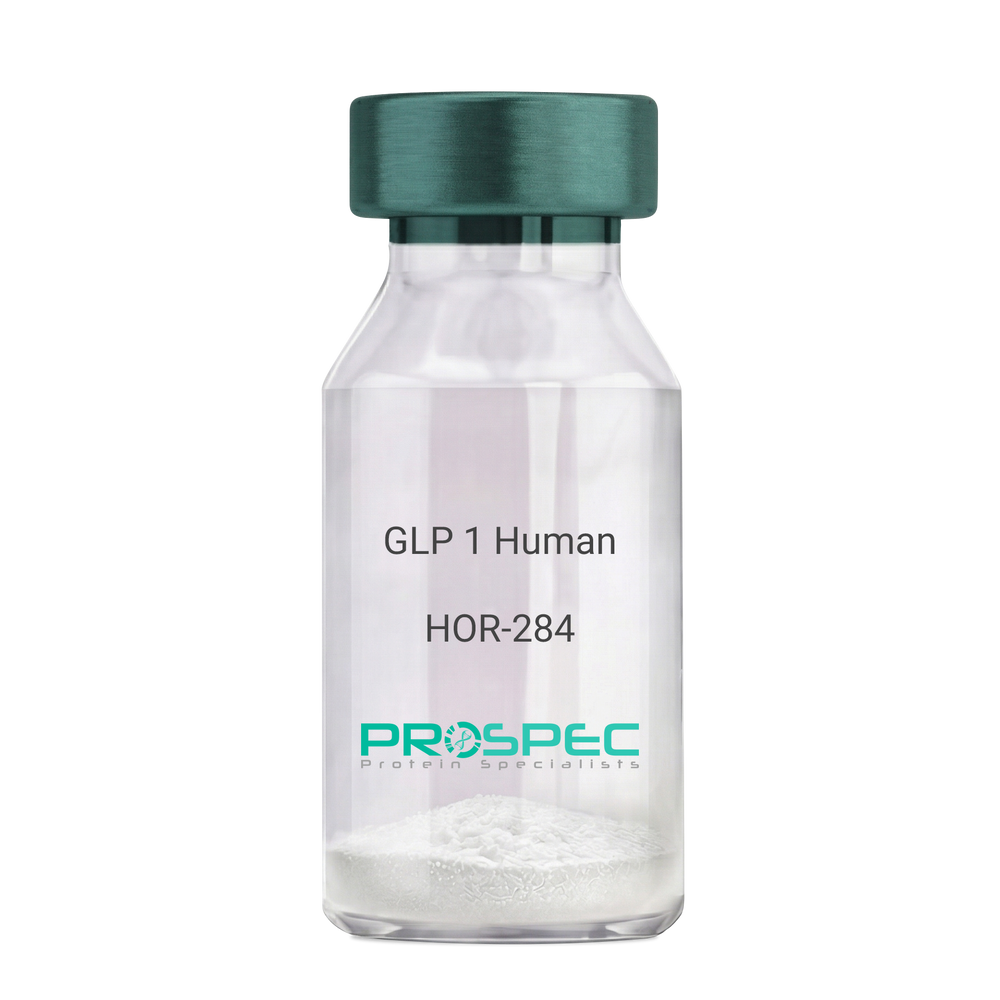
GLP 1 HumanDescription:
Human Glucagon Like Peptide-1
GLP1, Glucagon Like Peptide-1, Incretin hormone.
Product # :
HOR-284Price :
Quantity :
Shipping Method :
Shipped at Room temp
More Info
- description
- formulation
- purity
- More Info
Description
Glucagon Like Peptide-1 is a single, non-glycosylated, polypeptide chain containing 30 amino acids and having a molecular mass of 3297.7 Dalton.The GLP-1 is purified by proprietary chromatographic techniques.
Formulation
The GLP-1 peptide was lyophilized with no additives.
Purity
Greater than 96.0% as determined by RP-HPLC.
More Info
-
Introduction
Glucagon-like peptide-1 (GLP-1) is derived from the transcription product of the proglucagon gene. The major source of GLP-1 in the body is the intestinal L cell that secretes GLP-1 as a guthormone. The biologically active forms of GLP-1 are: GLP-1-(7-37) and GLP-1-(7-36)NH2.
GLP-1 secretion by L cells is dependent on the presence of nutrients in the lumen of the small intestine. The secretagogues (agents that causes or stimulates secretion) of this hormone include major nutrients like carbohydrate, proteinand lipid. Once in the circulation, GLP-1 has a half life of less than 2 minutes, due to rapid degradation by the enzyme dipeptidyl peptidase-4.
GLP-1 possesses several physiological properties that make it a subject of intensive investigation as a potential treatment of diabetes mellitus. The known physiological functions of GLP-1 include: Increases insulin secretion from the pancreas in a glucose-dependent manner, decreases glucagon secretion from the pancreas, increases beta cells mass and insulin gene expression, inhibits acid secretion and gastric emptying in the stomach, decreases food intake by increasing satiety. -
Synonyms
GLP1, Glucagon Like Peptide-1, Incretin hormone.
-
Physical Appearance
Sterile Filtered White lyophilized (freeze-dried) powder.
-
Stability
Lyophilized Glucagon Like Peptide-1 although stable at room temperature for 3 weeks, should be stored desiccated below -18°C. Upon reconstitution GLP-1 should be stored at 4°C between 2-7 days and for future use below -18°C. For long term storage it is recommended to add a carrier protein (0.1% HSA or BSA).Please prevent freeze-thaw cycles.
-
Solubility
It is recommended to reconstitute the lyophilized Glucagon Like Peptide-1 in sterile H2O at 0.5mg/ml, which can then be further diluted to other aqueous solutions.
-
Amino Acid Sequence
H-His-Ala-Glu-Gly-Thr-Phe-Thr-Ser-Asp-Val-Ser-Ser-Tyr-Leu-Glu-Gly-Gln-Ala-Ala-Lys-Glu-Phe-Ile-Ala-Trp-Leu-Val-Lys-Gly-Arg-NH2.
ProSpec's products are furnished for LABORATORY RESEARCH USE ONLY. The product may not be used as drugs, agricultural or pesticidal products, food additives or household chemicals.
- View Data Sheet
Name :
C4c HumanDescription:
Complement Component C4c Human
Complement C4c, Complement Component C4c, C4c.
Product # :
PRO-556Price :
Quantity :
Shipping Method :
Shipped with Ice Packs
More Info
- description
- source
- formulation
- purity
- More Info
Description
Human C4c produced in Human plasma having a molecular mass of 210 kDa.
Source
Human Plasma.
Formulation
The Human Complement C4c solution is in Sodium Phosphate buffer pH 7.0 containing 0.15M NaCl and 0.09% NaN3.
Purity
Greater than 99.0%.
More Info
-
Introduction
Complement C4c is a degradation product of C4b which is cleaved by C4b/C3b inactivator to yield C4d and C4c.
Complement C4c interacts with C1 and C2 to form C3 convertase of the classic activation pathway. Systemic lupus erythematosus (SLE) is often associated with congenital C4 deficiency. Reduced levels of components of the classic pathway (C1, C2, C4, C3) are common after such activation, e.g. in SLE, acute serum sickness and conditions associated with circulating immune complexes. -
Synonyms
Complement C4c, Complement Component C4c, C4c.
-
Physical Appearance
Sterile Filtered clear solution.
-
Stability
Human C4c although stable at 4°C for 1 week, should be stored at -15°C.Please avoid freeze-thaw cycles.
-
Human Virus Test
Plasma from each donor has been tested and found negative for HIV-1 & 2 antibodies, Hepatatis B surface antigen, and Hepatatis C antibodies.
ProSpec's products are furnished for LABORATORY RESEARCH USE ONLY. The product may not be used as drugs, agricultural or pesticidal products, food additives or household chemicals.
- View Data Sheet
Name :
C1R HumanDescription:
Complement C1r Human
Complement C1r subcomponent, Complement component 1 subcomponent r, C1R.
Product # :
ENZ-1163Price :
Quantity :
Shipping Method :
Shipped with Ice Packs
More Info
- description
- source
- formulation
- purity
- More Info
Description
Human Complement C1r produced in Human plasma having a molecular mass of 92 kDa.
Source
Human Plasma.
Formulation
(1mg/ml) 140mM NaCl, 10mM Imidazole and 8mM EDTA, pH 7.4.
Purity
Greater than 90.0% as determined by SDS-PAGE.
More Info
-
Introduction
C1r enzyme is the activated form of C1r proenzyme which is inactive zymogen until C1 is activated.C1r is a subunit of the C1 complex which is the first complement component in the classical pathway of complement. C1 is a non-covalent calcium-dependent complex containing1 C1q, 2 C1r and 2 C1s molecules.Each C1q bindsthe Fc domains of IgG or IgM by using2 or more of its six arms. The binding of multiple arms to immune complexes causesthe production of 2 proteases that cleave and activate the 2 C1s protease zymogens in the complex by the C1r proteins. The activation of C1r results from cleavage of C1r into 2 fragments of 57,000 and 35,000 Dalton.
-
Synonyms
Complement C1r subcomponent, Complement component 1 subcomponent r, C1R.
-
Physical Appearance
Sterile Filtered solution.
-
Stability
Human C1r is stable at 4°C if entire vial will be used within 2-4 weeks.Store, frozen below -20°C for longer periods of time.For long term storage it is recommended to add a carrier protein (0.1% HSA or BSA).Avoid multiple freeze-thaw cycles
-
Human Virus Test
Plasma from each donor has been tested and found negative for antibody to HIV-1, HIV-2, HCV, HTLV-I/II, STS and HBSAG.
ProSpec's products are furnished for LABORATORY RESEARCH USE ONLY. The product may not be used as drugs, agricultural or pesticidal products, food additives or household chemicals.
- View Data Sheet
Name :
TNNT3 HumanDescription:
Fast Skeletal Troponin T Human Recombinant
Troponin T, fast skeletal muscle, TnTf, Beta-TnTF, Fast skeletal muscle troponin T (fTnT).
Product # :
PRO-2792Price :
Quantity :
Shipping Method :
Shipped at Room temp
More Info
- description
- source
- formulation
- purity
- More Info
Description
TNNT3 Human produced in E.Coli is a single, non-glycosylated polypeptide chain containing 261 a.a and having a molecular mass of 30964 Dalton.
Source
Escherichia Coli.
Formulation
TNNT3 was lyophilized in 0.01M HCl, pH 2.0.
Purity
Greater than 95.0% as determined by SDS-PAGE.
More Info
-
Synonyms
Troponin T, fast skeletal muscle, TnTf, Beta-TnTF, Fast skeletal muscle troponin T (fTnT).
-
Physical Appearance
Sterile Filtered White lyophilized (freeze-dried) powder.
-
Stability
Lyophilized Fast Skeletal Troponin T although stable at room temperature for 3 weeks, should be stored desiccated below -18°C. Upon reconstitution TNNT3 should be stored at 4°C between 2-7 days and for future use below -18°C. For long term storage it is recommended to add a carrier protein (0.1% HSA or BSA). Please prevent freeze-thaw cycles.
-
Solubility
It is recommended to reconstitute the lyophilized TNNT3 in buffer containing 0.01M HCl, pH 2.0 not less than 100µg/ml, which can then be further diluted to other aqueous solutions.
-
Background
Troponin T3 Fast Skeletal Type (TNNT3) is a pivotal protein in the regulation of muscle contraction, specifically in fast-twitch skeletal muscles. The TNNT3 human recombinant, a product of cutting-edge biotechnology, offers a powerful tool to delve into the intricate molecular characteristics and multifaceted roles of this essential protein. This research aims to shed light on the molecular intricacies of TNNT3 and explore its significance beyond muscle contraction.
At its core, TNNT3 plays a central role in the troponin complex, which orchestrates muscle contraction by modulating the interaction between actin and myosin filaments. This molecular complex regulates muscle contraction in response to calcium ions (Ca²⁺), a fundamental process underpinning skeletal muscle function. TNNT3, as a specific isoform of troponin T, contributes to the fine-tuning of muscle contractility.
The importance of TNNT3 extends beyond its role in muscle physiology. It is primarily expressed in fast-twitch skeletal muscles responsible for rapid, forceful movements, such as sprinting and jumping. Consequently, TNNT3 is critical for the precise control of fast muscle contractions, ensuring optimal performance during activities requiring speed and power.
Furthermore, TNNT3 has gained recognition for its potential relevance in various neuromuscular disorders. Mutations in the TNNT3 gene have been associated with congenital myopathies, highlighting its clinical significance. Investigating the structural and functional properties of TNNT3 can provide valuable insights into the molecular basis of these disorders and potentially pave the way for therapeutic interventions.
The TNNT3 human recombinant serves as a gateway to comprehending the intricacies of this protein's molecular functions and its potential applications beyond muscle contraction. Researchers can leverage this recombinant protein to unravel TNNT3's structural properties, interactions with other muscle proteins, and its implications in neuromuscular diseases.
This research endeavor aims to offer a comprehensive analysis of the TNNT3 human recombinant, emphasizing its molecular characteristics, functional roles in muscle contraction, and its relevance in the context of neuromuscular disorders. By exploring the multifaceted nature of TNNT3, we aspire to contribute to a deeper understanding of muscle biology and the potential development of novel therapeutic strategies.
ProSpec's products are furnished for LABORATORY RESEARCH USE ONLY. The product may not be used as drugs, agricultural or pesticidal products, food additives or household chemicals.
- View Data Sheet
Name :
MUSTN1 HumanDescription:
Musculoskeletal, Embryonic Nuclear Protein 1 Human Recombinant
Musculoskeletal, Embryonic Nuclear Protein 1, MUSTANG, Musculoskeletal Embryonic Nuclear Protein 1, Musculoskeletal Temporally Activated Novel, MUSTN1.
Product # :
PRO-2061Price :
Quantity :
Shipping Method :
Shipped with Ice Packs
More Info
- description
- source
- formulation
- purity
- More Info
Description
MUSTN1 Human Recombinant produced in E.Coli is a single, non-glycosylated polypeptide chain containing 105 amino acids (1-82 a.a) and having a molecular mass of 11.3kDa. MUSTN1 is fused to a 23 amino acid His-tag at N-terminus & purified by proprietary chromatographic techniques.
Source
Escherichia Coli.
Formulation
MUSTN1 protein solution (0.25 mg/ml) containing 20mM Tris-HCl buffer (pH 8.0), 0.15M NaCl, 10% glycerol and 1mM DTT.
Purity
Greater than 80.0% as determined by SDS-PAGE.
More Info
-
Introduction
Musculoskeletal, Embryonic Nuclear Protein 1, also known as MUSTN1 is a member of the MUSTANG family. MUSTN1 is implicated in the development andregeneration of the musculoskeletal system.
-
Synonyms
Musculoskeletal, Embryonic Nuclear Protein 1, MUSTANG, Musculoskeletal Embryonic Nuclear Protein 1, Musculoskeletal Temporally Activated Novel, MUSTN1.
-
Physical Appearance
Sterile Filtered clear solution.
-
Stability
Store at 4°C if entire vial will be used within 2-4 weeks. Store, frozen at -20°C for longer periods of time. For long term storage it is recommended to add a carrier protein (0.1% HSA or BSA).Avoid multiple freeze-thaw cycles.
-
Amino Acid Sequence
MGSSHHHHHH SSGLVPRGSH MGSMSQAGAQ EAPIKKKRPP VKEEDLKGAR GNLTKNQEIK SKTYQVMREC EQAGSAAPSV FSRTRTGTET VFEKPKAGPT KSVFG.
ProSpec's products are furnished for LABORATORY RESEARCH USE ONLY. The product may not be used as drugs, agricultural or pesticidal products, food additives or household chemicals.
- View Data Sheet
Name :
KLK8 MouseDescription:
Kallikrein-8 Mouse Recombinant
Ovasin, PRSS19, TADG14, NRPN, NP, Kallikrein 8 (Neuropsin/Ovasin) 2 EC 3.4.21.118, Kallikrein-8, Neuropsin, EC 3.4.21 61, HNP, HK8
Product # :
ENZ-1014Price :
Quantity :
Shipping Method :
Shipped with Ice Packs
More Info
- description
- source
- formulation
- purity
- More Info
Description
KLK8 Mouse Recombinant produced in Sf9 is a single, glycosylated polypeptide chain containing 240 amino acids (29-260) and having a molecular mass of 26.5kDa (Molecular size on SDS-PAGE will appear at approximately 28-40kDa). The KLK8 is fused to an 8 amino acid His-Tag at C-terminus and purified by proprietary chromatographic techniques.
Source
Sf9, Baculovirus cells.
Formulation
KLK8 protein 0.5mg/ml is supplied in PBS, pH-7.4, and 10% Glycerol.
Purity
Greater than 95% as determined by SDS-PAGE.
More Info
-
Introduction
Kallikrein-8 is a serine protease which degrades various proteins such as casein, fibrinogen, kininogen, fibronectin and collagen type IV. Kallikrein-8 takes part in the formation and maturation of orphan and small synaptic boutons in the Schaffer-collateral pathway, regulates Schaffer-collateral long-term potentiation in the hippocampus and is essential for memory acquisition and synaptic plasticity. Kallikrein-8 participates in the secondary phase of pathogenesis following spinal cord injury and also takes part in skin desquamation and keratinocyte proliferation.
-
Synonyms
Ovasin, PRSS19, TADG14, NRPN, NP, Kallikrein 8 (Neuropsin/Ovasin) 2 EC 3.4.21.118, Kallikrein-8, Neuropsin, EC 3.4.21 61, HNP, HK8
-
Physical Appearance
Sterile Filtered colorless solution.
-
Stability
Store at 4°C if entire vial will be used within 2-4 weeks. Store, frozen at -20°C for longer periods of time. For long term storage it is recommended to add a carrier protein (0.1% HSA or BSA).Avoid multiple freeze-thaw cycles.
-
Amino Acid Sequence
QGSKILEGRE CIPHSQPWQA ALFQGERLIC GGVLVGDRWV LTAAHCKKQK YSVRLGDHSL QSRDQPEQEI QVAQSIQHPC YNNSNPEDHS HDIMLIRLQN SANLGDKVKP VQLANLCPKV GQKCIISGWG TVTSPQENFP NTLNCAEVKI YSQNKCERAY PGKITEGMVC AGSSNGADTC QGDSGGPLVC DGMLQGITSW GSDPCGKPEK PGVYTKICRY TTWIKKTMDN RDLEHHHHHH.
ProSpec's products are furnished for LABORATORY RESEARCH USE ONLY. The product may not be used as drugs, agricultural or pesticidal products, food additives or household chemicals.
- View Data Sheet
Name :
CTF2P MouseDescription:
Neuropoietin Mouse Recombinant
Cardiotrophin-2, CT-2, Neuropoietin, Np, Ctf2, Gm494.
Product # :
CYT-1128Price :
Quantity :
Shipping Method :
Shipped at Room temp
More Info
- description
- source
- formulation
- purity
- biological activity
- More Info
Description
Neuropoietin Mouse Recombinant produced in E.Coli is a non-glycosylated polypeptide chain containing 182 amino acids and having a molecular mass of approximately 19.7kDa.CTF2P is purified by proprietary chromatographic techniques.
Source
Escherichia Coli.
Formulation
Lyophilized from a 0.2μm filtered concentrated solution in 20mM Tris-HCl, pH 8.0, 0.5mM DTT and 500mM NaCl.
Purity
Greater than 98.0% as determined by:
(a) Analysis by RP-HPLC.
(b) Analysis by SDS-PAGE.Biological Activity
The ED50 as determined by a cell proliferation assay using human TF-1 cells is < 200 ng/ml, corresponding to a specific activity of > 5000 IU/mg.
More Info
-
Introduction
CTF2P, aka Neuropoietinis a part of the IL-6 family of cytokines. CTF2P is the outcome of a gene duplication event involving cardiotrophin-1 (CT-1) and it helps to define a subfamily within the IL-6 family that includes CT-1, CLC and CTNF. CTF2P Increases the platelet count associated with splenomegaly and takes part in neuronal precursor development and maturation.
-
Synonyms
Cardiotrophin-2, CT-2, Neuropoietin, Np, Ctf2, Gm494.
-
Physical Appearance
Sterile Filtered White lyophilized (freeze-dried) powder.
-
Stability
Lyophilized CTF2P although stable at room temperature for 3 weeks, should be stored desiccated below -18°C. Upon reconstitution Neuropoietin should be stored at 4°C between 2-7 days and for future use below -18°C. Please prevent freeze-thaw cycles.
-
Solubility
It is recommended to reconstitute the lyophilized Neuropoietin in sterile PBS not less than 100µg/ml, which can then be further diluted to other aqueous solutions.
-
Amino Acid Sequence
APISPSEPIG QAYSLALYMQ KNTSALLQTY LQHQGSPFSD PGFSAPELQL STLPSAAVSF KTWHAMEDAE RLSRAQGAFL ALTQHLQLVG DDQSYLNPGS PILLAQLGAA RLRAQGLLGN MAAIMTALGL PIPPEEDTLG FVPFGASAFE RKCRGYIVTR EYGHWTDRAV RDLALLKAKY SA.
-
Background
What is the molecular weight/Mw of CTF2P Protein?
CTF2P Protein has a total Mw of 19.7kDa.
What is the source or expression system of CTF2P Protein?
Escherichia Coli.
What is the Purity of CTF2P Protein?
CTF2P Protein is >98% pure as determined by SDS-PAGE.
What is the Biological Activity of CTF2P Protein?
The ED50 as determined by a cell proliferation assay using human TF-1 cells is < 200 ng/ml, corresponding to a specific activity of > 5000 IU/mg.
What is the amino acid sequence of CTF2P Protein?
APISPSEPIG QAYSLALYMQ KNTSALLQTY LQHQGSPFSD PGFSAPELQL STLPSAAVSF KTWHAMEDAE RLSRAQGAFL ALTQHLQLVG DDQSYLNPGS PILLAQLGAA RLRAQGLLGN MAAIMTALGL PIPPEEDTLG FVPFGASAFE RKCRGYIVTR EYGHWTDRAV RDLALLKAKY SA.
What applications can CTF2P Protein be used in?
CTF2P Protein can probably be used in western blot, ELISA and Lateral Flow.
What is the endotoxin level for CTF2P Protein?
The endotoxin level is minimal, CTF2P Protein was purified using conventional chromatography techniques.
ProSpec's products are furnished for LABORATORY RESEARCH USE ONLY. The product may not be used as drugs, agricultural or pesticidal products, food additives or household chemicals.
- View Data Sheet
Name :
CFD HumanDescription:
Complement Factor D Human
Complement factor D, Adipsin, C3 convertase activator, Properdin factor D, CFD, DF, PFD.
Product # :
PRO-2699Price :
Quantity :
Shipping Method :
Shipped with Ice Packs
More Info
- description
- source
- formulation
- purity
- More Info
Description
Human Complement Factor D produced in Human plasma is glycosylated polypeptide chain having a total molecular mass of 24kDa.
Source
Human Plasma.
Formulation
CFD protein solution contains 10mM Sodium phosphate and 145mM NaCl, pH 7.3.
Purity
Greater than 95.0% as determined by SDS-PAGE.
More Info
-
Introduction
CFD is an important component of the alternative pathway of complement activation. CFD cleaves and activates factor B when it binds C3b or a C3b-like protein such as C3 or CVF. CFD is a serine protease that exists as a mature protease, but it exhibits a highly restricted specificity and it appears to be substrate activated. CFD cleaves factor B bound to C3b leading to the release of the Ba fragment and leaving the Bb fragment bound to C3b. The C3b,Bb complex is called a C3 or C5 convertase because it converts these proteins to their active forms by cleaving off the small peptides C3a and C5a, respectively.
-
Synonyms
Complement factor D, Adipsin, C3 convertase activator, Properdin factor D, CFD, DF, PFD.
-
Physical Appearance
Sterile filtered solution.
-
Stability
CFD Human is stable at 4°C if entire vial will be used within 2-4 weeks.Store, frozen below -20°C for longer periods of time.For long term storage it is recommended to add a carrier protein (0.1% HSA or BSA).Avoid multiple freeze-thaw cycles.
-
Human Virus Test
Plasma from each donor has been tested and found negative for antibody to HIV-1, HIV-2, HCV and HBSAG.
ProSpec's products are furnished for LABORATORY RESEARCH USE ONLY. The product may not be used as drugs, agricultural or pesticidal products, food additives or household chemicals.
- View Data Sheet
Name :
CEA ProteinDescription:
Carcinoembryonic Antigen Human
CEACAM5, Meconium Antigen 100, Carcinoembryonic Antigen, CD66e Antigen, CD66e, Carcinoembryonic Antigen, CEA, oncofetal antigen.
Product # :
PRO-2801Price :
Quantity :
Shipping Method :
Shipped with Ice Packs
More Info
- description
- source
- formulation
- purity
- More Info
Description
CEA produced from patient source colon carcinoma liver metastatic tissue can be used as general marker in screening and monitoring malignant disease states.
Source
Liver tissue.
Formulation
CEA protein solution contains 0.1M PBS, pH 7.4, 0.09 % NaN3 and 2 % methyl-mannoside.
Purity
Greater than 95.0% as determined by SDS-PAGE.
More Info
-
Synonyms
CEACAM5, Meconium Antigen 100, Carcinoembryonic Antigen, CD66e Antigen, CD66e, Carcinoembryonic Antigen, CEA, oncofetal antigen.
-
Physical Appearance
Sterile Filtered colorless solution.
-
Stability
Store at 4°C if entire vial will be used within 2-4 weeks. Store, frozen at -20°C for longer periods of time. For long term storage it is recommended to add a carrier protein (0.1% HSA or BSA). Avoid multiple freeze-thaw cycles.
-
Applications
Blood samples from tissue donors were tested and found to be negative for HBsAg, HIV-1 and HIV-2 antibodies and HCV.
-
Background
Carcinoembryonic Antigen, commonly known as CEA, is a glycoprotein that was initially identified as a tumor marker. Over the years, research into CEA has unveiled its intricate involvement in various physiological processes, not only in cancer but also in the context of normal development and inflammatory conditions. This research aims to delve into the multifaceted roles of CEA, exploring its structural intricacies, regulatory mechanisms, and its implications in health, disease, and beyond.
Structural Complexity of CEA:
CEA, belonging to the immunoglobulin superfamily, is a complex glycoprotein featuring multiple structural domains. Its diverse forms and glycosylation patterns contribute to its functional versatility. CEA is primarily expressed in fetal tissues, but its presence is often detected in adults under pathological conditions, especially in various types of cancer.
CEA in Cancer Biology:
CEA was first recognized as a biomarker for colorectal cancer, but its overexpression is not limited to this context. Elevated CEA levels have been associated with several other malignancies, including breast, lung, and pancreatic cancers. CEA’s involvement in cancer biology ranges from promoting angiogenesis and metastasis to inhibiting immune responses, making it a critical player in tumor progression and evasion.
Beyond Cancer: CEA in Development and Inflammation:
While CEA’s role in cancer is prominent, recent studies have uncovered its participation in normal physiological processes. During embryonic development, CEA is involved in cell adhesion, contributing to tissue organization and morphogenesis. Additionally, CEA expression can be induced in inflammatory conditions, suggesting its involvement in immune responses and tissue repair mechanisms.
CEA as a Diagnostic and Therapeutic Target:
The diverse expression patterns of CEA in various diseases make it a valuable diagnostic tool. CEA assays are widely used for cancer screening, monitoring disease progression, and assessing treatment efficacy. Moreover, CEA’s presence on the surface of cancer cells has made it a target for immunotherapy, enabling the development of targeted therapies aimed at specifically eradicating CEA-positive tumor cells.
ProSpec's products are furnished for LABORATORY RESEARCH USE ONLY. The product may not be used as drugs, agricultural or pesticidal products, food additives or household chemicals.
- View Data Sheet
Name :
Hepsin HumanDescription:
Hepsin Human Recombinant
HPN, TMPRSS1, Serine protease hepsin, Transmembrane protease serine 1.
Product # :
PRO-2846Price :
Quantity :
Shipping Method :
Shipped at Room temp
More Info
- description
- source
- formulation
- purity
- biological activity
- More Info
Description
Hepsin Human Recombinant produced in Cho cells is a covalently-linked heterodimer having a total molecular mass of 43.0kDa. Hepsin is fused to a 6 amino acid His-tag at C-terminus & purified by proprietary chromatographic techniques.
Source
CHO cells.
Formulation
The Hepsin protein was Lyophilized from a 0.2µm filtered concentrated solution in 20mM Tris and 150mM NaCl, pH 8.0.
Purity
Greater than 95.0% as determined by SDS-PAGE.
Biological Activity
The specific activity is >20,000 pmol/min/μg and was measured by its ability to cleave tert-butoxycarbonyl-Gln-Arg-Arg-7-amino-4-methylcoumarin (Boc-QRR-AMC).More Info
-
Synonyms
HPN, TMPRSS1, Serine protease hepsin, Transmembrane protease serine 1.
-
Physical Appearance
Filtered White lyophilized (freeze-dried) powder.
-
Stability
Lyophilized Hepsin Active although stable at room temperature for 3 weeks, should be stored desiccated below -18°C. Upon reconstitution Hepsin Active should be stored at 4°C between 2-7 days and for future use below -18°C.
Please prevent freeze-thaw cycles. -
Solubility
It is recommended to reconstitute the lyophilized Hepsin in sterile 18MΩ-cm H2O not less than 100µg/ml, which can then be further diluted to other aqueous solutions.
-
Amino Acid Sequence
Light Chain (Non-catalytic Chain)
RSDQEPLYPV QVSSADARLM VFDKTEGTWR LLCSSRSNAR VAGLSCEEMG FLRALTHSEL DVRTAGANGT SGFFCVDEGR LPHTQRLLE VISVCDCPRGR FLAAICQDCG RRKLPVDR.
Heavy Chain (Catalytic Chain)
IVGGRDTSLG RWPWQVSLRY DGAHLCGGSL LSGDWVLTAA HCFPERNRVL SRWRVFAGAV AQASPHGLQL GVQAVVYHGG YLPFRDPNSE ENSNDIALVH LSSPLPLTEY IQPVCLPAAG QALVDGKICT VTGWGNTQYY GQQAGVLQEA RVPIISNDVC NGADFYGNQI KPKMFCAGYP EGGIDACQGD SGGPFVCEDS ISRTPRWRLC GIVSWGTGCA LAQKPGVYTK VSDFREWIFQ AIKTHSEASG MVTQLHHHHH H.
-
Background
Hepsin is a type II transmembrane serine protease expressed primarily on epithelial cells, it takes part in extracellular proteolysis by activating precursor proteins such as pro-HGF and contributes to tissue remodeling, cell signaling, and normal epithelial function. Recombinant Hepsin is used to study prostate cancer, extracellular matrix remodelling, protease signalling pathways, tumor invasion, metastasis, HGF/MET signaling, and for screening inhibitors that target serine proteases
What is the molecular weight / Mw of Hepsin Protein?
Hepsin Protein has a total Mw of 43kDa.
What is the source or expression system of Hepsin Protein?
CHO Cells
What is the Purity of Hepsin Protein?
Hepsin Protein is >95% pure as determined by SDS-PAGE.
What is the Biological Activity of Hepsin Protein?
The enzymatic activity was measured by its ability to cleave tert-butoxycarbonyl-Gln-Arg-Arg-7-amino-4-methylcoumarin (Boc-QRR-AMC). The specific activity is >20,000 pmol/min/μg.
What is the amino acid sequence of Hepsin Protein?
Light Chain (Non-catalytic Chain)
RSDQEPLYPV QVSSADARLM VFDKTEGTWR LLCSSRSNAR VAGLSCEEMG FLRALTHSEL DVRTAGANGT SGFFCVDEGR LPHTQRLLE VISVCDCPRGR FLAAICQDCG RRKLPVDR
Heavy Chain (Catalytic Chain)
IVGGRDTSLG RWPWQVSLRY DGAHLCGGSL LSGDWVLTAA HCFPERNRVL SRWRVFAGAV AQASPHGLQL GVQAVVYHGG YLPFRDPNSE ENSNDIALVH LSSPLPLTEY IQPVCLPAAG QALVDGKICT VTGWGNTQYY GQQAGVLQEA RVPIISNDVC NGADFYGNQI KPKMFCAGYP EGGIDACQGD SGGPFVCEDS ISRTPRWRLC GIVSWGTGCA LAQKPGVYTK VSDFREWIFQ AIKTHSEASG MVTQLHHHHH H
What applications can Hepsin Protein be used in?
Hepsin Protein can probably be used in western blot, ELISA and Lateral Flow.
What is the endotoxin level for Hepsin Protein?
The endotoxin level is minimal, Hepsin Protein was purified using conventional chromatography techniques.
ProSpec's products are furnished for LABORATORY RESEARCH USE ONLY. The product may not be used as drugs, agricultural or pesticidal products, food additives or household chemicals.
- View Data Sheet
Name :
C3 HumanDescription:
Complement C3 Human
Complement C3, C3 and PZP-like alpha-2-macroglobulin domain-containing protein 1, C3, CPAMD1.
Product # :
PRO-2684Price :
Quantity :
Shipping Method :
Shipped with Ice Packs
More Info
- description
- source
- formulation
- purity
- More Info
Description
Human Complement C3 produced in Human plasma having a molecular mass of 185 kDa.
Source
Human Plasma.
Formulation
C3 solution contains phosphate buffer saline.
Purity
Greater than 94.0% as determined by SDS-PAGE.
More Info
-
Introduction
Native human C3 is a naturally glycosylated polypeptide containing two disulfide-linked chains. C3 is central to the activation of all 3 pathways of complement activation. Initiation of each pathway generates proteolytic enzyme complexes which binds the target surface. These enzymes cleave a peptide bond in C3 releasing the anaphylatoxin C3a and activating C3b. Most of the C3 activated during complement activation never attaches to the surface due to its thioester reaction with water forming fluid phase C3b which is rapidly inactivated by factors H and I forming iC3b. Surface-bound C3b is necessary in all 3 pathways for efficient activation of C5 and formation of C5b-9 complexes that lyse the target cell membrane.
-
Synonyms
Complement C3, C3 and PZP-like alpha-2-macroglobulin domain-containing protein 1, C3, CPAMD1.
-
Physical Appearance
Sterile Filtered solution.
-
Stability
C3 Human is stable at 4°C if entire vial will be used within 2-4 weeks.Store, frozen below -20°C for longer periods of time.For long term storage it is recommended to add a carrier protein (0.1% HSA or BSA).Avoid multiple freeze-thaw cycles.
-
Human Virus Test
Plasma from each donor has been tested and found negative for antibody to HIV-1,HIV-2, HCV, HTLV-I &II, STS and HBSAG.
ProSpec's products are furnished for LABORATORY RESEARCH USE ONLY. The product may not be used as drugs, agricultural or pesticidal products, food additives or household chemicals.
- View Data Sheet
Name :
C6 HumanDescription:
Complement C6 Human
Complement component C6, C6.
Product # :
PRO-2693Price :
Quantity :
Shipping Method :
Shipped with Ice Packs
More Info
- description
- source
- formulation
- purity
- More Info
Description
Human Complement C6 produced in Human plasma having a molecular mass of 105 kDa.
Source
Human Plasma.
Formulation
C6 protein solution contains phosphate buffer saline, pH 7.2.
Purity
Greater than 90.0% as determined by SDS-PAGE.
More Info
-
Introduction
C6 is necessary for membrane attack complex formation and is activated by binding to recently-formed C5b at the cell membrane.Each pathway of complement activation generates proteolytic enzyme complexes which are bound to the target surface. These enzymes cleave a peptide bond in the larger alpha chain of C5 releasing C5a and activating C5b. Although C5b is unstable it remains bound to the activating complex for a few minutes during which it binds a single C6 from the surrounding fluid or it decays and is no longer capable of forming MAC. The C5b,6 complex may also remain connected to the C3/C5 convertase where the binding of a single C7 exposes a membrane-binding region and C5b,6,7 can enter into the bilipid layer of the target cell.
-
Synonyms
Complement component C6, C6.
-
Physical Appearance
Sterile filtered solution.
-
Stability
C6 Human is stable at 4°C if entire vial will be used within 2-4 weeks.Store, frozen below -20°C for longer periods of time.For long term storage it is recommended to add a carrier protein (0.1% HSA or BSA).Avoid multiple freeze-thaw cycles.
-
Human Virus Test
Plasma from each donor has been tested and found negative for antibody to HIV-1, HIV-2, HCV and HBSAG.
ProSpec's products are furnished for LABORATORY RESEARCH USE ONLY. The product may not be used as drugs, agricultural or pesticidal products, food additives or household chemicals.
- View Data Sheet
Name :
StreptavidinDescription:
Streptavidin Recombinant
Product # :
PRO-791Price :
Quantity :
Shipping Method :
Shipped at Room temp
More Info
- description
- source
- formulation
- purity
- More Info
Description
Streptavidin Streptomyces Avidinii Recombinant produced in E.Coli. The molecular weight per tetramer is approximately 52kDa.
Source
Escherichia Coli.
Formulation
Lyophilized in 10mM potassium phosphate buffer pH 6.5.
Purity
Greater than 98.0% as determined by SDS-PAGE and HPLC.
More Info
-
Introduction
Streptavidin is a tetrameric protein secreted by Streptomyces avidinii which binds firmly to biotin. Streptavidin is widely used in molecular biology through its unique high affinity for the vitamin biotin. The dissociation constant (Kd) of the biotin-streptavidin complex is about ~10-15 mol/L. The strong affinity recognition of biotin and biotinylated molecules has made streptavidin one of the most important components in diagnostics and laboratory kits. The streptavidin/biotin system has one of the biggest free energies of association of yet observed for noncovalent binding of a protein and small ligand in aqueous solution (K_assoc = 10**14). The complexes are also extremely stable over a wide range of temperature and pH.
-
Physical Appearance
Sterile Filtered White lyophilized (freeze-dried) powder.
-
Stability
Streptavidin is shipped at ambient temperature, upon arrival store at -20°C.
-
Solubility
It is recommended to reconstitute the lyophilized Streptavidin in sterile 18MΩ-cm H2O not less than 0.5mg/ml, which can then be further diluted to other aqueous solutions.
-
Amino Acid Sequence
MAEAGITGTWYNQLGSTFIVTAGADGALTGTYESAVGNAESRYVLT
GRYDSAPATDGSGTALGWTVAWKNNYRNAHSATTWSGQYVGGA
EARINTQWLLTSGTTEANAWKSTLVGHDTFTKVKPSAAS. -
Proteolytic Activity
< 10-3 U/mg protein (Azocoll, 25 °C, 24 h, pH 8.0).
-
Specific Activity
> 17U/mg (one unit binds 1 μg D-biotin).
ProSpec's products are furnished for LABORATORY RESEARCH USE ONLY. The product may not be used as drugs, agricultural or pesticidal products, food additives or household chemicals.
- View Data Sheet
Name :
CNRIP1 HumanDescription:
Cannabinoid Receptor Interacting Protein 1 Human Recombinant
Cannabinoid Receptor Interacting Protein 1, Cannabinoid Receptor CB1-Interacting Protein 1, Chromosome 2 Open Reading Frame 32, C2orf32, CRIP1, CRIP1a, CRIP1b, DKFZP566K1924.
Product # :
PRO-1214Price :
Quantity :
Shipping Method :
Shipped with Ice Packs
More Info
- description
- source
- formulation
- purity
- More Info
Description
CNRIP1 Human Recombinant produced in E. coli is a single polypeptide chain containing 187 amino acids (1-164) and having a molecular mass of 21.0 kDa.CNRIP1 is fused to a 23 amino acid His-tag at N-terminus & purified by proprietary chromatographic techniques.
Source
E.coli.
Formulation
The CNRIP1 solution (1mg/1ml) contains 20mM Tris-HCl buffer (pH 8.0), 0.1M NaCl, 1mM DTT and 20% glycerol.
Purity
Greater than 90% as determined by SDS-PAGE.
More Info
-
Introduction
CB1 cannabinoid receptor-interacting protein 1 (CNRIP1) is a 164 amino acid protein and G-protein coupled receptor which is a member of the CNRIP family. The CNRIP1 interacts with the C-terminal tail of cannabinoid receptor 1. CNRIP1 is expressed in the brain tissue and, at low levels, in the testis. CNRIP1 is involved in appetite, synaptic plasticity, neuroprotection and analgesia.
-
Synonyms
Cannabinoid Receptor Interacting Protein 1, Cannabinoid Receptor CB1-Interacting Protein 1, Chromosome 2 Open Reading Frame 32, C2orf32, CRIP1, CRIP1a, CRIP1b, DKFZP566K1924.
-
Physical Appearance
Sterile Filtered colorless solution.
-
Stability
Store at 4°C if entire vial will be used within 2-4 weeks. Store, frozen at -20°C for longer periods of time. For long term storage it is recommended to add a carrier protein (0.1% HSA or BSA).Avoid multiple freeze-thaw cycles.
-
Amino Acid Sequence
MGSSHHHHHH SSGLVPRGSH MGSMGDLPGL VRLSIALRIQ PNDGPVFYKV DGQRFGQNRT IKLLTGSSYK VEVKIKPSTL QVENISIGGV LVPLELKSKE PDGDRVVYTG TYDTEGVTPT KSGERQPIQI TMPFTDIGTF ETVWQVKFYN YHKRDHCQWG SPFSVIEYEC KPNETRSLMW VNKESFL
ProSpec's products are furnished for LABORATORY RESEARCH USE ONLY. The product may not be used as drugs, agricultural or pesticidal products, food additives or household chemicals.
- View Data Sheet
Name :
Rantes RatDescription:
Rantes Rat Recombinant (CCL5)
Small inducible cytokine A5, CCL5, T-cell-specific RANTES protein, SIS-delta, T cell- specific protein P228, TCP228, chemokine (C-C motif) ligand 5, SISd, SCYA5, RANTES, D17S136E, MGC17164.
Product # :
CHM-352Price :
Quantity :
Shipping Method :
Shipped at Room temp
More Info
- description
- source
- formulation
- purity
- biological activity
- More Info
Description
Rantes Rat Recombinant produced in E.Coli is a single, non-glycosylated polypeptide chain containing 68 amino acids and having a molecular mass of 7876 Dalton. The Rat Rantes is purified by proprietary chromatographic techniques.
Source
Escherichia Coli.
Formulation
Lyophilized from a concentrated (1mg/ml) solution in water containing no additives.
Purity
Greater than 95.0% as determined by:
(a) Analysis by RP-HPLC.
(b) Analysis by SDS-PAGE.Biological Activity
The activity is determined by the ability to chemoattract total human lymphocytes and murine T-cells at a concentration between 1-10ng/ml corresponding to a Specific Activity of 100,000-1,000,000IU/mg.More Info
-
Introduction
Regulated upon Activation, Normal T-cell Expressed, and Secreted or RANTES is an 8 kDa protein classified as a chemotactic cytokine or chemokine. It has recently been renamed CCL5. RANTES is chemotactic for T cells, eosinophils and basophils and plays an active role in recruiting leukocytes into inflammatory sites. With the help of particular cytokines (i.e. IL-2 and IFN-?) that are released by T cells, RANTES also induces the proliferation and activation of certain natural killer (NK) cells to form CHAK (CC-Chemokine-activated killer) cells. It is also a HIV-suppressive factor released from CD8+ T cells. This chemokine has been localized to chromosome 17 in humans.
-
Synonyms
Small inducible cytokine A5, CCL5, T-cell-specific RANTES protein, SIS-delta, T cell- specific protein P228, TCP228, chemokine (C-C motif) ligand 5, SISd, SCYA5, RANTES, D17S136E, MGC17164.
-
Physical Appearance
Sterile Filtered White lyophilized (freeze-dried) powder.
-
Stability
Lyophilized Rat Rantes although stable at room temperature for 3 weeks, should be stored desiccated below -18°C. Upon reconstitution Rat CCL5 should be stored at 4°C between 2-7 days and for future use below -18°C.For long term storage it is recommended to add a carrier protein (0.1% HSA or BSA).Please prevent freeze-thaw cycles.
-
Solubility
It is recommended to reconstitute the lyophilized Rantes in sterile 18MΩ-cm H2O not less than 100µg/ml, which can then be further diluted to other aqueous solutions.
-
Amino Acid Sequence
SPYGSDTTPC CFAYLSLALP RAHVKEYFYT SSKCSNLAVV FVTRRNRQVC ANPEKKWVQE YINYLEMS.
ProSpec's products are furnished for LABORATORY RESEARCH USE ONLY. The product may not be used as drugs, agricultural or pesticidal products, food additives or household chemicals.
- View Data Sheet
Name :
CRABP1 HumanDescription:
Cellular Retinoic Acid binding Protein 1 Human Recombinant
Cellular retinoic acid-binding protein 1, Cellular retinoic acid-binding protein I, CRABP-I, CRABP1, RBP5, CRABP, CRABPI.
Product # :
PRO-710Price :
Quantity :
Shipping Method :
Shipped with Ice Packs
More Info
- description
- source
- formulation
- purity
- More Info
Description
CRABP1 Human Recombinant produced in E.Coli is a single, non-glycosylated polypeptide chain containing 137 amino acids and having a molecular mass of 15.5kDa. The CRABP1 is purified by proprietary chromatographic techniques.
Source
Escherichia Coli.
Formulation
The CRABP1 (1mg/ml) protein solution contains 20mM Tris-HCl buffer (pH8.0) and 10% glycerol.
Purity
Greater than 95.0% as determined by SDS-PAGE.
More Info
-
Introduction
CRABP1 is a member of special carrier proteins for members of the vitamin A family. It is believed that CRABP1 has an essential role in retinoic acid-mediated differentiation and proliferation processes. Though, CRABP1 is structurally similar to the cellular retinol-binding proteins, it binds only retinoic acid at specific sites within the nucleus, which may contribute to vitamin A-directed differentiation in epithelial tissue. CRABP1 is constitutively expressed and is thought to have different functions in the cell than the related CRABP2. CRABP1 forms a beta-barrel structure which accommodates hydrophobic ligands in its interior.
Loss of CRABP1 function as a result of hypermethylation of its promoter leads to pathogenesis of papillary thyroid carcinoma. Furthermore, frequent methylation-associated silencing of CRABP1 is linked to esophageal squamous-cell carcinoma. -
Synonyms
Cellular retinoic acid-binding protein 1, Cellular retinoic acid-binding protein I, CRABP-I, CRABP1, RBP5, CRABP, CRABPI.
-
Physical Appearance
Sterile Filtered colorless solution.
-
Stability
Store at 4°C if entire vial will be used within 2-4 weeks. Store, frozen at -20°C for longer periods of time. For long term storage it is recommended to add a carrier protein (0.1% HSA or BSA).Avoid multiple freeze-thaw cycles.
-
Amino Acid Sequence
MPNFAGTWKM RSSENFDELL KALGVNAMLR KVAVAAASKP HVEIRQDGDQ FYIKTSTTVR TTEINFKVGE GFEEETVDGR KCRSLATWEN ENKIHCTQTL LEGDGPKTYW TRELANDELI LTFGADDVVC TRIYVRE.
ProSpec's products are furnished for LABORATORY RESEARCH USE ONLY. The product may not be used as drugs, agricultural or pesticidal products, food additives or household chemicals.