Search results
1000 results found for “Epimerase”
Name
Description
Product #
Price
Quantity
Shipping Method
- View Data Sheet
Name :
ALDOA HumanDescription:
Aldolase-A Human Recombinant
Fructose-bisphosphate aldolase A, Muscle-type aldolase, Lung cancer antigen NY-LU-1, ALDOA, ALDA, EC 4.1.2.13, GSD12, MGC10942, MGC17716, MGC17767, Aldolase-A.
Product # :
ENZ-486Price :
Quantity :
Shipping Method :
Shipped with Ice Packs
More Info
- description
- source
- formulation
- purity
- More Info
Description
ALDOA Human Recombinant fused to 20 amino acid His Tag at N-terminal produced in E.Coli is a single, non-glycosylated, polypeptide chain containing 384 amino acids (1-364 a.a.) and having a molecular mass of 41.5 kDa. The ALDOA is purified by proprietary chromatographic techniques.
Source
Escherichia Coli.
Formulation
The ALDOA solution contains 20mM Tris-HCl pH-8, 0.1M NaCl and 20% glycerol.
Purity
Greater than 95.0% as determined by SDS-PAGE.
More Info
-
Introduction
Aldolase A (ALDOA) is a glycolytic enzyme, which catalyzes the reversible conversion of fructose-1,6-bisphosphate to glyceraldehyde 3-phosphate and dihydroxyacetone phosphate. ALDOA is found in the developing embryo and is produced in even greater amounts in adult muscle. ALDOA expression is repressed in the adult liver, kidney and intestine and similar to ALDOC levels in the brain and other nervous tissue. ALDOA deficiency has been linked with myopathy and hemolytic anemia.
-
Synonyms
Fructose-bisphosphate aldolase A, Muscle-type aldolase, Lung cancer antigen NY-LU-1, ALDOA, ALDA, EC 4.1.2.13, GSD12, MGC10942, MGC17716, MGC17767, Aldolase-A.
-
Physical Appearance
Sterile Filtered clear colorless solution.
-
Stability
Store at 4°C if entire vial will be used within 2-4 weeks. Store, frozen at -20°C for longer periods of time. For long term storage it is recommended to add a carrier protein (0.1% HSA or BSA).Avoid multiple freeze-thaw cycles.
-
Amino Acid Sequence
MGSSHHHHHH SSGLVPRGSH MPYQYPALTP EQKKELSDIA HRIVAPGKGI LAADESTGSI AKRLQSIGTE NTEENRRFYR QLLLTADDRV NPCIGGVILF HETLYQKADD GRPFPQVIKS KGGVVGIKVD KGVVPLAGTN GETTTQGLDG LSERCAQYKK DGADFAKWRC VLKIGEHTPS ALAIMENANV LARYASICQQ NGIVPIVEPE ILPDGDHDLK RCQYVTEKVL AAVYKALSDH HIYLEGTLLK PNMVTPGHAC TQKFSHEEIA MATVTALRRT VPPAVTGITF LSGGQSEEEA SINLNAINKC PLLKPWALTF SYGRALQASA LKAWGGKKEN LKAAQEEYVK RALANSLACQ GKYTPSGQAG AAASESLFVS NHAY.
ProSpec's products are furnished for LABORATORY RESEARCH USE ONLY. The product may not be used as drugs, agricultural or pesticidal products, food additives or household chemicals.
- View Data Sheet
Name :
UBE2K HumanDescription:
Ubiquitin-Conjugating Enzyme E2K Human Recombinant
HIP2, Huntingtin Interacting protein 2, HYPG, Ubiquitin-conjugating enzyme E2-25K kDa, Ubiquitin-protein ligase, Ubiquitin carrier protein, LIG, HIP-2, E2(25K), DKFZp686J24237, OTTHUMP00000218440, EC 6.3.2.19.
Product # :
ENZ-339Price :
Quantity :
Shipping Method :
Shipped with Ice Packs
More Info
- description
- source
- formulation
- purity
- More Info
Description
UBE2K produced in E.Coli is a single, non-glycosylated polypeptide chain containing 236 amino acids (1-200a.a.) and having a molecular mass of 26.5kDa.UBE2K is fused to a 36 amino acid His-tag at N-terminus & purified by proprietary chromatographic techniques.
Source
Escherichia Coli.
Formulation
The UBE2K protein solution (1mg/1ml) is formulated in 20mM Tris-HCl buffer (pH7.5) 1mM DTT, 50mM NaCl, and 10% glycerol.
Purity
Greater than 95.0% as determined by: (a) Analysis by RP-HPLC.
(b) Analysis by SDS-PAGE.More Info
-
Introduction
UBE2K belongs to the ubiquitin-conjugating enzymes family which take part in many cellular processes such as selective protein degradation, DNA repair, cell cycle control, and sporulation. In this mechanism, the ATP-coupled activation and subsequent ligation of ubiquitin are catalyzed by separate enzymes functionally linked by ubiquitin carrier protein UBC1. In addition, UBE2K is involved in Alzheimer's disease, Huntington's disease and antigen processing through its interaction with amyloid-?, huntingtin, and MHC-heavy chain proteins.
-
Synonyms
HIP2, Huntingtin Interacting protein 2, HYPG, Ubiquitin-conjugating enzyme E2-25K kDa, Ubiquitin-protein ligase, Ubiquitin carrier protein, LIG, HIP-2, E2(25K), DKFZp686J24237, OTTHUMP00000218440, EC 6.3.2.19.
-
Physical Appearance
Sterile Filtered clear solution.
-
Stability
Store at 4°C if entire vial will be used within 2-4 weeks. Store, frozen at -20°C for longer periods of time.Please avoid freeze thaw cycles.
-
Amino Acid Sequence
MRGSHHHHHH GMASMTGGQQ MGRDLYDDDD KDRWGSMANI AVQRIKREFK EVLKSEETSK NQIKVDLVDE NFTELRGEIA GPPDTPYEGG RYQLEIKIPE TYPFNPPKVR FITKIWHPNI SSVTGAICLD ILKDQWAAAM TLRTVLLSLQ ALLAAAEPDD PQDAVVANQY KQNPEMFKQT ARLWAHVYAG APVSSPEYTK KIENLCAMGF DRNAVIVALS SKSWDVETAT ELLLSN.
ProSpec's products are furnished for LABORATORY RESEARCH USE ONLY. The product may not be used as drugs, agricultural or pesticidal products, food additives or household chemicals.
- View Data Sheet
Name :
TSTA3 HumanDescription:
Tissue Specific Transplantation Antigen P35B Human Recombinant
FX, P35B, SDR4E1, GDP-L-fucose synthase, Protein FX .
Product # :
ENZ-259Price :
Quantity :
Shipping Method :
Shipped with Ice Packs
More Info
- description
- source
- formulation
- purity
- More Info
Description
TSTA3 Recombinant Human produced in E.Coli is a single, non-glycosylated polypeptide chain containing 341 amino acids (1-321 a.a.) and having a molecular mass of 38 kDa. The TSTA3 is fused to 20 amino acid His-Tag at N-terminus and purified by proprietary chromatographic techniques.
Source
Escherichia Coli.
Formulation
0.5mg/ml solution containing 20mM Tris pH-8, 2mM DTT, 50mM NaCl & 10% glycerol.
Purity
Greater than 90.0% as determined by SDS-PAGE.
More Info
-
Introduction
TSTA3 is a NADP(H)-binding protein. TSTA3 catalyzes the two-step epimerase and the reductase reactions in GDP-D-mannose metabolism, converting GDP-4-keto-6-D-deoxymannose to GDP-L-fucose. GDP-L-fucose is the substrate of numerous fucosyltransferases that take part in the expression of several glycoconjugates, including blood group ABH antigens and developmental adhesion antigens.
Mutations in TSTA3 result in leukocyte adhesion deficiency, type II. -
Synonyms
FX, P35B, SDR4E1, GDP-L-fucose synthase, Protein FX .
-
Physical Appearance
Sterile filtered colorless solution.
-
Stability
Store at 4°C if entire vial will be used within 2-4 weeks. Store, frozen at -20°C for longer periods of time. For long term storage it is recommended to add a carrier protein (0.1% HSA or BSA). Avoid multiple freeze-thaw cycles.
-
Amino Acid Sequence
MGSSHHHHHH SSGLVPRGSH MGEPQGSMRI LVTGGSGLVG KAIQKVVADG AGLPGEDWVF VSSKDADLTD TAQTRALFEK VQPTHVIHLA AMVGGLFRNI KYNLDFWRKN VHMNDNVLHS AFEVGARKVV SCLSTCIFPD KTTYPIDETM IHNGPPHNSN FGYSYAKRMI DVQNRAYFQQ YGCTFTAVIP TNVFGPHDNF NIEDGHVLPG LIHKVHLAKS SGSALTVWGT GNPRRQFIYS LDLAQLFIWV LREYNEVEPI ILSVGEEDEV SIKEAAEAVV EAMDFHGEVT FDTTKSDGQF KKTASNSKLR TYLPDFRFTP FKQAVKETCA WFTDNYEQAR K.
ProSpec's products are furnished for LABORATORY RESEARCH USE ONLY. The product may not be used as drugs, agricultural or pesticidal products, food additives or household chemicals.
- View Data Sheet
Name :
UBE2E3 HumanDescription:
Ubiquitin Conjugating Enzyme E2E3 Human Recombinant
UBCH9, UbcM2, UBE2E3, Ubiquitin-Conjugating Enzyme E2E 3, UBCH9, Ubiquitin-Conjugating Enzyme E2E 3 (Homologous To Yeast UBC4/5), Ubiquitin-Conjugating Enzyme E2E 3 (UBC4/5 Homolog, Yeast), Ubiquitin-Conjugating Enzyme E2-23 KDa, Ubiquitin Carrier Protein E3, Ubiquitin-Protein Ligase E3, EC 6.3.2.19, Ubiquitin-Conjugating Enzyme E2 E3, UBCE4.
Product # :
ENZ-906Price :
Quantity :
Shipping Method :
Shipped with Ice Packs
More Info
- description
- source
- formulation
- purity
- More Info
Description
UBE2E3 Human Recombinant produced in E.Coli is a single, non-glycosylated polypeptide chain containing 230 amino acids (1-207 a.a) and having a molecular mass of 25.3kDa.UBE2E3 is fused to a 23 amino acid His-tag at N-terminus & purified by proprietary chromatographic techniques.
Source
Escherichia Coli.
Formulation
UBE2E3 protein solution (1mg/ml) containing Phosphate buffered saline (pH7.4), 20% glycerol and 1mM DTT.
Purity
Greater than 80.0% as determined by SDS-PAGE.
More Info
-
Introduction
UBE2E3 or Ubiquitin-conjugating enzyme E2 E3 is part of the E2 ubiquitinconjugating enzyme family. UBE2E3 is needed for the destruction of mitotic cyclins and for cell cycle progression. The ubiquitination process covalently attaches to a short protein of 76 amino acids called ubiquitin, to a lysine residue on the target protein. When a protein has been tagged with one ubiquitin molecule, other rounds of ubiquitination form a polyubiquitin chain that is recognized by the proteasome's 19S regulatory particle, triggering the ATP-dependent unfolding of the target protein which grants passage into the proteasome's 20S core particle, where proteases degrade the target into short peptide fragments for recycling by the cell.
-
Synonyms
UBCH9, UbcM2, UBE2E3, Ubiquitin-Conjugating Enzyme E2E 3, UBCH9, Ubiquitin-Conjugating Enzyme E2E 3 (Homologous To Yeast UBC4/5), Ubiquitin-Conjugating Enzyme E2E 3 (UBC4/5 Homolog, Yeast), Ubiquitin-Conjugating Enzyme E2-23 KDa, Ubiquitin Carrier Protein E3, Ubiquitin-Protein Ligase E3, EC 6.3.2.19, Ubiquitin-Conjugating Enzyme E2 E3, UBCE4.
-
Physical Appearance
Sterile filtered colorless solution.
-
Stability
Store at 4°C if entire vial will be used within 2-4 weeks. Store, frozen at -20°C for longer periods of time. For long term storage it is recommended to add a carrier protein (0.1% HSA or BSA).Avoid multiple freeze-thaw cycles.
-
Amino Acid Sequence
MGSSHHHHHH SSGLVPRGSH MGSMSSDRQR SDDESPSTSS GSSDADQRDP AAPEPEEQEE RKPSATQQKK NTKLSSKTTA KLSTSAKRIQ KELAEITLDP PPNCSAGPKG DNIYEWRSTI LGPPGSVYEG GVFFLDITFS SDYPFKPPKV TFRTRIYHCN INSQGVICLD ILKDNWSPAL TISKVLLSIC SLLTDCNPAD PLVGSIATQY LTNRAEHDRI ARQWTKRYAT.
ProSpec's products are furnished for LABORATORY RESEARCH USE ONLY. The product may not be used as drugs, agricultural or pesticidal products, food additives or household chemicals.
- View Data Sheet
Name :
UBE2T HumanDescription:
Ubiquitin-Conjugating Enzyme E2T Human Recombinant
EC 6.3.2.19, HSPC150, PIG50, Ubiquitin-conjugating enzyme E2 T, Ubiquitin-protein ligase T, Ubiquitin carrier protein T, Cell proliferation-inducing gene 50 protein, UBE2T.
Product # :
ENZ-506Price :
Quantity :
Shipping Method :
Shipped with Ice Packs
More Info
- description
- source
- formulation
- purity
- More Info
Description
UBE2T Human Recombinant produced in E.Coli is a single, non-glycosylated, polypeptide chain containing 205 amino acids (1-197 a.a.) and having a molecular mass of 23.6 kDa. The UBE2T is fused to an 8 amino acid His Tag at C-Terminus and purified by proprietary chromatographic techniques.
Source
Escherichia Coli.
Formulation
The UBE2T solution contains 20mM Tris-HCl pH-8, 1mM DTT, and 10% glycerol.
Purity
Greater than 95% as determined by SDS-PAGE.
More Info
-
Introduction
UBE2T is part of the E2 ubiquitin-conjugating enzyme family that participates in the protein degradation pathway. UBE2T catalyzes the ATP-dependent attachment of ubiquitin to target proteins, thus tagging them for subsequent destruction by the proteasome. UBE2T is an important factor of the Faconi anemia pathway of DNA damage repair and, upon self-inactivation, may negatively regulate the Faconi pathway.
-
Synonyms
EC 6.3.2.19, HSPC150, PIG50, Ubiquitin-conjugating enzyme E2 T, Ubiquitin-protein ligase T, Ubiquitin carrier protein T, Cell proliferation-inducing gene 50 protein, UBE2T.
-
Physical Appearance
Sterile Filtered clear colorless solution.
-
Stability
Store at 4°C if entire vial will be used within 2-4 weeks. Store, frozen at -20°C for longer periods of time. For long term storage it is recommended to add a carrier protein (0.1% HSA or BSA).Avoid multiple freeze-thaw cycles.
-
Amino Acid Sequence
MQRASRLKRE LHMLATEPPP GITCWQDKDQ MDDLRAQILG GANTPYEKGV FKLEVIIPER YPFEPPQIRF LTPIYHPNID SAGRICLDVL KLPPKGAWRP SLNIATVLTS IQLLMSEPNP DDPLMADISS EFKYNKPAFL KNARQWTEKH ARQKQKADEE EMLDNLPEAG DSRVHNSTQK RKASQLVGIE KKFHPDVLEH HHHHH.
ProSpec's products are furnished for LABORATORY RESEARCH USE ONLY. The product may not be used as drugs, agricultural or pesticidal products, food additives or household chemicals.
- View Data Sheet
Name :
PDHX HumanDescription:
Pyruvate Dehydrogenase Complex, Component X Human Recombinant
DLDBP, E3BP, OPDX, PDX1, proX, Pyruvate Dehydrogenase Complex, Component X, Dihydrolipoamide Dehydrogenase-Binding Protein Of Pyruvate Dehydrogenase Complex, Lipoyl-Containing Pyruvate Dehydrogenase Complex Component X, Pyruvate Dehydrogenase Complex, Lipoyl-Containing Component X, Pyruvate Dehydrogenase Complex, E3-Binding Protein Subunit, Pyruvate Dehydrogenase Protein X Component, Mitochondrial, E3-Binding Protein.
Product # :
ENZ-843Price :
Quantity :
Shipping Method :
Shipped with Ice Packs
More Info
- description
- source
- formulation
- purity
- More Info
Description
PDHX Human Recombinant produced in E.Coli is a single, non-glycosylated polypeptide chain containing 471 amino acids (54-501 a.a) and having a molecular mass of 50.4kDa. PDHX is fused to a 23 amino acid His-tag at N-terminus & purified by proprietary chromatographic techniques.
Source
Escherichia Coli.
Formulation
PDHX protein solution (0.25mg/ml) containing Phosphate buffer saline (pH 7.4) and 20% glycerol, 1mM DTT.
Purity
Greater than 85.0% as determined by SDS-PAGE.
More Info
-
Introduction
Pyruvate Dehydrogenase Complex, Component X, also known as PDHX, encodes the E3 binding protein subunit of the PDH complex which contains 3 catalytic subunits. PDHX tethers E3 dimers to the E2 core of the pyruvate dehydrogenase complexes of eukaryotes. This specific binding is critical for a functional PDH complex.
-
Synonyms
DLDBP, E3BP, OPDX, PDX1, proX, Pyruvate Dehydrogenase Complex, Component X, Dihydrolipoamide Dehydrogenase-Binding Protein Of Pyruvate Dehydrogenase Complex, Lipoyl-Containing Pyruvate Dehydrogenase Complex Component X, Pyruvate Dehydrogenase Complex, Lipoyl-Containing Component X, Pyruvate Dehydrogenase Complex, E3-Binding Protein Subunit, Pyruvate Dehydrogenase Protein X Component, Mitochondrial, E3-Binding Protein.
-
Physical Appearance
Sterile Filtered clear solution.
-
Stability
Store at 4°C if entire vial will be used within 2-4 weeks. Store, frozen at -20°C for longer periods of time. For long term storage it is recommended to add a carrier protein (0.1% HSA or BSA).Avoid multiple freeze-thaw cycles.
-
Amino Acid Sequence
MGSSHHHHHH SSGLVPRGSH MGSGDPIKIL MPSLSPTMEE GNIVKWLKKE GEAVSAGDAL CEIETDKAVV TLDASDDGIL AKIVVEEGSK NIRLGSLIGL IVEEGEDWKH VEIPKDVGPP PPVSKPSEPR PSPEPQISIP VKKEHIPGTL RFRLSPAARN ILEKHSLDAS QGTATGPRGI FTKEDALKLV QLKQTGKITE SRPTPAPTAT PTAPSPLQAT AGPSYPRPVI PPVSTPGQPN AVGTFTEIPA SNIRRVIAKR LTESKSTVPH AYATADCDLG AVLKVRQDLV KDDIKVSVND FIIKAAAVTL KQMPDVNVSW DGEGPKQLPF IDISVAVATD KGLLTPIIKD AAAKGIQEIA DSVKALSKKA RDGKLLPEEY QGGSFSISNL GMFGIDEFTA VINPPQACIL AVGRFRPVLK LTEDEEGNAK LQQRQLITVT MSSDSRVVDD ELATRFLKSF KANLENPIRL A.
ProSpec's products are furnished for LABORATORY RESEARCH USE ONLY. The product may not be used as drugs, agricultural or pesticidal products, food additives or household chemicals.
- View Data Sheet
Name :
UGDH MouseDescription:
UDP-Glucose Dehydrogenase Mouse Recombinant
GDH, UDP-GlcDH, UDPGDH, UGD, EC=1.1.1.22, UDP-Glc dehydrogenase, UDP-glucose 6-dehydrogenase, UGDH.
Product # :
ENZ-1070Price :
Quantity :
Shipping Method :
Shipped with Ice Packs
More Info
- description
- source
- formulation
- purity
- biological activity
- More Info
Description
UGDH Mouse Recombinant produced in E.Coli is a single, non-glycosylated polypeptide chain containing 516 amino acids (1-493a.a.) and having a molecular mass of 57.2kDa. UGDH is fused to a 23 amino acid His-tag at N-terminus & purified by proprietary chromatographic techniques.
Source
Escherichia Coli.
Formulation
UGDH protein solution (0.5mg/ml) containing 20mM MES buffer (pH5.0), 20% glycerol 150mM NaCl and 1mM EDTA.
Purity
Greater than 90.0% as determined by SDS-PAGE.
Biological Activity
Specific activity is > 2,500 pmol/min/ug, and is defined as the amount of enzyme that converts 1.0 pmole of UDP-glucose to UDP-glucuronate per minute at pH 8.7 at 37°C.
More Info
-
Introduction
UGDH is part of the UDP-glucose/GDP-mannose dehydrogenase family and is a widely expressed enzyme localized in the liver. UGDH transfers UDP-glucose to UDP-glucuronate and thus takes part in the biosynthesis of glycosaminoglycans such as hyaluronan, chondroitin sulfate, and heparan sulfate. These glycosylated products are ordinary molecules of the extracellular matrix and participate in signal transduction, cell migration, and cancer growth and metastasis.
-
Synonyms
GDH, UDP-GlcDH, UDPGDH, UGD, EC=1.1.1.22, UDP-Glc dehydrogenase, UDP-glucose 6-dehydrogenase, UGDH.
-
Physical Appearance
Sterile Filtered colorless solution.
-
Stability
Store at 4°C if entire vial will be used within 2-4 weeks. Store, frozen at -20°C for longer periods of time. For long term storage it is recommended to add a carrier protein (0.1% HSA or BSA). Please prevent freeze-thaw cycles.
-
Amino Acid Sequence
MGSSHHHHHH SSGLVPRGSH MGSMVEIKKI CCIGAGYVGG PTCSVIAHMC PEIRVTVVDV NEARINAWNS PTLPIYEPGL KEVVESCRGK NLFFSTNIDD AIREADLVFI SVNTPTKTYG MGKGRAADLK YIEACARRIV QNSNGYKIVT EKSTVPVRAA ESIRRIFDAN TKPNLNLQVL SNPEFLAEGT AIKDLKNPDR VLIGGDETPE GQKAVRALCA VYEHWVPKEK ILTTNTWSSE LSKLAANAFL AQRISSINSI SALCEATGAD VEEVATAIGM DQRIGNKFLK ASVGFGGSCF QKDVLNLVYL CEALNLPEVA RYWQQVIDMN DYQRRRFASR IIDSLFNTVT DKKIAILGFA FKKDTGDTRE SSSIYISKYL MDEGAHLHIY DPKVPREQIV VDLSHPGVSA DDQVSRLVTI SKDPYEACDG AHALVICTEW DMFKELDYER IHKKMLKPAF IFDGRRVLDG LHSELQTIGF QIETIGKKVS SKRIPYTPGE IPKFSLQDPP NKKPKV.
ProSpec's products are furnished for LABORATORY RESEARCH USE ONLY. The product may not be used as drugs, agricultural or pesticidal products, food additives or household chemicals.
- View Data Sheet
Name :
EPCAM HumanDescription:
Epithelial Cell Adhesion Molecule Human Recombinant
Epithelial cell adhesion molecule, Ep-CAM, Adenocarcinoma-associated antigen, Cell surface glycoprotein Trop-1, Epithelial cell surface antigen, Epithelial glycoprotein, EGP, Epithelial glycoprotein 314, EGP314, hEGP314, KS 1/4 antigen, KSA, Major gastrointestinal tumor-associated protein GA733-2, Tumor-associated calcium signal transducer 1, CD326, EPCAM, GA733-2, M1S2, M4S1, MIC18, TACSTD1, TROP1, ESA, MK-1, DIAR5, EGP-2, EGP40, KS1/4, HNPCC8.
Product # :
PRO-1322Price :
Quantity :
Shipping Method :
Shipped with Ice Packs
More Info
- description
- source
- formulation
- purity
- More Info
Description
EPCAM Human Recombinant produced in E.Coli is a single, non-glycosylated polypeptide chain containing 267 amino acids (24-265 a.a.) and having a molecular mass of 30.1kDa.EPCAM is fused to a 25 amino acid His-tag at N-terminus & purified by proprietary chromatographic techniques.
Source
Escherichia Coli.
Formulation
EPCAM protein solution (1mg/ml) containing 20mM Tris-HCl buffer (pH 8.0), 0.4M urea and 10% glycerol.
Purity
Greater than 85.0% as determined by SDS-PAGE.
More Info
-
Introduction
EPCAM is a carcinoma-associated antigen and belongs to a family which includes at least 2 type I membrane proteins. The EPCAM protein has a role in embryonic stem cells proliferation and differentiation. EPCAM is used as a target for immunotherapy treatment of human carcinomas. EPCAM is expressed on most normal epithelial cells and gastrointestinal carcinomas and acts as a homotypic calcium-independent cell adhesion molecule. Epithelial cell adhesion molecules (EPCAM) can act as a physical homophilic interaction molecule between intestinal epithelial cells (IECs) and intraepithelial lymphocytes (IELs) at the mucosal epithelium for supplying immunological barrier as a first line of defense against mucosal infection. EPCAM gene mutations result in congenital tufting enteropathy.
-
Synonyms
Epithelial cell adhesion molecule, Ep-CAM, Adenocarcinoma-associated antigen, Cell surface glycoprotein Trop-1, Epithelial cell surface antigen, Epithelial glycoprotein, EGP, Epithelial glycoprotein 314, EGP314, hEGP314, KS 1/4 antigen, KSA, Major gastrointestinal tumor-associated protein GA733-2, Tumor-associated calcium signal transducer 1, CD326, EPCAM, GA733-2, M1S2, M4S1, MIC18, TACSTD1, TROP1, ESA, MK-1, DIAR5, EGP-2, EGP40, KS1/4, HNPCC8.
-
Physical Appearance
Sterile Filtered colorless solution.
-
Stability
Store at 4°C if entire vial will be used within 2-4 weeks. Store, frozen at -20°C for longer periods of time. For long term storage it is recommended to add a carrier protein (0.1% HSA or BSA).Avoid multiple freeze-thaw cycles.
-
Amino Acid Sequence
MGSSHHHHHH SSGLVPRGSH MGSHMQEECV CENYKLAVNC FVNNNRQCQC TSVGAQNTVI CSKLAAKCLV MKAEMNGSKL GRRAKPEGALQNNDGLYDPD CDESGLFKAK QCNGTSMCWC VNTAGVRRTD KDTEITCSER VRTYWIIIEL KHKAREKPYD SKSLRTALQK EITTRYQLDP KFITSILYEN NVITIDLVQN SSQKTQNDVD IADVAYYFEK DVKGESLFHS KKMDLTVNGE QLDLDPGQTL IYYVDEKAPE FSMQGLK.
ProSpec's products are furnished for LABORATORY RESEARCH USE ONLY. The product may not be used as drugs, agricultural or pesticidal products, food additives or household chemicals.
- View Data Sheet
Name :
NMNAT2 HumanDescription:
Nicotinamide Nucleotide Adenylyltransferase 2 Human Recombinant
Nicotinamide Nucleotide Adenylyltransferase 2, C1orf15, Nicotinate-Nucleotide Adenylyltransferase 2, NaMN Adenylyltransferase 2, NMN Adenylyltransferase 2, PNAT2, KIAA0479, Chromosome 1 Open Reading Frame 15, Nicotinamide Mononucleotide Adenylyltransferase 2, Pyridine Nucleotide Adenylyltransferase 2, EC 2.7.7.1, EC 2.7.7.18.
Product # :
ENZ-509Price :
Quantity :
Shipping Method :
Shipped with Ice Packs
More Info
- description
- source
- formulation
- purity
- biological activity
- More Info
Description
NMNAT2 Human Recombinant produced in E.Coli is a single, non-glycosylated polypeptide chain containing 327 amino acids (1-307 a.a) and having a molecular mass of 36.6kDa.NMNAT2 is fused to a 20 amino acid His-tag at N-terminus & purified by proprietary chromatographic techniques.
Source
Escherichia Coli.
Formulation
NMNAT2 protein solution (0.25mg/ml) containing 20mM Tris-HCl buffer (pH 8.0), 0.15M NaCl, 20% glycerol and 1mM DTT.
Purity
Greater than 85.0% as determined by SDS-PAGE.
Biological Activity
Specific activity: > 500 pmol/min/ug. One unit will convert 1.0 pmole of beta-NADH per minute to beta-NAD at PH 8.0 at 37°C.More Info
-
Introduction
Nicotinamide Nucleotide Adenylyltransferase 2, also known as NMNAT2 is a member of the nicotinamide mononucleotide adenylyltransferase (NMNAT) enzyme family, members of which catalyze a vital step in NAD (NADP) biosynthetic pathway. Unlike the other human family member, which is localized to the nucleus, and is ubiquitously expressed; NMNAT2 is cytoplasmic, and is predominantly expressed in the brain. Two transcript variants encoding different isoforms have been found for NMNAT2. Among the diseases associated with NMNAT2 are tauopathy, and systemic lupus erythematosus.
-
Synonyms
Nicotinamide Nucleotide Adenylyltransferase 2, C1orf15, Nicotinate-Nucleotide Adenylyltransferase 2, NaMN Adenylyltransferase 2, NMN Adenylyltransferase 2, PNAT2, KIAA0479, Chromosome 1 Open Reading Frame 15, Nicotinamide Mononucleotide Adenylyltransferase 2, Pyridine Nucleotide Adenylyltransferase 2, EC 2.7.7.1, EC 2.7.7.18.
-
Physical Appearance
Sterile Filtered clear solution.
-
Stability
Store at 4°C if entire vial will be used within 2-4 weeks. Store, frozen at -20°C for longer periods of time. For long term storage it is recommended to add a carrier protein (0.1% HSA or BSA).Avoid multiple freeze-thaw cycles.
-
Amino Acid Sequence
MGSSHHHHHH SSGLVPRGSH MTETTKTHVI LLACGSFNPI TKGHIQMFER ARDYLHKTGR FIVIGGIVSP VHDSYGKQGL VSSRHRLIMC QLAVQNSDWI RVDPWECYQD TWQTTCSVLE HHRDLMKRVT GCILSNVNTP SMTPVIGQPQ NETPQPIYQN SNVATKPTAA KILGKVGESL SRICCVRPPV ERFTFVDENA NLGTVMRYEE IELRILLLCG SDLLESFCIP GLWNEADMEV IVGDFGIVVV PRDAADTDRI MNHSSILRKY KNNIMVVKDD INHPMSVVSS TKSRLALQHG DGHVVDYLSQ PVIDYILKSQ LYINASG
ProSpec's products are furnished for LABORATORY RESEARCH USE ONLY. The product may not be used as drugs, agricultural or pesticidal products, food additives or household chemicals.
- View Data Sheet
Name :
tPA Human, Sf9Description:
Tissue Plasminogen Activator Human Recombinant, Sf9
Tissue-type plasminogen activator, EC 3.4.21.68, tPA, t-PA, t-plasminogen activator, TPA, T-PA, DKFZp686I03148, PLAT and tPA.
Product # :
ENZ-1011Price :
Quantity :
Shipping Method :
Shipped with Ice Packs
More Info
- description
- source
- formulation
- purity
- More Info
Description
tPA Human Recombinant produced in Sf9 Baculovirus cells is a single, glycosylated polypeptide chain containing 545 amino acids (24-562 a.a) and having a molecular mass of 61.3kDa (Molecular size on SDS-PAGE will appear at approximately 50-70kDa).tPA is fused to a 6 amino acid His-tag at C-terminus & purified by proprietary chromatographic techniques.
Source
Sf9, Baculovirus cells.
Formulation
tPA protein solution (0.25mg/ml) containing 50mM MES buffer(pH 5.5 ), 40% glycerol, 5mM CaCl2, 1mM DTT and 0.5M NaCl.
Purity
Greater than 90.0% as determined by SDS-PAGE.
More Info
-
Introduction
Tissue plasminogen activator (abbreviated PLAT or tPA) is a secreted serine proteasewhich converts the proenzymeplasminogento plasmin, a fibrinolyticenzyme. Plasminogen is synthesized as a single chain which is cleaved by PLAT into the two chain disulfide linked plasmin.
This enzyme plays a role in cell migrationand tissue remodeling. Increased enzymatic activity causes hyperfibrinolysis, which manifests as excessive bleeding; decreased activity leads to hypofibrinolysiswhich can result in thrombosisor embolism. -
Synonyms
Tissue-type plasminogen activator, EC 3.4.21.68, tPA, t-PA, t-plasminogen activator, TPA, T-PA, DKFZp686I03148, PLAT and tPA.
-
Physical Appearance
Sterile Filtered colorless solution.
-
Stability
Store at 4°C if entire vial will be used within 2-4 weeks. Store, frozen at -20°C for longer periods of time. For long term storage it is recommended to add a carrier protein (0.1% HSA or BSA).Avoid multiple freeze-thaw cycles.
-
Amino Acid Sequence
QEIHARFRRG ARSYQVICRD EKTQMIYQQH QSWLRPVLRS NRVEYCWCNS GRAQCHSVPV KSCSEPRCFN GGTCQQALYF SDFVCQCPEG FAGKCCEIDT RATCYEDQGI SYRGTWSTAE SGAECTNWNS SALAQKPYSG RRPDAIRLGL GNHNYCRNPD RDSKPWCYVF KAGKYSSEFC STPACSEGNS DCYFGNGSAY RGTHSLTESG ASCLPWNSMI LIGKVYTAQN PSAQALGLGK HNYCRNPDGD AKPWCHVLKN RRLTWEYCDV PSCSTCGLRQ YSQPQFRIKG GLFADIASHP WQAAIFAKHR RSPGERFLCG GILISSCWIL SAAHCFQERF PPHHLTVILG RTYRVVPGEE EQKFEVEKYI VHKEFDDDTY DNDIALLQLK SDSSRCAQES SVVRTVCLPP ADLQLPDWTE CELSGYGKHE ALSPFYSERL KEAHVRLYPS SRCTSQHLLN RTVTDNMLCA GDTRSGGPQA NLHDACQGDS GGPLVCLNDG RMTLVGIISW GLGCGQKDVP GVYTKVTNYL DWIRDNMRPHHHHHH.
ProSpec's products are furnished for LABORATORY RESEARCH USE ONLY. The product may not be used as drugs, agricultural or pesticidal products, food additives or household chemicals.
- View Data Sheet
Name :
ABHD10 HumanDescription:
Abhydrolase Domain Containing 10 Human Recombinant
Abhydrolase domain containing 10 mitochondrial2, FLJ11342, EC 3.4.-.-.
Product # :
ENZ-612Price :
Quantity :
Shipping Method :
Shipped with Ice Packs
More Info
- description
- source
- formulation
- purity
- More Info
Description
ABHD10 Human Recombinant produced in E. coli is a single polypeptide chain containing 279 amino acids (53-306) and having a molecular mass of 30.9kDa.ABHD10 is fused to a 25 amino acid His-tag at N-terminus & purified by proprietary chromatographic techniques.
Source
E.coli.
Formulation
The ABHD10 solution (1mg/1ml) contains 20mM Tris-HCl buffer (pH 8.0), 1mM DTT, 100mM NaCl and 20% glycerol.
Purity
Greater than 85% as determined by SDS-PAGE.
More Info
-
Introduction
Abhydrolase domain-containing protein 10 (ABHD10) is a member of the AB hydrolase superfamily.
-
Synonyms
Abhydrolase domain containing 10 mitochondrial2, FLJ11342, EC 3.4.-.-.
-
Physical Appearance
Sterile Filtered colorless solution.
-
Stability
Store at 4°C if entire vial will be used within 2-4 weeks. Store, frozen at -20°C for longer periods of time. For long term storage it is recommended to add a carrier protein (0.1% HSA or BSA).Avoid multiple freeze-thaw cycles.
-
Amino Acid Sequence
MGSSHHHHHH SSGLVPRGSH MGSHMTSLSF LNRPDLPNLA YKKLKGKSPG IIFIPGYLSY MNGTKALAIE EFCKSLGHAC IRFDYSGVGS SDGNSEESTL GKWRKDVLSI IDDLADGPQI LVGSSLGGWL MLHAAIARPE KVVALIGVAT AADTLVTKFN QLPVELKKEV EMKGVWSMPS KYSEEGVYNV QYSFIKEAEH HCLLHSPIPV NCPIRLLHGM KDDIVPWHTS MQVADRVLST DVDVILRKHS DHRMREKADI QLLVYTIDDL IDKLSTIVN
ProSpec's products are furnished for LABORATORY RESEARCH USE ONLY. The product may not be used as drugs, agricultural or pesticidal products, food additives or household chemicals.
- View Data Sheet
Name :
G6PD HumanDescription:
Glucose-6-Phosphate Dehydrogenase Human Recombinant
G6PD, G6PD1, Glucose-6-phosphate 1-dehydrogenase.
Product # :
ENZ-608Price :
Quantity :
Shipping Method :
Shipped with Ice Packs
More Info
- description
- source
- formulation
- purity
- biological activity
- More Info
Description
G6PD Human Recombinant produced in Hi-5 cells is a single polypeptide chain containing 535 amino acids (1-515) and having a molecular mass of 61.4kDa.G6PD is fused to a 20 amino acid His-tag at N-terminus & purified by proprietary chromatographic techniques.
Source
Hi-5 cells.
Formulation
The G6PD solution (0.5mg/1ml) contains 20mM Tris-HCl buffer (pH 8.0), 0.1mM PMSF, 2mM EDTA, 2mM DTT, 200mM NaCl and 20% glycerol.
Purity
Greater than 95% as determined by SDS-PAGE.
Biological Activity
Specific activity is > 45 units/ml obtained by measuring the increase of beta-NADPH in absorbance at 340 nm resulting from the reduction of beta NADP. One unit oxidizes 1.0 umole D-glucose-6-phosphate to 6-phospho-D-gluconate per min in the presence of beta-NADP at pH 7.4 at 25C.
More Info
-
Introduction
G6PD is the rate-limiting enzyme of the pentose phosphate pathway, a metabolic pathway that supplies reducing energy to cells by maintaining the level of NADPH. G6PD converts glucose-6-phosphate into 6-phosphoglucono-?-lactone and at the same time produces NADPH. The NADPH maintains the level of glutathione in these cells that helps protect the red blood cells against oxidative damage. G6PD deficiency causes acute hemolytic anemia, neonatal jaundice or acute hemolysis. G6PD is a cytosolic enzyme encoded by an X-linked gene whose main function is to produce NADPH, a crucial electron donor in the defense against oxidizing agents and in reductive biosynthetic reactions. G6PD produces pentose sugars for nucleic acid synthesis and is a main producer of NADPH reducing power.
-
Synonyms
G6PD, G6PD1, Glucose-6-phosphate 1-dehydrogenase.
-
Physical Appearance
Sterile Filtered colorless solution.
-
Stability
Store at 4°C if entire vial will be used within 2-4 weeks. Store, frozen at -20°C for longer periods of time. For long term storage it is recommended to add a carrier protein (0.1% HSA or BSA).Avoid multiple freeze-thaw cycles.
-
Amino Acid Sequence
MGSSHHHHHH SSGLVPRGSH MAEQVALSRT QVCGILREEL FQGDAFHQSD THIFIIMGAS GDLAKKKIYP TIWWLFRDGL LPENTFIVGY ARSRLTVADI RKQSEPFFKA TPEEKLKLED FFARNSYVAG QYDDAASYQR LNSHMNALHL GSQANRLFYL ALPPTVYEAV TKNIHESCMS QIGWNRIIVE KPFGRDLQSS DRLSNHISSL FREDQIYRID HYLGKEMVQN LMVLRFANRI FGPIWNRDNI ACVILTFKEP FGTEGRGGYF DEFGIIRDVM QNHLLQMLCL VAMEKPASTN SDDVRDEKVK VLKCISEVQA NNVVLGQYVG NPDGEGEATK GYLDDPTVPR GSTTATFAAV VLYVENERWD GVPFILRCGK ALNERKAEVR LQFHDVAGDI FHQQCKRNEL VIRVQPNEAV YTKMMTKKPG MFFNPEESEL DLTYGNRYKN VKLPDAYERL ILDVFCGSQM HFVRSDELRE AWRIFTPLLH QIELEKPKPI PYIYGSRGPT EADELMKRVG FQYEGTYKWV NPHKL
ProSpec's products are furnished for LABORATORY RESEARCH USE ONLY. The product may not be used as drugs, agricultural or pesticidal products, food additives or household chemicals.
- View Data Sheet
Name :
CTSD HumanDescription:
Cathepsin-D Human Recombinant
Cathepsin D, EC 3.4.23.5, CTSD, CPSD, CLN10, MGC2311.
Product # :
ENZ-378Price :
Quantity :
Shipping Method :
Shipped with Ice Packs
More Info
- description
- source
- formulation
- purity
- More Info
Description
CTSD produced in HEK293 cells is a single, glycosylated polypeptide chain containing 398 amino acids (21-412 a.a.) and having a molecular mass of 43.4kDa. CTSD is expressed with a 6 amino acid His tag at C-Terminus and purified by proprietary chromatographic techniques.
Source
HEK293 Cells
Formulation
CTSD at 1mg/ml in 50mM MES, pH5.5, 100mM NaCl and 20% Glycerol.
Purity
Greater than 95% as determined by SDS-PAGE
More Info
-
Introduction
Cathepsin D is synthesized as a 54kDa precursor, which is proteolytically processed to an intermediate 48kDa single chain, which matures into more stable 34kDa and 14kDa two chain form. It is an estrogen-regulated lysosomal protease that has been suggested to facilitate cancer cell migration and invasion by digesting the basement membrane, extracellular matrix, and xonnective tissue. Because of its mitogenic and proteolytic activities, it has been implicated as a prognostic marker in many tumor types. Cathepsin D is expressed in epithelial cells as well as in macrophages.
-
Synonyms
Cathepsin D, EC 3.4.23.5, CTSD, CPSD, CLN10, MGC2311.
-
Physical Appearance
Sterile Filtered clear solution.
-
Stability
Store vial at -20°C to -80°C. When stored at the recommended temperature, this protein is stable for 12 months.Please prevent freeze-thaw cycles.
-
Amino Acid Sequence
LVRIPLHKFT SIRRTMSEVG GSVEDLIAKG PVSKYSQAVP AVTEGPIPEV LKNYMDAQYY GEIGIGTPPQ CFTVVFDTGS SNLWVPSIHC KLLDIACWIH HKYNSDKSST YVKNGTSFDI HYGSGSLSGY LSQDTVSVPC QSASSASALG GVKVERQVFG EATKQPGITF IAAKFDGILG MAYPRISVNN VLPVFDNLMQ QKLVDQNIFS FYLSRDPDAQ PGGELMLGGT DSKYYKGSLS YLNVTRKAYW QVHLDQVEVA SGLTLCKEGC EAIVDTGTSL MVGPVDEVRE LQKAIGAVPL IQGEYMIPCE KVSTLPAITL KLGGKGYKLS PEDYTLKVSQ AGKTLCLSGF MGMDIPPPSG PLWILGDVFI GRYYTVFDRD NNRVGFAEAA RLHHHHHH
-
Enzymatic Activity
> 20 pmol/min/ug, defined as the amount of enzyme which cleaves 1pmol of Mca-PLGLDpa-AR-NH2/min at pH-3.5 at 25C.
ProSpec's products are furnished for LABORATORY RESEARCH USE ONLY. The product may not be used as drugs, agricultural or pesticidal products, food additives or household chemicals.
- View Data Sheet
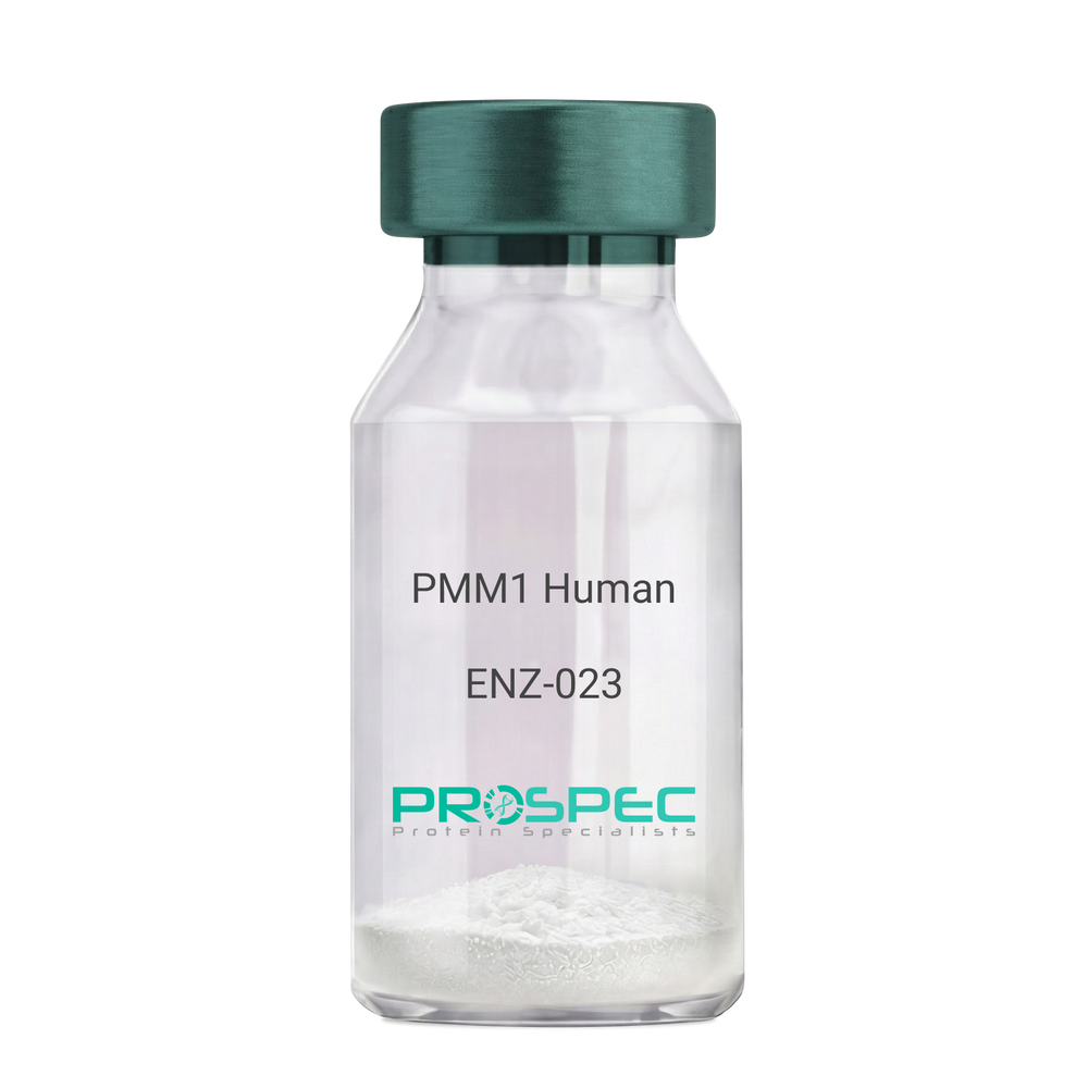
Name :
PMM1 HumanDescription:
Phosphomannomutase 1 Human Recombinant
Phosphomannomutase 1, PMM 1, PMMH-22, PMM1, PMMH22, Sec53.
Product # :
ENZ-023Price :
Quantity :
Shipping Method :
Shipped with Ice Packs
More Info
- description
- source
- formulation
- purity
- More Info
Description
PMM1 Human Recombinant fused with a 20 amino acid His tag at N-terminus produced in E.Coli is a single, non-glycosylated, polypeptide chain containing 282 amino acids (1-262 a.a.) and having a molecular mass of 31.9kDa. The PMM1 is purified by proprietary chromatographic techniques.
Source
Escherichia Coli.
Formulation
The PMM1 solution (0.5mg/ml) contains 20mM Tris-HCl buffer (pH8.0), 10% glycerol, 2mM DTT, 100mM NaCl and 0.1mM PMSF.
Purity
Greater than 90.0% as determined by SDS-PAGE.
More Info
-
Introduction
Phosphomannomutase 1 (PMM1) is an enzyme involved in the synthesis of the GDP-mannose and dolichol-phosphate-mannose required for a number of critical mannosyl transfer reactions. PMM1 catalyzes the conversion between D-mannose 6-phosphate and D-mannose 1-phosphate which is a substrate for GDP-mannose synthesis. GDP-mannose is used for the synthesis of dolichol-phosphate-mannose, which is crucial for N-linked glycosylation and accordingly the secretion of several glycoproteins as well as for the synthesis of glycosyl-phosphatidyl-inositol (GPI) anchored proteins. Additionally, PMM1 may be responsible for the degradation of glucose-1,6-bisphosphate in ischemic brain.
-
Synonyms
Phosphomannomutase 1, PMM 1, PMMH-22, PMM1, PMMH22, Sec53.
-
Physical Appearance
Sterile Filtered colorless solution.
-
Stability
Store at 4°C if entire vial will be used within 2-4 weeks. Store, frozen at -20°C for longer periods of time. For long term storage it is recommended to add a carrier protein (0.1% HSA or BSA).Avoid multiple freeze-thaw cycles.
-
Amino Acid Sequence
MGSSHHHHHH SSGLVPRGSH MAVTAQAARR KERVLCLFDV DGTLTPARQK IDPEVAAFLQ KLRSRVQIGV VGGSDYCKIA EQLGDGDEVI EKFDYVFAEN GTVQYKHGRL LSKQTIQNHL GEELLQDLIN FCLSYMALLR LPKKRGTFIE FRNGMLNISP IGRSCTLEER IEFSELDKKE KIREKFVEAL KTEFAGKGLR FSRGGMISFD VFPEGWDKRY CLDSLDQDSF DTIHFFGNET SPGGNDFEIF ADPRTVGHSV VSPQDTVQRC REIFFPETAH EA.
ProSpec's products are furnished for LABORATORY RESEARCH USE ONLY. The product may not be used as drugs, agricultural or pesticidal products, food additives or household chemicals.
- View Data Sheet
Name :
GAD2 HumanDescription:
Glutamate Decarboxylase 2 Human Recombinant
Glutamate Decarboxylase 2 (Pancreatic Islets And Brain, 65kDa), Glutamate Decarboxylase 65 KDa Isoform, 65 KDa Glutamic Acid Decarboxylase, EC 4.1.1.15, GAD-65, GAD65, Glutamate Decarboxylase 2 (Pancreatic Islets And Brain, 65kD), Glutamate Decarboxylase-2 (Pancreas), EC 4.1.1, GAD2.
Product # :
ENZ-937Price :
Quantity :
Shipping Method :
Shipped with Ice Packs
More Info
- description
- source
- formulation
- purity
- More Info
Description
Recombinant Human GAD2 produced in SF9 is a glycosylated, polypeptide chain having a calculated molecular mass of 61 kDa. GAD2 is expressed with a 6xHis tag at N-terminus and purified by proprietary chromatographic techniques.
Source
Sf9 insect cells.
Formulation
GAD2 protein solution is supplied in 20mM Sodium phosphate pH-7.4, 150mM NaCl, 0.016mM Pyridoxal-5’-Phosphate, 0.05% Tergitol 15-S-9 and 20% glycerol.
Purity
Greater than 95.0% as determined by SDS-PAGE.
More Info
-
Introduction
Glutamate Decarboxylase 2 (GAD2) is one of several forms of glutamic acid decarboxylase, identified as a main autoantigen in type II diabetes. The GAD2 enzyme is responsible for catalyzing the production of gamma-aminobutyric acid from L-glutamic acid. A pathogenic role for the GAD2 enzyme has been characterized in the human pancreas since it has been identified as an autoantibody and an autoreactive T cell target in type II diabetes. GAD2 gene may also have a role in the stiff man syndrome. In addition, GAD2 catalyzes the production of GABA.
-
Synonyms
Glutamate Decarboxylase 2 (Pancreatic Islets And Brain, 65kDa), Glutamate Decarboxylase 65 KDa Isoform, 65 KDa Glutamic Acid Decarboxylase, EC 4.1.1.15, GAD-65, GAD65, Glutamate Decarboxylase 2 (Pancreatic Islets And Brain, 65kD), Glutamate Decarboxylase-2 (Pancreas), EC 4.1.1, GAD2.
-
Physical Appearance
Sterile Filtered clear solution.
-
Stability
Store at 4°C if entire vial will be used within 2-4 weeks. Store, frozen at -20°C for longer periods of time. Avoid multiple freeze-thaw cycles.
ProSpec's products are furnished for LABORATORY RESEARCH USE ONLY. The product may not be used as drugs, agricultural or pesticidal products, food additives or household chemicals.
- View Data Sheet
Name :
MMP 9 HumanDescription:
Matrix Metalloproteinase-9 Human Recombinant
Matrix metalloproteinase-9, MMP-9, 92 kDa gelatinase, Gelatinase B, GELB, MMP9, CLG4B.
Product # :
ENZ-438Price :
Quantity :
Shipping Method :
Shipped with Ice Packs
More Info
- description
- source
- formulation
- purity
- More Info
Description
MMP-9 Human Recombinant produced in E.Coli is single, a non-glycosylated, Polypeptide chain containing 338 amino acids fragment (113-450) corresponding to the catalytic domain of the protein, having a total molecular mass of 42.03kDa and fused with a 4.5kDa amino-terminal hexahistidine tag. The MMP-9 is purified by proprietary chromatographic techniques.
Source
Escherichia Coli.
Formulation
MMP-9 protein is supplied in 20mM Tris-HCl pH 8.0 and 50% glycerol.
Purity
Greater than 95.0% as determined by SDS-PAGE.
More Info
-
Introduction
Matrix metalloproteinases are a family of zinc and calcium-dependent endopeptidases that break down extracellular matrix proteins. The MMP9 is secreted as a 92kDa zymogen. Cleavage of ProMMP-9 results in the active enzyme, having a molecular weight of approximately 82kDa. MMP9 is composed of the following domains: a gelatin-binding domain consisting of three fibronectin type II units, a catalytic domain containing the zinc-binding site, a proline-rich type V collagen-homologous domain and a hemopexin-like domain. MMP9 is produced by the several cell types: monocytes, macrophages, neutrophils, keratinocytes, fibroblasts, osteoclasts and endothelial cells. MMP9 is involved in inflammatory responses, tissue remodeling, wound healing, tumor growth and metastasis. MMP9 may also play an important part in local proteolysis of the extracellular matrix and in leukocyte migration, as well as in bone osteoclastic resorption. MMP9 cleaves type IV and type V collagens into large C-terminal three quarter fragments and shorter N-terminal one quarter fragments. MMP9 can also degrade fibronectin but not laminin or Pz-peptide.
MMP9 defects may be a cause of susceptibility to intervertebral disc disease (IDD), also known as lumbar disk herniation (LDH). -
Synonyms
Matrix metalloproteinase-9, MMP-9, 92 kDa gelatinase, Gelatinase B, GELB, MMP9, CLG4B.
-
Physical Appearance
Sterile Filtered clear solution.
-
Stability
Store at 4°C if entire vial will be used within 2-4 weeks. Store, frozen at -20°C for longer periods of time. Please avoid freeze thaw cycles.
-
Amino Acid Sequence
4.5kDa His Tag-DLKWHHHNITYWIQNYSEDLPRAVIDDAFARAFALWSAVTPLTFTRVYSRDAD
IVIQFGVAEHGDGYPFDGKDGLLAHAFPPGPGIQGDAHFDDDELWSLGKGVVVPTRFGNADGAACHFP
FIFEGRSYSACTTDGRSDGLPWCSTTANYDTDDRFGFCPSERLYTRDGNADGKPCQFPFIFQGQSYSA
CTTDGRSDGYRWCATTANYDRDKLFGFCPTRADSTVMGGNSAGELCVFPFTFLGKEYSTCTSEGRGDG
RLWCATTSNFDSDKKWGFCPDQGYSLFLVAAHEFGHALGLDHSSVPEALMYPMYRFTEGPPLHKDDVN
GIRHLYGP.
ProSpec's products are furnished for LABORATORY RESEARCH USE ONLY. The product may not be used as drugs, agricultural or pesticidal products, food additives or household chemicals.
- View Data Sheet
Name :
CTSE HumanDescription:
Cathepsin-E Human Recombinant
Cathepsin E, EC 3.4.23.34, CATE, Erythrocyte Membrane Aspartic Proteinase, Slow-Moving Proteinase, EC 3.4.23.
Product # :
ENZ-776Price :
Quantity :
Shipping Method :
Shipped with Ice Packs
More Info
- description
- source
- formulation
- purity
- More Info
Description
CTSE Human Recombinant produced in E.Coli is a single, non-glycosylated polypeptide chain containing 330 amino acids (57-363 a.a) and having a molecular mass of 35.4kDa.CTSE is fused to a 23 amino acid His-tag at N-terminus & purified by proprietary chromatographic techniques.
Source
Escherichia Coli.
Formulation
CTSE protein solution (1mg/ml) containing 20mM Tris-HCl buffer (pH 8.0), 0.4M urea and 10% glycerol.
Purity
Greater than 85% as determined by SDS-PAGE.
More Info
-
Introduction
Cathepsin-E also known as CTSE is a gastric aspartyl protease which functions as a disulfide-linked homodimer. CTSE belongs to the peptidase C1 family; furthermore it has specificity similar to pepsin A and cathepsin D. CTSE is an intracellular proteinase which does not seem to be involved in the digestion of dietary protein and is found in the uppermost concentration in the surface of epithelial mucus-producing cells of the stomach. CTSE is the first aspartic proteinaseexpressed in the fetal stomach and is discovered in more than half of gastric cancers. For that reason CTSE is anoncofetal antigen. In addition, transcript variants utilizing alternative polyadenylation signals and two transcript variantsencoding different isoforms exist for this gene.
-
Synonyms
Cathepsin E, EC 3.4.23.34, CATE, Erythrocyte Membrane Aspartic Proteinase, Slow-Moving Proteinase, EC 3.4.23.
-
Physical Appearance
Sterile Filtered clear solution.
-
Stability
Store at 4°C if entire vial will be used within 2-4 weeks. Store, frozen at -20°C for longer periods of time.For long term storage it is recommended to add a carrier protein (0.1% HSA or BSA).Please avoid freeze thaw cycles.
-
Amino Acid Sequence
MGSSHHHHHH SSGLVPRGSH MGSTESCSMD QSAKEPLINY LDMEYFGTIS IGSPPQNFTV IFDTGSSNLW VPSVYCTSPA CKTHSRFQPS QSSTYSQPGQ SFSIQYGTGS LSGIIGADQV SVEGLTVVGQ QFGESVTEPG QTFVDAEFDG ILGLGYPSLA VGGVTPVFDN MMAQNLVDLP MFSVYMSSNP EGGAGSELIF GGYDHSHFSG SLNWVPVTKQ AYWQIALDNM LWSVPTLTSC RMSPSPLTES PIPSAQLPTP YWTSWMECSS AAVAFKDLTS TLQLGPSGSW GMSSFDSFTQ SLTVGITVWD WPQQSPKEGP CVCACLSDRP
ProSpec's products are furnished for LABORATORY RESEARCH USE ONLY. The product may not be used as drugs, agricultural or pesticidal products, food additives or household chemicals.
- View Data Sheet
Name :
GlpK E. coliDescription:
Glycerol kinase E. Coli Recombinant
Glycerol kinase, glycerol 3-phosphotransferase, Glycerokinase, GK.
Product # :
PKA-038Price :
Quantity :
Shipping Method :
Shipped with Ice Packs
More Info
- description
- source
- formulation
- purity
- More Info
Description
GlpK E. Coli Recombinant produced in E. coli is a single, non-glycosylated polypeptide chain containing 525 amino acids (1-502 a.a) and having a molecular mass of 58.6 kDa.GlpK is fused to a 23 amino acid His-tag at N-terminus& purified by proprietary chromatographic techniques.
Source
Escherichia Coli.
Formulation
GlpK protein solution (1mg/ml) containing Phosphate buffered saline (pH7.4),10% glycerol and 1mM DTT.
Purity
Greater than 95.0% as determined by SDS-PAGE.
More Info
-
Introduction
GlpK also known as glycerol kinase, is a member of the FGGY kinase family. GlpK catalyzes the transfer of a phosphate group from ATP to glycerol, thereby forming glycerol phosphate. Furthermore, this intermediate can then be converted to dihydroxyacetone phosphate (DHAP), which is utilized in either glycolysis or gluconeogenesis. The activity of GlpK is affected by numerous metabolites. The non-competitive allosteric inhibition by fructose 1,6-bisphosphate (FBP) triggers modifications in the quaternary structure of Glpk.
-
Synonyms
Glycerol kinase, glycerol 3-phosphotransferase, Glycerokinase, GK.
-
Physical Appearance
Sterile Filtered clear solution.
-
Stability
Store at 4°C if entire vial will be used within 2-4 weeks.Store, frozen at -20°C for longer periods of time. For long term storage it is recommended to add a carrier protein (0.1% HSA or BSA).Avoid multiple freeze-thaw cycles.
-
Amino Acid Sequence
MGSSHHHHHH SSGLVPRGSH MGSMTEKKYI VALDQGTTSS RAVVMDHDAN IISVSQREFE QIYPKPGWVE HDPMEIWATQ SSTLVEVLAK ADISSDQIAA IGITNQRETT IVWEKETGKP IYNAIVWQCR RTAEICEHLK RDGLEDYIRS NTGLVIDPYF SGTKVKWILD HVEGSRERAR RGELLFGTVD TWLIWKMTQG RVHVTDYTNA SRTMLFNIHT LDWDDKMLEV LDIPREMLPE VRRSSEVYGQ TNIGGKGGTR IPISGIAGDQ QAALFGQLCV KEGMAKNTYG TGCFMLMNTG EKAVKSENGL LTTIACGPTG EVNYALEGAV FMAGASIQWL RDEMKLINDA YDSEYFATKV QNTNGVYVVP AFTGLGAPYW DPYARGAIFG LTRGVNANHI IRATLESIAY QTRDVLEAMQ ADSGIRLHAL RVDGGAVANN FLMQFQSDIL GTRVERPEVR EVTALGAAYL AGLAVGFWQN LDELQEKAVI EREFRPGIET TERNYRYAGW KKAVKRAMAW EEHDE.
ProSpec's products are furnished for LABORATORY RESEARCH USE ONLY. The product may not be used as drugs, agricultural or pesticidal products, food additives or household chemicals.
- View Data Sheet
Name :
PDE6H HumanDescription:
Phosphodiesterase 6H cGMP-Specific Cone Gamma Human Recombinant
Phosphodiesterase 6H, CGMP-Specific, Cone, Gamma, Retinal Cone Rhodopsin-Sensitive CGMP 3',5'-Cyclic Phosphodiesterase, Subunit Gamma, EC 3.1.4.35, EC 3.1.4.17, RCD3, GMP-PDE Gamma, ACHM6, PDE6H.
Product # :
ENZ-819Price :
Quantity :
Shipping Method :
Shipped with Ice Packs
More Info
- description
- source
- formulation
- purity
- More Info
Description
PDE6H Human Recombinant produced in E.Coli is a single, non-glycosylated polypeptide chain containing 106 amino acids (1-83 a.a) and having a molecular mass of 11.5 kDa. PDE6H is fused to a 23 amino acid His-tag at N-terminus & purified by proprietary chromatographic techniques.
Source
Escherichia Coli.
Formulation
PDE6H protein solution (0.5mg/ml) containing 20mM Tris-HCl buffer (pH 8.0), 0.2M NaCl, 40% glycerol, 2mM DTT and 0.1mM PMSF.
Purity
Greater than 80% as determined by SDS-PAGE.
More Info
-
Introduction
PDE6H belongs to the rod/cone cGMP-PDE gamma subunit family, which selectively catalyze the hydrolysis of 3 cyclic phosphate bonds in adenosine and/or guanine 3,5 cyclic monophosphate (cAMP and/or cGMP). This family regulates the cellular levels, localization and duration of action of these second messengers by controlling the rate of their degradation. PDE6H is the inhibitory (or gamma) subunit of the cone-specific cGMP phosphodiesterase, which is atetramer composed of two catalytic chains (alpha and beta), and two inhibitory chains (gamma). PDE6H is particularly expressed in the retina, and is implicated in the transmission and amplification of the visual signal. Mutations in PDE6H have been associated with retinal cone dystrophy type 3A.
-
Synonyms
Phosphodiesterase 6H, CGMP-Specific, Cone, Gamma, Retinal Cone Rhodopsin-Sensitive CGMP 3',5'-Cyclic Phosphodiesterase, Subunit Gamma, EC 3.1.4.35, EC 3.1.4.17, RCD3, GMP-PDE Gamma, ACHM6, PDE6H.
-
Physical Appearance
Sterile Filtered colorless solution.
-
Stability
Store at 4°C if entire vial will be used within 2-4 weeks. Store, frozen at -20°C for longer periods of time. For long term storage it is recommended to add a carrier protein (0.1% HSA or BSA).Avoid multiple freeze-thaw cycles.
-
Amino Acid Sequence
MGSSHHHHHH SSGLVPRGSH MGSMSDNTTL PAPASNQGPT TPRKGPPKFK QRQTRQFKSK PPKKGVKGFG DDIPGMEGLG TDITVICPWE AFSHLELHEL AQFGII.
ProSpec's products are furnished for LABORATORY RESEARCH USE ONLY. The product may not be used as drugs, agricultural or pesticidal products, food additives or household chemicals.
- View Data Sheet
Name :
TNAA E.ColiDescription:
Tryptophanase E.Coli Recombinant
Tryptophanase/L-cysteine desulfhydrase, PLP-dependent, Tryptophanase, TNAA, L-tryptophan indole-lyase, TNase, tnaA, ind.
Product # :
ENZ-852Price :
Quantity :
Shipping Method :
Shipped with Ice Packs
More Info
- description
- source
- formulation
- purity
- More Info
Description
TNAA Recombinant produced in E.coli is a single, non-glycosylated polypeptide chain containing 494 amino acids (1-471) and having a molecular mass of 55.2kDa.TNAA is fused to a 23 amino acid His-tag at N-terminus & purified by proprietary chromatographic techniques.
Source
E.coli.
Formulation
The TNAA solution (0.5mg/ml) contains Phosphate buffered saline (pH7.4), 10% glycerol and 1mM DTT.
Purity
Greater than 95% as determined by SDS-PAGE.
More Info
-
Introduction
Tryptophanase, also known as tnaA, catalyzes chemical reaction using 2 substrates which are L-tryptophan and H2O. tnaA protein's three products are indole, pyruvate, and NH3.
-
Synonyms
Tryptophanase/L-cysteine desulfhydrase, PLP-dependent, Tryptophanase, TNAA, L-tryptophan indole-lyase, TNase, tnaA, ind.
-
Physical Appearance
Sterile Filtered colorless solution.
-
Stability
Store at 4°C if entire vial will be used within 2-4 weeks. Store, frozen at -20°C for longer periods of time. For long term storage it is recommended to add a carrier protein (0.1% HSA or BSA).Avoid multiple freeze-thaw cycles.
-
Amino Acid Sequence
MGSSHHHHHH SSGLVPRGSH MGSMENFKHL PEPFRIRVIE PVKRTTRAYR EEAIIKSGMN PFLLDSEDVF IDLLTDSGTG AVTQSMQAAM MRGDEAYSGS RSYYALAESV KNIFGYQYTI PTHQGRGAEQ IYIPVLIKKR EQEKGLDRSK MVAFSNYFFD TTQGHSQING CTVRNVYIKE AFDTGVRYDF KGNFDLEGLE RGIEEVGPNN VPYIVATITS NSAGGQPVSL ANLKAMYSIA KKYDIPVVMD SARFAENAYF IKQREAEYKD WTIEQITRET YKYADMLAMS AKKDAMVPMG GLLCMKDDSF FDVYTECRTL CVVQEGFPTY GGLEGGAMER LAVGLYDGMN LDWLAYRIAQ VQYLVDGLEE IGVVCQQAGG HAAFVDAGKL LPHIPADQFP AQALACELYK VAGIRAVEIG SFLLGRDPKT GKQLPCPAEL LRLTIPRATY TQTHMDFIIE AFKHVKENAA NIKGLTFTYE PKVLRHFTAK LKEV.
ProSpec's products are furnished for LABORATORY RESEARCH USE ONLY. The product may not be used as drugs, agricultural or pesticidal products, food additives or household chemicals.
- View Data Sheet
Name :
ALDOB HumanDescription:
Aldolase B Fructose-Bisphosphate Human Recombinant
Fructose-bisphosphate aldolase B, Liver-type aldolase, ALDOB, ALDB, Aldolase B fructose-bisphosphate, ALDO2, aldolase 2, Aldolase B fructose-bisphosphatase.
Product # :
ENZ-245Price :
Quantity :
Shipping Method :
Shipped with Ice Packs
More Info
- description
- source
- formulation
- purity
- More Info
Description
ALDOB Human Recombinant produced in E.coli is a single, non-glycosylated polypeptide chain containing 388 amino acids (1-364) and having a molecular mass of 42kDa.ALDOB is fused to a 24 amino acid His-tag at N-terminus & purified by proprietary chromatographic techniques.
Source
Escherichia Coli.
Formulation
The ALDOB solution (1mg/ml) contains 20mM Tris-HCl buffer (pH 8.0), 1mM DTT, 10% glycerol and 0.1M NaCl.
Purity
Greater than 95.0% as determined by SDS-PAGE.
More Info
-
Introduction
ALDOB is a tetrameric glycolytic enzyme which catalyzes the reversible cleavage of fructose 1-phosphate into dihydroxyacetone phosphate and glyceraldehyde. Fructose-bisphosphate aldolase B (ALDOB) is one of 3 known aldolase isoenzymes, and is located in the kidney and the small adult intestine where it is linked with aldolases A or C. ALDOB is regulated by Insulin and glucagon and is implicated in hereditary fructose intolerance disease.
-
Synonyms
Fructose-bisphosphate aldolase B, Liver-type aldolase, ALDOB, ALDB, Aldolase B fructose-bisphosphate, ALDO2, aldolase 2, Aldolase B fructose-bisphosphatase.
-
Physical Appearance
Sterile Filtered clear colorless solution.
-
Stability
Store at 4°C if entire vial will be used within 2-4 weeks. Store, frozen at -20°C for longer periods of time. For long term storage it is recommended to add a carrier protein (0.1% HSA or BSA).Avoid multiple freeze-thaw cycles.
-
Amino Acid Sequence
MGSSHHHHHH SSGLVPRGSH MGSHMAHRFP ALTQEQKKEL SEIAQSIVAN GKGILAADES VGTMGNRLQR IKVENTEENR RQFREILFSV DSSINQSIGG VILFHETLYQ KDSQGKLFRN ILKEKGIVVG IKLDQGGAPL AGTNKETTIQ GLDGLSERCA QYKKDGVDFG KWRAVLRIAD
QCPSSLAIQE NANALARYAS ICQQNGLVPI VEPEVIPDGD HDLEHCQYVT EKVLAAVYKA LNDHHVYLEG TLLKPNMVTA GHACTKKYTP EQVAMATVTA LHRTVPAAVP GICFLSGGMS EEDATLNLNA INLCPLPKPW KLSFSYGRAL QASALAAWGG KAANKEATQE AFMKRAMANC
QAAKGQYVHT GSSGAASTQS LFTACYTY.
ProSpec's products are furnished for LABORATORY RESEARCH USE ONLY. The product may not be used as drugs, agricultural or pesticidal products, food additives or household chemicals.
- View Data Sheet
Name :
TPMT HumanDescription:
Thiopurine S-methyltransferase Human Recombinant
TPMT, Thiopurine S-methyltransferase, EC 2.1.1.67, Thiopurine methyltransferase.
Product # :
PKA-255Price :
Quantity :
Shipping Method :
Shipped with Ice Packs
More Info
- description
- source
- formulation
- purity
- More Info
Description
TPMT Human Recombinant produced in E.Coli is a single, non-glycosylated, polypeptide chain containing 245 amino acids (1-245) and having a molecular mass of 28 kDa. Thiopurine S-methyltransferase is purified by proprietary chromatographic techniques.
Source
Escherichia Coli.
Formulation
1 mg/ml solution containing 20mM Tris 8.0, 0.2mM PMSF and 2mM EDTA.
Purity
Greater than 95.0% as determined by SDS-PAGE.
More Info
-
Introduction
TPMT, thiopurine S-methyltransferase, is a cytosolic enzyme that metabolizes thiopurine drugs via S-adenosyl-L-methionine as the S-methyl donor and S-adenosyl-L-homocysteine as a byproduct. TPMT activity exhibits autosomal codominant genetic polymorphism, and patients inheriting TPMT-deficiency are at high risk of potentially fatal hematopoietic toxicity.
-
Synonyms
TPMT, Thiopurine S-methyltransferase, EC 2.1.1.67, Thiopurine methyltransferase.
-
Physical Appearance
Sterile filtered colorless solution.
-
Stability
Store at 4°C if entire vial will be used within 2-4 weeks. Store, frozen at -20°C for longer periods of time. For long term storage it is recommended to add a carrier protein (0.1% HSA or BSA).Avoid multiple freeze-thaw cycles.
-
Amino Acid Sequence
MDGTRTSLDI EEYSDTEVQK NQVLTLEEWQ DKWVNGKTAF HQEQGHQLLK KHLDTFLKGKSGLRVFFPLC GKAVEMKWFA DRGHSVVGVE ISELGIQEFF TEQNLSYSEE PITEIPGTKVFKSSSGNISL YCCSIFDLPR TNIGKFDMIW DRGALVAINP GDRKCYADTM FSLLGKKFQY LLCVLSYDPT KHPGPPFYVP HAEIERLFGK ICNIRRLEKV DAFEERHKSW GIDCLFEKLYLLTEK.
ProSpec's products are furnished for LABORATORY RESEARCH USE ONLY. The product may not be used as drugs, agricultural or pesticidal products, food additives or household chemicals.
- View Data Sheet
Name :
HPD MouseDescription:
4-Hydroxyphenylpyruvate Dioxygenase Mouse Recombinant
4-hydroxyphenylpyruvate dioxygenase, 4-hydroxyphenylpyruvic acid oxidase, 4HPPD, HPD, HPPDase, F Alloantigen, F protein.
Product # :
ENZ-1067Price :
Quantity :
Shipping Method :
Shipped with Ice Packs
More Info
- description
- source
- formulation
- purity
- More Info
Description
HPD Mouse Recombinant produced in E.Coli is a single, non-glycosylated polypeptide chain containing 416 amino acids (1-393 a.a) and having a molecular mass of 47.4kDa.HPD is fused to a 23 amino acid His-tag at N-terminus & purified by proprietary chromatographic techniques.
Source
Escherichia Coli.
Formulation
HPD protein solution (0.5mg/ml) contains 10% glycerol & 20mM Tris-HCl (pH 8.0).
Purity
Greater than 90% as determined by SDS-PAGE.
More Info
-
Introduction
HPGD is the essential enzyme of prostaglandin degradation. 15-PGDH protein strongly decreases the biologic activity of these molecules by catalyzing the oxidation of the 15-hydroxyl group of prostaglandins to a keto group. GDH1 is involved in numerous physiologic and cellular processes, for instance inflammation.
-
Synonyms
4-hydroxyphenylpyruvate dioxygenase, 4-hydroxyphenylpyruvic acid oxidase, 4HPPD, HPD, HPPDase, F Alloantigen, F protein.
-
Physical Appearance
Sterile Filtered clear solution.
-
Stability
Store at 4°C if entire vial will be used within 2-4 weeks. Store, frozen at -20°C for longer periods of time. For long term storage it is recommended to add a carrier protein (0.1% HSA or BSA).Avoid multiple freeze-thaw cycles.
-
Amino Acid Sequence
MGSSHHHHHH SSGLVPRGSH MGSMTTYNNK GPKPERGRFL HFHSVTFWVG NAKQAASFYC NKMGFEPLAY RGLETGSREV VSHVIKQGKI VFVLCSALNP WNKEMGDHLV KHGDGVKDIA FEVEDCDHIV QKARERGAKI VREPWVEQDK FGKVKFAVLQ TYGDTTHTLV EKINYTGRFL PGFEAPTYKD TLLPKLPRCN LEIIDHIVGN QPDQEMQSAS EWYLKNLQFH RFWSVDDTQV HTEYSSLRSI VVTNYEESIK MPINEPAPGR KKSQIQEYVD YNGGAGVQHI ALKTEDIITA IRHLRERGTE FLAAPSSYYK LLRENLKSAK IQVKESMDVL EELHILVDYD EKGYLLQIFT KPMQDRPTLF LEVIQRHNHQ GFGAGNFNSL FKAFEEEQAL RGNLTDLEPN GVRSGM.
ProSpec's products are furnished for LABORATORY RESEARCH USE ONLY. The product may not be used as drugs, agricultural or pesticidal products, food additives or household chemicals.
- View Data Sheet
Name :
FUCA1 HumanDescription:
Fucosidase Alpha-L- 1 Plasma Human Recombinant
Fucosidase, Alpha-L- 1, Tissue, Alpha-L-Fucoside Fucohydrolase 1, Alpha-L-Fucosidase 1, Alpha-L-Fucosidase I, EC 3.2.1.51, Tissue Alpha-L-Fucosidase, EC 3.2.1, FUCA, Tissue alpha-L-fucosidase.
Product # :
ENZ-921Price :
Quantity :
Shipping Method :
Shipped with Ice Packs
More Info
- description
- source
- formulation
- purity
- More Info
Description
FUCA1 produced in Sf9 Baculovirus cells is a single, glycosylated polypeptide chain containing 445 amino acids (28-466a.a.) and having a molecular mass of 51.7kDa. (Molecular size on SDS-PAGE will appear at approximately 50-70kDa). FUCA1 is expressed with a 6 amino acid His tag at C-Terminus and purified by proprietary chromatographic techniques.
Source
Sf9, Baculovirus cells.
Formulation
FUCA1 protein solution (0.5mg/ml) contains Phosphate Buffered Saline (pH 7.4) and 10% glycerol.
Purity
Greater than 90% as determined by SDS-PAGE.
More Info
-
Introduction
Fucosidase Alpha-L- 1 Plasma, also known as FUCA1 is a member of the glycosyl hydrolase 29 family which is responsible for hydrolyzing the alpha-1,6-linked fucose joined to the reducing-end N-acetylglucosamine of the carbohydrate moieties of glycoproteins. Fucosidosis is an autosomal recessive lysosomal storage disease caused by the absence of alpha-L-fucosidase activity.
-
Synonyms
Fucosidase, Alpha-L- 1, Tissue, Alpha-L-Fucoside Fucohydrolase 1, Alpha-L-Fucosidase 1, Alpha-L-Fucosidase I, EC 3.2.1.51, Tissue Alpha-L-Fucosidase, EC 3.2.1, FUCA, Tissue alpha-L-fucosidase.
-
Physical Appearance
Sterile Filtered colorless solution.
-
Stability
Store at 4°C if entire vial will be used within 2-4 weeks. Store, frozen at -20°C for longer periods of time. For long term storage it is recommended to add a carrier protein (0.1% HSA or BSA).Avoid multiple freeze-thaw cycles.
-
Amino Acid Sequence
VRRAQPPRRY TPDWPSLDSR PLPAWFDEAK FGVFIHWGVF SVPAWGSEWF WWHWQGEGRP QYQRFMRDNY PPGFSYADFG PQFTARFFHP EEWADLFQAA GAKYVVLTTK HHEGFTNWPS PVSWNWNSKD VGPHRDLVGE LGTALRKRNI RYGLYHSLLE WFHPLYLLDK KNGFKTQHFV SAKTMPELYD LVNSYKPDLI WSDGEWECPD TYWNSTNFLS WLYNDSPVKD EVVVNDRWGQ NCSCHHGGYY NCEDKFKPQS LPDHKWEMCT SIDKFSWGYR RDMALSDVTE ESEIISELVQ TVSLGGNYLL NIGPTKDGLI VPIFQERLLA VGKWLSINGE AIYASKPWRV QWEKNTTSVW YTSKGSAVYA IFLHWPENGV LNLESPITTS TTKITMLGIQ GDLKWSTDPD KGLFISLPQL PPSAVPAEFA WTIKLTGVKH HHHHH.
ProSpec's products are furnished for LABORATORY RESEARCH USE ONLY. The product may not be used as drugs, agricultural or pesticidal products, food additives or household chemicals.