Search results
1000 results found for “fsh”
Name
Description
Product #
Price
Quantity
Shipping Method
- View Data Sheet
Name :
SF20 Mouse, HisDescription:
MYDGF Mouse Recombinant, His Tag
D17Wsu104e, Il25, Ly6elg, MYDGF, Interleukin-25, IL-25, Stromal cell-derived growth factor SF20.
Product # :
CYT-1040Price :
Quantity :
Shipping Method :
Shipped with Ice Packs
More Info
- description
- source
- formulation
- purity
- More Info
- sds-page
- Citations
Description
MYDGF Mouse Recombinant produced in E.Coli is a single, non-glycosylated polypeptide chain containing 165 amino acids (25-166 a.a) and having a molecular mass of 18.1kDa. MYDGF is fused to a 23 amino acid His-tag at N-terminus & purified by proprietary chromatographic techniques.
Source
Escherichia Coli.
Formulation
MYDGF protein solution (0.5mg/ml) containing 20mM Tris-HCl (pH8.0), 0.1M NaCl and 10% glycerol.
Purity
Greater than 95.0% as determined by SDS-PAGE.
sds-page
Citations
More Info
-
Introduction
Myeloid-derived growth factor (Mydgf) is a paracrine-acting protein and a bone marrow-derived monocyte which stimulates cardiac myocyte survival and adaptive angiogenesis for cardiac protection and repair after myocardial infarction. Mydgf induces endothelial cell proliferation through a MAPK1/3-, STAT3- and CCND1-mediated signaling lane. When comparing wild-type mice to mice with a Mydgf-deficiency, the later develop larger infarct scars and more acute contractile dysfunction.
-
Synonyms
D17Wsu104e, Il25, Ly6elg, MYDGF, Interleukin-25, IL-25, Stromal cell-derived growth factor SF20.
-
Physical Appearance
Sterile Filtered clear solution.
-
Stability
Store at 4°C if entire vial will be used within 2-4 weeks. Store, frozen at -20°C for longer periods of time. For long term storage it is recommended to add a carrier protein (0.1% HSA or BSA).Avoid multiple freeze-thaw cycles.
-
Amino Acid Sequence
MGSSHHHHHH SSGLVPRGSH MGSVSEPTTV PFDVRPGGVV HSFSQDVGPG NKFTCTFTYA SQGGTNEQWQ MSLGTSEDSQ HFTCTIWRPQ GKSYLYFTQF KAELRGAEIE YAMAYSKAAF ERESDVPLKS EEFEVTKTAV SHRPGAFKAE LSKLVIVAKA ARSEL.
ProSpec's products are furnished for LABORATORY RESEARCH USE ONLY. The product may not be used as drugs, agricultural or pesticidal products, food additives or household chemicals.
- View Data Sheet
Description:
Vasoactive Intestinal Peptide
Product # :
HOR-043Price :
Quantity :
Shipping Method :
Shipped at Room temp
More Info
- description
- formulation
- purity
- More Info
- Citations
Description
Vasoactive Intestinal Peptide Synthetic is a single, non-glycosylated polypeptide chain containing 28 amino acids, having a molecular mass of 3325 Dalton and a Molecular formula of C147H238N44O42S .
Formulation
The protein was lyophilized with no additives.
Purity
Greater than 97.0% as determined by analysis by RP-HPLC.
Citations
More Info
-
Physical Appearance
Sterile Filtered White lyophilized (freeze-dried) powder.
-
Stability
Lyophilized Vasoactive Intestinal Peptide although stable at room temperature for 3 weeks, should be stored desiccated below -18°C. Upon reconstitution Vasoactive Intestinal Peptide should be stored at 4°C between 2-7 days and for future use below -18°C. For long term storage it is recommended to add a carrier protein (0.1% HSA or BSA). Please prevent freeze-thaw cycles.
-
Solubility
It is recommended to reconstitute the lyophilized Vasoactive Intestinal Peptide in sterile 18MΩ-cm H2O not less than 100 µg/ml, which can then be further diluted to other aqueous solutions.
-
Amino Acid Sequence
H-His-Ser-Asp-Ala-Val-Phe-Thr-Asp-Asn-Tyr-Thr-Arg-Leu-Arg-Lys-Gln-Met-Ala-Val-Lys-Lys-Tyr-Leu-Asn-Ser-Ile-Leu-Asn-NH2.
-
Background
Vasoactive Intestinal Peptide (VIP), a prominent member of the glucagon-secretin peptide superfamily, plays a multifaceted role as a neuropeptide and neurotransmitter. This research paper aims to provide an extensive analysis of VIP, exploring its biochemical properties, diverse physiological functions, and potential therapeutic applications in various disease conditions.
Vasoactive Intestinal Peptide (VIP), originally identified for its potent vasodilatory properties in the gastrointestinal system, has since been recognized for its wide-ranging physiological effects throughout the body. As a neuropeptide and neurotransmitter.
VIP, a 28-amino acid peptide, exhibits a diverse range of biological activities due to its interactions with multiple G protein-coupled receptors (GPCRs) from the VIP/PACAP receptor family (Harmar et al., 2012). These receptors are expressed in various tissues and cells, enabling VIP to exert its pleiotropic effects.
VIP is involved in the regulation of numerous physiological processes. It functions as a potent vasodilator, modulates smooth muscle activity in the gastrointestinal tract, and participates in neurotransmission in the central and peripheral nervous systems (Lelievre et al., 2008). Additionally, VIP has immunomodulatory properties, influencing immune cell functions and inflammatory responses (Delgado et al., 2004).
VIP serves as a neurotransmitter in the central nervous system, where it contributes to various neuronal functions, including learning, memory, and sensory perception (Deng et al., 2015). Moreover, VIP has demonstrated neuroprotective effects in neurodegenerative disorders, making it a potential therapeutic target for neuroprotection and neuroregeneration (Delgado et al., 2002).
VIP's diverse physiological functions offer potential therapeutic applications in various disease contexts. Research has explored its potential in the treatment of inflammatory diseases, neurodegenerative disorders, and gastrointestinal disorders (Delgado et al., 2013). Furthermore, VIP-based therapies are being investigated for their neuroprotective potential in brain injuries and stroke.
As the research on VIP continues to progress, further investigation is warranted to uncover the precise mechanisms of action and potential clinical applications in medicine. VIP stands as a remarkable example of a neuropeptide with extensive physiological roles and therapeutic potential in various disease conditions.
What is the molecular weight/Mw of VASOACTIVE INTESTINAL PEPTIDE Protein?
VASOACTIVE INTESTINAL PEPTIDE Protein has a total Mw of 3.32kDa.
What is the Purity of VASOACTIVE INTESTINAL PEPTIDE Protein?
VASOACTIVE INTESTINAL PEPTIDE Protein is >97% pure as determined by SDS-PAGE.
What is the Biological Activity of VASOACTIVE INTESTINAL PEPTIDE Protein?
The biological functionality of VASOACTIVE INTESTINAL PEPTIDE Protein will be determined in the future.
What is the amino acid sequence of VASOACTIVE INTESTINAL PEPTIDE Protein?
H-His-Ser-Asp-Ala-Val-Phe-Thr-Asp-Asn-Tyr-Thr-Arg-Leu-Arg-Lys-Gln-Met-Ala-Val-Lys-Lys-Tyr-Leu-Asn-Ser-Ile-Leu-Asn-NH2.
What applications can VASOACTIVE INTESTINAL PEPTIDE Protein be used in?
VASOACTIVE INTESTINAL PEPTIDE Protein can probably be used in western blot, ELISA and Lateral Flow.
What is the endotoxin level for VASOACTIVE INTESTINAL PEPTIDE Protein?
The endotoxin level is minimal, VASOACTIVE INTESTINAL PEPTIDE Protein was purified using conventional chromatography techniques.
ProSpec's products are furnished for LABORATORY RESEARCH USE ONLY. The product may not be used as drugs, agricultural or pesticidal products, food additives or household chemicals.
- View Data Sheet
Name :
EFNB2 MouseDescription:
Ephrin- B2 Mouse Recombinant
Ephrin-B2, ELF-2, EPH-related receptor tyrosine kinase ligand 5, LERK-5, HTK ligand, HTK-L, Elf2, Eplg5, Htkl, Lerk5, Efnb2, ELF-2, Epl5, Eplg5, Htk-L, Lerk5, NLERK-1, EFNB2.
Product # :
PRO-2246Price :
Quantity :
Shipping Method :
Shipped with Ice Packs
More Info
- description
- source
- formulation
- purity
- More Info
- Citations
Description
EFNB2 Mouse Recombinant produced in Sf9 Baculovirus cells is a single polypeptide chain containing 212 amino acids (29-232) and having a molecular mass of 23.4kDa.(Molecular size on SDS-PAGE will appear at approximately 28-40KDa).EFNB2 is fused to 8 amino acid His-tag at C-terminus & purified by proprietary chromatographic techniques.
Source
Sf9, Baculovirus cells.
Formulation
The EFNB2 solution (0.5mg/1ml) contains phosphate buffered saline (pH7.4) and 10% glycerol.
Purity
Greater than 90.0% as determined by SDS-PAGE.
Citations
More Info
-
Introduction
Ephrin-B2 belongs to the ephrin (EPH) family. The ephrins and EPH-related receptors contain the largest subfamily of receptor protein-tyrosine kinases and have been associated with mediating developmental events, particularly in the nervous system and in erythropoiesis. Based upon their structures and sequence relationships, ephrins are allocated into the ephrin-A (EFNA) class, which are anchored to the membrane by a glycosylphosphatidylinositol linkage, and the ephrin-B (EFNB) class, which are transmembrane proteins. Ephrin-B2 binds to the EPHB4 and EPHA3 receptors.
-
Synonyms
Ephrin-B2, ELF-2, EPH-related receptor tyrosine kinase ligand 5, LERK-5, HTK ligand, HTK-L, Elf2, Eplg5, Htkl, Lerk5, Efnb2, ELF-2, Epl5, Eplg5, Htk-L, Lerk5, NLERK-1, EFNB2.
-
Physical Appearance
Sterile Filtered clear solution.
-
Stability
Store at 4°C if entire vial will be used within 2-4 weeks. Store, frozen at -20°C for longer periods of time. For long term storage it is recommended to add a carrier protein (0.1% HSA or BSA).Avoid multiple freeze-thaw cycles.
-
Amino Acid Sequence
RSIVLEPIYW NSSNSKFLPG QGLVLYPQIG DKLDIICPKV DSKTVGQYEY YKVYMVDKDQ ADRCTIKKEN TPLLNCARPD QDVKFTIKFQ EFSPNLWGLE FQKNKDYYII STSNGSLEGL DNQEGGVCQT RAMKILMKVG QDASSAGSAR NHGPTRRPEL EAGTNGRSST TSPFVKPNPG SSTDGNSAGH SGNNLLGSEV ALFALEHHHH HH.
ProSpec's products are furnished for LABORATORY RESEARCH USE ONLY. The product may not be used as drugs, agricultural or pesticidal products, food additives or household chemicals.
- View Data Sheet
Name :
BDH1 HumanDescription:
3-Hydroxybutyrate Dehydrogenase, Type 1 Human Recombinant
D-beta-hydroxybutyrate dehydrogenase, mitochondrial, BDH, 3-hydroxybutyrate dehydrogenase, BDH1, SDR9C1.
Product # :
ENZ-215Price :
Quantity :
Shipping Method :
Shipped with Ice Packs
More Info
- description
- source
- formulation
- purity
- More Info
- Citations
Description
BDH1 Human Recombinant produced in E.coli is a single, non-glycosylated polypeptide chain containing 318 amino acids (47-343) and having a molecular mass of 35.3kDa.BDH1 is fused to a 21 amino acid His-tag at N-terminus & purified by proprietary chromatographic techniques.
Source
E.coli.
Formulation
The BDH1 solution (0.25mg/ml) contains 20mM Tris-HCl buffer (pH8.0), 30% glycerol, 0.2M NaCl, 2mM DTT and 0.1mM PMSF.
Purity
Greater than 85% as determined by SDS-PAGE.
Citations
More Info
-
Introduction
BDH1 belongs to the short-chain dehydrogenase/reductase gene family. BDH1 forms a homotetrameric lipid-requiring enzyme of the mitochondrial membrane and has a specific necessity for phosphatidylcholine for optimal enzymatic activity. BDH1 catalyzes the interconversion of acetoacetate and (R)-3-hydroxybutyrate, the 2 main ketone bodies formed during fatty acid catabolism.
-
Synonyms
D-beta-hydroxybutyrate dehydrogenase, mitochondrial, BDH, 3-hydroxybutyrate dehydrogenase, BDH1, SDR9C1.
-
Physical Appearance
Sterile Filtered colorless solution.
-
Stability
Store at 4°C if entire vial will be used within 2-4 weeks. Store, frozen at -20°C for longer periods of time. For long term storage it is recommended to add a carrier protein (0.1% HSA or BSA).Avoid multiple freeze-thaw cycles.
-
Amino Acid Sequence
MGSSHHHHHH SSGLVPRGSH MASAAEPVGS KAVLVTGCDS GFGFSLAKHL HSKGFLVFAG CLMKDKGHDG VKELDSLNSD RLRTVQLNVC SSEEVEKVVE IVRSSLKDPE KGMWGLVNNA GISTFGEVEF TSLETYKQVA EVNLWGTVRM TKSFLPLIRR AKGRVVNISS MLGRMANPAR SPYCITKFGV EAFSDCLRYE MYPLGVKVSV VEPGNFIAAT SLYSPESIQA IAKKMWEELP EVVRKDYGKK YFDEKIAKME TYCSSGSTDT SPVIDAVTHA LTATTPYTRY HPMDYYWWLR MQIMTHLPGA ISDMIYIR.
ProSpec's products are furnished for LABORATORY RESEARCH USE ONLY. The product may not be used as drugs, agricultural or pesticidal products, food additives or household chemicals.
- View Data Sheet
Name :
SSR1 HumanDescription:
Signal Sequence Receptor, Alpha Human Recombinant
Signal Sequence Receptor Alpha, Translocon-Associated Protein Alpha, SSR Alpha Subunit, TRAP-alpha, TRAPA.
Product # :
PRO-1772Price :
Quantity :
Shipping Method :
Shipped with Ice Packs
More Info
- description
- source
- formulation
- purity
- More Info
- Citations
Description
SSR1 Human Recombinant produced in E.Coli is a single, non-glycosylated polypeptide chain topological domain containing 209 amino acids (22-207 a.a) and having a molecular mass of 23.1kDa. SSR1 is fused to a 23 amino acid His-tag at N-terminus & purified by proprietary chromatographic techniques.
Source
Escherichia Coli.
Formulation
SSR1 protein solution (0.25mg/ml) containing 20mM Tris-HCl buffer (pH8.0), 0.1M NaCl, 1mM DTT and 20% glycerol.
Purity
Greater than 90.0% as determined by SDS-PAGE.
Citations
More Info
-
Introduction
SSR is a glycosylated endoplasmic reticulum (ER) membrane receptor involved in protein translocation across the ER membrane. SSR is formed of 2 subunits, a 34-kD glycoprotein encoded by this gene and a 22-kD glycoprotein. SSR gene creates a number of mRNA species due to complex alternative polyadenylation. SSR is unique since it exploits arrays of polyA signal sequences which are mostly non-canonical.
-
Synonyms
Signal Sequence Receptor Alpha, Translocon-Associated Protein Alpha, SSR Alpha Subunit, TRAP-alpha, TRAPA.
-
Physical Appearance
Sterile Filtered clear solution.
-
Stability
Store at 4°C if entire vial will be used within 2-4 weeks. Store, frozen at -20°C for longer periods of time.For long term storage it is recommended to add a carrier protein (0.1% HSA or BSA).Please avoid freeze thaw cycles.
-
Amino Acid Sequence
MGSSHHHHHH SSGLVPRGSH MGSRGGPRGL LAVAQDLTED EETVEDSIIE DEDDEAEVEE DEPTDLVEDK EEEDVSGEPE ASPSADTTIL FVKGEDFPAN NIVKFLVGFT NKGTEDFIVE SLDASFRYPQ DYQFYIQNFT ALPLNTVVPP QRQATFEYSF IPAEPMGGRP FGLVINLNYK DLNGNVFQDA VFNQTVTVIE REDGLDGET
ProSpec's products are furnished for LABORATORY RESEARCH USE ONLY. The product may not be used as drugs, agricultural or pesticidal products, food additives or household chemicals.
- View Data Sheet
Name :
sRANKL Human, GSTDescription:
Soluble RANK Ligand Human Recombinant, GST tag
Soluble Receptor Activator of NFkB Ligand, TNFSF11, TRANCE, TNF-related activation-induced cytokine, OPGL, ODF, Osteoclast differentiation factor, Tumor necrosis factor ligand superfamily member 11, Receptor activator of nuclear factor kappa B ligand, RANKL, Osteoprotegerin ligand, CD254 antigen, sRANKL, sOdf, hRANKL2.
Product # :
CYT-631Price :
Quantity :
Shipping Method :
Shipped with Ice Packs
More Info
- description
- source
- formulation
- purity
- More Info
- Citations
Description
RANKL Human Recombinant fused to GST tag produced in E.Coli is a single, non-glycosylated polypeptide having a molecular mass of 47 kDa. RANKL is purified by proprietary chromatographic techniques.
Source
Escherichia Coli.
Formulation
The soluble RANKL protein solution contains 1mM EDTA and PBS pH-7.4.
Purity
Greater than 95.0% as determined by SDS-PAGE.
Citations
More Info
-
Introduction
RANKL binds to tnfrsf11b/opg and to tnfrsf11a/rank. Osteoclast differentiation and activation factor. Augments the ability of dendritic cells to stimulate naive t-cell proliferation. May be an important regulator of interactions between t-cells and dendritic cells and may play a role in the regulation of the t-cell-dependent immune response. sRANKL may also play an important role in enhanced bone-resorption in humoral hypercalcemia of malignancy. By injecting soluble RANKL, novel osteopenia model mice were established in only 50 hours. Degree of bone loss can be controlled by changing doses of sRANKL. RANKL can be used in establishing osteopenia model for in vivo screening of drugs for osteoporosis, determination of effect and mechanism, evaluation of bone anabolic drugs.
-
Synonyms
Soluble Receptor Activator of NFkB Ligand, TNFSF11, TRANCE, TNF-related activation-induced cytokine, OPGL, ODF, Osteoclast differentiation factor, Tumor necrosis factor ligand superfamily member 11, Receptor activator of nuclear factor kappa B ligand, RANKL, Osteoprotegerin ligand, CD254 antigen, sRANKL, sOdf, hRANKL2.
-
Physical Appearance
Sterile Filtered colorelss clear solution.
-
Stability
Store at 4°C if entire vial will be used within 2-4 weeks. Store, frozen at -20°C for longer periods of time. Please avoid freeze thaw cycles.
ProSpec's products are furnished for LABORATORY RESEARCH USE ONLY. The product may not be used as drugs, agricultural or pesticidal products, food additives or household chemicals.
- View Data Sheet
Name :
FAS AntibodyDescription:
FAS Blocking/Activating antibody (CD95), Mouse anti Human
FASLG receptor, Apoptosis-mediating surface antigen FAS, Apo-1 antigen, CD95, Tumor necrosis factor receptor superfamily member 6 TNR6, APT1, FAS1, TNFRSF6.
Product # :
ANT-204Price :
Quantity :
Shipping Method :
Shipped at Room temp
More Info
- formulation
- More Info
- Citations
Formulation
1 mg/ml in PBS (after reconstitution).
Citations
More Info
-
Introduction
The Fas receptor (CD95) mediates apoptotic signaling by Fas-ligand expressed on the surface of other cells. The Fas-FasL interaction plays an important role in the immune system and lack of this system leads to autoimmunity, indicating that Fas-mediated apoptosis removes self-reactive lymphocytes. Fas signaling is also involved in immune surveillance to remove transformed cells and virus infected cells. Binding of FAS to oligimerized FasL on another cell activates apoptotic signaling through a cytoplasmic domain termed the death domain that interacts with signaling adaptors including FAF, FADD and DAX to activate the caspase proteolytic cascade. Caspase-8 and caspase-10 are first activated, to then cleave and activate downstream caspases, and a variety of cellular substrates that lead to cell death. Caspases cleave nuclear lamins, causing the nucleus to break down and lose its normal structure and another caspase substrate is DFF, inducing cleavage and degradation of the genome. Other caspase substrates are involved in cytoskeletal structure, cell cycle regulation and signaling pathways. Activation of JNK kinase, activation of Jun, and production of ceramide may also play roles in Fas-mediated apoptosis. Activation of fas-mediated apoptosis is opposed by I-FLICE and FAP. Viruses and tumors may escape immune surveillance in part through suppression of fas-mediated apoptosis using similar mechanisms.
-
Synonyms
FASLG receptor, Apoptosis-mediating surface antigen FAS, Apo-1 antigen, CD95, Tumor necrosis factor receptor superfamily member 6 TNR6, APT1, FAS1, TNFRSF6.
-
Solubility
Reconstitute with H20. Mix gently, wash the sides of the vial and wait 30-60 seconds before use.
-
Immunogen
Recombinant Human FAS.
-
Ig Subclass
mouse IgG1.
-
Clone
NYRhFAS.
-
Note
This antibody is a BLOCKING antibody when in soluble form and will block FAS-mediated apoptosis. It will ACTIVATE FAS-mediated killing when immobilized.
-
Titer
By direct ELISA, 1:10,000 dilution will yield 0.5 O.D using alkaline phosphatase conjugated rabbit anti-mouse Ig (Jackson Laboratories).
-
Shipping Conditions
Antibody is shipped lyophilized at ambient temperature.
-
Type
Mouse Anti Human Monoclonal.
-
Storage Procedures
In lyophilized form, for long periods, store at 4°C in a dry environment. After reconstitution, if not intended for use within a month, aliquot and store at -20°C.
-
Purification Method
Protein-A column.
ProSpec's products are furnished for LABORATORY RESEARCH USE ONLY. The product may not be used as drugs, agricultural or pesticidal products, food additives or household chemicals.
- View Data Sheet
Name :
CSTF1 Human, sf9Description:
Cleavage Stimulation Factor 1 Human Recombinant, sf9
Cleavage Stimulation Factor Subunit 1, Cleavage Stimulation Factor, 3 Pre-RNA, Subunit 1, 50kD, Cleavage Stimulation Factor 50 KDa Subunit, CF-1 50 KDa Subunit, CSTF 50 KDa Subunit, CstF-50, Cleavage Stimulation Factor, 3 Pre-RNA, Subunit 1, 50kDa, Cleavage Stimulation Factor, 3 Pre-RNA, Subunit 1, CstFp50.
Product # :
PRO-2436Price :
Quantity :
Shipping Method :
Shipped with Ice Packs
More Info
- description
- source
- formulation
- purity
- More Info
- Citations
Description
CSTF1 Human Recombinant produced in Sf9 Baculovirus cells is a single, glycosylated polypeptide chain containing 437 amino acids (1-431a.a.) and having a molecular mass of 49.1kDa (Molecular size on SDS-PAGE will appear at approximately 40-57kDa).CSTF1 is expressed with a 6 amino acids His tag at C-Terminus and purified by proprietary chromatographic techniques.
Source
Sf9, Baculovirus cells.
Formulation
CSTF1 protein solution (0.5mg/ml) contains Phosphate Buffered Saline (pH 7.4), 1mM DTT and 20% glycerol.
Purity
Greater than 90.0% as determined by SDS-PAGE.
Citations
More Info
-
Introduction
Cleavage stimulation factor subunit 1 (CSTF1) is involved in the polyadenylation and 3'end cleavage of pre-mRNAs. The CSTF1 gene encodes one of three subunits which merge to produce cleavage stimulation factor (CSTF). Like the mammalian G protein beta subunits, CSTF1 contains transducin-like repeats. CSTF1 is one of the numerous factors necessary for polyadenylation and 3'-end cleavage of mammalian pre-mRNAs. CSTF1 is responsible for the interaction of CSTF with other factors to create a stable complex on the pre-mRNA.
-
Synonyms
Cleavage Stimulation Factor Subunit 1, Cleavage Stimulation Factor, 3 Pre-RNA, Subunit 1, 50kD, Cleavage Stimulation Factor 50 KDa Subunit, CF-1 50 KDa Subunit, CSTF 50 KDa Subunit, CstF-50, Cleavage Stimulation Factor, 3 Pre-RNA, Subunit 1, 50kDa, Cleavage Stimulation Factor, 3 Pre-RNA, Subunit 1, CstFp50.
-
Physical Appearance
Sterile Filtered colorless solution.
-
Stability
Store at 4°C if entire vial will be used within 2-4 weeks. Store, frozen at -20°C for longer periods of time. For long term storage it is recommended to add a carrier protein (0.1% HSA or BSA). Avoid multiple freeze-thaw cycles.
-
Amino Acid Sequence
MYRTKVGLKD RQQLYKLIIS QLLYDGYISI ANGLINEIKP QSVCAPSEQL LHLIKLGMEN DDTAVQYAIG RSDTVAPGTG IDLEFDADVQ TMSPEASEYE TCYVTSHKGP CRVATYSRDG QLIATGSADA SIKILDTERM LAKSAMPIEV MMNETAQQNM ENHPVIRTLY DHVDEVTCLA FHPTEQILAS GSRDYTLKLF DYSKPSAKRA FKYIQEAEML RSISFHPSGD FILVGTQHPT LRLYDINTFQ CFVSCNPQDQ HTDAICSVNY NSSANMYVTG SKDGCIKLWD GVSNRCITTF EKAHDGAEVC SAIFSKNSKY ILSSGKDSVA KLWEISTGRT LVRYTGAGLS GRQVHRTQAV FNHTEDYVLL PDERTISLCC WDSRTAERRN LLSLGHNNIV RCIVHSPTNP GFMTCSDDFR ARFWYRRSTT DHHHHHH.
ProSpec's products are furnished for LABORATORY RESEARCH USE ONLY. The product may not be used as drugs, agricultural or pesticidal products, food additives or household chemicals.
- View Data Sheet
Name :
OSCAR Human, Sf9Description:
Osteoclast Associated, Immunoglobulin-Like Receptor Human Recombinant, Sf9
OSCAR, PIgR-3, PIGR3, hOSCAR, PIgR-3, Poly-Ig Receptor 3.
Product # :
PRO-2546Price :
Quantity :
Shipping Method :
Shipped with Ice Packs
More Info
- description
- source
- formulation
- purity
- More Info
- Citations
Description
OSCAR produced in Sf9 Baculovirus cells is a single, glycosylated polypeptide chain (19-282 a.a.) and fused to a 6 aa His Tag at C-terminus containing a total of 270 amino acids and having a molecular mass of 29.2kDa.OSCAR shows multiple bands between 40-57kDa on SDS-PAGE, reducing conditions and purified by proprietary chromatographic techniques.
Source
Sf9, Baculovirus cells.
Formulation
OSCAR protein solution (0.25mg/ml) contains Phosphate Buffered Saline (pH 7.4) & 10% glycerol.
Purity
Greater than 90.0% as determined by SDS-PAGE.
Citations
More Info
-
Introduction
Osteoclast-associated immunoglobulin-like receptor isoform 6 or OSCAR, is a receptor for surfactant protein D that can be found on human myeloid cells. OSCAR is part of the TNF-α release from human CCR2+ inflammatory monocytes process. The protein is involved in antigen presentation and activation of dendritic cells. Preosteoclasts or mature osteoclasts express the OSCAR protein. Patients that suffer from RA (rheumatoid arthritis) has upregulations on monocytes which present increased proosteoclastogenic potential.
-
Synonyms
OSCAR, PIgR-3, PIGR3, hOSCAR, PIgR-3, Poly-Ig Receptor 3.
-
Physical Appearance
Sterile Filtered colorless solution.
-
Stability
Store at 4°C if entire vial will be used within 2-4 weeks. Store, frozen at -20°C for longer periods of time. For long term storage it is recommended to add a carrier protein (0.1% HSA or BSA). Avoid multiple freeze-thaw cycles.
-
Amino Acid Sequence
DITPSVPPAS YHPKPWLGAQ PATVVTPGVN VTLRCRAPQP AWRFGLFKPG EIAPLLFRDV SSELAEFFLE EVTPAQGGSY RCCYRRPDWG PGVWSQPSDV LELLVTEELP RPSLVALPGP VVGPGANVSL RCAGRLRNMS FVLYREGVAA PLQYRHSAQP WADFTLLGAR APGTYSCYYH TPSAPYVLSQ RSEVLVISWE GEGPEARPAS SAPGMQAPGP PPSDPGAQAP SLSSFRPRGL VLQPLLPQTQ DSWDPAPPPS DPGVHHHHHH.
ProSpec's products are furnished for LABORATORY RESEARCH USE ONLY. The product may not be used as drugs, agricultural or pesticidal products, food additives or household chemicals.
- View Data Sheet
Name :
CARHSP1 HumanDescription:
Calcium Regulated Heat Stable Protein 1 Human Recombinant
Calcium-regulated heat stable protein 1, Calcium-regulated heat-stable protein of 24 kDa, CRHSP-24, CARHSP1, asCSDC1, MGC111446.
Product # :
PRO-257Price :
Quantity :
Shipping Method :
Shipped with Ice Packs
More Info
- description
- source
- formulation
- purity
- More Info
- Citations
Description
CRAHSP1 Human Recombinant fused with a 20 amino acid His tag at N-terminus produced in E.Coli is a single, non-glycosylated, polypeptide chain containing 167 amino acids (1-147 a.a.) and having a molecular mass of 18kDa. The CRAHSP1 is purified by proprietary chromatographic techniques.
Source
Escherichia Coli.
Formulation
The CRAHSP1 solution (0.5 mg/ml) contains 20mM Tris-HCl buffer (pH8.0) containing 10% glycerol and 0.1M NaCl.
Purity
Greater than 90.0% as determined by SDS-PAGE.
Citations
More Info
-
Introduction
CRAHSP1 is a serine phosphoprotein initially identified as a physiological substrate for the Ca2+-calmodulin regulated protein phosphatase calcineurin (PP2B). CRAHSP1 interacts with the STYX/dead phosphate protein in developing spermatids and is a paralog of the brain-specific mRNA-binding protein PIPPIN. It is believed to have a common role in calcium-mediated signal transduction resulting from phosphorylation on serine residues.
-
Synonyms
Calcium-regulated heat stable protein 1, Calcium-regulated heat-stable protein of 24 kDa, CRHSP-24, CARHSP1, asCSDC1, MGC111446.
-
Physical Appearance
Sterile Filtered colorless solution.
-
Stability
Store at 4°C if entire vial will be used within 2-4 weeks. Store, frozen at -20°C for longer periods of time. For long term storage it is recommended to add a carrier protein (0.1% HSA or BSA).Avoid multiple freeze-thaw cycles.
-
Amino Acid Sequence
MGSSHHHHHH SSGLVPRGSH MSSEPPPPPQ PPTHQASVGL LDTPRSRERS PSPLRGNVVP SPLPTRRTRT FSATVRASQG PVYKGVCKCF CRSKGHGFIT PADGGPDIFL HISDVEGEYV PVEGDEVTYK MCSIPPKNEK LQAVEVVITH LAPGTKHETW SGHVISS.
ProSpec's products are furnished for LABORATORY RESEARCH USE ONLY. The product may not be used as drugs, agricultural or pesticidal products, food additives or household chemicals.
- View Data Sheet
Name :
GHBP OvineDescription:
Growth Hormone Binding Protein Ovine Recombinant
GHR, GHBP, GH receptor, Somatotropin receptor.
Product # :
CYT-470Price :
Quantity :
Shipping Method :
Shipped at Room temp
More Info
- description
- source
- formulation
- purity
- biological activity
- More Info
- Citations
Description
Growth Hormone Binding Protein Ovine Recombinant produced in E.Coli is a single, non-glycosylated, polypeptide chain containing 244 amino acids and having a molecular mass of 28 kDa. GHBP is purified by proprietary chromatographic techniques.
Source
Escherichia Coli.
Formulation
GHBP was lyophilized from a concentrated (1mg/ml) solution with 0.0045mM NaHCO3.
Purity
Greater than 98.0% as determined by:
(a) Gel filtration analysis.
(b) Analysis by SDS-PAGE.Biological Activity
fully biologically active as evidenced by its ability of forming 2:1 complex with Human GH.
Citations
More Info
-
Introduction
GHBP is a transmembrane receptor for growth hormone. Binding of growth hormone to the receptor leads to receptor dimerization and the activation of an intra- and intercellular signal transduction pathway leading to growth. A common alternate allele of this gene, called GHRd3, lacks exon three and has been well-characterized. Mutations in this gene have been associated with Laron syndrome, also known as the growth hormone insensitivity syndrome (GHIS), a disorder characterized by short stature. Other splice variants, including one encoding a soluble form of the protein (GHRtr), have been observed but have not been thoroughly characterized.
-
Synonyms
GHR, GHBP, GH receptor, Somatotropin receptor.
-
Physical Appearance
Sterile Filtered White lyophilized (freeze-dried) powder.
-
Stability
Lyophilized Growth Hormone Binding Protein although stable at room temperature for 3 weeks, should be stored desiccated below -18°C. Upon reconstitution GHBP should be stored at 4°C between 2-7 days and for future use below -18°C.For long term storage it is recommended to add a carrier protein (0.1% HSA or BSA).Please prevent freeze-thaw cycles.
-
Solubility
It is recommended to reconstitute the lyophilized GHBP Ovine in DDW or sterile 0.4% NaHCO3 adjusted to pH 10, not less than 100µg/ml, which can then be further diluted to other aqueous solutions preferably in presence of carrier proteins such as BSA or HSA.
-
Amino Acid Sequence
The sequence of the first five N-terminal amino acids was determined and was found to be Ala-Phe-Ser-Gly-Ser.
ProSpec's products are furnished for LABORATORY RESEARCH USE ONLY. The product may not be used as drugs, agricultural or pesticidal products, food additives or household chemicals.
- View Data Sheet
Name :
FOLH1 MouseDescription:
Folate Hydrolase 1 Mouse Recombinant
Folh1, GCP2, mopsm, Glutamate carboxypeptidase 2, Folate hydrolase 1, Folylpoly-gamma-glutamate carboxypeptidase, FGCP, Glutamate carboxypeptidase II, GCPII, Naalad1.
Product # :
ENZ-957Price :
Quantity :
Shipping Method :
Shipped with Ice Packs
More Info
- description
- source
- formulation
- purity
- More Info
- Citations
Description
FOLH1 Mouse Recombinant produced in in Sf9 Baculovirus cells is a single, non-glycosylated polypeptide chain containing 717 amino acids (45-752a.a) and having a molecular mass of 80.5kDa (Migrates at 70-100kDa on SDS-PAGE under reducing conditions). FOLH1 is fused to a 6 amino acid His-tag at C-Terminus and purified by proprietary chromatographic techniques.
Source
Sf9, Baculovirus cells.
Formulation
The FOLH1 solution (0.5mg/ml) contains Phosphate Buffered Saline (pH 7.4) and 10% glycerol.
Purity
Greater than 85% as determined by SDS-PAGE.
Citations
More Info
-
Introduction
Folate Hydrolase 1 (Folh1) is a single pass type 2 membrane protein which is expressed mainly in prostate epithelium. Folh1 which is a part of the peptidase M28 family and M28B subfamily has both folate hydrolase and N-acetylated-alpha-linked-acidic dipeptidase activity. Folh1 can be found in urinary bladder, kidney, testis, ovary, stomach, small intestine colon, and the capillary endothelium of various tumors. Therefore, Folh1 plays a role in directed imaging and therapy of recurrent of metastatic disease.
-
Synonyms
Folh1, GCP2, mopsm, Glutamate carboxypeptidase 2, Folate hydrolase 1, Folylpoly-gamma-glutamate carboxypeptidase, FGCP, Glutamate carboxypeptidase II, GCPII, Naalad1.
-
Physical Appearance
Sterile Filtered colorless solution.
-
Stability
Store at 4°C if entire vial will be used within 2-4 weeks. Store, frozen at -20°C for longer periods of time. For long term storage it is recommended to add a carrier protein (0.1% HSA or BSA). Avoid multiple freeze-thaw cycles.
-
Amino Acid Sequence
ADPKPSNEAT GNVSHSGMKK EFLHELKAEN IKKFLYNFTR TPHLAGTQNN FELAKQIHDQ WKEFGLDLVE LSHYDVLLSY PNKTHPNYIS IINEDGNEIF KTSLSEQPPP GYENISDVVP PYSAFSPQGT PEGDLVYVNY ARTEDFFKLE REMKISCSGK IVIARYGKVF RGNMVKNAQL AGAKGMILYS DPADYFVPAV KSYPDGWNLP GGGVQRGNVL NLNGAGDPLT PGYPANEHAY RHELTNAVGL PSIPVHPIGY DDAQKLLEHM GGPAPPDSSW KGGLKVPYNV GPGFAGNFST QKVKMHIHSY TKVTRIYNVI GTLKGALEPD RYVILGGHRD AWVFGGIDPQ SGAAVVHEIV RSFGTLKKKG RRPRRTILFA SWDAEEFGLL GSTEWAEEHS RLLQERGVAY INADSSIEGN YTLRVDCTPL MYSLVYNLTK ELQSPDEGFE GKSLYDSWKE KSPSPEFIGM PRISKLGSGN DFEVFFQRLG IASGRARYTK NWKTNKVSSY PLYHSVYETY ELVVKFYDPT FKYHLTVAQV RGAMVFELAN SIVLPFDCQS YAVALKKYAD TIYNISMKHP QEMKAYMISF DSLFSAVNNF TDVASKFNQR LQELDKSNPI LLRIMNDQLM YLERAFIDPL GLPGRPFYRH IIYAPSSHNK YAGESFPGIY DALFDISSKV NASKAWNEVK RQISIATFTV QAAAETLREV AHHHHHH.
ProSpec's products are furnished for LABORATORY RESEARCH USE ONLY. The product may not be used as drugs, agricultural or pesticidal products, food additives or household chemicals.
- View Data Sheet
Name :
IFNG Mouse, HisDescription:
Interferon-gamma Mouse Recombinant, His Tag
Immune Interferon, type II interferon, T cell interferon, MAF, IFNG, IFG, IFI, IFN-gamma.
Product # :
CYT-1001Price :
Quantity :
Shipping Method :
Shipped with Ice Packs
More Info
- description
- source
- formulation
- purity
- More Info
- SDS-PAGE
- Citations
Description
Interferon-gamma Mouse Recombinant produced in E.Coli is a single, non-glycosylated polypeptide chain containing 158 amino acids (23-155a.a.) and having a molecular mass of 18.2kDa.IFNG is fused to a 25 amino acid His-tag at N-terminus & purified by proprietary chromatographic techniques.
Source
Escherichia Coli.
Formulation
IFNG protein solution (0.5mg/ml) containing 20mM MES buffer (pH5.0), 1mM DTT, 0.2M NaCl & 40% glycerol.
Purity
Greater than 90% as determined by SDS-PAGE.
SDS-PAGE
Citations
More Info
-
Introduction
IFN-gamma produced by lymphocytes activated by specific antigens or mitogens.
IFN-gamma, in addition to having antiviral activity, has important immunoregulatory functions, it is a potent activator of macrophages, and has antiproliferative effects on transformed cells and it can potentiate the antiviral and antitumor effects of the type I interferons. -
Synonyms
Immune Interferon, type II interferon, T cell interferon, MAF, IFNG, IFG, IFI, IFN-gamma.
-
Physical Appearance
Sterile Filtered clear solution.
-
Stability
Store at 4°C if entire vial will be used within 2-4 weeks. Store, frozen at -20°C for longer periods of time.For long term storage it is recommended to add a carrier protein (0.1% HSA or BSA).Please avoid freeze thaw cycles.
-
Amino Acid Sequence
MGSSHHHHHH SSGLVPRGSH MGSHMHGTVI ESLESLNNYF NSSGIDVEEK SLFLDIWRNW QKDGDMKILQ SQIISFYLRL FEVLKDNQAI SNNISVIESH LITTFFSNSK AKKDAFMSIA KFEVNNPQVQ RQAFNELIRV VHQLLPESSL RKRKRSRC.
-
Background
What is the molecular weight/Mw of IFNG MOUSE, HIS Protein?
IFNG MOUSE, HIS Protein has a total Mw of 18.2kDa.
What is the source or expression system of IFNG MOUSE, HIS Protein?
Escherichia Coli.
What is the Purity of IFNG MOUSE, HIS Protein?
IFNG MOUSE, HIS Protein is >90% pure as determined by SDS-PAGE.
What is the Biological Activity of IFNG MOUSE, HIS Protein?
The biological functionality of IFNG MOUSE, HIS Protein will be determined in the future.
What is the amino acid sequence of IFNG MOUSE, HIS Protein?
MGSSHHHHHH SSGLVPRGSH MGSHMHGTVI ESLESLNNYF NSSGIDVEEK SLFLDIWRNW QKDGDMKILQ SQIISFYLRL FEVLKDNQAI SNNISVIESH LITTFFSNSK AKKDAFMSIA KFEVNNPQVQ RQAFNELIRV VHQLLPESSL RKRKRSRC.
What applications can IFNG MOUSE, HIS Protein be used in?
IFNG MOUSE, HIS Protein can probably be used in western blot, ELISA and Lateral Flow.
What is the endotoxin level for IFNG MOUSE, HIS Protein?
The endotoxin level is minimal, IFNG MOUSE, HIS Protein was purified using conventional chromatography techniques.
ProSpec's products are furnished for LABORATORY RESEARCH USE ONLY. The product may not be used as drugs, agricultural or pesticidal products, food additives or household chemicals.
- View Data Sheet
Name :
USH1C HumanDescription:
Usher Syndrome 1C Human Recombinant
Harmonin, Usher syndrome type-1C protein, Autoimmune enteropathy-related antigen AIE-75, Antigen NY-CO-38/NY-CO-37, PDZ-73 protein, Renal carcinoma antigen NY-REN-3, USH1C, AIE75, PDZ73, AIE-75, DFNB18, PDZ-45, NY-CO-37, NY-CO-38, ush1cpst, PDZ-73/NY-CO-38.
Product # :
PRO-706Price :
Quantity :
Shipping Method :
Shipped with Ice Packs
More Info
- description
- source
- formulation
- purity
- More Info
- Citations
Description
USH1C Human Recombinant fused with a 37 amino acid His tag at N-terminus produced in E.Coli is a single, non-glycosylated, polypeptide chain containing 570 amino acids (1-533 a.a.) and having a molecular mass of 64.6kDa.The USH1C is purified by proprietary chromatographic techniques.
Source
Escherichia Coli.
Formulation
USH1C protein solution (1mg/ml) containing 20mM Tris-HCl buffer (pH 8.0) and 20% glycerol.
Purity
Greater than 95.0% as determined by SDS-PAGE.
Citations
More Info
-
Introduction
USH1C gene product Harmonin, is a scaffold protein which functions in the assembly of Usher protein complexes. Harmonin is able to attach to various proteins in cell membranes and coordinate their activities. Harmonin contains PDZ domains, a coiled-coil region with a bipartite nuclear localization signal and a PEST degradation sequence. USH1C is expressed in the small intestine, colon, kidney, eye, vestibule of the inner ear and weakly in the pancreas.
Mutations in the USH1C gene cause the Usher syndrome type I which is an autosomal recessive sensory defect involving congenital profound sensorineural deafness, vestibular dysfunction, and blindness due to progressive retinitis pigmentosa. Sensorineural deafness is caused by damage to the neural receptors of the inner ear, the nerve pathways to the brain, or the area of the brain that receives sound information. The 3 types of the Usher syndrome (1- 3) are distinguished by age at onset and differences in auditory and vestibular function. USH1C gene defects cause of non-syndromic sensorineural deafness autosomal recessive type 18 (DFNB18), is a form of sensorineural hearing loss. -
Synonyms
Harmonin, Usher syndrome type-1C protein, Autoimmune enteropathy-related antigen AIE-75, Antigen NY-CO-38/NY-CO-37, PDZ-73 protein, Renal carcinoma antigen NY-REN-3, USH1C, AIE75, PDZ73, AIE-75, DFNB18, PDZ-45, NY-CO-37, NY-CO-38, ush1cpst, PDZ-73/NY-CO-38.
-
Physical Appearance
Sterile Filtered colorless solution.
-
Stability
Store at 4°C if entire vial will be used within 2-4 weeks. Store, frozen at -20°C for longer periods of time. For long term storage it is recommended to add a carrier protein (0.1% HSA or BSA).Avoid multiple freeze-thaw cycles.
-
Amino Acid Sequence
MRGSHHHHHH GMASMTGGQQ MGRDLYDDDD KDRWGSHMDR KVAREFRHKV DFLIENDAEK DYLYDVLRMY HQTMDVAVLV GDLKLVINEP SRLPLFDAIR PLIPLKHQVE YDQLTPRRSR KLKEVRLDRL HPEGLGLSVR GGLEFGCGLF ISHLIKGGQA DSVGLQVGDE IVRINGYSIS SCTHEEVINL IRTKKTVSIK VRHIGLIPVK SSPDEPLTWQ YVDQFVSESG GVRGSLGSPG NRENKEKKVF ISLVGSRGLG CSISSGPIQK PGIFISHVKP GSLSAEVGLE IGDQIVEVNG VDFSNLDHKE GRELFMTDRE RLAEARQREL QRQELLMQKR LAMESNKILQ EQQEMERQRR KEIAQKAAEE NERYRKEMEQ IVEEEEKFKK QWEEDWGSKE QLLLPKTITA EVHPVPLRKP KYDQGVEPEL EPADDLDGGT EEQGEQDFRK YEEGFDPYSM FTPEQIMGKD VRLLRIKKEG SLDLALEGGV DSPIGKVVVS AVYERGAAER HGGIVKGDEI MAINGKIVTD YTLAEADAAL QKAWNQGGDW IDLVVAVCPP KEYDDELTFF.
ProSpec's products are furnished for LABORATORY RESEARCH USE ONLY. The product may not be used as drugs, agricultural or pesticidal products, food additives or household chemicals.
- View Data Sheet
Name :
CRHBP (22-322), HumanDescription:
Corticotropin Releasing Hormone Binding Protein (22-322 a.a.) Human Recombinant
Corticotropin releasing hormone binding protein, CRF-BP, CRH-BP, CRF-binding protein.
Product # :
HOR-025Price :
Quantity :
Shipping Method :
Shipped with Ice Packs
More Info
- description
- source
- formulation
- purity
- More Info
- Citations
Description
CRHBP Human Recombinant produced in E.Coli is a single, non-glycosylated polypeptide chain containing 324 amino acids (22-322 a.a) and having a molecular mass of 36.1kDa.CRHBP is fused to a 23 amino acid His-tag at N-terminus & purified by proprietary chromatographic techniques.
Source
Escherichia Coli.
Formulation
CRHBP protein (1mg/ml) contains 10% glycerol, 0.4M urea and 20mM Tris-HCl buffer (pH 8.0).
Purity
Greater than 85.0% as determined by SDS-PAGE.
Citations
More Info
-
Introduction
Corticotropin Releasing Hormone Binding Protein or CRHBP is a hormone that stimulates the preopiomelanocortin-derived peptides secretion & synthesis. In general, the levels of CRHBP in the human plasma are relatively low. There are times when we can find high levels of the hormone, mainly in pregnancy, after parturition the levels drop back, the high levels are thought to be derived from the placenta. CRH-binding protein can be found in the plasma, this protein inactivates CRH, in order to avoid inappropriate pituitary-adrenal stimulation during gestation.
-
Synonyms
Corticotropin releasing hormone binding protein, CRF-BP, CRH-BP, CRF-binding protein.
-
Physical Appearance
Sterile Filtered colorless solution.
-
Stability
Store at 4°C if entire vial will be used within 2-4 weeks. Store, frozen at -20°C for longer periods of time. For long term storage it is recommended to add a carrier protein (0.1% HSA or BSA).Avoid multiple freeze-thaw cycles.
-
Amino Acid Sequence
MGSSHHHHHH SSGLVPRGSH MGSESRYLEL REAADYDPFL LFSANLKREL AGEQPYRRAL RCLDMLSLQG QFTFTADRPQ LHCAAFFISE PEEFITIHYD QVSIDCQGGD FLKVFDGWIL KGEKFPSSQD HPLPSAERYI DFCESGLSRR SIRSSQNVAM IFFRVHEPGN GFTLTIKTDP NLFPCNVISQ TPNGKFTLVV PHQHRNCSFS IIYPVVIKIS DLTLGHVNGL QLKKSSAGCE GIGDFVELLG GTGLDPSKMT PLADLCYPFH GPAQMKVGCD NTVVRMVSSG KHVNRVTFEY RQLEPYELEN PNGNSIGEFC LSGL
ProSpec's products are furnished for LABORATORY RESEARCH USE ONLY. The product may not be used as drugs, agricultural or pesticidal products, food additives or household chemicals.
- View Data Sheet
Name :
FABP3 AntibodyDescription:
Mouse Anti Human Fatty Acid Binding Protein-3
Fatty acid-binding protein heart, H-FABP, Heart-type fatty acid-binding protein, Muscle fatty acid-binding protein, M-FABP, Mammary-derived growth inhibitor, MDGI, FABP3, FABP11, O-FABP.
Product # :
ANT-698Price :
Quantity :
Shipping Method :
Shipped with Ice Packs
More Info
- formulation
- More Info
- Citations
Formulation
1mg/ml containing PBS, pH-7.4, 10% Glycerol and 0.02% Sodium Azide.
Citations
More Info
-
Introduction
Recombinant Fatty Acid Binding Protein is a newly introduced plasma marker of acute myocardial infarction (AMI). The plasma kinetics of FABP (15kD) closely resemble those of myoglobin in that elevated plasma concentrations are found within 2 hours after AMI and return to normal generally within 18 to 24 hours. But the concentration of FABP in the skeletal muscle is 20 times lower than in cardiac tissue (for myoglobin the same content for cardiac and skeletal tissue), that makes FABP to be more cardiac specific than myoglobin. This makes FABP a useful biochemical marker for the early assessment or exclusion of AMI. FABP also appears to be a useful plasma marker for the estimation of myocardial infarct size.
-
Synonyms
Fatty acid-binding protein heart, H-FABP, Heart-type fatty acid-binding protein, Muscle fatty acid-binding protein, M-FABP, Mammary-derived growth inhibitor, MDGI, FABP3, FABP11, O-FABP.
-
Physical Appearance
Sterile filtered colorless solution.
-
Immunogen
Anti-human FABP3 mAb, is derived from hybridization of mouse F0 myeloma cells with spleen cells from BALB/c mice immunized with a recombinant human FABP3 protein 1-133 amino acids purified from E. coli.
-
Ig Subclass
Mouse IgG1 heavy chain and k light chain.
-
Clone
PAT9F10AT.
-
Applications
FABP3 antibody has been tested by ELISA and Western blot analysis to assure specificity and reactivity. Since application varies, however, each investigation should be titrated by the reagent to obtain optimal results.
-
Type
Mouse Anti Human Monoclonal.
-
Storage Procedures
For periods up to 1 month store at 4°C, for longer periods of time, store at -20°C. Prevent freeze thaw cycles.
-
Purification Method
FABP3 antibody was purified from mouse ascitic fluids by protein-A affinity chromatography.
ProSpec's products are furnished for LABORATORY RESEARCH USE ONLY. The product may not be used as drugs, agricultural or pesticidal products, food additives or household chemicals.
- View Data Sheet
Description:
SARS-Associated Coronavirus Nuclecapsid (340-390 a.a.) Recombinant, His Tag
Product # :
SARS-006Price :
Quantity :
Shipping Method :
Shipped with Ice Packs
More Info
- description
- source
- formulation
- purity
- More Info
- Citations
Description
The E.coli derived recombinant protein contains the Nucleocapsid C-Terminal protein 340-390 amino acids immunodominant regions fused to 6xHis tag at C-terminal.
Source
Escherichia Coli.
Formulation
SARS Nucleocapsid protein solution is supplied in PBS.
Purity
Protein is >90% pure as determined SDS-PAGE.
Citations
More Info
-
Introduction
SARS-Associated Coronavirus Nuclecapsid is an enveloped viral componentthat contains three external structural antigens, the mains are membrane (M), envelope (E), and spike (S) proteins. Spike (S)-glycoprotein of the virus interacts with a membrenal receptor and mediates membrane fusion to grant viral entry into the target cells. therefore, S-protein has a crucial part in virus infection cycle and is the main target of neutralizing antibodies.
-
Physical Appearance
Sterile Filtered clear solution.
-
Stability
Protein is shipped on ice packs. Upon arrival, Store at -20°C.
-
Specificity
Immunoreactive with sera of SARS-infected individuals.
-
Purification Method
Purified by proprietary chromatographic technique.
ProSpec's products are furnished for LABORATORY RESEARCH USE ONLY. The product may not be used as drugs, agricultural or pesticidal products, food additives or household chemicals.
- View Data Sheet
Name :
HCV Core 22kDa, FLSCNDescription:
Hepatitis C Virus Core 22kDa, FLSCN Recombinant
Product # :
HCV-261Price :
Quantity :
Shipping Method :
Shipped with Ice Packs
More Info
- description
- formulation
- purity
- More Info
- Citations
Description
The E.coli derived recombinant protein contains the HCV core nucleocapsid immunodominant regions, amino acids 2-192. The FLSCN labeled protein is fused with b-galactosidase (114 kDa) at N-terminus.
Formulation
20mM Tris-Hcl pH 8, 8M urea and 10mM B-ME.
Purity
HCV-Core protein is >95% pure as determined by 10% PAGE (coomassie staining).
Citations
More Info
-
Introduction
HCV is a small 50nm, enveloped, single-stranded, positive sense RNAvirus in the family Flaviviridae.
HCV has a high rate of replication with approximately one trillion particles produced each day in an infected individual. Due to lack of proofreading by the HCV RNA polymerase, the HCV has an exceptionally high mutation rate, a factor that may help it elude the host's immune response. Hepatitis C virus is classified into six genotypes(1-6) with several subtypes within each genotype. The preponderance and distribution of HCV genotypes varies globally. Genotype is clinically important in determining potential response to interferon-based therapy and the required duration of such therapy. Genotypes 1 and 4 are less responsive to interferon-based treatment than are the other genotypes (2, 3, 5 and 6). -
Stability
HCV-Core although stable at 4°C for 1 week, should be stored below -18°C. Please prevent freeze thaw cycles.
-
Applications
HCV-Core antigen is suitable for ELISA and Western blots, excellent antigen for detection of HCV with minimal specificity problems.
-
Specificity
Immunoreactive with sera of HCV-infected individuals.
-
Purification Method
HCV-Core protein was purified by proprietary chromatographic technique.
ProSpec's products are furnished for LABORATORY RESEARCH USE ONLY. The product may not be used as drugs, agricultural or pesticidal products, food additives or household chemicals.
- View Data Sheet
Name :
SemaglutideDescription:
Semaglutide
Product # :
HOR-048Price :
Quantity :
Shipping Method :
Shipped at Room temp
More Info
- description
- formulation
- purity
- More Info
- HPLC, MS
- Citations
Description
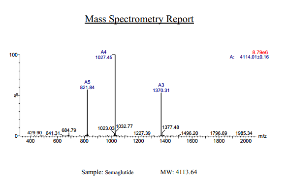
Semaglutide Synthetic is a single, non-glycosylated polypeptide chain containing 31 amino acids, having a molecular mass of 4113 Dalton and a Molecular formula of C187H291N45O59.
Formulation
The protein was lyophilized with no additives.
Purity
Greater than 97.0% as determined by analysis by RP-HPLC.
HPLC, MS
Citations
More Info
-
Physical Appearance
Sterile Filtered White lyophilized (freeze-dried) powder.
-
Stability
Lyophilized Semaglutide although stable at room temperature for 3 weeks, should be stored desiccated below -18°C. Upon reconstitution Semaglutide should be stored at 4°C between 2-7 days and for future use below -18°C. For long term storage it is recommended to add a carrier protein (0.1% HSA or BSA). Please prevent freeze-thaw cycles.
-
Solubility
It is recommended to reconstitute the lyophilized Semaglutide in sterile 18MΩ-cm H2O not less than 100 µg/ml, which can then be further diluted to other aqueous solutions.
-
Amino Acid Sequence
H-His-Aib-Glu-Gly-Thr-Phe-Thr-Ser-Asp-Val-Ser-Ser-Tyr-Leu-Glu-Gly-Gln-Ala-Ala-Lys(AEEAc-AEEAc-γ-Glu-17-carboxyheptadecanoyl)-Glu-Phe-Ile-Ala-Trp-Leu-Val-Arg-Gly-Arg-Gly-OH.
-
Background
Semaglutide, a glucagon-like peptide-1 receptor agonist (GLP-1 RA), has emerged as a breakthrough in the field of diabetes management and metabolic disorders. It is recognized for its potent glucose-lowering effects, weight management properties, and cardiovascular benefits. Beyond diabetes, semaglutide is under investigation for its potential applications in obesity treatment and other related conditions. This research aims to comprehensively explore semaglutide, shedding light on its multifaceted mechanisms of action and its broader therapeutic implications.
The primary objective of this research is to elucidate the mechanisms underlying the glucose-lowering and metabolic effects of semaglutide. In vitro and in vivo experiments will be conducted to investigate how semaglutide interacts with GLP-1 receptors, influences insulin secretion, and modulates glucose homeostasis. Understanding these mechanisms is crucial for harnessing the full therapeutic potential of semaglutide.
The second objective is to assess the clinical relevance of semaglutide in diabetes management. Clinical trials involving individuals with type 2 diabetes will be conducted to evaluate the efficacy, safety, and long-term outcomes of semaglutide treatment. These investigations may provide valuable insights into its use as a monotherapy or adjunct therapy in diabetes care.
The third objective is to explore the potential applications of semaglutide beyond diabetes. Research will investigate its role in obesity treatment, cardiovascular risk reduction, and non-alcoholic fatty liver disease (NAFLD) management. Understanding the multifaceted properties of semaglutide may open new avenues for therapeutic interventions in various metabolic and cardiovascular conditions.
By delving into the diverse functions of semaglutide, this research aims to expand our understanding of its therapeutic potential and clinical applications. The findings may contribute to improved treatment strategies for individuals affected by diabetes, obesity, and related metabolic disorders.
ProSpec's products are furnished for LABORATORY RESEARCH USE ONLY. The product may not be used as drugs, agricultural or pesticidal products, food additives or household chemicals.
- View Data Sheet
Name :
TFRC NativeDescription:
Transferrin Receptor Human
Transferrin receptor protein 1, TR, TfR, TfR1, Trfr, T9, p90, CD_antigen: CD71, Transferrin receptor, serum form, sTfR, TFRC, CD71
Product # :
PRO-2742Price :
Quantity :
Shipping Method :
Shipped with Ice Packs
More Info
- description
- source
- formulation
- More Info
- Citations
Description
Human Transferrin Receptor Protein produced in human serum tissue having a molecular mass of 85kDa.
Source
Human serum.
Formulation
TFRC Native solution (0.2µm filtered) contains 250mM TRIS-HCl buffer, 0.15M NaCl, 0.09% NaN3, pH 8.0.
Citations
More Info
-
Introduction
Transferrin receptor protein 1 (TFRC) is required for iron delivery from transferring to cells. The TFRC protein is a transmembrane glycoprotein comprised of 2 disulfide-linked monomers joined by 2 disulfide bonds. Each monomer will bind one holo-transferrin molecule producing an iron-Tf-TfR complex which enters the cell by endocytosis.
-
Synonyms
Transferrin receptor protein 1, TR, TfR, TfR1, Trfr, T9, p90, CD_antigen: CD71, Transferrin receptor, serum form, sTfR, TFRC, CD71
-
Physical Appearance
Sterile Filtered brown solution.
-
Stability
Store at 4°C if entire vial will be used within 2-4 weeks. Store, frozen at -20°C for longer periods of time. For long term storage it is recommended to add a carrier protein (0.1% HSA or BSA). Avoid multiple freeze-thaw cycles.
-
Human Virus Test
Starting material donor has been tested and certified negative for antibodies to HIV-1, HIV-2, HCV, HBSAG, Parvovirus B19, Syphilis and HIV/HBV/HCV (PCR).
ProSpec's products are furnished for LABORATORY RESEARCH USE ONLY. The product may not be used as drugs, agricultural or pesticidal products, food additives or household chemicals.
- View Data Sheet
Name :
FAS Blocking AntibodyDescription:
FAS Blocking Antibody (CD95), Mouse anti Human
FASLG receptor, Apoptosis-mediating surface antigen FAS, Apo-1 antigen, CD95, Tumor necrosis factor receptor superfamily member 6 TNR6, APT1, FAS1, TNFRSF6.
Product # :
ANT-205Price :
Quantity :
Shipping Method :
Shipped at Room temp
More Info
- More Info
- Citations
Citations
More Info
-
Introduction
The Fas receptor (CD95) mediates apoptotic signaling by Fas-ligand expressed on the surface of other cells. The Fas-FasL interaction plays an important role in the immune system and lack of this system leads to autoimmunity, indicating that Fas-mediated apoptosis removes self-reactive lymphocytes. Fas signaling is also involved in immune surveillance to remove transformed cells and virus infected cells. Binding of FAS to oligimerized FasL on another cell activates apoptotic signaling through a cytoplasmic domain termed the death domain that interacts with signaling adaptors including FAF, FADD and DAX to activate the caspase proteolytic cascade. Caspase-8 and caspase-10 are first activated, to then cleave and activate downstream caspases, and a variety of cellular substrates that lead to cell death. Caspases cleave nuclear lamins, causing the nucleus to break down and lose its normal structure and another caspase substrate is DFF, inducing cleavage and degradation of the genome. Other caspase substrates are involved in cytoskeletal structure, cell cycle regulation and signaling pathways. Activation of JNK kinase, activation of Jun, and production of ceramide may also play roles in Fas-mediated apoptosis. Activation of fas-mediated apoptosis is opposed by I-FLICE and FAP. Viruses and tumors may escape immune surveillance in part through suppression of fas-mediated apoptosis using similar mechanisms.
-
Synonyms
FASLG receptor, Apoptosis-mediating surface antigen FAS, Apo-1 antigen, CD95, Tumor necrosis factor receptor superfamily member 6 TNR6, APT1, FAS1, TNFRSF6.
-
Solubility
Reconstitute with H20. Mix gently, wash the sides of the vial and wait 30-60 seconds before use.
-
Immunogen
Recombinant Human FAS.
-
Ig Subclass
mouse IgG1.
-
Clone
NYRhFAS.
-
Note
This is a BLOCKING antibody and will block FAS-mediated apoptosis.
-
Titer
By direct ELISA, 1:10,000 dilution will yield 0.5 O.D using alkaline phosphatase conjugated rabbit anti-mouse Ig (Jackson Laboratories).
-
Shipping Conditions
Antibody is shipped lyophilized at ambient temperature.
-
Type
Mouse Anti Human Monoclonal.
-
Storage Procedures
In lyophilized form, for long periods, store at 4°C in a dry environment. After reconstitution, if not intended for use within a month, aliquot and store at -20°C.
-
Purification Method
Ion exchange column Protein concentration1 mg/ml in PBS (after reconstitution).
ProSpec's products are furnished for LABORATORY RESEARCH USE ONLY. The product may not be used as drugs, agricultural or pesticidal products, food additives or household chemicals.
- View Data Sheet
Name :
FCGR2A Human, HEKDescription:
CD32a Human Recombinant, HEK
CD32, CD32A, CDw32, FcGR, IGFR2, MGC23887, MGC30032, Low affinity immunoglobulin gamma Fc region receptor II-a, IgG Fc receptor II-a, Fc-gamma-RIIa, FcRII-a, FCG2, FCGR2A1.
Product # :
PRO-2775Price :
Quantity :
Shipping Method :
Shipped with Ice Packs
More Info
- description
- source
- formulation
- purity
- biological activity
- More Info
- Citations
Description
FCGR2A Human Recombinant is a single, glycosylated, polypeptide chain (30-218 a.a) containing a total of 195 amino acids, having a molecular mass of 21.6kDa. FCGR2A is fused to a 6 amino acid His-tag at C-terminus and is purified by proprietary chromatographic techniques.
Source
HEK293 Cells.
Formulation
FCGR2A protein solution (1mg/ml) containing 10% Glycerol and Phosphate-Buffered Saline (pH 7.4).
Purity
Greater than 95.0% as determined by SDS-PAGE.
Biological Activity
The ED50 range ≤5 ug/ml. and is measured by its binding ability in a functional ELISA with Human IgG1 Fc (CAT# pro-2763).
Citations
More Info
-
Synonyms
CD32, CD32A, CDw32, FcGR, IGFR2, MGC23887, MGC30032, Low affinity immunoglobulin gamma Fc region receptor II-a, IgG Fc receptor II-a, Fc-gamma-RIIa, FcRII-a, FCG2, FCGR2A1.
-
Physical Appearance
Sterile Filtered colorless solution.
-
Stability
Store at 4°C if entire vial will be used within 2-4 weeks. Store, frozen at -20°C for longer periods of time. For long term storage it is recommended to add a carrier protein (0.1% HSA or BSA). Avoid multiple freeze-thaw cycles.
-
Amino Acid Sequence
SADSQAAAPP KAVLKLEPPW INVLQEDSVT LTCQGARSPE SDSIQWFHNG NLIPTHTQPS YRFKANNNDS GEYTCQTGQT SLSDPVHLTV LSEWLVLQTP HLEFQEGETI MLRCHSWKDK PLVKVTFFQN GKSQKFSHLD PTFSIPQANH SHSGDYHCTG NIGYTLFSSK PVTITVQVPS MGSSSPMGIH HHHHH.
-
Background
The immune system is a complex network of cells and molecules that play a crucial role in protecting the body against pathogens and maintaining homeostasis. Fc gamma receptors (FcγRs) are a family of cell surface receptors that mediate the effector functions of antibodies, particularly immunoglobulin G (IgG). Among these receptors, FCGR2A, also known as CD32a, is a prominent member expressed on the surface of various immune cells. This research aims to investigate the structure, function, and implications of FCGR2A protein in immune responses, with a focus on its relevance to human health and therapeutic applications.
Structure and Expression of FCGR2A:
FCGR2A is a transmembrane glycoprotein composed of two extracellular immunoglobulin-like domains, a transmembrane domain, and a cytoplasmic tail. It is primarily expressed on immune cells such as macrophages, monocytes, and dendritic cells. The unique structure of FCGR2A allows it to bind to the Fc portion of IgG antibodies, initiating immune effector functions.
Role of FCGR2A in Immune Responses:
FCGR2A plays a crucial role in modulating immune responses through its involvement in antibody-dependent cellular cytotoxicity (ADCC) and phagocytosis. Upon binding to IgG antibodies, FCGR2A triggers intracellular signaling cascades that activate immune effector cells, leading to the elimination of antibody-bound pathogens or targeted cells. Furthermore, FCGR2A is involved in immune complex clearance, regulation of cytokine production, and modulation of cellular activation, thus contributing to the overall immune response.
Implications of FCGR2A in Human Health:
The dysregulation of FCGR2A has been implicated in various immune-related diseases. Genetic variations in FCGR2A have been associated with increased susceptibility to autoimmune disorders, such as systemic lupus erythematosus and rheumatoid arthritis. Altered FCGR2A expression or function has also been observed in infectious diseases, including HIV and malaria, affecting disease progression and immune control. Understanding the role of FCGR2A in these conditions is crucial for developing targeted therapeutic strategies.
Therapeutic Potential of FCGR2A:
The unique properties of FCGR2A make it an attractive target for therapeutic interventions. Modulating FCGR2A activity and function can potentially enhance immune responses against pathogens or tumor cells. For example, monoclonal antibodies that target FCGR2A have shown promising results in cancer immunotherapy by promoting ADCC and phagocytosis of tumor cells. Furthermore, strategies aimed at selectively activating or inhibiting FCGR2A signaling could lead to the development of novel immunotherapeutic approaches.
Challenges and Future Perspectives:
While the study of FCGR2A holds great promise, several challenges need to be addressed. Further research is required to unravel the intricate interplay between FCGR2A and other immune receptors and signaling molecules. Additionally, the development of therapeutic agents that selectively target FCGR2A without compromising normal immune functions remains a challenge.
Conclusion:
The investigation of FCGR2A protein provides insights into its pivotal role in immune responses and its implications for human health and therapeutics. Understanding the structure, function, and dysregulation of FCGR2A in immune-related diseases opens avenues for the development of targeted therapies. Continued research on FCGR2A and its interactions within the immune system will contribute to advancements in immunotherapy and personalized medicine.
ProSpec's products are furnished for LABORATORY RESEARCH USE ONLY. The product may not be used as drugs, agricultural or pesticidal products, food additives or household chemicals.
- View Data Sheet
Name :
HUS1 HumanDescription:
HUS1 Checkpoint Homolog Human Recombinant
HUS1 Checkpoint Homolog (S. pombe), Checkpoint Protein HUS1, Hus1+-like protein.
Product # :
PRO-039Price :
Quantity :
Shipping Method :
Shipped with Ice Packs
More Info
- description
- source
- formulation
- purity
- More Info
- Citations
Description
HUS1 produced in E.Coli is a single, non-glycosylated polypeptide chain containing 300 amino acids (1-280a.a.) and having a molecular mass of 33.8kDa.HUS1 is fused to a 20 amino acid His-tag at N-terminus & purified by proprietary chromatographic techniques.
Source
Escherichia Coli.
Formulation
The HUS1 protein solution (0.25mg/ml) is formulated in 20 mM Tris-HCl Buffer (pH 8.0), 100 mM NaCl and 40% Glycerol.
Purity
Greater than 95% as determined by SDS-PAGE.
Citations
More Info
-
Introduction
HUS1 is a component of an evolutionarily conserved, genotoxin-activated checkpoint complex. HUS1 protein connects with Rad9 and Rad1 to form the 9-1-1(RAD9-RAD1-HUS1) complex, that confines to DNA lesions and promotes DNA damage signaling and repair or apoptosis, cell cycle arrest. The trimeric complex is structurally similar to the proliferating cell nuclear antigen (PCNA) sliding clamp and interacts with Rad17 as a clamp-clamp loader pair during the DNA damage response.
-
Synonyms
HUS1 Checkpoint Homolog (S. pombe), Checkpoint Protein HUS1, Hus1+-like protein.
-
Physical Appearance
Sterile Filtered clear solution.
-
Stability
Store at 4°C if entire vial will be used within 2-4 weeks. Store, frozen at -20°C for longer periods of time.Please avoid freeze thaw cycles.
-
Amino Acid Sequence
MGSSHHHHHH SSGLVPRGSH MKFRAKIVDG ACLNHFTRIS NMIAKLAKTC TLRISPDKLN FILCDKLANG GVSMWCELEQ ENFFNEFQME GVSAENNEIY LELTSENLSR ALKTAQNARA LKIKLTNKHF PCLTVSVELL SMSSSSRIVT HDIPIKVIPR KLWKDLQEPV VPDPDVSIYL PVLKTMKSVV EKMKNISNHL VIEANLDGEL NLKIETELVC VTTHFKDLGN PPLASESTHE DRNVEHMAEV HIDIRKLLQF LAGQQVNPTK ALCNIVNNKM VHFDLLHEDV SLQYFIPALS
ProSpec's products are furnished for LABORATORY RESEARCH USE ONLY. The product may not be used as drugs, agricultural or pesticidal products, food additives or household chemicals.
- View Data Sheet
Name :
FTCD HumanDescription:
Formiminotransferase Cyclodeaminase Human Recombinant
Formiminotransferase-cyclodeaminase, FTCD, LCHC1, LC-1.
Product # :
ENZ-302Price :
Quantity :
Shipping Method :
Shipped with Ice Packs
More Info
- description
- source
- formulation
- purity
- More Info
- Citations
Description
Formiminotransferase Cyclodeaminase Human Recombinant (also called liver cytosol type 1) produced in SF9, is a glycosylated, polypeptide chain having a molecular mass of 59,749 Dalton.The FTCD is expressed with a -6xHis tag and purified by proprietary chromatographic techniques.
Source
Sf9 insect cells.
Formulation
FTCD is supplied in 16mM HEPES buffer pH-7.6, 240mM sodium chloride, and 20% glycerol.
Purity
Greater than 95% as determined by SDS-PAGE.
Citations
More Info
-
Introduction
Formiminotransferase cyclodeaminase is an enzyme which catalyzes the conversion of formiminoglutamateand tetrahydrofolateinto formiminotetrahydrofolateand glutamate.
Formiminotransferase cyclodeaminase, a bifunctional enzyme of tetrahydrofolate synthesis, is the target antigen of anti-LC1 (liver cytosol antigen type 1) autoantibodies. Presence of LC1 autoantibodies is a marker for type 2 autoimmune hepatitis (for which anti-LKM 1/cytochrome P450 2D6 autoantibodies are a further marker).
Serum LC1 autoantibody concentrations appear to fluctuate in parallel with aminotransferase levels, a particularly intriguing observation that suggests a possible role of LC1 autoreactivity in the pathogenic mechanism leading to hepatocyte injury. -
Synonyms
Formiminotransferase-cyclodeaminase, FTCD, LCHC1, LC-1.
-
Stability
Store at 4°C if entire vial will be used within 2-4 weeks. Store, frozen at -20°Cfor longer periods of time. Avoid multiple freeze-thaw cycles.
-
Immunological Functions
1. Binds IgG-type human auto-antibodies.2. Standard ELISA test (checker-board analysis of positive/negative sera panels, immuno-dot test).
-
coating concentration
0.35-0.7 µg/ml (depending on the type of ELISA plate and coating buffer).Suitable for biotinylation and iodination.
ProSpec's products are furnished for LABORATORY RESEARCH USE ONLY. The product may not be used as drugs, agricultural or pesticidal products, food additives or household chemicals.