Skip to content

High-quality research proteins.

  • 1-800-805-6531

  • Mon - Sat 8.00-18.00, Sun - Closed

  • info@prospecbio.com

  • PO Box 6591 East Brunswick NJ 08816

  • Products
    • Cytokines
    • Growth factors
    • Chemokines
    • Cd antigens
    • Neurotrophins
    • Hormones
    • Enzymes
    • Viral antigens
    • Recombinant proteins
    • Natural proteins
    • Monoclonal antibodies
    • Polyclonal antibodies
    • Thermal Packaging
  • About
    • Company
    • Founder
    • Contribution
  • Customers
    • Eastern Asia
    • Eastern Europe
    • North America
    • South America
    • South East Asia
    • Southern Asia
    • Western Europe
  • Ordering
    • Ordering Method
    • Ordering - Credit Card
    • Ordering - PO
    • Shipping Fees
    • FAQs
  • Services
    • DNA Cloning
    • Protein Expression
    • Monoclonal Antibody
    • Fermentation
    • Technology License
  • Contact us
Log in

Country/region

  • USD $ | Albania
  • USD $ | Andorra
  • USD $ | Argentina
  • USD $ | Armenia
  • USD $ | Australia
  • USD $ | Austria
  • USD $ | Azerbaijan
  • USD $ | Belarus
  • USD $ | Belgium
  • USD $ | Bolivia
  • USD $ | Brazil
  • USD $ | Bulgaria
  • USD $ | Cambodia
  • USD $ | Canada
  • USD $ | Chile
  • USD $ | China
  • USD $ | Colombia
  • USD $ | Costa Rica
  • USD $ | Croatia
  • USD $ | Cyprus
  • USD $ | Czechia
  • USD $ | Denmark
  • USD $ | Estonia
  • USD $ | Finland
  • USD $ | France
  • USD $ | Georgia
  • USD $ | Germany
  • USD $ | Greece
  • USD $ | Hong Kong SAR
  • USD $ | Hungary
  • USD $ | Iceland
  • USD $ | India
  • USD $ | Ireland
  • USD $ | Israel
  • USD $ | Italy
  • USD $ | Japan
  • USD $ | Kazakhstan
  • USD $ | Latvia
  • USD $ | Liechtenstein
  • USD $ | Lithuania
  • USD $ | Madagascar
  • USD $ | Malta
  • USD $ | Mexico
  • USD $ | Moldova
  • USD $ | Monaco
  • USD $ | Netherlands
  • USD $ | New Zealand
  • USD $ | North Macedonia
  • USD $ | Norway
  • USD $ | Panama
  • USD $ | Paraguay
  • USD $ | Peru
  • USD $ | Philippines
  • USD $ | Poland
  • USD $ | Portugal
  • USD $ | Romania
  • USD $ | Singapore
  • USD $ | Slovakia
  • USD $ | Slovenia
  • USD $ | South Africa
  • USD $ | South Korea
  • USD $ | Spain
  • USD $ | Sri Lanka
  • USD $ | Sweden
  • USD $ | Switzerland
  • USD $ | Taiwan
  • USD $ | Thailand
  • USD $ | Türkiye
  • USD $ | Ukraine
  • USD $ | United Kingdom
  • USD $ | United States
  • USD $ | Uruguay
  • USD $ | Vatican City
  • USD $ | Venezuela
  • USD $ | Vietnam
  • Facebook
  • Instagram
logoProSpec - Recombinant Protein Manufacturer for academic and R&D industry
Become a distributor
Account Log in
Wishlist
Cart Cart(0)
  • Products
    +
    • Cytokines
    • Growth factors
    • Chemokines
    • Cd antigens
    • Neurotrophins
    • Hormones
    • Enzymes
    • Viral antigens
    • Recombinant proteins
    • Natural proteins
    • Monoclonal antibodies
    • Polyclonal antibodies
    • Thermal Packaging
  • About
    +
    • Company
    • Founder
    • Contribution
  • Customers
    +
    • Eastern Asia
    • Eastern Europe
    • North America
    • South America
    • South East Asia
    • Southern Asia
    • Western Europe
  • Ordering
    +
    • Ordering Method
    • Ordering - Credit Card
    • Ordering - PO
    • Shipping Fees
    • FAQs
  • Services
    +
    • DNA Cloning
    • Protein Expression
    • Monoclonal Antibody
    • Fermentation
    • Technology License
  • Contact us

Item added to your cart

View cart
View All
  • Cytokines
  • Growth factors
  • Chemokines
  • Cd antigens
  • Neurotrophins
  • Hormones
  • Enzymes
  • Viral antigens
  • Recombinant proteins
  • Natural proteins
  • Monoclonal antibodies
  • Polyclonal antibodies
  • Thermal Packaging
  • Tumor Necrosis Factor

    Tumor Necrosis Factor

    View All
  • 4-1BB

    4-1BB

    View All
  • Adiponectin

    Adiponectin

    View All
  • AIF1

    AIF1

    View All
  • Other Cytokines

    Other Cytokines

    View All
  • AITRL

    AITRL

    View All
  • Angiopoietin

    Angiopoietin

    View All
  • Apolipoprotein

    Apolipoprotein

    View All
  • B-Cell Activating Factor

    B-Cell Activating Factor

    View All
  • Beta Defensin

    Beta Defensin

    View All
  • Bone Morphogenetic Protein

    Bone Morphogenetic Protein

    View All
  • B type Natriuretic Peptide

    B type Natriuretic Peptide

    View All
  • BST

    BST

    View All
  • Betacellulin

    Betacellulin

    View All
  • Cardiotrophin

    Cardiotrophin

    View All
  • Activin

    Activin

    View All
  • Fibroblast Growth Factor

    Fibroblast Growth Factor

    View All
  • Other Growth Factors

    Other Growth Factors

    View All
  • CSF

    CSF

    View All
  • CTGF

    CTGF

    View All
  • IGFBP

    IGFBP

    View All
  • VEGF Protein

    VEGF Protein

    View All
  • EGF

    EGF

    View All
  • Epigen

    Epigen

    View All
  • Erythropoietin

    Erythropoietin

    View All
  • Insulin-Like Growth Factor

    Insulin-Like Growth Factor

    View All
  • Growth Hormone

    Growth Hormone

    View All
  • HDGF

    HDGF

    View All
  • Hepatocyte Growth Factor

    Hepatocyte Growth Factor

    View All
  • Insulin

    Insulin

    View All
  • BCA-1/ BLC (CXCL13)

    BCA-1/ BLC (CXCL13)

    View All
  • BRAK (CXCL14)

    BRAK (CXCL14)

    View All
  • C-10 (CCL6)

    C-10 (CCL6)

    View All
  • Other Chemokines

    Other Chemokines

    View All
  • MEC (CCL28)

    MEC (CCL28)

    View All
  • LD78-beta (CCL3L1)

    LD78-beta (CCL3L1)

    View All
  • CTACK (CCL27)

    CTACK (CCL27)

    View All
  • CXCL16

    CXCL16

    View All
  • CXCL17

    CXCL17

    View All
  • Platelet Factor-4 (CXCL4)

    Platelet Factor-4 (CXCL4)

    View All
  • ENA-78 (CXCL5)

    ENA-78 (CXCL5)

    View All
  • Eotaxin (CCL11,24,26)

    Eotaxin (CCL11,24,26)

    View All
  • Exodus-2 (CCL21)

    Exodus-2 (CCL21)

    View All
  • Fractalkine (CX3CL1)

    Fractalkine (CX3CL1)

    View All
  • CXCL6

    CXCL6

    View All
  • Other CD Antigens

    Other CD Antigens

    View All
  • CD14

    CD14

    View All
  • CD164

    CD164

    View All
  • CD1

    CD1

    View All
  • CD2

    CD2

    View All
  • CD200

    CD200

    View All
  • CD207

    CD207

    View All
  • CD226

    CD226

    View All
  • CD23

    CD23

    View All
  • CD244

    CD244

    View All
  • CD247

    CD247

    View All
  • CD27

    CD27

    View All
  • CD274

    CD274

    View All
  • CD300

    CD300

    View All
  • CD33

    CD33

    View All
  • Other Neurotrophins

    Other Neurotrophins

    View All
  • Beta-NGF

    Beta-NGF

    View All
  • BDNF

    BDNF

    View All
  • CDNF

    CDNF

    View All
  • CNTF

    CNTF

    View All
  • GDNF

    GDNF

    View All
  • Glia Maturation Factor

    Glia Maturation Factor

    View All
  • MANF

    MANF

    View All
  • Midkine

    Midkine

    View All
  • Neuroglobin

    Neuroglobin

    View All
  • Neurotrophic factors

    Neurotrophic factors

    View All
  • Neuregulin

    Neuregulin

    View All
  • Neuropilin

    Neuropilin

    View All
  • Pigment Epithelium-Derived Factor

    Pigment Epithelium-Derived Factor

    View All
  • Pleiotrophin

    Pleiotrophin

    View All
  • Peptide Hormones

    Peptide Hormones

    View All
  • Other Hormones

    Other Hormones

    View All
  • HCG

    HCG

    View All
  • Endothelin

    Endothelin

    View All
  • Exendin

    Exendin

    View All
  • FSH

    FSH

    View All
  • Glucagon

    Glucagon

    View All
  • GHRP

    GHRP

    View All
  • GLP

    GLP

    View All
  • Thyrostimulin

    Thyrostimulin

    View All
  • Inhibin A

    Inhibin A

    View All
  • LHRH

    LHRH

    View All
  • Melanotan

    Melanotan

    View All
  • Procalcitonin

    Procalcitonin

    View All
  • PTH

    PTH

    View All
  • Peroxiredoxin

    Peroxiredoxin

    View All
  • Peptidase

    Peptidase

    View All
  • Synthetase

    Synthetase

    View All
  • Transferase

    Transferase

    View All
  • Hydrolase

    Hydrolase

    View All
  • Dehydrogenase

    Dehydrogenase

    View All
  • ACE2

    ACE2

    View All
  • Esterase

    Esterase

    View All
  • Protein Kinases

    Protein Kinases

    View All
  • Other Enzymes

    Other Enzymes

    View All
  • Phosphatase

    Phosphatase

    View All
  • Deaminase

    Deaminase

    View All
  • Oxygenase

    Oxygenase

    View All
  • Adenylate Kinase

    Adenylate Kinase

    View All
  • Lyase

    Lyase

    View All
  • Chagas

    Chagas

    View All
  • SARS

    SARS

    View All
  • Influenza

    Influenza

    View All
  • ASFV

    ASFV

    View All
  • Astrovirus

    Astrovirus

    View All
  • Borrelia

    Borrelia

    View All
  • Helicobacter Pylori

    Helicobacter Pylori

    View All
  • Chikungunya

    Chikungunya

    View All
  • Chlamydia

    Chlamydia

    View All
  • Cytomegalo

    Cytomegalo

    View All
  • Dengue

    Dengue

    View All
  • EBV

    EBV

    View All
  • Ebola

    Ebola

    View All
  • Feline Leukemia Virus

    Feline Leukemia Virus

    View All
  • Hepatitis A

    Hepatitis A

    View All
  • Other

    Other

    View All
  • Actin

    Actin

    View All
  • ADAM

    ADAM

    View All
  • Complement Component

    Complement Component

    View All
  • Ag85

    Ag85

    View All
  • Eukaryotic Translation Initiation Factor

    Eukaryotic Translation Initiation Factor

    View All
  • Anterior Gradient Protein

    Anterior Gradient Protein

    View All
  • Heat Shock Protein

    Heat Shock Protein

    View All
  • Alpha-2-HS-Glycoprotein

    Alpha-2-HS-Glycoprotein

    View All
  • Hemoglobin

    Hemoglobin

    View All
  • Allergy

    Allergy

    View All
  • Anaplasma

    Anaplasma

    View All
  • Angiogenin

    Angiogenin

    View All
  • Ankyrin Repeat Domain

    Ankyrin Repeat Domain

    View All
  • Annexin

    Annexin

    View All
  • Other Natural Proteins

    Other Natural Proteins

    View All
  • Aprotinin

    Aprotinin

    View All
  • Transferrin

    Transferrin

    View All
  • Avidin

    Avidin

    View All
  • Anti Coagulation Factors

    Anti Coagulation Factors

    View All
  • Natural Albumin

    Natural Albumin

    View All
  • Natural Coagulation Factors

    Natural Coagulation Factors

    View All
  • Fibronectin

    Fibronectin

    View All
  • Thrombin

    Thrombin

    View All
  • Anti Human Enzyme

    Anti Human Enzyme

    View All
  • Other Monoclonal Antibodies

    Other Monoclonal Antibodies

    View All
  • Anti Human Cytokine

    Anti Human Cytokine

    View All
  • Anti Human Heat Shock Protein

    Anti Human Heat Shock Protein

    View All
  • Anti Mouse Lymphocyte

    Anti Mouse Lymphocyte

    View All
  • Anti Human Chemokine

    Anti Human Chemokine

    View All
  • Anti Human Lymphocyte

    Anti Human Lymphocyte

    View All
  • Anti Viral Monoclonal

    Anti Viral Monoclonal

    View All
  • Anti Mouse Cytokine

    Anti Mouse Cytokine

    View All
  • Anti-GST Monoclonal

    Anti-GST Monoclonal

    View All
  • Other Polyclonal Antibodies

    Other Polyclonal Antibodies

    View All
  • Anti Viral

    Anti Viral

    View All
  • Anti-GST Monoclonal

    Anti-GST Monoclonal

    View All
Back

Search results

505 results found for “density lipoprotein”

Name

Description

Product #

Price

Quantity

Shipping Method

  • View Data Sheet

    Name :

    Tissue Factor Human

    Description:

    Coagulation Factor III Human Recombinant

    Tissue factor, Coagulation factor III, Thromboplastin, CD142, TF, F3, TFA.

    Product # :

    PRO-312

    Price :

    Quantity :

    Shipping Method :

    Ice Icon

    Shipped with Ice Packs

    Add To Cart

    More Info

    • description
    • source
    • formulation
    • purity
    • More Info
    • sds-page

    Description

    Tissue factor Human Recombinant produced in E.Coli is single, a non-glycosylated, Polypeptide chain containing 219 amino acids of the extracellular domain (33-251) having a molecular mass of 29.39 kDa and fused with a 4.5kDa amino-terminal hexahistidine tag. The Tissue factor is purified by proprietary chromatographic techniques.

    Source

    Escherichia Coli.

    Formulation

    Tissue factor protein is supplied in 20mM Tris-HCl pH8.0, 1mM EDTA and 50% glycerol.

    Purity

    Greater than 95.0% as determined by SDS-PAGE.

    sds-page

    tissue factor human sds-page - Product image 1

    More Info

    • Introduction

      Tissue factor is well-known as the main cellular initiator of blood coagulation. The Tissue factor gene encodes coagulation factor III which is a cell surface glycoprotein that enables cells to initiate the blood coagulation cascades, and functions as the high-affinity receptor for the coagulation factor VII. Following vessel injury, the Tissue Factor and Factor VIIa complex activates the coagulation protease cascade, which leads to fibrin deposition and activation of platelets. The ensuing complex presents a catalytic event, which is responsible for initiation of the coagulation protease cascades by specific limited proteolysis. Therefore, Tissue factor has a role in normal hemostasis by initiating the cell-surface assembly and propagation of the coagulation protease cascade. Tissue Factor can also be stimulated by the inflammatory mediators interleukin 1 and TNF, as well as by endotoxin, to appear on monocytes and vascular endothelial cells as a component of cellular immune response.
      Tissue factor is the only one in the coagulation pathway for which a congenital deficiency has not been described. Certain levels of Tissue Factor are essential for the maintained viability and growth of endothelium and Tissue Factor-expressing tumor cells. Additionally, abnormal Tissue Factor expression inside the vasculature initiates life threatening thrombosis in various diseases, for example sepsis, atherosclerosis, and cancer. Alternative spliced Tissue Factor expression advances tumor growth, and is linked to increased tumor cell proliferation and angiogenesis in pancreatic cancer.

    • Synonyms

      Tissue factor, Coagulation factor III, Thromboplastin, CD142, TF, F3, TFA.

    • Physical Appearance

      Sterile Filtered clear solution.

    • Stability

      Store at 4°C if entire vial will be used within 2-4 weeks. Store, frozen at -20°C for longer periods of time. Please avoid freeze thaw cycles.

    ProSpec's products are furnished for LABORATORY RESEARCH USE ONLY. The product may not be used as drugs, agricultural or pesticidal products, food additives or household chemicals.

    Tissue Factor Human
  • View Data Sheet

    Name :

    FGL1 Human

    Description:

    Fibrinogen Like 1 Human Recombinant

    Fibrinogen-like protein 1, HP-041, Hepassocin, Hepatocyte-derived fibrinogen-related protein 1, HFREP-1, Liver fibrinogen-related protein 1, LFIRE-1, FGL1, HFREP1.

    Product # :

    PRO-2580

    Price :

    Quantity :

    Shipping Method :

    Room Temp Icon

    Shipped at Room temp

    Add To Cart

    More Info

    • description
    • source
    • formulation
    • purity
    • More Info

    Description

    FGL1 Human Recombinant produced in E.Coli is a single, non-glycosylated, polypeptide chain (a.a 23-312) containing 300 amino acids including a 10 a.a N-terminal His tag. The total molecular mass is 35.3kDa (calculated).

    Source

    Escherichia Coli.

    Formulation

    FGL1 filtered (0.4 µm) and lyophilized from 0.5mg/ml in in 100 mM carbonate buffer, pH 9.2.

    Purity

    Greater than 90.0% as determined by SDS-PAGE.

    More Info

    • Introduction

      FGL1 belongs to the fibrinogen family. Fibrinogen Like 1 is homologous to the carboxy terminus of the fibrinogen beta- and gamma- subunits which comprises the 4 conserved cysteines of fibrinogens and fibrinogen related proteins. FGL1 lacks the platelet-binding site, cross-linking region and a thrombin-sensitive site which are necessary for fibrin clot formation. Fibrinogen takes part in the development of hepatocellular carcinomas.

    • Synonyms

      Fibrinogen-like protein 1, HP-041, Hepassocin, Hepatocyte-derived fibrinogen-related protein 1, HFREP-1, Liver fibrinogen-related protein 1, LFIRE-1, FGL1, HFREP1.

    • Physical Appearance

      Filtered White lyophilized (freeze-dried) powder.

    • Stability

      Store lyophilized protein at -20°C. Aliquot the product after reconstitution to avoid repeated freezing/thawing cycles. Reconstituted protein can be stored at 4°C for a limited period of time; it does not show any change after two weeks at 4°C.

    • Solubility

      It is recommended to add deionized water to prepare a working stock solution of approximately 0.5mg/ml and let the lyophilized pellet dissolve completely. FGL1 is not sterile! Please filter the product by an appropriate sterile filter before using it in the cell culture.

    • Amino Acid Sequence

      MKHHHHHHAS LEDCAQEQMR LRAQVRLLET RVKQQQVKIK QLLQENEVQF LDKGDENTVI DLGSKRQYAD CSEIFNDGYK LSGFYKIKPL QSPAEFSVYC DMSDGGGWTV IQRRSDGSEN FNRGWKDYEN GFGNFVQKHG EYWLGNKNLH FLTTQEDYTL KIDLADFEKN SRYAQYKNFK VGDEKNFYEL NIGEYSGTAG DSLAGNFHPE VQWWASHQRM KFSTWDRDHD NYEGNCAEED QSGWWFNRCH SANLNGVYYS GPYTAKTDNG IVWYTWHGWW YSLKSVVMKI RPNDFIPNVI.

    ProSpec's products are furnished for LABORATORY RESEARCH USE ONLY. The product may not be used as drugs, agricultural or pesticidal products, food additives or household chemicals.

    Fgl1 Human
  • View Data Sheet

    Name :

    TPM4 Human

    Description:

    Tropomyosin-4 Human Recombinant

    Tropomyosin alpha-4 chain, TM30p1, Tropomyosin-4, TPM4.

    Product # :

    PRO-187

    Price :

    Quantity :

    Shipping Method :

    Ice Icon

    Shipped with Ice Packs

    Add To Cart

    More Info

    • description
    • source
    • formulation
    • purity
    • More Info

    Description

    TPM4 produced in E.Coli is a single, non-glycosylated polypeptide chain containing 268 amino acids (1-248 a.a.) and having a molecular mass of 30.7kDa.TPM4 is fused to a 20 amino acid His-tag at N-terminus & purified by proprietary chromatographic techniques.

    Source

    Escherichia Coli.

    Formulation

    The TPM4 solution (1 mg/ml) contains 20mM Tris-HCl buffer (pH 8.0), 10% glycerol, 2mM DTT and 0.1M NaCl.

    Purity

    Greater than 85.0% as determined by SDS-PAGE.

    More Info

    • Introduction

      TPM4 is a member of the tropomyosin family. Tropomyosins exist in practically all eukaryotic cells (both muscle and nonmuscle), where they bind actin filaments and function to modulate actin-myosin interaction and stabilize actin filament structure. TPM4 binds to actin filaments in muscle and nonmuscle cells and plays a central role, in connection with the troponin complex, in the calcium dependent regulation of vertebrate striated muscle contraction.

    • Synonyms

      Tropomyosin alpha-4 chain, TM30p1, Tropomyosin-4, TPM4.

    • Physical Appearance

      Sterile Filtered colorless solution.

    • Stability

      Store at 4°C if entire vial will be used within 2-4 weeks. Store, frozen at -20°C for longer periods of time. For long term storage it is recommended to add a carrier protein (0.1% HSA or BSA).Avoid multiple freeze-thaw cycles.

    • Amino Acid Sequence

      MGSSHHHHHH SSGLVPRGSH MAGLNSLEAV KRKIQALQQQ ADEAEDRAQG LQRELDGERE RREKAEGDVA ALNRRIQLVE EELDRAQERL ATALQKLEEA EKAADESERG MKVIENRAMK DEEKMEIQEM QLKEAKHIAE EADRKYEEVA RKLVILEGEL ERAEERAEVS ELKCGDLEEE LKNVTNNLKS LEAASEKYSE KEDKYEEEIK LLSDKLKEAE TRAEFAERTV AKLEKTIDDL EEKLAQAKEE NVGLHQTLDQ TLNELNCI.

    ProSpec's products are furnished for LABORATORY RESEARCH USE ONLY. The product may not be used as drugs, agricultural or pesticidal products, food additives or household chemicals.

    Tpm4 Human
  • View Data Sheet

    Name :

    GLDA E.coli, Active

    Description:

    Glycerol dehydrogenase E.coli Recombinant, Active

    ECK3937, JW5556, Glycerol dehydrogenase, GDH, GLDH, b3945.

    Product # :

    ENZ-904

    Price :

    Quantity :

    Shipping Method :

    Ice Icon

    Shipped with Ice Packs

    Add To Cart

    More Info

    • description
    • source
    • formulation
    • purity
    • biological activity
    • More Info

    Description

    GLDA E.coli Recombinant produced in E.Coli is a single, non-glycosylated polypeptide chain containing 390 amino acids (1-367 a.a) and having a molecular mass of 41.1kDa. GLDA is fused to a 23 amino acid His-tag at N-terminus & purified by proprietary chromatographic techniques.

    Source

    Escherichia Coli.

    Formulation

    GLDA protein solution (1mg/ml) containing Phosphate buffered saline (pH7.4) and 10% glycerol.

    Purity

    Greater than 95.0% as determined by SDS-PAGE.

    Biological Activity

    Specific activity: > 14 Units/ml. One unit will oxidize 1.0 umole of glycerol to dihydroxyacetone per minute at pH 8.0 at 25C.

    More Info

    • Introduction

      Glycerol dehydrogenase (GldA) catalyzes the NAD-dependent oxidation of glycerol to dihydroxyacetone (glycerone). The GldA protein allows microorganisms to use glycerol as a source of carbon under anaerobic conditions. Furthermore, in E.coli GldA has an imperative role by regulating the intracellular level of dihydroxyacetone by catalyzing the reverse reaction, i.e. the conversion of dihydroxyacetone into glycerol. GldA possesses an extensive substrate specificity, due to its ability to oxidize 1,2-propanediol and to reduce glycolaldehyde, methylglyoxal and hydroxyacetone into ethylene glycol, lactaldehyde and 1,2-propanediol, respectively.

    • Synonyms

      ECK3937, JW5556, Glycerol dehydrogenase, GDH, GLDH, b3945.

    • Physical Appearance

      Sterile Filtered colorless solution.

    • Stability

      Store at 4°C if entire vial will be used within 2-4 weeks. Store, frozen at -20°C for longer periods of time. For long term storage it is recommended to add a carrier protein (0.1% HSA or BSA).Avoid multiple freeze-thaw cycles.

    • Amino Acid Sequence

      MGSSHHHHHH SSGLVPRGSH MGSMDRIIQS PGKYIQGADV INRLGEYLKP LAERWLVVGD KFVLGFAQST VEKSFKDAGL VVEIAPFGGE CSQNEIDRLR GIAETAQCGA ILGIGGGKTL DTAKALAHFM GVPVAIAPTI ASTDAPCSAL SVIYTDEGEF DRYLLLPNNP NMVIVDTKIV AGAPARLLAA GIGDALATWF EARACSRSGA TTMAGGKCTQ AALALAELCY NTLLEEGEKA MLAAEQHVVT PALERVIEAN TYLSGVGFES GGLAAAHAVH NGLTAIPDAH HYYHGEKVAF GTLTQLVLEN APVEEIETVA ALSHAVGLPI TLAQLDIKED VPAKMRIVAE AACAEGETIH NMPGGATPDQ VYAALLVADQ YGQRFLQEWE.

    ProSpec's products are furnished for LABORATORY RESEARCH USE ONLY. The product may not be used as drugs, agricultural or pesticidal products, food additives or household chemicals.

    Glda Ecoli Active
  • View Data Sheet

    Name :

    CoV-229E

    Description:

    Coronavirus 229E Recombinant

    Product # :

    SARS-001

    Price :

    Quantity :

    Shipping Method :

    Ice Icon

    Shipped with Ice Packs

    Add To Cart

    More Info

    • description
    • source
    • formulation
    • purity
    • More Info

    Description

    The Recombinant Human Coronavirus 229E, E.Coli derived, 359 amino acids, contains the nucleocapsid immunodominant regions. The protein is fused to a 6xHis tag at C-terminal and purified by standard chromatography techniques.

    Source

    Escherichia Coli.

    Formulation

    CoV-229E (0.88mg/1ml) contains PBS and 25Mm K2CO3.

    Purity

    Protein is >95% pure as determined by 12% SDS-PAGE (coomassie staining).

    More Info

    • Introduction

      Human coronavirus 229E is a single-stranded, positive-sense, RNA virus species in the Alphacoronavirus genus of the subfamily Coronavirinae, in the family Coronaviridae, of the order Nidovirales. There are four globally distributed known human coronaviruses - HCoV-229E, HCoV-HKU1, HC0V-NL63 and HCoV-OC43, which are found in different locations around the world at different times of the year. Coronavirus 229E and Human coronavirus OC43 are known to be the cause for the common cold. HCoV-229E is related to large range of respiratory symptoms, from the common cold to high-morbidity diseases such as pneumonia and bronchiolitis. Additionally, between the Coronaviruses, HCoV-229E is the most frequently co-detected with other respiratory viruses, mainly with HRSV (Human respiratory syncytial virus).

    • Physical Appearance

      Sterile Filtered clear solution.

    • Stability

      Coronavirus 229E Recombinant although stable at 4°C for 1 week, should be stored below -18°C. Please prevent freeze thaw cycles.

    • Amino Acid Sequence

      P RNLVPINKKD KNKLIGYWNV QKRFRTRKGK RVDLSPKLHF YYLGTGPHKD AKFRERVEGV VWVAVDGAKT EPTGYGVRRK NSEPEIPHFN QKLPNGVTVV EEPDSRAPSR SQSRSQSRGR GESKPQSRNP SSDRNHNSQD DIMKAVAAAL KSLGFDKPQE KDKKSAKTGT PKPSRNQSPA SSQTSAKSLA RSQSSETKEQ KHEMQKPRWK RQPNDDVTSN VTQCFGPRDL DHNFGSAGVV ANGVKAKGYP QFAELVPSTA AMLFDSHIVS KESGNTVVLT FTTRVTVPKD HPHLGKFLEE LNAFTREMQQ

    • Applications

      Coronavirus 229E nucleocapsid has high immunogenicity, reactive to its specific antibody produced by infected individuals. Recombinant 229E nucleocapsid has been used as a diagnostic agent to detect anti-coronavirus antibody in clinical study.

    ProSpec's products are furnished for LABORATORY RESEARCH USE ONLY. The product may not be used as drugs, agricultural or pesticidal products, food additives or household chemicals.

    product_image.jpg
  • View Data Sheet

    Name :

    GDF11 Human, His

    Description:

    Growth and Differentiation factor 11 Human Recombinant, His Tag

    Growth Differentiation Factor 11, BMP11, Bone Morphogenetic Protein 11, BMP-11, GDF-11, Growth/Differentiation Factor 11, Growth/differentiation factor 11.

    Product # :

    CYT-887

    Price :

    Quantity :

    Shipping Method :

    Ice Icon

    Shipped with Ice Packs

    Add To Cart

    More Info

    • description
    • source
    • formulation
    • purity
    • More Info

    Description

    GDF11 Human Recombinant produced in E.Coli is a single, non-glycosylated polypeptide chain containing 132 amino acids (299-407a.a) and having a molecular mass of 14.8kDa. GDF11 is fused to a 23 amino acid His-tag at N-terminus & purified by proprietary chromatographic techniques.

    Source

    Escherichia Coli.

    Formulation

    GDF11 protein solution (1mg/ml) containing 20mM Tris-HCl (pH8.0) and 10% glycerol.

    Purity

    Greater than 85.0% as determined by SDS-PAGE.

    More Info

    • Introduction

      GDF-11 belongs to the bone morphogenetic protein (BMP) family and the TGF-beta superfamily. GDF-11 is a central developmental factor which controls muscular and neural development. In adults, GDF-11 encourages cardiac hypertrophy reverse by the revival of cardiomyocytes.

    • Synonyms

      Growth Differentiation Factor 11, BMP11, Bone Morphogenetic Protein 11, BMP-11, GDF-11, Growth/Differentiation Factor 11, Growth/differentiation factor 11.

    • Physical Appearance

      Sterile Filtered colorless solution.

    • Stability

      Store at 4°C if entire vial will be used within 2-4 weeks. Store, frozen at -20°C for longer periods of time. For long term storage it is recommended to add a carrier protein (0.1% HSA or BSA).Avoid multiple freeze-thaw cycles.

    • Amino Acid Sequence

      MGSSHHHHHH SSGLVPRGSH MGSNLGLDCD EHSSESRCCR YPLTVDFEAF GWDWIIAPKR YKANYCSGQC EYMFMQKYPH THLVQQANPR GSAGPCCTPT KMSPINMLYF NDKQQIIYGK IPGMVVDRCG CS.

    • Background

      What is the molecular weight/Mw of GDF11 HUMAN, HIS Protein?
      GDF11 HUMAN, HIS Protein has a total Mw of 14.8kDa.

      What is the source or expression system of GDF11 HUMAN, HIS Protein?
      Escherichia Coli.

      What is the Purity of GDF11 HUMAN, HIS Protein?
      GDF11 HUMAN, HIS Protein is >85% pure as determined by SDS-PAGE.

      What is the Biological Activity of GDF11 HUMAN, HIS Protein?
      The biological functionality of GDF11 HUMAN, HIS Protein will be determined in the future.

      What is the amino acid sequence of GDF11 HUMAN, HIS Protein?
      MGSSHHHHHH SSGLVPRGSH MGSNLGLDCD EHSSESRCCR YPLTVDFEAF GWDWIIAPKR YKANYCSGQC EYMFMQKYPH THLVQQANPR GSAGPCCTPT KMSPINMLYF NDKQQIIYGK IPGMVVDRCG CS.

      What applications can GDF11 HUMAN, HIS Protein be used in?
      GDF11 HUMAN, HIS Protein can probably be used in western blot, ELISA and Lateral Flow.

      What is the endotoxin level for GDF11 HUMAN, HIS Protein?
      The endotoxin level is minimal, GDF11 HUMAN, HIS Protein was purified using conventional chromatography techniques.


    ProSpec's products are furnished for LABORATORY RESEARCH USE ONLY. The product may not be used as drugs, agricultural or pesticidal products, food additives or household chemicals.

    Gdf11 Human His
  • View Data Sheet

    Name :

    CYTH3 Human

    Description:

    Cytohesin 3 Human Recombinant

    Cytohesin 3, PSCD3, ARNO3, GRP1, PH SEC7 and Coiled-Coil Domain-Containing Protein 3 , Pleckstrin Homology Sec7 and Coiled-Coil Domains 3, General Receptor of Phosphoinositides 1 , ARF Nucleotide-Binding Site Opener 3 , Protein ARNO3 , Cytohesin-3, CYTH3.

    Product # :

    PRO-1951

    Price :

    Quantity :

    Shipping Method :

    Ice Icon

    Shipped with Ice Packs

    Add To Cart

    More Info

    • description
    • source
    • formulation
    • purity
    • More Info

    Description

    CYTH3 Human Recombinant produced in E.coli is a single, non-glycosylated polypeptide chain containing 422 amino acids (1-399) and having a molecular mass of 48.7 kDa.CYTH3 is fused to a 23 amino acid His-tag at N-terminus & purified by proprietary chromatographic techniques.

    Source

    E.coli.

    Formulation

    The CYTH3 solution (0.25mg/ml) contains 20mM Tris-HCl buffer (pH 8.0), 0.15M NaCl, 20% glycerol and 1mM DTT.

    Purity

    Greater than 90% as determined by SDS-PAGE.

    More Info

    • Introduction

      Cytohesin 3 (CYTH3) belongs to the PSCD family, whose members have identical structural organization which consists of an N-terminal coiled-coil motif, a central Sec7 domain, and a C-terminal pleckstrin homology (PH) domain, and seem to mediate the regulation of protein sorting and membrane trafficking. CYTH3 is involved in the regulation of Golgi structure and function, and it might have a physiological role in regulating ADP-ribosylation factor protein 6 (ARF) functions, in addition to acting on ARF1.

    • Synonyms

      Cytohesin 3, PSCD3, ARNO3, GRP1, PH SEC7 and Coiled-Coil Domain-Containing Protein 3 , Pleckstrin Homology Sec7 and Coiled-Coil Domains 3, General Receptor of Phosphoinositides 1 , ARF Nucleotide-Binding Site Opener 3 , Protein ARNO3 , Cytohesin-3, CYTH3.

    • Physical Appearance

      Sterile Filtered colorless solution.

    • Stability

      Store at 4°C if entire vial will be used within 2-4 weeks. Store, frozen at -20°C for longer periods of time. For long term storage it is recommended to add a carrier protein (0.1% HSA or BSA).Avoid multiple freeze-thaw cycles.

    • Amino Acid Sequence

      MGSSHHHHHH SSGLVPRGSH MGSMDEDGGG EGGGVPEDLS LEEREELLDI RRRKKELIDD IERLKYEIAE VMTEIDNLTS VEESKTTQRN KQIAMGRKKF NMDPKKGIQF LIENDLLQSS PEDVAQFLYK GEGLNKTVIG DYLGERDEFN IKVLQAFVEL HEFADLNLVQ ALRQFLWSFR LPGEAQKIDR MMEAFASRYC LCNPGVFQST DTCYVLSFAI IMLNTSLHNH NVRDKPTAER FIAMNRGINE GGDLPEELLR NLYESIKNEP FKIPEDDGND LTHTFFNPDR EGWLLKLGGR VKTWKRRWFI LTDNCLYYFE YTTDKEPRGI IPLENLSIRE VEDPRKPNCF ELYNPSHKGQ VIKACKTEAD GRVVEGNHVV YRISAPSPEE KEEWMKSIKA SISRDPFYDM LATRKRRIAN KK.

    ProSpec's products are furnished for LABORATORY RESEARCH USE ONLY. The product may not be used as drugs, agricultural or pesticidal products, food additives or household chemicals.

    Cyth3 Human
  • View Data Sheet

    Name :

    IDE Human, Active

    Description:

    Insulin-Degrading Enzyme Human Recombinant

    Insulin-Degrading Enzyme, Abeta-Degrading Protease, Insulysin, EC 3.4.24.56, Insulinase, Insulin Protease, INSULYSIN, EC 3.4.24, IDE, insulin-degrading enzyme isoform 1. 

    Product # :

    ENZ-1192

    Price :

    Quantity :

    Shipping Method :

    Ice Icon

    Shipped with Ice Packs

    Add To Cart

    More Info

    • description
    • source
    • formulation
    • purity
    • biological activity
    • More Info

    Description

    IDE Human, Active Human Recombinant produced in E.Coli is a single, non-glycosylated, polypeptide chain (42-1019 a.a) containing a total of 984 amino acids, having a molecular mass of 114 kDa. IDE is fused to a 6 amino acid His-tag at C-terminus and is purified by proprietary chromatographic techniques.

    Source

    Escherichia Coli.

    Formulation

    The IDE solution (0.5mg/ml) contains 10% Glycerol, 100mM NaCl, 0.05% Brij35 and 20mM Tris-HCl buffer (pH 7.5).

    Purity

    Greater than 90.0% as determined by SDS-PAGE.

    Biological Activity

    Specific activity is greater than 3,000 pmol/min/ug in which 1 unit will convert 1.0 pmole of Mca-RPPGFSAFK(Dnp)-OH to MCA-Pro-Leu-OH per minute at pH 7.5 at 25°C.

    More Info

    • Synonyms

      Insulin-Degrading Enzyme, Abeta-Degrading Protease, Insulysin, EC 3.4.24.56, Insulinase, Insulin Protease, INSULYSIN, EC 3.4.24, IDE, insulin-degrading enzyme isoform 1.

    • Physical Appearance

      Filtered colorless solution.

    • Stability

      Store at 4°C if entire vial will be used within 2-4 weeks. Store, frozen at -20°C for longer periods of time. For long term storage it is recommended to add a carrier protein (0.1% HSA or BSA). Avoid multiple freeze-thaw cycles.

    • Amino Acid Sequence

      MNNPAIKRIG NHITKSPEDK REYRGLELAN GIKVLLISDP TTDKSSAALD VHIGSLSDPP NIAGLSHFCE HMLFLGTKKY PKENEYSQFL SEHAGSSNAF TSGEHTNYYF DVSHEHLEGA LDRFAQFFLC PLFDESCKDR EVNAVDSEHE KNVMNDAWRL FQLEKATGNP KHPFSKFGTG NKYTLETRPN QEGIDVRQEL LKFHSAYYSS NLMAVCVLGR ESLDDLTNLV VKLFSEVENK NVPLPEFPEH PFQEEHLKQL YKIVPIKDIR NLYVTFPIPD LQKYYKSNPG HYLGHLIGHE GPGSLLSELK SKGWVNTLVG GQKEGARGFM FFIINVDLTE EGLLHVEDII LHMFQYIQKL RAEGPQEWVF QECKDLNAVA FRFKDKERPR GYTSKIAGIL HYYPLEEVLT AEYLLEEFRP DLIEMVLDKL RPENVRVAIV SKSFEGKTDR TEEWYGTQYK QEAIPDEVIK KWQNADLNGK FKLPTKNEFI PTNFEILPLE KEATPYPALI KDTAMSKLWF KQDDKFFLPK ACLNFEFFSP FAYVDPLHCN MAYLYLELLK DSLNEYAYAA ELAGLSYDLQ NTIYGMYLSV KGYNDKQPIL LKKIIEKMAT FEIDEKRFEI IKEAYMRSLN NFRAEQPHQH AMYYLRLLMT EVAWTKDELK EALDDVTLPR LKAFIPQLLS RLHIEALLHG NITKQAALGI MQMVEDTLIE HAHTKPLLPS
      QLVRYREVQL PDRGWFVYQQ RNEVHNNCGI EIYYQTDMQS TSENMFLELF CQIISEPCFN TLRTKEQLGY IVFSGPRRAN GIQGLRFIIQ SEKPPHYLES RVEAFLITME KSIEDMTEEA FQKHIQALAI RRLDKPKKLS AECAKYWGEI ISQQYNFDRD NTEVAYLKTL TKEDIIKFYK EMLAVDAPRR HKVSVHVLAR EMDSCPVVGE FPCQNDINLS QAPALPQPEV IQNMTEFKRG LPLFPLVKPH INFMAAKLHH HHHH.

    • Background

      Insulin-degrading enzyme (IDE) is a crucial protease that plays a significant role in maintaining glucose homeostasis by degrading insulin and other bioactive peptides. Dysregulation of IDE has been implicated in various metabolic disorders, particularly type 2 diabetes mellitus. IDE is also associated with the clearance of amyloid-beta peptides in the brain, making it relevant to Alzheimer's disease pathology. Studying the recombinant form of IDE is fundamental to understanding its functional mechanisms and exploring potential avenues for therapeutic interventions.

      The primary goal of this research is to express and purify recombinant IDE using diverse expression systems. Recombinant DNA techniques will be employed to construct expression vectors containing the IDE gene, followed by expression in bacterial, yeast, or mammalian cell-based systems. The recombinant IDE will be purified using affinity chromatography or other appropriate methods, facilitating subsequent biochemical and biophysical characterization.

      The second objective is to investigate the substrate specificity and catalytic activity of the purified IDE. In vitro enzymatic assays will be conducted to analyse the ability of the recombinant IDE to degrade insulin and other potential substrates. The effects of various factors, such as pH, temperature, and potential modulators, on IDE activity will be evaluated. Additionally, the interactions between IDE and its substrates will be explored using binding assays.

      The third objective is to elucidate the three-dimensional structure of the IDE recombinant using techniques like X-ray crystallography or nuclear magnetic resonance (NMR) spectroscopy. Structural insights into the active site and binding pockets of IDE will provide valuable information for understanding its substrate recognition and catalytic mechanisms. This knowledge could be instrumental in designing targeted therapeutic compounds.

      By characterizing the IDE recombinant, this research aims to contribute to our understanding of its role in insulin metabolism, glucose regulation, and potential therapeutic applications. The findings from this study may have implications for the development of novel treatments for diabetes and other related disorders.

    ProSpec's products are furnished for LABORATORY RESEARCH USE ONLY. The product may not be used as drugs, agricultural or pesticidal products, food additives or household chemicals.

    Ide Human Active
  • View Data Sheet

    Name :

    SDF 1b Human

    Description:

    Stromal Cell Derived Factor-1 Beta Human Recombinant (CXCL12)

    SDF-1, CXCL12, Pre-B cell growth-stimulating factor, PBSF, hIRH, chemokine (C-X-C motif) ligand 12, SDF1, SDF1B, TPAR1, SCYB12, SDF-1b, TLSF-b.

    Product # :

    CHM-325

    Price :

    Quantity :

    Shipping Method :

    Room Temp Icon

    Shipped at Room temp

    Add To Cart

    More Info

    • description
    • source
    • formulation
    • purity
    • biological activity
    • More Info

    Description

    Stromal Cell-Derived Factor-1 beta Human Recombinant produced in E.Coli is a non-glycosylated, Polypeptide chain containing 72 amino acids and having a molecular mass of 8508 Dalton. The SDF-1b is purified by proprietary chromatographic techniques.

    Source

    Escherichia Coli.

    Formulation

    The CXCL12 protein was lyophilized from a concentrated (1mg/ml) sterile solution containing no additives.

    Purity

    Greater than 98.0% as determined by:
    (a) Analysis by RP-HPLC.
    (b) Analysis by SDS-PAGE.

    Biological Activity

    The specific activity as determined by its ability to chemoattract human peripheral T cells activated with PHA and IL-2 using a concentation of 20-80ng/ml corresponding to a Specific Activity of 12,500-50,000IU/mg.

    More Info

    • Introduction

      SDF-1 (stromal cell-derived factor-1) is small cytokine belonging to the chemokine family that is officially designated Chemokine (C-X-C motif) ligand 12 (CXCL12). It is produced in two forms, SDF-1?/CXCL12a and SDF-1?/CXCL12b, by alternate splicing of the same gene. Chemokines are characterized by the presence of four conserved cysteines, which form two disulfide bonds. The CXCL12 proteins belong to the group of CXC chemokines, whose initial pair of cysteines are separated by one intervening amino acid. CXCL12 is strongly chemotactic for lymphocytes and has been implicated as an important cell co-ordinator during development. During embryogenesis it directs the migration of hematopoietic cells from foetal liver to bone marrow. Mice which were knocked-out for CXCL12 gene were lethal before the birth or within just 1 hour of life. As another role, CXCL12a alters also the electrophysiology of neurons. CXCL12 was shown to be expressend in many tissues in mice (including brain, thymus, heart, lung, liver, kidney, spleen and bone marrow).
      The receptor for this chemokine is CXCR4, which was previously called fusin. This CXCL12-CXCR4 interaction used to be considered exclusive (unlike for other chemokines and their receptors), but recently it was suggested that CXCL12 is also bound by CXCR7 receptor.
      The gene for CXCL12 is located on human chromosome 10. In human and mouse both CXCL12 and CXCR4 show high identity of sequence: 99% and 90%, respectively.

    • Synonyms

      SDF-1, CXCL12, Pre-B cell growth-stimulating factor, PBSF, hIRH, chemokine (C-X-C motif) ligand 12, SDF1, SDF1B, TPAR1, SCYB12, SDF-1b, TLSF-b.

    • Physical Appearance

      Sterile Filtered White lyophilized (freeze-dried) powder.

    • Stability

      Lyophilized SDF-1b although stable at room temperature for 3 weeks, should be stored desiccated below -18°C. Upon reconstitution CXCL12 should be stored at 4°C between 2-7 days and for future use below -18°C. For long term storage it is recommended to add a carrier protein (0.1% HSA or BSA).Please prevent freeze-thaw cycles.

    • Solubility

      It is recommended to reconstitute the lyophilized Stromal Cell-Derived Factor-1b in sterile 18MΩ-cm H2O not less than 100µg/ml, which can then be further diluted to other aqueous solutions.

    • Amino Acid Sequence

      The sequence of the first five N-terminal amino acids was determined and was found to be Lys-Pro-Val-Ser-Leu.

    • Protein content

      Protein quantitation was carried out by two independent methods: 1. UV spectroscopy at 280 nm using the absorbency value of 1.06 as the extinction coefficient for a 0.1% (1mg/ml) solution. This value is calculated by the PC GENE computer analysis program of protein sequences (IntelliGenetics). 2. Analysis by RP-HPLC, using a standard solution of SDF-1b as a Reference Standard.

    ProSpec's products are furnished for LABORATORY RESEARCH USE ONLY. The product may not be used as drugs, agricultural or pesticidal products, food additives or household chemicals.

    Sdf 1 B Human
  • View Data Sheet

    Name :

    SNCA NACP112 Human

    Description:

    Alpha Synuclein NACP112 Human Recombinant

    Alpha-synuclein, Non-A beta component of AD amyloid, Non-A4 component of amyloid precursor, NACP, PD1, PARK1, PARK4, MGC110988, a-Synuclein, SNCA.

    Product # :

    PRO-162

    Price :

    Quantity :

    Shipping Method :

    Ice Icon

    Shipped with Ice Packs

    Add To Cart

    More Info

    • description
    • source
    • formulation
    • purity
    • More Info

    Description

    A-Synuclein NACP112 Human Recombinant which is an alternatively spliced (103-129) form of a-Synuclein, produced in E.Coli is a single, non-glycosylated polypeptide chain of 112 amino acids having a molecular mass of 11.3kDa. The Recombinant Human a-Synuclein NACP112 is purified by proprietary chromatographic techniques.

    Source

    Escherichia Coli.

    Formulation

    The SNCA NACP112 protein solution (1mg/ml) contains 20mM Tris-HCl buffer pH 7.5 and 100mM NaCl.

    Purity

    Greater than 95.0% as determined by SDS-PAGE.

    More Info

    • Introduction

      a-Synuclein (amino acids 1-140), an acidic neuronal protein of 140 amino acids, is extremely heat-resistant and is natively unfolded with an extended structure primarily composed of random coils. a-synuclein has been suggested to be implicated in the pathogenesis of Parkinson’s disease and related neurodegenerative disorders, and more recently, to be an important regulatory component of vesicular transport in neuronal cells. Moreover, recent studies have shown that a-synuclein has chaperone activity and that this activity is lost upon removing its C-terminal acidic tail (amino acids 96-140).

    • Synonyms

      Alpha-synuclein, Non-A beta component of AD amyloid, Non-A4 component of amyloid precursor, NACP, PD1, PARK1, PARK4, MGC110988, a-Synuclein, SNCA.

    • Physical Appearance

      Sterile filtered colorless solution.

    • Stability

      Store at 4°C if entire vial will be used within 2-4 weeks. Store, frozen at -20°C for longer periods of time. For long term storage it is recommended to add a carrier protein (0.1% HSA or BSA). Avoid multiple freeze-thaw cycles.

    • Amino Acid Sequence

      MDVFMKGLSK AKEGVVAAAE KTKQGVAEAA GKTKEGVLYV GSKTKEGVVH GVATVAEKTK EQVTNVGGAV VTGVTAVAQK TVEGAGSIAA ATGFVKKDQL GKEGYQDYEP EA.

    ProSpec's products are furnished for LABORATORY RESEARCH USE ONLY. The product may not be used as drugs, agricultural or pesticidal products, food additives or household chemicals.

    Snca Nacp112 Human
  • View Data Sheet

    Name :

    TNFRSF8 Mouse

    Description:

    CD30 Ligand Receptor Mouse Recombinant

    Tumor Necrosis Factor Receptor Superfamily, Member 8, Lymphocyte Activation Antigen CD30, CD30L Receptor, Ki-1 Antigen, D1S166E, CD30, Cytokine Receptor CD30, CD30 Antigen, Ki-1

    Product # :

    CYT-1165

    Price :

    Quantity :

    Shipping Method :

    Ice Icon

    Shipped with Ice Packs

    Add To Cart

    More Info

    • description
    • source
    • formulation
    • purity
    • More Info

    Description

    TNFRSF8 Mouse Recombinant produced in Baculovirus is a single glycosylated polypeptide chain containing 479 amino acids (19-258 aa) and having a molecular mass of 52.2kDa.TNFRSF8 is fused to a 239 amino acid hIgG-His tag at C-terminus and purified by proprietary chromatographic techniques.

    Source

    Sf9, Baculovirus cells.

    Formulation

    TNFRSF8 protein (1mg/ml) contains 10% glycerol and Phosphate-Buffered Saline (pH 7.4).

    Purity

    Greater than 90.0% as determined by SDS-PAGE.

    More Info

    • Introduction

      Tumor necrosis factor receptor superfamily member 8 (TNFRSF8) is a receptor for TNFSF8/CD30L. TNFRSF8 has a role in the regulation of cellular growth and transformation of activated lymphoblasts. In addition, the TNFRSF8 protein regulates gene expression via activation of NF-kappa-B. TNFRSF8 being a regulator of apoptosis, induces cell death or proliferation, depending on the cell type.

    • Synonyms

      Tumor Necrosis Factor Receptor Superfamily, Member 8, Lymphocyte Activation Antigen CD30, CD30L Receptor, Ki-1 Antigen, D1S166E, CD30, Cytokine Receptor CD30, CD30 Antigen, Ki-1

    • Physical Appearance

      Sterile Filtered colorless solution.

    • Stability

      Store at 4°C if entire vial will be used within 2-4 weeks. Store, frozen at -20°C for longer periods of time. For long term storage it is recommended to add a carrier protein (0.1% HSA or BSA).Avoid multiple freeze-thaw cycles.

    • Amino Acid Sequence

      FPTDRPLKTT CAGDLSHYPG EAARNCCYQC PSGLSPTQPC PRGPAHCRKQ CAPDYYVNED GKCTACVTCL PGLVEKAPCS GNSPRICECQ PGMHCCTPAV NSCARCKLHC SGEEVVKSPG TAKKDTICEL PSSGSGPNCS NPGDRKTLTS HATPQAMPTL ESPANDSARS LLPMRVTNLV QEDATELVKV PESSSSKARE PSPDPGNAEK NMTLELPSPG TLPDISTSEN SKEPASTAST LEPKSCDKTH TCPPCPAPEL LGGPSVFLFP PKPKDTLMIS RTPEVTCVVV DVSHEDPEVK FNWYVDGVEV HNAKTKPREE QYNSTYRVVS VLTVLHQDWL NGKEYKCKVS NKALPAPIEK TISKAKGQPR EPQVYTLPPS RDELTKNQVS LTCLVKGFYP SDIAVEWESN GQPENNYKTT PPVLDSDGSF FLYSKLTVDK SRWQQGNVFS CSVMHEALHN HYTQKSLSLS PGKHHHHHH

    ProSpec's products are furnished for LABORATORY RESEARCH USE ONLY. The product may not be used as drugs, agricultural or pesticidal products, food additives or household chemicals.

    Cd30 Mouse
  • View Data Sheet

    Name :

    CNRIP1 Human

    Description:

    Cannabinoid Receptor Interacting Protein 1 Human Recombinant

    Cannabinoid Receptor Interacting Protein 1, Cannabinoid Receptor CB1-Interacting Protein 1, Chromosome 2 Open Reading Frame 32, C2orf32, CRIP1, CRIP1a, CRIP1b, DKFZP566K1924.

    Product # :

    PRO-1214

    Price :

    Quantity :

    Shipping Method :

    Ice Icon

    Shipped with Ice Packs

    Add To Cart

    More Info

    • description
    • source
    • formulation
    • purity
    • More Info

    Description

    CNRIP1 Human Recombinant produced in E. coli is a single polypeptide chain containing 187 amino acids (1-164) and having a molecular mass of 21.0 kDa.CNRIP1 is fused to a 23 amino acid His-tag at N-terminus & purified by proprietary chromatographic techniques.

    Source

    E.coli.

    Formulation

    The CNRIP1 solution (1mg/1ml) contains 20mM Tris-HCl buffer (pH 8.0), 0.1M NaCl, 1mM DTT and 20% glycerol.

    Purity

    Greater than 90% as determined by SDS-PAGE.

    More Info

    • Introduction

      CB1 cannabinoid receptor-interacting protein 1 (CNRIP1) is a 164 amino acid protein and G-protein coupled receptor which is a member of the CNRIP family. The CNRIP1 interacts with the C-terminal tail of cannabinoid receptor 1. CNRIP1 is expressed in the brain tissue and, at low levels, in the testis. CNRIP1 is involved in appetite, synaptic plasticity, neuroprotection and analgesia.

    • Synonyms

      Cannabinoid Receptor Interacting Protein 1, Cannabinoid Receptor CB1-Interacting Protein 1, Chromosome 2 Open Reading Frame 32, C2orf32, CRIP1, CRIP1a, CRIP1b, DKFZP566K1924.

    • Physical Appearance

      Sterile Filtered colorless solution.

    • Stability

      Store at 4°C if entire vial will be used within 2-4 weeks. Store, frozen at -20°C for longer periods of time. For long term storage it is recommended to add a carrier protein (0.1% HSA or BSA).Avoid multiple freeze-thaw cycles.

    • Amino Acid Sequence

      MGSSHHHHHH SSGLVPRGSH MGSMGDLPGL VRLSIALRIQ PNDGPVFYKV DGQRFGQNRT IKLLTGSSYK VEVKIKPSTL QVENISIGGV LVPLELKSKE PDGDRVVYTG TYDTEGVTPT KSGERQPIQI TMPFTDIGTF ETVWQVKFYN YHKRDHCQWG SPFSVIEYEC KPNETRSLMW VNKESFL

    ProSpec's products are furnished for LABORATORY RESEARCH USE ONLY. The product may not be used as drugs, agricultural or pesticidal products, food additives or household chemicals.

    Cnrip1 Human
  • View Data Sheet

    Name :

    PfHSP70

    Description:

    Plasmodium Falciparum HSP70 Recombinant

    Product # :

    MAL-001

    Price :

    Quantity :

    Shipping Method :

    Ice Icon

    Shipped with Ice Packs

    Add To Cart

    More Info

    • description
    • formulation
    • purity
    • More Info

    Description

    The E.Coli derived recombinant Plasmodium falciparum contains the Plasmodium falciparum HSP70 protein epitopes 33-114 amino acids.

    Formulation

    150mM Imidazole, pH 8.0, 150mM NaCl, 150mM Sodium Phosphate and 50% glycerol.

    Purity

    Protein is >95% pure as determined by 10% PAGE (coomassie staining) and RP-HPLC.

    More Info

    • Introduction

      Plasmodium falciparum heat shock proteins (PfHSPs) contribute to immunopathological alterations in infected hosts PfHSP70s may have a significant function during parasite adaptation in its environments. They have also been a focus of considerable attention due to their immunodominant antigenic nature and their properties as mediators of protective immunity.

    • Physical Appearance

      Sterile Filtered clear solution.

    • Stability

      PfHSP70 although stable at 4°C for 1 week, should be stored below -18°C. Please prevent freeze thaw cycles.

    • Applications

      Use as an antigen in ELISA and Western Blots.

    • Purification Method

      Purified by proprietary chromatographic technique

    ProSpec's products are furnished for LABORATORY RESEARCH USE ONLY. The product may not be used as drugs, agricultural or pesticidal products, food additives or household chemicals.

    Pfhsp70
  • View Data Sheet

    Name :

    ACADS Antibody

    Description:

    Acyl-Coenzyme A Dehydrogenase, C-2 to C-3, Mouse Anti Human

    ACAD3, SCAD, EC 1.3.99.2, Short-chain specific acyl-CoA dehydrogenase, mitochondrial, Butyryl-CoA dehydrogenase, ACADS.

    Product # :

    ANT-689

    Price :

    Quantity :

    Shipping Method :

    Ice Icon

    Shipped with Ice Packs

    Add To Cart

    More Info

    • formulation
    • More Info

    Formulation

    1mg/ml containing PBS, pH-7.4, 10% Glycerol and 0.02% Sodium Azide.

    More Info

    • Introduction

      ACADS is a tetrameric mitochondrial flavoprotein, which is part of the acyl-CoA dehydrogenase family. ACADS catalyzes the first step of the mitochondrial fatty acid beta-oxidation pathway. Mutations in ACADS have been associated with Short Chain Acyl-CoA Dehydrogenase Deficiency.

    • Synonyms

      ACAD3, SCAD, EC 1.3.99.2, Short-chain specific acyl-CoA dehydrogenase, mitochondrial, Butyryl-CoA dehydrogenase, ACADS.

    • Physical Appearance

      Sterile filtered colorless solution.

    • Immunogen

      Anti-human ACADS mAb, is derived from hybridization of mouse F0 myeloma cells with spleen cells from BALB/c mice immunized with recombinant human ACADS amino acids 25-412 purified from E. coli.

    • Ig Subclass

      Mouse IgG2a heavy chain and k light chain.

    • Clone

      PAT8B10AT.

    • Applications

      ACADS antibody has been tested by ELISA, Western blot analysis, Flow cytometry and ICC/IF to assure specificity and reactivity. Since application varies, however, each investigation should be titrated by the reagent to obtain optimal results.

    • Type

      Mouse Anti Human Monoclonal.

    • Storage Procedures

      For periods up to 1 month store at 4°C, for longer periods of time, store at -20°C. Prevent freeze thaw cycles.

    • Purification Method

      ACADS antibody was purified from mouse ascitic fluids by protein-A affinity chromatography.

    ProSpec's products are furnished for LABORATORY RESEARCH USE ONLY. The product may not be used as drugs, agricultural or pesticidal products, food additives or household chemicals.

    Acads Antibody
  • View Data Sheet

    Name :

    DHFR Antibody

    Description:

    Dihydrofolate Reductase, Mouse Anti Human

    Dihydrofolate reductase, DHFR, DHFRP1.

    Product # :

    ANT-596

    Price :

    Quantity :

    Shipping Method :

    Ice Icon

    Shipped with Ice Packs

    Add To Cart

    More Info

    • formulation
    • More Info

    Formulation

    1mg/ml containing PBS, pH-7.4, 10% Glycerol and 0.02% Sodium Azide.

    More Info

    • Introduction

      Dihydrofolate reductase (DHFR) is an enzyme that reduces dihydrofolic acid to tetrahydrofolic acid, with NADPH as electron donor that can be converted to the kinds of tetrahydrofolate cofactors applied in 1-carbon transfer chemistry. DHFR converts dihydrofolate into tetrahydrofolate, which is a methyl group shuttle required for the de novo synthesis of purines, thymidylic acid, and specific amino acids. Even though the functional DHFR gene is mapped to chromosome 5, numerous intronless processed pseudogenes or dihydrofolate reductase-like genes are identified on separate chromosomes.
      DHFR deficiency is associated with megaloblastic anemia.DHFR knockdown plays a role in the anticancer activity of 2-hydroxyoleic acid.
      DHFR gene insertion/deletion polymorphism is linked to variation in serum and red blood cell folate concentrations in women.

    • Synonyms

      Dihydrofolate reductase, DHFR, DHFRP1.

    • Physical Appearance

      Sterile filtered colorless solution.

    • Immunogen

      Anti-human DHFR mAb, is derived from hybridization of mouse F0 myeloma cells with spleen cells from BALB/c mice immunized with recombinant human DHFR amino acids 1-187 purified from E. coli.

    • Ig Subclass

      Mouse IgG1 heavy chain and k light chain.

    • Clone

      PAT5B2AT.

    • Applications

      DHFR antibody has been tested by ELISA, Western blot analysis and Flow cytometry to assure specificity and reactivity. Since application varies, however, each investigation should be titrated by the reagent to obtain optimal results. Recommended starting dilution for Western blot analysis is 1:1000.

    • Type

      Mouse Anti Human Monoclonal.

    • Storage Procedures

      For periods up to 1 month store at 4°C, for longer periods of time, store at -20°C. Prevent freeze thaw cycles.

    • Purification Method

      DHFR antibody was purified from mouse ascitic fluids by protein-A affinity chromatography.

    ProSpec's products are furnished for LABORATORY RESEARCH USE ONLY. The product may not be used as drugs, agricultural or pesticidal products, food additives or household chemicals.

    Dhfr Antibody
  • View Data Sheet

    Name :

    XPA Antibody

    Description:

    Xeroderma Pigmentosum Complementation Group A, Mouse Anti Human

    XP1, XPAC, DNA repair protein complementing XP-A cells.

    Product # :

    ANT-504

    Price :

    Quantity :

    Shipping Method :

    Ice Icon

    Shipped with Ice Packs

    Add To Cart

    More Info

    • formulation
    • More Info

    Formulation

    1mg/ml containing PBS, pH-7.4, 10% Glycerol and 0.02% Sodium Azide.

    More Info

    • Introduction

      DNA repair protein complementing XP-A cells, (XPA), is a member of the XPA family. XPA protein takes a part in DNA excision repair. It Inductees repair by binding to damaged sites with different affinities depending on the photoproduct and the transcriptional state of the region. Defects in XPA is the reason of xeroderma pigmentosum complementation group A (XP-A), which is infrequent human autosomal recessive disease which characterized by solar sensitivity, high predisposition for developing cancers on areas exposed to sunlight, also may cause to neurological abnormalities.

    • Synonyms

      XP1, XPAC, DNA repair protein complementing XP-A cells.

    • Physical Appearance

      Sterile filtered colorless solution.

    • Immunogen

      Anti-human XPA mAb, clone PAT71H3, is derived from hybridization of mouse F0 myeloma cells with spleen cells from BALB/c mice immunized with a recombinant human XPA protein 1-273 amino acids purified from E. coli.

    • Ig Subclass

      Mouse IgG1 heavy chain and k light chain.

    • Clone

      PAT71H3AT.

    • Applications

      The antibody has been tested by ELISA, Western blot analysis, ICC/IF to assure specificity and reactivity. Since application varies, however, each investigation should be titrated by the reagent to obtain optimal results.

    • Type

      Mouse Anti Human Monoclonal.

    • Storage Procedures

      For periods up to 1 month store at 4°C, for longer periods of time, store at -20°C. Prevent freeze thaw cycles.

    • Purification Method

      XPA antibody was purified from mouse ascitic fluids by protein-A affinity chromatography.

    ProSpec's products are furnished for LABORATORY RESEARCH USE ONLY. The product may not be used as drugs, agricultural or pesticidal products, food additives or household chemicals.

    Xpa Antibody
  • View Data Sheet

    Name :

    Tesamorelin

    Description:

    Tesamorelin

    Product # :

    HOR-062

    Price :

    Quantity :

    Shipping Method :

    Room Temp Icon

    Shipped at Room temp

    Add To Cart

    More Info

    • description
    • formulation
    • purity
    • More Info

    Description

    Tesamorelin is a synthetic single, non-glycosylated polypeptide chain containing 44 amino acids, having a molecular mass of 5135.78 Dalton and a Molecular formula of C221H366N72O67S.

    Formulation

    The protein was lyophilized with no additives.

    Purity

    Greater than 97.0% as determined by analysis by RP-HPLC.

    More Info

    • Physical Appearance

      Sterile Filtered White lyophilized (freeze-dried) powder.

    • Stability

      Lyophilized Tesamorelin although stable at room temperature for 3 weeks, should be stored desiccated below -18°C. Upon reconstitution Tesamorelin should be stored at 4°C between 2-7 days and for future use below -18°C.
      For long term storage it is recommended to add a carrier protein (0.1% HSA or BSA).
      Please prevent freeze-thaw cycles.

    • Solubility

      It is recommended to reconstitute the lyophilized Tesamorelin in sterile 18MΩ-cm H2O not less than 100 µg/ml, which can then be further diluted to other aqueous solutions.

    • Amino Acid Sequence

      trans-3-hexenoyl-Tyr-Ala-Asp-Ala-IlePhe-Thr-Asn-Ser-Tyr-Arg-Lys-Val-Leu-Gly-Gln-Leu-Ser-Ala-Arg-LysLeu-Leu-Gln-Asp-Ile-Met-Ser-Arg-Gln-Gln-Gly-Glu-Ser-Asn-Gln-GluArg-Gly-Ala-Arg-Ala-Arg-Leu-NH2.

    • Background

      Tesamorelin is a growth hormone-releasing hormone (GHRH) analogue. Tesamorelin stimulates the pituitary gland to produce endogenous growth hormone, which targets visceral adipose tissue. Tesamorelin has evolved from a HIV treatment into an effective metabolic and regenerative therapy.

    ProSpec's products are furnished for LABORATORY RESEARCH USE ONLY. The product may not be used as drugs, agricultural or pesticidal products, food additives or household chemicals.

    Tesamorelin
  • View Data Sheet

    Name :

    PECAM1 Human

    Description:

    Platelet Endothelial Cell Adhesion Molecule 1 Human Recombinant

    Platelet endothelial cell adhesion molecule, PECAM1, CD31, CD31/EndoCAM, endoCAM, GPIIA', PECA1, PECAM-1, Platelet Endothelial Cell Adhesion Molecule 1.

    Product # :

    CYT-924

    Price :

    Quantity :

    Shipping Method :

    Ice Icon

    Shipped with Ice Packs

    Add To Cart

    More Info

    • description
    • source
    • formulation
    • purity
    • More Info

    Description

    PECAM1 Human Recombinant produced in Sf9 Baculovirus is a single, non-glycosylated polypeptide chain containing 582 amino acids (28-601a.a.) and having a molecular mass of 65.5kDa (Migrates at 70-100kDa on SDS-PAGE under reducing conditions).PECAM1 is fused to an 8 amino acid His-tag at C-terminus & purified by proprietary chromatographic techniques.

    Source

    Sf9, Baculovirus cells.

    Formulation

    PECAM1 protein solution (0.25mg/ml) containing Phosphate Buffered Saline (pH 7.4) and 10% glycerol.

    Purity

    Greater than 95.0% as determined by SDS-PAGE.

    More Info

    • Introduction

      Platelet endothelial cell adhesion molecule (PECAM1) induces susceptibility to atherosclerosis. PECAM1 averts phagocyte ingestion of closely apposed viable cells by transmitting detachment signals, and transforms function upon apoptosis, thus promoting tethering of dying cells to phagocytes. The encounter of a viable cell with a phagocyte via the homophilic interaction of PECAM1 on both cell surfaces as a result causing the viable cell's active repulsion from the phagocyte.

    • Synonyms

      Platelet endothelial cell adhesion molecule, PECAM1, CD31, CD31/EndoCAM, endoCAM, GPIIA', PECA1, PECAM-1, Platelet Endothelial Cell Adhesion Molecule 1.

    • Physical Appearance

      Sterile filtered colorless solution.

    • Stability

      Store at 4°C if entire vial will be used within 2-4 weeks. Store, frozen at -20°C for longer periods of time. For long term storage it is recommended to add a carrier protein (0.1% HSA or BSA).Avoid multiple freeze-thaw cycles.

    • Amino Acid Sequence

      QENSFTINSV DMKSLPDWTV QNGKNLTLQC FADVSTTSHV KPQHQMLFYK DDVLFYNISS MKSTESYFIP EVRIYDSGTY KCTVIVNNKE KTTAEYQVLV EGVPSPRVTL DKKEAIQGGI VRVNCSVPEE KAPIHFTIEK LELNEKMVKL KREKNSRDQN FVILEFPVEE QDRVLSFRCQ ARIISGIHMQ TSESTKSELV TVTESFSTPK FHISPTGMIM EGAQLHIKCT IQVTHLAQEF PEIIIQKDKA IVAHNRHGNK AVYSVMAMVE HSGNYTCKVE SSRISKVSSI VVNITELFSK PELESSFTHL DQGERLNLSC SIPGAPPANF TIQKEDTIVS QTQDFTKIAS KSDSGTYICT AGIDKVVKKS NTVQIVVCEM LSQPRISYDA QFEVIKGQTI EVRCESISGT LPISYQLLKT SKVLENSTKN SNDPAVFKDN PTEDVEYQCV ADNCHSHAKM LSEVLRVKVI APVDEVQISI LSSKVVESGE DIVLQCAVNE GSGPITYKFY REKEGKPFYQ MTSNATQAFW TKQKASKEQE GEYYCTAFNR ANHASSVPRS KILTVRVILA PWKKVEHHHH HH.

    ProSpec's products are furnished for LABORATORY RESEARCH USE ONLY. The product may not be used as drugs, agricultural or pesticidal products, food additives or household chemicals.

    Pecam1 Human
  • View Data Sheet

    Name :

    UGDH Human

    Description:

    UDP-Glucose Dehydrogenase Human Recombinant

    GDH, UDP-GlcDH, UDPGDH, UGD, EC=1.1.1.22, UDP-Glc dehydrogenase, UDP-glucose 6-dehydrogenase, UGDH.

    Product # :

    ENZ-535

    Price :

    Quantity :

    Shipping Method :

    Ice Icon

    Shipped with Ice Packs

    Add To Cart

    More Info

    • description
    • source
    • formulation
    • purity
    • biological activity
    • More Info

    Description

    UGDH Recombinant Human produced in E.Coli is a single, non-glycosylated polypeptide chain containing 533 amino acids (1-494 a.a.) and having a molecular mass of 59.5 kDa. The UGDH is fused to a 39 amino acid His-Tag at N-terminus and purified by proprietary chromatographic techniques.

    Source

    Escherichia Coli.

    Formulation

    UGDH Human solution (0.5mg/ml) containing 20mM Tris-HCl pH-8, 1mM DTT, 1mM EDTA, 0.1M NaCl & 20% glycerol.

    Purity

    Greater than 95.0% as determined by SDS-PAGE.

    Biological Activity

    Specific activity is > 2500pmol/min/ug, and is defined as the amount of enzyme that convert 1.0pmole of UDPglucose to UDP-glucuronate per minute at pH 8.7 at 37C.

    More Info

    • Introduction

      UGDH is part of the UDP-glucose/GDP-mannose dehydrogenase family and is a widely expressed enzyme localized in the liver. UGDH transfers UDP-glucose to UDP-glucuronate and thus takes part in the biosynthesis of glycosaminoglycans such as hyaluronan, chondroitin sulfate, and heparan sulfate. These glycosylated products are ordinary molecules of the extracellular matrix and participate in signal transduction, cell migration, and cancer growth and metastasis.

    • Synonyms

      GDH, UDP-GlcDH, UDPGDH, UGD, EC=1.1.1.22, UDP-Glc dehydrogenase, UDP-glucose 6-dehydrogenase, UGDH.

    • Physical Appearance

      Sterile filtered colorless solution.

    • Stability

      Store at 4°C if entire vial will be used within 2-4 weeks. Store, frozen at -20°C for longer periods of time. For long term storage it is recommended to add a carrier protein (0.1% HSA or BSA).Avoid multiple freeze-thaw cycles.

    • Amino Acid Sequence

      MRGSHHHHHH GMASMTGGQQ MGRDLYDDDD KDRWGSELEM FEIKKICCIG AGYVGGPTCS VIAHMCPEIR VTVVDVNESR INAWNSPTLP IYEPGLKEVV ESCRGKNLFF STNIDDAIKE ADLVFISVNT PTKTYGMGKG RAADLKYIEA CARRIVQNSN GYKIVTEKST VPVRAAESIR RIFDANTKPN LNLQVLSNPE FLAEGTAIKD LKNPDRVLIG GDETPEGQRA VQALCAVYEH WVPREKILTT NTWSSELSKL AANAFLAQRI SSINSISALC EATGADVEEV ATAIGMDQRI GNKFLKASVG FGGSCFQKDV LNLVYLCEAL NLPEVARYWQ QVIDMNDYQR RRFASRIIDS LFNTVTDKKI AILGFAFKKD TGDTRESSSI YISKYLMDEG AHLHIYDPKV PREQIVVDLS HPGVSEDDQV SRLVTISKDP YEACDGAHAV VICTEWDMFK ELDYERIHKK MLKPAFIFDG RRVLDGLHNE LQTIGFQIET IGKKVSSKRI PYAPSGEIPK FSLQDPPNKK PKV.

    ProSpec's products are furnished for LABORATORY RESEARCH USE ONLY. The product may not be used as drugs, agricultural or pesticidal products, food additives or household chemicals.

    Ugdh Human
  • View Data Sheet

    Name :

    WIF1 Mouse

    Description:

    WNT Inhibitory Factor 1 Mouse Recombinant

    Wnt inhibitory factor 1, WIF-1, Wif1.

    Product # :

    PRO-2248

    Price :

    Quantity :

    Shipping Method :

    Ice Icon

    Shipped with Ice Packs

    Add To Cart

    More Info

    • description
    • source
    • formulation
    • purity
    • More Info

    Description

    WIF1 produced in Sf9 Insect cells is a single, glycosylated polypeptide chain containing 359 amino acids (29-379a.a.) and having a molecular mass of 39.4kDa. (Molecular size on SDS-PAGE will appear at approximately 40-57kDa).WIF1 is expressed with an 8 amino acid His tag at C-Terminus and purified by proprietary chromatographic techniques.

    Source

    Sf9, Insect cells.

    Formulation

    WIF1 protein solution (1mg/ml) contains 20mM MES (pH5.5), 1mM DTT, 1mM PMSF and 30% glycerol.

    Purity

    Greater than 90.0% as determined by SDS-PAGE.

    More Info

    • Introduction

      WIF1 binds to wnt proteins and inhibits their activities. WIF1 plays a role in mesoderm segmentation. WNT proteins are extracellular signaling molecules that take part in the control of embryonic development & cancer. WIF1 protein contains a WNT inhibitory factor (WIF) domain and 5 epidermal growth factor (EGF)-like domains. WIF1 takes part in mesoderm segmentation. WIF1 protein is found to be present in fish, amphibia and mammals. WIF1 is a recurrent target in human salivary gland oncogenesis. Downregulation of WIF1 takes part in the development and progression of pleomorphic adenomas. WIF1 is a tumor suppressor, specifically in nonfunctioning pituitary tumors.

    • Synonyms

      Wnt inhibitory factor 1, WIF-1, Wif1.

    • Physical Appearance

      Sterile filtered colorless solution.

    • Stability

      Store at 4°C if entire vial will be used within 2-4 weeks.Store, frozen at -20°C for longer periods of time. For long term storage it is recommended to add a carrier protein (0.1% HSA or BSA).Avoid multiple freeze-thaw cycles.

    • Amino Acid Sequence

      GQPPEESLYL WIDAHQARVL IGFEEDILIV SEGKMAPFTH DFRKAQQRMP AIPVNIHSMN FTWQAAGQAE YFYEFLSLRS LDKGIMADPT VNVPLLGTVP HKASVVQVGF PCLGKQDGVA AFEVNVIVMN SEGNTILRTP QNAIFFKTCQ QAECPGGCRN GGFCNERRVC ECPDGFYGPH
      CEKALCIPRC MNGGLCVTPG FCICPPGFYG VNCDKANCST TCFNGGTCFY PGKCICPPGL EGEQCELSKC PQPCRNGGKC IGKSKCKCPK GYQGDLCSKP VCEPGCGAHG TCHEPNKCQC REGWHGRHCN KRYGASLMHA PRPAGAGLER HTPSLKKAED RRDPPESNYI WVEHHHHHH.

    ProSpec's products are furnished for LABORATORY RESEARCH USE ONLY. The product may not be used as drugs, agricultural or pesticidal products, food additives or household chemicals.

    Wif1 Mouse
  • View Data Sheet

    Name :

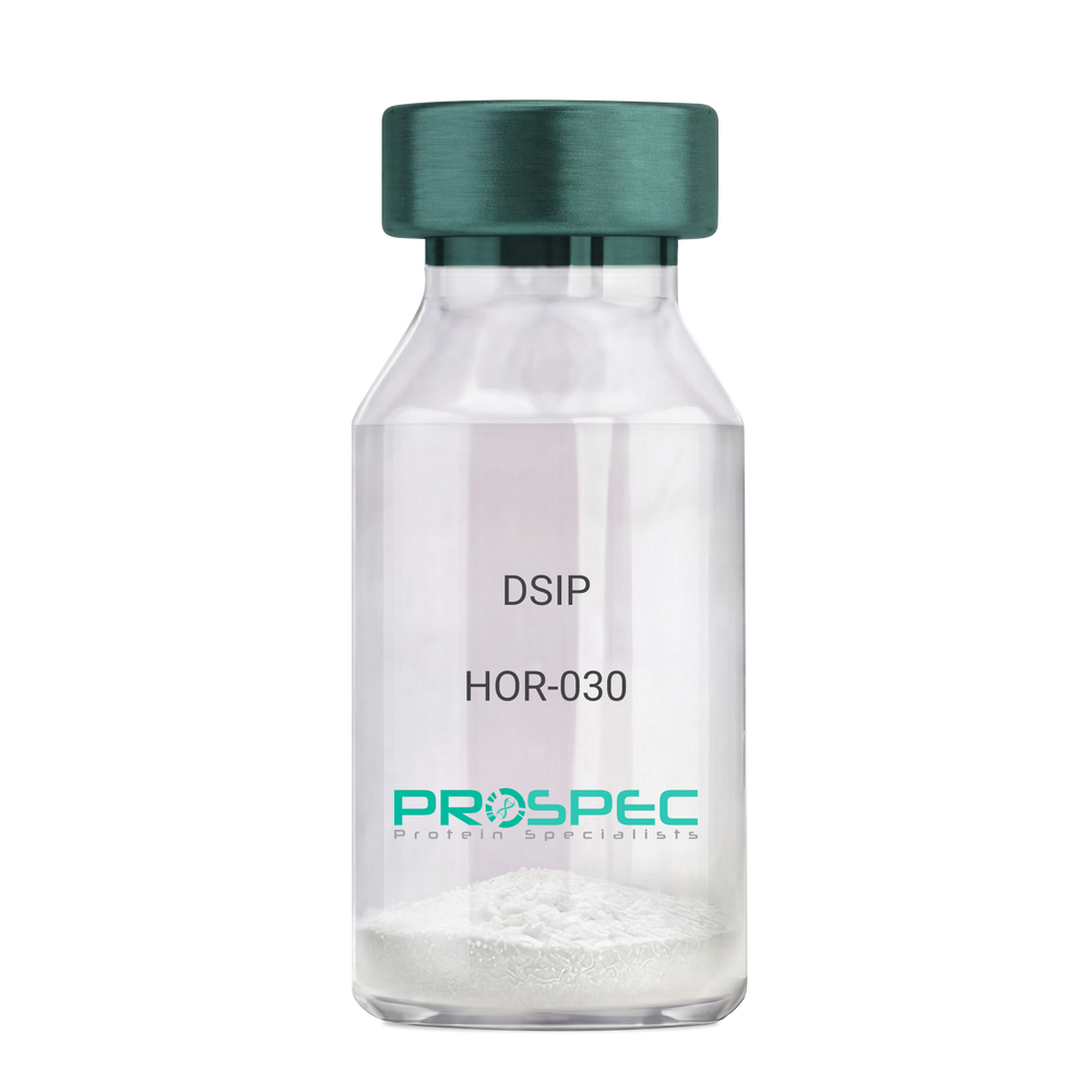
    DSIP

    Description:

    Delta Sleep Inducing Peptide

    Product # :

    HOR-030

    Price :

    Quantity :

    Shipping Method :

    Room Temp Icon

    Shipped at Room temp

    Add To Cart

    More Info

    • description
    • formulation
    • purity
    • More Info

    Description

    DSIP Synthetic is a single, non-glycosylated polypeptide chain containing 15 amino acids, having a molecular mass of 1419.55 Dalton and a Molecular formula of C62H98N16O22.

    Formulation

    The protein was lyophilized with no additives.

    Purity

    Greater than 97.0% as determined by analysis by RP-HPLC.

    More Info

    • Physical Appearance

      Sterile Filtered White lyophilized (freeze-dried) powder.

    • Stability

      Lyophilized DSIP although stable at room temperature for 3 weeks, should be stored desiccated below -18°C. Upon reconstitution DSIP should be stored at 4°C between 2-7 days and for future use below -18°C. For long term storage it is recommended to add a carrier protein (0.1% HSA or BSA). Please prevent freeze-thaw cycles.

    • Solubility

      It is recommended to reconstitute the lyophilized DSIP in sterile 18MΩ-cm H2O not less than 100 µg/ml, which can then be further diluted to other aqueous solutions.

    • Amino Acid Sequence

      H-Gly-Glu-Pro-Pro-Pro-Gly-Lys-Pro-Ala-Asp-Asp-Ala-Gly-Leu-Val-OH

    • Background

      Delta Sleep-Inducing Peptide (DSIP), also known as Sleep-Promoting Peptide, is a neuropeptide that has been the subject of extensive research due to its potential role in sleep regulation, stress response, and neuroprotection. This nonapeptide, first isolated from the cerebral venous blood of rabbits during sleep, has been shown to induce slow-wave sleep, modulate pain perception, and exhibit potential antioxidant and immunomodulatory properties.

      DSIP's primary function is its interaction with the sleep regulatory system. By modulating the release of certain neurotransmitters, DSIP can influence sleep patterns, particularly promoting slow-wave sleep, the most restorative stage of sleep. Studies by Kovalzon et al. (2011) have demonstrated that DSIP can enhance sleep quality in rats, suggesting potential applications in sleep disorders and the promotion of healthy sleep patterns.

      In addition to its sleep-inducing effects, DSIP has been shown to possess neuroprotective properties. Research by Zolotarev et al. (2014) found that DSIP could protect neurons from oxidative stress, suggesting potential applications in the treatment of neurodegenerative diseases such as Alzheimer's and Parkinson's disease.

      Given its sleep-inducing and neuroprotective effects, DSIP has been proposed as a potential therapeutic agent for a variety of conditions, including sleep disorders, chronic pain, and neurodegenerative diseases. For instance, a study by Spong et al. (2016) found that DSIP could improve sleep quality in patients with chronic insomnia, indicating its potential as a therapeutic agent in the treatment of sleep disorders.

      While research on DSIP is promising, it is important to note that most studies have been conducted in animals or in vitro. More research is needed to fully understand the potential effects and applications of DSIP in humans. However, the existing body of research suggests that DSIP could be a promising tool in the treatment of sleep disorders, chronic pain, and neurodegenerative diseases.

    ProSpec's products are furnished for LABORATORY RESEARCH USE ONLY. The product may not be used as drugs, agricultural or pesticidal products, food additives or household chemicals.

    Dsip
  • View Data Sheet

    Name :

    CSNK2B Protein

    Description:

    Casein Kinase 2b Human Recombinant

    Casein kinase II subunit beta, CK II beta, Phosvitin, G5a, CK2B, CK2N, CSK2B, MGC138222, MGC138224.

    Product # :

    PKA-223

    Price :

    Quantity :

    Shipping Method :

    Ice Icon

    Shipped with Ice Packs

    Add To Cart

    More Info

    • description
    • source
    • formulation
    • purity
    • More Info

    Description

    CSNK2B Human Recombinant produced in E.Coli is a single, non-glycosylated, polypeptide chain containing 215 amino acids and having a total molecular mass of 24.9kDa. CK2 beta is purified by proprietary chromatographic techniques.

    Source

    Escherichia Coli.

    Formulation

    The CSNK2B protein (1 mg/ml) contains 20mM Tris-HCl pH 8.0, 200mM NaCl, 1mM DTT, 1mM EDTA, 1uM leupeptin and 40% glycerol.

    Purity

    Greater than 95.0% as determinedAnalysis by SDS-PAGE.

    More Info

    • Introduction

      Casein Kinase 2 also called CK2 (also called PKCK2) is a ubiquitous Ser/Thr kinase expressed in all eukaryotes. CK2 is a tetramer composed of two catalytic kinase domains, alpha subunits, and two identical regulatory beta subunits. It has been implicated in cell cycle control, DNA repair, regulation of the circadian rhythm, and other cellular processes. The beta subunit itself does not have kinase activity, but confers stability to the CK2 alpha subunit and is involved in activity and substrate specificity.

    • Synonyms

      Casein kinase II subunit beta, CK II beta, Phosvitin, G5a, CK2B, CK2N, CSK2B, MGC138222, MGC138224.

    • Physical Appearance

      Sterile filtered colorless solution.

    • Stability

      Store at 4°C if entire vial will be used within 2-4 weeks.Store, frozen at -20°C for longer periods of time. For long term storage it is recommended to add a carrier protein (0.1% HSA or BSA). Please avoid freeze-thaw cycles.

    • Amino Acid Sequence

      MSSSEEVSWI SWFCGLRGNE FFCEVDEDYI QDKFNLTGLN EQVPHYRQAL DMILDLEPDE ELEDNPNQSD LIEQAAEMLY GLIHARYILT NRGIAQMLEK YQQGDFGYCP RVYCENQPML
      PIGLSDIPGE AMVKLYCPKC MDVYTPKSSR HHHTDGAYFG TGFPHMLFMV HPEYRPKRPA NQFVPRLYGF KIHPMAYQLQ LQAASNFKSP VKTIR.

    ProSpec's products are furnished for LABORATORY RESEARCH USE ONLY. The product may not be used as drugs, agricultural or pesticidal products, food additives or household chemicals.

    Csnk2B Human
  • View Data Sheet

    Name :

    MBP (27-392) E.Coli

    Description:

    Maltose Binding Protein E.coli Recombinant

    Product # :

    PRO-616

    Price :

    Quantity :

    Shipping Method :

    Ice Icon

    Shipped with Ice Packs

    Add To Cart

    More Info

    • description
    • source
    • formulation
    • purity
    • More Info

    Description

    Maltose Binding Protein E.Coli Recombinant produced in E.Coli is a single, non-glycosylated polypeptide chain containing 387 amino acids (27-392) with additional 20aa linker and having a molecular mass of 42kDa.

    Source

    Escherichia Coli.

    Formulation

    The Maltose Binding Protein solution (1 mg/ml) contains 20mM Tris-HCl pH-8.

    Purity

    Greater than 95.0% as determined by SDS-PAGE.

    More Info

    • Introduction

      Maltose Binding Protein is a member of the maltose/maltodextrin system of E.Coli, which is accountable for the uptake and efficient catabolism of maltodextrins. The maltose/maltodextrin is a complex regulatory and transport system involving many proteins and protein complexes.
      MBP elevates the yield of its fusion partner in many cases and is often able to promote the solubility of polypeptides to which it is fused.

    • Physical Appearance

      Sterile Filtered colorless solution.

    • Stability

      Store at 4°C if entire vial will be used within 2-4 weeks. Store, frozen at -20°C for longer periods of time. For long term storage it is recommended to add a carrier protein (0.1% HSA or BSA).Avoid multiple freeze-thaw cycles.

    • Amino Acid Sequence

      MKIEEGKLVI WINGDKGYNG LAEVGKKFEK DTGIKVTVEH PDKLEEKFPQ VAATGDGPDI IFWAHDRFGG YAQSGLLAEI TPDKAFQDKL YPFTWDAVRY NGKLIAYPIAVEALSLIYNK DLLPNPPKTW EEIPALDKEL KAKGKSALMF NLQEPYFTWP LIAADGGYAF KYENGKYDIK DVGVDNAGAK AGLTFLVDLI KNKHMNADTD YSIAEAAFNK GETAMTINGP WAWSNIDTSK VNYGVTVLPT FKGQPSKPFV GVLSAGINAA SPNKELAKEF LENYLLTDEG LEAVNKDKPL GAVALKSYEE ELAKDPRIAA TMENAQKGEI MPNIPQMSAF WYAVRTAVIN AASGRQTVDE ALKDAQTNSS SNNNNNNNNN NLGIEGR.

    ProSpec's products are furnished for LABORATORY RESEARCH USE ONLY. The product may not be used as drugs, agricultural or pesticidal products, food additives or household chemicals.

    Maltose Binding Protein
  • View Data Sheet

    Name :

    SPP1 Mouse

    Description:

    Osteopontin 1 Mouse Recombinant

    Secreted Phosphoprotein-1, OPN, BNSP, BSPI, ETA-1, MGC110940, SPP-1, Osteopontin, Bone sialoprotein 1, Urinary stone protein, Nephropontin, Uropontin, SPP1

    Product # :

    CYT-1201

    Price :

    Quantity :

    Shipping Method :

    Ice Icon

    Shipped with Ice Packs

    Add To Cart

    More Info

    • description
    • source
    • formulation
    • purity
    • biological activity
    • More Info

    Description

    SPP1 Mouse Recombinant produced in HEK293 Cells is a single, glycosylated polypeptide chain containing 287 amino acids (17-294 a.a) and having a molecular mass of 31.8kDa.SPP1 is expressed with a 6 amino acid His tag at C-Terminus and purified by proprietary chromatographic techniques.

    Source

    HEK293 Cells.

    Formulation

    The SPP1 solution (0.25mg/1ml) contains phosphate buffered saline (pH7.4) and 10% glycerol.

    Purity

    Greater than 90.0% as determined by SDS-PAGE.

    Biological Activity

    Measured by the ability of the immobilized protein to support the adhesion of HEK293 HEK cells when the cells are added to mouse SPP1 coated plates.  The ED50 range is ≤ 1.5 ug/ml.

    More Info

    • Introduction

      Osteopontin is a glycoprotein that was first identified in osteoblasts and is involved in bone remodeling, immune functions in fibroblasts, macrophages, and lymphocytes during inflammation and wound healing. SPP1 binds tightly to hydroxyapatite. SPP1 forms an integral part of the mineralized matrix. SPP1 is vital to cell-matrix interaction. Secreted Phosphoprotein-1 protects against cardiac ischemia-reperfusion injury via late preconditioning. Expression of both Ostepontin and CD44 in hepatocellular carcinoma is linked with advanced tumor stage and contributes to prognosis information. SPP1 is the most over-expressed gene in intrahepatic cholangiocarcinoma. Secreted Phosphoprotein-1 overexpression is related with interstitial lung diseases.

    • Synonyms

      Secreted Phosphoprotein-1, OPN, BNSP, BSPI, ETA-1, MGC110940, SPP-1, Osteopontin, Bone sialoprotein 1, Urinary stone protein, Nephropontin, Uropontin, SPP1

    • Physical Appearance

      Sterile Filtered colorless solution.

    • Stability

      Store at 4°C if entire vial will be used within 2-4 weeks.Store, frozen at -20°C for longer periods of time.For long term storage it is recommended to add a carrier protein (0.1% HSA or BSA).Avoid multiple freeze-thaw cycles.

    • Amino Acid Sequence

      DGSLPVKVTD SGSSEEKLYS LHPDPIATWL VPDPSQKQNL LAPQNAVSSE EKDDFKQETL PSNSNESHDH MDDDDDDDDD DGDHAESEDS VDSDESDESH HSDESDETVT ASTQADTFTP IVPTVDVPNG RGDSLAYGLR SKSRSFQVSD EQYPDATDED LTSHMKSGES KESLDVIPVA QLLSMPSDQD NNGKGSHESS QLDEPSLETH RLEHSKESQE SADQSDVIDS QASSKASLEH QSHKFHSHKD KLVLDPKSKE DDRYLKFRIS HELESSSSEV NHHHHHH

    ProSpec's products are furnished for LABORATORY RESEARCH USE ONLY. The product may not be used as drugs, agricultural or pesticidal products, food additives or household chemicals.

    Osteopontin Mouse
  • 1
  • …
  • 18
  • 19
  • 20
  • 21
  • 22
Your browser does not support the video tag.

Products

  • Cytokines
  • Growth Factors
  • Chemokines
  • CD Antigens
  • Neurotrophins
  • Hormones
  • Enzymes
  • Viral Antigens
  • Recombinant Proteins
  • Natural Proteins
  • Monoclonal Antibodies
  • Polyclonal Antibodies
  • Thermal Packaging

About

  • Company
  • Founder
  • Contribution
  • Contact Us
  • My Account

Ordering

  • Ordering Method
  • Ordering - Credit Card
  • Ordering PO
  • Shipping Fees
  • FAQ's

Legal

  • Terms & Conditions
  • Privacy
  • Site Map
  • info@prospecbio.com
  • Facebook
  • Instagram
  • Linkedin Profile

© 2026 ProSpec-Tany TechnoGene Ltd. All Rights Reserved.

  • Choosing a selection results in a full page refresh.
  • Opens in a new window.