Search results
460 results found for “mesoderm development candidate”
Name
Description
Product #
Price
Quantity
Shipping Method
- View Data Sheet
Name :
HK2 AntibodyDescription:
Hexokinase-2, Mouse Anti Human
Hexokinase-2, EC 2.7.1.1, HK2, Hexokinase type II, HK II, Muscle form hexokinase, HXK2, DKFZp686M1669.
Product # :
ANT-378Price :
Quantity :
Shipping Method :
Shipped with Ice Packs
More Info
- formulation
- More Info
Formulation
1mg/ml containing PBS, pH-7.4, & 0.1% Sodium Azide.
More Info
-
Introduction
Hexokinases phosphorylate glucose to produce glucose-6-phosphate, thus committing glucose to the glycolytic pathway. Hexokinase 2 is the predominant form found in skeletal muscle. It localizes to the outer membrane of mitochondria. Expression of this gene is insulin-responsive, and studies in rat suggest that it is involved in the increased rate of glycolysis seen in rapidly growing cancer cells.
-
Synonyms
Hexokinase-2, EC 2.7.1.1, HK2, Hexokinase type II, HK II, Muscle form hexokinase, HXK2, DKFZp686M1669.
-
Immunogen
Anti-human Hexokinase-2 mAb is derived from hybridization of mouse F0 myeloma cells with spleen cells from BALB/c mice immunized with recombinant human Hexokinase-2 amino acids 1-917 purified from E. coli.
-
Ig Subclass
Mouse IgG1 heavy chain and κ light chain.
-
Clone
P1A7AT.
-
Applications
Hexokinase-2 antibody has been tested by ELISA and Western blot analysis to assure specificity and reactivity. Since application varies, however, each investigation should be titrated by the reagent to obtain optimal results. Recommended dilution range for Western blot analysis is 1:1,000 ~ 3,000. Recommended starting dilution is 1:2,000.
-
Type
Mouse Anti Human Monoclonal.
-
Storage Procedures
For periods up to 1 month store at 4°C, for longer periods of time, store at -20°C. Prevent freeze thaw cycles.
-
Purification Method
Hexokinase-2 antibody was purified from mouse ascitic fluids by protein-G affinity chromatography.
ProSpec's products are furnished for LABORATORY RESEARCH USE ONLY. The product may not be used as drugs, agricultural or pesticidal products, food additives or household chemicals.
- View Data Sheet
Name :
IL 7 MouseDescription:
Interleukin-7 Mouse Recombinant
Lymphopoietin 1 (LP-1), pre-B cell factor, IL-7.
Product # :
CYT-372Price :
Quantity :
Shipping Method :
Shipped at Room temp
More Info
- description
- source
- formulation
- purity
- biological activity
- More Info
Description
Interleukin-7 Mouse Recombinant produced in E.Coli is a single, non-glycosylated polypeptide chain containing 129 amino acids and having a molecular mass of 14.9kDa.
Source
Escherichia Coli.
Formulation
Filtered (0.2µm) and lyophilized from a concentrated (1mg/ml) solution in 1×PBS, pH7.4 and 2% trehalose.
Purity
Greater than 96.0% as determined by:
(a) Analysis by RP-HPLC.
(b) Analysis by SDS-PAGE.Biological Activity
The ED50was determined by the dose-dependent stimulation of the proliferation of murine 2E8 cells is less than 0.2 ng/ml, corresponding to a specific activity of >5.0×10⁶ IU/mg.More Info
-
Introduction
Interleukin-7 is a potent lymphoid cell growth factor produced primarily by stromal cells. IL-7 has been shown to support the proliferation and differentiation of pre B- and early T cells as well as displaying a biological effect on cells of NK and myeloid lineages.
-
Synonyms
Lymphopoietin 1 (LP-1), pre-B cell factor, IL-7.
-
Physical Appearance
Sterile Filtered White lyophilized (freeze-dried) powder.
-
Stability
Lyophilized Interleukin-7 although stable at room temperature for 3 weeks, should be stored desiccated below -18°C. Upon reconstitution IL7 should be stored at 4°C between 2-7 days and for future use below -18°C.For long term storage it is recommended to add a carrier protein (0.1% HSA or BSA).Please prevent freeze-thaw cycles.
-
Solubility
It is recommended to reconstitute the lyophilized Interleukin 7 in sterile 18MΩ-cm H2O not less than 100µg/ml, which can then be further diluted to other aqueous solutions.
-
Amino Acid Sequence
ECHIKDKEGK AYESVLMISI DELDKMTGTD SNCPNNEPNF FRKHVCDDTK EAAFLNRAAR KLKQFLKMNI SEEFNVHLLT VSQGTQTLVN CTSKEEKNVK EQKKNDACFL KRLLREIKTC WNKILKGSI.
ProSpec's products are furnished for LABORATORY RESEARCH USE ONLY. The product may not be used as drugs, agricultural or pesticidal products, food additives or household chemicals.
- View Data Sheet
Name :
HCV Core Genotype-4Description:
Hepatitis C Virus Core Genotype-4 Recombinant
Product # :
HCV-216Price :
Quantity :
Shipping Method :
Shipped with Ice Packs
More Info
- description
- formulation
- purity
- More Info
Description
The E.coli derived recombinant protein contains the HCV core nucleocapsid immunodominant regions, amino acids 2-119. The protein is fused to a GST tag at N-Terminus.
Formulation
50mM Tris-HCl, pH-8, 60mM NaCl, 10mM glutathione, 0.25% sarkosil & 50% glycerol.
Purity
HCV Core Genotype-4 protein is >95% pure as determined by 10% PAGE (coomassie staining).
More Info
-
Introduction
HCV is a small 50nm, enveloped, single-stranded, positive sense RNAvirus in the family Flaviviridae.
HCV has a high rate of replication with approximately one trillion particles produced each day in an infected individual. Due to lack of proofreading by the HCV RNA polymerase, the HCV has an exceptionally high mutation rate, a factor that may help it elude the host's immune response. Hepatitis C virus is classified into six genotypes(1-6) with several subtypes within each genotype. The preponderance and distribution of HCV genotypes varies globally. Genotype is clinically important in determining potential response to interferon-based therapy and the required duration of such therapy. Genotypes 1 and 4 are less responsive to interferon-based treatment than are the other genotypes (2, 3, 5 and 6). -
Stability
HCV Core Genotype-4 although stable at 4°C for 1 week, should be stored below -18°C. Please prevent freeze thaw cycles.
-
Applications
HCV Core Genotype-4 atigen is suitable for ELISA and Western blots, excellent antigen for detection of HCV with minimal specificity problems.
-
Specificity
Immunoreactive with sera of HCV-infected individuals.
-
Purification Method
HCV Core Genotype-4 protein was purified by proprietary chromatographic technique.
ProSpec's products are furnished for LABORATORY RESEARCH USE ONLY. The product may not be used as drugs, agricultural or pesticidal products, food additives or household chemicals.
- View Data Sheet
Name :
EPHB2 HumanDescription:
EPH Receptor B2 Human Recombinant
EPHB2, CAPB, DRT, EK5, EPHT3, ERK, Hek5, PCBC, Tyro5, Developmentally-regulated Eph-related tyrosine kinase, ELK-related tyrosine kinase, EPH tyrosine kinase 3, EPH-like kinase 5, hEK5, Renal carcinoma antigen NY-REN-47, Tyrosine-protein kinase TYRO5, Tyrosine-protein kinase receptor EPH-3.
Product # :
PRO-2379Price :
Quantity :
Shipping Method :
Shipped with Ice Packs
More Info
- description
- source
- formulation
- purity
- More Info
Description
EPHB2 Human Recombinant produced in Sf9 Baculovirus cells is a single, glycosylated polypeptide chain containing 533 amino acids (19-543a.a) and having a molecular mass of 59.1kDa (Molecular size on SDS-PAGE will appear at approximately 50-70kDa). EPHB2 is fused to a 8 amino acid His-tag at C-terminus & purified by proprietary chromatographic techniques.
Source
Sf9, Baculovirus cells.
Formulation
EPHB2 protein solution (0.5mg/ml) containing Phosphate Buffered Saline (pH 7.4), 1mM DTT and 20% glycerol.
Purity
Greater than 90% as determined by SDS-PAGE.
More Info
-
Introduction
EPH Receptor B2 (EPHB2) is a part of the transmembrane Eph receptor tyrosine kinase family (RTKs) which binds proteins of the Ephrin family on adjacent cells. The interaction leads to contact-dependent bidirectional signaling into neighboring cells. Hippocampal neurons can release vesicles containing full length EPHB2, and these are taken up by neighboring glial cells. EPHB2 takes part in the guidance of commissural axons through the embryonic midline and regulates dendritic spines development and maturation and stimulates the formation of excitatory synapses.
-
Synonyms
EPHB2, CAPB, DRT, EK5, EPHT3, ERK, Hek5, PCBC, Tyro5, Developmentally-regulated Eph-related tyrosine kinase, ELK-related tyrosine kinase, EPH tyrosine kinase 3, EPH-like kinase 5, hEK5, Renal carcinoma antigen NY-REN-47, Tyrosine-protein kinase TYRO5, Tyrosine-protein kinase receptor EPH-3.
-
Physical Appearance
Sterile Filtered colorless solution.
-
Stability
Store at 4°C if entire vial will be used within 2-4 weeks. Store, frozen at -20°C for longer periods of time. For long term storage it is recommended to add a carrier protein (0.1% HSA or BSA).Avoid multiple freeze-thaw cycles.
-
Amino Acid Sequence
VEETLMDSTT ATAELGWMVH PPSGWEEVSG YDENMNTIRT YQVCNVFESS QNNWLRTKFI RRRGAHRIHV EMKFSVRDCS SIPSVPGSCK ETFNLYYYEA DFDSATKTFP NWMENPWVKV DTIAADESFS QVDLGGRVMK INTEVRSFGP VSRSGFYLAF QDYGGCMSLI AVRVFYRKCP RIIQNGAIFQ ETLSGAESTS LVAARGSCIA NAEEVDVPIK LYCNGDGEWL VPIGRCMCKA GFEAVENGTV CRGCPSGTFK ANQGDEACTH CPINSRTTSE GATNCVCRNG YYRADLDPLD MPCTTIPSAP QAVISSVNET SLMLEWTPPR DSGGREDLVY NIICKSCGSG RGACTRCGDN VQYAPRQLGL TEPRIYISDL LAHTQYTFEI QAVNGVTDQS PFSPQFASVN ITTNQAAPSA VSIMHQVSRT VDSITLSWSQ PDQPNGVILD YELQYYEKEL SEYNATAIKS PTNTVTVQGL KAGAIYVFQV RARTVAGYGR YSGKMYFQTM TEAEYQTSIQ EKLPLLEHHH HHH.
ProSpec's products are furnished for LABORATORY RESEARCH USE ONLY. The product may not be used as drugs, agricultural or pesticidal products, food additives or household chemicals.
- View Data Sheet
Name :
SARS MERS Spike S1Description:
Mouse Anti SARS MERS Spike S1
Product # :
ANT-766Price :
Quantity :
Shipping Method :
Shipped with Ice Packs
More Info
- description
- formulation
- purity
- More Info
Description
SARS MERS Spike-S1 antibody is specific for the S1 domain of the spike protein of the MERS virus.
Formulation
1mg/ml in PBS and 0.1% NaN3.
Purity
Greater than 90% as determined by SDS-PAGE gel.
More Info
-
Introduction
SARS Coronavirus is an enveloped virus containing three outer structural proteins, namely the membrane (M), envelope (E), and spike (S) proteins. Spike (S)-glycoprotein of the virus interacts with a cellular receptor and mediates membrane fusion to allow viral entry into susceptible target cells. Accordingly, S-protein plays an important role in virus infection cycle and is the primary target of neutralizing antibodies.It has recently been shown that SARS is caused by a human coronavirus. Human coronaviruses are the major cause of upper respiratory tract illness in humans, such as the common cold. Coronaviruses are positive-stranded RNA viruses, featuring the largest viral RNA genomes known to date (27-31 kb). The first step in coronavirus infection is binding of the viral spike protein, a 139-kDa protein, to certain receptors on host cells. The spike protein is the main surface antigen of the coronavirus. The most prominent protein in the culture supernatants infected with SARS virus is a 46 kDa nucleocapsid protein. This suggests that the nucleocapsid protein is a major immunogen that may be useful for early diagnostics.
-
Physical Appearance
Sterile Filtered liquid formulation.
-
Stability
Store at 4°C, stable for 2 weeks. For long-term storage, store at -20°C.
-
Applications
ELISA.
-
Isotype
IgG1.
-
Type
Mouse antibody Monoclonal.
-
Purification Method
Protein A affinity purified.
ProSpec's products are furnished for LABORATORY RESEARCH USE ONLY. The product may not be used as drugs, agricultural or pesticidal products, food additives or household chemicals.
- View Data Sheet
Name :
TGFB2 MouseDescription:
Transforming Growth Factor-Beta 2 Mouse Recombinant
Transforming growth factor beta-2, TGF-beta-2, G-TSF, Tgfb-2, TGFbeta2.
Product # :
CYT-1266Price :
Quantity :
Shipping Method :
Shipped at Room temp
More Info
- description
- source
- formulation
- purity
- biological activity
- More Info
Description
Transforming Growth Factor-Beta 2 Mouse Recombinant produced in CHO is a homodimer, polypeptide chain containing 2 x 112 amino acids and having a total molecular mass of 25.4kDa.
TGFB2 Mouse Recombinant is purified by proprietary chromatographic techniques.Source
CHO Cells.
Formulation
The protein was lyophilized with 0.1% (v/v) TFA and 35% (v/v) Acetonitrile.
Purity
Greater than 97.0% as determined by SDS-PAGE and SEC-HPLC analyses.
Biological Activity
The biological activity was determined by TGFB2 ability to inhibit the mouse IL-4-dependent proliferation of mouse HT-2 cells. The expected ED50 for this effect is <0.05ng/ml, corresponding to a specific activity of ≥ 2.0 × 10⁷ units/mg.More Info
-
Synonyms
Transforming growth factor, beta 2, cetermin, Glioblastoma-derived T-cell suppressor factor, polyergin, G-TSF, TGF-beta2, TGF-beta-2, transforming growth factor beta-2, BSC-1 cell growth inhibitor, TGFB-2.
-
Physical Appearance
Sterile Filtered White lyophilized (freeze-dried) powder.
-
Stability
Lyophilized TGFB2 although stable at room temperature for 3 weeks, should be stored desiccated below -18°C. Upon reconstitution Transforming Growth Factor-Beta 2 should be stored at 4°C between 2-7 days and for future use below -18°C.
For long term storage it is recommended to add a carrier protein (0.1% HSA or BSA).
Please prevent freeze-thaw cycles.
-
Solubility
It is recommended to reconstitute the lyophilized Transforming Growth Factor-Beta 2 in sterile 4mM HCl not less than 100µg/ml, which can then be further diluted to other aqueous solutions.
-
Amino Acid Sequence
ALDAAYCFRN VQDNCCLRPL YIDFKRDLGW KWIHEPKGYN ANFCAGACPY LWSSDTQHTK VLSLYNTINP EASASPCCVS QDLEPLTILY YIGNTPKIEQ LSNMIVKSCK CS.
-
Background
TGFB2 differ from TGFB1 in the tissue distribution, receptor interactions, and several biological roles.
TGFB2 requires TGFBR3 for its binding to TGFBR2 while TGFB1 binds directly to the receptor TGFBR2.
TGFB2 is crucial for embryonic development ocular biology, neural development, and tissue morphogenesis while TGFB1 is crucial for immune regulation and fibrosis.
TGFB2 exhibits more tissue-specific developmental expression Vs TGFB1.
What is the source or expression system of Mouse TGFB2 Protein?
CHO Cells
What is the Purity of Mouse TGFB2 Protein?
Mouse TGFB2 Protein is >97% pure as determined by SDS-PAGE.
What is the molecular weight / Mw of Mouse TGFB2 Protein?
Mouse TGFB2 Protein having a total Mw of 25.6kDa.
What is the Biological Activity of Mouse TGFB2 Protein?
The biological functionality of Mouse TGFB2 Protein is determined by mouse HT-2 cells.
What is the endotoxin level for Mouse TGFB2 Protein?
The endotoxin level is minimal, Mouse TGFB2 Protein was purified using conventional chromatography techniques.
What is the amino acid sequence of Mouse TGFB2 Protein?
ALDAAYCFRN VQDNCCLRPL YIDFKRDLGW KWIHEPKGYN ANFCAGACPY LWSSDTQHTK VLSLYNTINP EASASPCCVS QDLEPLTILY YIGNTPKIEQ LSNMIVKSCK CS.
Is TGFB2 a homodimer / homodimeric protein?
Yes, TGFB2 is homo dimer consisting of 2 identical chains.
ProSpec's products are furnished for LABORATORY RESEARCH USE ONLY. The product may not be used as drugs, agricultural or pesticidal products, food additives or household chemicals.
- View Data Sheet
Name :
DHFR AntibodyDescription:
Dihydrofolate Reductase, Mouse Anti Human
Dihydrofolate reductase, DHFR, DHFRP1.
Product # :
ANT-596Price :
Quantity :
Shipping Method :
Shipped with Ice Packs
More Info
- formulation
- More Info
Formulation
1mg/ml containing PBS, pH-7.4, 10% Glycerol and 0.02% Sodium Azide.
More Info
-
Introduction
Dihydrofolate reductase (DHFR) is an enzyme that reduces dihydrofolic acid to tetrahydrofolic acid, with NADPH as electron donor that can be converted to the kinds of tetrahydrofolate cofactors applied in 1-carbon transfer chemistry. DHFR converts dihydrofolate into tetrahydrofolate, which is a methyl group shuttle required for the de novo synthesis of purines, thymidylic acid, and specific amino acids. Even though the functional DHFR gene is mapped to chromosome 5, numerous intronless processed pseudogenes or dihydrofolate reductase-like genes are identified on separate chromosomes.
DHFR deficiency is associated with megaloblastic anemia.DHFR knockdown plays a role in the anticancer activity of 2-hydroxyoleic acid.
DHFR gene insertion/deletion polymorphism is linked to variation in serum and red blood cell folate concentrations in women. -
Synonyms
Dihydrofolate reductase, DHFR, DHFRP1.
-
Physical Appearance
Sterile filtered colorless solution.
-
Immunogen
Anti-human DHFR mAb, is derived from hybridization of mouse F0 myeloma cells with spleen cells from BALB/c mice immunized with recombinant human DHFR amino acids 1-187 purified from E. coli.
-
Ig Subclass
Mouse IgG1 heavy chain and k light chain.
-
Clone
PAT5B2AT.
-
Applications
DHFR antibody has been tested by ELISA, Western blot analysis and Flow cytometry to assure specificity and reactivity. Since application varies, however, each investigation should be titrated by the reagent to obtain optimal results. Recommended starting dilution for Western blot analysis is 1:1000.
-
Type
Mouse Anti Human Monoclonal.
-
Storage Procedures
For periods up to 1 month store at 4°C, for longer periods of time, store at -20°C. Prevent freeze thaw cycles.
-
Purification Method
DHFR antibody was purified from mouse ascitic fluids by protein-A affinity chromatography.
ProSpec's products are furnished for LABORATORY RESEARCH USE ONLY. The product may not be used as drugs, agricultural or pesticidal products, food additives or household chemicals.
- View Data Sheet
Name :
Monkeypox B5RDescription:
Monkeypox B5R Recombinant
Product # :
MPV-002Price :
Quantity :
Shipping Method :
Shipped with Ice Packs
More Info
- description
- source
- formulation
- purity
- More Info
Description
The E.Coli derived recombinant B5R protein contains the Envelope Monkeypox immunodominant regions, having an Mw of 32kDa. The Monkeypox protein is fused to a 6xHis tag at C-terminus and purified by proprietary chromatographic technique
Source
Escherichia Coli.
Formulation
Recombinant Monkeypox protein solution contains PBS, 0.05% sodium nitrate and 25mM K2CO3
Purity
Protein is >95% pure as determined by 12% PAGE (coomassie staining).
More Info
-
Introduction
Monkeypox virus is transmitted to humans from animalssuch as rodents and primates having similar symptoms similar of smallpox although less severe. Monkeypox is an enveloped double-stranded DNA virus approximately 190 kb which is part of the Orthopoxvirus genus of the Poxviridae family. 2monkeypox genetic clades: central African which is more transmissible& west African clade. Monkeypox virus spreadsfrom animal to human &human to humanvia animal bite or by direct body fluid contact. Incubation period lasts12 days. The MPV common ssymptoms include fever, rash, headache, swelling of lymph nodes and muscle pain,
-
Physical Appearance
Sterile Filtered clear solution.
-
Stability
Upon arrival, Store at -20°C. Avoid multiple freeze-thaw cycles.
ProSpec's products are furnished for LABORATORY RESEARCH USE ONLY. The product may not be used as drugs, agricultural or pesticidal products, food additives or household chemicals.
- View Data Sheet
Name :
MG HumanDescription:
Menopausal Gonadotropin Human
Product # :
HOR-251Price :
Quantity :
Shipping Method :
Shipped at Room temp
More Info
- description
- source
- formulation
- biological activity
- More Info
Description
Menopausal Gonadotropin Human is produced from a sterile preparation of placental glucoprotein urine of post-menopausal women.The MG is purified by proprietary chromatographic techniques.
Source
Urine of post-menopausal women.
Formulation
The Human MG was lyophilized from a concentrated (1mg/ml) solution with no additives.
Biological Activity
100 IU/mg of FSH and 100 IU/mg LH.More Info
-
Physical Appearance
Sterile Filtered White lyophilized (freeze-dried) powder.
-
Stability
Lyophilized Menopausal Gonadotropin Human although stable at room temperature for 3 weeks, should be stored desiccated below -18°C. Upon reconstitution MG should be stored at 4°C between 2-7 days and for future use below -18°C.For long term storage it is recommended to add a carrier protein (0.1% HSA or BSA).Please prevent freeze-thaw cycles.
-
Solubility
It is recommended to reconstitute the lyophilized MG in sterile 18MΩ-cm H2O not less than 100 µg/ml, which can then be further diluted to other aqueous solutions.
-
Contaminants
Free of: HbsAg, Hepatitis B surface antigen and antibodies to HIV, Hepatitis C and HIV.
ProSpec's products are furnished for LABORATORY RESEARCH USE ONLY. The product may not be used as drugs, agricultural or pesticidal products, food additives or household chemicals.
- View Data Sheet
Name :
SARS MERS, HEKDescription:
SARS MERS Spike Glycoprotein-S1, Recombinant
Product # :
SARS-023Price :
Quantity :
Shipping Method :
Shipped with Ice Packs
More Info
- description
- source
- formulation
- purity
- More Info
Description
The HEK293 derived recombinant protein contains the SARS MERS Spike Glycoprotein S1, amino acids 18-725 fused to dimeric Fc tag at N-terminal having a total Mw of 215.7 kDa.
Source
HEK293
Formulation
SARS MERS S1 protein solution is supplied in PBS.
Purity
Protein is >85% pure as determined SDS-PAGE.
More Info
-
Introduction
SARS Coronavirus is an enveloped virus containing three outer structural proteins, namely the membrane (M), envelope (E), and spike (S) proteins. Spike (S)-glycoprotein of the virus interacts with a cellular receptor and mediates membrane fusion to allow viral entry into susceptible target cells. Accordingly, S-protein plays an important role in virus infection cycle and is the primary target of neutralizing antibodies.
-
Physical Appearance
Sterile Filtered clear solution.
-
Stability
SARS MERS Protein is shipped on ice packs. Upon arrival, Store at -20°C.
-
Purification Method
Purified by Protein-G chromatographic technique.
ProSpec's products are furnished for LABORATORY RESEARCH USE ONLY. The product may not be used as drugs, agricultural or pesticidal products, food additives or household chemicals.
- View Data Sheet
Name :
GH Gilthead SeabreamDescription:
Growth Hormone Gilthead Seabream Recombinant
GH1, GH, GHN, GH-N, hGH-N, Pituitary growth hormone, Growth hormone 1, Somatotropin.
Product # :
CYT-529Price :
Quantity :
Shipping Method :
Shipped at Room temp
More Info
- description
- source
- formulation
- purity
- biological activity
- More Info
Description
Somatotropin Gilthead Seabream Recombinant Sparus Aurata produced in E.Coli is a single, non-glycosylated polypeptide chain containing 188 amino acids with an additional Ala at the N-terminus and having a molecular mass of 21.4 kDa. The Gilthead Seabream Growth-Hormone Recombinant is purified by proprietary chromatographic techniques.
Source
Escherichia Coli.
Formulation
The protein was lyophilized from a concentrated (1mg/ml) solution with 0.02% NaHCO3.
Purity
Greater than 98.0% as determined by:
(a) Analysis by SEC-HPLC.
(b) Analysis by SDS-PAGE.Biological Activity
Binding assays of the 125I-labeled gilthead seabream GH to dolphin fish liver microsomal fraction resulted in high specific binding characterized by a Ka of 1.93 nM and a Bmax of 540 fmol/mg microsomal fraction protein. Recombinant gilthead seabream Growth Hormone, like ovine placental lactogen, exhibited growth-stimulating activity when applied orally to Sparus aurata larvae or intraperitoneally to juvenile fish.More Info
-
Introduction
GH is a member of the somatotropin/prolactin family of hormones which play an important role in growth control. The gene, along with four other related genes, is located at the growth hormone locus on chromosome 17 where they are interspersed in the same transcriptional orientation; an arrangement which is thought to have evolved by a series of gene duplications. The five genes share a remarkably high degree of sequence identity. Alternative splicing generates additional isoforms of each of the five growth hormones, leading to further diversity and potential for specialization. This particular family member is expressed in the pituitary but not in placental tissue as is the case for the other four genes in the growth hormone locus. Mutations in or deletions of the gene lead to growth hormone deficiency and short stature.
-
Synonyms
GH1, GH, GHN, GH-N, hGH-N, Pituitary growth hormone, Growth hormone 1, Somatotropin.
-
Physical Appearance
Sterile Filtered White lyophilized (freeze-dried) powder.
-
Stability
Lyophilized Growth-Hormone Gilthead Seabream although stable at room temperature for at least two weeks, should be stored desiccated below -18°C. Upon reconstitution and filter sterilization GH can be stored at 4°C, pH 9 for up to 4 weeks. For long term storage and more diluted solutions it is recommended to add a carrier protein (0.1% HSA or BSA). Please prevent freeze-thaw cycles.
-
Solubility
It is recommended to reconstitute the lyophilized Growth-Hormone Gilthead Seabream in 0.4% NaHCO3 or water adjusted to pH 8-9, not less than 100µg/ml, which can then be further diluted to other aqueous solutions, preferably in a presence of a carrier protein such as BSA or similar.
-
Amino Acid Sequence
AQPITDGQRLFSIAVSRVQHLHLLAQRLFSDFESSLQTEEQPQLNKIFLQ
DFCNCDYIISPIDKHETQRSSVLKLLSISYRLVESWEFPSRSLSGGSAPR
NQISPKLSELKTGIHLLIRANEDGAEIFPDRSALQLAPYGNYYQSLGTDE
SLRRTYELLACFKKDMHKVETYLTVAKCRLSPEANCTL
ProSpec's products are furnished for LABORATORY RESEARCH USE ONLY. The product may not be used as drugs, agricultural or pesticidal products, food additives or household chemicals.
- View Data Sheet
Name :
PPP1CA AntibodyDescription:
Protein Phosphatase 1 Catalytic Subunit Alpha, Mouse Anti Human
Serine/threonine-protein phosphatase PP1-alpha catalytic subunit, PP-1A, PPP1CA, PPP1A, protein phosphatase 1 catalytic subunit alpha isozyme, MGC1674, MGC15877, PP1alpha.
Product # :
ANT-415Price :
Quantity :
Shipping Method :
Shipped with Ice Packs
More Info
- formulation
- More Info
Formulation
1mg/ml containing PBS, pH-7.4, & 0.1% Sodium Azide.
More Info
-
Introduction
PPP1CA is one of the 3 catalytic subunits of protein phosphatase 1 (PP1). PP1 is a serine/threonine specific protein phosphatase which is involved in the regulation of a variety of cellular processes, such as cell division, glycogen metabolism, muscle contractility, protein synthesis, and HIV-1 viral transcription. Increased PP1 activity is detected in the end stage of heart failure. Studies in both human and mice suggest that PP1 is a significant regulator of cardiac function. Mouse studies also propose that PP1 functions as a suppressor of learning and memory.
-
Synonyms
Serine/threonine-protein phosphatase PP1-alpha catalytic subunit, PP-1A, PPP1CA, PPP1A, protein phosphatase 1 catalytic subunit alpha isozyme, MGC1674, MGC15877, PP1alpha.
-
Physical Appearance
Sterile Filtered clear solution.
-
Immunogen
Anti-human PPP1CA mAb, is derived from hybridization of mouse FO myeloma cells with spleen cells from BALB/c mice immunized with recombinant human PPP1CA amino acids 30-299 purified from E. coli.
-
Ig Subclass
Mouse IgG2a heavy chain and κ light chain.
-
Clone
P4G3AT.
-
Applications
PPP1CA antibody has been tested by ELISA and Western blot analysis to assure specificity and reactivity. Since application varies, however, each investigation should be titrated by the reagent to obtain optimal results. Recommended dilution range for Western blot analysis is 1:500 ~ 2,000. Recommended starting dilution is 1:1,000.
-
Type
Mouse Anti Human Monoclonal.
-
Storage Procedures
For periods up to 1 month store at 4°C, for longer periods of time, store at -20°C. Prevent freeze thaw cycles.
-
Purification Method
PPP1CA antibody was purified from mouse ascitic fluids by protein-G affinity chromatography.
ProSpec's products are furnished for LABORATORY RESEARCH USE ONLY. The product may not be used as drugs, agricultural or pesticidal products, food additives or household chemicals.
- View Data Sheet
Name :
CAPNS1 AntibodyDescription:
Calpain, Small Subunit 1, Mouse Anti Human
Calpain Small Subunit 1, CAPN4, Calcium-Activated Neutral Proteinase Small Subunit, Calcium-Dependent Protease Small Subunit 1, Calpain Regulatory Subunit, CANP Small Subunit, CDPS, CSS1, Calcium-Dependent Protease Small Subunit, Calcium-Dependent Protease Small Subunit, Calpain 4 Small Subunit (30K), Calpain Small Polypeptide, Calpain Small Subunit 1, CALPAIN4, CANPS, CAPNS, CANP, CAPNS1.
Product # :
ANT-587Price :
Quantity :
Shipping Method :
Shipped with Ice Packs
More Info
- formulation
- More Info
Formulation
1mg/ml containing PBS, pH-7.4, 10% Glycerol and 0.02% Sodium Azide.
More Info
-
Introduction
Calpain, Small Subunit 1 (CAPNS1) belongs to the calpain small subunit family. Calpains are a ubiquitous, well-conserved family of calcium-dependent, cysteine proteases, widely distributed in mammalian cells. Calpain families are implicated in neurodegenerative processes, considering that their activation can be triggered by calcium influx and oxidative stress. Calpains function as heterodimers, comprising a specific large catalytic subunit (calpain 1 subunit in Calpain I, and calpain 2 subunit in Calpain II), and a common small regulatory subunit encoded by the CAPNS1 gene. The CAPNS1 protein is vital for the stability and function of both calpain heterodimers, whose proteolytic activities influence numerous cellular functions including apoptosis, proliferation, migration, adhesion, and autophagy.
-
Synonyms
Calpain Small Subunit 1, CAPN4, Calcium-Activated Neutral Proteinase Small Subunit, Calcium-Dependent Protease Small Subunit 1, Calpain Regulatory Subunit, CANP Small Subunit, CDPS, CSS1, Calcium-Dependent Protease Small Subunit, Calcium-Dependent Protease Small Subunit, Calpain 4 Small Subunit (30K), Calpain Small Polypeptide, Calpain Small Subunit 1, CALPAIN4, CANPS, CAPNS, CANP, CAPNS1.
-
Physical Appearance
Sterile filtered colorless solution.
-
Immunogen
Anti-human CAPNS1 mAb, is derived from hybridization of mouse F0 myeloma cells with spleen cells from BALB/c mice immunized with a recombinant human CAPNS1 protein 84-268 amino acids purified from E. coli.
-
Ig Subclass
Mouse IgG1 heavy chain and k light chain.
-
Clone
PAT1D11AT.
-
Applications
CAPNS1 antibody has been tested by ELISA, Western blot analysis, ICC/IF and Flow cytometry to assure specificity and reactivity. Since application varies, however, each investigation should be titrated by the reagent to obtain optimal results.
-
Type
Mouse Anti Human Monoclonal.
-
Storage Procedures
For periods up to 1 month store at 4°C, for longer periods of time, store at -20°C. Prevent freeze thaw cycles.
-
Purification Method
CAPNS1 antibody was purified from mouse ascitic fluids by protein-A affinity chromatography.
ProSpec's products are furnished for LABORATORY RESEARCH USE ONLY. The product may not be used as drugs, agricultural or pesticidal products, food additives or household chemicals.
- View Data Sheet
Name :
BAFF AntibodyDescription:
B-cell Activating Factor, Mouse Anti Human
BAFF, BLYS, CD257, TALL1, THANK, ZTNF4, TALL-1, TNFSF20, TNFSF13B.
Product # :
ANT-367Price :
Quantity :
Shipping Method :
Shipped with Ice Packs
More Info
- formulation
- More Info
Formulation
1mg/ml containing PBS, pH-7.4, & 0.1% Sodium Azide.
More Info
-
Introduction
Binds to tnfrsf13b/taci and tnfrsf17/bcma. tnfsf13/april binds to the same 2 receptors, together, they form a 2 ligands -2 receptors pathway involved in the stimulation of b- and t-cell function and the regulation of humoral immunity. A third b-cell specific baff-receptor (baffr/br3) promotes the survival of mature b-cells and the b-cell response. B Lymphocyte Stimulator functions as a potent B-cell growth factor in costimulation assays. Administration of BAFF Human recombinant to mice disrupts splenic B-cell and T-cell zones and results in elevated levels of serum immunoglobulin.
-
Synonyms
BAFF, BLYS, CD257, TALL1, THANK, ZTNF4, TALL-1, TNFSF20, TNFSF13B.
-
Physical Appearance
Sterile Filtered colorless solution.
-
Immunogen
Anti-human BAFF mAb, is derived from hybridization of mouse F0 myeloma cells with spleen cells from BALB/c mice immunized with Recombinant human BAFF amino acids 134-285 purified from E. coli.
-
Ig Subclass
Mouse IgG3 heavy chain and κ light chain.
-
Clone
PH4C7AT.
-
Applications
BAFF antibody has been tested by ELISA and Western blot analysis to assure specificity and reactivity. Since application varies, however, each investigation should be titrated by the reagent to obtain optimal results. Recommended dilution range for Western blot analysis is 1:500 ~ 1,000. Recommended starting dilution is 1:500.
-
Type
Mouse Anti Human Monoclonal.
-
Storage Procedures
For periods up to 1 month store at 4°C, for longer periods of time, store at -20°C. Prevent freeze thaw cycles.
-
Purification Method
BAFF antibody was purified from mouse ascitic fluids by protein-G affinity chromatography.
ProSpec's products are furnished for LABORATORY RESEARCH USE ONLY. The product may not be used as drugs, agricultural or pesticidal products, food additives or household chemicals.
- View Data Sheet
Name :
KIR2DL3 AntibodyDescription:
Killer Cell Immunoglobulin-Like Receptor 2 Domains Long Cytoplasmic Tail 3, Mouse Anti Human
Killer cell immunoglobulin-like receptor 2DL3, MHC class I NK cell receptor, Natural killer-associated transcript 2, NKAT-2, NKAT2a, NKAT2b, p58 natural killer cell receptor clone CL-6, p58 NK receptor, p58.2 MHC class-I-specific NK receptor, Killer inhibitory receptor cl 2-3, KIR-023GB, CD158 antigen-like family member B2, CD158b2 antigen, KIR2DL3, CD158B2, KIRCL23, NKAT2, p58, NKAT, GL183, CD158b, KIR-K7b, KIR-K7c, MGC129943.
Product # :
ANT-300Price :
Quantity :
Shipping Method :
Shipped with Ice Packs
More Info
- formulation
- More Info
Formulation
1mg/ml containing PBS, pH-7.4, & 0.1% Sodium Azide.
More Info
-
Introduction
Killer-cell immunoglobulin-like receptors (KIRs), are a family of cell surface glycoproteins found on Natural Killer (NK) Cells, which are important cells of the immune system. They control the killing function of these cells by interacting with MHC class I molecules, which are expressed on all cell types. This interaction allows them to identify virally infected cells or tumor cells that have a distinctive low level of Class I MHC on their surface. The majority of KIRs are inhibitory, which means that their recognition of MHC suppresses the cytotoxic activity of their NK cell. Only a limited number of KIRs have the capacity to activate cells. The KIR genes are found in a cluster on chromosome 19q13.4 within the 1 Mb leukocyte receptor complex (LRC). KIR molecules are extremely polymorphic, meaning their gene sequences differ significantly between individuals, so that different individuals have different arrays/repertoires of KIR genes. The KIR proteins are categorized by the number of extracellular immunoglobulin domains (2D or 3D) and by whether they have a long (L) or short (S) cytoplasmic domain. KIR proteins with the long cytoplasmic domain transduce inhibitory signals upon ligand binding via an immune tyrosine-based inhibitory motif (ITIM). Whereas KIR proteins with the short cytoplasmic domain lack the ITIM motif and instead associate with the TYRO protein tyrosine kinase binding protein to transduce activating signals. KIR2DL3 is an inhibitory Killer Cell Ig-like Receptor (KIR, previously called p58 KIR, cl-6, NKAT2 or KIR-K7), which recognizes class I MHC molecules (HLA-Cw1, -Cw3, -Cw7, and Cw8). KIR2DL3 inhibits the activity of NK cells thus preventing cell lysis.
-
Synonyms
Killer cell immunoglobulin-like receptor 2DL3, MHC class I NK cell receptor, Natural killer-associated transcript 2, NKAT-2, NKAT2a, NKAT2b, p58 natural killer cell receptor clone CL-6, p58 NK receptor, p58.2 MHC class-I-specific NK receptor, Killer inhibitory receptor cl 2-3, KIR-023GB, CD158 antigen-like family member B2, CD158b2 antigen, KIR2DL3, CD158B2, KIRCL23, NKAT2, p58, NKAT, GL183, CD158b, KIR-K7b, KIR-K7c, MGC129943.
-
Physical Appearance
Sterile Filtered colorless solution.
-
Immunogen
Anti-human KIR2DL3 mAb, is derived from hybridization of mouse P3-x63-Ag8.653 myeloma cells with spleen cells from BALB/c mice immunized with recombinant human KIR2DL3 amino acids 19-161 purified from E. coli.
-
Ig Subclass
Mouse IgG2a heavy chains and κ light chain.
-
Clone
P190IIC311AT.
-
Applications
KIR2DL3 antibody has been tested by ELISA and Western blot immunofluorescent staining with flow cytometric analysis to assure specificity. Since application varies, however, each investigation should be titrated by the reagent to obtain optimal results. Applications include flow cytometry (5-10g/1×1000000), ELISA (1:1,000 when tested against the immunized protein) and western blot analysis (1μg/ml).
-
Type
Mouse Anti Human Monoclonal.
-
Storage Procedures
For periods up to 1 month store at 4°C, for longer periods of time, store at -20°C. Prevent freeze thaw cycles.
-
Purification Method
KIR2DL3 antibody was purified from mouse ascitic fluids by protein-G affinity chromatography.
ProSpec's products are furnished for LABORATORY RESEARCH USE ONLY. The product may not be used as drugs, agricultural or pesticidal products, food additives or household chemicals.
- View Data Sheet
Name :
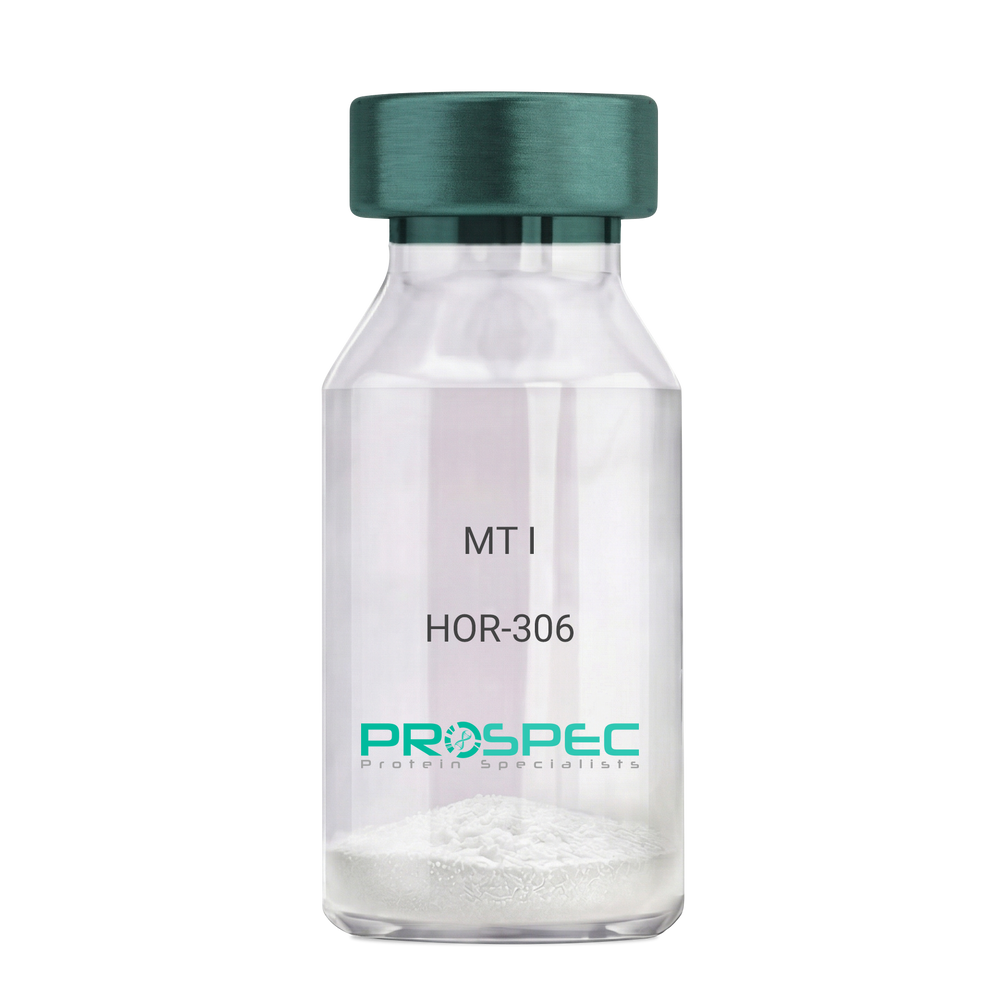
MT IDescription:
Melanotan-I
Melanotan-I, MT-I, Melanotan-1.
Product # :
HOR-306Price :
Quantity :
Shipping Method :
Shipped at Room temp
More Info
- description
- formulation
- purity
- More Info
Description
Melanotan-I has the amino acid sequence of Ser-Tyr-Ser-Nle-Glu-His-D-Phe-Arg-Trp-Gly-Lys-Pro-Val and a molecular weight of 1647.4 Dalton.
Formulation
The protein (1mg/ml) was lyophilized with no additives.
Purity
Greater than 98.0% as determined by RP-HPLC.
More Info
-
Synonyms
Melanotan-I, MT-I, Melanotan-1.
-
Physical Appearance
Sterile Filtered off-White lyophilized (freeze-dried) powder.
-
Stability
Lyophilized Melanotan-I although stable at room temperature for 3 weeks, should be stored desiccated below -18°C. Upon reconstitution MT-I should be stored at 4°C between 2-7 days and for future use below -18°C.For long term storage it is recommended to add a carrier protein (0.1% HSA or BSA).Please prevent freeze-thaw cycles.
-
Solubility
It is recommended to reconstitute the lyophilized Melanotan-I in sterile 1% acetic acid not less than 100 µg/ml, which can then be further diluted to other aqueous solutions.
ProSpec's products are furnished for LABORATORY RESEARCH USE ONLY. The product may not be used as drugs, agricultural or pesticidal products, food additives or household chemicals.
- View Data Sheet
Name :
PPM1A AntibodyDescription:
Protein Phosphatase 1A Alpha Isoform, Mouse Anti Human
Protein phosphatase 1A, EC 3.1.3.16, Protein phosphatase 2C isoform alpha, PP2C-alpha, IA, PPM1A, PP2CA, MGC9201.
Product # :
ANT-309Price :
Quantity :
Shipping Method :
Shipped with Ice Packs
More Info
- formulation
- More Info
Formulation
1mg/ml containing PBS, pH-7.4, & 0.1% Sodium Azide.
More Info
-
Introduction
Protein Phosphatase 2C alpha is a member of the PP2C family of Ser/Thr protein phosphatases. PP2C family members are known to be negative regulators of cell stress response pathways. This phosphatase dephosphorylates, and negatively regulates the activities of, MAP kinases and MAP kinase kinases. It has been shown to inhibit the activation of p38 and JNK kinase cascades induced by environmental stresses. This phosphatase can also dephosphorylate cyclin-dependent kinases, and thus may be involved in cell cycle control. Overexpression of this phosphatase is reported to activate the expression of the tumor suppressor gene TP53/p53, which leads to G2/M cell cycle arrest and apoptosis. Three alternatively spliced transcript variants encoding two distinct isoforms have been described. Protein phosphatase 2C(PP2Cα) is a Mn2+- or Mg2+-dependent protein serine/threonine phosphatase that is essential for regulating cellular stress response in eukaryotes.
-
Synonyms
Protein phosphatase 1A, EC 3.1.3.16, Protein phosphatase 2C isoform alpha, PP2C-alpha, IA, PPM1A, PP2CA, MGC9201.
-
Immunogen
Anti-human PPM1A mAb, is derived from hybridization of mouse SP2/O myeloma cells with spleen cells from BALB/c mice immunized with recombinant human PPM1A amino acids 1-382 purified from E. coli.
-
Ig Subclass
Mouse IgG2b heavy chain and κ light chain.
-
Clone
Pp6c7AT.
-
Applications
PPM1A antibody has been tested by ELISA and Western blot analysis to assure specificity and reactivity. Since application varies, however, each investigation should be titrated by the reagent to obtain optimal results. Recommended dilution range for Western blot analysis is 1:250 ~ 500. Recommended starting dilution is 1:250.
-
Type
Mouse Anti Human Monoclonal.
-
Storage Procedures
For periods up to 1 month store at 4°C, for longer periods of time, store at -20°C. Prevent freeze thaw cycles.
-
Purification Method
PPM1A antibody was purified from mouse ascitic fluids by protein-G affinity chromatography.
ProSpec's products are furnished for LABORATORY RESEARCH USE ONLY. The product may not be used as drugs, agricultural or pesticidal products, food additives or household chemicals.
- View Data Sheet
Name :
HCV NS3 Genotype-6Description:
Hepatitis C Virus NS3 Genotype-6, (1356-1459 a.a.) Recombinant
Product # :
HCV-254Price :
Quantity :
Shipping Method :
Shipped with Ice Packs
More Info
- description
- formulation
- purity
- More Info
Description
The E.coli derived recombinant protein contains the HCV NS3 immunodominant regions, amino acids 1356-1459. The protein is fused to a GST tag at N-Terminus.
Formulation
1.5M urea, 25mM Tris-HCl pH-8, 0.2% Triton-X & 50% Glycerol.
Purity
HCV NS3 Genotype-6 protein is >95% pure as determined by 10% PAGE (coomassie staining).
More Info
-
Introduction
HCV is a small 50nm, enveloped, single-stranded, positive sense RNAvirus in the family Flaviviridae.
HCV has a high rate of replication with approximately one trillion particles produced each day in an infected individual. Due to lack of proofreading by the HCV RNA polymerase, the HCV has an exceptionally high mutation rate, a factor that may help it elude the host's immune response. Hepatitis C virus is classified into six genotypes(1-6) with several subtypes within each genotype. The preponderance and distribution of HCV genotypes varies globally. Genotype is clinically important in determining potential response to interferon-based therapy and the required duration of such therapy. Genotypes 1 and 4 are less responsive to interferon-based treatment than are the other genotypes (2, 3, 5 and 6). -
Stability
HCV NS3 Genotype-6 although stable at 4°C for 1 week, should be stored below -18°C. Please prevent freeze thaw cycles.
-
Applications
HCV NS3 Genotype-6 antigen is suitable for ELISA and Western blots, excellent antigen for detection of HCV with minimal specificity problems.
-
Specificity
Immunoreactive with sera of HCV-infected individuals.
-
Purification Method
HCV NS3 Genotype-6 protein was Purified by proprietary chromatographic technique.
ProSpec's products are furnished for LABORATORY RESEARCH USE ONLY. The product may not be used as drugs, agricultural or pesticidal products, food additives or household chemicals.
- View Data Sheet
Name :
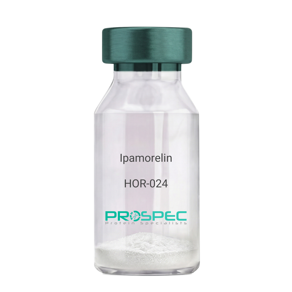
IpamorelinDescription:
Ipamorelin
Ipamorelin
Product # :
HOR-024Price :
Quantity :
Shipping Method :
Shipped at Room temp
More Info
- description
- formulation
- purity
- More Info
Description
Ipamorelin Synthetic is a single, non-glycosylated polypeptide chain containing 4 amino acids, having a molecular mass of 711.85 Dalton and a Molecular formula of C38H49N9O5.
Formulation
The protein was lyophilized with no additives.
Purity
Greater than 97.0% as determined by analysis by RP-HPLC.
More Info
-
Introduction
Ipamorelin is a peptide selective agonist of the ghrelin/growth hormone secretagogue receptor and a growth hormone secretagogue. Ipamorelin is a pentapeptide that was derived from GHRP1. Ipamorelin significantly increases plasma growth hormone levels in both animals and humans. Like pralmorelin and GHRP-6, ipamorelin does not affect prolactin, FSH, LH or TSH levels. However, unlike GHRP2 and GHRP6, but as growth hormone-releasing hormone (GHRH), ipamorelin does not stimulate the secretion of adrenocorticotropic hormone (ACTH) or cortisol, and is highly selective for inducing the secretion only of GH.
-
Synonyms
Ipamorelin
-
Physical Appearance
Sterile Filtered White lyophilized (freeze-dried) powder.
-
Stability
Lyophilized Ipamorelin although stable at room temperature for 3 weeks, should be stored desiccated below -18°C. Upon reconstitution Ipamorelin should be stored at 4°C between 2-7 days and for future use below -18°C. For long term storage it is recommended to add a carrier protein (0.1% HSA or BSA). Please prevent freeze-thaw cycles.
-
Solubility
It is recommended to reconstitute the lyophilized Ipamorelin in sterile 18MΩ-cm H2O not less than 100 µg/ml, which can then be further diluted to other aqueous solutions.
-
Amino Acid Sequence
Aib-His-D-2-Nal-D-Phe-Lys-NH2.
ProSpec's products are furnished for LABORATORY RESEARCH USE ONLY. The product may not be used as drugs, agricultural or pesticidal products, food additives or household chemicals.
- View Data Sheet
Name :
CoV2 Paired AntibodyDescription:
Mouse Anti SARS CoV-2 Paired
Product # :
ANT-770Price :
Quantity :
Shipping Method :
Shipped with Ice Packs
More Info
- description
- formulation
- purity
- More Info
Description
SARS CoV2 antibodies, coating C865 and conjugating C866 which target CoV2 nucleoprotein. SARS CoV2 antibodies, coating and conjugating are paired for the preparation of CoV2 antigen rapid test for the detection of CoV-2 nucleoprotein. SARS CoV2 antibodies, coating and conjugating do not cross-react with CoV nucleoproteins: 229E, HKU1, NL63 and OC43. CoV2 antigen rapid test prepared by SARS CoV2 antibodies, coating and conjugating can detect 5ng/ml of recombinant SARS-CoV2 nucleoprotein. Please note that when ordering for example: 100µg antibody we ship 50µg from each of the antibodies (100µg in total).
Formulation
PBS pH-7.4
Purity
Greater than 90%.
More Info
-
Introduction
SARS Coronavirus is an enveloped virus containing three outer structural proteins, namely the membrane (M), envelope (E), and spike (S) proteins. Spike (S)-glycoprotein of the virus interacts with a cellular receptor and mediates membrane fusion to allow viral entry into susceptible target cells. Accordingly, S-protein plays an important role in virus infection cycle and is the primary target of neutralizing antibodies.It has recently been shown that SARS is caused by a human coronavirus. Human coronaviruses are the major cause of upper respiratory tract illness in humans, such as the common cold. Coronaviruses are positive-stranded RNA viruses, featuring the largest viral RNA genomes known to date (27-31 kb). The first step in coronavirus infection is binding of the viral spike protein, a 139-kDa protein, to certain receptors on host cells. The spike protein is the main surface antigen of the coronavirus. The most prominent protein in the culture supernatants infected with SARS virus is a 46 kDa nucleocapsid protein. This suggests that the nucleocapsid protein is a major immunogen that may be useful for early diagnostics.
-
Physical Appearance
2 vials of sterile filtered clear colorless solution.
-
Stability
SARS CoV2 antibodies, coating and conjugating although stable at 4°C for 1 week, should be stored below -18°C. For long term storage it is recommended to add a carrier protein (0.1% HSA or BSA). Please prevent freeze-thaw cycles.
-
Applications
Lateral flow rapid test.
-
Type
Mouse antibody Monoclonal.
-
Purification Method
Purified monoclonal IgG by protein A chromatography.
ProSpec's products are furnished for LABORATORY RESEARCH USE ONLY. The product may not be used as drugs, agricultural or pesticidal products, food additives or household chemicals.
- View Data Sheet
Name :
Dengue Envelope-1 & 3Description:
Dengue Virus Subtype 1 & 3 fused Envelope 58kDa Recombinant
Product # :
DEN-001Price :
Quantity :
Shipping Method :
Shipped with Ice Packs
More Info
- description
- source
- formulation
- purity
- More Info
Description
The E.coli derived recombinant 58kDa protein is a genetically engineered peptide which is derived from Dengue Type-1 and 3 to be expressed as a fused envelope, each part in this fusion contains 170 a.a (positions 46-217), it is used in ELISA assay. This fusion protein is connected to a 6xHis Tag. Dengue Type-1 and 3 is purified by proprietary chromatographic technique.
Source
Escherichia Coli.
Formulation
The protein solution contains Phosphate buffered saline, pH-7.4 and 0.05% sodium azide.
Purity
Protein is >95% pure as determined by 12% PAGE (coomassie staining).
More Info
-
Introduction
Caused by one of four closely related virus serotypes of the genus Flavivirus, family Flaviviridae, each serotype is sufficiently different that there is no cross-protection and epidemics caused by multiple serotypes (hyperendemicity) can occur. In cell culture experiments and mice Morpholino antisense oligos have shown specific activity against Dengue virus.
-
Physical Appearance
Sterile filtered colorless solution.
-
Stability
Dengue Envelope-1 & 3 Recombinant although stable at 4°C for 1 week, should be stored below -18°C. Please prevent freeze thaw cycles.
ProSpec's products are furnished for LABORATORY RESEARCH USE ONLY. The product may not be used as drugs, agricultural or pesticidal products, food additives or household chemicals.
- View Data Sheet
Name :
Dengue Envelope-4, InsectDescription:
Dengue Virus Subtype 4 Recombinant, Insect Cells
Product # :
DEN-036Price :
Quantity :
Shipping Method :
Shipped with Ice Packs
More Info
- description
- source
- formulation
- purity
- More Info
Description
Recombinant Dengue Virus Subtype 4 produced in Insect Cells is a polypeptide chain containing amino acids 280-674 and having a molecular weight of approximately 50kDa. Dengue Envelope-4 is purified by proprietary chromatographic technique.
Source
Insect cells.
Formulation
Dengue Envelope-4 protein solution in 1xD-PBS, pH7.4, 0.1% Thimerosal, 5mM EDTA, 1µg/ml of Leupeptin, Aprotinin and Pepstatin A.
Purity
Protein is >95% pure as determined by 12.5% SDS-PAGE.
More Info
-
Introduction
Caused by one of four closely related virus serotypes of the genus Flavivirus, family Flaviviridae, each serotype is sufficiently different that there is no cross-protection and epidemics caused by multiple serotypes (hyperendemicity) can occur. In cell culture experiments and mice Morpholino antisense oligos have shown specific activity against Dengue virus.
-
Physical Appearance
Sterile filtered colorless solution.
-
Stability
Store at 4°C if entire vial will be used within 2-4 weeks. Store, frozen at -20°C for longer periods of time. For long term storage it is recommended to add a carrier protein (0.1% HSA or BSA).Avoid multiple freeze-thaw cycles.
ProSpec's products are furnished for LABORATORY RESEARCH USE ONLY. The product may not be used as drugs, agricultural or pesticidal products, food additives or household chemicals.
- View Data Sheet
Name :
CoV2 Nucleocapsid PolyclonalDescription:
Full Length CoV2 Nucleocapsid, Polyclonal Antibody
Product # :
ANT-769Price :
Quantity :
Shipping Method :
Shipped at Room temp
More Info
- formulation
- More Info
Formulation
Lyophilized from PBS.
More Info
-
Introduction
SARS Coronavirus is an enveloped virus containing three outer structural proteins, namely the membrane (M), envelope (E), and spike (S) proteins. Spike (S)-glycoprotein of the virus interacts with a cellular receptor and mediates membrane fusion to allow viral entry into susceptible target cells. Accordingly, S-protein plays an important role in virus infection cycle and is the primary target of neutralizing antibodies.
It has recently been shown that SARS is caused by a human coronavirus. Human coronaviruses are the major cause of upper respiratory tract illness in humans, such as the common cold. Coronaviruses are positive-stranded RNA viruses, featuring the largest viral RNA genomes known to date (27-31 kb). The first step in coronavirus infection is binding of the viral spike protein, a 139-kDa protein, to certain receptors on host cells. The spike protein is the main surface antigen of the coronavirus. The most prominent protein in the culture supernatants infected with SARS virus is a 46 kDa nucleocapsid protein. This suggests that the nucleocapsid protein is a major immunogen that may be useful for early diagnostics. -
Stability
Lyophilized Full Length CoV-2 Polyclonal Antibody although stable at room temperature for 3 weeks, should be stored desiccated below -18°C. Upon reconstitution CoV-2 Polyclonal Antibody should be stored at 4°C between 2-7 days and for future use below -18°C. Please prevent freeze-thaw cycles.
-
Solubility
It is recommended to reconstitute the lyophilized Full Length CoV-2 Polyclonal Antibody in sterile PBS not less than 100µg/ml, which can then be further diluted to other aqueous solutions.
-
Immunogen
The antibody was developed by immunizing rabbits with recombinant Full Length SARS COV-2 Nucleocapsid Protein.
-
Isotype
Rabbit IgG.
-
Type
Polyclonal Rabbit Antibody.
-
Purification Method
Protein-A S-Sepharose.
ProSpec's products are furnished for LABORATORY RESEARCH USE ONLY. The product may not be used as drugs, agricultural or pesticidal products, food additives or household chemicals.
- View Data Sheet
Name :
Dengue-2 C-terminalDescription:
Dengue-2 Envelope Hydrophobic Domain Recombinant
Product # :
DEN-039Price :
Quantity :
Shipping Method :
Shipped with Ice Packs
More Info
- description
- source
- formulation
- purity
- More Info
Description
The Recombinant Dengue 2 Envelope Hydrophobic Domain e.coli derived contains 225 amino acids (228-485) from dengue 2 envelope. The protein is fused to a His tag at C-terminal and purified by standard chromatography techniques.
Source
E.coli.
Formulation
PBS.
Purity
Protein is >95% pure as determined by 10% PAGE (coomassie staining).
More Info
-
Introduction
Caused by one of four closely related virus serotypes of the genus Flavivirus, family Flaviviridae, each serotype is sufficiently different that there is no cross-protection and epidemics caused by multiple serotypes (hyperendemicity) can occur. In cell culture experiments and mice Morpholino antisense oligos have shown specific activity against Dengue virus.
-
Physical Appearance
Sterile Filtered clear solution.
-
Stability
Dengue 2 Envelope Recombinant although stable at 4°C for 1 week, should be stored below -18°C. Please prevent freeze thaw cycles.
-
Applications
Immunoassay.
ProSpec's products are furnished for LABORATORY RESEARCH USE ONLY. The product may not be used as drugs, agricultural or pesticidal products, food additives or household chemicals.