Skip to content

High-quality research proteins.

  • 1-800-805-6531

  • Mon - Sat 8.00-18.00, Sun - Closed

  • info@prospecbio.com

  • PO Box 6591 East Brunswick NJ 08816

  • Products
    • Cytokines
    • Growth factors
    • Chemokines
    • Cd antigens
    • Neurotrophins
    • Hormones
    • Enzymes
    • Viral antigens
    • Recombinant proteins
    • Natural proteins
    • Monoclonal antibodies
    • Polyclonal antibodies
    • Thermal Packaging
  • About
    • Company
    • Founder
    • Contribution
  • Customers
    • Eastern Asia
    • Eastern Europe
    • North America
    • South America
    • South East Asia
    • Southern Asia
    • Western Europe
  • Ordering
    • Ordering Method
    • Ordering - Credit Card
    • Ordering - PO
    • Shipping Fees
    • FAQs
  • Services
    • DNA Cloning
    • Protein Expression
    • Monoclonal Antibody
    • Fermentation
    • Technology License
  • Contact us
Log in

Country/region

  • USD $ | Albania
  • USD $ | Andorra
  • USD $ | Argentina
  • USD $ | Armenia
  • USD $ | Australia
  • USD $ | Austria
  • USD $ | Azerbaijan
  • USD $ | Belarus
  • USD $ | Belgium
  • USD $ | Bolivia
  • USD $ | Brazil
  • USD $ | Bulgaria
  • USD $ | Cambodia
  • USD $ | Canada
  • USD $ | Chile
  • USD $ | China
  • USD $ | Colombia
  • USD $ | Costa Rica
  • USD $ | Croatia
  • USD $ | Cyprus
  • USD $ | Czechia
  • USD $ | Denmark
  • USD $ | Estonia
  • USD $ | Finland
  • USD $ | France
  • USD $ | Georgia
  • USD $ | Germany
  • USD $ | Greece
  • USD $ | Hong Kong SAR
  • USD $ | Hungary
  • USD $ | Iceland
  • USD $ | India
  • USD $ | Ireland
  • USD $ | Israel
  • USD $ | Italy
  • USD $ | Japan
  • USD $ | Kazakhstan
  • USD $ | Latvia
  • USD $ | Liechtenstein
  • USD $ | Lithuania
  • USD $ | Madagascar
  • USD $ | Malta
  • USD $ | Mexico
  • USD $ | Moldova
  • USD $ | Monaco
  • USD $ | Netherlands
  • USD $ | New Zealand
  • USD $ | North Macedonia
  • USD $ | Norway
  • USD $ | Panama
  • USD $ | Paraguay
  • USD $ | Peru
  • USD $ | Philippines
  • USD $ | Poland
  • USD $ | Portugal
  • USD $ | Romania
  • USD $ | Singapore
  • USD $ | Slovakia
  • USD $ | Slovenia
  • USD $ | South Africa
  • USD $ | South Korea
  • USD $ | Spain
  • USD $ | Sri Lanka
  • USD $ | Sweden
  • USD $ | Switzerland
  • USD $ | Taiwan
  • USD $ | Thailand
  • USD $ | Türkiye
  • USD $ | Ukraine
  • USD $ | United Kingdom
  • USD $ | United States
  • USD $ | Uruguay
  • USD $ | Vatican City
  • USD $ | Venezuela
  • USD $ | Vietnam
  • Facebook
  • Instagram
logoProSpec - Recombinant Protein Manufacturer for academic and R&D industry
Become a distributor
Account Log in
Wishlist
Cart Cart(0)
  • Products
    +
    • Cytokines
    • Growth factors
    • Chemokines
    • Cd antigens
    • Neurotrophins
    • Hormones
    • Enzymes
    • Viral antigens
    • Recombinant proteins
    • Natural proteins
    • Monoclonal antibodies
    • Polyclonal antibodies
    • Thermal Packaging
  • About
    +
    • Company
    • Founder
    • Contribution
  • Customers
    +
    • Eastern Asia
    • Eastern Europe
    • North America
    • South America
    • South East Asia
    • Southern Asia
    • Western Europe
  • Ordering
    +
    • Ordering Method
    • Ordering - Credit Card
    • Ordering - PO
    • Shipping Fees
    • FAQs
  • Services
    +
    • DNA Cloning
    • Protein Expression
    • Monoclonal Antibody
    • Fermentation
    • Technology License
  • Contact us

Item added to your cart

View cart
View All
  • Cytokines
  • Growth factors
  • Chemokines
  • Cd antigens
  • Neurotrophins
  • Hormones
  • Enzymes
  • Viral antigens
  • Recombinant proteins
  • Natural proteins
  • Monoclonal antibodies
  • Polyclonal antibodies
  • Thermal Packaging
  • Tumor Necrosis Factor

    Tumor Necrosis Factor

    View All
  • 4-1BB

    4-1BB

    View All
  • Adiponectin

    Adiponectin

    View All
  • AIF1

    AIF1

    View All
  • Other Cytokines

    Other Cytokines

    View All
  • AITRL

    AITRL

    View All
  • Angiopoietin

    Angiopoietin

    View All
  • Apolipoprotein

    Apolipoprotein

    View All
  • B-Cell Activating Factor

    B-Cell Activating Factor

    View All
  • Beta Defensin

    Beta Defensin

    View All
  • Bone Morphogenetic Protein

    Bone Morphogenetic Protein

    View All
  • B type Natriuretic Peptide

    B type Natriuretic Peptide

    View All
  • BST

    BST

    View All
  • Betacellulin

    Betacellulin

    View All
  • Cardiotrophin

    Cardiotrophin

    View All
  • Activin

    Activin

    View All
  • Fibroblast Growth Factor

    Fibroblast Growth Factor

    View All
  • Other Growth Factors

    Other Growth Factors

    View All
  • CSF

    CSF

    View All
  • CTGF

    CTGF

    View All
  • IGFBP

    IGFBP

    View All
  • VEGF Protein

    VEGF Protein

    View All
  • EGF

    EGF

    View All
  • Epigen

    Epigen

    View All
  • Erythropoietin

    Erythropoietin

    View All
  • Insulin-Like Growth Factor

    Insulin-Like Growth Factor

    View All
  • Growth Hormone

    Growth Hormone

    View All
  • HDGF

    HDGF

    View All
  • Hepatocyte Growth Factor

    Hepatocyte Growth Factor

    View All
  • Insulin

    Insulin

    View All
  • BCA-1/ BLC (CXCL13)

    BCA-1/ BLC (CXCL13)

    View All
  • BRAK (CXCL14)

    BRAK (CXCL14)

    View All
  • C-10 (CCL6)

    C-10 (CCL6)

    View All
  • Other Chemokines

    Other Chemokines

    View All
  • MEC (CCL28)

    MEC (CCL28)

    View All
  • LD78-beta (CCL3L1)

    LD78-beta (CCL3L1)

    View All
  • CTACK (CCL27)

    CTACK (CCL27)

    View All
  • CXCL16

    CXCL16

    View All
  • CXCL17

    CXCL17

    View All
  • Platelet Factor-4 (CXCL4)

    Platelet Factor-4 (CXCL4)

    View All
  • ENA-78 (CXCL5)

    ENA-78 (CXCL5)

    View All
  • Eotaxin (CCL11,24,26)

    Eotaxin (CCL11,24,26)

    View All
  • Exodus-2 (CCL21)

    Exodus-2 (CCL21)

    View All
  • Fractalkine (CX3CL1)

    Fractalkine (CX3CL1)

    View All
  • CXCL6

    CXCL6

    View All
  • Other CD Antigens

    Other CD Antigens

    View All
  • CD14

    CD14

    View All
  • CD164

    CD164

    View All
  • CD1

    CD1

    View All
  • CD2

    CD2

    View All
  • CD200

    CD200

    View All
  • CD207

    CD207

    View All
  • CD226

    CD226

    View All
  • CD23

    CD23

    View All
  • CD244

    CD244

    View All
  • CD247

    CD247

    View All
  • CD27

    CD27

    View All
  • CD274

    CD274

    View All
  • CD300

    CD300

    View All
  • CD33

    CD33

    View All
  • Other Neurotrophins

    Other Neurotrophins

    View All
  • Beta-NGF

    Beta-NGF

    View All
  • BDNF

    BDNF

    View All
  • CDNF

    CDNF

    View All
  • CNTF

    CNTF

    View All
  • GDNF

    GDNF

    View All
  • Glia Maturation Factor

    Glia Maturation Factor

    View All
  • MANF

    MANF

    View All
  • Midkine

    Midkine

    View All
  • Neuroglobin

    Neuroglobin

    View All
  • Neurotrophic factors

    Neurotrophic factors

    View All
  • Neuregulin

    Neuregulin

    View All
  • Neuropilin

    Neuropilin

    View All
  • Pigment Epithelium-Derived Factor

    Pigment Epithelium-Derived Factor

    View All
  • Pleiotrophin

    Pleiotrophin

    View All
  • Peptide Hormones

    Peptide Hormones

    View All
  • Other Hormones

    Other Hormones

    View All
  • HCG

    HCG

    View All
  • Endothelin

    Endothelin

    View All
  • Exendin

    Exendin

    View All
  • FSH

    FSH

    View All
  • Glucagon

    Glucagon

    View All
  • GHRP

    GHRP

    View All
  • GLP

    GLP

    View All
  • Thyrostimulin

    Thyrostimulin

    View All
  • Inhibin A

    Inhibin A

    View All
  • LHRH

    LHRH

    View All
  • Melanotan

    Melanotan

    View All
  • Procalcitonin

    Procalcitonin

    View All
  • PTH

    PTH

    View All
  • Peroxiredoxin

    Peroxiredoxin

    View All
  • Peptidase

    Peptidase

    View All
  • Synthetase

    Synthetase

    View All
  • Transferase

    Transferase

    View All
  • Hydrolase

    Hydrolase

    View All
  • Dehydrogenase

    Dehydrogenase

    View All
  • ACE2

    ACE2

    View All
  • Esterase

    Esterase

    View All
  • Protein Kinases

    Protein Kinases

    View All
  • Other Enzymes

    Other Enzymes

    View All
  • Phosphatase

    Phosphatase

    View All
  • Deaminase

    Deaminase

    View All
  • Oxygenase

    Oxygenase

    View All
  • Adenylate Kinase

    Adenylate Kinase

    View All
  • Lyase

    Lyase

    View All
  • Chagas

    Chagas

    View All
  • SARS

    SARS

    View All
  • Influenza

    Influenza

    View All
  • ASFV

    ASFV

    View All
  • Astrovirus

    Astrovirus

    View All
  • Borrelia

    Borrelia

    View All
  • Helicobacter Pylori

    Helicobacter Pylori

    View All
  • Chikungunya

    Chikungunya

    View All
  • Chlamydia

    Chlamydia

    View All
  • Cytomegalo

    Cytomegalo

    View All
  • Dengue

    Dengue

    View All
  • EBV

    EBV

    View All
  • Ebola

    Ebola

    View All
  • Feline Leukemia Virus

    Feline Leukemia Virus

    View All
  • Hepatitis A

    Hepatitis A

    View All
  • Other

    Other

    View All
  • Actin

    Actin

    View All
  • ADAM

    ADAM

    View All
  • Complement Component

    Complement Component

    View All
  • Ag85

    Ag85

    View All
  • Eukaryotic Translation Initiation Factor

    Eukaryotic Translation Initiation Factor

    View All
  • Anterior Gradient Protein

    Anterior Gradient Protein

    View All
  • Heat Shock Protein

    Heat Shock Protein

    View All
  • Alpha-2-HS-Glycoprotein

    Alpha-2-HS-Glycoprotein

    View All
  • Hemoglobin

    Hemoglobin

    View All
  • Allergy

    Allergy

    View All
  • Anaplasma

    Anaplasma

    View All
  • Angiogenin

    Angiogenin

    View All
  • Ankyrin Repeat Domain

    Ankyrin Repeat Domain

    View All
  • Annexin

    Annexin

    View All
  • Other Natural Proteins

    Other Natural Proteins

    View All
  • Aprotinin

    Aprotinin

    View All
  • Transferrin

    Transferrin

    View All
  • Avidin

    Avidin

    View All
  • Anti Coagulation Factors

    Anti Coagulation Factors

    View All
  • Natural Albumin

    Natural Albumin

    View All
  • Natural Coagulation Factors

    Natural Coagulation Factors

    View All
  • Fibronectin

    Fibronectin

    View All
  • Thrombin

    Thrombin

    View All
  • Anti Human Enzyme

    Anti Human Enzyme

    View All
  • Other Monoclonal Antibodies

    Other Monoclonal Antibodies

    View All
  • Anti Human Cytokine

    Anti Human Cytokine

    View All
  • Anti Human Heat Shock Protein

    Anti Human Heat Shock Protein

    View All
  • Anti Mouse Lymphocyte

    Anti Mouse Lymphocyte

    View All
  • Anti Human Chemokine

    Anti Human Chemokine

    View All
  • Anti Human Lymphocyte

    Anti Human Lymphocyte

    View All
  • Anti Viral Monoclonal

    Anti Viral Monoclonal

    View All
  • Anti Mouse Cytokine

    Anti Mouse Cytokine

    View All
  • Anti-GST Monoclonal

    Anti-GST Monoclonal

    View All
  • Other Polyclonal Antibodies

    Other Polyclonal Antibodies

    View All
  • Anti Viral

    Anti Viral

    View All
  • Anti-GST Monoclonal

    Anti-GST Monoclonal

    View All
Back

Search results

460 results found for “mesoderm development candidate”

Name

Description

Product #

Price

Quantity

Shipping Method

  • View Data Sheet

    Name :

    RNH1 Antibody

    Description:

    Ribonuclease inhibitor, Mouse Anti Human

    Ribonuclease inhibitor, Ribonuclease/angiogenin inhibitor 1, Placental ribonuclease inhibitor, Placental RNase inhibitor, RAI, RNH, MGC4569, MGC18200, MGC54054, RNH1, PRI.

    Product # :

    ANT-445

    Price :

    Quantity :

    Shipping Method :

    Ice Icon

    Shipped with Ice Packs

    Add To Cart

    More Info

    • formulation
    • More Info

    Formulation

    1mg/ml containing PBS, pH-7.4, 0.02% Sodium Azide & 10% glycerol.

    More Info

    • Introduction

      RNH1 belongs to the proteinaceous cytoplasmic RNase inhibitors family which occur in many tissues and binds to both intracellular and extracellular RNases. In addition to control of intracellular RNases, the inhibitor may have a part in the regulation of angiogenin. The 50kDa RNH1 binds to ribonucleases and holds them in a latent form. Since neutral and alkaline ribonucleases possibly play a critical role in the turnover of RNA in eukaryotic cells, RNH1 may be vital for control of mRNA turnover; the interaction of eukaryotic cells with ribonuclease may be reversible in vivo.

    • Synonyms

      Ribonuclease inhibitor, Ribonuclease/angiogenin inhibitor 1, Placental ribonuclease inhibitor, Placental RNase inhibitor, RAI, RNH, MGC4569, MGC18200, MGC54054, RNH1, PRI.

    • Immunogen

      Anti-human RNH1 mAb, is derived from hybridization of mouse F0 myeloma cells with spleen cells from BALB/c mice immunized with recombinant human RNH1 amino acids 7-461 purified from E. coli.

    • Ig Subclass

      Mouse IgG2a heavy chain and κ light chain.

    • Clone

      PAT1H23AT.

    • Applications

      RNH1 antibody has been tested by ELISA and Western blot analysis to assure specificity and reactivity. Since application varies, however, each investigation should be titrated by the reagent to obtain optimal results.

    • Type

      Mouse Anti Human Monoclonal.

    • Storage Procedures

      For periods up to 1 month store at 4°C, for longer periods of time, store at -20°C. Prevent freeze thaw cycles.

    • Purification Method

      RNH1 antibody was purified from mouse ascitic fluids by protein-G affinity chromatography.

    ProSpec's products are furnished for LABORATORY RESEARCH USE ONLY. The product may not be used as drugs, agricultural or pesticidal products, food additives or household chemicals.

    Rnh1 Antibody
  • View Data Sheet

    Name :

    MMP14 Human, His

    Description:

    Matrix Metalloproteinase-14 Human Recombinant, His Tag

    Matrix Metallopeptidase 14, Matrix Metallopeptidase 14 (Membrane-Inserted), Membrane-Type-1 Matrix Metalloproteinase, Membrane Type 1 Metalloprotease, EC 3.4.24.80, MT-MMP 1, MT1-MMP, MMP-14, MMP-X1, MT1MMP, MTMMP1, Matrix Metalloproteinase 14 (Membrane-Inserted), Membrane-Type Matrix Metalloproteinase 1, Matrix Metalloproteinase-14, EC 3.4.24, MT-MMP, WNCHRS, Matrix metalloproteinase-14, Membrane-type matrix metalloproteinase 1.

    Product # :

    ENZ-1003

    Price :

    Quantity :

    Shipping Method :

    Ice Icon

    Shipped with Ice Packs

    Add To Cart

    More Info

    • description
    • source
    • formulation
    • purity
    • More Info

    Description

    MMP14 Human Recombinant produced in Sf9 Baculovirus cells is a single, non-glycosylated polypeptide chain containing 527 amino acids (21-538a.a) and having a molecular mass of 59.9kDa (Molecular size on SDS-PAGE will appear at approximately 35-70kDa). MMP14 is fused to a 6 amino acid His-tag at C-terminus & purified by proprietary chromatographic techniques.

    Source

    Sf9, Baculovirus cells.

    Formulation

    MMP14 protein solution (0.25mg/ml) containing Phosphate Buffered Saline (pH 7.4) and 10% glycerol.

    Purity

    Greater than 90% as determined by SDS-PAGE.

    More Info

    • Introduction

      Matrix metalloproteinase-14 (MMP14), is a membrane-anchored zinc-binding endopeptidase which is expressed at the leading edge of different invasive carcinomas and also promotes tumor cell invasion through degradation of the extracellular matrix. MMP14 takes a vital part in extracellular matrix, ECM, remodeling by having the capability to degrade type I collagen, activate pro-MMP-2 and process cell adhesion molecules for instance CD44 and integrin alpha V. MMP14 is a key enzyme in many physiological as well as pathological processes for example angiogenesis & tumor invasion.

    • Synonyms

      Matrix Metallopeptidase 14, Matrix Metallopeptidase 14 (Membrane-Inserted), Membrane-Type-1 Matrix Metalloproteinase, Membrane Type 1 Metalloprotease, EC 3.4.24.80, MT-MMP 1, MT1-MMP, MMP-14, MMP-X1, MT1MMP, MTMMP1, Matrix Metalloproteinase 14 (Membrane-Inserted), Membrane-Type Matrix Metalloproteinase 1, Matrix Metalloproteinase-14, EC 3.4.24, MT-MMP, WNCHRS, Matrix metalloproteinase-14, Membrane-type matrix metalloproteinase 1.

    • Physical Appearance

      Sterile Filtered colorless solution.

    • Stability

      Store at 4°C if entire vial will be used within 2-4 weeks. Store, frozen at -20°C for longer periods of time. For long term storage it is recommended to add a carrier protein (0.1% HSA or BSA). Avoid multiple freeze-thaw cycles.

    • Amino Acid Sequence

      ADLALASLGS AQSSSFSPEA WLQQYGYLPP GDLRTHTQRS PQSLSAAIAA MQKFYGLQVT GKADADTMKA MRRPRCGVPD KFGAEIKANV RRKRYAIQGL KWQHNEITFC IQNYTPKVGE YATYEAIRKA FRVWESATPL RFREVPYAYI REGHEKQADI MIFFAEGFHG DSTPFDGEGG FLAHAYFPGP NIGGDTHFDS AEPWTVRNED LNGNDIFLVA VHELGHALGL EHSSDPSAIM APFYQWMDTE NFVLPDDDRR GIQQLYGGES GFPTKMPPQP RTTSRPSVPD KPKNPTYGPN ICDGNFDTVA MLRGEMFVFK ERWFWRVRNN QVMDGYPMPI GQFWRGLPAS INTAYERKDG KFVFFKGDKH WVFDEASLEP GYPKHIKELG RGLPTDKIDA ALFWMPNGKT YFFRGNKYYR FNEELRAVDS EYPKNIKVWE GIPESPRGSF MGSDEVFTYF YKGNKYWKFN NQKLKVEPGY PKSALRDWMG CPSGGRPDEG TEEETEVIII EVDEEGGGAV SHHHHHH.

    ProSpec's products are furnished for LABORATORY RESEARCH USE ONLY. The product may not be used as drugs, agricultural or pesticidal products, food additives or household chemicals.

    Mmp14 Human
  • View Data Sheet

    Name :

    LY6D Human

    Description:

    Lymphocyte Antigen 6 Complex, Locus D Human Recombinant

    Lymphocyte Antigen 6 Complex Locus D, E48 Antigen, Ly-6D, Lymphocyte Antigen 6D.

    Product # :

    PRO-1838

    Price :

    Quantity :

    Shipping Method :

    Ice Icon

    Shipped with Ice Packs

    Add To Cart

    More Info

    • description
    • source
    • formulation
    • purity
    • More Info

    Description

    LY6D Human Recombinant produced in E.coli is a single, non-glycosylated polypeptide chain containing 101 amino acids (21-98) and having a molecular mass of 10.8 kDa. LY6D is fused to a 23 amino acid His-tag at N-terminus.

    Source

    Escherichia Coli.

    Formulation

    The LY6D solution (0.5mg/ml) contains 20mM Tris-HCl buffer (pH 8.0), 0.15M NaCl and 50% glycerol.

    Purity

    Greater than 90% as determined by SDS-PAGE.

    More Info

    • Introduction

      LY6D encloses 1 UPAR/Ly6 domain and is only expressed at the keratinocyte of stratified squamous epithelia and the outer cell surface of transitional epithelia. LY6D performs as a specification marker at earliest stage specification of lymphocytes between B- and T-cell developments.

    • Synonyms

      Lymphocyte Antigen 6 Complex Locus D, E48 Antigen, Ly-6D, Lymphocyte Antigen 6D.

    • Physical Appearance

      Sterile Filtered clear solution.

    • Stability

      Store at 4°C if entire vial will be used within 2-4 weeks. Store, frozen at -20°C for longer periods of time. For long term storage it is recommended to add a carrier protein (0.1% HSA or BSA).Avoid multiple freeze-thaw cycles.

    • Amino Acid Sequence

      MGSSHHHHHH SSGLVPRGSH MGSLRCHVCT SSSNCKHSVV CPASSRFCKT TNTVEPLRGN LVKKDCAESC TPSYTLQGQV SSGTSSTQCC QEDLCNEKLH N

    ProSpec's products are furnished for LABORATORY RESEARCH USE ONLY. The product may not be used as drugs, agricultural or pesticidal products, food additives or household chemicals.

    Ly6D Human
  • View Data Sheet

    Name :

    ANGPTL2 Human

    Description:

    Angiopoietin-like Protein 2 Human Recombinant

    Angiopoietin-related protein 2, Angiopoietin-like protein 2, ANGPTL2, ARP2, HARP.

    Product # :

    CYT-765

    Price :

    Quantity :

    Shipping Method :

    Ice Icon

    Shipped with Ice Packs

    Add To Cart

    More Info

    • description
    • source
    • formulation
    • purity
    • More Info
    • sds-page

    Description

    ANGPTL2 Human Recombinant produced in E.Coli is a single, non-glycosylated, polypeptide chain containing 493 amino acids ( 22-493 a.a.) including a 20 a.a N-terminal His tag. The total molecular mass is 57.1kDa (calculated).

    Source

    Escherichia Coli.

    Formulation

    ANGPTL2 protein solution (0.5mg/ml) contains 20mM Tris HCL (pH7-8) and 10% glycerol.

    Purity

    Greater than 85.0% as determined by SDS-PAGE.

    sds-page

    ANGPTL2-sds-page - Product image 1

    More Info

    • Introduction

      Angiopoietins belong to the vascular endothelial growth factor family and the only known growth factors largely specific for vascular endothelium. Angiopoietins-1, 2 and 4 partake in the formation of blood vessels. ANGPTL2 displays angiogenic effects. Angiopoietin-like Protein 2 (ANGPTL2) is an anti-diabetic factor. ANGPTL2 induces sprouting in endothelial cells through an autocrine and paracrine action. ANGPTL2 increases insulin sensitivity in adipocytes. In addition, ANGPTL2 is a mediator of chronic adipose tissue inflammation. ANGPTL2 is widely expressed in the heart, small intestine, spleen and stomach. ANGPTL2 is also found in lower levels in the colon, ovary, adrenal gland, skeletal muscle and in prostate.

    • Synonyms

      Angiopoietin-related protein 2, Angiopoietin-like protein 2, ANGPTL2, ARP2, HARP.

    • Physical Appearance

      Sterile filtered colorless solution.

    • Stability

      Store at 4°C if entire vial will be used within 2-4 weeks.Store, frozen at -20°C for longer periods of time.For long term storage it is recommended to add a carrier protein (0.1% HSA or BSA).Avoid multiple freeze-thaw cycles.

    • Amino Acid Sequence

      MGSSHHHHHH SSGLVPRGSH MGQEDGFEGT EEGSPREFIY LNRYKRAGES QDKCTYTFIV PQQRVTGAIC VNSKEPEVLL ENRVHKQELE LLNNELLKQK RQIETLQQLV EVDGGIVSEV KLLRKESRNM NSRVTQLYMQ LLHEIIRKRD NALELSQLEN RILNQTADML QLASKYKDLE HKYQHLATLA HNQSEIIAQL EEHCQRVPSA RPVPQPPPAA PPRVYQPPTY NRIINQISTN EIQSDQNLKV LPPPLPTMPT LTSLPSSTDK PSGPWRDCLQ ALEDGHDTSS IYLVKPENTN RLMQVWCDQR HDPGGWTVIQ RRLDGSVNFF RNWETYKQGF GNIDGEYWLG LENIYWLTNQ GNYKLLVTME DWSGRKVFAE YASFRLEPES EYYKLRLGRY HGNAGDSFTW HNGKQFTTLD RDHDVYTGNC AHYQKGGWWY NACAHSNLNG VWYRGGHYRS RYQDGVYWAE FRGGSYSLKK VVMMIRPNPN TFH

    • Background

      Angiopoietin-like Protein 2 Human Recombinant: A Potential Therapeutic Target for Metabolic and Cardiovascular Disorders

      Abstract:

      Angiopoietin-like protein 2 (ANGPTL2) has emerged as a crucial regulator in metabolic and cardiovascular disorders. This multifunctional protein is involved in various biological processes, including angiogenesis, adipose tissue function, and inflammation. The availability of human recombinant ANGPTL2 protein has provided researchers with a valuable tool to explore its therapeutic potential. This concise review provides an overview of the role of ANGPTL2 in metabolic and cardiovascular health and discusses the potential of ANGPTL2 human recombinant protein as a therapeutic target.

      Introduction:

      Metabolic disorders, such as obesity and type 2 diabetes, are closely associated with cardiovascular diseases and pose significant global health challenges. ANGPTL2, a member of the angiopoietin-like protein family, has gained attention for its involvement in metabolic regulation and cardiovascular homeostasis. Through interactions with various receptors and signaling pathways, ANGPTL2 influences lipid metabolism, insulin sensitivity, inflammation, and vascular integrity.

      Mechanisms of ANGPTL2 Action:

      ANGPTL2 acts through binding to integrins, toll-like receptors (TLRs), and other receptors on different cell types, including adipocytes, endothelial cells, and immune cells. By influencing angiogenesis, inflammation, and extracellular matrix remodeling, ANGPTL2 affects adipose tissue function, lipid metabolism, and insulin signaling.

      Role of ANGPTL2 in Metabolic Regulation:

      ANGPTL2 plays a critical role in metabolic regulation and the development of metabolic disorders. It promotes adipose tissue inflammation, impairs adipogenesis, and alters adipokine secretion, contributing to metabolic dysfunction. Furthermore, ANGPTL2 modulates lipid metabolism by regulating lipoprotein lipase activity, affecting triglyceride clearance, and promoting hepatic lipid accumulation.

      ANGPTL2 in Cardiovascular Health and Disease:

      Increasing evidence suggests that ANGPTL2 is implicated in cardiovascular diseases, including atherosclerosis and heart failure. ANGPTL2 promotes vascular inflammation, endothelial dysfunction, and smooth muscle cell proliferation, which contribute to atherosclerotic plaque progression and vascular remodeling. Additionally, ANGPTL2 influences cardiac remodeling and fibrosis, impacting heart failure development.

      Therapeutic Potential of ANGPTL2 Human Recombinant Protein:

      The availability of ANGPTL2 human recombinant protein opens avenues for therapeutic interventions targeting metabolic and cardiovascular disorders. Preclinical studies employing ANGPTL2 blockade or supplementation have shown promising results in improving metabolic parameters, reducing atherosclerosis, and preserving cardiac function. However, further research is needed to optimize the clinical application of ANGPTL2 human recombinant protein, including dosage, timing, and delivery methods.

      Conclusion:

      ANGPTL2 holds promise as a therapeutic target for metabolic and cardiovascular disorders. Its involvement in key biological processes makes it an attractive candidate for interventions aiming to improve metabolic health and prevent cardiovascular complications. The development of ANGPTL2 human recombinant protein provides a valuable tool for investigating its therapeutic potential further.

      What is the molecular weight/Mw of ANGPTL2 Protein?
      ANGPTL2 Protein has a total Mw of 57.1kDa.

      What is the source or expression system of ANGPTL2 Protein?
      Escherichia Coli.

      What is the Purity of ANGPTL2 Protein?
      ANGPTL2 Protein is >85% pure as determined by SDS-PAGE.

      What is the Biological Activity of ANGPTL2 Protein?
      The biological functionality of ANGPTL2 Protein will be determined in the future.

      What is the amino acid sequence of ANGPTL2 Protein?
      MGSSHHHHHH SSGLVPRGSH MGQEDGFEGT EEGSPREFIY LNRYKRAGES QDKCTYTFIV PQQRVTGAIC VNSKEPEVLL ENRVHKQELE LLNNELLKQK RQIETLQQLV EVDGGIVSEV KLLRKESRNM NSRVTQLYMQ LLHEIIRKRD NALELSQLEN RILNQTADML QLASKYKDLE HKYQHLATLA HNQSEIIAQL EEHCQRVPSA RPVPQPPPAA PPRVYQPPTY NRIINQISTN EIQSDQNLKV LPPPLPTMPT LTSLPSSTDK PSGPWRDCLQ ALEDGHDTSS IYLVKPENTN RLMQVWCDQR HDPGGWTVIQ RRLDGSVNFF RNWETYKQGF GNIDGEYWLG LENIYWLTNQ GNYKLLVTME DWSGRKVFAE YASFRLEPES EYYKLRLGRY HGNAGDSFTW HNGKQFTTLD RDHDVYTGNC AHYQKGGWWY NACAHSNLNG VWYRGGHYRS RYQDGVYWAE FRGGSYSLKK VVMMIRPNPN TFH

      What applications can ANGPTL2 Protein be used in?
      ANGPTL2 Protein can probably be used in western blot, ELISA and Lateral Flow.

      What is the endotoxin level for ANGPTL2 Protein?
      The endotoxin level is minimal, ANGPTL2 Protein was purified using conventional chromatography techniques.

    ProSpec's products are furnished for LABORATORY RESEARCH USE ONLY. The product may not be used as drugs, agricultural or pesticidal products, food additives or household chemicals.

    Angptl2 Human
  • View Data Sheet

    Name :

    ANGPTL4 Human

    Description:

    Angiopoietin-like Protein 4 Human Recombinant

    ANGPTL4, NL2, ARP4, FIAF, PGAR, HFARP, pp1158, ANGPTL2, Fasting- Induced Adipose Factor, Hepatic Fibrinogen/Angiopoietin-Related Protein, PPARG Angiopoietin-Related Protein.

    Product # :

    CYT-249

    Price :

    Quantity :

    Shipping Method :

    Room Temp Icon

    Shipped at Room temp

    Add To Cart

    More Info

    • description
    • source
    • formulation
    • purity
    • More Info

    Description

    The ANGPTL4 Human Recombinant is manufactured with N-terminal fusion of His Tag. The Angiopoietin-like Protein 4 His -Tagged Fusion Protein is 25 kDa protein containing 204 amino acid residues of the Angiopoietin-like Protein 4 and 16 additional amino acid residues - His Tag (underlined).

    Source

    Escherichia Coli.

    Formulation

    Filtered and lyophilized from 0.5 mg/ml in 0.05M Acetate buffer pH-4.

    Purity

    Greater than 95% as determined by SDS-PAGE.

    More Info

    • Introduction

      FIAF (fasting-induced adipose factor) a.k.a ANGPTL4 or PGAR or HFARP is an adipocytokine up-regulated by fasting, by peroxisome proliferator-activated receptor agonists, and by hypoxia. ANGPTL4 is found in human and mouse blood plasma both as a native protein and in a truncated form. In human white adipose tissue and SGBS adipocytes, only the native form of ANGPTL4 could be detected, whereas in mice the differentiation of mouse 3T3-L1 adipocytes is associated with the production of truncated ANGPTL4. However, the truncated ANGPTL4 is produced by human liver. In human blood plasma FIAF is mainly presented in a truncated form (FIAF-S2), whose levels treatment increases (as shown by experimental data). There is an inter individual variation in ANGPTL4 levels of both the truncated and the native form, however those levels were not influenced by prolonged semistarvation and are not associated with body mass index.

    • Synonyms

      ANGPTL4, NL2, ARP4, FIAF, PGAR, HFARP, pp1158, ANGPTL2, Fasting- Induced Adipose Factor, Hepatic Fibrinogen/Angiopoietin-Related Protein, PPARG Angiopoietin-Related Protein.

    • Physical Appearance

      Filtered White lyophilized (freeze-dried) powder.

    • Stability

      Store lyophilized Angiopoietin-like Protein 4 Human recombinant at -20°C. Aliquot the ANGPTL4 after reconstitution to avoid repeated freezing/thawing cycles. Reconstituted ANGPTL4 can be stored at 4°C for a limited period of time; it does not show any change after two weeks at 4°C.

    • Solubility

      Add 0.1M Acetate buffer pH4 and let the lyophilized pellet dissolve completely. For conversion into higher pH value, we recommend intensive dilution by relevant buffer to a concentration of 10μg/ml. In higher concentrations the solubility of this antigen is limited.

    • Amino Acid Sequence

      MRGSHHHHHH GMASHMGPVQ SKSPRFASWD EMNVLAHGLL QLGQGLREHA ERTRSQLSAL ERRLSACGSA CQGTEGSTDL PLAPESRVDP EVLHSLQTQL KAQNSRIQQL FHKVAQQQRH LEKQHLRIQH LQSQFGLLDH KHLDHEVAKP ARRKRLPEMA QPVDPAHNVS RLHRLPRDCQ ELFQVGERQS GLFEIQPQGS PPFLVNCKMT SDGGWTVIQR.

    • Background

      Angiopoietin-like Protein 4 Human Recombinant: A Promising Therapeutic Approach for Metabolic Disorders

      Abstract:


      Angiopoietin-like protein 4 (ANGPTL4) is a multifunctional protein involved in the regulation of lipid metabolism and energy homeostasis. It has emerged as a potential therapeutic target for metabolic disorders, including obesity, dyslipidemia, and insulin resistance. The availability of human recombinant ANGPTL4 protein has provided researchers with a valuable tool to investigate its biological functions and explore its therapeutic potential. This concise review provides an overview of the role of ANGPTL4 in metabolic regulation and discusses the potential of ANGPTL4 human recombinant protein as a therapeutic intervention.

      Introduction:


      Metabolic disorders are a global health challenge with significant implications for cardiovascular health. ANGPTL4, a member of the angiopoietin-like protein family, has gained attention for its role in lipid metabolism and energy balance. ANGPTL4 influences the uptake, storage, and utilization of lipids, as well as glucose metabolism, making it an attractive target for therapeutic interventions.

      Mechanisms of ANGPTL4 Action:


      ANGPTL4 exerts its effects through multiple mechanisms. It inhibits lipoprotein lipase (LPL) activity, reducing the hydrolysis of triglycerides and leading to elevated plasma triglyceride levels. ANGPTL4 also promotes adipose tissue lipolysis, facilitating the release of fatty acids for energy utilization. Moreover, ANGPTL4 affects glucose metabolism by modulating insulin signaling and glucose uptake in various tissues.

      Role of ANGPTL4 in Metabolic Regulation:


      ANGPTL4 plays a critical role in lipid and glucose metabolism. It regulates plasma lipid levels by modulating LPL activity and lipid uptake in adipose tissue and skeletal muscle. ANGPTL4 also influences energy homeostasis by promoting lipid mobilization from adipocytes and regulating fatty acid oxidation. Additionally, ANGPTL4 impacts glucose metabolism by modulating insulin sensitivity and glucose uptake in skeletal muscle and adipose tissue.

      Therapeutic Potential of ANGPTL4 Human Recombinant Protein:


      The availability of ANGPTL4 human recombinant protein opens new avenues for therapeutic interventions targeting metabolic disorders. Administration of ANGPTL4 recombinant protein or modulation of ANGPTL4 activity offers potential strategies to improve lipid profiles, reduce adiposity, and enhance insulin sensitivity. Preclinical studies have shown promising results, demonstrating the potential of ANGPTL4 as a therapeutic target for metabolic disorders.

      Conclusion:


      ANGPTL4 represents a promising target for therapeutic interventions in metabolic disorders. Its involvement in the regulation of lipid and glucose metabolism makes it an attractive candidate for the development of novel treatment approaches. The availability of ANGPTL4 human recombinant protein enables further exploration of its therapeutic potential and may contribute to the development of effective interventions for metabolic disorders.

      What is the molecular weight/Mw of ANGPTL4 Protein?
      ANGPTL4 Protein has a total Mw of 25kDa.

      What is the source or expression system of ANGPTL4 Protein?
      Escherichia Coli.

      What is the Purity of ANGPTL4 Protein?
      ANGPTL4 Protein is >95% pure as determined by SDS-PAGE.

      What is the Biological Activity of ANGPTL4 Protein?
      The biological functionality of ANGPTL4 Protein will be determined in the future.

      What is the amino acid sequence of ANGPTL4 Protein?
      MRGSHHHHHH GMASHMGPVQ SKSPRFASWD EMNVLAHGLL QLGQGLREHA ERTRSQLSAL ERRLSACGSA CQGTEGSTDL PLAPESRVDP EVLHSLQTQL KAQNSRIQQL FHKVAQQQRH LEKQHLRIQH LQSQFGLLDH KHLDHEVAKP ARRKRLPEMA QPVDPAHNVS RLHRLPRDCQ ELFQVGERQS GLFEIQPQGS PPFLVNCKMT SDGGWTVIQR.

      What applications can ANGPTL4 Protein be used in?
      ANGPTL4 Protein can probably be used in western blot, ELISA and Lateral Flow.

      What is the endotoxin level for ANGPTL4 Protein?
      The endotoxin level is minimal, ANGPTL4 Protein was purified using conventional chromatography techniques.

    ProSpec's products are furnished for LABORATORY RESEARCH USE ONLY. The product may not be used as drugs, agricultural or pesticidal products, food additives or household chemicals.

    Angptl4 Human
  • View Data Sheet

    Name :

    Lymphotactin Human

    Description:

    Lymphotactin Human Recombinant (XCL1)

    XCL1, Cytokine SCM-1, ATAC, Lymphotaxin, SCM-1-alpha, Small inducible cytokine C1, XC chemokine ligand 1, LTN, LPTN, SCM1, SCM-1, SCYC1, SCM-1a.

    Product # :

    CHM-314

    Price :

    Quantity :

    Shipping Method :

    Room Temp Icon

    Shipped at Room temp

    Add To Cart

    More Info

    • description
    • source
    • formulation
    • purity
    • biological activity
    • More Info

    Description

    Lymphotactin Human Recombinant produced in E.Coli is a single, non-glycosylated, polypeptide chain containing 92 amino acids and having a molecular mass of 10007 Dalton. The Lymphotactin is purified by proprietary chromatographic techniques.

    Source

    Escherichia Coli.

    Formulation

    The XCL1 was lyophilized from a concentrated (1mg/ml) solution in water containing no additives.

    Purity

    Greater than 99.0% as determined by:
    (a) Analysis by RP-HPLC.
    (b) Analysis by SDS-PAGE.

    Biological Activity

    The Biological activity is calculated by its ability to chemoattract human T cells at 10-100ng/ml corresponding to a Specific Activity of 10,000-100,000IU/mg.

    More Info

    • Introduction

      Chemokine (C motif) ligand (XCL1) is a small cytokine belonging to the XC chemokine family that is also known as lymphotactin. It is found in high levels in spleen, thymus, intestine and peripheral blood leukocytes, and at lower levels in lung, prostate gland and ovary. Cellular sources for XCL1 include activated thymic and peripheral blood CD8+ T cells. This chemokine attracts T cells. In humans, XCL1 is closely related to another chemokine called XCL2, whose geneis found at the same locus on chromosome 1. XCL1 induces it chemotactic function by binding to a chemokine receptor called XCR1.

    • Synonyms

      XCL1, Cytokine SCM-1, ATAC, Lymphotaxin, SCM-1-alpha, Small inducible cytokine C1, XC chemokine ligand 1, LTN, LPTN, SCM1, SCM-1, SCYC1, SCM-1a.

    • Physical Appearance

      Sterile Filtered White lyophilized (freeze-dried) powder.

    • Stability

      Lyophilized Lymphotactin although stable at room temperature for 3 weeks, should be stored desiccated below -18°C. Upon reconstitution XCL1 should be stored at 4°C between 2-7 days and for future use below -18°C. For long term storage it is recommended to add a carrier protein (0.1% HSA or BSA). Please avoid freeze-thaw cycles.

    • Solubility

      It is recommended to reconstitute the lyophilized Lymphotactin in sterile 18MΩ-cm H2O not less than 100µg/ml, which can then be further diluted to other aqueous solutions.

    • Amino Acid Sequence

      The sequence of the first five N-terminal amino acids was determined and was found to be Gln-Ser-Glu-Val-Ser.

    ProSpec's products are furnished for LABORATORY RESEARCH USE ONLY. The product may not be used as drugs, agricultural or pesticidal products, food additives or household chemicals.

    Lymphotactin Human
  • View Data Sheet

    Name :

    TARDBP Human

    Description:

    TAR DNA Binding Protein Human Recombinant

    ALS10, TDP43, TAR DNA-binding protein 43, TDP-43, TARDBP.

    Product # :

    PRO-1410

    Price :

    Quantity :

    Shipping Method :

    Ice Icon

    Shipped with Ice Packs

    Add To Cart

    More Info

    • description
    • source
    • formulation
    • purity
    • More Info

    Description

    TARDBP Human Recombinant produced in E.Coli is a single, non-glycosylated polypeptide chain containing 296 amino acids (1-260 a.a) and having a molecular mass of 33.6kDa. TARDBP is fused to a 36 amino acid His-tag at N-terminus & purified by proprietary chromatographic techniques.

    Source

    E.coli.

    Formulation

    TARDBP protein solution (0.5mg/ml) containing 20mM Tris-HCl buffer (pH8.0), 10% glycerol and 0.1M NaCl.

    Purity

    Greater than 90% as determined by SDS-PAGE.

    More Info

    • Introduction

      TARDBP binds both DNA and RNA and have numerous roles in transcriptional repression, pre-mRNA splicing and translational regulation. TARDBP was initially identified as a transcriptional repressor that binds to chromosomally integrated TAR DNA and represses HIV-1 transcription. TARDBP has also been identified in individuals diagnosed with chronic traumatic ncephalopathy, a condition which frequently mimics ALS and that has been associated with athletes who have experienced multiple concussions and other types of head injury. TARDBP may also be involved in microRNA biogenesis, apoptosis and cell division.

    • Synonyms

      ALS10, TDP43, TAR DNA-binding protein 43, TDP-43, TARDBP.

    • Physical Appearance

      Sterile Filtered colorless solution.

    • Stability

      Store at 4°C if entire vial will be used within 2-4 weeks. Store, frozen at -20°C for longer periods of time. For long term storage it is recommended to add a carrier protein (0.1% HSA or BSA).Avoid multiple freeze-thaw cycles.

    • Amino Acid Sequence

      MRGSHHHHHH GMASMTGGQQ MGRDLYDDDD KDRWGSMSEY IRVTEDENDE PIEIPSEDDG TVLLSTVTAQ FPGACGLRYR NPVSQCMRGV RLVEGILHAP DAGWGNLVYV VNYPKDNKRK MDETDASSAV KVKRAVQKTS DLIVLGLPWK TTEQDLKEYF STFGEVLMVQ VKKDLKTGHS KGFGFVRFTE YETQVKVMSQ RHMIDGRWCD CKLPNSKQSQ DEPLRSRKVF VGRCTEDMTE DELREFFSQY GDVMDVFIPK PFRAFAFVTF ADDQIAQSLC GEDLIIKGIS VHISNA.

    ProSpec's products are furnished for LABORATORY RESEARCH USE ONLY. The product may not be used as drugs, agricultural or pesticidal products, food additives or household chemicals.

    Tardbp Human
  • View Data Sheet

    Name :

    Retatrutide

    Description:

    Retatrutide

    Product # :

    HOR-058

    Price :

    Quantity :

    Shipping Method :

    Room Temp Icon

    Shipped at Room temp

    Add To Cart

    More Info

    • description
    • formulation
    • purity
    • More Info

    Description

    Retatrutide  is a synthetic single, non-glycosylated polypeptide chain containing 39 amino acids, having a molecular mass of 4731 Dalton and a Molecular formula of C221H342N46O68.               

    Formulation

    The protein was lyophilized with no additives.

    Purity

    Greater than 97.0% as determined by analysis by RP-HPLC.

    More Info

    • Physical Appearance

      Sterile Filtered White lyophilized (freeze-dried) powder.

    • Stability

      Lyophilized Retatrutide although stable at room temperature for 3 weeks, should be stored desiccated below -18°C. Upon reconstitution Retatrutide should be stored at 4°C between 2-7 days and for future use below -18°C.
      For long term storage it is recommended to add a carrier protein (0.1% HSA or BSA).
      Please prevent freeze-thaw cycles.

    • Solubility

      It is recommended to reconstitute the lyophilized Retatrutide in sterile 18MΩ-cm H2O not less than 100 µg/ml, which can then be further diluted to other aqueous solutions.

    • Amino Acid Sequence

      H-Tyr-Aib-Gln-Gly-Thr-Phe-Thr-SerAsp-Tyr-Ser-Ile-α-Me-Leu-Leu-Asp-Lys-{diacid-C20-gamma-Glu- (AEEA)-Lys}-Ala-Gln-Aib-Ala-Phe-Ile-Glu-Tyr-Leu-Leu-Glu-Gly-GlyPro-Ser-Ser-Gly-Ala-Pro-Pro-Pro-Ser-NH2.

    • Background

      Retatrutide is atriple agonist targeting the GIP, GLP-1, and glucagon receptors which offers a distinct metabolic profile that goes beyond previous dual agonists like Tirzepatide. Retatrutide takes a very important part in metabolic pharmacotherapy, achieving high weight loss of nearly 30% in Phase 3 trials. Retatrutide’s triple-receptor agonism—mainly the inclusion of the glucagon receptor—provides a superior metabolic advantage in clearing liver fat and increasing thermogenesis compared to dual agonists.

    ProSpec's products are furnished for LABORATORY RESEARCH USE ONLY. The product may not be used as drugs, agricultural or pesticidal products, food additives or household chemicals.

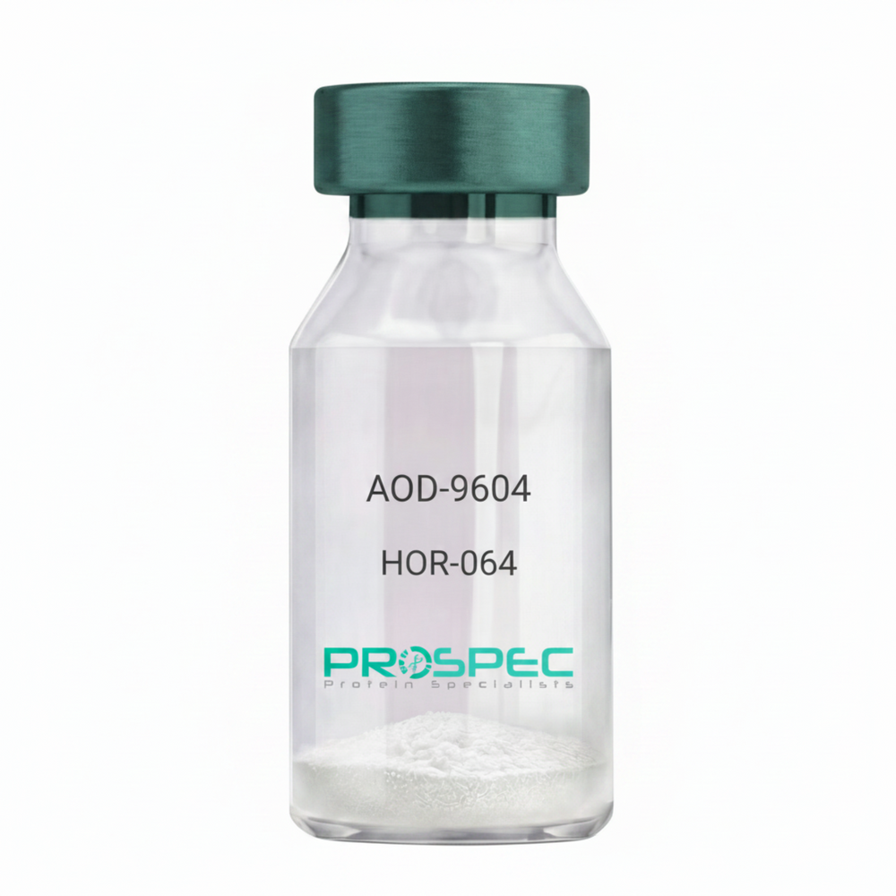
    AOD-9604
  • View Data Sheet

    Name :

    FLAG PAT3G6AT antibody

    Description:

    FLAG peptide Clone PAT3G6AT, Mouse Anti Human

    Product # :

    ANT-736

    Price :

    Quantity :

    Shipping Method :

    Ice Icon

    Shipped with Ice Packs

    Add To Cart

    More Info

    • formulation
    • More Info

    Formulation

    1mg/ml containing PBS, pH-7.4, 10% Glycerol and 0.02% Sodium Azide.

    More Info

    • Introduction

      A fusion tag called FLAG consists of eight amino acids (AspTyrLysAspAspAspAspLys) including an enterokinase-cleavage site. FLAG is specifically designed for immunoaffinity chromatography. It allows elution under non-denaturing conditions. Several antibodies against this peptide have been developed. One antibody denoted as M1 binds the peptide in the presence of bivalent metal cations preferably Ca(+). Elution is effected by chelating agents. Another strategy is competitive elution with excess of free Flag Peptide. The FLAG peptide purifies and detects recombinant fusion proteins. FLAG peptide is useful in Western blotting, immunocytochemistry, immunoprecipitation, flow cytometry, protein purification, and in the study of protein-protein interactions, cell ultrastructure, and protein localization. Flag peptide is a hydrophilic tag which significantly improves the detection and purification of recombinant fusion proteins.

    • Physical Appearance

      Sterile filtered colorless solution.

    • Immunogen

      Anti-human FLAG mAb, is derived from hybridization of mouse F0 myeloma cells with spleen cells from BALB/c mice immunized with a Synthetic peptide (DYKDDDDKC)-KLH.

    • Ig Subclass

      Mouse IgG1 heavy chain and k light chain.

    • Clone

      PAT3G6AT.

    • Applications

      The antibody has been tested by ELISA, Western blot analysis, Flow cytometry and ICC/IF to assure specificity and reactivity. Since application varies, however, each investigation should be titrated by the reagent to obtain optimal results.

    • Type

      Mouse Anti Human Monoclonal.

    • Storage Procedures

      For periods up to 1 month store at 4°C, for longer periods of time, store at -20°C. Prevent freeze thaw cycles.

    • Purification Method

      FLAG antibody was purified from mouse ascitic fluids by protein-A affinity chromatography.

    ProSpec's products are furnished for LABORATORY RESEARCH USE ONLY. The product may not be used as drugs, agricultural or pesticidal products, food additives or household chemicals.

    Flag Antibody
  • View Data Sheet

    Name :

    SARS MERS RBD, Active

    Description:

    SARS MERS Spike Receptor Binding Domain Recombinant, Active

    Middle East respiratory syndrome coronavirus, Human betacoronavirus 2c EMC/2012, MERS-CoV, MERS, MERSCoV RBD, MERS RBD, receptor binding domain, RBD, Spike RBD protein, Spike glycoprotein, S glycoprotein, E2, Peplomer protein

    Product # :

    SARS-060

    Price :

    Quantity :

    Shipping Method :

    Ice Icon

    Shipped with Ice Packs

    Add To Cart

    More Info

    • description
    • source
    • formulation
    • purity
    • biological activity
    • More Info

    Description

    SARS MERS RBD Recombinant produced in Sf9 Baculovirus cells is a single, glycosylated polypeptide chain containing 258 amino acids (358-606 aa) and having a molecular mass of 28.2kDa. SARS MERS RBD is fused to a 6 amino acid His-tag at C-terminus & purified by proprietary chromatographic techniques.

    Source

    Sf9, Baculovirus cells.

    Formulation

    The SARS MERS RBD solution (0.5mg/ml) contains 10% glycerol and Phosphate-Buffered Saline (pH 7.4).

    Purity

    Greater than 90.0% as determined by SDS-PAGE.

    Biological Activity

    Measured by its binding ability in a functional ELISA with Human DPPIV/CD26 (CAT# enz-1187).

    More Info

    • Synonyms

      Middle East respiratory syndrome coronavirus, Human betacoronavirus 2c EMC/2012, MERS-CoV, MERS, MERSCoV RBD, MERS RBD, receptor binding domain, RBD, Spike RBD protein, Spike glycoprotein, S glycoprotein, E2, Peplomer protein

    • Physical Appearance

      Sterile filtered colorless solution.

    • Stability

      Store at 4°C if entire vial will be used within 2-4 weeks.Store, frozen at -20°C for longer periods of time.For long term storage it is recommended to add a carrier protein (0.1% HSA or BSA).Avoid multiple freeze-thaw cycles.

    • Amino Acid Sequence

      ADPSGVYSVS SFEAKPSGSV VEQAEGVECD FSPLLSGTPP QVYNFKRLVF TNCNYNLTKL LSLFSVNDFT CSQISPAAIA SNCYSSLILD YFSYPLSMKS DLSVSSAGPI SQFNYKQSFS NPTCLILATV PHNLTTITKP LKYSYINKCS RLLSDDRTEV PQLVNANQYS PCVSIVPSTV WEDGDYYRKQ LSPLEGGGWL VASGSTVAMT EQLQMGFGIT VQYGTDTNSV CPKLEFANDT KIASQLGNCV EYHHHHHH.

    • Background

      The severe acute respiratory syndrome coronavirus (SARS-CoV) and Middle East respiratory syndrome coronavirus (MERS-CoV) have posed significant global health threats in recent years. Central to their pathogenesis is the interaction between the viral spike proteins and host cell receptors. This research aims to investigate the receptor binding domain (RBD) of SARS and MERS spike proteins and its implications for viral entry and the development of therapeutic interventions. Understanding the molecular mechanisms underlying viral-host interactions can pave the way for targeted therapeutic strategies against these deadly coronaviruses.

      Structure and Function of SARS and MERS Spike RBD:

      The spike proteins of SARS-CoV and MERS-CoV are critical for viral entry into host cells. These proteins consist of two subunits: S1, responsible for receptor binding, and S2, involved in membrane fusion. The receptor binding domain (RBD) within the S1 subunit specifically interacts with host cell receptors, enabling viral attachment and entry. The RBDs of SARS and MERS spike proteins exhibit unique structural features and binding affinities for their respective receptors.

      Interaction with ACE2 and DPP4 Receptors:

      The SARS-CoV spike protein RBD interacts with the angiotensin-converting enzyme 2 (ACE2) receptor, which is abundantly expressed in the respiratory tract. The binding of SARS-CoV RBD to ACE2 facilitates viral entry into host cells. On the other hand, the MERS-CoV spike protein RBD interacts with the dipeptidyl peptidase 4 (DPP4) receptor, predominantly expressed in the lungs and other tissues. The ACE2 and DPP4 receptors play crucial roles in determining the host range and tissue tropism of SARS and MERS coronaviruses.

      Implications for Viral Pathogenesis:

      The binding of SARS and MERS spike RBDs to their respective receptors triggers conformational changes in the spike protein, leading to membrane fusion and subsequent viral entry. This process is crucial for viral replication and the spread of infection within the host. The specificity and affinity of the RBD-receptor interaction influence viral tropism, tissue damage, and disease severity. Understanding the determinants of RBD-receptor binding can provide insights into viral pathogenesis and potential therapeutic targets.

      Development of Therapeutic Interventions:

      The RBD of SARS and MERS spike proteins represents a promising target for the development of antiviral therapeutics. Several strategies have been explored, including monoclonal antibodies and small molecule inhibitors, to disrupt the RBD-receptor interaction and inhibit viral entry. These approaches aim to block the binding interface between the spike RBD and the host receptor, thereby preventing viral attachment and entry. Additionally, vaccine development efforts have focused on generating neutralizing antibodies against the RBD to elicit protective immune responses.

      Conclusion:

      The investigation of the receptor binding domain (RBD) of SARS and MERS spike proteins sheds light on the molecular mechanisms underlying viral entry and pathogenesis. The specific interactions between the spike RBD and host cell receptors play a crucial role in determining viral tropism and tissue damage. Targeting the RBD-receptor interaction holds promise for the development of effective therapeutics against SARS-CoV, MERS-CoV, and potentially other related coronaviruses. Further research and development efforts are needed to exploit the potential of the spike RBD as a therapeutic target and to combat future coronavirus outbreaks.

    ProSpec's products are furnished for LABORATORY RESEARCH USE ONLY. The product may not be used as drugs, agricultural or pesticidal products, food additives or household chemicals.

    product_image.jpg
  • View Data Sheet

    Name :

    FSCN1 Antibody

    Description:

    Fascin Actin-Bundling Protein 1, Mouse Anti Human

    Strongylocentrotus Purpuratus, actin-bundling protein, FAN1, HSN, SNL.

    Product # :

    ANT-582

    Price :

    Quantity :

    Shipping Method :

    Ice Icon

    Shipped with Ice Packs

    Add To Cart

    More Info

    • formulation
    • More Info

    Formulation

    1mg/ml containing PBS, pH-7.4, 10% Glycerol and 0.02% Sodium Azide.

    More Info

    • Introduction

      FSCN1 is an actin-bundling protein that is responsible for rigidity to filopodial bundles to efficiently push the membrane forward during cytoskeleton remodeling and cell migration. FSCN1 takes part in the gathering of actin filament bundles present in microspikes, membrane ruffles, and stress fibers. FSCN1 is absent from most normal epithelia though is upregulated in multiple forms of human carcinoma, where its expression associates clinically with a poor prognosis. FSCN1 expression is abundantly found in neurons, glial cells, and endothelial cells.

    • Synonyms

      Strongylocentrotus Purpuratus, actin-bundling protein, FAN1, HSN, SNL.

    • Physical Appearance

      Sterile filtered colorless solution.

    • Immunogen

      Anti-human FSCN1 mAb, is derived from hybridization of mouse F0 myeloma cells with spleen cells from BALB/c mice immunized with a recombinant human FSCN1 protein 1-493 amino acids purified from E. coli.

    • Ig Subclass

      Mouse IgG2b heavy chain and k light chain.

    • Clone

      PAT13D2AT.

    • Applications

      FSCN1 antibody has been tested by ELISA, Western blot analysis, ICC/IF and Flow cytometry to assure specificity and reactivity. Since application varies, however, each investigation should be titrated by the reagent to obtain optimal results.

    • Type

      Mouse Anti Human Monoclonal.

    • Storage Procedures

      For periods up to 1 month store at 4°C, for longer periods of time, store at -20°C. Prevent freeze thaw cycles.

    • Purification Method

      FSCN1 antibody was purified from mouse ascitic fluids by protein-A affinity chromatography.

    ProSpec's products are furnished for LABORATORY RESEARCH USE ONLY. The product may not be used as drugs, agricultural or pesticidal products, food additives or household chemicals.

    Fscn1 Antibody
  • View Data Sheet

    Name :

    Tesamorelin

    Description:

    Tesamorelin

    Product # :

    HOR-062

    Price :

    Quantity :

    Shipping Method :

    Room Temp Icon

    Shipped at Room temp

    Add To Cart

    More Info

    • description
    • formulation
    • purity
    • More Info

    Description

    Tesamorelin is a synthetic single, non-glycosylated polypeptide chain containing 44 amino acids, having a molecular mass of 5135.78 Dalton and a Molecular formula of C221H366N72O67S.

    Formulation

    The protein was lyophilized with no additives.

    Purity

    Greater than 97.0% as determined by analysis by RP-HPLC.

    More Info

    • Physical Appearance

      Sterile Filtered White lyophilized (freeze-dried) powder.

    • Stability

      Lyophilized Tesamorelin although stable at room temperature for 3 weeks, should be stored desiccated below -18°C. Upon reconstitution Tesamorelin should be stored at 4°C between 2-7 days and for future use below -18°C.
      For long term storage it is recommended to add a carrier protein (0.1% HSA or BSA).
      Please prevent freeze-thaw cycles.

    • Solubility

      It is recommended to reconstitute the lyophilized Tesamorelin in sterile 18MΩ-cm H2O not less than 100 µg/ml, which can then be further diluted to other aqueous solutions.

    • Amino Acid Sequence

      trans-3-hexenoyl-Tyr-Ala-Asp-Ala-IlePhe-Thr-Asn-Ser-Tyr-Arg-Lys-Val-Leu-Gly-Gln-Leu-Ser-Ala-Arg-LysLeu-Leu-Gln-Asp-Ile-Met-Ser-Arg-Gln-Gln-Gly-Glu-Ser-Asn-Gln-GluArg-Gly-Ala-Arg-Ala-Arg-Leu-NH2.

    • Background

      Tesamorelin is a growth hormone-releasing hormone (GHRH) analogue. Tesamorelin stimulates the pituitary gland to produce endogenous growth hormone, which targets visceral adipose tissue. Tesamorelin has evolved from a HIV treatment into an effective metabolic and regenerative therapy.

    ProSpec's products are furnished for LABORATORY RESEARCH USE ONLY. The product may not be used as drugs, agricultural or pesticidal products, food additives or household chemicals.

    Tesamorelin
  • View Data Sheet

    Name :

    LRPAP1 Antibody

    Description:

    Low Density Lipoprotein Receptor-Related Protein Associated Protein 1, Mouse Anti Human

    Low density lipoprotein-related protein-associated protein 1 (alpha-2-macroglobulin receptor-associated protein 1), A2MRAP, HBP44, RAP, A2RAP, Alpha-2-MRAP, LRPAP1.

    Product # :

    ANT-547

    Price :

    Quantity :

    Shipping Method :

    Ice Icon

    Shipped with Ice Packs

    Add To Cart

    More Info

    • formulation
    • More Info

    Formulation

    1mg/ml containing PBS, pH-7.4, 10% Glycerol and 0.02% Sodium Azide.

    More Info

    • Introduction

      LRPAP1 is a member of the alpha-2-MRAP family. LRPAP1 cooperates with LRP1/alpha-2-macroglobulin receptor and glycoprotein 330. LRPAP1, located on the cell surface, creates a complex with the alpha 2 macroglobulin receptor light and heavy chains and has part in the pathogenesis of membrane glomerular nephritis.

    • Synonyms

      Low density lipoprotein-related protein-associated protein 1 (alpha-2-macroglobulin receptor-associated protein 1), A2MRAP, HBP44, RAP, A2RAP, Alpha-2-MRAP, LRPAP1.

    • Physical Appearance

      Sterile filtered colorless solution.

    • Immunogen

      Anti-human LRPAP1 mAb, is derived from hybridization of mouse F0 myeloma cells with spleen cells from BALB/c mice immunized with a recombinant human LRPAP1 protein 35-357 amino acids purified from E. coli.

    • Ig Subclass

      Mouse IgG1 heavy chain and k light chain.

    • Clone

      PAT8G8AT.

    • Applications

      LRPAP1 antibody has been tested by ELISA, Western blot analysis to assure specificity and reactivity. Since application varies, however, each investigation should be titrated by the reagent to obtain optimal results. Recommended starting dilution is 1:500.

    • Type

      Mouse Anti Human Monoclonal.

    • Storage Procedures

      For periods up to 1 month store at 4°C, for longer periods of time, store at -20°C. Prevent freeze thaw cycles.

    • Purification Method

      LRPAP1 antibody was purified from mouse ascitic fluids by protein-A affinity chromatography.

    ProSpec's products are furnished for LABORATORY RESEARCH USE ONLY. The product may not be used as drugs, agricultural or pesticidal products, food additives or household chemicals.

    Lrpap1 Antibody
  • View Data Sheet

    Name :

    NRAS Antibody

    Description:

    Neuroblastoma RAS Viral Oncogene Homolog, Mouse Anti Human

    GTPase NRas, HRAS1, ALPS4, N-ras, NRAS1, NS6, Transforming protein N-Ras.

    Product # :

    ANT-047

    Price :

    Quantity :

    Shipping Method :

    Ice Icon

    Shipped with Ice Packs

    Add To Cart

    More Info

    • formulation
    • More Info

    Formulation

    1mg/ml containing PBS, pH-7.4, & 0.1% Sodium Azide.

    More Info

    • Introduction

      NRAS binds GDP/GTP and has intrinsic GTPase activity. NRAS is a membrane protein that shuttles between the Golgi apparatus and the plasma membrane. This transport is regulated through palmitoylation and depalmitoylation by the ZDHHC9-GOLGA7 complex. NRAS is activated to a GTP-bound form by a GTPase activating protein and inactivated to a GDP-bound form by a guanine nucleotide-exchange factor. Defects in this NRAS gene result in juvenile myelomonocytic leukemia.

    • Synonyms

      GTPase NRas, HRAS1, ALPS4, N-ras, NRAS1, NS6, Transforming protein N-Ras.

    • Immunogen

      Anti-human NRAS mAb, is derived from hybridization of mouse FO myeloma cells with spleen cells from BALB/c mice immunized with recombinant human NRAS amino acids 1-186 purified from E. coli.

    • Ig Subclass

      Mouse IgG2a heavy chain and k light chain.

    • Clone

      PAT2G9AT.

    • Applications

      NRAS antibody has been tested by ELISA and Western blot analysis to assure specificity and reactivity. Since application varies, however, each investigation should be titrated by the reagent to obtain optimal results. Recommended dilution range for Western blot analysis is 1:500 ~ 1:5000.
      Recommended starting dilution is 1:1000.

    • Type

      Mouse Anti Human Monoclonal.

    • Storage Procedures

      For periods up to 1 month store at 4°C, for longer periods of time, store at -20°C. Prevent freeze thaw cycles.

    • Purification Method

      NRAS antibody was purified from mouse ascitic fluids by protein-G affinity chromatography.

    ProSpec's products are furnished for LABORATORY RESEARCH USE ONLY. The product may not be used as drugs, agricultural or pesticidal products, food additives or household chemicals.

    Nras Antibody
  • View Data Sheet

    Name :

    GARS Antibody

    Description:

    Glycyl-TRNA Synthetase, Mouse Anti Human

    Glycine--tRNA ligase, Diadenosine tetraphosphate synthetase, AP-4-A synthetase, Glycyl-tRNA synthetase, GlyRS, GARS, CMT2D, DSMAV, HMN5, SMAD1.

    Product # :

    ANT-544

    Price :

    Quantity :

    Shipping Method :

    Ice Icon

    Shipped with Ice Packs

    Add To Cart

    More Info

    • formulation
    • More Info

    Formulation

    1mg/ml containing PBS, pH-7.4, 10% Glycerol and 0.02% Sodium Azide.

    More Info

    • Introduction

      GARS is an (alpha)2 dimer which is a member of the class II family of tRNA synthetases. GARS is a glycyl-tRNA synthetase, one of the aminoacyl-tRNA synthetases which charge tRNAs with their cognate amino acids. GARS catalyzes the attachment of glycine to tRNA(Gly). In addition, GARS is able to produce diadenosine tetraphosphate (Ap4A), which is a universal pleiotropic signaling molecule required for cell regulation pathways, by direct condensation of two ATPs. GARS has been demonstrated to be a target of autoantibodies in the human autoimmune diseases, polymyositis or dermatomyositis.

    • Synonyms

      Glycine--tRNA ligase, Diadenosine tetraphosphate synthetase, AP-4-A synthetase, Glycyl-tRNA synthetase, GlyRS, GARS, CMT2D, DSMAV, HMN5, SMAD1.

    • Physical Appearance

      Sterile filtered colorless solution.

    • Immunogen

      Anti-human GARS mAb, is derived from hybridization of mouse F0 myeloma cells with spleen cells from BALB/c mice immunized with recombinant human GARS amino acids 43-289 purified from E. coli.

    • Ig Subclass

      Mouse IgG1 heavy chain and k light chain.

    • Clone

      PAT4E10AT.

    • Applications

      GARS antibody has been tested by ELISA, Western blot analysis to assure specificity and reactivity. Since application varies, however, each investigation should be titrated by the reagent to obtain optimal results. Recommended starting dilution is 1:500.

    • Type

      Mouse Anti Human Monoclonal.

    • Storage Procedures

      For periods up to 1 month store at 4°C, for longer periods of time, store at -20°C. Prevent freeze thaw cycles.

    • Purification Method

      GARS antibody was purified from mouse ascitic fluids by protein-A affinity chromatography.

    ProSpec's products are furnished for LABORATORY RESEARCH USE ONLY. The product may not be used as drugs, agricultural or pesticidal products, food additives or household chemicals.

    Gars Antibody
  • View Data Sheet

    Name :

    ARF1 Antibody

    Description:

    ADP-Ribosylation Factor 1, Mouse Anti Human

    ARF-1, ADP-ribosylation factor 1, ARF1.

    Product # :

    ANT-058

    Price :

    Quantity :

    Shipping Method :

    Ice Icon

    Shipped with Ice Packs

    Add To Cart

    More Info

    • formulation
    • More Info

    Formulation

    1mg/ml containing PBS, pH-7.4, & 0.1% Sodium Azide.

    More Info

    • Introduction

      ARF1 is a small guanine nucleotide-binding protein that increases the enzymatic activities of cholera toxin. ARF1 is necessary and ubiquitous in eukaryotes. ARF1 takes part in vesicular transport and functioning via phospholipase D activation. ARF1 is involved in membrane traffic and organelle integrity which are closely tied to their reversible association with membranes and distinct interactions with membrane phospholipids.

    • Synonyms

      ARF-1, ADP-ribosylation factor 1, ARF1.

    • Immunogen

      Anti-human ARF1 mAb, is derived from hybridization of mouse FO myeloma cells with spleen cells from BALB/c mice immunized with recombinant human ARF1 amino acids 1-181 purified from E. coli.

    • Ig Subclass

      Mouse IgG1 heavy chain and k light chain.

    • Clone

      P1B3AT.

    • Applications

      ARF1 antibody has been tested by ELISA, Western blot analysis to assure specificity and reactivity. Since application varies, however, each investigation should be titrated by the reagent to obtain optimal results. Recommended dilution range for Western blot analysis is 1:1,000.

    • Type

      Mouse Anti Human Monoclonal.

    • Storage Procedures

      For periods up to 1 month store at 4°C, for longer periods of time, store at -20°C. Prevent freeze thaw cycles.

    ProSpec's products are furnished for LABORATORY RESEARCH USE ONLY. The product may not be used as drugs, agricultural or pesticidal products, food additives or household chemicals.

    Arf1 Antibody
  • View Data Sheet

    Name :

    CD163 Porcine

    Description:

    CD163 Porcine Recombinant

    CD-163, Hemoglobin scavenger receptor, macrophage-associated antigen, M130, sCD163, CD163, MM130.

    Product # :

    PRO-856

    Price :

    Quantity :

    Shipping Method :

    Room Temp Icon

    Shipped at Room temp

    Add To Cart

    More Info

    • description
    • source
    • formulation
    • purity
    • More Info

    Description

    CD163 Porcine Recombinant produced in E.Coli is a single, non-glycosylated, polypeptide chain containing 805 amino acids and having a molecular mass of 87kDa.The CD163 is fused to an 8 amino acid His Tag at C-terminus and purified by proprietary chromatographic techniques.

    Source

    Escherichia Coli.

    Formulation

    The protein was lyophilized from a 0.2µm filtered concentrated solution in 1×PBS, pH 7.4, containing 4M Urea.

    Purity

    Greater than 95.0% as determined by: (a) Analysis by HPLC. (b) Analysis by SDS-PAGE.

    More Info

    • Introduction

      CD163 is an acute phase-regulated receptor which participates in the removal and endocytosis of hemoglobin/haptoglobin complexes by macrophages and thus keeps tissues from free hemoglobin-mediated oxidative damage. Furthermore, CD163 partakes in the uptake and recycling of iron, through endocytosis of hemoglobin/haptoglobin and ensuing breakdown of heme. In addition, CD163 binds hemoglobin/haptoglobin complexes in a calcium-dependent and pH-dependent way. CD163 demonstrates greater affinity for complexes of hemoglobin and multimeric haptoglobin of HP-1F phenotype than for complexes of hemoglobin and dimeric haptoglobin of HP-1S phenotype. Moreover, CD163 stimulates a cascade of intracellular signals which involves tyrosine kinase-dependent calcium recruitment, inositol triphosphate formation and secretion of IL-6 & CSF-1.

    • Synonyms

      CD-163, Hemoglobin scavenger receptor, macrophage-associated antigen, M130, sCD163, CD163, MM130.

    • Physical Appearance

      Sterile Filtered White lyophilized (freeze-dried) powder.

    • Stability

      Lyophilized CD163 although stable at room temperature for 3 weeks, should be stored desiccated below -18°C. Upon reconstitution CD163 should be stored at 4°C between 2-7 days and for future use below -18°C. For long term storage it is recommended to add a carrier protein (0.1% HSA or BSA). Please prevent freeze-thaw cycles.

    • Solubility

      It is recommended to reconstitute the lyophilized CD163 in sterile 18MΩ-cm H2O not less than 100µg/ml, which can then be further diluted to other aqueous solutions.

    • Amino Acid Sequence

      MDKLRMVLHE NSGSADLKLR VVDGVTECSG RLEVKFQGEW GTICDDGWDS DDAAVACKQL GCPTAVTAIG RVNASEGTGH IWLDSVSCHG HESALWQCRH HEWGKHYCNH NEDAGVTCSD GSDLELRLKG GGSHCAGTVE VEIQKLVGKV CDRSWGLKEA DVVCRQLGCG SALKTSYQVY SKTKATNTWL FVSSCNGNET SLWDCKNWQW GGLSCDHYDE AKITCSAHRK PRLVGGDIPC SGRVEVQHGD TWGTVCDSDF SLEAASVLCR ELQCGTVVSL LGGAHFGEGS GQIWAEEFQC EGHESHLSLC PVAPRPDGTC SHSRDVGVVC SRYTQIRLVN GKTPCEGRVE LNILGSWGSL CNSHWDMEDA HVLCQQLKCG VALSIPGGAP FGKGSEQVWR HMFHCTGTEK HMGDCSVTAL GASLCSSGQV ASVICSGNQS QTLSPCNSSS SDPSSSIISE ENGVACIGSG QLRLVDGGGR CAGRVEVYHE GSWGTICDDS WDLNDAHVVC KQLSCGWAIN ATGSAHFGEG TGPIWLDEIN CNGKESHIWQ CHSHGWGRHN CRHKEDAGVI CSEFMSLRLI SENSRETCAG RLEVFYNGAW GSVGKNSMSP ATVGVVCRQL GCADRGDISP ASSDKTVSRH MWVDNVQCPK GPDTLWQCPS SPWKKRLASP SEETWITCAN KIRLQEGNTN CSGRVEIWYG GSWGTVCDDS WDLEDAQVVC RQLGCGSALE AGKEAAFGQG TGPIWLNEVK CKGNETSLWD CPARSWGHSD CGHKEDAAVT CSEIAKSRES LHATGRSHHH HHHHH.

    ProSpec's products are furnished for LABORATORY RESEARCH USE ONLY. The product may not be used as drugs, agricultural or pesticidal products, food additives or household chemicals.

    Cd163 Porcine
  • View Data Sheet

    Name :

    QTRTD1 Human

    Description:

    Queuine TRNA-Ribosyltransferase Domain Containing 1 Human Recombinant

     Queuine TRNA-Ribosyltransferase Domain Containing 1, EC 2.4.2.29, Queuine TRNA-Ribosyltransferase Domain-Containing Protein 1, QTRTD1.

    Product # :

    ENZ-833

    Price :

    Quantity :

    Shipping Method :

    Ice Icon

    Shipped with Ice Packs

    Add To Cart

    More Info

    • description
    • source
    • formulation
    • purity
    • More Info

    Description

    QTRTD1 Human Recombinant produced in E.Coli is a single, non-glycosylated polypeptide chain containing 438 amino acids (1-415 a.a) and having a molecular mass of 49.1kDa.QTRTD1 is fused to a 23 amino acid His-tag at N-terminus & purified by proprietary chromatographic techniques.

    Source

    Escherichia Coli.

    Formulation

    QTRTD1 protein solution (0.25mg/ml) containing Phosphate buffered saline (pH7.4) 30% glycerol, 2mM DTT, 1mM EDTA and 0.1mM PMSF.

    Purity

    Greater than 95.0% as determined by SDS-PAGE.

    More Info

    • Introduction

      Queuine TRNA-Ribosyltransferase Domain Containing 1, also known as QTRTD1 act together with QTRT1 to form an active queuine tRNA-ribosyltransferase. QTRTD1 exchanges queuine for the guanine at the wobble position of tRNAs along with GUN anticodons (tRNA-Asp,-Asn,-His and -Tyr), in that way forming the hypermodified nucleoside queuosine.

    • Synonyms

      Queuine TRNA-Ribosyltransferase Domain Containing 1, EC 2.4.2.29, Queuine TRNA-Ribosyltransferase Domain-Containing Protein 1, QTRTD1.

    • Physical Appearance

      Sterile Filtered colorless solution.

    • Stability

      Store at 4°C if entire vial will be used within 2-4 weeks. Store, frozen at -20°C for longer periods of time. For long term storage it is recommended to add a carrier protein (0.1% HSA or BSA).Avoid multiple freeze-thaw cycles.

    • Amino Acid Sequence

      MGSSHHHHHH SSGLVPRGSH MGSMKLSLTK VVNGCRLGKI KNLGKTGDHT MDIPGCLLYT KTGSAPHLTH HTLHNIHGVP AMAQLTLSSL AEHHEVLTEY KEGVGKFIGM PESLLYCSLH DPVSPCPAGY VTNKSVSVWS VAGRVEMTVS KFMAIQKALQ PDWFQCLSDG EVSCKEATSI KRVRKSVDRS LLFLDNCLRL QEESEVLQKS VIIGVIEGGD VMEERLRSAR ETAKRPVGGF LLDGFQGNPT TLEARLRLLS SVTAELPEDK PRLISGVSRP DEVLECIERG VDLFESFFPY QVTERGCALT FSFDYQPNPE ETLLQQNGTQ EEIKCMDQIK KIETTGCNQE ITSFEINLKE KKYQEDFNPL VRGCSCYCCK NHTRAYIHHL LVTNELLAGV LLMMHNFEHY FGFFHYIREA LKSDKLAQLK ELIHRQAS.

    ProSpec's products are furnished for LABORATORY RESEARCH USE ONLY. The product may not be used as drugs, agricultural or pesticidal products, food additives or household chemicals.

    Qtrtd1 Human
  • View Data Sheet

    Name :

    RNASE1 Human

    Description:

    Ribonuclease 1 Human Recombinant

    RNASE1, ribonuclease A family member 1, pancreatic, RAC1, RIB1, RNS1, ribonuclease pancreatic, HP-RNase, RIB-1, RNase UpI-1, RNase 1, Ribonuclease A, RNase A, Ribonuclease 1.

    Product # :

    ENZ-1168

    Price :

    Quantity :

    Shipping Method :

    Ice Icon

    Shipped with Ice Packs

    Add To Cart

    More Info

    • description
    • source
    • formulation
    • purity
    • biological activity
    • More Info

    Description

    RNASE1 Human Recombinant produced in HEK cells is a single, glycosylated, polypeptide chain (29-156 a.a) containing a total of 134 amino acids, having a molecular mass of 15.3kDa. RNASE1 is fused to a 6 amino acid His-tag at C-terminus,and is purified by proprietary chromatographic techniques.

    Source

    HEK293 Cells.

    Formulation

    The RNASE1 solution (1mg/ml) contains 10% Glycerol and Phosphate-Buffered Saline (pH 7.4).

    Purity

    Greater than 90.0% as determined by SDS-PAGE.

    Biological Activity

    Specific activity is >  3 X 10^6 unit/mg. Defined by the amount of enzyme that  hydrolyzes 1nmole of RNA per minute at 25˚C.

    More Info

    • Introduction

      Ribonuclease 1R (NASE1) is a small protein which is a part of pancreatic ribonuclease enzyme family. NASE1 has 4 disulfide bonds in its native state and cleaves speciallyafter pyrimidine nucleotides.Cleavage takes place in 2 steps: first, the 3’,5’-phosphodiester bond is cleaved to craete a 2’,3’-cyclic phosphodiester intermediate; than, the cyclic phosphodiester is hydrolyzed to a 3’-monophosphate. NASE1is activated the most with single stranded RNA.NASE1inhibited by alkylation of His12 and His119 and activated by potassium and sodium salts.NASE1hydrolyzes RNA from protein samples.

    • Synonyms

      RNASE1, ribonuclease A family member 1, pancreatic, RAC1, RIB1, RNS1, ribonuclease pancreatic, HP-RNase, RIB-1, RNase UpI-1, RNase 1, Ribonuclease A, RNase A, Ribonuclease 1.

    • Physical Appearance

      Sterile Filtered colorless solution.

    • Stability

      Store at 4°C if entire vial will be used within 2-4 weeks.Store, frozen at -20°C for longer periods of time.For long term storage it is recommended to add a carrier protein (0.1% HSA or BSA).Avoid multiple freeze-thaw cycles.

    • Amino Acid Sequence

      KESRAKKFQR QHMDSDSSPS SSSTYCNQMM RRRNMTQGRC KPVNTFVHEP LVDVQNVCFQ EKVTCKNGQG NCYKSNSSMH ITDCRLTNGS RYPNCAYRTS PKERHIIVAC EGSPYVPVHF DASVEDSTHH HHHH

    ProSpec's products are furnished for LABORATORY RESEARCH USE ONLY. The product may not be used as drugs, agricultural or pesticidal products, food additives or household chemicals.

    Rnase1 Human
  • View Data Sheet

    Name :

    Rantes Human

    Description:

    Rantes Human Recombinant (CCL5)

    Small inducible cytokine A5, CCL5, T-cell-specific RANTES protein, SIS-delta, T cell- specific protein P228, TCP228, chemokine (C-C motif) ligand 5, SISd, SCYA5, RANTES, D17S136E, MGC17164.

    Product # :

    CHM-328

    Price :

    Quantity :

    Shipping Method :

    Room Temp Icon

    Shipped at Room temp

    Add To Cart

    More Info

    • description
    • source
    • formulation
    • purity
    • biological activity
    • More Info

    Description

    Rantes Human Recombinant produced in E.Coli is a single, non-glycosylated polypeptide chain containing 68 amino acids and having a molecular mass of 7.8 kDa.
    The Rantes is purified by proprietary chromatographic techniques.

    Source

    Escherichia Coli.

    Formulation

    Lyophilized from a 0.2µm filtered solution in 35% (v/v) Acetonitrile and 0.1% (v/v) TFA.

    Purity

    Greater than 97.0% as determined by:
    (a) Analysis by RP-HPLC.
    (b) Analysis by SDS-PAGE.

    Biological Activity

    The activity is determined by the chemoattract of human blood monocytes at a concentration between 1-10 ng/ml.

    More Info

    • Introduction

      Regulated upon Activation, Normal T-cell Expressed, and Secreted or RANTES is an 8 kDa protein classified as a chemotactic cytokine or chemokine. Rantes has recently been renamed CCL5. RANTES is chemotactic for T cells, eosinophils and basophils and plays an active role in recruiting leukocytes into inflammatory sites. With the help of particular cytokines (i.e. IL-2 and IFN-g) that are released by T cells, RANTES also induces the proliferation and activation of certain natural killer (NK) cells to form CHAK (CC-Chemokine-activated killer) cells. Rantes is also an HIV-suppressive factor released from CD8+ T cells. The Rantes chemokine has been localized to chromosome 17 in humans.

    • Synonyms

      Small inducible cytokine A5, CCL5, T-cell-specific RANTES protein, SIS-delta, T cell- specific protein P228, TCP228, chemokine (C-C motif) ligand 5, SISd, SCYA5, RANTES, D17S136E, MGC17164.

    • Physical Appearance

      Sterile Filtered White lyophilized (freeze-dried) powder.

    • Stability

      Lyophilized Rantes although stable at room temperature for 3 weeks, should be stored desiccated below -18°C. Upon reconstitution Rantes should be stored at 4°C between 2-7 days and for future use below -18°C. For long term storage it is recommended to add a carrier protein (0.1% HSA or BSA).Please prevent freeze-thaw cycles.

    • Solubility

      It is recommended to reconstitute the lyophilized Rantes in sterile 18MΩ-cm H2O not less than 100µg/ml, which can then be further diluted to other aqueous solutions.

    • Amino Acid Sequence

      SPYSSDTTPC CFAYIARPLP RAHIKEYFYT SGKCSNPAVV FVTRKNRQVC ANPEKKWVRE YINSLEMS.

    ProSpec's products are furnished for LABORATORY RESEARCH USE ONLY. The product may not be used as drugs, agricultural or pesticidal products, food additives or household chemicals.

    Rantes Human
  • View Data Sheet

    Name :

    HK1/2/3 Antibody

    Description:

    Hexokinase-1/2/3, Mouse Anti Human

    Hexokinase-3, EC 2.7.1.1, Hexokinase type III, HK III, HXK3, HK3.

    Product # :

    ANT-726

    Price :

    Quantity :

    Shipping Method :

    Ice Icon

    Shipped with Ice Packs

    Add To Cart

    More Info

    • formulation
    • More Info

    Formulation

    1mg/ml containing PBS, pH-7.4, 10% Glycerol and 0.02% Sodium Azide.

    More Info

    • Introduction

      Hexokinases phosphorylate glucose to produce glucose-6-phosphate, thus committing glucose to the glycolytic pathway. HK3 encodes hexokinase 3. Similar to hexokinases 1 and 2, this allosteric enzyme is inhibited by its product glucose-6-phosphate. Hexokinase3 lacks the hydrophobic N-terminal sequence critical for targeting to mitochondria. Hexpkinase3 may have anabolic functions, providing H6P for glycogen or lipid synthesis.

    • Synonyms

      Hexokinase-3, EC 2.7.1.1, Hexokinase type III, HK III, HXK3, HK3.

    • Physical Appearance

      Sterile filtered colorless solution.

    • Immunogen

      Anti-human HK1/2/3mAb, is derived from hybridization of mouse F0 myeloma cells with spleen cells from BALB/c mice immunized with recombinant human HK1/2/3amino acids 1-923 purified from E. coli.

    • Ig Subclass

      Mouse IgG1 heavy chain and κ light chain.

    • Clone

      PAT4C12AT.

    • Applications

      HK1/2/3antibody has been tested by ELISA and Western blot analysis to assure specificity and reactivity. Since application varies, however, each investigation should be titrated by the reagent to obtain optimal results.

    • Type

      Mouse Anti Human Monoclonal.

    • Storage Procedures

      For periods up to 1 month store at 4°C, for longer periods of time, store at -20°C. Prevent freeze thaw cycles.

    • Purification Method

      HK1/2/3 antibody was purified from mouse ascitic fluids by protein-A affinity chromatography.

    ProSpec's products are furnished for LABORATORY RESEARCH USE ONLY. The product may not be used as drugs, agricultural or pesticidal products, food additives or household chemicals.

    Hk123 Antibody
  • View Data Sheet

    Name :

    TBCA Antibody

    Description:

    Tubulin Folding Cofactor A, Mouse Anti Human

    Tubulin-specific chaperone A, Tubulin-folding cofactor A, CFA, TCP1-chaperonin cofactor A, TBCA.

    Product # :

    ANT-362

    Price :

    Quantity :

    Shipping Method :

    Ice Icon

    Shipped with Ice Packs

    Add To Cart

    More Info

    • formulation
    • More Info

    Formulation

    1mg/ml containing PBS, pH-7.4, 10% Glycerol and 0.02% Sodium Azide.

    More Info

    • Introduction

      TBCA is a tubulin-folding protein which is involved in the early step of the tubulin folding pathway. TBCA is one of four proteins (cofactors A, D, E, and C) implicated in the pathway directing to properly folded beta-tubulin from folding intermediates. Cofactors A and D are thought to be a factor in capturing and stabilizing beta-tubulin in a quasi-native confirmation. TBCA is crucial for cell viability, if reduced it causes a decrease in the amount of soluble tubulin, alterations in microtubules and G1 cell cycle arrest. Cofactor E attaches to the cofactor D-tubulin complex, afterward, interaction with cofactor C triggers the release of tubulin polypeptides that are committed to the native state.

    • Synonyms

      Tubulin-specific chaperone A, Tubulin-folding cofactor A, CFA, TCP1-chaperonin cofactor A, TBCA.

    • Immunogen

      Anti-human TBCA mAb is derived from hybridization of mouse F0 myeloma cells with spleen cells from BALB/c mice immunized with recombinant human TBCA amino acids 1-108 purified from E. coli.

    • Ig Subclass

      Mouse IgG2b heavy chain and κ light chain.

    • Clone

      PAT1A5AT.

    • Applications

      TBCA antibody has been tested by ELISA, Western blot, ICC/IF and FACS analysis to assure specificity and reactivity. Since application varies, however, each investigation should be titrated by the reagent to obtain optimal results.

    • Type

      Mouse Anti Human Monoclonal.

    • Storage Procedures

      For periods up to 1 month store at 4°C, for longer periods of time, store at -20°C. Prevent freeze thaw cycles.

    • Purification Method

      TBCA antibody was purified from mouse ascitic fluids by protein-G affinity chromatography.

    ProSpec's products are furnished for LABORATORY RESEARCH USE ONLY. The product may not be used as drugs, agricultural or pesticidal products, food additives or household chemicals.

    Tbca Antibody
  • View Data Sheet

    Name :

    XPA Antibody

    Description:

    Xeroderma Pigmentosum Complementation Group A, Mouse Anti Human

    XP1, XPAC, DNA repair protein complementing XP-A cells.

    Product # :

    ANT-504

    Price :

    Quantity :

    Shipping Method :

    Ice Icon

    Shipped with Ice Packs

    Add To Cart

    More Info

    • formulation
    • More Info

    Formulation

    1mg/ml containing PBS, pH-7.4, 10% Glycerol and 0.02% Sodium Azide.

    More Info

    • Introduction

      DNA repair protein complementing XP-A cells, (XPA), is a member of the XPA family. XPA protein takes a part in DNA excision repair. It Inductees repair by binding to damaged sites with different affinities depending on the photoproduct and the transcriptional state of the region. Defects in XPA is the reason of xeroderma pigmentosum complementation group A (XP-A), which is infrequent human autosomal recessive disease which characterized by solar sensitivity, high predisposition for developing cancers on areas exposed to sunlight, also may cause to neurological abnormalities.

    • Synonyms

      XP1, XPAC, DNA repair protein complementing XP-A cells.

    • Physical Appearance

      Sterile filtered colorless solution.

    • Immunogen

      Anti-human XPA mAb, clone PAT71H3, is derived from hybridization of mouse F0 myeloma cells with spleen cells from BALB/c mice immunized with a recombinant human XPA protein 1-273 amino acids purified from E. coli.

    • Ig Subclass

      Mouse IgG1 heavy chain and k light chain.

    • Clone

      PAT71H3AT.

    • Applications

      The antibody has been tested by ELISA, Western blot analysis, ICC/IF to assure specificity and reactivity. Since application varies, however, each investigation should be titrated by the reagent to obtain optimal results.

    • Type

      Mouse Anti Human Monoclonal.

    • Storage Procedures

      For periods up to 1 month store at 4°C, for longer periods of time, store at -20°C. Prevent freeze thaw cycles.

    • Purification Method

      XPA antibody was purified from mouse ascitic fluids by protein-A affinity chromatography.

    ProSpec's products are furnished for LABORATORY RESEARCH USE ONLY. The product may not be used as drugs, agricultural or pesticidal products, food additives or household chemicals.

    Xpa Antibody
  • View Data Sheet

    Name :

    NBL1 Antibody

    Description:

    Neuroblastoma 1, Mouse Anti Human

    D1S1733E, DAN, DAND1, NB, NO3, Neuroblastoma suppressor of tumorigenicity 1, DAN domain family member 1, Zinc finger protein DAN, NBL1.

    Product # :

    ANT-488

    Price :

    Quantity :

    Shipping Method :

    Ice Icon

    Shipped with Ice Packs

    Add To Cart

    More Info

    • formulation
    • More Info

    Formulation

    1mg/ml containing PBS, pH-7.4, 10% Glycerol and 0.02% Sodium Azide.

    More Info

    • Introduction

      Neuroblastoma suppressor of tumorigenicity 1 (NBL1) is a part of the evolutionarily conserved CAN (Cerberus and DAN) family of proteins, which contain a domain resembling the CTCK (C-terminal cystine knot-like) motif found in several signaling molecules. NBL1 is a is a tumor suppressor of neuroblastoma and takes part in preventing cells from entering the final stage (G1/S) of the transformation process. NBL1 is produced in small neurons of the dorsal root ganglion. The expression of NBL1 is triggered by MATH-1.

    • Synonyms

      D1S1733E, DAN, DAND1, NB, NO3, Neuroblastoma suppressor of tumorigenicity 1, DAN domain family member 1, Zinc finger protein DAN, NBL1.

    • Physical Appearance

      Sterile filtered colorless solution.

    • Immunogen

      Anti-human NBL1 mAb, clone PAT38G8AT, is derived from hybridization of mouse F0 myeloma cells with spleen cells from BALB/c mice immunized with a recombinant human NBL1 protein 18-181 amino acids purified from E. coli.

    • Ig Subclass

      Mouse IgG1 heavy chain and Kappa light chain.

    • Clone

      PAT38G8AT.

    • Applications

      The antibody has been tested by ELISA, Western blot analysis to assure specificity and reactivity. Since application varies, however, each investigation should be titrated by the reagent to obtain optimal results. Recommended starting dilution is 1:1000.

    • Type

      Mouse Anti Human Monoclonal.

    • Storage Procedures

      For periods up to 1 month store at 4°C, for longer periods of time, store at -20°C. Prevent freeze thaw cycles.

    • Purification Method

      NBL1 antibody was purified from mouse ascitic fluids by protein-A affinity chromatography.

    ProSpec's products are furnished for LABORATORY RESEARCH USE ONLY. The product may not be used as drugs, agricultural or pesticidal products, food additives or household chemicals.

    Nbl1 Antibody
  • 1
  • …
  • 16
  • 17
  • 18
  • 19
  • 20
Your browser does not support the video tag.

Products

  • Cytokines
  • Growth Factors
  • Chemokines
  • CD Antigens
  • Neurotrophins
  • Hormones
  • Enzymes
  • Viral Antigens
  • Recombinant Proteins
  • Natural Proteins
  • Monoclonal Antibodies
  • Polyclonal Antibodies
  • Thermal Packaging

About

  • Company
  • Founder
  • Contribution
  • Contact Us
  • My Account

Ordering

  • Ordering Method
  • Ordering - Credit Card
  • Ordering PO
  • Shipping Fees
  • FAQ's

Legal

  • Terms & Conditions
  • Privacy
  • Site Map
  • info@prospecbio.com
  • Facebook
  • Instagram
  • Linkedin Profile

© 2026 ProSpec-Tany TechnoGene Ltd. All Rights Reserved.

  • Choosing a selection results in a full page refresh.
  • Opens in a new window.