Skip to content

High-quality research proteins.

  • 1-800-805-6531

  • Mon - Sat 8.00-18.00, Sun - Closed

  • info@prospecbio.com

  • PO Box 6591 East Brunswick NJ 08816

  • Products
    • Cytokines
    • Growth factors
    • Chemokines
    • Cd antigens
    • Neurotrophins
    • Hormones
    • Enzymes
    • Viral antigens
    • Recombinant proteins
    • Natural proteins
    • Monoclonal antibodies
    • Polyclonal antibodies
    • Thermal Packaging
  • About
    • Company
    • Founder
    • Contribution
  • Customers
    • Eastern Asia
    • Eastern Europe
    • North America
    • South America
    • South East Asia
    • Southern Asia
    • Western Europe
  • Ordering
    • Ordering Method
    • Ordering - Credit Card
    • Ordering - PO
    • Shipping Fees
    • FAQs
  • Services
    • DNA Cloning
    • Protein Expression
    • Monoclonal Antibody
    • Fermentation
    • Technology License
  • Contact us
Log in

Country/region

  • USD $ | Albania
  • USD $ | Andorra
  • USD $ | Argentina
  • USD $ | Armenia
  • USD $ | Australia
  • USD $ | Austria
  • USD $ | Azerbaijan
  • USD $ | Belarus
  • USD $ | Belgium
  • USD $ | Bolivia
  • USD $ | Brazil
  • USD $ | Bulgaria
  • USD $ | Cambodia
  • USD $ | Canada
  • USD $ | Chile
  • USD $ | China
  • USD $ | Colombia
  • USD $ | Costa Rica
  • USD $ | Croatia
  • USD $ | Cyprus
  • USD $ | Czechia
  • USD $ | Denmark
  • USD $ | Estonia
  • USD $ | Finland
  • USD $ | France
  • USD $ | Georgia
  • USD $ | Germany
  • USD $ | Greece
  • USD $ | Hong Kong SAR
  • USD $ | Hungary
  • USD $ | Iceland
  • USD $ | India
  • USD $ | Ireland
  • USD $ | Israel
  • USD $ | Italy
  • USD $ | Japan
  • USD $ | Kazakhstan
  • USD $ | Latvia
  • USD $ | Liechtenstein
  • USD $ | Lithuania
  • USD $ | Madagascar
  • USD $ | Malta
  • USD $ | Mexico
  • USD $ | Moldova
  • USD $ | Monaco
  • USD $ | Netherlands
  • USD $ | New Zealand
  • USD $ | North Macedonia
  • USD $ | Norway
  • USD $ | Panama
  • USD $ | Paraguay
  • USD $ | Peru
  • USD $ | Philippines
  • USD $ | Poland
  • USD $ | Portugal
  • USD $ | Romania
  • USD $ | Singapore
  • USD $ | Slovakia
  • USD $ | Slovenia
  • USD $ | South Africa
  • USD $ | South Korea
  • USD $ | Spain
  • USD $ | Sri Lanka
  • USD $ | Sweden
  • USD $ | Switzerland
  • USD $ | Taiwan
  • USD $ | Thailand
  • USD $ | Türkiye
  • USD $ | Ukraine
  • USD $ | United Kingdom
  • USD $ | United States
  • USD $ | Uruguay
  • USD $ | Vatican City
  • USD $ | Venezuela
  • USD $ | Vietnam
  • Facebook
  • Instagram
logoProSpec - Recombinant Protein Manufacturer for academic and R&D industry
Become a distributor
Account Log in
Wishlist
Cart Cart(0)
  • Products
    +
    • Cytokines
    • Growth factors
    • Chemokines
    • Cd antigens
    • Neurotrophins
    • Hormones
    • Enzymes
    • Viral antigens
    • Recombinant proteins
    • Natural proteins
    • Monoclonal antibodies
    • Polyclonal antibodies
    • Thermal Packaging
  • About
    +
    • Company
    • Founder
    • Contribution
  • Customers
    +
    • Eastern Asia
    • Eastern Europe
    • North America
    • South America
    • South East Asia
    • Southern Asia
    • Western Europe
  • Ordering
    +
    • Ordering Method
    • Ordering - Credit Card
    • Ordering - PO
    • Shipping Fees
    • FAQs
  • Services
    +
    • DNA Cloning
    • Protein Expression
    • Monoclonal Antibody
    • Fermentation
    • Technology License
  • Contact us

Item added to your cart

View cart
View All
  • Cytokines
  • Growth factors
  • Chemokines
  • Cd antigens
  • Neurotrophins
  • Hormones
  • Enzymes
  • Viral antigens
  • Recombinant proteins
  • Natural proteins
  • Monoclonal antibodies
  • Polyclonal antibodies
  • Thermal Packaging
  • Tumor Necrosis Factor

    Tumor Necrosis Factor

    View All
  • 4-1BB

    4-1BB

    View All
  • Adiponectin

    Adiponectin

    View All
  • AIF1

    AIF1

    View All
  • Other Cytokines

    Other Cytokines

    View All
  • AITRL

    AITRL

    View All
  • Angiopoietin

    Angiopoietin

    View All
  • Apolipoprotein

    Apolipoprotein

    View All
  • B-Cell Activating Factor

    B-Cell Activating Factor

    View All
  • Beta Defensin

    Beta Defensin

    View All
  • Bone Morphogenetic Protein

    Bone Morphogenetic Protein

    View All
  • B type Natriuretic Peptide

    B type Natriuretic Peptide

    View All
  • BST

    BST

    View All
  • Betacellulin

    Betacellulin

    View All
  • Cardiotrophin

    Cardiotrophin

    View All
  • Activin

    Activin

    View All
  • Fibroblast Growth Factor

    Fibroblast Growth Factor

    View All
  • Other Growth Factors

    Other Growth Factors

    View All
  • CSF

    CSF

    View All
  • CTGF

    CTGF

    View All
  • IGFBP

    IGFBP

    View All
  • VEGF Protein

    VEGF Protein

    View All
  • EGF

    EGF

    View All
  • Epigen

    Epigen

    View All
  • Erythropoietin

    Erythropoietin

    View All
  • Insulin-Like Growth Factor

    Insulin-Like Growth Factor

    View All
  • Growth Hormone

    Growth Hormone

    View All
  • HDGF

    HDGF

    View All
  • Hepatocyte Growth Factor

    Hepatocyte Growth Factor

    View All
  • Insulin

    Insulin

    View All
  • BCA-1/ BLC (CXCL13)

    BCA-1/ BLC (CXCL13)

    View All
  • BRAK (CXCL14)

    BRAK (CXCL14)

    View All
  • C-10 (CCL6)

    C-10 (CCL6)

    View All
  • Other Chemokines

    Other Chemokines

    View All
  • MEC (CCL28)

    MEC (CCL28)

    View All
  • LD78-beta (CCL3L1)

    LD78-beta (CCL3L1)

    View All
  • CTACK (CCL27)

    CTACK (CCL27)

    View All
  • CXCL16

    CXCL16

    View All
  • CXCL17

    CXCL17

    View All
  • Platelet Factor-4 (CXCL4)

    Platelet Factor-4 (CXCL4)

    View All
  • ENA-78 (CXCL5)

    ENA-78 (CXCL5)

    View All
  • Eotaxin (CCL11,24,26)

    Eotaxin (CCL11,24,26)

    View All
  • Exodus-2 (CCL21)

    Exodus-2 (CCL21)

    View All
  • Fractalkine (CX3CL1)

    Fractalkine (CX3CL1)

    View All
  • CXCL6

    CXCL6

    View All
  • Other CD Antigens

    Other CD Antigens

    View All
  • CD14

    CD14

    View All
  • CD164

    CD164

    View All
  • CD1

    CD1

    View All
  • CD2

    CD2

    View All
  • CD200

    CD200

    View All
  • CD207

    CD207

    View All
  • CD226

    CD226

    View All
  • CD23

    CD23

    View All
  • CD244

    CD244

    View All
  • CD247

    CD247

    View All
  • CD27

    CD27

    View All
  • CD274

    CD274

    View All
  • CD300

    CD300

    View All
  • CD33

    CD33

    View All
  • Other Neurotrophins

    Other Neurotrophins

    View All
  • Beta-NGF

    Beta-NGF

    View All
  • BDNF

    BDNF

    View All
  • CDNF

    CDNF

    View All
  • CNTF

    CNTF

    View All
  • GDNF

    GDNF

    View All
  • Glia Maturation Factor

    Glia Maturation Factor

    View All
  • MANF

    MANF

    View All
  • Midkine

    Midkine

    View All
  • Neuroglobin

    Neuroglobin

    View All
  • Neurotrophic factors

    Neurotrophic factors

    View All
  • Neuregulin

    Neuregulin

    View All
  • Neuropilin

    Neuropilin

    View All
  • Pigment Epithelium-Derived Factor

    Pigment Epithelium-Derived Factor

    View All
  • Pleiotrophin

    Pleiotrophin

    View All
  • Peptide Hormones

    Peptide Hormones

    View All
  • Other Hormones

    Other Hormones

    View All
  • HCG

    HCG

    View All
  • Endothelin

    Endothelin

    View All
  • Exendin

    Exendin

    View All
  • FSH

    FSH

    View All
  • Glucagon

    Glucagon

    View All
  • GHRP

    GHRP

    View All
  • GLP

    GLP

    View All
  • Thyrostimulin

    Thyrostimulin

    View All
  • Inhibin A

    Inhibin A

    View All
  • LHRH

    LHRH

    View All
  • Melanotan

    Melanotan

    View All
  • Procalcitonin

    Procalcitonin

    View All
  • PTH

    PTH

    View All
  • Peroxiredoxin

    Peroxiredoxin

    View All
  • Peptidase

    Peptidase

    View All
  • Synthetase

    Synthetase

    View All
  • Transferase

    Transferase

    View All
  • Hydrolase

    Hydrolase

    View All
  • Dehydrogenase

    Dehydrogenase

    View All
  • ACE2

    ACE2

    View All
  • Esterase

    Esterase

    View All
  • Protein Kinases

    Protein Kinases

    View All
  • Other Enzymes

    Other Enzymes

    View All
  • Phosphatase

    Phosphatase

    View All
  • Deaminase

    Deaminase

    View All
  • Oxygenase

    Oxygenase

    View All
  • Adenylate Kinase

    Adenylate Kinase

    View All
  • Lyase

    Lyase

    View All
  • Chagas

    Chagas

    View All
  • SARS

    SARS

    View All
  • Influenza

    Influenza

    View All
  • ASFV

    ASFV

    View All
  • Astrovirus

    Astrovirus

    View All
  • Borrelia

    Borrelia

    View All
  • Helicobacter Pylori

    Helicobacter Pylori

    View All
  • Chikungunya

    Chikungunya

    View All
  • Chlamydia

    Chlamydia

    View All
  • Cytomegalo

    Cytomegalo

    View All
  • Dengue

    Dengue

    View All
  • EBV

    EBV

    View All
  • Ebola

    Ebola

    View All
  • Feline Leukemia Virus

    Feline Leukemia Virus

    View All
  • Hepatitis A

    Hepatitis A

    View All
  • Other

    Other

    View All
  • Actin

    Actin

    View All
  • ADAM

    ADAM

    View All
  • Complement Component

    Complement Component

    View All
  • Ag85

    Ag85

    View All
  • Eukaryotic Translation Initiation Factor

    Eukaryotic Translation Initiation Factor

    View All
  • Anterior Gradient Protein

    Anterior Gradient Protein

    View All
  • Heat Shock Protein

    Heat Shock Protein

    View All
  • Alpha-2-HS-Glycoprotein

    Alpha-2-HS-Glycoprotein

    View All
  • Hemoglobin

    Hemoglobin

    View All
  • Allergy

    Allergy

    View All
  • Anaplasma

    Anaplasma

    View All
  • Angiogenin

    Angiogenin

    View All
  • Ankyrin Repeat Domain

    Ankyrin Repeat Domain

    View All
  • Annexin

    Annexin

    View All
  • Other Natural Proteins

    Other Natural Proteins

    View All
  • Aprotinin

    Aprotinin

    View All
  • Transferrin

    Transferrin

    View All
  • Avidin

    Avidin

    View All
  • Anti Coagulation Factors

    Anti Coagulation Factors

    View All
  • Natural Albumin

    Natural Albumin

    View All
  • Natural Coagulation Factors

    Natural Coagulation Factors

    View All
  • Fibronectin

    Fibronectin

    View All
  • Thrombin

    Thrombin

    View All
  • Anti Human Enzyme

    Anti Human Enzyme

    View All
  • Other Monoclonal Antibodies

    Other Monoclonal Antibodies

    View All
  • Anti Human Cytokine

    Anti Human Cytokine

    View All
  • Anti Human Heat Shock Protein

    Anti Human Heat Shock Protein

    View All
  • Anti Mouse Lymphocyte

    Anti Mouse Lymphocyte

    View All
  • Anti Human Chemokine

    Anti Human Chemokine

    View All
  • Anti Human Lymphocyte

    Anti Human Lymphocyte

    View All
  • Anti Viral Monoclonal

    Anti Viral Monoclonal

    View All
  • Anti Mouse Cytokine

    Anti Mouse Cytokine

    View All
  • Anti-GST Monoclonal

    Anti-GST Monoclonal

    View All
  • Other Polyclonal Antibodies

    Other Polyclonal Antibodies

    View All
  • Anti Viral

    Anti Viral

    View All
  • Anti-GST Monoclonal

    Anti-GST Monoclonal

    View All
Back

Search results

462 results found for “glycogen synthase kinase”

Name

Description

Product #

Price

Quantity

Shipping Method

  • View Data Sheet

    Name :

    EG VEGF Human

    Description:

    Endocrine Gland Vascular Endothelial Growth Factor Human Recombinant

    PK1, PRK1, Prokineticin 1, EG-VEGF.

    Product # :

    CYT-338

    Price :

    Quantity :

    Shipping Method :

    Room Temp Icon

    Shipped at Room temp

    Add To Cart

    More Info

    • description
    • source
    • formulation
    • purity
    • biological activity
    • More Info

    Description

    EG-VEGF Human Recombinant produced in E.Coli is a single, non-glycosylated, polypeptide chain containing 86 amino acids and having a molecular mass of 9.7kDa. The EG-VEGF is purified by proprietary chromatographic techniques.

    Source

    Escherichia Coli.

    Formulation

    The protein was lyophilized from a concentrated (1mg/ml) solution containing 0.1% Trifluoroacetic Acid (TFA).

    Purity

    Greater than 95.0% as determined by SDS-PAGE.

    Biological Activity

    The activity as determined by the dose-dependent proliferation of MIA PaCa-2 cells is typically 1-4 μg/ml.

    More Info

    • Introduction

      Endocrine gland-derived vascular endothelial growth factor (EG-VEGF) induces proliferation, migration, and fenestration in capillary endothelial cells derived from endocrine glands. Its expression is induced by hypoxia and is restricted to the steroidogenic glands (ovary, testis, adrenal, and placenta). Its expression is often complementary to the expression of VEGF (MIM 192240), suggesting that these molecules function in a coordinated manner. EG-VEGF potently contracts gastrointestinal (gi) smooth muscle. Induces proliferation, migration and fenestration (the formation of membrane discontinuities) in capillary endothelial cells derived from endocrine glands. Has little or no effect on a variety of other endothelial and non-endothelial cell types.

    • Synonyms

      PK1, PRK1, Prokineticin 1, EG-VEGF.

    • Physical Appearance

      Sterile Filtered White lyophilized (freeze-dried) powder.

    • Stability

      Lyophilized EG-VEGF Human Recombinant although stable at room temperature for 3 weeks, should be stored desiccated below -18°C. Upon reconstitution EG-VEGF should be stored at 4°C between 2-7 days and for future use below -18°C.Please prevent freeze-thaw cycles.

    • Solubility

      It is recommended to reconstitute the lyophilized Endocrine Gland Vascular Endothelial Growth Factor in sterile 18MΩ-cm H2O not less than 100µg/ml, which can then be further diluted to other aqueous solutions.

    • Amino Acid Sequence

      AVITGACERD VQCGAGTCCA ISLWLRGLRM CTPLGREGEE CHPGSHKVPF FRKRKHHTCP CLPNLLCSRF PDGRYRCSMD LKNINF.

    • Background

      Title: Endocrine Gland Vascular Endothelial Growth Factor Human Recombinant: Insights into its Role in Endocrine Disorders and Therapeutic Applications

      Abstract:


      Endocrine Gland Vascular Endothelial Growth Factor (EG-VEGF) is a unique angiogenic factor that plays a crucial role in the development and function of endocrine glands. This research paper provides a comprehensive analysis of human recombinant EG-VEGF, focusing on its production, characterization, and potential applications in endocrine disorders. The paper highlights the significance of EG-VEGF in endocrine gland angiogenesis and explores its role in the pathogenesis of endocrine-related diseases. Furthermore, it discusses ongoing research and clinical trials investigating the therapeutic potential of recombinant EG-VEGF in endocrine disorders and related conditions. The information presented in this paper aims to enhance our understanding of human recombinant EG-VEGF and its utility as a research tool and a potential therapeutic agent.

      Introduction:


      Endocrine Gland Vascular Endothelial Growth Factor (EG-VEGF) is a growth factor specifically expressed in endocrine tissues. Human recombinant EG-VEGF, produced through genetic engineering techniques, offers a valuable tool for studying its angiogenic properties and exploring its potential therapeutic applications in endocrine disorders.

      Production and Characterization:


      Recombinant EG-VEGF is typically generated using mammalian cell expression systems. The protein is then purified and characterized to ensure its structural integrity and functional activity. Rigorous quality control measures are implemented to confirm the specificity and potency of the recombinant EG-VEGF.

      Role in Endocrine Disorders:


      EG-VEGF is involved in the regulation of endocrine gland angiogenesis, which is critical for their development, hormone secretion, and overall function. Dysregulation of EG-VEGF signaling has been implicated in various endocrine disorders, including preeclampsia, gestational trophoblastic diseases, and adrenal disorders. Recombinant EG-VEGF serves as a valuable tool for investigating the mechanisms underlying EG-VEGF-mediated angiogenesis and its potential implications in endocrine-related diseases.

      Therapeutic Implications:


      Manipulation of angiogenesis holds promise as a therapeutic approach in various endocrine disorders. Recombinant EG-VEGF offers potential therapeutic applications in promoting neovascularization and restoring endocrine gland function. Ongoing research and clinical trials are investigating the therapeutic potential of recombinant EG-VEGF in conditions such as hypopituitarism, ovarian disorders, and other endocrine-related pathologies.

      Conclusion:


      Human recombinant EG-VEGF represents a valuable research tool and a potential therapeutic agent. Its production, characterization, and applications in endocrine disorders contribute to our understanding of endocrine gland angiogenesis and the development of targeted therapeutic interventions. Continued research and clinical trials exploring the therapeutic potential of recombinant EG-VEGF offer promising avenues for improving outcomes in endocrine disorders and related conditions.

      What is the molecular weight/Mw of EG-VEGF Protein?
      EG-VEGF Protein has a total Mw of 9.7kDa.

      What is the source or expression system of EG-VEGF Protein?
      Escherichia Coli.

      What is the Purity of EG-VEGF Protein?
      EG-VEGF Protein is >95% pure as determined by SDS-PAGE.

      What is the Biological Activity of EG-VEGF Protein?
      The activity as determined by the dose-dependent proliferation of MIA PaCa-2 cells is typically 1-4 μg/ml.

      What is the amino acid sequence of EG-VEGF Protein?
      AVITGACERD VQCGAGTCCA ISLWLRGLRM CTPLGREGEE CHPGSHKVPF FRKRKHHTCP CLPNLLCSRF PDGRYRCSMD LKNINF.

      What applications can EG-VEGF Protein be used in?
      EG-VEGF Protein can probably be used in western blot, ELISA and Lateral Flow.

      What is the endotoxin level for EG-VEGF Protein?
      The endotoxin level is minimal, EG-VEGF Protein was purified using conventional chromatography techniques.

    ProSpec's products are furnished for LABORATORY RESEARCH USE ONLY. The product may not be used as drugs, agricultural or pesticidal products, food additives or household chemicals.

    Eg Vegf Human
  • View Data Sheet

    Name :

    NUDT10 Human

    Description:

    Nudix Type Motif 10 Human Recombinant

    Diphosphoinositol polyphosphate phosphohydrolase 3-alpha, DIPP-3-alpha, DIPP3-alpha, hDIPP3alpha, Diadenosine 5',5'''-P1,P6-hexaphosphate hydrolase 3-alpha, Nucleoside diphosphate-linked moiety X motif 10, Nudix motif 10, hAps2, NUDT10, APS2, DIPP3A.

    Product # :

    ENZ-126

    Price :

    Quantity :

    Shipping Method :

    Ice Icon

    Shipped with Ice Packs

    Add To Cart

    More Info

    • description
    • source
    • formulation
    • purity
    • More Info

    Description

    NUDT10 produced in E.Coli is a single, non-glycosylated polypeptide chain containing 172 amino acids (1-164 a.a.) and having a molecular mass of 19.5kDa.NUDT10 is fused to an 8 amino acid His-tag at C-terminus & purified by proprietary chromatographic techniques.

    Source

    Escherichia Coli.

    Formulation

    NUDT10 protein solution (0.5mg/ml) containing 20mM Tris-HCl buffer (pH8.0), 20% glycerol, 0.1M NaCl and 1mM DTT.

    Purity

    Greater than 95.0% as determined by SDS-PAGE.

    More Info

    • Introduction

      NUDT10 belongs to the Nudix hydrolase family of pyrophosphatases. Nudix hydrolases contain a characteristic Nudix domain and are responsible for catalyzing the hydrolysis of nucleoside diphosphate derivatives. NUDT10 functions as a manganese-dependent polyphosphate phosphohydrolase with an optimum pH of 8.5. NUDT10 protein specifically metabolizes diadendosine-polyphosphates and, to a lesser extent, diphosphoinositol polyphosphates.

    • Synonyms

      Diphosphoinositol polyphosphate phosphohydrolase 3-alpha, DIPP-3-alpha, DIPP3-alpha, hDIPP3alpha, Diadenosine 5',5'''-P1,P6-hexaphosphate hydrolase 3-alpha, Nucleoside diphosphate-linked moiety X motif 10, Nudix motif 10, hAps2, NUDT10, APS2, DIPP3A.

    • Physical Appearance

      Sterile Filtered colorless solution.

    • Stability

      NUDT10 Human Recombinant although stable at 4°C for 1 week, should be stored below -18°C. Please prevent freeze thaw cycles.

    • Amino Acid Sequence

      MKCKPNQTRT YDPEGFKKRA ACLCFRSERE DEVLLVSSSR YPDRWIVPGG GMEPEEEPGG AAVREVYEEA GVKGKLGRLL GVFEQNQDPK HRTYVYVLTV TELLEDWEDS VSIGRKREWF KVEDAIKVLQ CHKPVHAEYL EKLKLGGSPT NGNSMAPSSP DSDPLEHHHH HH.

    ProSpec's products are furnished for LABORATORY RESEARCH USE ONLY. The product may not be used as drugs, agricultural or pesticidal products, food additives or household chemicals.

    Nudt10 Human
  • View Data Sheet

    Name :

    GLSA1 E.Coli

    Description:

    Glutaminase 1 E.Coli Recombinant

    Glutaminase 1, ybaS.

    Product # :

    ENZ-133

    Price :

    Quantity :

    Shipping Method :

    Ice Icon

    Shipped with Ice Packs

    Add To Cart

    More Info

    • description
    • source
    • formulation
    • purity
    • More Info

    Description

    GLSA1 E.Coli Recombinant fused with a 20 amino acid His tag at N-terminus produced in E.Coli is a single, non-glycosylated, polypeptide chain containing 330 amino acids (1-310a.a.) and having a molecular mass of 35.0kDa. The GLSA1 is purified by proprietary chromatographic techniques.

    Source

    Escherichia Coli.

    Formulation

    The GLSA1 solution (1mg/1ml) contains 20mM Tris-HCl buffer (pH8.0), 0.1M NaCl, 1mM DTT and 10% glycerol.

    Purity

    Greater than 95.0% as determined by SDS-PAGE.

    More Info

    • Introduction

      GlsA1 is a member of the great superfamily of serine-dependent beta-lactamases and penicillin-binding proteins which exist almost in every bacteria and eukaryotes and catalyze the hydrolytic deamidation of l-glutamine to l-glutamate and free NH4+.

    • Synonyms

      Glutaminase 1, ybaS.

    • Physical Appearance

      Sterile Filtered colorless solution.

    • Stability

      Store at 4°C if entire vial will be used within 2-4 weeks. Store, frozen at -20°C for longer periods of time. For long term storage it is recommended to add a carrier protein (0.1% HSA or BSA).Avoid multiple freeze-thaw cycles.

    • Amino Acid Sequence

      MGSSHHHHHH SSGLVPRGSH MLDANKLQQA VDQAYTQFHS LNGGQNADYI PFLANVPGQL AAVAIVTCDG NVYSAGDSDY RFALESISKV CTLALALEDV GPQAVQDKIG ADPTGLPFNS VIALELHGGK PLSPLVNAGA IATTSLINAE NVEQRWQRIL HIQQQLAGEQ VALSDEVNQS EQTTNFHNRA IAWLLYSAGY LYCDAMEACD VYTRQCSTLL NTIELATLGA TLAAGGVNPL THKRVLQADN VPYILAEMMM EGLYGRSGDW AYRVGLPGKS GVGGGILAVV PGVMGIAAFS PPLDEDGNSV RGQKMVASVA KQLGYNVFKG

    ProSpec's products are furnished for LABORATORY RESEARCH USE ONLY. The product may not be used as drugs, agricultural or pesticidal products, food additives or household chemicals.

    Glsa1 Ecoli
  • View Data Sheet

    Name :

    MTHSFD Human

    Description:

    Methenyltetrahydrofolate Synthetase Domain Containing Human Recombinant

    Methenyltetrahydrofolate synthase domain-containing protein, Methenyltetrahydrofolate synthase domaincontaining protein, Methenyltetrahydrofolate synthase

    Product # :

    PRO-2646

    Price :

    Quantity :

    Shipping Method :

    Ice Icon

    Shipped with Ice Packs

    Add To Cart

    More Info

    • description
    • source
    • formulation
    • purity
    • More Info

    Description

    MTHSFD Human Recombinant produced in E.Coli is a single, non-glycosylated polypeptide chain containing 406 amino acids (1-383 a.a.) and having a molecular mass of 44.5kDa. MTHSFD is fused to a 23 amino acid His tag at N-Terminus and purified by proprietary chromatographic techniques.

    Source

    Escherichia Coli.

    Formulation

    The MTHSFD solution (0.25mg/ml) contains 30% glycerol, 1mM DTT and Phosphate-Buffered Saline (pH 7.4).

    Purity

    Greater than 90.0% as determined by SDS-PAGE.

    More Info

    • Introduction

      MTHSFD, also known as Methenyltetrahydrofolate Synthetase Domain Containing protein, is linked to Pancreas, Annular and Gastrointestinal Defects. It has also been associated with Immunodeficiency Syndrome. Some of the Gene Ontology annotations that are related to the MTHSFD gene are nucleic acid binding and nucleotide binding.

    • Synonyms

      Methenyltetrahydrofolate synthase domain-containing protein, Methenyltetrahydrofolate synthase domaincontaining protein, Methenyltetrahydrofolate synthase

    • Physical Appearance

      Sterile Filtered colorless solution.

    • Stability

      Store at 4°C if entire vial will be used within 2-4 weeks. Store, frozen at -20°C for longer periods of time. For long term storage it is recommended to add a carrier protein (0.1% HSA or BSA).Avoid multiple freeze-thaw cycles.

    • Amino Acid Sequence

      MGSSHHHHHH SSGLVPRGSH MGSMEPRAVG VSKQDIREQI WGYMESQNLA DFPRPVHHRI PNFKGASHAA EQLPRLQAFK TARTIKVNPD APQKSARFFV LESKKTLLVP TPRLRTGLFN KITPPPGATK DILRKCATSQ GVRNYSVPIG LDSRVLVDLV VVGSVAVSEK GWRIGKGEGY ADLEYAMMVS MGAVSKETPV VTIVHDCQVV DIPEELVEEH DITVDYILTP TRVIATGCKR PKPMGITWFK ISLEMMEKIP ILRSLRAREQ QAGKDVTLQG EHQHLPEPGC QQTVPLSVGR RPPDTPGPET NSMEAAPGSP PGEGAPLAAD VYVGNLPGDA RVSDLKRALR ELGSVPLRLT WQGPRRRAFL HYPDSAAAQQ AVSCLQGLRL GTDTLRVALA RQQRDK

    ProSpec's products are furnished for LABORATORY RESEARCH USE ONLY. The product may not be used as drugs, agricultural or pesticidal products, food additives or household chemicals.

    Mthsfd Human
  • View Data Sheet

    Name :

    UNG Antibody

    Description:

    Uracil DNA Glycosilase, Mouse Anti Human

    Uracil DNA Glycosilase, Uracil DNA Glycosylase, UNG.

    Product # :

    ANT-374

    Price :

    Quantity :

    Shipping Method :

    Ice Icon

    Shipped with Ice Packs

    Add To Cart

    More Info

    • formulation
    • More Info

    Formulation

    1mg/ml containing PBS, pH-7.4, & 0.1% Sodium Azide.

    More Info

    • Introduction

      E.Coli Uracil DNA Glycosilase (UNG) catalyses the release of free Uracil from Uracil-containing DNA. UNG efficiently hydrolyzes uracil from signle-stranded or double-stranded DNA, but not from oligomers (6 fewer bases).

    • Synonyms

      Uracil DNA Glycosilase, Uracil DNA Glycosylase, UNG.

    • Physical Appearance

      Sterile Filtered clear solution.

    • Immunogen

      Anti-human UNG mAb is derived from hybridization of mouse SP2/O myeloma cells with spleen cells from BALB/c mice immunized with recombinant human UNG amino acids 1-313 purified from E. coli.

    • Ig Subclass

      Mouse IgG2b heavy chain and κ light chain.

    • Clone

      Pk1C12AT.

    • Applications

      UNG antibody has been tested by ELISA and Western blot analysis to assure specificity and reactivity. Since application varies, however, each investigation should be titrated by the reagent to obtain optimal results. Recommended dilution range for Western blot analysis is 1:1,000 ~ 2,000. Recommended starting dilution is 1:1,000.

    • Type

      Mouse Anti Human Monoclonal.

    • Storage Procedures

      For periods up to 1 month store at 4°C, for longer periods of time, store at -20°C. Prevent freeze thaw cycles.

    • Purification Method

      UNG antibody was purified from mouse ascitic fluids by protein-G affinity chromatography.

    ProSpec's products are furnished for LABORATORY RESEARCH USE ONLY. The product may not be used as drugs, agricultural or pesticidal products, food additives or household chemicals.

    Ung Antibody
  • View Data Sheet

    Name :

    GFRA3 Human

    Description:

    GDNF Family Receptor Alpha 3 Human Recombinant

    GDNF Family Receptor Alpha3, GDNFR-alpha-3, GFR-alpha-3, GDNF Receptor Alpha-3, GDNFR3, GDNF Family Receptor Alpha-3, Glial Cell Line-Derived Neurotrophic Factor Receptor Alpha-3, GPI-Linked Receptor.

    Product # :

    CYT-399

    Price :

    Quantity :

    Shipping Method :

    Ice Icon

    Shipped with Ice Packs

    Add To Cart

    More Info

    • description
    • source
    • formulation
    • purity
    • More Info
    • sds-page

    Description

    GFRA3 Human Recombinant produced in E.Coli is a single, non-glycosylated polypeptide chain containing 366 amino acids (32-374a.a) and having a molecular mass of 40.7kDa.GFRA3 is fused to a 23 amino acid His-tag at N-terminus & purified by proprietary chromatographic techniques.

    Source

    Escherichia Coli.

    Formulation

    GFRA3 protein solution (1mg/ml) containing 20mM Tris-HCl buffer (pH 8.0) 0.4M urea and 10% glycerol.

    Purity

    Greater than 85% as determined by SDS-PAGE.

    sds-page

    GFRA3 Human - Product image 1

    More Info

    • Introduction

      GDNF Family Receptor Alpha 3 (GFRA3) belongs to the GDNF receptor family. GFRA3 creates a signaling receptor complex with RET tyrosine kinase receptor and binds the ligand, artemin (ARTN).

    • Synonyms

      GDNF Family Receptor Alpha3, GDNFR-alpha-3, GFR-alpha-3, GDNF Receptor Alpha-3, GDNFR3, GDNF Family Receptor Alpha-3, Glial Cell Line-Derived Neurotrophic Factor Receptor Alpha-3, GPI-Linked Receptor.

    • Physical Appearance

      Sterile Filtered clear solution.

    • Stability

      Store at 4°C if entire vial will be used within 2-4 weeks. Store, frozen at -20°C for longer periods of time.For long term storage it is recommended to add a carrier protein (0.1% HSA or BSA).Please avoid freeze thaw cycles.

    • Amino Acid Sequence

      MGSSHHHHHH SSGLVPRGSH MGSDPLPTES RLMNSCLQAR RKCQADPTCS AAYHHLDSCT SSISTPLPSE EPSVPADCLE AAQQLRNSSL IGCMCHRRMK NQVACLDIYW TVHRARSLGN YELDVSPYED TVTSKPWKMN LSKLNMLKPD SDLCLKFAML CTLNDKCDRL RKAYGEACSGPHCQRHVCLR QLLTFFEKAA EPHAQGLLLC PCAPNDRGCG ERRRNTIAPN CALPPVAPNC LELRRLCFSD PLCRSRLVDF QTHCHPMDIL GTCATEQSRC LRAYLGLIGT AMTPNFVSNV NTSVALSCTC RGSGNLQEEC EMLEGFFSHN PCLTEAIAAK MRFHSQLFSQ DWPHPTFAVMAHQNEN

    • Background

      What is the molecular weight/Mw of GFRA3 HUMAN Protein?
      GFRA3 HUMAN Protein has a total Mw of 40.7kDa.

      What is the source or expression system of GFRA3 HUMAN Protein?
      Escherichia Coli.

      What is the Purity of GFRA3 HUMAN Protein?
      GFRA3 HUMAN Protein is >85% pure as determined by SDS-PAGE.

      What is the Biological Activity of GFRA3 HUMAN Protein?
      The biological functionality of GFRA3 HUMAN Protein will be determined in the future.

      What is the amino acid sequence of GFRA3 HUMAN Protein?
      MGSSHHHHHH SSGLVPRGSH MGSDPLPTES RLMNSCLQAR RKCQADPTCS AAYHHLDSCT SSISTPLPSE EPSVPADCLE AAQQLRNSSL IGCMCHRRMK NQVACLDIYW TVHRARSLGN YELDVSPYED TVTSKPWKMN LSKLNMLKPD SDLCLKFAML CTLNDKCDRL RKAYGEACSGPHCQRHVCLR QLLTFFEKAA EPHAQGLLLC PCAPNDRGCG ERRRNTIAPN CALPPVAPNC LELRRLCFSD PLCRSRLVDF QTHCHPMDIL GTCATEQSRC LRAYLGLIGT AMTPNFVSNV NTSVALSCTC RGSGNLQEEC EMLEGFFSHN PCLTEAIAAK MRFHSQLFSQ DWPHPTFAVMAHQNEN

      What applications can GFRA3 HUMAN Protein be used in?
      GFRA3 HUMAN Protein can probably be used in western blot, ELISA and Lateral Flow.

      What is the endotoxin level for GFRA3 HUMAN Protein?
      The endotoxin level is minimal, GFRA3 HUMAN Protein was purified using conventional chromatography techniques.


    ProSpec's products are furnished for LABORATORY RESEARCH USE ONLY. The product may not be used as drugs, agricultural or pesticidal products, food additives or household chemicals.

    Gfra3 Human
  • View Data Sheet

    Name :

    PDZK1 Antibody

    Description:

    PDZ Domain Containing 1, Mouse Anti Human

    PDZ Domain Containing 1, PDZ-Containing Kidney Protein 1, Na(+)/H(+) Exchange Regulatory Cofactor NHE-RF3, Na/Pi Cotransporter C-Terminal-Associated Protein 1, Sodium-Hydrogen Exchanger Regulatory Factor 3, CFTR-Associated Protein Of 70 KDa, NHERF-3, CAP70, PDZD1, CLAMP, naPi-Cap1. 

    Product # :

    ANT-460

    Price :

    Quantity :

    Shipping Method :

    Ice Icon

    Shipped with Ice Packs

    Add To Cart

    More Info

    • formulation
    • More Info

    Formulation

    1mg/ml containing PBS, pH-7.4, 10% Glycerol and 0.02% Sodium Azide.

    More Info

    • Introduction

      PDZK1 is a PDZ domain-containing scaffolding protein which facilitates localization of cell surface proteins and has a key part in cholesterol metabolism by regulating the HDL receptor, scavenger receptor class B type 1. Single nucleotide polymorphism in this gene is linked to metabolic syndrome, and overexpression results in drug resistance of multiple myeloma.

    • Synonyms

      PDZ Domain Containing 1, PDZ-Containing Kidney Protein 1, Na(+)/H(+) Exchange Regulatory Cofactor NHE-RF3, Na/Pi Cotransporter C-Terminal-Associated Protein 1, Sodium-Hydrogen Exchanger Regulatory Factor 3, CFTR-Associated Protein Of 70 KDa, NHERF-3, CAP70, PDZD1, CLAMP, naPi-Cap1.

    • Physical Appearance

      Sterile Filtered colorless solution.

    • Immunogen

      Anti-human PDZK1 mAb, clone PAT1A2A, is derived from hybridization of mouse F0 myeloma cells with spleen cells from BALB/c mice immunized with a recombinant human PDZK1 protein 1-519 amino acids purified from E. coli.

    • Ig Subclass

      Mouse IgG2b heavy chain and Kappa light chain.

    • Clone

      PAT1A2AT.

    • Applications

      The antibody has been tested by ELISA and Western blot analysis to assure specificity and reactivity. Since application varies, however, each investigation should be titrated by the reagent to obtain optimal results. Recommended starting dilution is 1:1000.

    • Type

      Mouse Anti Human Monoclonal.

    • Storage Procedures

      For periods up to 1 month store at 4°C, for longer periods of time, store at -20°C. Prevent freeze thaw cycles.

    • Purification Method

      PDZK1 antibody was purified from mouse ascitic fluids by protein-A affinity chromatography.

    ProSpec's products are furnished for LABORATORY RESEARCH USE ONLY. The product may not be used as drugs, agricultural or pesticidal products, food additives or household chemicals.

    Pdzk1 Antibody
  • View Data Sheet

    Name :

    PPP1CA Antibody

    Description:

    Protein Phosphatase 1 Catalytic Subunit Alpha, Mouse Anti Human

    Serine/threonine-protein phosphatase PP1-alpha catalytic subunit, PP-1A, PPP1CA, PPP1A, protein phosphatase 1 catalytic subunit alpha isozyme, MGC1674, MGC15877, PP1alpha.

    Product # :

    ANT-415

    Price :

    Quantity :

    Shipping Method :

    Ice Icon

    Shipped with Ice Packs

    Add To Cart

    More Info

    • formulation
    • More Info

    Formulation

    1mg/ml containing PBS, pH-7.4, & 0.1% Sodium Azide.

    More Info

    • Introduction

      PPP1CA is one of the 3 catalytic subunits of protein phosphatase 1 (PP1). PP1 is a serine/threonine specific protein phosphatase which is involved in the regulation of a variety of cellular processes, such as cell division, glycogen metabolism, muscle contractility, protein synthesis, and HIV-1 viral transcription. Increased PP1 activity is detected in the end stage of heart failure. Studies in both human and mice suggest that PP1 is a significant regulator of cardiac function. Mouse studies also propose that PP1 functions as a suppressor of learning and memory.

    • Synonyms

      Serine/threonine-protein phosphatase PP1-alpha catalytic subunit, PP-1A, PPP1CA, PPP1A, protein phosphatase 1 catalytic subunit alpha isozyme, MGC1674, MGC15877, PP1alpha.

    • Physical Appearance

      Sterile Filtered clear solution.

    • Immunogen

      Anti-human PPP1CA mAb, is derived from hybridization of mouse FO myeloma cells with spleen cells from BALB/c mice immunized with recombinant human PPP1CA amino acids 30-299 purified from E. coli.

    • Ig Subclass

      Mouse IgG2a heavy chain and κ light chain.

    • Clone

      P4G3AT.

    • Applications

      PPP1CA antibody has been tested by ELISA and Western blot analysis to assure specificity and reactivity. Since application varies, however, each investigation should be titrated by the reagent to obtain optimal results. Recommended dilution range for Western blot analysis is 1:500 ~ 2,000. Recommended starting dilution is 1:1,000.

    • Type

      Mouse Anti Human Monoclonal.

    • Storage Procedures

      For periods up to 1 month store at 4°C, for longer periods of time, store at -20°C. Prevent freeze thaw cycles.

    • Purification Method

      PPP1CA antibody was purified from mouse ascitic fluids by protein-G affinity chromatography.

    ProSpec's products are furnished for LABORATORY RESEARCH USE ONLY. The product may not be used as drugs, agricultural or pesticidal products, food additives or household chemicals.

    Ppp1Ca Antibody
  • View Data Sheet

    Name :

    GCSF Monkey

    Description:

    Granulocyte Colony Stimulating Factor Recombinant Rhesus Macaque

    CSF3, MGI-1G, GM-CSF beta, Pluripoietin, G-CSF, GCSF.

    Product # :

    CYT-1121

    Price :

    Quantity :

    Shipping Method :

    Room Temp Icon

    Shipped at Room temp

    Add To Cart

    More Info

    • description
    • source
    • formulation
    • purity
    • biological activity
    • More Info

    Description

    Granulocyte Colony Stimulating Rhesus Macaque Recombinant produced in E.Coli is a non-glycosylated polypeptide chain containing 174 amino acids and having a molecular mass of approximately 18.9kDa.GCSF is purified by proprietary chromatographic techniques.

    Source

    Escherichia Coli.

    Formulation

    Lyophilized from a 0.2μm filtered concentrated solution in PBS, pH7.4.

    Purity

    Greater than 98.0% as determined by:
    (a) Analysis by RP-HPLC.
    (b) Analysis by SDS-PAGE.

    Biological Activity

    The ED50 as determined by a cell proliferation assay using murine NFS-60 cells is <   0.05 ng/ml, corresponding to a specific activity of > 2.0 × 10⁷ IU/mg.

    More Info

    • Introduction

      GCSF is a cytokine that controls the production, differentiation, and function of granulocytes. The active protein is found extracellularly. 3 transcript variants encoding 3 different isoforms have been found for the GCSF gene. Granulocyte/macrophage colony-stimulating factors are cytokines that take part in hematopoiesis by controlling the production, differentiation, and function of 2 related white cell populations of the blood, the granulocytes and the monocytes-macrophages. This csf induces granulocytes.

    • Synonyms

      CSF3, MGI-1G, GM-CSF beta, Pluripoietin, G-CSF, GCSF.

    • Physical Appearance

      Sterile Filtered White lyophilized (freeze-dried) powder.

    • Stability

      Lyophilized GCSF although stable at room temperature for 3 weeks, should be stored desiccated below -18°C. Upon reconstitution Granulocyte Colony Stimulating Rhesus Macaque should be stored at 4°C between 2-7 days and for future use below -18°C.Please prevent freeze-thaw cycles.

    • Solubility

      It is recommended to reconstitute the lyophilized Granulocyte Colony Stimulating Rhesus Macaque in sterile 18MΩ-cm H2O not less than 100µg/ml, which can then be further diluted to other aqueous solutions.

    • Amino Acid Sequence

      TPLGPASSLP QSFLLKCLEQ VRKIQGDGAA LQEKLCATYK LCHPEELVLL RHSLGIPWAP LSSCPSQALQ LTGCLSQLHS SLFLYQGLLQ ALEGISPELS PTLDTLQLDI ADFATTIWQQ MEDLGMAPAL QPTQGAMPAF TSAFQRRAGG VLVASHLQRF LELAYRVLRH LAQS.

    • Background

      What is the molecular weight/Mw of GCSF MONKEY Protein?
      GCSF MONKEY Protein has a total Mw of 18.9kDa.

      What is the source or expression system of GCSF MONKEY Protein?
      Escherichia Coli.

      What is the Purity of GCSF MONKEY Protein?
      GCSF MONKEY Protein is >98% pure as determined by SDS-PAGE.

      What is the Biological Activity of GCSF MONKEY Protein?
      The ED50 as determined by a cell proliferation assay using murine NFS-60 cells is < 0.05 ng/ml, corresponding to a specific activity of > 2.0 × 10⁷ IU/mg.

      What is the amino acid sequence of GCSF MONKEY Protein?
      TPLGPASSLP QSFLLKCLEQ VRKIQGDGAA LQEKLCATYK LCHPEELVLL RHSLGIPWAP LSSCPSQALQ LTGCLSQLHS SLFLYQGLLQ ALEGISPELS PTLDTLQLDI ADFATTIWQQ MEDLGMAPAL QPTQGAMPAF TSAFQRRAGG VLVASHLQRF LELAYRVLRH LAQS.

      What applications can GCSF MONKEY Protein be used in?
      GCSF MONKEY Protein can probably be used in western blot, ELISA and Lateral Flow.

      What is the endotoxin level for GCSF MONKEY Protein?
      The endotoxin level is minimal, GCSF MONKEY Protein was purified using conventional chromatography techniques.


    ProSpec's products are furnished for LABORATORY RESEARCH USE ONLY. The product may not be used as drugs, agricultural or pesticidal products, food additives or household chemicals.

    Gcsf Monkey
  • View Data Sheet

    Name :

    MMP9 Human, HEK

    Description:

    Matrix Metalloproteinase-9 Human Recombinant, HEK

    Matrix metalloproteinase-9, MMP-9, 92 kDa gelatinase, Gelatinase B, GELB, MMP9, CLG4B.

    Product # :

    ENZ-1084

    Price :

    Quantity :

    Shipping Method :

    Room Temp Icon

    Shipped at Room temp

    Add To Cart

    More Info

    • description
    • source
    • formulation
    • purity
    • More Info

    Description

    MMP9 Human Recombinant is a single, glycosylated polypeptide chain containing 694 amino acids (20-707a.a) and having a molecular mass of 77.2kDa (calculated). MMP9 is fused to a 6 a.a His tag at C-terminal.

    Source

    HEK293 Cells.

    Formulation

    MMP9 filtered (0.4 µm) and lyophilized from 0.5mg/ml solution in PBS, pH7.5 and 5% (w/v) Threalose.

    Purity

    Greater than 95.0% as determined by SDS-PAGE.

    More Info

    • Introduction

      Matrix metalloproteinases are a family of zinc and calcium-dependent endopeptidases that break down extracellular matrix proteins. The MMP9 is secreted as a 92kDa zymogen. Cleavage of ProMMP-9 results in the active enzyme, having a molecular weight of approximately 82kDa. MMP9 is composed of the following domains: a gelatin-binding domain consisting of three fibronectin type II units, a catalytic domain containing the zinc-binding site, a proline-rich type V collagen-homologous domain and a hemopexin-like domain. MMP9 is produced by the several cell types: monocytes, macrophages, neutrophils, keratinocytes, fibroblasts, osteoclasts and endothelial cells. MMP9 is involved in inflammatory responses, tissue remodeling, wound healing, tumor growth and metastasis. MMP9 may also play an important part in local proteolysis of the extracellular matrix and in leukocyte migration, as well as in bone osteoclastic resorption. MMP9 cleaves type IV and type V collagens into large C-terminal three quarter fragments and shorter N-terminal one quarter fragments. MMP9 can also degrade fibronectin but not laminin or Pz-peptide.
      MMP9 defects may be a cause of susceptibility to intervertebral disc disease (IDD), also known as lumbar disk herniation (LDH).

    • Synonyms

      Matrix metalloproteinase-9, MMP-9, 92 kDa gelatinase, Gelatinase B, GELB, MMP9, CLG4B.

    • Physical Appearance

      Filtered White lyophilized (freeze-dried) powder.

    • Stability

      Store lyophilized protein at -20°C. Aliquot the product after reconstitution to avoid repeated freezing/thawing cycles. Reconstituted protein can be stored at 4°C for a limited period of time; it does not show any change after two weeks at 4°C.

    • Solubility

      It is recommended to add deionized water to prepare a working stock solution of approximately 0.5mg/ml and let the lyophilized pellet dissolve completely.

    • Amino Acid Sequence

      APRQRQSTLVLFPGDLRTNLTDRQLAEEYLYRYGYTRVAEMRGESKSLGPALLLLQKQLSLPET

      GELDSATLKAMRTPRCGVPDLGRFQTFEGDLKWHHHNITYWIQNYSEDLPRAVIDDAFARAF

      ALWSAVTPLTFTRVYSRDADIVIQFGVAEHGDGYPFDGKDGLLAHAFPPGPGIQGDAHFDDD

      ELWSLGKGVVVPTRFGNADGAACHFPFIFEGRSYSACTTDGRSDGLPWCSTTANYDTDDRFG

      FCPSERLYTRDGNADGKPCQFPFIFQGQSYSACTTDGRSDGYRWCATTANYDRDKLFGFCPTR

      ADSTVMGGNSAGELCVFPFTFLGKEYSTCTSEGRGDGRLWCATTSNFDSDKKWGFCPDQ

      GYSLFLVAAHEFGHALGLDHSSVPEALMYPMYRFTEGPPLHKDDVNGIRHLYGPRPEPEPRPPTTTT

      PQPTAPPTVCPTGPPTVHPSERPTAGPTGPPSAGPTGPPTAGPSTATTVPLSPVDDACNVNIFDAIAE

      IGNQLYLFKDGKYWRFSEGRGSRPQGPFLIADKWPALPRKLDSVFEERLSKKLFFFSGRQVWVYTGAS

      VLGPRRLDKLGLGADVAQVTGALRSGRGKMLLFSGRRLWRFDVKAQMVDPRSASEVDRMFPGVPLD

      THDVFQYREKAYFCQDRFYWRVSSRSELNQVDQVGYVTYDILQCPEDHHHHHH.

    ProSpec's products are furnished for LABORATORY RESEARCH USE ONLY. The product may not be used as drugs, agricultural or pesticidal products, food additives or household chemicals.

    Mmp9 Protein
  • View Data Sheet

    Name :

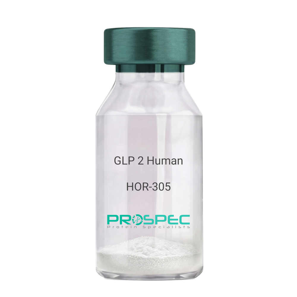
    GLP 2 Human

    Description:

    Human GLP-2

    GLP2, GLP-2, GLP 2.

    Product # :

    HOR-305

    Price :

    Quantity :

    Shipping Method :

    Room Temp Icon

    Shipped at Room temp

    Add To Cart

    More Info

    • description
    • formulation
    • purity
    • More Info

    Description

    GLP-2 contains 34 amino acids having a molecular mass of 3922.35 Dalton.

    Formulation

    The protein (1mg/ml) was lyophilized with no additives.

    Purity

    Greater than 98.0% as determined by RP-HPLC.

    More Info

    • Introduction

      GLP-2 functions as an intestinal growth factor, which stimulates intestinal epithelial growth. GLP2 is involved in diabetes-associated bowel growth. GLP2 enhances cell differentiation, playing a role as a cytokine and in tissue regeneration, and mediating cytoprotection. GLP2 is invloded numerous therapeutic applications. GLP2 regulates signaling pathways coupled to cell proliferation and cell death by apoptosis.
      GLP-2 is produced by specific post-translational proteolytic cleavage of proGLP. GLP-2 is manufactured by the intestinal endocrine L cell and by several neurons in the central nervous system.

    • Synonyms

      GLP2, GLP-2, GLP 2, Glucagon Like Peptide-2.

    • Physical Appearance

      Sterile Filtered White lyophilized (freeze-dried) powder.

    • Stability

      Lyophilized GLP2 although stable at room temperature for 3 weeks, should be stored desiccated below -18°C. Upon reconstitution GLP2 should be stored at 4°C between 2-7 days and for future use below -18°C.For long term storage it is recommended to add a carrier protein (0.1% HSA or BSA).Please prevent freeze-thaw cycles.

    • Solubility

      It is recommended to reconstitute the lyophilized GLP-2 in sterile 18MΩ-cm H2O not less than 100 µg/ml, which can then be further diluted to other aqueous solutions.

    • Amino Acid Sequence

      His-Ala-Asp-Gly-Ser-Phe-Ser-Asp-Glu-Met-Asn-Thr-Ile-Leu-Asp-Asn-Leu-Ala-Ala-Arg-Asp-Phe-Ile-Asn-Trp-Leu-Ile-Gln-Thr-Lys-Ile-Thr-Asp-Arg.

    • Background

      What is the molecular weight/Mw of GLP 2 HUMAN Protein?
      GLP 2 HUMAN Protein has a total Mw of 3.92kDa.


      What is the Purity of GLP 2 HUMAN Protein?
      GLP 2 HUMAN Protein is >95% pure as determined by SDS-PAGE.

      What is the Biological Activity of GLP 2 HUMAN Protein?
      The biological functionality of GLP 2 HUMAN Protein will be determined in the future.

      What is the amino acid sequence of GLP 2 HUMAN Protein?
      His-Ala-Asp-Gly-Ser-Phe-Ser-Asp-Glu-Met-Asn-Thr-Ile-Leu-Asp-Asn-Leu-Ala-Ala-Arg-Asp-Phe-Ile-Asn-Trp-Leu-Ile-Gln-Thr-Lys-Ile-Thr-Asp-Arg.

      What applications can GLP 2 HUMAN Protein be used in?
      GLP 2 HUMAN Protein can probably be used in western blot, ELISA and Lateral Flow.

      What is the endotoxin level for GLP 2 HUMAN Protein?
      The endotoxin level is minimal, GLP 2 HUMAN Protein was purified using conventional chromatography techniques.


    ProSpec's products are furnished for LABORATORY RESEARCH USE ONLY. The product may not be used as drugs, agricultural or pesticidal products, food additives or household chemicals.

    Glp 2 Human
  • View Data Sheet

    Name :

    SNCA 1-95, Human

    Description:

    Alpha-Synuclein 1-95 Human Recombinant

    SNCA, NACP, PARK1, alpha-Synuclein, Non-A beta component of AD amyloid, Non-A4 component of amyloid precursor, Alpha synuclein, Alpha-synuclein isoform NACP140, alphaSYN, MGC105443, MGC110988, MGC127560, MGC64356, Non A beta component of AD amyloid, Non A4 component of amyloid precursor, Non-A-beta component of alzheimers disease amyloid, precursor of PARK 1, PARK 4, PARK4, Parkinson disease familial 1, PD 1, PD1, Synuclein alpha.

    Product # :

    PRO-2625

    Price :

    Quantity :

    Shipping Method :

    Ice Icon

    Shipped with Ice Packs

    Add To Cart

    More Info

    • description
    • source
    • formulation
    • purity
    • More Info

    Description

    SNCA Human Recombinant produced in E.Coli is a single, non-glycosylated polypeptide chain containing 95 amino acids (1-95 a.a.) and having a molecular mass of 9.3kDa.SNCA is purified by proprietary chromatographic techniques.

    Source

    E.coli.

    Formulation

    SNCA protein solution (1mg/ml) containing 20 mM Tris-HCl buffer (pH 7.5) and 0.1 M NaCl.

    Purity

    Greater than 95.0% as determined by SDS-PAGE.

    More Info

    • Introduction

      Alpha-synuclein or SNCA is a synuclein protein. SNCA mainly found in the brain, small concentration of the protein can also be located in other tissues such as heart and muscle. When looking in the brain tissue, SNCA is located in the end of the neuron, in an area called presynaptic terminal. In the presynaptic terminal SNCA has interaction with phospholipids & other proteins. Neurotransmitters are released from the synaptic vesicles in the Presynaptic terminals and act as messengers. Once released, the neurotransmitters send signals across the neurons that are crucial for the brain’s operation.

    • Synonyms

      SNCA, NACP, PARK1, alpha-Synuclein, Non-A beta component of AD amyloid, Non-A4 component of amyloid precursor, Alpha synuclein, Alpha-synuclein isoform NACP140, alphaSYN, MGC105443, MGC110988, MGC127560, MGC64356, Non A beta component of AD amyloid, Non A4 component of amyloid precursor, Non-A-beta component of alzheimers disease amyloid, precursor of PARK 1, PARK 4, PARK4, Parkinson disease familial 1, PD 1, PD1, Synuclein alpha.

    • Physical Appearance

      Sterile Filtered colorless solution.

    • Stability

      Store at 4°C if entire vial will be used within 2-4 weeks. Store, frozen at -20°C for longer periods of time. For long term storage it is recommended to add a carrier protein (0.1% HSA or BSA).Avoid multiple freeze-thaw cycles.

    • Amino Acid Sequence

      MDVFMKGLSK AKEGVVAAAE KTKQGVAEAA GKTKEGVLYV GSKTKEGVVH GVATVAEKTK EQVTNVGGAV VTGVTAVAQK TVEGAGSIAA ATGFV

    ProSpec's products are furnished for LABORATORY RESEARCH USE ONLY. The product may not be used as drugs, agricultural or pesticidal products, food additives or household chemicals.

    Alpha Synuclein
  • View Data Sheet

    Name :

    GAPDH Antibody

    Description:

    Glyceraldehyde-3-Phosphate Dehydrogenase, Mouse Anti Human

    G3PD, GAPD, MGC88685, GAPDH, Glyceraldehyde-3-Phosphate Dehydrogenase.

    Product # :

    ANT-005

    Price :

    Quantity :

    Shipping Method :

    Ice Icon

    Shipped with Ice Packs

    Add To Cart

    More Info

    • formulation
    • More Info

    Formulation

    1mg/ml containing PBS, pH-7.4, & 0.1% Sodium Azide.

    More Info

    • Introduction

      GAPDH is a catalytic enzyme normally known to play a role in glycolysis. GAPDH exists as a tetramer composed of 36-kDa subunits and has a range of intracellular functions. GAPDH catalyzes the reversible reduction of 1,3-bisphosphoglycerate to glyceraldehyde 3-phosphophate in the presence of NADPH. Besides functioning as a glycolytic enzyme in cytoplasm, GAPDH has function in intracellular processes such as membrane fusion, microtubule bundling, phosphotransferase activity, nuclear RNA export, DNA replication and DNA repair. GAPDH catalyzes a vital energy-yielding step in carbohydrate metabolism, the reversible oxidative phosphorylation of glyceraldehyde-3-phosphate in the presence of inorganic phosphate and nicotinamide adenine dinucleotide (NAD). The enzyme exists as a tetramer of identical chains.

    • Synonyms

      G3PD, GAPD, MGC88685, GAPDH, Glyceraldehyde-3-Phosphate Dehydrogenase.

    • Physical Appearance

      Sterile Filtered clear solution.

    • Immunogen

      Anti-human GAPDH mAb, is derived from hybridization of mouse F0 myeloma cells with spleen cells from BALB/c mice immunized with recombinant human GAPDH amino acids 1-335 purified from E. coli.

    • Ig Subclass

      Mouse IgG2b heavy chain and κ light chain.

    • Clone

      PAT8G4AT.

    • Applications

      GAPDH antibody has been tested by ELISA, Western blot and Immunofluorescence analysis to assure specificity and reactivity. Since application varies, however, each investigation should be titrated by the reagent to obtain optimal results. Recommended dilution range for Western blot analysis and Immunofluorescence is 1:500 ~ 10,000.
      Recommended starting dilution is 1:1,000.

    • Type

      Mouse Anti Human Monoclonal.

    • Storage Procedures

      For periods up to 1 month store at 4°C, for longer periods of time, store at -20°C. Prevent freeze thaw cycles.

    • Purification Method

      GAPDH antibody was purified from mouse ascitic fluids by protein-G affinity chromatography.

    ProSpec's products are furnished for LABORATORY RESEARCH USE ONLY. The product may not be used as drugs, agricultural or pesticidal products, food additives or household chemicals.

    Gapdh Antibody
  • View Data Sheet

    Name :

    G CSF Human, HEK

    Description:

    Granulocyte-Colony Stimulating Factor Human Recombinant, HEK

    CSF-3, MGI-1G, GM-CSF beta, Pluripoietin, Filgrastim, Lenograstim, G-CSF, MGC45931, GCSF.

    Product # :

    CYT-088

    Price :

    Quantity :

    Shipping Method :

    Room Temp Icon

    Shipped at Room temp

    Add To Cart

    More Info

    • description
    • source
    • formulation
    • purity
    • biological activity
    • More Info

    Description

    G-CSF Human Recombinant produced in HEK cells is a glycosylated monomer, having a molecular weight range of 21-25kDa due to glycosylation.The G-CSF is purified by proprietary chromatographic techniques.

    Source

    HEK.

    Formulation

    The G-CSF was lyophilized from 1mg/ml in 1xPBS.

    Purity

    Greater than 95% as obsereved by SDS-PAGE.

    Biological Activity

    The specific activity was determined by the dose-dependent stimulation of the proliferation of murine M-NFS-60 cells (Mouse Myeloid Leukemia indicator cell line), the ED50 is <0.01ng/ml.

    More Info

    • Introduction

      GCSF is a cytokine that controls the production, differentiation, and function of granulocytes. The active protein is found extracellularly. Three transcript variants encoding three different isoforms have been found for the GCSF gene. Granulocyte/macrophage colony-stimulating factors are cytokines that act in hematopoiesis by controlling the production, differentiation, and function of 2 related white cell populations of the blood, the granulocytes and the monocytes-macrophages. This csf induces granulocytes.

    • Synonyms

      CSF-3, MGI-1G, GM-CSF beta, Pluripoietin, Filgrastim, Lenograstim, G-CSF, MGC45931, GCSF.

    • Physical Appearance

      Sterile Filtered White lyophilized (freeze-dried) powder.

    • Stability

      Lyophilized G-CSF although stable at room temperature for 3 weeks, should be stored desiccated below -18°C. Upon reconstitution G-CSF should be stored at 4°C between 2-7 days and for future use below -18°C.For long term storage it is recommended to add a carrier protein (0.1% HSA or BSA).Please prevent freeze-thaw cycles.

    • Solubility

      It is recommended to reconstitute the lyophilized G-CSF in sterile 1xPBS containing 0.1% endotoxin-free recombinant HSA.

    • Background

      What is the molecular weight/Mw of G CSF HUMAN, HEK Protein?
      G CSF HUMAN, HEK Protein has a total Mw of 23kDa.

      What is the source or expression system of G CSF HUMAN, HEK Protein?
      HEK.

      What is the Purity of G CSF HUMAN, HEK Protein?
      G CSF HUMAN, HEK Protein is >95% pure as determined by SDS-PAGE.

      What is the Biological Activity of G CSF HUMAN, HEK Protein?
      The specific activity was determined by the dose-dependent stimulation of the proliferation of murine M-NFS-60 cells (Mouse Myeloid Leukemia indicator cell line), the ED50 is <0.01ng/ml.

      What applications can G CSF HUMAN, HEK Protein be used in?
      G CSF HUMAN, HEK Protein can probably be used in western blot, ELISA and Lateral Flow.

      What is the endotoxin level for G CSF HUMAN, HEK Protein?
      The endotoxin level is minimal, G CSF HUMAN, HEK Protein was purified using conventional chromatography techniques.


    ProSpec's products are furnished for LABORATORY RESEARCH USE ONLY. The product may not be used as drugs, agricultural or pesticidal products, food additives or household chemicals.

    G Csf Human Hek
  • View Data Sheet

    Name :

    HADHB Human

    Description:

    2-Enoyl-Coenzyme A (CoA) Hydratase, Beta Human Recombinant

    Hydroxyacyl-CoA Dehydrogenase/3-Ketoacyl-CoA Thiolase/Enoyl-CoA Hydratase (Trifunctional Protein) Beta Subunit, Hydroxyacyl-Coenzyme A Dehydrogenase/3-Ketoacyl-Coenzyme A Thiolase/Enoyl-Coenzyme A Hydratase (Trifunctional Protein) Beta Subunit, TP-BETA, 3-Ketoacyl-Coenzyme A (CoA) Thiolase Of Mitochondrial Trifunctional Protein Beta Subunit, 2-Enoyl-Coenzyme A (CoA) Hydratase Beta Subunit, Trifunctional Enzyme Subunit Beta Mitochondrial, Mitochondrial Trifunctional Protein, Acetyl-CoA Acyltransferase, Beta-Ketothiolase, Beta Subunit, EC 2.3.1.16, EC 2.3.1, MSTP029, ECHB, MTPB.

    Product # :

    ENZ-845

    Price :

    Quantity :

    Shipping Method :

    Ice Icon

    Shipped with Ice Packs

    Add To Cart

    More Info

    • description
    • source
    • formulation
    • purity
    • More Info

    Description

    HADHB Human Recombinant produced in E.Coli is a single, non-glycosylated polypeptide chain containing 464 amino acids (34-474 a.a) and having a molecular mass of 49.9kDa. HADHB is fused to a 23 amino acid His-tag at N-terminus & purified by proprietary chromatographic techniques.

    Source

    Escherichia Coli.

    Formulation

    HADHB protein solution (0. 5mg/ml) containing 20mM Tris-HCl (pH8.0) and 10% glycerol.

    Purity

    Greater than 90.0% as determined by SDS-PAGE.

    More Info

    • Introduction

      2-Enoyl-Coenzyme A (CoA) Hydratase, Beta (HADHB) is the beta subunit of the mitochondrial trifunctional protein, that catalyzes the last 3 phases of mitochondrial beta-oxidation of long chain fatty acids. HADHB binds RNA and reduces the stability of various mRNAs. Mutations in HADHB cause trifunctional protein deficiency.

    • Synonyms

      Hydroxyacyl-CoA Dehydrogenase/3-Ketoacyl-CoA Thiolase/Enoyl-CoA Hydratase (Trifunctional Protein) Beta Subunit, Hydroxyacyl-Coenzyme A Dehydrogenase/3-Ketoacyl-Coenzyme A Thiolase/Enoyl-Coenzyme A Hydratase (Trifunctional Protein) Beta Subunit, TP-BETA, 3-Ketoacyl-Coenzyme A (CoA) Thiolase Of Mitochondrial Trifunctional Protein Beta Subunit, 2-Enoyl-Coenzyme A (CoA) Hydratase Beta Subunit, Trifunctional Enzyme Subunit Beta Mitochondrial, Mitochondrial Trifunctional Protein, Acetyl-CoA Acyltransferase, Beta-Ketothiolase, Beta Subunit, EC 2.3.1.16, EC 2.3.1, MSTP029, ECHB, MTPB.

    • Physical Appearance

      Sterile Filtered clear solution.

    • Stability

      Store at 4°C if entire vial will be used within 2-4 weeks. Store, frozen at -20°C for longer periods of time. For long term storage it is recommended to add a carrier protein (0.1% HSA or BSA).Avoid multiple freeze-thaw cycles.

    • Amino Acid Sequence

      MGSSHHHHHH SSGLVPRGSH MGSAAPAVQT KTKKTLAKPN IRNVVVVDGV RTPFLLSGTS YKDLMPHDLA RAALTGLLHR TSVPKEVVDY IIFGTVIQEV KTSNVAREAA LGAGFSDKTP AHTVTMACIS ANQAMTTGVG LIASGQCDVI VAGGVELMSD VPIRHSRKMR KLMLDLNKAK SMGQRLSLIS KFRFNFLAPE LPAVSEFSTS ETMGHSADRL AAAFAVSRLE QDEYALRSHS LAKKAQDEGL LSDVVPFKVP GKDTVTKDNG IRPSSLEQMA KLKPAFIKPY GTVTAANSSF LTDGASAMLI MAEEKALAMG YKPKAYLRDF MYVSQDPKDQ LLLGPTYATP KVLEKAGLTM NDIDAFEFHE AFSGQILANF KAMDSDWFAE NYMGRKTKVG LPPLEKFNNW GGSLSLGHPF GATGCRLVMA AANRLRKEGG QYGLVAACAA GGQGHAMIVE AYPK.

    ProSpec's products are furnished for LABORATORY RESEARCH USE ONLY. The product may not be used as drugs, agricultural or pesticidal products, food additives or household chemicals.

    Hadhb Human
  • View Data Sheet

    Name :

    AIMP1 Antibody

    Description:

    Aminoacyl tRNA Synthetase Complex-Interacting Multifunctional Protein 1, Mouse Anti Human

    Aminoacyl tRNA synthase complex-interacting multifunctional protein 1, Multisynthase complex auxiliary component p43, AIMP1, EMAP2, SCYE1, p43, EMAPII.

    Product # :

    ANT-619

    Price :

    Quantity :

    Shipping Method :

    Ice Icon

    Shipped with Ice Packs

    Add To Cart

    More Info

    • formulation
    • More Info

    Formulation

    1mg/ml containing PBS, pH-7.4, 10% Glycerol and 0.02% Sodium Azide.

    More Info

    • Introduction

      AIMP1 (EMPA2 or p43) is a cytokine that is specifically induced by apoptosis, and it is involved in the control of angiogenesis, inflammation, and wound healing. The release of the AIMP1 cytokine renders the tumor-associated vasculature sensitive to tumor necrosis factor. Furthermore, AIMP1 is involved in the stimulation of inflammatory responses after proteolytic cleavage in tumor cells.

    • Synonyms

      Aminoacyl tRNA synthase complex-interacting multifunctional protein 1, Multisynthase complex auxiliary component p43, AIMP1, EMAP2, SCYE1, p43, EMAPII.

    • Physical Appearance

      Sterile filtered colorless solution.

    • Immunogen

      Anti-human AIMP1 mAb, is derived from hybridization of mouse F0 myeloma cells with spleen cells from BALB/c mice immunized with a recombinant human AIMP1 protein 1-336 amino acids purified from E. coli.

    • Ig Subclass

      Mouse IgG2a heavy chain and Kappa light chain.

    • Clone

      PAT6D10AT.

    • Applications

      The antibody has been tested by ELISA, Western blot analysis, Flow cytometry and ICC/IF to assure specificity and reactivity. Since application varies, however, each investigation should be titrated by the reagent to obtain optimal results.

    • Type

      Mouse Anti Human Monoclonal.

    • Storage Procedures

      For periods up to 1 month store at 4°C, for longer periods of time, store at -20°C. Prevent freeze thaw cycles.

    • Purification Method

      AIMP1 antibody was purified by protein-A affinity chromatography.

    ProSpec's products are furnished for LABORATORY RESEARCH USE ONLY. The product may not be used as drugs, agricultural or pesticidal products, food additives or household chemicals.

    Aimp1 Antibody
  • View Data Sheet

    Name :

    ARG1 Human, Active

    Description:

    Arginase-1, Active Human Recombinant

    Arginase-1 liver, Arginase-1, liver, Arginase-1, liver A I, Al, ARG 1, ARG1, Arginase 1, Arginase liver, Arginase type I, Arginase1, Liver type arginase, Type I arginase.

    Product # :

    ENZ-1120

    Price :

    Quantity :

    Shipping Method :

    Ice Icon

    Shipped with Ice Packs

    Add To Cart

    More Info

    • description
    • source
    • formulation
    • purity
    • biological activity
    • More Info

    Description

    ARG1 Human Recombinant produced in E.Coli is a single, non-glycosylated, polypeptide chain containing 330 amino acids ( 1-322aa ) and having a molecular mass of 35.8 kDa. ARG1 is fused to a 8 amino acid His tag at C-terminus and purified by proprietary chromatographic techniques.

    Source

    Escherichia Coli.

    Formulation

    ARG1 protein solution (0.5mg/ml) containing 20mM Tris-HCl buffer (pH8.0) containing 20% glycerol, 2mM DTT and 100mM NaCl.

    Purity

    Greater than 85.0% as determined by SDS-PAGE.

    Biological Activity

    Specific activity is > 150,000 pmol/min/ug, and is defined as the amount of enzyme that hydrolyze 1.0 pmole of arginine to urea per minute at pH 10.5 at 37C.

    More Info

    • Introduction

      Arginase-1 is part of the urea cycle, it catalyzes the hydrolysis of arginine to ornithine and urea. There are two isoforms of mammalian arginase which differ in their tissue location, subcellular localization, immunologic crossreactivity & physiologic role. Arginase-1is a cytosolic enzyme and expressed primarily in the liver tissue. Inherited deficiency in this enzyme may lead toargininemia, which is an autosomal recessive disease in which hyperammonemia is detected.

    • Synonyms

      Arginase-1 liver, Arginase-1, liver, Arginase-1, liver A I, Al, ARG 1, ARG1, Arginase 1, Arginase liver, Arginase type I, Arginase1, Liver type arginase, Type I arginase.

    • Physical Appearance

      Sterile Filtered colorless solution.

    • Stability

      Store at 4°C if entire vial will be used within 2-4 weeks. Store, frozen at -20°C for longer periods of time. For long term storage it is recommended to add a carrier protein (0.1% HSA or BSA).Avoid multiple freeze-thaw cycles.

    • Amino Acid Sequence

      MSAKSRTIGI IGAPFSKGQP RGGVEEGPTV LRKAGLLEKL KEQECDVKDY GDLPFADIPN DSPFQIVKNP RSVGKASEQL AGKVAEVKKN GRISLVLGGD HSLAIGSISG HARVHPDLGV IWVDAHTDIN TPLTTTSGNL HGQPVSFLLK ELKGKIPDVP GFSWVTPCIS AKDIVYIGLR DVDPGEHYIL KTLGIKYFSM TEVDRLGIGK VMEETLSYLL GRKKRPIHLS FDVDGLDPSF TPATGTPVVG GLTYREGLYI TEEIYKTGLL SGLDIMEVNP SLGKTPEEVT RTVNTAVAIT LACFGLAREG NHKPIDYLNP PKLEHHHHHH.

    ProSpec's products are furnished for LABORATORY RESEARCH USE ONLY. The product may not be used as drugs, agricultural or pesticidal products, food additives or household chemicals.

    Arginase 1
  • View Data Sheet

    Name :

    B3GNT2 Human

    Description:

    Beta-1,3-N-Acetylglucosaminyltransferase 2 Human Recombinant

    B3GNT2, B3GN-T2, B3GNT, B3GNT-2, B3GNT1, BETA3GNT, BGnT-2, BGNT2, N-acetyllactosaminide beta-1,3-N-acetylglucosaminyltransferase 2, Beta-1,3-N-acetylglucosaminyltransferase 1, BGnT-1, Beta-1,3-Gn-T1, Beta3Gn-T1, Beta-1,3-galactosyltransferase 7, Beta-1,3-GalTase 7, Beta3Gal-T7, Beta3GalT7, b3Gal-T7, Beta-3-Gx-T7, beta-GlcNAc beta-1,3-galactosyltransferase 7, betaGal beta-1,3-N-acetylglucosaminyltransferase 2, BGnT-2, Beta-1,3-Gn-T2, Beta-1,3-N-acetylglucosaminyltransferase 2, Beta3Gn-T2, beta-N-acetylglucosamine beta-1,3-galactosyltransferase 7.

    Product # :

    ENZ-973

    Price :

    Quantity :

    Shipping Method :

    Ice Icon

    Shipped with Ice Packs

    Add To Cart

    More Info

    • description
    • source
    • formulation
    • purity
    • More Info

    Description

    B3GNT2 produced in Sf9 Baculovirus cells is a single, glycosylated polypeptide chain containing 375 amino acids (29-397a.a.) and having a molecular mass of 43.5kDa (Molecular size on SDS-PAGE will appear at approximately 40-57kDa). B3GNT2 is expressed with a 6 amino acid His tag at C-Terminus and purified by proprietary chromatographic techniques.

    Source

    Sf9, Baculovirus cells.

    Formulation

    B3GNT2 protein solution (0.5mg/ml) contains Phosphate Buffered Saline (pH 7.4) and 10% glycerol.

    Purity

    Greater than 90.0% as determined by SDS-PAGE.

    More Info

    • Introduction

      Beta-1,3-N-Acetylglucosaminyltransferase 2 (B3GNT2) is a part of the beta-1,3-N-acetylglucosaminyltransferase family which takes part in the synthesis of poly-N-acetyllactosamine. B3GNT2 is a type II transmembrane protein which prefers the substrate of lacto-N-neotetraose. B3GNT2 catalyzes the initiation and elongation of poly-N-acetyllactosamine chains and comprises the main polylactosamine synthase.

    • Synonyms

      B3GNT2, B3GN-T2, B3GNT, B3GNT-2, B3GNT1, BETA3GNT, BGnT-2, BGNT2, N-acetyllactosaminide beta-1,3-N-acetylglucosaminyltransferase 2, Beta-1,3-N-acetylglucosaminyltransferase 1, BGnT-1, Beta-1,3-Gn-T1, Beta3Gn-T1, Beta-1,3-galactosyltransferase 7, Beta-1,3-GalTase 7, Beta3Gal-T7, Beta3GalT7, b3Gal-T7, Beta-3-Gx-T7, beta-GlcNAc beta-1,3-galactosyltransferase 7, betaGal beta-1,3-N-acetylglucosaminyltransferase 2, BGnT-2, Beta-1,3-Gn-T2, Beta-1,3-N-acetylglucosaminyltransferase 2, Beta3Gn-T2, beta-N-acetylglucosamine beta-1,3-galactosyltransferase 7.

    • Physical Appearance

      Sterile Filtered colorless solution.

    • Stability

      Store at 4°C if entire vial will be used within 2-4 weeks. Store, frozen at -20°C for longer periods of time. For long term storage it is recommended to add a carrier protein (0.1% HSA or BSA). Avoid multiple freeze-thaw cycles.

    • Amino Acid Sequence

      ADPKSSSQEK NGKGEVIIPK EKFWKISTPP EAYWNREQEK LNRQYNPILS MLTNQTGEAG RLSNISHLNY CEPDLRVTSV VTGFNNLPDR FKDFLLYLRC RNYSLLIDQP DKCAKKPFLL LAIKSLTPHF ARRQAIRESW GQESNAGNQT VVRVFLLGQT PPEDNHPDLS DMLKFESEKH QDILMWNYRD TFFNLSLKEV LFLRWVSTSC PDTEFVFKGD DDVFVNTHHI LNYLNSLSKT KAKDLFIGDV IHNAGPHRDK KLKYYIPEVV YSGLYPPYAG GGGFLYSGHL ALRLYHITDQ VHLYPIDDVY TGMCLQKLGL VPEKHKGFRT FDIEEKNKNN ICSYVDLMLV HSRKPQEMID IWSQLQSAHL KCHHHHHH.

    ProSpec's products are furnished for LABORATORY RESEARCH USE ONLY. The product may not be used as drugs, agricultural or pesticidal products, food additives or household chemicals.

    B3Gnt2 Human
  • View Data Sheet

    Name :

    NTRK2 Human

    Description:

    Neurotrophic Receptor Tyrosine Kinase 2 Human Recombinant

    GP145-TrkB, trk-B, TRKB, NTRK2

    Product # :

    CYT-1159

    Price :

    Quantity :

    Shipping Method :

    Ice Icon

    Shipped with Ice Packs

    Add To Cart

    More Info

    • description
    • source
    • formulation
    • purity
    • More Info

    Description

    NTRK2 Human produced in Sf9 Baculovirus cells is a single, glycosylated polypeptide chain containing 407 amino acids (32-430a.a) and having a molecular mass of 45.2kDa.NTRK2 is fused to an 8 amino acid His tag at C-terminus and purified by proprietary chromatographic techniques.

    Source

    Sf9, Baculovirus cells.

    Formulation

    The NTRK2 solution (0. 5mg/ml) contains 10% Glycerol and Phosphate-Buffered Saline (pH 7.4).

    Purity

    Greater than 95% as determined by SDS-PAGE.

    More Info

    • Introduction

      Neurotrophic Receptor Tyrosine Kinase 2, also referred to NTRK2, is a tyrosine-protein kinase receptor which takes part in the development & the maturation of the central and the peripheral nervous systems through regulation of neuron survival, migration, proliferation, differentiation and synapse formation & plasticity. NTRK2 acts in learning and memory by regulating both short term synaptic function and long-term potentiation. The substrates that are known for the TRK family receptors are: SHC1, PI-3 kinase and PLC-gamma-1.

    • Synonyms

      GP145-TrkB, trk-B, TRKB, NTRK2

    • Physical Appearance

      Sterile Filtered colorless solution.

    • Stability

      Store at 4°C if entire vial will be used within 2-4 weeks. Store, frozen at -20°C for longer periods of time. For long term storage it is recommended to add a carrier protein (0.1% HSA or BSA).Avoid multiple freeze-thaw cycles.

    • Amino Acid Sequence

      CPTSCKCSAS RIWCSDPSPG IVAFPRLEPN SVDPENITEI FIANQKRLEI INEDDVEAYV GLRNLTIVDS GLKFVAHKAF LKNSNLQHIN FTRNKLTSLS RKHFRHLDLS ELILVGNPFT CSCDIMWIKT LQEAKSSPDT QDLYCLNESS KNIPLANLQI PNCGLPSANL AAPNLTVEEG KSITLSCSVA GDPVPNMYWD VGNLVSKHMN ETSHTQGSLR ITNISSDDSG KQISCVAENL VGEDQDSVNL TVHFAPTITF LESPTSDHHW CIPFTVKGNP KPALQWFYNG AILNESKYIC TKIHVTNHTE YHGCLQLDNP THMNNGDYTL IAKNEYGKDE KQISAHFMGW PGIDDGANPN YPDVIYEDYG TAANDIGDTT NRSNEIPSTD VTDKTGREHL EHHHHHH

    ProSpec's products are furnished for LABORATORY RESEARCH USE ONLY. The product may not be used as drugs, agricultural or pesticidal products, food additives or household chemicals.

    Ntrk2 Human
  • View Data Sheet

    Name :

    GPD1L Antibody

    Description:

    Glycerol-3-Phosphate Dehydrogenase 1 Like, Mouse Anti Human

    Glycerol-3-phosphate dehydrogenase 1-like protein, GPD1-L, GPD1L, KIAA0089.

    Product # :

    ANT-638

    Price :

    Quantity :

    Shipping Method :

    Ice Icon

    Shipped with Ice Packs

    Add To Cart

    More Info

    • formulation
    • More Info

    Formulation

    1mg/ml containing PBS, pH-7.4, 10% Glycerol and 0.02% Sodium Azide.

    More Info

    • Introduction

      Glycerol-3-phosphate dehydrogenase 1-like protein (GPD1L) converts sn-glycerol 3-phosphate to glycerone phosphate. GPD1L is found in the cytoplasm, associated with the plasma membrane, where it binds the sodium channel, voltage-gated, type V, alpha subunit (SCN5A). Mutations in the GPD1L gene are the cause of SIDS (sudden infant death syndrome) and Brugada syndrome type 2 (an autosomal dominant tachyarrhythmia).

    • Synonyms

      Glycerol-3-phosphate dehydrogenase 1-like protein, GPD1-L, GPD1L, KIAA0089.

    • Physical Appearance

      Sterile filtered colorless solution.

    • Immunogen

      Anti-human GPD1L mAb, is derived from hybridization of mouse F0 myeloma cells with spleen cells from BALB/c mice immunized with recombinant human GPD1L amino acids 1-351 purified from E. coli.

    • Ig Subclass

      Mouse IgG1 heavy chain and k light chain.

    • Clone

      PAT14E2AT.

    • Applications

      GPD1L antibody has been tested by ELISA, Western blot analysis, Flow cytometry and ICC/IF to assure specificity and reactivity. Since application varies, however, each investigation should be titrated by the reagent to obtain optimal results.

    • Type

      Mouse Anti Human Monoclonal.

    • Storage Procedures

      For periods up to 1 month store at 4°C, for longer periods of time, store at -20°C. Prevent freeze thaw cycles.

    • Purification Method

      GPD1L antibody was purified from mouse ascitic fluids by protein-A affinity chromatography.

    ProSpec's products are furnished for LABORATORY RESEARCH USE ONLY. The product may not be used as drugs, agricultural or pesticidal products, food additives or household chemicals.

    Gpd1L Antibody
  • View Data Sheet

    Name :

    EGFR Human, CHO

    Description:

    Epidermal Growth Factor Receptor, CHO Human Recombinant

    Epidermal Growth Factor Receptor, Receptor Tyrosine-Protein Kinase ErbB-1, Erb-B2 Receptor Tyrosine Kinase, Proto-Oncogene C-ErbB-1, EC 2.7.10.1, ERBB1, ERBB, HER1, Epidermal Growth Factor Receptor (Avian Erythroblastic Leukemia Viral (V-Erb-B) Oncogene Homolog), Erythroblastic Leukemia Viral (V-Erb-B) Oncogene Homolog (Avian), Avian Erythroblastic Leukemia Viral (V-Erb-B) Oncogene Homolog, Cell Proliferation-Inducing Protein 61, Cell Growth Inhibiting Protein 40, EC 2.7.10, NISBD2, PIG61, MENA.

    Product # :

    PKA-086

    Price :

    Quantity :

    Shipping Method :

    Ice Icon

    Shipped with Ice Packs

    Add To Cart

    More Info

    • description
    • source
    • formulation
    • purity
    • More Info

    Description

    EGFR produced in CHO cells is a single, glycosylated polypeptide chain containing 860 amino acids (25-645 a.a.) and having a molecular mass of 95.5 kDa (Migrates at 100-150 on SDS-PAGE under reducing conditions). EGFR is expressed with a 239 amino acid hIgG-His tag at C-Terminus and purified by proprietary chromatographic techniques.

    Source

    Chinese Hamster Ovary cells.

    Formulation

    EGFR protein solution (0.25mg/ml) contains Phosphate Buffered Saline (pH 7.4) and 10% glycerol.

    Purity

    Greater than 90.0% as determined by SDS-PAGE.

    More Info

    • Introduction

      The epidermal growth factor receptor (EGF R) subfamily of receptor tyrosine kinases comprises four members: EGF R (also known as HER1, ErbB1 or ErbB), ErbB2 (Neu, HER-2), ErbB3 (HER-3), and ErbB4 (HER-4). All family members are type I transmembrane glycoprotein that has an extracellular domain which contains two cysteine-rich domains separated by a spacer region that is involved in ligand-binding, and a cytoplasmic domain which has a membrane-proximal tyrosine kinase domain and a C-terminal tail with multiple tyrosine autophosphorylation sites. The human EGF R gene encodes a 1210 amino acid (aa) residue precursor with a 24 aa putative signal peptide, a 621 aa extracellular domain, a 23 aa transmembrane domain, and a 542 aa cytoplasmic domain. EGF R has been shown to bind a subset of the EGF family ligands, including EGF, amphiregulin, TGFa , betacellulin, epiregulin, heparin-binding EGF and neuregulin-2 in the absence of a co-receptor. Ligand binding induces EGF R homodimerization as well as heterdimerization with ErbB2, resulting in kinase activation, tyrosine phosphorylation and cell signaling. EGF R can also be recruited to form heterodimers with the ligand-activated ErbB3 or ErbB4. EGF R signaling has been shown to regulate multiple biological functions including cell proliferation, differentiation, motility and apoptosis. In addition, EGF R signaling has also been shown to play a role in carcinogenesis.

    • Synonyms

      Epidermal Growth Factor Receptor, Receptor Tyrosine-Protein Kinase ErbB-1, Erb-B2 Receptor Tyrosine Kinase, Proto-Oncogene C-ErbB-1, EC 2.7.10.1, ERBB1, ERBB, HER1, Epidermal Growth Factor Receptor (Avian Erythroblastic Leukemia Viral (V-Erb-B) Oncogene Homolog), Erythroblastic Leukemia Viral (V-Erb-B) Oncogene Homolog (Avian), Avian Erythroblastic Leukemia Viral (V-Erb-B) Oncogene Homolog, Cell Proliferation-Inducing Protein 61, Cell Growth Inhibiting Protein 40, EC 2.7.10, NISBD2, PIG61, MENA.

    • Physical Appearance

      Sterile Filtered colorless solution.

    • Stability

      Store at 4°C if entire vial will be used within 2-4 weeks. Store, frozen at -20°C for longer periods of time. For long term storage it is recommended to add a carrier protein (0.1% HSA or BSA). Avoid multiple freeze-thaw cycles.

    • Amino Acid Sequence

      LEEKKVCQGT SNKLTQLGTF EDHFLSLQRM FNNCEVVLGN LEITYVQRNY DLSFLKTIQE VAGYVLIALN TVERIPLENL QIIRGNMYYE NSYALAVLSN YDANKTGLKE LPMRNLQEIL HGAVRFSNNP ALCNVESIQW RDIVSSDFLS NMSMDFQNHL GSCQKCDPSC PNGSCWGAGE ENCQKLTKII CAQQCSGRCR GKSPSDCCHN QCAAGCTGPR ESDCLVCRKF RDEATCKDTC PPLMLYNPTT YQMDVNPEGK YSFGATCVKK CPRNYVVTDH GSCVRACGAD SYEMEEDGVR KCKKCEGPCR KVCNGIGIGE FKDSLSINAT NIKHFKNCTS ISGDLHILPV AFRGDSFTHT PPLDPQELDI LKTVKEITGF LLIQAWPENR TDLHAFENLE IIRGRTKQHG QFSLAVVSLN ITSLGLRSLK EISDGDVIIS GNKNLCYANT INWKKLFGTS GQKTKIISNR GENSCKATGQ VCHALCSPEG CWGPEPRDCV SCRNVSRGRE CVDKCNLLEG EPREFVENSE CIQCHPECLP QAMNITCTGR GPDNCIQCAH YIDGPHCVKT CPAGVMGENN TLVWKYADAG HVCHLCHPNC TYGCTGPGLE GCPTNGPKIP SRSPKSCDKT HTCPPCPAPE LLGGPSVFLF PPKPKDTLMI SRTPEVTCVV VDVSHEDPEV KFNWYVDGVE VHNAKTKPRE EQYNSTYRVV SVLTVLHQDW LNGKEYKCKV SNKALPAPIE KTISKAKGQP REPQVYTLPP SRDELTKNQV SLTCLVKGFY PSDIAVEWES NGQPENNYKT TPPVLDSDGS FFLYSKLTVD KSRWQQGNVF SCSVMHEALH NHYTQKSLSL SPGKHHHHHH.

    ProSpec's products are furnished for LABORATORY RESEARCH USE ONLY. The product may not be used as drugs, agricultural or pesticidal products, food additives or household chemicals.

    Egfr Human Cho
  • View Data Sheet

    Name :

    L-Asparaginase

    Description:

    L-Asparaginase

    Product # :

    ENZ-287

    Price :

    Quantity :

    Shipping Method :

    Room Temp Icon

    Shipped at Room temp

    Add To Cart

    More Info

    • description
    • source
    • formulation
    • purity
    • biological activity
    • More Info

    Description

    L-asparaginase was purified from E.coli ASI.357.

    Source

    Escherichia Coli.

    Formulation

    The enzyme was lyophilized with no additives.

    Purity

    Greater than 96.0% as determined by SDS-PAGE.

    Biological Activity

    One IU of L- Asparaginase is defined as that amount of enzyme required to generate 1 µmol of ammonia per minute at pH 7.3 and 37°C.

    More Info

    • Introduction

      L-Asparaginase is an enzyme that depletes L-Asparagine "an important nutrient for cancer cells" resulting in cancer/tumor cell starvation. L-asparaginase is an anti-tumor agent derived from E.coli.,which can inhibit the growth of malignant cells. It is used mainly for the induction of remission in acute lymphoblastic leukaemia. Because of the lymph node origin of malignant B cells in Multiple Myeloma, L-Asparagine is an essential amino acid for their cell metabolism, and, consequently, L-Asparaginase may be of value in managing the disease.
      The rationale behind asparaginase is that it takes advantage of the fact that ALL cellsare unable to synthesize the non-essential amino acidasparaginewhereas normal cells are able to make their own asparagine. These leukemic cells depend on circulating asparagine. Asparaginase however catalyzes the conversion of L-asparagine to aspartic acidand ammonia. This deprives the leukemic cell of circulating asparagine.

    • Physical Appearance

      Sterile Filtered White lyophilized (freeze-dried) powder.

    • Stability

      Lyophilized L-Asparaginase although stable at room temperature for 3 weeks, should be stored desiccated below -18°C. Upon reconstitution L-Asparaginase should be stored at 4°C between 2-7 days and for future use below -18°C. Please prevent freeze-thaw cycles.

    • Solubility

      It is recommended to reconstitute the lyophilized L-Asparaginase in 18M-cm H2O at 1mg/ml.

    • Background

      L-Asparaginase Enzyme: Unraveling Its Therapeutic Potential in Cancer Treatment

      Abstract: L-Asparaginase is an enzyme that plays a crucial role in cancer therapy by depleting the levels of asparagine in the blood, thereby inhibiting the growth of cancer cells.

      This research paper provides a comprehensive analysis of L-Asparaginase, including its biochemical properties, mechanisms of action, therapeutic applications, and clinical implications.

      The paper explores the significance of L-Asparaginase as a key enzyme in cancer treatment and highlights its potential for improving patient outcomes.

      1. Introduction L-Asparaginase is an enzyme widely used in cancer therapy, particularly in the treatment of acute lymphoblastic leukemia (ALL). This section introduces L-Asparaginase and its role in cancer treatment, emphasizing its importance in medical research and clinical practice.
      2. Biochemical Properties of L-Asparaginase L-Asparaginase belongs to the class of enzymes known as hydrolases and catalyzes the hydrolysis of asparagine to aspartic acid and ammonia. This section discusses the biochemical properties of L-Asparaginase, including its structure, catalytic mechanism, and factors influencing its activity and stability.
      3. Mechanisms of Action L-Asparaginase exerts its anti-cancer effects by depleting circulating asparagine, an essential amino acid for cancer cell survival. This section delves into the mechanisms of action of L-Asparaginase, including its ability to selectively target cancer cells and induce metabolic stress, leading to cell death. The impact of asparagine deprivation on cancer cell metabolism and survival is also explored.
      4. Therapeutic Applications of L-Asparaginase L-Asparaginase has demonstrated therapeutic efficacy in the treatment of various malignancies, including ALL and certain solid tumors. This section provides an overview of the therapeutic applications of L-Asparaginase, highlighting its use as a first-line treatment in ALL and its potential in other cancer types. The challenges and limitations associated with L-Asparaginase therapy are also discussed.
      5. Clinical Implications and Future Perspectives L-Asparaginase therapy has shown promising results in improving patient outcomes, but it is not without side effects and challenges. This section discusses the clinical implications of L-Asparaginase treatment, including its impact on patient survival, toxicity profile, and the development of resistance. Additionally, the future prospects of L-Asparaginase therapy, such as the development of novel formulations and combination strategies, are explored.
      6. Conclusion L-Asparaginase is a vital enzyme in cancer treatment, particularly in the management of ALL. This research paper has provided a comprehensive analysis of L-Asparaginase, highlighting its biochemical properties, mechanisms of action, therapeutic applications, and clinical implications. Further research on L-Asparaginase and its optimization in cancer therapy will enhance our understanding and pave the way for improved treatments.

    • Unit Definition

      One unit of enzyme catalyzes hydrolyzation of 10 nanomoles of dUTP to dUMP in one hour at 85 Centigrade.

    • Specific Activity

      250IU/mg.

    ProSpec's products are furnished for LABORATORY RESEARCH USE ONLY. The product may not be used as drugs, agricultural or pesticidal products, food additives or household chemicals.

    L Asparaginase
  • View Data Sheet

    Name :

    UBE2M Human

    Description:

    Ubiquitin Conjugating Enzyme E2M Human Recombinant

    NEDD8-conjugating enzyme Ubc12, Ubiquitin-conjugating enzyme E2 M, NEDD8 protein ligase, NEDD8 carrier protein, UBC12, hUbc12, UBC-RS2.

    Product # :

    ENZ-345

    Price :

    Quantity :

    Shipping Method :

    Room Temp Icon

    Shipped at Room temp

    Add To Cart

    More Info

    • description
    • source
    • formulation
    • purity
    • More Info

    Description

    UBE2M Human Recombinant produced E. coli is a single polypeptide chain containing 207 amino acids (1-183) and having a molecular mass of 23.5kDa.UBE2M is fused to a 24 amino acid His-tag at N-terminus & purified by proprietary chromatographic techniques.

    Source

    Escherichia Coli.

    Formulation

    The UBE2M solution (1mg/1ml) contains 20mM Tris-HCl buffer (pH 8.0), 0.2M NaCl, 1mM DTT and 20% glycerol.

    Purity

    Greater than 95% as determined by SDS-PAGE.

    More Info

    • Introduction

      UbcH12 is functional in in vitro NEDDylation reactions. It has been shown to form a thioester linkage with NEDD8 in the presence of the NEDD8 activating enzyme complex Uba3/APP-BP1. APP-BP1 binds to the amyloid precursor protein (APP) carboxy terminal domain and is important in conjunction with Uba3 and UbcH12 in driving cells through the S to M checkpoint. It was demonstrated to be the E2 responsible for the NEDDylation of the Cul-1 component of the SCF (?-TRCP) complex which is important as the E3-ligase in the ubiquitinylation of I?B?. NEDDylation of Cul-1 is essential for conjugation and processing of NF-?B p105 by SCF (?-TRCP) following phosphorylation of the complex. A dominant negative form of UbcH12, previously demonstrated to sequester NEDD8 and inhibit its conjugation, inhibits both conjugation and processing of p105, which is alleviated by wild-type UbcH12.

    • Synonyms

      NEDD8-conjugating enzyme Ubc12, Ubiquitin-conjugating enzyme E2 M, NEDD8 protein ligase, NEDD8 carrier protein, UBC12, hUbc12, UBC-RS2.

    • Physical Appearance

      Sterile filtered colorless solution.

    • Stability

      Store at 4°C if entire vial will be used within 2-4 weeks. Store, frozen at -20°C for longer periods of time. For long term storage it is recommended to add a carrier protein (0.1% HSA or BSA).Avoid multiple freeze-thaw cycles.

    • Amino Acid Sequence

      MGSSHHHHHH SSGLVPRGSH MGSHMIKLFS LKQQKKEEES AGGTKGSSKK ASAAQLRIQK DINELNLPKT CDISFSDPDD LLNFKLVICP DEGFYKSGKF VFSFKVGQGY PHDPPKVKCE TMVYHPNIDL EGNVCLNILR EDWKPVLTIN SIIYGLQYLF LEPNPEDPLN KEAAEVLQNN RRLFEQNVQR SMRGGYIGST YFERCLK

    ProSpec's products are furnished for LABORATORY RESEARCH USE ONLY. The product may not be used as drugs, agricultural or pesticidal products, food additives or household chemicals.

    Ube2M Human
  • View Data Sheet

    Name :

    SIRPA Human

    Description:

    Signal-Regulatory Protein Alpha Human Recombinant

    Signal-Regulatory Protein Alpha, SHPS1, CD172 Antigen-Like Family Member A, Inhibitory Receptor SHPS-1, Macrophage Fusion Receptor, PTPNS1, SIRP, P84, BIT, MFR, Brain-Immunoglobulin-Like Molecule With Tyrosine-Based Activation Motifs, Brain Ig-Like Molecule With Tyrosine-Based Activation Motifs, Protein Tyrosine Phosphatase, Non-Receptor Type Substrate 1, Tyrosine-Protein Phosphatase Non-Receptor Type Substrate 1, Tyrosine Phosphatase SHP Substrate 1, Signal-Regulatory Protein Alpha-1 , ignal-Regulatory Protein Alpha-2, Signal-Regulatory Protein Alpha-3, SHP Substrate 1, CD172a Antigen, Myd-1 Antigen, MyD-1 Antigen, Sirp-Alpha-1, Sirp-Alpha-2, Sirp-Alpha-3, CD172A, SHPS-1, MYD-1, MYD1, Tyrosine-protein phosphatase non-receptor type substrate 1.

    Product # :

    PRO-2080

    Price :

    Quantity :

    Shipping Method :

    Ice Icon

    Shipped with Ice Packs

    Add To Cart

    More Info

    • description
    • source
    • formulation
    • purity
    • More Info

    Description

    SIRPA Human Recombinant produced in E.Coli is a single, non-glycosylated polypeptide chain containing 370 amino acids (27-373 a.a) and having a molecular mass of 40.4kDa. SIRPA is fused to a 23 amino acid His-tag at N-terminus & purified by proprietary chromatographic techniques.

    Source

    Escherichia Coli.

    Formulation

    SIRPA protein solution (0.5mg/ml) containing PBS buffer (pH 7.4), 10% glycerol and 1mM DTT.

    Purity

    Greater than 90.0% as determined by SDS-PAGE.

    More Info

    • Introduction

      Signal-Regulatory Protein Alpha, SIRPA belongs to the signal-regulatory-protein (SIRP) family, as well as theimmunoglobulin super family. The members of the SIRP family are receptor-type transmembrane glycoproteins which areinvolved in the negative regulation of receptor tyrosine kinase-coupled signaling processes. SIRPA can bephosphorylated by tyrosine kinases. The phospho-tyrosine residues of this PTP have been shown to recruit SH2domain containing tyrosine phosphatases (PTP), and perform as substrates of PTPs. SIRPA take part insignal transduction mediated by a variety of growth factor receptors. CD47 has been shown to be a ligand for SIRPA.

    • Synonyms

      Signal-Regulatory Protein Alpha, SHPS1, CD172 Antigen-Like Family Member A, Inhibitory Receptor SHPS-1, Macrophage Fusion Receptor, PTPNS1, SIRP, P84, BIT, MFR, Brain-Immunoglobulin-Like Molecule With Tyrosine-Based Activation Motifs, Brain Ig-Like Molecule With Tyrosine-Based Activation Motifs, Protein Tyrosine Phosphatase, Non-Receptor Type Substrate 1, Tyrosine-Protein Phosphatase Non-Receptor Type Substrate 1, Tyrosine Phosphatase SHP Substrate 1, Signal-Regulatory Protein Alpha-1 , ignal-Regulatory Protein Alpha-2, Signal-Regulatory Protein Alpha-3, SHP Substrate 1, CD172a Antigen, Myd-1 Antigen, MyD-1 Antigen, Sirp-Alpha-1, Sirp-Alpha-2, Sirp-Alpha-3, CD172A, SHPS-1, MYD-1, MYD1, Tyrosine-protein phosphatase non-receptor type substrate 1.

    • Physical Appearance

      Sterile Filtered colorless solution.

    • Stability

      Store at 4°C if entire vial will be used within 2-4 weeks. Store, frozen at -20°C for longer periods of time. For long term storage it is recommended to add a carrier protein (0.1% HSA or BSA).Avoid multiple freeze-thaw cycles.

    • Amino Acid Sequence

      MGSSHHHHHH SSGLVPRGSH MGSGVAGEEE LQVIQPDKSV LVAAGETATL RCTATSLIPV GPIQWFRGAG PGRELIYNQK EGHFPRVTTV SDLTKRNNMD FSIRIGNITP ADAGTYYCVK FRKGSPDDVE FKSGAGTELS VRAKPSAPVV SGPAARATPQ HTVSFTCESH GFSPRDITLK WFKNGNELSD FQTNVDPVGE SVSYSIHSTA KVVLTREDVH SQVICEVAHV TLQGDPLRGT ANLSETIRVP PTLEVTQQPV RAENQVNVTC QVRKFYPQRL QLTWLENGNV SRTETASTVT ENKDGTYNWM SWLLVNVSAH RDDVKLTCQV EHDGQPAVSK SHDLKVSAHP KEQGSNTAAE NTGSNERNIY.

    ProSpec's products are furnished for LABORATORY RESEARCH USE ONLY. The product may not be used as drugs, agricultural or pesticidal products, food additives or household chemicals.

    Sirpa Human
  • 1
  • …
  • 16
  • 17
  • 18
  • 19
  • 20
Your browser does not support the video tag.

Products

  • Cytokines
  • Growth Factors
  • Chemokines
  • CD Antigens
  • Neurotrophins
  • Hormones
  • Enzymes
  • Viral Antigens
  • Recombinant Proteins
  • Natural Proteins
  • Monoclonal Antibodies
  • Polyclonal Antibodies
  • Thermal Packaging

About

  • Company
  • Founder
  • Contribution
  • Contact Us
  • My Account

Ordering

  • Ordering Method
  • Ordering - Credit Card
  • Ordering PO
  • Shipping Fees
  • FAQ's

Legal

  • Terms & Conditions
  • Privacy
  • Site Map
  • info@prospecbio.com
  • Facebook
  • Instagram
  • Linkedin Profile

© 2026 ProSpec-Tany TechnoGene Ltd. All Rights Reserved.

  • Choosing a selection results in a full page refresh.
  • Opens in a new window.