Search results
453 results found for “glycogen synthase kinase”
Name
Description
Product #
Price
Quantity
Shipping Method
- View Data Sheet
Name :
SUGT1 HumanDescription:
SGT1 Recombinant Human
SGT1, suppressor of G2 allele of SKP1 (S. cerevisiae), SGT1, Putative 40-6-3 protein, SGT1B protein.
Product # :
PRO-096Price :
Quantity :
Shipping Method :
Shipped with Ice Packs
More Info
- description
- source
- formulation
- purity
- More Info
Description
SGT1 produced in E.Coli is a single, non-glycosylated polypeptide chain containing 252 amino acids (115-365.a.a) and having a molecular mass of 30.7kDa. SGT1 is fused to a 21 amino acid His-tag at N-terminus & purified by proprietary chromatographic techniques.
Source
Escherichia Coli.
Formulation
The SGT1 protein solution (0.5mg/1ml) is formulated in 20 mM Tris-HCl buffer (pH8.0), 0.1M NaCl, 1mM DTT and 20% glycerol.
Purity
Greater than 90% as determined by SDS-PAGE.
More Info
-
Introduction
SUGT1 is homolog to yeast protein SGT1, a regulator of the cell cycle which is vital for G1/S and G2/M transitions. SUGT1 holds a CS domain, a SGS domain, a p23 domain and three tetratricopeptide repeats (TPR). SUGT1 protein associates with Skp1 p19 and CUL-1, subunits of the SCF ubiquitin ligase complex, and is believed to take a part in protein degradation. Furthermore, SUGT1 is essential for the kinetochores assembly, and has a role as a co-chaperone for HSP90.
-
Synonyms
SGT1, suppressor of G2 allele of SKP1 (S. cerevisiae), SGT1, Putative 40-6-3 protein, SGT1B protein.
-
Physical Appearance
Sterile Filtered clear solution.
-
Stability
Store at 4°C if entire vial will be used within 2-4 weeks. Store, frozen at -20°C for longer periods of time.Please avoid freeze thaw cycles.
-
Amino Acid Sequence
MGSSHHHHHH SSGLVPRGSH MHRVGQAGLQ LLTSSDPPAL DSQSAGITGA DANFSVWIKR CQEAQNGSES EVWTHQSKIK YDWYQTESQVVITLMIKNVQ KNDVNVEFSE KELSALVKLP SGEDYNLKLE LLHPIIPEQS TFKVLSTKIE IKLKKPEAVR WEKLEGQGDV PTPKQFVADVKNLYPSSSPY TRNWDKLVGE IKEEEKNEKL EGDAALNRLF QQIYSDGSDE VKRAMNKSFM ESGGTVLSTN WSDVGKRKVE INPPDDMEWKKY
ProSpec's products are furnished for LABORATORY RESEARCH USE ONLY. The product may not be used as drugs, agricultural or pesticidal products, food additives or household chemicals.
- View Data Sheet
Name :
FAHD1 HumanDescription:
Fumarylacetoacetate Hydrolase Domain Containing 1 Human Recombinant
Fumarylacetoacetate hydrolase domain-containing protein 1, YisK-like protein, FAHD1, C16orf36, YISKL, MGC74876, DKFZp566J2046.
Product # :
ENZ-067Price :
Quantity :
Shipping Method :
Shipped with Ice Packs
More Info
- description
- source
- formulation
- purity
- More Info
Description
FAHD1 Human Recombinant fused with a 20 amino acid His tag at N-terminus produced in E.Coli is a single, non-glycosylated, polypeptide chain containing 244 amino acids (1-224 a.a.) and having a molecular mass of 27kDa. The FAHD1 is purified by proprietary chromatographic techniques.
Source
Escherichia Coli.
Formulation
The FAHD1 solution (1 mg/ml) contains 20mM Tris-HCl buffer (pH 8.0), 1mM DTT, 10% glycerol and 0.1M NaCl.
Purity
Greater than 95.0% as determined by SDS-PAGE.
More Info
-
Introduction
Fumarylacetoacetate hydrolase domain-containing protein 1 (FAHD1) is a member of the FAH family. FAHD1 is considered to have hydrolase activity and it uses Magnesium and Calcium as cofactors. It seems quite likely that the metal binding sites are involved in an enzymatic activity connected to the catabolism of aromatic amino acids.
-
Synonyms
Fumarylacetoacetate hydrolase domain-containing protein 1, YisK-like protein, FAHD1, C16orf36, YISKL, MGC74876, DKFZp566J2046.
-
Physical Appearance
Sterile Filtered colorless solution.
-
Stability
Store at 4°C if entire vial will be used within 2-4 weeks. Store, frozen at -20°C for longer periods of time. For long term storage it is recommended to add a carrier protein (0.1% HSA or BSA).Avoid multiple freeze-thaw cycles.
-
Amino Acid Sequence
MGSSHHHHHH SSGLVPRGSH MGIMAASRPL SRFWEWGKNI VCVGRNYADH VREMRSAVLS EPVLFLKPST AYAPEGSPIL MPAYTRNLHH ELELGVVMGK RCRAVPEAAA MDYVGGYALC LDMTARDVQD ECKKKGLPWT LAKSFTASCP VSAFVPKEKI PDPHKLKLWL KVNGELRQEG ETSSMIFSIP YIISYVSKII TLEEGDIILT GTPKGVGPVK ENDEIEAGIH GLVSMTFKVE KPEY.
ProSpec's products are furnished for LABORATORY RESEARCH USE ONLY. The product may not be used as drugs, agricultural or pesticidal products, food additives or household chemicals.
- View Data Sheet
Description:
Adiponectin Globular Recombinant, His Tag
Acrp30, AdipoQ, GBP-28, APM-1, ACDC.
Product # :
CYT-277Price :
Quantity :
Shipping Method :
Shipped at Room temp
More Info
- description
- source
- formulation
- purity
- More Info
Description
Acrp30 Human has a total of 171 amino acids. N-terminal underlined amino acids are His-tag and the protease cleavage site (31AA-Underlined). The AA sequence of Acrp30 Human is homologous to the 105-244 amino acid sequence of the Human full-length Adiponectin (Swiss-prot entry Q15848).
Source
Escherichia Coli.
Formulation
Acrp30 Human is a filtered powder, lyophilized from 0.6mg/ml in PBS buffer.
Purity
Purity of Acrp30 Human is greater than 95% as determined by SDS PAGE.
More Info
-
Introduction
Adiponectin is a protein exclusively secreted from adipose tissue. In the circulation, adiponectin is present as three different oligomeric complexes, including the high molecular weight (HMW), the middle molecular weight (MMW, also called hexamer) and low molecular weigh (LMW, also called trimer) forms. Different oligomeric complex of adiponectin activates different signaling pathways and exerts distinct functions.
-
Synonyms
Acrp30, AdipoQ, GBP-28, APM-1, ACDC.
-
Stability
For long term, store lyophilized Acrp30 Human at -20°C. Aliquot the product after reconstitution to avoid repeated freezing/thawing cycles. Reconstituted Acrp30 Human can be stored at 4°C for a limited period of time; it does not show any change after two weeks at 4°C. The lyophilized Acrp30 Human remains stable for 24 months when stored at -20°C.
-
Solubility
Add deionized water and let the lyophilized pellet of Acrp30 Human dissolve completely.
-
Amino Acid Sequence
MSWWHHHHHH NWNIPTTQDT TQDLWFEGAM GGEGAYVYRS FSVGLETYV TIPNMPIRFT KIFYNQQNHYDGSTGKFHCN IPGLYYFAYH ITVYMKDVKV SLFKKDKAML FTYDQYQENN VDQASGSVLL HEVGDQVWLQVYGEGERNGL YADNDNDSTF TGFLLYHDTN.
-
Background
What is the molecular weight/Mw of ADIPONECTIN Protein?
ADIPONECTIN Protein has a total Mw of 16.7kDa.
What is the source or expression system of ADIPONECTIN Protein?
Escherichia Coli.
What is the Purity of ADIPONECTIN Protein?
ADIPONECTIN Protein is >95% pure as determined by SDS-PAGE.
What is the Biological Activity of ADIPONECTIN Protein?
The biological functionality of ADIPONECTIN Protein will be determined in the future.
What is the amino acid sequence of ADIPONECTIN Protein?
MSWWHHHHHH NWNIPTTQDT TQDLWFEGAM GGEGAYVYRS FSVGLETYV TIPNMPIRFT KIFYNQQNHYDGSTGKFHCN IPGLYYFAYH ITVYMKDVKV SLFKKDKAML FTYDQYQENN VDQASGSVLL HEVGDQVWLQVYGEGERNGL YADNDNDSTF TGFLLYHDTN.
What applications can ADIPONECTIN Protein be used in?
ADIPONECTIN Protein can probably be used in western blot, ELISA and Lateral Flow.
What is the endotoxin level for ADIPONECTIN Protein?
The endotoxin level is minimal, ADIPONECTIN Protein was purified using conventional chromatography techniques
ProSpec's products are furnished for LABORATORY RESEARCH USE ONLY. The product may not be used as drugs, agricultural or pesticidal products, food additives or household chemicals.
- View Data Sheet
Name :
UBE2E3 HumanDescription:
Ubiquitin Conjugating Enzyme E2E3 Human Recombinant
UBCH9, UbcM2, UBE2E3, Ubiquitin-Conjugating Enzyme E2E 3, UBCH9, Ubiquitin-Conjugating Enzyme E2E 3 (Homologous To Yeast UBC4/5), Ubiquitin-Conjugating Enzyme E2E 3 (UBC4/5 Homolog, Yeast), Ubiquitin-Conjugating Enzyme E2-23 KDa, Ubiquitin Carrier Protein E3, Ubiquitin-Protein Ligase E3, EC 6.3.2.19, Ubiquitin-Conjugating Enzyme E2 E3, UBCE4.
Product # :
ENZ-906Price :
Quantity :
Shipping Method :
Shipped with Ice Packs
More Info
- description
- source
- formulation
- purity
- More Info
Description
UBE2E3 Human Recombinant produced in E.Coli is a single, non-glycosylated polypeptide chain containing 230 amino acids (1-207 a.a) and having a molecular mass of 25.3kDa.UBE2E3 is fused to a 23 amino acid His-tag at N-terminus & purified by proprietary chromatographic techniques.
Source
Escherichia Coli.
Formulation
UBE2E3 protein solution (1mg/ml) containing Phosphate buffered saline (pH7.4), 20% glycerol and 1mM DTT.
Purity
Greater than 80.0% as determined by SDS-PAGE.
More Info
-
Introduction
UBE2E3 or Ubiquitin-conjugating enzyme E2 E3 is part of the E2 ubiquitinconjugating enzyme family. UBE2E3 is needed for the destruction of mitotic cyclins and for cell cycle progression. The ubiquitination process covalently attaches to a short protein of 76 amino acids called ubiquitin, to a lysine residue on the target protein. When a protein has been tagged with one ubiquitin molecule, other rounds of ubiquitination form a polyubiquitin chain that is recognized by the proteasome's 19S regulatory particle, triggering the ATP-dependent unfolding of the target protein which grants passage into the proteasome's 20S core particle, where proteases degrade the target into short peptide fragments for recycling by the cell.
-
Synonyms
UBCH9, UbcM2, UBE2E3, Ubiquitin-Conjugating Enzyme E2E 3, UBCH9, Ubiquitin-Conjugating Enzyme E2E 3 (Homologous To Yeast UBC4/5), Ubiquitin-Conjugating Enzyme E2E 3 (UBC4/5 Homolog, Yeast), Ubiquitin-Conjugating Enzyme E2-23 KDa, Ubiquitin Carrier Protein E3, Ubiquitin-Protein Ligase E3, EC 6.3.2.19, Ubiquitin-Conjugating Enzyme E2 E3, UBCE4.
-
Physical Appearance
Sterile filtered colorless solution.
-
Stability
Store at 4°C if entire vial will be used within 2-4 weeks. Store, frozen at -20°C for longer periods of time. For long term storage it is recommended to add a carrier protein (0.1% HSA or BSA).Avoid multiple freeze-thaw cycles.
-
Amino Acid Sequence
MGSSHHHHHH SSGLVPRGSH MGSMSSDRQR SDDESPSTSS GSSDADQRDP AAPEPEEQEE RKPSATQQKK NTKLSSKTTA KLSTSAKRIQ KELAEITLDP PPNCSAGPKG DNIYEWRSTI LGPPGSVYEG GVFFLDITFS SDYPFKPPKV TFRTRIYHCN INSQGVICLD ILKDNWSPAL TISKVLLSIC SLLTDCNPAD PLVGSIATQY LTNRAEHDRI ARQWTKRYAT.
ProSpec's products are furnished for LABORATORY RESEARCH USE ONLY. The product may not be used as drugs, agricultural or pesticidal products, food additives or household chemicals.
- View Data Sheet
Name :
PSMA2 HumanDescription:
Proteasome Subunit Alpha Type 2 Human Recombinant
Proteasome subunit alpha type-2, Macropain subunit C3, Multicatalytic endopeptidase complex subunit C3, Proteasome component C3, PSMA2, HC3, PSC3, MU, PSC2.
Product # :
ENZ-175Price :
Quantity :
Shipping Method :
Shipped with Ice Packs
More Info
- description
- source
- formulation
- purity
- More Info
Description
PSMA2 Human Recombinant produced in E.Coli is a single, non-glycosylated polypeptide chain containing 254 amino acids (1-234 a.a.) and having a molecular mass of 28kDa.PSMA2 is fused to a 20 amino acid His-tag at N-terminus & purified by proprietary chromatographic techniques.
Source
Escherichia Coli.
Formulation
PSMA2 protein solution (1mg/ml) containing 20mM Tris-HCl buffer (pH8.0), 30% glycerol, 0.1M NaCl, 1mM DTT and 0.1mM PMSF.
Purity
Greater than 95.0% as determined by SDS-PAGE.
More Info
-
Introduction
Proteasome subunit alpha type-2 (PSMA2) is a multicatalytic proteinase complex which is characterized by its ability to cleave peptides with Arg, Phe, Tyr, Leu, and Glu adjacent to the leaving group at neutral or slightly basic pH. PSMA2 is a 20S core alpha subunit of proteasome belonging to the peptidase T1A family. PSMA2 functions as a docking domain for the regulatory particles and exterior gates blocking unregulated access to the inner cavity.
-
Synonyms
Proteasome subunit alpha type-2, Macropain subunit C3, Multicatalytic endopeptidase complex subunit C3, Proteasome component C3, PSMA2, HC3, PSC3, MU, PSC2.
-
Physical Appearance
Sterile Filtered colorless solution.
-
Stability
Store at 4°C if entire vial will be used within 2-4 weeks. Store, frozen at -20°C for longer periods of time. For long term storage it is recommended to add a carrier protein (0.1% HSA or BSA).Avoid multiple freeze-thaw cycles.
-
Amino Acid Sequence
MGSSHHHHHH SSGLVPRGSH MAERGYSFSL TTFSPSGKLV QIEYALAAVA GGAPSVGIKA ANGVVLATEK KQKSILYDER SVHKVEPITK HIGLVYSGMG PDYRVLVHRA RKLAQQYYLV YQEPIPTAQL VQRVASVMQE YTQSGGVRPF GVSLLICGWN EGRPYLFQSD PSGAYFAWKA TAMGKNYVNG KTFLEKRYNE DLELEDAIHT AILTLKESFE GQMTEDNIEV GICNEAGFRR LTPTEVKDYL AAIA.
ProSpec's products are furnished for LABORATORY RESEARCH USE ONLY. The product may not be used as drugs, agricultural or pesticidal products, food additives or household chemicals.
- View Data Sheet
Name :
BLNK HumanDescription:
B-Cell Linker Human Recombinant
B-cell linker protein, B-cell adapter containing a SH2 domain protein, B-cell adapter containing a Src homology 2 domain protein, Cytoplasmic adapter protein, Src homology 2 domain-containing leukocyte protein of 65 kDa, SLP-65, BLNK, BASH, SLP65, AGM4, LY57, BLNK-S, MGC111051.
Product # :
PRO-102Price :
Quantity :
Shipping Method :
Shipped with Ice Packs
More Info
- description
- source
- formulation
- purity
- More Info
Description
BLNK Human Recombinant fused with a 20 amino acid His tag at N-terminus produced in E.Coli is a single, non-glycosylated, polypeptide chain containing 476 amino acids (1-456 a.a.) and having a molecular mass of 52.6kDa (Molecular weight on SDS-PAGE will appear higher). The BLNK is purified by proprietary chromatographic techniques.
Source
Escherichia Coli.
Formulation
The BLNK solution (1 mg/ml) contains 20mM Tris-HCl buffer (pH8.0), 20% glycerol, 0.1M NaCl, 1mM DTT and 0.1mM PMSF.
Purity
Greater than 90.0% as determined by SDS-PAGE.
More Info
-
Introduction
BLNK is a cytoplasmic linker or adaptor protein which has a significant role in B cell development. B-cell linker (BLNK) is essential for normal B-cell development. BLNK bridges B cell receptor-associated kinase activation with downstream signaling pathways, thus affecting different biological functions. BLNK associates with the effector proteins GRB2, Vav, NCK and PLC-g following activation of the B cell receptor. BLNK is phosphorylated by the Syk tyrosine kinase, which in turn permits activation of downstream effector proteins including GRB2 and PLC-g. Mutations in the BLNK gene cause hypoglobulinemia and absent B cells, a disease in which the pro- to pre-B-cell transition is developmentally blocked. Deficiency in the BLNK protein is seen in some cases of pre-B acute lymphoblastic leukemia.
-
Synonyms
B-cell linker protein, B-cell adapter containing a SH2 domain protein, B-cell adapter containing a Src homology 2 domain protein, Cytoplasmic adapter protein, Src homology 2 domain-containing leukocyte protein of 65 kDa, SLP-65, BLNK, BASH, SLP65, AGM4, LY57, BLNK-S, MGC111051.
-
Physical Appearance
Sterile Filtered colorless solution.
-
Stability
Store at 4°C if entire vial will be used within 2-4 weeks. Store, frozen at -20°C for longer periods of time. For long term storage it is recommended to add a carrier protein (0.1% HSA or BSA).Avoid multiple freeze-thaw cycles.
-
Amino Acid Sequence
MGSSHHHHHH SSGLVPRGSH MDKLNKITVP ASQKLRQLQK MVHDIKNNEG GIMNKIKKLK VKAPPSVPRR DYASESPADE EQQWSDDFDS DYENPDEHSD SEMYVMPAEE NADDSYEPPP VEQETRPVHP ALPFARGEYI DNRSSQRHSP PFSKTLPSKP SWPSEKARLT STLPALTALQ KPQVPPKPKG LLEDEADYVV PVEDNDENYI HPTESSSPPP EKAPMVNRST KPNSSTPASP PGTASGRNSG AWETKSPPPA APSPLPRAGK KPTTPLKTTP VASQQNASSV CEEKPIPAER HRGSSHRQEA VQSPVFPPAQ KQIHQKPIPL PRFTEGGNPT VDGPLPSFSS NSTISEQEAG VLCKPWYAGA CDRKSAEEAL HRSNKDGSFL IRKSSGHDSK QPYTLVVFFN KRVYNIPVRF IEATKQYALG RKKNGEEYFG SVAEIIRNHQ HSPLVLIDSQ NNTKDSTRLK YAVKVS.
ProSpec's products are furnished for LABORATORY RESEARCH USE ONLY. The product may not be used as drugs, agricultural or pesticidal products, food additives or household chemicals.
- View Data Sheet
Name :
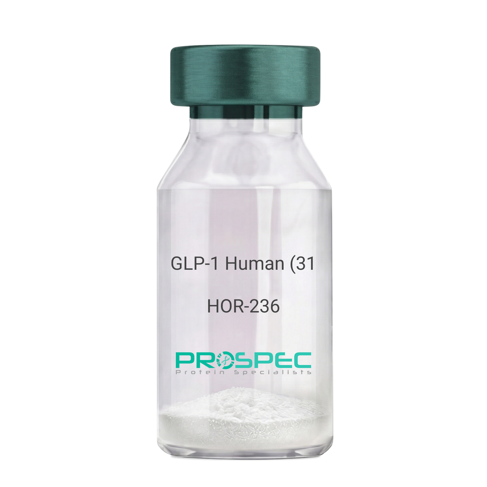
GLP-1 Human (31 a.a.)Description:
Glucagon Like Peptide-1 (31 a.a.) Human Recombinant
GLP1, GLP2, GRPP.
Product # :
HOR-236Price :
Quantity :
Shipping Method :
Shipped at Room temp
More Info
- description
- source
- formulation
- purity
- More Info
Description
Glucagon Like Peptide-1 Human Recombinant produced in E.Coli is a single, non-glycosylated, polypeptide chain containing 31 amino acids and having a molecular mass of 3.3 kDa. The GLP-1 is purified by proprietary chromatographic techniques.
Source
Escherichia Coli.
Formulation
The Recombinant GLP-1 concentrated solution (1mg/ml) was lyophilized after extensive dialyses against 0.1mg sodium phosphate monobasic & 1.6mg sodium phosphate dibasic.
Purity
Greater than 98.0% as determined by:
(a) Analysis by RP-HPLC.
(b) Analysis by SDS-PAGE.More Info
-
Introduction
Glucagon-like peptide-1 (GLP-1) is derived from the transcription product of the proglucagon gene. The major source of GLP-1 in the body is the intestinal L cell that secretes GLP-1 as a guthormone. The biologically active forms of GLP-1 are: GLP-1-(7-37) and GLP-1-(7-36)NH2.
GLP-1 secretion by L cells is dependent on the presence of nutrients in the lumen of the small intestine. The secretagogues (agents that causes or stimulates secretion) of this hormone include major nutrients like carbohydrate, proteinand lipid. Once in the circulation, GLP-1 has a half life of less than 2 minutes, due to rapid degradation by the enzyme dipeptidyl peptidase-4.
GLP-1 possesses several physiological properties that make it a subject of intensive investigation as a potential treatment of diabetes mellitus. The known physiological functions of GLP-1 include: Increases insulin secretion from the pancreas in a glucose-dependent manner, decreases glucagon secretion from the pancreas, increases beta cells mass and insulin gene expression, inhibits acid secretion and gastric emptying in the stomach, decreases food intake by increasing satiety. -
Synonyms
GLP1, GLP2, GRPP.
-
Physical Appearance
Sterile Filtered White lyophilized (freeze-dried) powder.
-
Stability
Lyophilized Glucagon Like Peptide-1 although stable at room temperature for 3 weeks, should be stored desiccated below -18°C. Upon reconstitution GLP-1 should be stored at 4°C between 2-7 days and for future use below -18°C. For long term storage it is recommended to add a carrier protein (0.1% HSA or BSA).Please prevent freeze-thaw cycles.
-
Solubility
It is recommended to reconstitute the lyophilized Glucagon Like Peptide-1 in sterile 18MΩ-cm H2O not less than 100µg/ml, which can then be further diluted to other aqueous solutions.
-
Amino Acid Sequence
HAEGTFTSDV SSYLEGQAAK EFIAWLVKGR G.
ProSpec's products are furnished for LABORATORY RESEARCH USE ONLY. The product may not be used as drugs, agricultural or pesticidal products, food additives or household chemicals.
- View Data Sheet
Name :
NTRK2 Human , HEKDescription:
Neurotrophic Receptor Tyrosine Kinase 2, HEK Human Recombinant
GP145-TrkB, trk-B, TRKB, NTRK2.
Product # :
CYT-1230Price :
Quantity :
Shipping Method :
Shipped with Ice Packs
More Info
- description
- source
- formulation
- purity
- More Info
Description
NTRK2 Human Recombinant is a single, glycosylated, polypeptide chain (32-430 a.a) containing a total of 632 amino acids and having a molecular mass of 70.3 kDa. NTRK2 is fused to a 233 a.a hIgG-His-Tag at C-terminus and is purified by proprietary chromatographic techniques.
Source
HEK293 Cells.
Formulation
The NTRK2 solution (1mg/ml) contains 10% Glycerol and Phosphate-Buffered Saline (pH 7.4).
Purity
Greater than 90.0% as determined by SDS-PAGE.
More Info
-
Synonyms
GP145-TrkB, trk-B, TRKB, NTRK2.
-
Physical Appearance
Sterile Filtered colorless solution.
-
Stability
Store at 4°C if entire vial will be used within 2-4 weeks. Store, frozen at -20°C for longer periods of time. For long term storage it is recommended to add a carrier protein (0.1% HSA or BSA). Avoid multiple freeze-thaw cycles.
-
Amino Acid Sequence
CPTSCKCSAS RIWCSDPSPG IVAFPRLEPN SVDPENITEI FIANQKRLEI INEDDVEAYV GLRNLTIVDS GLKFVAHKAF LKNSNLQHIN FTRNKLTSLS RKHFRHLDLS ELILVGNPFT CSCDIMWIKT LQEAKSSPDT QDLYCLNESS KNIPLANLQI PNCGLPSANL AAPNLTVEEG KSITLSCSVA GDPVPNMYWD VGNLVSKHMN ETSHTQGSLR ITNISSDDSG KQISCVAENL VGEDQDSVNL TVHFAPTITF LESPTSDHHW CIPFTVKGNP KPALQWFYNG AILNESKYIC TKIHVTNHTE YHGCLQLDNP THMNNGDYTL IAKNEYGKDE KQISAHFMGW PGIDDGANPN YPDVIYEDYG TAANDIGDTT NRSNEIPSTD VTDKTGREH LEPKSCDKTHT CPPCPAPELL GGPSVFLFPP KPKDTLMISR TPEVTCVVVD VSHEDPEVKF NWYVDGVEVH NAKTKPREEQ YNSTYRVVSV LTVLHQDWLN GKEYKCKVSN KALPAPIEKT ISKAKGQPRE PQVYTLPPSR DELTKNQVSL TCLVKGFYPS DIAVEWESNG QPENNYKTTP PVLDSDGSFF LYSKLTVDKS RWQQGNVFSC SVMHEALHNH YTQKSLSLSP GK.
-
Background
NTRK2, also known as TrkB, is a high-affinity receptor for BDNF and other neurotrophins such as neurotrophin-4 (NT-4) and neurotrophin-3 (NT-3). It plays a crucial role in the development, function, and plasticity of the nervous system. Dysregulation of NTRK2 signaling is associated with various neurological disorders, including neurodegenerative diseases, psychiatric disorders, and cancers. NTRK2 has been developed to facilitate research into its biological functions and potential therapeutic applications. This paper provides a comprehensive overview of the scientific research on recombinant human NTRK2, focusing on its biological functionality, detailed description, and physical properties.
NTRK2 is critical for synaptic plasticity, which underlies learning and memory. Research with rhNTRK2 has demonstrated that BDNF-NTRK2 signaling enhances synaptic strength and facilitates long-term potentiation (LTP) in hippocampal neuron.
ProSpec's products are furnished for LABORATORY RESEARCH USE ONLY. The product may not be used as drugs, agricultural or pesticidal products, food additives or household chemicals.
- View Data Sheet
Name :
EIF3K (50-94) HumanDescription:
Eukaryotic Translation Initiation Factor 3K (50-94 a.a.) Human Recombinant
PLAC-24, eIF3k, eIF-3 p25, eIF-3 p28, EIF3S12.
Product # :
PRO-2833Price :
Quantity :
Shipping Method :
Shipped at Room temp
More Info
- description
- source
- formulation
- purity
- More Info
Description
The EIF3KHuman is created as a recombinant protein with a 4kda N-terminal fusion of His Tag. The EIF3KHis-Tagged Fusion Protein, produced in E. coli, is a 10kDa protein containing 45 amino acid residues of the EIF3KHuman, 50-94 amino acids.
Source
Escherichia Coli.
Formulation
Each mg was lyophilized with 1xPBS, 0.4% SDS and 4mM DTT.
Purity
Greater than 90% as determined by SDS-PAGE.
More Info
-
Synonyms
PLAC-24, eIF3k, eIF-3 p25, eIF-3 p28, EIF3S12.
-
Physical Appearance
Filtered White lyophilized (freeze-dried) powder.
-
Stability
Store lyophilized EIF3Kat -20°C. Aliquot the product after reconstitution to avoid repeated freezing/thawing cycles. Reconstituted protein can be stored at 4°C for a limited period of time; it does not show any change after two weeks at 4°C.
-
Solubility
It is recommended to add deionized water to prepare a working stock solution of approximately 0.5mg/ml and let the lyophilized pellet dissolve completely. Product is not sterile! Please filter the product by an appropriate sterile filter before using it on cell culture.
-
Background
Eukaryotic Translation Initiation Factor 3K (EIF3K) is a part of the eIF3 subunit K family. EIF3K is the smallest subunit of eIF3 and it interacts with a few other subunits of the 40S ribosomal subunit and eIF3. EIF3K is found both in nucleus and cytoplasm and colocalized with cyclin D3, a regulatory subunit of cyclin-dependent kinase 4. EIF3K is conserved among high eukaryotes, including mammals, plants and insects and is expressed in human tissues.
ProSpec's products are furnished for LABORATORY RESEARCH USE ONLY. The product may not be used as drugs, agricultural or pesticidal products, food additives or household chemicals.
- View Data Sheet
Name :
PIM1 HumanDescription:
PIM1 Human Recombinant
PIM, Serine/threonine-protein kinase pim-1, PIM1.
Product # :
PRO-1389Price :
Quantity :
Shipping Method :
Shipped with Ice Packs
More Info
- description
- source
- formulation
- purity
- More Info
Description
PIM1 Human Recombinant produced in E.Coli is a single, non-glycosylated polypeptide chain containing 274 amino acids (38-290 a.a.) and having a molecular mass of 31.4kDa. PIM1 is fused to a 21 amino acid His-tag at N-terminus & purified by proprietary chromatographic techniques.
Source
E.coli.
Formulation
PIM1 protein solution (0.5mg/ml) contains 20mM Tris-HCl buffer (pH 8.0), 0.15M NaCl, 10% glycerol and 1mM DTT.
Purity
Greater than 90% as determined by SDS-PAGE.
More Info
-
Introduction
PIM1, which is a part of the Ser/Thr protein kinase family and PIM subfamily, is expressed primarily in B-lymphoid and myeloid cell lines, and is overexpressed in hematopoietic malignancies and in prostate cancer. PIM1 functions as Proto-oncogene with serine/threonine kinase activity involved in cell survival and cell proliferation, consequently providing a selective advantage in tumorigenesis. Both human and orthologous mouse genes have been reported to encode two isoforms resulting from the use of alternative in-frame translation initiation codons, the upstream non-AUG (CUG) and downstream AUG codons.
-
Synonyms
PIM, Serine/threonine-protein kinase pim-1, PIM1.
-
Physical Appearance
Sterile Filtered colorless solution.
-
Stability
Store at 4°C if entire vial will be used within 2-4 weeks. Store, frozen at -20°C for longer periods of time. For long term storage it is recommended to add a carrier protein (0.1% HSA or BSA).Avoid multiple freeze-thaw cycles.
-
Amino Acid Sequence
MGSSHHHHHH SSGLVPRGSH MYQVGPLLGS GGFGSVYSGI RVSDNLPVAI KHVEKDRISD WGELPNGTRV PMEVVLLKKV SSGFSGVIRL LDWFERPDSF VLILERPEPV QDLFDFITER GALQEELARS FFWQVLEAVR HCHNCGVLHR DIKDENILID LNRGELKLID FGSGALLKDT VYTDFDGTRV YSPPEWIRYH RYHGRSAAVW SLGILLYDMV CGDIPFEHDE EIIRGQVFFR QRVSSECQHL IRWCLALRPS DRPTFEEIQN HPWM.
ProSpec's products are furnished for LABORATORY RESEARCH USE ONLY. The product may not be used as drugs, agricultural or pesticidal products, food additives or household chemicals.
- View Data Sheet
Name :
HS3ST1 HumanDescription:
Heparan Sulfate 3-O-Sulfotransferase 1 Human Recombinant
Heparan sulfate glucosamine 3-O-sulfotransferase 1, Heparan sulfate D-glucosaminyl 3-O-sulfotransferase 1, 3-OST-1, Heparan sulfate 3-O-sulfotransferase 1, h3-OST-1, HS3ST1, 3OST, 3OST1, HS3S1.
Product # :
ENZ-744Price :
Quantity :
Shipping Method :
Shipped with Ice Packs
More Info
- description
- source
- formulation
- purity
- More Info
Description
HS3ST1 Human Recombinant produced in E.Coli is a single, non-glycosylated polypeptide chain containing 310 amino acids (21-307 a.a) and having a molecular mass of 36.2kDa.HS3ST1 is fused to a 23 amino acid His-tag at N-terminus & purified by proprietary chromatographic techniques.
Source
Escherichia Coli.
Formulation
HS3ST1 protein solution (0.25mg/ml) containing 20mM Tris-HCl buffer (pH 8.0), 0.2M NaCl, 40% glycerol and 2mM DTT.
Purity
Greater than 90.0% as determined by SDS-PAGE.
More Info
-
Introduction
Heparan Sulfate 3-O-Sulfotransferase 1 (HS3ST1), is sulfotransferase which uses 3'-phospho-5'-adenylyl sulfate (PAPS) to catalyze the transfer of a sulfo group to position 3 of glucosamine residues in heparan. HS3ST1 catalyzes the rate limiting step in the biosynthesis of heparan sulfate (HSact). This modification is a vital part in the biosynthesis of anticoagulant heparan sulfate since it concludes the structure of the antithrombin pentasaccharide binding site.
-
Synonyms
Heparan sulfate glucosamine 3-O-sulfotransferase 1, Heparan sulfate D-glucosaminyl 3-O-sulfotransferase 1, 3-OST-1, Heparan sulfate 3-O-sulfotransferase 1,
h3-OST-1, HS3ST1, 3OST, 3OST1, HS3S1. -
Physical Appearance
Sterile Filtered colorless solution.
-
Stability
Store at 4°C if entire vial will be used within 2-4 weeks. Store, frozen at -20°C for longer periods of time. For long term storage it is recommended to add a carrier protein (0.1% HSA or BSA).Avoid multiple freeze-thaw cycles.
-
Amino Acid Sequence
MGSSHHHHHH SSGLVPRGSH MGSRPAELGQ QELLRKAGTL QDDVRDGVAP NGSAQQLPQT IIIGVRKGGT RALLEMLSLH PDVAAAENEV HFFDWEEHYS HGLGWYLSQM PFSWPHQLTV EKTPAYFTSP KVPERVYSMN PSIRLLLILR DPSERVLSDY TQVFYNHMQK HKPYPSIEEF LVRDGRLNVD YKALNRSLYH VHMQNWLRFF PLRHIHIVDG DRLIRDPFPE IQKVERFLKL SPQINASNFY FNKTKGFYCL RDSGRDRCLH ESKGRAHPQV DPKLLNKLHE YFHEPNKKFF ELVGRTFDWH.
ProSpec's products are furnished for LABORATORY RESEARCH USE ONLY. The product may not be used as drugs, agricultural or pesticidal products, food additives or household chemicals.
- View Data Sheet
Name :
EOGT MouseDescription:
EGF Domain-Specific O-Linked N-Acetylglucosamine Transferase Mouse Recombinant
EGF domain-specific O-linked N-acetylglucosamine transferase, Extracellular O-linked N-acetylglucosamine transferase.
Product # :
ENZ-946Price :
Quantity :
Shipping Method :
Shipped with Ice Packs
More Info
- description
- source
- formulation
- purity
- More Info
Description
EOGT produced in Sf9 Baculovirus cells is a single, glycosylated polypeptide chain containing 516 amino acids (20-527 a.a.) and having a molecular mass of 60.4kDa (Migrates at 50-70kDa on SDS-PAGE under reducing conditions). EOGT is expressed with an 8 amino acid His tag at C-Terminus and purified by proprietary chromatographic techniques.
Source
Sf9, Baculovirus cells.
Formulation
EOGT protein solution (0.5mg/ml) contains Phosphate Buffered Saline (pH 7.4) and 10% glycerol.
Purity
Greater than 85.0% as determined by SDS-PAGE.
More Info
-
Introduction
EGF Domain-Specific O-Linked N-Acetylglucosamine Transferase (EOGT) takes part in the regulation of Notch receptor. EOGT catalyzes the transfer of a single N-acetylglucosamine from UDP-GlcNAc to a serine/ threonine residue in extracellular proteins resulting in their modification with a beta-linked N-acetylglucosamine (O-GlcNAc). EOGT mainly glycosylates the Thr residue positioned between the fifth and sixth conserved cysteines of folded EGF-like domains.
-
Synonyms
EGF domain-specific O-linked N-acetylglucosamine transferase, Extracellular O-linked N-acetylglucosamine transferase.
-
Physical Appearance
Sterile Filtered colorless solution.
-
Stability
Store at 4°C if entire vial will be used within 2-4 weeks. Store, frozen at -20°C for longer periods of time. For long term storage it is recommended to add a carrier protein (0.1% HSA or BSA). Avoid multiple freeze-thaw cycles.
-
Amino Acid Sequence
DKAHSEADDA PGKALYDYSS LRLPAEHIPF FLHNNRHVAS VCREDSHCPY KKHLENLNYC WGYEKSCAPE FRFGSPVCSY VDLGWTDTLE SAQDMFWRQA DFGYARERLG EIRTICQPER ASDSSLVCSR YLQYCRATGL YLDLRNIKRN HDRFKEDFLQ GGEIGGYCKL DSHALVSEGQ RKSPLQSWFA ELQGYTQLNF RPIEDAKCDI VVEKPTYFMK LDAGINMYHH FCDFLNLYLT QHVNNSFSTD VYIVMWDTST YGYGDLFSDT WKAFTDYDVI HLKTYDSKKV CFKEAVFSLL PRMRYGLFYN TPLISGCQNT GLFRAFSQHV LHRLNITQEG PKDGKVRVTI LARSTEYRKI LNQDELVNAL KTVSTFEVRV VDYKYRELGF LDQLRITHNT DIFIGMHGAG LTHLLFLPDW AAVFELYNCE DERCYLDLAR LRGIHYITWR KPSKVFPQDK GHHPTLGEHP KFTNYSFDVE EFMYLVLQAA EHVLQHPQWP FKKKHDELLE HHHHHH.
ProSpec's products are furnished for LABORATORY RESEARCH USE ONLY. The product may not be used as drugs, agricultural or pesticidal products, food additives or household chemicals.
- View Data Sheet
Name :
UBE2F HumanDescription:
Ubiquitin-Conjugating Enzyme E2F Human Recombinant
Ubiquitin-conjugating enzyme E2F (putative), NEDD8 carrier protein UBE2F, NEDD8 protein ligase UBE2F, NEDD8-conjugating enzyme 2, NEDD8-conjugating enzyme UBE2F, NCE2, EC 6.3.2.-.
Product # :
ENZ-625Price :
Quantity :
Shipping Method :
Shipped with Ice Packs
More Info
- description
- source
- formulation
- purity
- More Info
Description
UBE2F Human Recombinant produced in E. coli is a single polypeptide chain containing 209 amino acids (1-185) and having a molecular mass of 23.7 kDa.UBE2F is fused to a 24 amino acid His-tag at N-terminus & purified by proprietary chromatographic techniques.
Source
E.coli.
Formulation
The UBE2F solution (1mg/1ml) contains 20mM Tris-HCl buffer (pH 8.0), 200mM NaCl, 1mM DTT and 30% glycerol.
Purity
Greater than 95% as determined by SDS-PAGE.
More Info
-
Introduction
Ubiquitin-conjugating enzyme E2 F (UBE2F) is a member of the ubiquitin-conjugating enzyme family and UBE2F subfamily. Ubiquitin-conjugating enzymes or sometimes as ubiquitin-carrier enzymes, run the second step in the ubiquitination reaction which targets a protein for degradation using the proteasome. The UBE2F receives the ubiquitin-like protein NEDD8 from the UBA3-NAE1 E1 complex and catalyzes its covalent attachment to other proteins.
RBX2-UBE2F complex neddylates specific target proteins, for example CUL5 as can be seen from the specific interaction with the E3 ubiquitin ligase RBX2, but not RBX1. -
Synonyms
Ubiquitin-conjugating enzyme E2F (putative), NEDD8 carrier protein UBE2F, NEDD8 protein ligase UBE2F, NEDD8-conjugating enzyme 2, NEDD8-conjugating enzyme UBE2F, NCE2, EC 6.3.2.-.
-
Physical Appearance
Sterile Filtered colorless solution.
-
Stability
Store at 4°C if entire vial will be used within 2-4 weeks. Store, frozen at -20°C for longer periods of time. For long term storage it is recommended to add a carrier protein (0.1% HSA or BSA).Avoid multiple freeze-thaw cycles.
-
Amino Acid Sequence
MGSSHHHHHH SSGLVPRGSH MGSHMLTLAS KLKRDDGLKG SRTAATASDS TRRVSVRDKL LVKEVAELEA NLPCTCKVHF PDPNKLHCFQ LTVTPDEGYY QGGKFQFETE VPDAYNMVPP KVKCLTKIWH PNITETGEIC LSLLREHSID GTGWAPTRTL KDVVWGLNSL FTDLLNFDDP LNIEAAEHHL RDKEDFRNKV DDYIKRYAR
ProSpec's products are furnished for LABORATORY RESEARCH USE ONLY. The product may not be used as drugs, agricultural or pesticidal products, food additives or household chemicals.
- View Data Sheet
Name :
GPI AntibodyDescription:
Glucose-6-Phosphate Isomerase, Mouse Anti Human
Glucose-6-phosphate isomerase, Phosphoglucose isomerase, Phosphohexose isomerase, Autocrine motility factor, Neuroleukin, Sperm antigen 36, GPI, PGI, PHI, AMF, NLK, SA-36, GNPI.
Product # :
ANT-613Price :
Quantity :
Shipping Method :
Shipped with Ice Packs
More Info
- formulation
- More Info
Formulation
1mg/ml containing PBS, pH-7.4, 10% Glycerol and 0.02% Sodium Azide.
More Info
-
Introduction
Glucose-6-phosphate isomerase (GPI) is a part of the GPI family whose members encode multifunctional phosphoglucose isomerase proteins involved in energy pathways. GPI is a dimeric enzyme which catalyzes the reversible isomerization of glucose-6-phosphate and fructose-6-phosphate. Mammalian GPI also functions as a tumor-secreted cytokine and an angiogenic factor (AMF) which stimulates endothelial cell motility. In addition, GPI is a neurotrophic factor (Neuroleukin) for spinal and sensory neurons. GPI performs in different capacities inside and outside the cell. In the cytoplasm, GPI is involved in glycolysis and gluconeogenesis, while outside the cell it acts as a neurotrophic factor for spinal and sensory neurons.
Defects in the GPI gene cause the nonspherocytic hemolytic anemia and a severe enzyme deficiency can be linked to hydrops fetalis, immediate neonatal death and neurological impairment.
-
Synonyms
Glucose-6-phosphate isomerase, Phosphoglucose isomerase, Phosphohexose isomerase, Autocrine motility factor, Neuroleukin, Sperm antigen 36, GPI, PGI, PHI, AMF, NLK, SA-36, GNPI.
-
Physical Appearance
Sterile filtered colorless solution.
-
Immunogen
Anti-human GPI mAb, clone PAT22G2AT, is derived from hybridization of mouse F0 myeloma cells with spleen cells from BALB/c mice immunized with a recombinant human GPI protein 1-558 amino acids purified from E. coli.
-
Ig Subclass
Mouse IgG2a heavy chain and k light chain.
-
Clone
PAT22G2AT.
-
Applications
The antibody has been tested by ELISA, Western blot analysis, Flow cytometry and ICC/IF to assure specificity and reactivity. Since application varies, however, each investigation should be titrated by the reagent to obtain optimal results.
-
Type
Mouse Anti Human Monoclonal.
-
Storage Procedures
For periods up to 1 month store at 4°C, for longer periods of time, store at -20°C. Prevent freeze thaw cycles.
-
Purification Method
GPI antibody was purified from mouse ascitic fluids by protein-A affinity chromatography.
ProSpec's products are furnished for LABORATORY RESEARCH USE ONLY. The product may not be used as drugs, agricultural or pesticidal products, food additives or household chemicals.
- View Data Sheet
Name :
PRPS1 HumanDescription:
Phosphoribosyl Pyrophosphate Synthetase 1 Human Recombinant
ARTS, CMTX5, PPRibP, PRSI, DFN2, Ribose-phosphate pyrophosphokinase 1, DFNX1, Phosphoribosyl pyrophosphate synthase I, PRS-I, PRPS1, KIAA0967.
Product # :
PKA-361Price :
Quantity :
Shipping Method :
Shipped with Ice Packs
More Info
- description
- source
- formulation
- purity
- More Info
Description
PRPS1 Human Recombinant produced in E.Coli is a single, non-glycosylated polypeptide chain containing 338 amino acids (1-318 a.a.) and having a molecular weight of 36.9kDa.The PRPS1 is fused to 20 a.a His-Tag at N-terminus and purified by proprietary chromatographic techniques.
Source
Escherichia Coli.
Formulation
The PRPS1 protein solution contains 20mM Tris-HCl, pH-8, 100mM NaCl, 1mM DTT and 20% glycerol.
Purity
Greater than 90.0% as determined by SDS-PAGE.
More Info
-
Introduction
PRPS1 catalyzes the synthesis of phosphoribosylpyrophosphate (PRPP) that is essential for nucleotide synthesis. PRPS1 catalyzes the phosphoribosylation of ribose 5-phosphate to 5-phosphoribosyl-1-pyrophosphate, which is essential for purine metabolism and nucleotide biosynthesis. Defects in PRPS1 result in phosphoribosylpyrophosphate synthetase superactivity, Charcot-Marie-Tooth disease X-linked recessive type 5 and Arts Syndrome.
-
Synonyms
ARTS, CMTX5, PPRibP, PRSI, DFN2, Ribose-phosphate pyrophosphokinase 1, DFNX1, Phosphoribosyl pyrophosphate synthase I, PRS-I, PRPS1, KIAA0967.
-
Physical Appearance
Sterile filtered colorless solution.
-
Stability
Store at 4°C if entire vial will be used within 2-4 weeks. Store, frozen at -20°C for longer periods of time. For long term storage it is recommended to add a carrier protein (0.1% HSA or BSA). Avoid multiple freeze-thaw cycles.
-
Amino Acid Sequence
MGSSHHHHHH SSGLVPRGSH MPNIKIFSGS SHQDLSQKIA DRLGLELGKV VTKKFSNQET CVEIGESVRG EDVYIVQSGC GEINDNLMEL LIMINACKIA SASRVTAVIP CFPYARQDKK DKSRAPISAK LVANMLSVAG ADHIITMDLH ASQIQGFFDI PVDNLYAEPA VLKWIRENIS EWRNCTIVSP DAGGAKRVTS IADRLNVDFA LIHKERKKAN EVDRMVLVGD VKDRVAILVD DMADTCGTIC HAADKLLSAG ATRVYAILTH GIFSGPAISR INNACFEAVV VTNTIPQEDK MKHCSKIQVI DISMILAEAI RRTHNGESVS YLFSHVPL.
ProSpec's products are furnished for LABORATORY RESEARCH USE ONLY. The product may not be used as drugs, agricultural or pesticidal products, food additives or household chemicals.
- View Data Sheet
Name :
UBE2I HumanDescription:
Ubiquitin-Conjugating Enzyme E2I Human Recombinant
SUMO-conjugating enzyme UBC9, EC 6.3.2.-, SUMO-protein ligase, Ubiquitin-conjugating enzyme E2 I, Ubiquitin-protein ligase I, Ubiquitin carrier protein I, Ubiquitin carrier protein 9, p18, UBC9, C358B7.1.
Product # :
ENZ-341Price :
Quantity :
Shipping Method :
Shipped with Ice Packs
More Info
- description
- source
- formulation
- purity
- More Info
Description
UBE2I Human Recombinant produced in E.Coli is a single, non-glycosylated polypeptide chain containing 158 amino acids (1-158 aa) & having a molecular mass of 18.0 kDa. UBE2I is purified by proprietary chromatographic techniques.
Source
Escherichia Coli.
Formulation
UBE2I (1mg/ml) contains 50mM HEPES (pH7.4) 150mM NaCl, 1mM DTT and 10% glycerol.
Purity
Greater than 95.0% as determined by Analysis by SDS-PAGE.
More Info
-
Introduction
Human Ubc9 is homologous to ubiquitin-conjugating enzymes (E2s). However, instead of conjugating ubiquitin, it conjugates a ubiquitin homologue, small ubiquitin-like modifier 1(SUMO-1). And hUbc9 retains striking structural and functional conservation with yeast Ubc9. The ubiquitin-dependent protein degradation system has been recognized as a complete enzymatic pathway that is responsible for the selective degradation of abnormal and short-lived proteins. The conjugation of ubiquitin requires the activities of ubiquitin-activating (E1) and –conjugating (E2) enzymes.
-
Synonyms
SUMO-conjugating enzyme UBC9, EC 6.3.2.-, SUMO-protein ligase, Ubiquitin-conjugating enzyme E2 I, Ubiquitin-protein ligase I, Ubiquitin carrier protein I, Ubiquitin carrier protein 9, p18, UBC9, C358B7.1.
-
Physical Appearance
Sterile filtered colorless solution.
-
Stability
Store at 4°C if entire vial will be used within 2-4 weeks. Store, frozen at -20°C for longer periods of time. For long term storage it is recommended to add a carrier protein (0.1% HSA or BSA).Avoid multiple freeze-thaw cycles.
-
Amino Acid Sequence
MSGIALSRLA QERKAWRKDH PFGFVAVPTK NPDGTMNLMN WECAIPGKKG TPWEGGLFKL RMLFKDDYPS SPPKCKFEPP LFHPNVYPSG TVCLSILEED KDWRPAITIK QILLGIQELL NEPNIQDPAQ AEAYTIYCQN RVEYEKRVRA QAKKFAPS.
ProSpec's products are furnished for LABORATORY RESEARCH USE ONLY. The product may not be used as drugs, agricultural or pesticidal products, food additives or household chemicals.
- View Data Sheet
Name :
MIF Human, GSTDescription:
Macrophage Migration Inhibitor Factor Human Recombinant, GST tag
Macrophage Migration Inhibitory Factor (Glycosylation-Inhibiting Factor), Phenylpyruvate Tautomerase, L-Dopachrome Tautomerase, L-Dopachrome Isomerase, GLIF, MMIF, GIF, Macrophage Migration Inhibitory Factor, Glycosylation-Inhibiting Factor, EC 5.3.3.12, EC 5.3.2.1, Macrophage migration inhibitory factor.
Product # :
CYT-401Price :
Quantity :
Shipping Method :
Shipped with Ice Packs
More Info
- description
- source
- formulation
- purity
- More Info
Description
MIF Human Recombinant produced in E.Coli is a single, non-glycosylated polypeptide chain containing 345 amino acids (1-115 a.a) and having a molecular mass of 39.2kDa. MIF is fused to a 230 amino acid GST-tag at N-terminus & purified by proprietary chromatographic techniques.
Source
Escherichia Coli.
Formulation
MIF protein solution (1mg/ml) containing Phosphate Buffered Saline (pH7.4) and 10% glycerol.
Purity
Greater than 90.0% as determined by SDS-PAGE.
More Info
-
Introduction
The cytokine Macrophage migration inhibitory factor (MIF) has been identified to be secreted by the pituitary gland and the monocyte/macrophage and to play an important role in endotoxic shock. MIF has the unique property of being released from macrophages and T cells in response to physiological concentrations of glucocorticoids. The secretion of MIF is tightly regulated and decreases at high, anti-inflammatory steroid concentration.
-
Synonyms
Macrophage Migration Inhibitory Factor (Glycosylation-Inhibiting Factor), Phenylpyruvate Tautomerase, L-Dopachrome Tautomerase, L-Dopachrome Isomerase, GLIF, MMIF, GIF, Macrophage Migration Inhibitory Factor, Glycosylation-Inhibiting Factor, EC 5.3.3.12, EC 5.3.2.1, Macrophage migration inhibitory factor.
-
Physical Appearance
Sterile Filtered clear solution.
-
Stability
Store at 4°C if entire vial will be used within 2-4 weeks. Store, frozen at -20°C for longer periods of time. For long term storage it is recommended to add a carrier protein (0.1% HSA or BSA).Avoid multiple freeze-thaw cycles.
-
Amino Acid Sequence
MSPILGYWKI KGLVQPTRLL LEYLEEKYEE HLYERDEGDK WRNKKFELGL EFPNLPYYID GDVKLTQSMA IIRYIADKHN MLGGCPKERA EISMLEGAVL DIRYGVSRIA YSKDFETLKV DFLSKLPEML KMFEDRLCHK TYLNGDHVTH PDFMLYDALD VVLYMDPMCL DAFPKLVCFK KRIEAIPQID KYLKSSKYIA WPLQGWQATF GGGDHPPKSD LVPRGSPEFA MPMFIVNTNV PRASVPDGFL SELTQQLAQA TGKPPQYIAV HVVPDQLMAF GGSSEPCALC SLHSIGKIGG AQNRSYSKLL CGLLAERLRI SPDRVYINYY DMNAANVGWN NSTFA.
ProSpec's products are furnished for LABORATORY RESEARCH USE ONLY. The product may not be used as drugs, agricultural or pesticidal products, food additives or household chemicals.
- View Data Sheet
Name :
EG VEGF HumanDescription:
Endocrine Gland Vascular Endothelial Growth Factor Human Recombinant
PK1, PRK1, Prokineticin 1, EG-VEGF.
Product # :
CYT-338Price :
Quantity :
Shipping Method :
Shipped at Room temp
More Info
- description
- source
- formulation
- purity
- biological activity
- More Info
Description
EG-VEGF Human Recombinant produced in E.Coli is a single, non-glycosylated, polypeptide chain containing 86 amino acids and having a molecular mass of 9.7kDa. The EG-VEGF is purified by proprietary chromatographic techniques.
Source
Escherichia Coli.
Formulation
The protein was lyophilized from a concentrated (1mg/ml) solution containing 0.1% Trifluoroacetic Acid (TFA).
Purity
Greater than 95.0% as determined by SDS-PAGE.
Biological Activity
The activity as determined by the dose-dependent proliferation of MIA PaCa-2 cells is typically 1-4 μg/ml.
More Info
-
Introduction
Endocrine gland-derived vascular endothelial growth factor (EG-VEGF) induces proliferation, migration, and fenestration in capillary endothelial cells derived from endocrine glands. Its expression is induced by hypoxia and is restricted to the steroidogenic glands (ovary, testis, adrenal, and placenta). Its expression is often complementary to the expression of VEGF (MIM 192240), suggesting that these molecules function in a coordinated manner. EG-VEGF potently contracts gastrointestinal (gi) smooth muscle. Induces proliferation, migration and fenestration (the formation of membrane discontinuities) in capillary endothelial cells derived from endocrine glands. Has little or no effect on a variety of other endothelial and non-endothelial cell types.
-
Synonyms
PK1, PRK1, Prokineticin 1, EG-VEGF.
-
Physical Appearance
Sterile Filtered White lyophilized (freeze-dried) powder.
-
Stability
Lyophilized EG-VEGF Human Recombinant although stable at room temperature for 3 weeks, should be stored desiccated below -18°C. Upon reconstitution EG-VEGF should be stored at 4°C between 2-7 days and for future use below -18°C.Please prevent freeze-thaw cycles.
-
Solubility
It is recommended to reconstitute the lyophilized Endocrine Gland Vascular Endothelial Growth Factor in sterile 18MΩ-cm H2O not less than 100µg/ml, which can then be further diluted to other aqueous solutions.
-
Amino Acid Sequence
AVITGACERD VQCGAGTCCA ISLWLRGLRM CTPLGREGEE CHPGSHKVPF FRKRKHHTCP CLPNLLCSRF PDGRYRCSMD LKNINF.
-
Background
Title: Endocrine Gland Vascular Endothelial Growth Factor Human Recombinant: Insights into its Role in Endocrine Disorders and Therapeutic Applications
Abstract:
Endocrine Gland Vascular Endothelial Growth Factor (EG-VEGF) is a unique angiogenic factor that plays a crucial role in the development and function of endocrine glands. This research paper provides a comprehensive analysis of human recombinant EG-VEGF, focusing on its production, characterization, and potential applications in endocrine disorders. The paper highlights the significance of EG-VEGF in endocrine gland angiogenesis and explores its role in the pathogenesis of endocrine-related diseases. Furthermore, it discusses ongoing research and clinical trials investigating the therapeutic potential of recombinant EG-VEGF in endocrine disorders and related conditions. The information presented in this paper aims to enhance our understanding of human recombinant EG-VEGF and its utility as a research tool and a potential therapeutic agent.Introduction:
Endocrine Gland Vascular Endothelial Growth Factor (EG-VEGF) is a growth factor specifically expressed in endocrine tissues. Human recombinant EG-VEGF, produced through genetic engineering techniques, offers a valuable tool for studying its angiogenic properties and exploring its potential therapeutic applications in endocrine disorders.Production and Characterization:
Recombinant EG-VEGF is typically generated using mammalian cell expression systems. The protein is then purified and characterized to ensure its structural integrity and functional activity. Rigorous quality control measures are implemented to confirm the specificity and potency of the recombinant EG-VEGF.Role in Endocrine Disorders:
EG-VEGF is involved in the regulation of endocrine gland angiogenesis, which is critical for their development, hormone secretion, and overall function. Dysregulation of EG-VEGF signaling has been implicated in various endocrine disorders, including preeclampsia, gestational trophoblastic diseases, and adrenal disorders. Recombinant EG-VEGF serves as a valuable tool for investigating the mechanisms underlying EG-VEGF-mediated angiogenesis and its potential implications in endocrine-related diseases.Therapeutic Implications:
Manipulation of angiogenesis holds promise as a therapeutic approach in various endocrine disorders. Recombinant EG-VEGF offers potential therapeutic applications in promoting neovascularization and restoring endocrine gland function. Ongoing research and clinical trials are investigating the therapeutic potential of recombinant EG-VEGF in conditions such as hypopituitarism, ovarian disorders, and other endocrine-related pathologies.Conclusion:
Human recombinant EG-VEGF represents a valuable research tool and a potential therapeutic agent. Its production, characterization, and applications in endocrine disorders contribute to our understanding of endocrine gland angiogenesis and the development of targeted therapeutic interventions. Continued research and clinical trials exploring the therapeutic potential of recombinant EG-VEGF offer promising avenues for improving outcomes in endocrine disorders and related conditions.What is the molecular weight/Mw of EG-VEGF Protein?
EG-VEGF Protein has a total Mw of 9.7kDa.
What is the source or expression system of EG-VEGF Protein?
Escherichia Coli.
What is the Purity of EG-VEGF Protein?
EG-VEGF Protein is >95% pure as determined by SDS-PAGE.
What is the Biological Activity of EG-VEGF Protein?
The activity as determined by the dose-dependent proliferation of MIA PaCa-2 cells is typically 1-4 μg/ml.
What is the amino acid sequence of EG-VEGF Protein?
AVITGACERD VQCGAGTCCA ISLWLRGLRM CTPLGREGEE CHPGSHKVPF FRKRKHHTCP CLPNLLCSRF PDGRYRCSMD LKNINF.
What applications can EG-VEGF Protein be used in?
EG-VEGF Protein can probably be used in western blot, ELISA and Lateral Flow.
What is the endotoxin level for EG-VEGF Protein?
The endotoxin level is minimal, EG-VEGF Protein was purified using conventional chromatography techniques.
ProSpec's products are furnished for LABORATORY RESEARCH USE ONLY. The product may not be used as drugs, agricultural or pesticidal products, food additives or household chemicals.
- View Data Sheet
Name :
GLSA1 E.ColiDescription:
Glutaminase 1 E.Coli Recombinant
Glutaminase 1, ybaS.
Product # :
ENZ-133Price :
Quantity :
Shipping Method :
Shipped with Ice Packs
More Info
- description
- source
- formulation
- purity
- More Info
Description
GLSA1 E.Coli Recombinant fused with a 20 amino acid His tag at N-terminus produced in E.Coli is a single, non-glycosylated, polypeptide chain containing 330 amino acids (1-310a.a.) and having a molecular mass of 35.0kDa. The GLSA1 is purified by proprietary chromatographic techniques.
Source
Escherichia Coli.
Formulation
The GLSA1 solution (1mg/1ml) contains 20mM Tris-HCl buffer (pH8.0), 0.1M NaCl, 1mM DTT and 10% glycerol.
Purity
Greater than 95.0% as determined by SDS-PAGE.
More Info
-
Introduction
GlsA1 is a member of the great superfamily of serine-dependent beta-lactamases and penicillin-binding proteins which exist almost in every bacteria and eukaryotes and catalyze the hydrolytic deamidation of l-glutamine to l-glutamate and free NH4+.
-
Synonyms
Glutaminase 1, ybaS.
-
Physical Appearance
Sterile Filtered colorless solution.
-
Stability
Store at 4°C if entire vial will be used within 2-4 weeks. Store, frozen at -20°C for longer periods of time. For long term storage it is recommended to add a carrier protein (0.1% HSA or BSA).Avoid multiple freeze-thaw cycles.
-
Amino Acid Sequence
MGSSHHHHHH SSGLVPRGSH MLDANKLQQA VDQAYTQFHS LNGGQNADYI PFLANVPGQL AAVAIVTCDG NVYSAGDSDY RFALESISKV CTLALALEDV GPQAVQDKIG ADPTGLPFNS VIALELHGGK PLSPLVNAGA IATTSLINAE NVEQRWQRIL HIQQQLAGEQ VALSDEVNQS EQTTNFHNRA IAWLLYSAGY LYCDAMEACD VYTRQCSTLL NTIELATLGA TLAAGGVNPL THKRVLQADN VPYILAEMMM EGLYGRSGDW AYRVGLPGKS GVGGGILAVV PGVMGIAAFS PPLDEDGNSV RGQKMVASVA KQLGYNVFKG
ProSpec's products are furnished for LABORATORY RESEARCH USE ONLY. The product may not be used as drugs, agricultural or pesticidal products, food additives or household chemicals.
- View Data Sheet
Name :
MTHSFD HumanDescription:
Methenyltetrahydrofolate Synthetase Domain Containing Human Recombinant
Methenyltetrahydrofolate synthase domain-containing protein, Methenyltetrahydrofolate synthase domaincontaining protein, Methenyltetrahydrofolate synthase
Product # :
PRO-2646Price :
Quantity :
Shipping Method :
Shipped with Ice Packs
More Info
- description
- source
- formulation
- purity
- More Info
Description
MTHSFD Human Recombinant produced in E.Coli is a single, non-glycosylated polypeptide chain containing 406 amino acids (1-383 a.a.) and having a molecular mass of 44.5kDa. MTHSFD is fused to a 23 amino acid His tag at N-Terminus and purified by proprietary chromatographic techniques.
Source
Escherichia Coli.
Formulation
The MTHSFD solution (0.25mg/ml) contains 30% glycerol, 1mM DTT and Phosphate-Buffered Saline (pH 7.4).
Purity
Greater than 90.0% as determined by SDS-PAGE.
More Info
-
Introduction
MTHSFD, also known as Methenyltetrahydrofolate Synthetase Domain Containing protein, is linked to Pancreas, Annular and Gastrointestinal Defects. It has also been associated with Immunodeficiency Syndrome. Some of the Gene Ontology annotations that are related to the MTHSFD gene are nucleic acid binding and nucleotide binding.
-
Synonyms
Methenyltetrahydrofolate synthase domain-containing protein, Methenyltetrahydrofolate synthase domaincontaining protein, Methenyltetrahydrofolate synthase
-
Physical Appearance
Sterile Filtered colorless solution.
-
Stability
Store at 4°C if entire vial will be used within 2-4 weeks. Store, frozen at -20°C for longer periods of time. For long term storage it is recommended to add a carrier protein (0.1% HSA or BSA).Avoid multiple freeze-thaw cycles.
-
Amino Acid Sequence
MGSSHHHHHH SSGLVPRGSH MGSMEPRAVG VSKQDIREQI WGYMESQNLA DFPRPVHHRI PNFKGASHAA EQLPRLQAFK TARTIKVNPD APQKSARFFV LESKKTLLVP TPRLRTGLFN KITPPPGATK DILRKCATSQ GVRNYSVPIG LDSRVLVDLV VVGSVAVSEK GWRIGKGEGY ADLEYAMMVS MGAVSKETPV VTIVHDCQVV DIPEELVEEH DITVDYILTP TRVIATGCKR PKPMGITWFK ISLEMMEKIP ILRSLRAREQ QAGKDVTLQG EHQHLPEPGC QQTVPLSVGR RPPDTPGPET NSMEAAPGSP PGEGAPLAAD VYVGNLPGDA RVSDLKRALR ELGSVPLRLT WQGPRRRAFL HYPDSAAAQQ AVSCLQGLRL GTDTLRVALA RQQRDK
ProSpec's products are furnished for LABORATORY RESEARCH USE ONLY. The product may not be used as drugs, agricultural or pesticidal products, food additives or household chemicals.
- View Data Sheet
Name :
UNG AntibodyDescription:
Uracil DNA Glycosilase, Mouse Anti Human
Uracil DNA Glycosilase, Uracil DNA Glycosylase, UNG.
Product # :
ANT-374Price :
Quantity :
Shipping Method :
Shipped with Ice Packs
More Info
- formulation
- More Info
Formulation
1mg/ml containing PBS, pH-7.4, & 0.1% Sodium Azide.
More Info
-
Introduction
E.Coli Uracil DNA Glycosilase (UNG) catalyses the release of free Uracil from Uracil-containing DNA. UNG efficiently hydrolyzes uracil from signle-stranded or double-stranded DNA, but not from oligomers (6 fewer bases).
-
Synonyms
Uracil DNA Glycosilase, Uracil DNA Glycosylase, UNG.
-
Physical Appearance
Sterile Filtered clear solution.
-
Immunogen
Anti-human UNG mAb is derived from hybridization of mouse SP2/O myeloma cells with spleen cells from BALB/c mice immunized with recombinant human UNG amino acids 1-313 purified from E. coli.
-
Ig Subclass
Mouse IgG2b heavy chain and κ light chain.
-
Clone
Pk1C12AT.
-
Applications
UNG antibody has been tested by ELISA and Western blot analysis to assure specificity and reactivity. Since application varies, however, each investigation should be titrated by the reagent to obtain optimal results. Recommended dilution range for Western blot analysis is 1:1,000 ~ 2,000. Recommended starting dilution is 1:1,000.
-
Type
Mouse Anti Human Monoclonal.
-
Storage Procedures
For periods up to 1 month store at 4°C, for longer periods of time, store at -20°C. Prevent freeze thaw cycles.
-
Purification Method
UNG antibody was purified from mouse ascitic fluids by protein-G affinity chromatography.
ProSpec's products are furnished for LABORATORY RESEARCH USE ONLY. The product may not be used as drugs, agricultural or pesticidal products, food additives or household chemicals.
- View Data Sheet
Name :
PDZK1 AntibodyDescription:
PDZ Domain Containing 1, Mouse Anti Human
PDZ Domain Containing 1, PDZ-Containing Kidney Protein 1, Na(+)/H(+) Exchange Regulatory Cofactor NHE-RF3, Na/Pi Cotransporter C-Terminal-Associated Protein 1, Sodium-Hydrogen Exchanger Regulatory Factor 3, CFTR-Associated Protein Of 70 KDa, NHERF-3, CAP70, PDZD1, CLAMP, naPi-Cap1.
Product # :
ANT-460Price :
Quantity :
Shipping Method :
Shipped with Ice Packs
More Info
- formulation
- More Info
Formulation
1mg/ml containing PBS, pH-7.4, 10% Glycerol and 0.02% Sodium Azide.
More Info
-
Introduction
PDZK1 is a PDZ domain-containing scaffolding protein which facilitates localization of cell surface proteins and has a key part in cholesterol metabolism by regulating the HDL receptor, scavenger receptor class B type 1. Single nucleotide polymorphism in this gene is linked to metabolic syndrome, and overexpression results in drug resistance of multiple myeloma.
-
Synonyms
PDZ Domain Containing 1, PDZ-Containing Kidney Protein 1, Na(+)/H(+) Exchange Regulatory Cofactor NHE-RF3, Na/Pi Cotransporter C-Terminal-Associated Protein 1, Sodium-Hydrogen Exchanger Regulatory Factor 3, CFTR-Associated Protein Of 70 KDa, NHERF-3, CAP70, PDZD1, CLAMP, naPi-Cap1.
-
Physical Appearance
Sterile Filtered colorless solution.
-
Immunogen
Anti-human PDZK1 mAb, clone PAT1A2A, is derived from hybridization of mouse F0 myeloma cells with spleen cells from BALB/c mice immunized with a recombinant human PDZK1 protein 1-519 amino acids purified from E. coli.
-
Ig Subclass
Mouse IgG2b heavy chain and Kappa light chain.
-
Clone
PAT1A2AT.
-
Applications
The antibody has been tested by ELISA and Western blot analysis to assure specificity and reactivity. Since application varies, however, each investigation should be titrated by the reagent to obtain optimal results. Recommended starting dilution is 1:1000.
-
Type
Mouse Anti Human Monoclonal.
-
Storage Procedures
For periods up to 1 month store at 4°C, for longer periods of time, store at -20°C. Prevent freeze thaw cycles.
-
Purification Method
PDZK1 antibody was purified from mouse ascitic fluids by protein-A affinity chromatography.
ProSpec's products are furnished for LABORATORY RESEARCH USE ONLY. The product may not be used as drugs, agricultural or pesticidal products, food additives or household chemicals.
- View Data Sheet
Name :
PPP1CA AntibodyDescription:
Protein Phosphatase 1 Catalytic Subunit Alpha, Mouse Anti Human
Serine/threonine-protein phosphatase PP1-alpha catalytic subunit, PP-1A, PPP1CA, PPP1A, protein phosphatase 1 catalytic subunit alpha isozyme, MGC1674, MGC15877, PP1alpha.
Product # :
ANT-415Price :
Quantity :
Shipping Method :
Shipped with Ice Packs
More Info
- formulation
- More Info
Formulation
1mg/ml containing PBS, pH-7.4, & 0.1% Sodium Azide.
More Info
-
Introduction
PPP1CA is one of the 3 catalytic subunits of protein phosphatase 1 (PP1). PP1 is a serine/threonine specific protein phosphatase which is involved in the regulation of a variety of cellular processes, such as cell division, glycogen metabolism, muscle contractility, protein synthesis, and HIV-1 viral transcription. Increased PP1 activity is detected in the end stage of heart failure. Studies in both human and mice suggest that PP1 is a significant regulator of cardiac function. Mouse studies also propose that PP1 functions as a suppressor of learning and memory.
-
Synonyms
Serine/threonine-protein phosphatase PP1-alpha catalytic subunit, PP-1A, PPP1CA, PPP1A, protein phosphatase 1 catalytic subunit alpha isozyme, MGC1674, MGC15877, PP1alpha.
-
Physical Appearance
Sterile Filtered clear solution.
-
Immunogen
Anti-human PPP1CA mAb, is derived from hybridization of mouse FO myeloma cells with spleen cells from BALB/c mice immunized with recombinant human PPP1CA amino acids 30-299 purified from E. coli.
-
Ig Subclass
Mouse IgG2a heavy chain and κ light chain.
-
Clone
P4G3AT.
-
Applications
PPP1CA antibody has been tested by ELISA and Western blot analysis to assure specificity and reactivity. Since application varies, however, each investigation should be titrated by the reagent to obtain optimal results. Recommended dilution range for Western blot analysis is 1:500 ~ 2,000. Recommended starting dilution is 1:1,000.
-
Type
Mouse Anti Human Monoclonal.
-
Storage Procedures
For periods up to 1 month store at 4°C, for longer periods of time, store at -20°C. Prevent freeze thaw cycles.
-
Purification Method
PPP1CA antibody was purified from mouse ascitic fluids by protein-G affinity chromatography.
ProSpec's products are furnished for LABORATORY RESEARCH USE ONLY. The product may not be used as drugs, agricultural or pesticidal products, food additives or household chemicals.
- View Data Sheet
Name :
GCSF MonkeyDescription:
Granulocyte Colony Stimulating Factor Recombinant Rhesus Macaque
CSF3, MGI-1G, GM-CSF beta, Pluripoietin, G-CSF, GCSF.
Product # :
CYT-1121Price :
Quantity :
Shipping Method :
Shipped at Room temp
More Info
- description
- source
- formulation
- purity
- biological activity
- More Info
Description
Granulocyte Colony Stimulating Rhesus Macaque Recombinant produced in E.Coli is a non-glycosylated polypeptide chain containing 174 amino acids and having a molecular mass of approximately 18.9kDa.GCSF is purified by proprietary chromatographic techniques.
Source
Escherichia Coli.
Formulation
Lyophilized from a 0.2μm filtered concentrated solution in PBS, pH7.4.
Purity
Greater than 98.0% as determined by:
(a) Analysis by RP-HPLC.
(b) Analysis by SDS-PAGE.Biological Activity
The ED50 as determined by a cell proliferation assay using murine NFS-60 cells is < 0.05 ng/ml, corresponding to a specific activity of > 2.0 × 10⁷ IU/mg.
More Info
-
Introduction
GCSF is a cytokine that controls the production, differentiation, and function of granulocytes. The active protein is found extracellularly. 3 transcript variants encoding 3 different isoforms have been found for the GCSF gene. Granulocyte/macrophage colony-stimulating factors are cytokines that take part in hematopoiesis by controlling the production, differentiation, and function of 2 related white cell populations of the blood, the granulocytes and the monocytes-macrophages. This csf induces granulocytes.
-
Synonyms
CSF3, MGI-1G, GM-CSF beta, Pluripoietin, G-CSF, GCSF.
-
Physical Appearance
Sterile Filtered White lyophilized (freeze-dried) powder.
-
Stability
Lyophilized GCSF although stable at room temperature for 3 weeks, should be stored desiccated below -18°C. Upon reconstitution Granulocyte Colony Stimulating Rhesus Macaque should be stored at 4°C between 2-7 days and for future use below -18°C.Please prevent freeze-thaw cycles.
-
Solubility
It is recommended to reconstitute the lyophilized Granulocyte Colony Stimulating Rhesus Macaque in sterile 18MΩ-cm H2O not less than 100µg/ml, which can then be further diluted to other aqueous solutions.
-
Amino Acid Sequence
TPLGPASSLP QSFLLKCLEQ VRKIQGDGAA LQEKLCATYK LCHPEELVLL RHSLGIPWAP LSSCPSQALQ LTGCLSQLHS SLFLYQGLLQ ALEGISPELS PTLDTLQLDI ADFATTIWQQ MEDLGMAPAL QPTQGAMPAF TSAFQRRAGG VLVASHLQRF LELAYRVLRH LAQS.
-
Background
What is the molecular weight/Mw of GCSF MONKEY Protein?
GCSF MONKEY Protein has a total Mw of 18.9kDa.
What is the source or expression system of GCSF MONKEY Protein?
Escherichia Coli.
What is the Purity of GCSF MONKEY Protein?
GCSF MONKEY Protein is >98% pure as determined by SDS-PAGE.
What is the Biological Activity of GCSF MONKEY Protein?
The ED50 as determined by a cell proliferation assay using murine NFS-60 cells is < 0.05 ng/ml, corresponding to a specific activity of > 2.0 × 10⁷ IU/mg.
What is the amino acid sequence of GCSF MONKEY Protein?
TPLGPASSLP QSFLLKCLEQ VRKIQGDGAA LQEKLCATYK LCHPEELVLL RHSLGIPWAP LSSCPSQALQ LTGCLSQLHS SLFLYQGLLQ ALEGISPELS PTLDTLQLDI ADFATTIWQQ MEDLGMAPAL QPTQGAMPAF TSAFQRRAGG VLVASHLQRF LELAYRVLRH LAQS.
What applications can GCSF MONKEY Protein be used in?
GCSF MONKEY Protein can probably be used in western blot, ELISA and Lateral Flow.
What is the endotoxin level for GCSF MONKEY Protein?
The endotoxin level is minimal, GCSF MONKEY Protein was purified using conventional chromatography techniques.
ProSpec's products are furnished for LABORATORY RESEARCH USE ONLY. The product may not be used as drugs, agricultural or pesticidal products, food additives or household chemicals.